Embed Size (px)
Citation preview

Robotic LV Epicardial Lead PlacementRobotic LV Epicardial Lead Placement: : Indications and TechniqueIndications and Technique
Sandhya K. Balaram, M.D., Ph.D.Sandhya K. Balaram, M.D., Ph.D.Division of Cardiothoracic SurgeryDivision of Cardiothoracic Surgery
St. Luke’s-Roosevelt Hospital CenterSt. Luke’s-Roosevelt Hospital CenterAssistant Professor of Clinical SurgeryAssistant Professor of Clinical Surgery
Columbia University College of Physicians and SurgeonsColumbia University College of Physicians and Surgeons
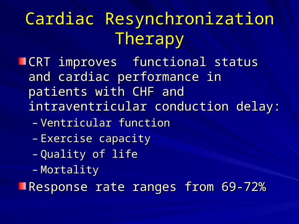
Cardiac Resynchronization Cardiac Resynchronization TherapyTherapy
CRT improves functional status and CRT improves functional status and cardiac performance in patients with CHF cardiac performance in patients with CHF and intraventricular conduction delay:and intraventricular conduction delay:– Ventricular functionVentricular function– Exercise capacityExercise capacity– Quality of lifeQuality of life– Mortality Mortality
Response rate ranges from 69-72%Response rate ranges from 69-72%
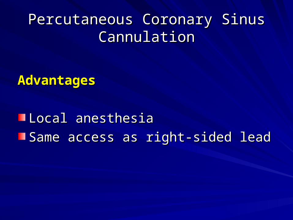
Percutaneous Coronary Sinus Percutaneous Coronary Sinus CannulationCannulation
AdvantagesAdvantages
Local anesthesiaLocal anesthesia
Same access as right-sided leadSame access as right-sided lead
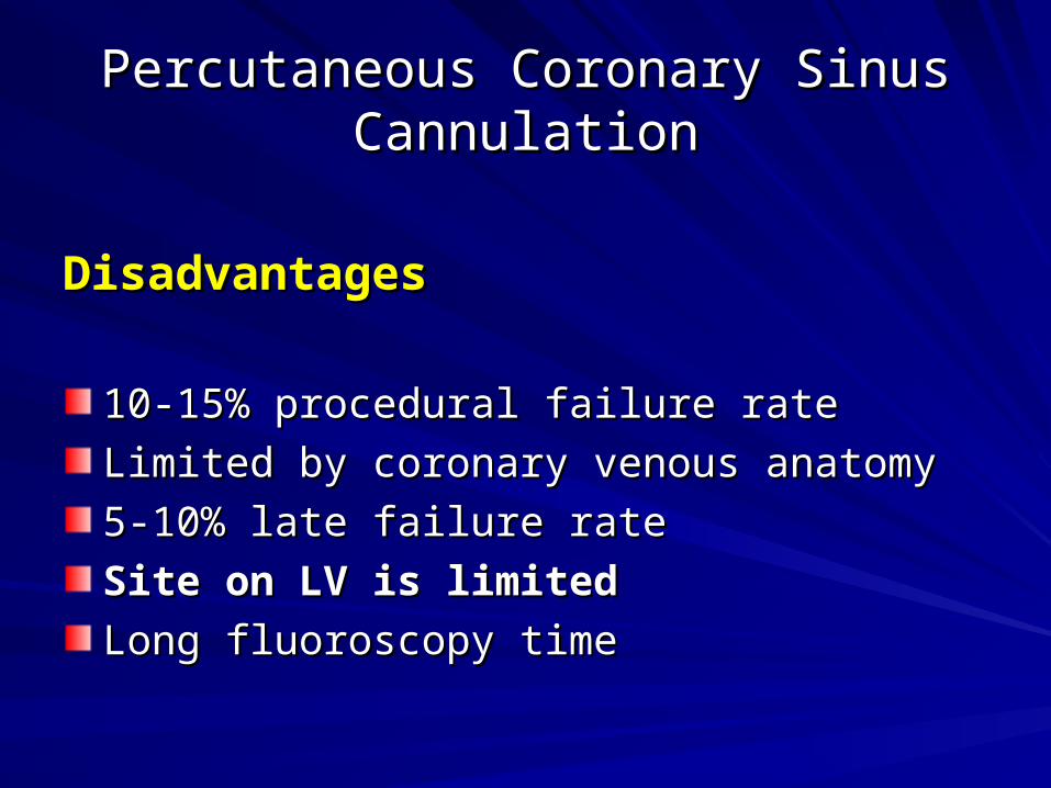
Percutaneous Coronary Sinus Percutaneous Coronary Sinus CannulationCannulation
DisadvantagesDisadvantages
10-15% procedural failure rate10-15% procedural failure rate
Limited by coronary venous anatomyLimited by coronary venous anatomy
5-10% late failure rate 5-10% late failure rate
Site on LV is limited Site on LV is limited
Long fluoroscopy timeLong fluoroscopy time

Surgical LV leads: Is there a need ?Surgical LV leads: Is there a need ?
2 million patients with NYHA class III-IV 2 million patients with NYHA class III-IV CHFCHF
30-50% have widened QRS (600,000-30-50% have widened QRS (600,000-1,000,000)1,000,000)
CS failure rate of 15% (90,000-150,000)CS failure rate of 15% (90,000-150,000)
Lead dislodgement rate of 7% (42,000-Lead dislodgement rate of 7% (42,000-70,000)70,000)
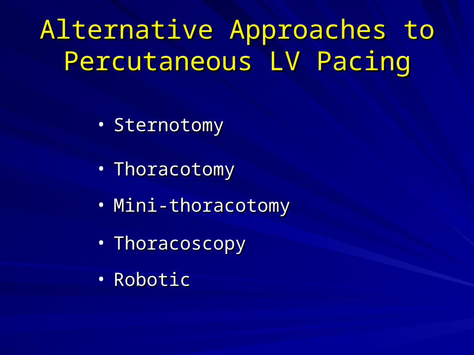
Alternative Approaches to Alternative Approaches to Percutaneous LV PacingPercutaneous LV Pacing
• SternotomySternotomy
• ThoracotomyThoracotomy
• Mini-thoracotomyMini-thoracotomy
• ThoracoscopyThoracoscopy
• RoboticRobotic

ThoracotomyThoracotomy
Most common incision used for CS lead Most common incision used for CS lead failurefailure
Morbidity includes:Morbidity includes:– Postoperative painPostoperative pain– Respiratory complicationsRespiratory complications– Atelectasis/pneumoniaAtelectasis/pneumonia– Several days of recoverySeveral days of recovery
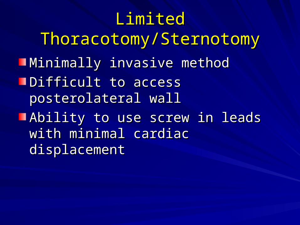
Limited Thoracotomy/SternotomyLimited Thoracotomy/Sternotomy
Minimally invasive methodMinimally invasive method
Difficult to access posterolateral wallDifficult to access posterolateral wall
Ability to use screw in leads with minimal Ability to use screw in leads with minimal cardiac displacementcardiac displacement
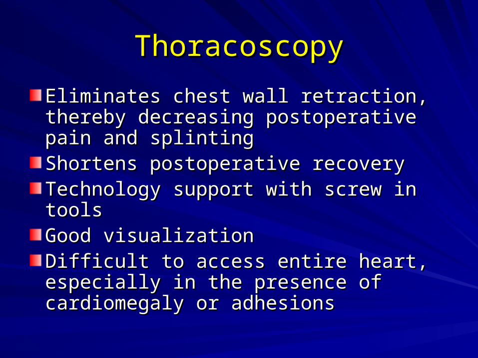
ThoracoscopyThoracoscopy
Eliminates chest wall retraction, thereby Eliminates chest wall retraction, thereby decreasing postoperative pain and decreasing postoperative pain and splintingsplintingShortens postoperative recoveryShortens postoperative recoveryTechnology support with screw in toolsTechnology support with screw in toolsGood visualizationGood visualizationDifficult to access entire heart, especially Difficult to access entire heart, especially in the presence of cardiomegaly or in the presence of cardiomegaly or adhesionsadhesions
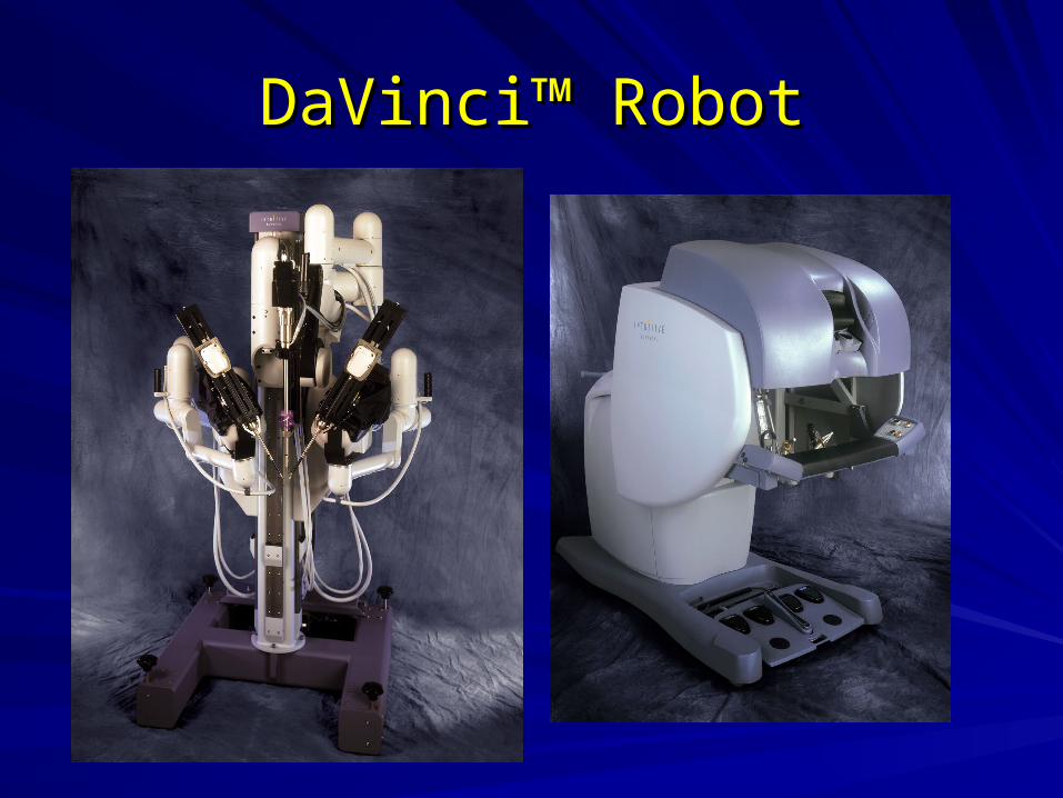
DaVinciDaVinci™™ Robot Robot

Robotic LV Epicardial LeadsRobotic LV Epicardial Leads
AdvantagesAdvantages
Direct placement on any portion of the LVDirect placement on any portion of the LV
Minimally invasive Minimally invasive
Site-directed approachSite-directed approach

Robotic LV Epicardial LeadsRobotic LV Epicardial Leads
DisadvantagesDisadvantages
General anesthesiaGeneral anesthesia
Single lung ventilationSingle lung ventilation
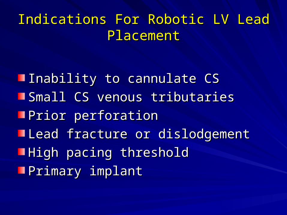
Indications For Robotic LV Lead Indications For Robotic LV Lead PlacementPlacement
Inability to cannulate CSInability to cannulate CS
Small CS venous tributariesSmall CS venous tributaries
Prior perforationPrior perforation
Lead fracture or dislodgementLead fracture or dislodgement
High pacing threshold High pacing threshold
Primary implantPrimary implant

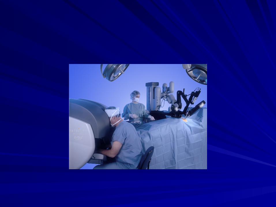
Technical Aspects of Robotic Lead Technical Aspects of Robotic Lead PlacementPlacement
Requires general anesthesiaRequires general anesthesia
Selective single lung ventilationSelective single lung ventilation
Preop pulmonary function testsPreop pulmonary function tests
Posterior approachPosterior approach
Hold anticoagulation (coumadin)Hold anticoagulation (coumadin)
Lead surveillance similar to CS leadsLead surveillance similar to CS leads
Back-up lead kept in device pocketBack-up lead kept in device pocket

Operative TechniqueOperative Technique::The Posterior ApproachThe Posterior Approach
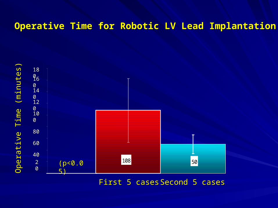
108 50Op
era
tive
Tim
e (m
inu
tes)
Op
era
tive
Tim
e (m
inu
tes)
First 5 casesFirst 5 cases Second 5 casesSecond 5 cases
180180
140140
120120
100100
8080
6060
4040
2020
160160
(p<0.05)(p<0.05)
Operative Time for Robotic LV Lead ImplantationOperative Time for Robotic LV Lead Implantation

Types of Epicardial LV LeadsTypes of Epicardial LV Leads
Steroid-eluting, sew-in leadsSteroid-eluting, sew-in leads
Screw in leadsScrew in leads

Can Robotic Pacing Improve Can Robotic Pacing Improve Clinical Outcomes of CRT ?Clinical Outcomes of CRT ?
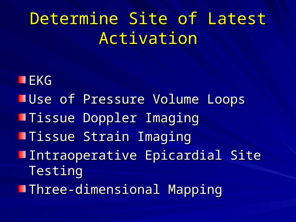
Determine Site of Latest ActivationDetermine Site of Latest Activation
EKGEKG
Use of Pressure Volume LoopsUse of Pressure Volume Loops
Tissue Doppler ImagingTissue Doppler Imaging
Tissue Strain ImagingTissue Strain Imaging
Intraoperative Epicardial Site TestingIntraoperative Epicardial Site Testing
Three-dimensional MappingThree-dimensional Mapping

Echocardiographic MappingEchocardiographic Mapping::Tissue Doppler ImagingTissue Doppler Imaging
Asynchrony CRT
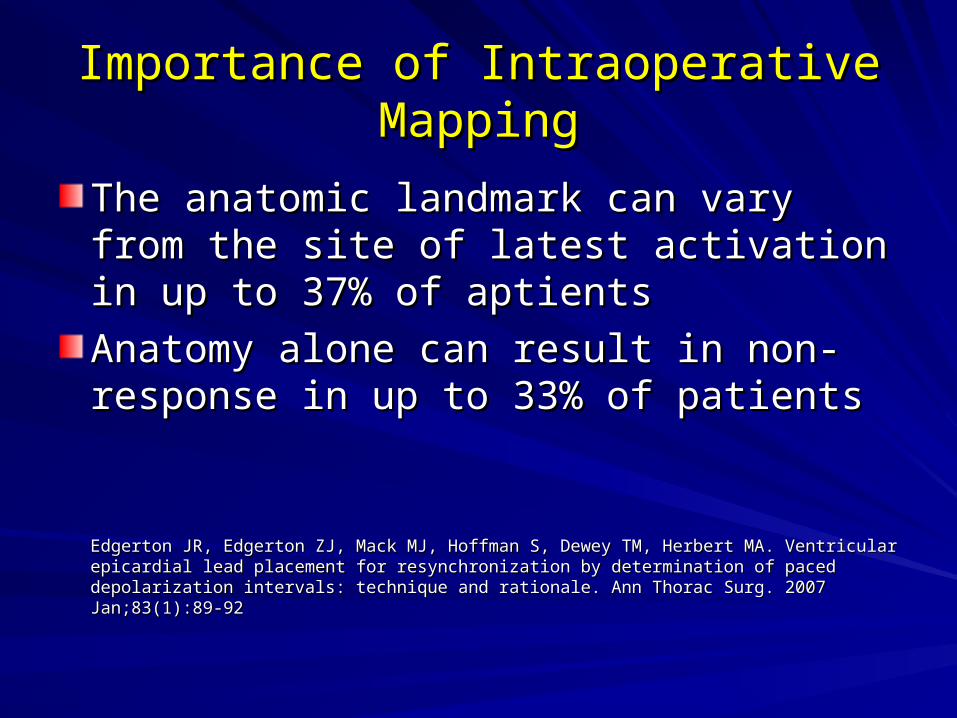
Importance of Intraoperative MappingImportance of Intraoperative Mapping
The anatomic landmark can vary from the The anatomic landmark can vary from the site of latest activation in up to 37% of site of latest activation in up to 37% of aptientsaptients
Anatomy alone can result in non-response Anatomy alone can result in non-response in up to 33% of patientsin up to 33% of patients
Edgerton JR, Edgerton ZJ, Mack MJ, Hoffman S, Dewey TM, Herbert MA. Ventricular epicardial Edgerton JR, Edgerton ZJ, Mack MJ, Hoffman S, Dewey TM, Herbert MA. Ventricular epicardial lead placement for resynchronization by determination of paced depolarization intervals: lead placement for resynchronization by determination of paced depolarization intervals: technique and rationale. Ann Thorac Surg. 2007 Jan;83(1):89-92 technique and rationale. Ann Thorac Surg. 2007 Jan;83(1):89-92
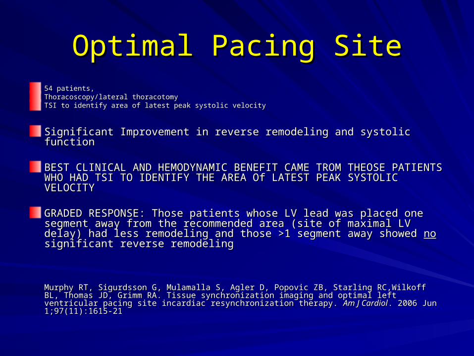
Optimal Pacing SiteOptimal Pacing Site54 patients,54 patients,Thoracoscopy/lateral thoracotomyThoracoscopy/lateral thoracotomyTSI to identify area of latest peak systolic velocityTSI to identify area of latest peak systolic velocity
Significant Improvement in reverse remodeling and systolic functionSignificant Improvement in reverse remodeling and systolic function
BEST CLINICAL AND HEMODYNAMIC BENEFIT CAME TROM THEOSE BEST CLINICAL AND HEMODYNAMIC BENEFIT CAME TROM THEOSE PATIENTS WHO HAD TSI TO IDENTIFY THE AREA Of LATEST PEAK PATIENTS WHO HAD TSI TO IDENTIFY THE AREA Of LATEST PEAK SYSTOLIC VELOCITYSYSTOLIC VELOCITY
GRADED RESPONSE: Those patients whose LV lead was placed one GRADED RESPONSE: Those patients whose LV lead was placed one segment away from the recommended area (site of maximal LV delay) had segment away from the recommended area (site of maximal LV delay) had less remodeling and those >1 segment away showed less remodeling and those >1 segment away showed nono significant reverse significant reverse remodelingremodeling
Murphy RT, Sigurdsson G, Mulamalla S, Agler D, Popovic ZB, Starling RC,Wilkoff BL, Thomas Murphy RT, Sigurdsson G, Mulamalla S, Agler D, Popovic ZB, Starling RC,Wilkoff BL, Thomas JD, Grimm RA. Tissue synchronization imaging and optimal left ventricular pacing site incardiac JD, Grimm RA. Tissue synchronization imaging and optimal left ventricular pacing site incardiac resynchronization therapy. resynchronization therapy. Am J CardiolAm J Cardiol. 2006 Jun 1;97(11):1615-21 . 2006 Jun 1;97(11):1615-21
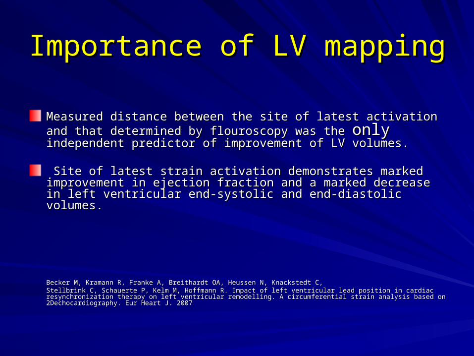
Importance of LV mappingImportance of LV mapping
Measured distance between the site of latest activation and that determined Measured distance between the site of latest activation and that determined by flouroscopy was the by flouroscopy was the onlyonly independent predictor of improvement of LV independent predictor of improvement of LV volumes. volumes.
Site of latest strain activation demonstrates marked improvement in Site of latest strain activation demonstrates marked improvement in ejection fraction and a marked decrease in left ventricular end-systolic and ejection fraction and a marked decrease in left ventricular end-systolic and end-diastolic volumes.end-diastolic volumes.
Becker M, Kramann R, Franke A, Breithardt OA, Heussen N, Knackstedt C,Becker M, Kramann R, Franke A, Breithardt OA, Heussen N, Knackstedt C,Stellbrink C, Schauerte P, Kelm M, Hoffmann R. Impact of left ventricular lead position in cardiac resynchronization therapy on left Stellbrink C, Schauerte P, Kelm M, Hoffmann R. Impact of left ventricular lead position in cardiac resynchronization therapy on left ventricular remodelling. A circumferential strain analysis based on 2Dechocardiography. Eur Heart J. 2007ventricular remodelling. A circumferential strain analysis based on 2Dechocardiography. Eur Heart J. 2007
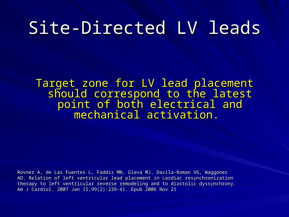
Site-Directed LV leadsSite-Directed LV leads
Target zone for LV lead placement should Target zone for LV lead placement should correspond to the latest point of both electrical correspond to the latest point of both electrical
and mechanical activation.and mechanical activation.
Rovner A, de Las Fuentes L, Faddis MN, Gleva MJ, Davila-Roman VG, WaggonerRovner A, de Las Fuentes L, Faddis MN, Gleva MJ, Davila-Roman VG, WaggonerAD. Relation of left ventricular lead placement in cardiac resynchronizationAD. Relation of left ventricular lead placement in cardiac resynchronizationtherapy to left ventricular reverse remodeling and to diastolic dyssynchrony.therapy to left ventricular reverse remodeling and to diastolic dyssynchrony.Am J Cardiol. 2007 Jan 15;99(2):239-41. Epub 2006 Nov 21Am J Cardiol. 2007 Jan 15;99(2):239-41. Epub 2006 Nov 21

Epicardial vs. Percutaneous LeadsEpicardial vs. Percutaneous Leads
No prospective randomized trials as yetNo prospective randomized trials as yet
Mair et al report a retrospective comparison of Mair et al report a retrospective comparison of – 79 patients with CS lead insertion 79 patients with CS lead insertion – 16 patients with epicardial lead placement through limited thoracotomy16 patients with epicardial lead placement through limited thoracotomy
Results:Results:– 100% patient with epicardial leads had posterolateral placement vs 100% patient with epicardial leads had posterolateral placement vs
70% in transvenous group70% in transvenous group– No statistically different length of stayNo statistically different length of stay– Percutaneous leads had higher thresholds over 16 month follow up. Percutaneous leads had higher thresholds over 16 month follow up.
Mair H, Sachweh J, Meuris B et al. Surgical epicardial left ventricular lead versus Mair H, Sachweh J, Meuris B et al. Surgical epicardial left ventricular lead versus coronary sinus lead placement in biventricular pacing.coronary sinus lead placement in biventricular pacing. Eur J Cardiothorac Surg Eur J Cardiothorac Surg 2005; 27: 235–242.2005; 27: 235–242.
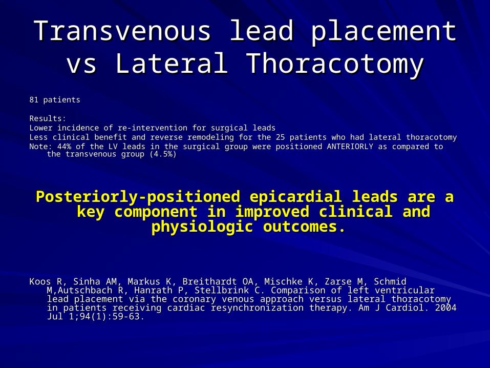
Transvenous lead placement vs Transvenous lead placement vs Lateral ThoracotomyLateral Thoracotomy
81 patients81 patients
Results:Results:Lower incidence of re-intervention for surgical leadsLower incidence of re-intervention for surgical leadsLess clinical benefit and reverse remodeling for the 25 patients who had lateral thoracotomyLess clinical benefit and reverse remodeling for the 25 patients who had lateral thoracotomyNote: 44% of the LV leads in the surgical group were positioned ANTERIORLY as compared to the transvenous group Note: 44% of the LV leads in the surgical group were positioned ANTERIORLY as compared to the transvenous group
(4.5%)(4.5%)
Posteriorly-positioned epicardial leads are a key Posteriorly-positioned epicardial leads are a key component in improved clinical and physiologic component in improved clinical and physiologic
outcomes.outcomes.
Koos R, Sinha AM, Markus K, Breithardt OA, Mischke K, Zarse M, Schmid M,Autschbach R, Hanrath Koos R, Sinha AM, Markus K, Breithardt OA, Mischke K, Zarse M, Schmid M,Autschbach R, Hanrath P, Stellbrink C. Comparison of left ventricular lead placement via the coronary venous approach P, Stellbrink C. Comparison of left ventricular lead placement via the coronary venous approach versus lateral thoracotomy in patients receiving cardiac resynchronization therapy. Am J Cardiol. versus lateral thoracotomy in patients receiving cardiac resynchronization therapy. Am J Cardiol. 2004 Jul 1;94(1):59-63. 2004 Jul 1;94(1):59-63.
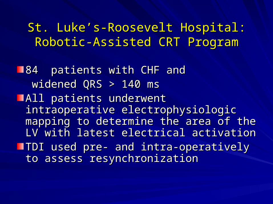
St. Luke’s-Roosevelt Hospital:St. Luke’s-Roosevelt Hospital:Robotic-Assisted CRT ProgramRobotic-Assisted CRT Program
84 patients with CHF and84 patients with CHF and widened QRS > 140 mswidened QRS > 140 ms
All patients underwent intraoperative All patients underwent intraoperative electrophysiologic mapping to determine electrophysiologic mapping to determine the area of the LV with latest electrical the area of the LV with latest electrical activationactivationTDI used pre- and intra-operatively to TDI used pre- and intra-operatively to assess resynchronizationassess resynchronization
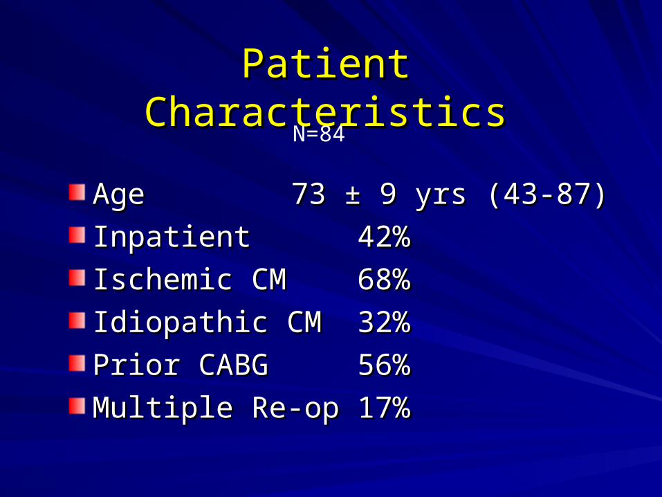
Patient CharacteristicsPatient Characteristics
AgeAge 73 ± 9 yrs (43-87)73 ± 9 yrs (43-87)
InpatientInpatient 42%42%
Ischemic CMIschemic CM 68%68%
Idiopathic CMIdiopathic CM 32%32%
Prior CABGPrior CABG 56%56%
Multiple Re-opMultiple Re-op 17%17%
N=84
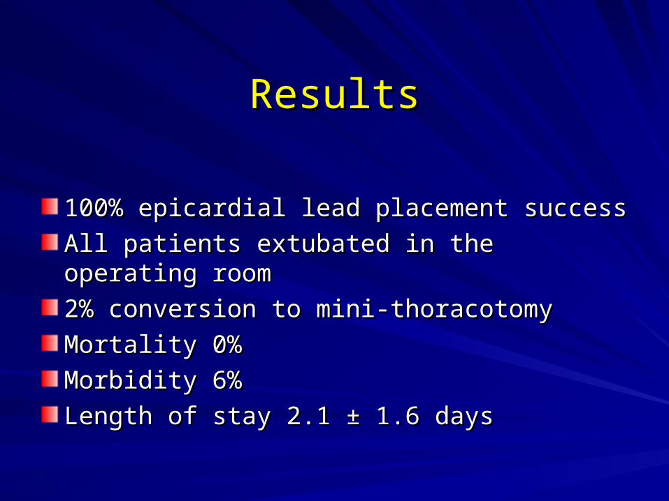
ResultsResults
100% epicardial lead placement success100% epicardial lead placement success
All patients extubated in the operating roomAll patients extubated in the operating room
2% conversion to mini-thoracotomy 2% conversion to mini-thoracotomy
Mortality 0%Mortality 0%
Morbidity 6%Morbidity 6%
Length of stay 2.1 Length of stay 2.1 ± 1.6 days± 1.6 days
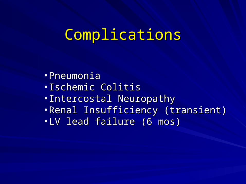
ComplicationsComplications
•PneumoniaPneumonia•Ischemic ColitisIschemic Colitis•Intercostal NeuropathyIntercostal Neuropathy•Renal Insufficiency (transient)Renal Insufficiency (transient)•LV lead failure (6 mos)LV lead failure (6 mos)
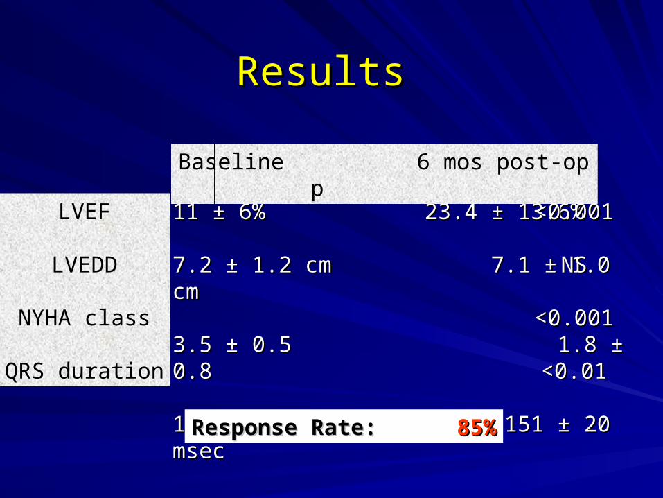
ResultsResults
11 ± 6%11 ± 6% 23.4 ± 13.6% 23.4 ± 13.6%
7.2 ± 1.2 cm7.2 ± 1.2 cm 7.1 ± 1.0 cm 7.1 ± 1.0 cm
3.5 ± 0.5 1.8 ± 0.83.5 ± 0.5 1.8 ± 0.8
184 ± 29 msec184 ± 29 msec 151 ± 20 msec 151 ± 20 msec
Baseline 6 mos post-op p
LVEF
LVEDD
NYHA class
QRS duration
<0.001<0.001
NSNS
<0.001<0.001
<0.01<0.01
Response Rate:Response Rate: 85%85%
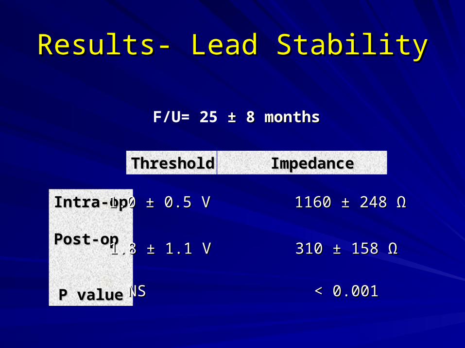
Results- Lead StabilityResults- Lead Stability
F/U= 25 ± 8 months± 8 months
Intra-opIntra-op
Post-op Post-op
P valueP value
ThresholdThreshold Impedance Impedance
1.0 ± 0.5 V1.0 ± 0.5 V 1160 ± 248 1160 ± 248 ΩΩ
1.8 1.8 ±± 1.1 V 1.1 V 310 ± 158 310 ± 158 ΩΩ
NSNS < 0.001 < 0.001
SummarySummary
Robotic LV lead implantation is safe and effectiveRobotic LV lead implantation is safe and effectiveExcellent minimally invasive option for failed CS Excellent minimally invasive option for failed CS cannulationcannulationOptimal portion of myocardium can be targetedOptimal portion of myocardium can be targetedPosterior approach particularly useful for re-opsPosterior approach particularly useful for re-opsEpicardial leads are stable over timeEpicardial leads are stable over timeRole in primary implants awaits randomized trialsRole in primary implants awaits randomized trials