Embed Size (px)
Citation preview
RESEARCH Open Access
ROCK2 deprivation leads to the inhibitionof tumor growth and metastatic potentialin osteosarcoma cells through themodulation of YAP activityCinzia Zucchini1*†, Maria Cristina Manara2†, Camilla Cristalli2, Marianna Carrabotta2, Sara Greco2,Rosa Simona Pinca2, Cristina Ferrari2, Lorena Landuzzi2, Michela Pasello2, Pier-Luigi Lollini1, Marco Gambarotti3,Davide Maria Donati4,5 and Katia Scotlandi2*
Abstract
Background: The treatment of metastatic osteosarcoma (OS) remains a challenge for oncologists, and noveltherapeutic strategies are urgently needed. An understanding of the pathways that regulate OS dissemination isrequired for the design of novel treatment approaches. We recently identified Rho-associated coiled-coil containingprotein kinase 2 (ROCK2) as a crucial driver of OS cell migration. In this study, we explored the impact of ROCK2disruption on the metastatic capabilities of OS cells and analyzed its functional relationship with Yes-associatedprotein-1 (YAP), the main transcriptional mediator of mechanotransduction signaling.
Methods: The effects of ROCK2 depletion on metastasis were studied in NOD Scid gamma (NSG) mice injectedwith U-2OS cells in which ROCK2 expression had been stably silenced. Functional studies were performed in vitroin human U-2OS cells and in three novel cell lines derived from patient-derived xenografts (PDXs) by usingstandard methods to evaluate malignancy parameters and signaling transduction. The nuclear immunostaining ofYAP and the evaluation of its downstream targets Cysteine Rich Angiogenic Inducer 6, Connective Tissue GrowthFactor and Cyclin D1 by quantitative PCR were performed to analyze YAP activity. The effect of the expression andactivity of ROCK2 and YAP on tumor progression was analyzed in 175 OS primary tumors.
Results: The silencing of ROCK2 markedly reduced tumor growth and completely abolished the metastatic abilityof U-2OS cells. The depletion of ROCK2, either by pharmacological inhibition or silencing, induced a dose- andtime-dependent reduction in the nuclear expression and transcriptional activity of YAP. The nuclear expression ofYAP was observed in 80/175 (46%) tumor samples and was significantly correlated with worse patient prognosisand a higher likelihood of metastasis and death. The use of verteporfin, a molecule that specifically inhibits theTEAD–YAP association, remarkably impaired the growth and migration of OS cells in vitro. Moreover to inhibitingYAP activity, our findings indicate that verteporfin also affects the ROCK2 protein and its functions.
(Continued on next page)
© The Author(s). 2019 Open Access This article is distributed under the terms of the Creative Commons Attribution 4.0International License (http://creativecommons.org/licenses/by/4.0/), which permits unrestricted use, distribution, andreproduction in any medium, provided you give appropriate credit to the original author(s) and the source, provide a link tothe Creative Commons license, and indicate if changes were made. The Creative Commons Public Domain Dedication waiver(http://creativecommons.org/publicdomain/zero/1.0/) applies to the data made available in this article, unless otherwise stated.
* Correspondence: [email protected]; [email protected]†Cinzia Zucchini and Maria Cristina Manara contributed equally to this work.1Department of Experimental, Diagnostic and Specialty Medicine, (DIMES),University of Bologna, Via Massarenti 9, 40126 Bologna, BO, Italy2Experimental Oncology Laboratory, IRCCS Istituto Ortopedico Rizzoli, via diBarbiano 1/10, 40136 Bologna, ItalyFull list of author information is available at the end of the article
Zucchini et al. Journal of Experimental & Clinical Cancer Research (2019) 38:503 https://doi.org/10.1186/s13046-019-1506-3
(Continued from previous page)
Conclusions: We describe the functional connection between ROCK2 and YAP in the regulation of OS cellmigration and metastasis formation. These data provide support for the use of verteporfin as a possible therapeuticoption to prevent OS cell dissemination.
Keywords: Osteosarcoma, ROCK2, YAP, Verteporfin, Metastasis,
BackgroundOsteosarcoma (OS), a highly aggressive malignant tumorthat develops in the bone, preferentially occurs in chil-dren and young adults. The prognosis for patients hasimproved greatly during the past three decades due tothe advancement of neoadjuvant and adjuvant chemo-therapy in conjunction with surgery, and at present, the5-year event-free survival rate has reached approximately70% for patients with localized disease [1–3]. However,the prognosis for metastatic patients remains grim, andthe survival rates for patients who present metastases atthe time of diagnosis are below 30% [4]. Thus, treatingmetastatic OS remains a challenge for oncologists, and adeeper understanding of the biology underlying metasta-sis in OS is an urgent need for the development of noveland more targeted therapeutic options.The ability of cancer cells to spread to secondary organs
outside of the primary tumor site requires mechanicalforces exerted via actin cytoskeleton dynamics. The actinstatus is used as a signaling intermediary by a variety ofpathways associated with cancer cell dissemination andmetastasis, including the Hippo signaling pathway, anoncosuppressive pathway that plays multiple critical rolesin the control of cellular malignancy. Canonical Hippotransduction involves a cascade of serine/threonine ki-nases that phosphorylate and inhibit Yes-associatedprotein-1 (YAP) and its coactivator TAZ, promoting theircytoplasmic retention and/or subsequent degradation.When Hippo signaling is ‘off’, YAP and TAZ translocateto the nucleus, where they interact with the transcriptionfactors TEAD1–4 to induce the expression of target genesresponsible for cellular proliferation, differentiation andsurvival [5]. The dysregulation of Hippo signaling and/orYAP activity occurs frequent in a variety of human can-cers [6], including OS, as YAP is highly expressed in bothhuman and mouse OS. YAP suppression sharply decreasescell proliferation, cancer stemness and tumorigenicity [7–9], thereby acting as a potential therapeutic target for tu-mors. In addition to operating in Hippo signaling, YAPalso senses and mediates the integrity of the actomyosincytoskeleton and the intracellular mechanotransductionpathway [10–13]. The actin status is also controlled by theRho/Rho-associated coiled-coil containing protein kinase(ROCK) pathway, which can sustain and promote YAP ac-tivity through the phosphorylation of several moleculartargets that are induced by Rho-associated coiled-coil
containing protein kinase 1 and 2 (ROCK1 and ROCK2)[11, 14]. Thus, the Hippo pathway, the cytoskeleton, Rho/ROCK and YAP/TAZ may form a complex molecular net-work of multilayered interactions with feedback mecha-nisms, whose connections are still poorly understood andmay differ in diverse cellular contexts. In OS, we have pre-viously highlighted the importance of ROCK2, rather thanROCK1, as a crucial mediator of cell migration and inva-sion [15]. In this study, we analyzed the impact of ROCK2depletion on OS metastasis and its functional connectionswith YAP activity. We also tested verteporfin, a small mol-ecule that specifically inhibits the TEAD–YAP association[16], as a potential therapeutic agent for OS.
MethodsCell linesThe U-2OS OS cell line was obtained from theAmerican Type Culture Collection (ATCC). Theprimary cultures PDX-OS#2-C1, PDX-OS#16-C2 andPDX-OS#25-C1 were recently obtained from OSpatient-derived xenografts (PDXs) after one or twopassages in animals [17]. Patient informed consentwas obtained for the establishment of the PDXmodels. All cell lines were tested for mycoplasmacontamination (Mycoalert Mycoplasma Detection Kit,Lonza) before use. Cell lines were immediately ex-panded to generate liquid nitrogen stocks and werenever passaged for more than 1 month after thawing.Cells were grown in Iscove’s modified Dulbecco’smedium (IMDM) supplemented with 10% inactivatedfetal bovine serum (FBS) (Euroclone), 100 units/mlpenicillin and 100 μg/ml streptomycin (Sigma). Cellswere maintained at 37 °C in a humidified 5% CO2
atmosphere.
Stable silencingFor stable silencing, a short hairpin RNA (shRNA) plasmid(pSilencer 2.1-U6 Neo vector; Ambion) expressing ROCK2siRNA (Fw: 5′-GATCCCGGCAACTGGCTCGTTCAATTTTCAAGAGA TTAACTTGCTCGGTCAACGTTTTTTGGAA-3′; Rw: 5′-AGCTTTTCCAAAAAACGTTGACCGAGCAAGTTAATCTCTTGAAAATTGAACGAGCCAGTTGCCGG-3′) was created, and U-2OS cells were transfectedusing the calcium phosphate transfection method (Life Tech-nologies). Stable transfectants expressing shRNA-ROCK2(U-2/shROCK2#78 and #46) or nontargeting shRNA
Zucchini et al. Journal of Experimental & Clinical Cancer Research (2019) 38:503 Page 2 of 14
sequences (U-2/SCR pool) were obtained after selection inneomycin (500 μg/ml) (Sigma).
TreatmentsFor transient ROCK2 silencing, cells were transfected withsmall interfering RNA (siRNA) sequences targeting ROCK2(ON-TARGETplus SMARTpool, Human ROCK2, Dharma-con) or irrelevant targets (ON-TARGETplus Non-targetingsiRNA). For ROCK2 inhibition, the ROCK2 inhibitor N-(2-(2-(dimethylamino)ethoxy)-4-(1H-pyrazol-4-yl)phenyl)-2,3dihydrobenzo[b]1, 4 dioxine-2-carboxamide (StemoleculeROCK2 Inhibitor, Stemgent) was used. To inhibit YAP ac-tivity, tests were performed with the YAP inhibitor vertepor-fin (Sigma). Both compounds were dissolved in dimethylsulfoxide (DMSO; Sigma-Aldrich). Working solutions wereprepared in IMDM immediately before use.
Motility assayCells (1 × 10⁵) were pretreated with or without the YAPinhibitor verteporfin (2 μM) for 24 h, after which theywere analyzed for their migration ability. A motilityassay was performed using Transwell chambers (Costar)with 8-μm pore size polyvinylpyrrolidone-free polycar-bonate filters (Nucleopore). Cells were seeded in IMDMwith 10% FBS in the upper compartment and were incu-bated for 18 h at 37 °C. The number of cells that mi-grated toward the filter to reach the lower chamber wascounted after fixation with methanol and staining withGiemsa (Sigma).
Wound-healing assayA total of 2 × 10⁵ U-2OS cells were seeded in 60-mmPetri-dish well plates. Cells were allowed to grow to100% confluence. The cell monolayer was scraped in astraight line to create a scratch with a p200 pipet tip.The debris was removed, and the medium was replacedwith IMDM with 10% FBS with or without 2 μM verte-porfin. Cells were kept in a tissue culture incubator at37 °C, and pictures were taken at 0, 3 and 6 h.
Cell growth inhibitionTo perform cell culture experiments, OS cells (2X10⁵/well for U-2OS or 4 × 10⁵/well for PDX-OS primary cul-tures) were plated, and verteporfin (0.1–10 μM) wasadded after 24 h. Cells were exposed to the drug for upto 96 h before being counted by Trypan blue vital dyeexclusion (Sigma). In parallel, cells were treated withDMSO-containing medium as a control. The highestfinal concentration of DMSO in the medium was < 0.3%,and DMSO had no effect on cell growth.Anchorage-independent growth was measured in
0.33% agarose (Sea-Plaque; Lonza) with a 0.5% agaroseunderlay. OS cells (10,000 for U-2OS or 100,000 forPDX-OS#16-C2) were plated in semisolid medium with
or without verteporfin (2 μM) and were incubated at37 °C in a humidified 5% CO2 atmosphere. Colonieswere counted after 10 and 14 days for U-2OS or PDX-OS#16-C2, respectively.
ImmunofluorescenceCells grown on coverslips were treated with verteporfinas described above. Cells were fixed in 4% paraformalde-hyde were permeabilized with 0.15% Triton X-100(Sigma) in phosphate-buffered saline or in methanol andwere incubated with the following antibodies: anti-YAP(sc-271134, dilution 1:25), anti-β-catenin (sc-7963, dilu-tion 1:50), and anti-ROCK2 (sc-398,519, dilution 1:50)that were all purchased from Santa Cruz Biotechnolo-gies; and anti-N-cadherin (BD Transduction Labs,610921, dilution 1:100). Anti-mouse FITC (Thermo Sci-entific, #31569, dilution 1:100) or anti-goat IgG NL493(FITC equivalent R&D, #NL003, dilution 1:50) were usedas secondary antibodies. Nuclei were counterstainedwith Hoechst 33256 (Sigma). Images were acquiredusing a Nikon ECLIPSE 90i microscope and were thenanalyzed with NIS-Elements software (Nikon).
In vivo experimentsFemale, 5 weeks old, immunodeficient NOD Scidgamma (NSG) mice were obtained from Charles River,Italy. Groups of 6 mice received injections of 107 U-2OS cells subcutaneously. Tumor growth was measuredweekly and tumor volumes were calculated as π/2·[√(a·b)]3/6, where a and b are the two maximal diame-ters. After 9–10 weeks, animals were sacrificed by CO2
inhalation and cervical dislocation, and an accuratenecropsy was performed. Tumors were removed forfurther studies; lungs were perfused with black Indiaink and fixed. Lung metastases were then countedunder a dissecting microscope.
RNA extraction and qPCRTotal RNA from snap-frozen tissue samples and cell lineswas isolated using TRIzol Reagent (Thermo Fisher Scientific- Life Technologies). RNA quality and quantity were assessedby NanoDrop analysis (NanoDrop ND1000, Thermo Scien-tific) and by electrophoresis. Total RNA from each samplewas reverse transcribed into complementary DNA (cDNA)using a High-Capacity cDNA Reverse Transcription Kit(Thermo Scientific - Applied Biosystems, #4368814) accord-ing to the manufacturer’s protocols. Quantitative PCR(qPCR) was performed on a ViiA7 system (Life Technolo-gies) using TaqMan Universal PCR Master Mix (ThermoFisher Scientific - Applied Biosystems, #4304437) and SYBRGreen PCR Master Mix (Thermo Fisher Scientific - AppliedBiosystems, #4312704). Predesigned TaqMan probes(Thermo Fisher Scientific - Applied Biosystems) were usedfor Connective Tissue Growth Factor (CTGF) (Hs00170014)
Zucchini et al. Journal of Experimental & Clinical Cancer Research (2019) 38:503 Page 3 of 14
Cysteine Rich Angiogenic Inducer 61 (CYR61) (Hs00155479)and Cyclin D1 (CCND1) (Hs00765553). The primers usedare ROCK2 forward 5′- CAACTGTGAGGCTTGTATGAAG-3′ and reverse 5′-TGCAAGGTGCTATAATCTCCTC-3′; GAPDH forward: 5′-GAAGGTGAAGGTCGGAGTC-3′, reverse: 5′-GAAGATGGTGATGGGATTTC-3′.Relativequantification was performed in tumor samples with theΔCT method (relative abundance, RA= 2- ΔCT) while theΔΔCT method (relative quantification, RQ= 2- ΔΔCT) wasused for cell line analysis. The expression levels of the targetgenes were normalized to those of the housekeeping geneGAPDH (Hs99999905_m1). Untreated cells (CTRL) or cellsexposed to an shRNA against irrelevant targets (SCR) wereused as controls.
Western blottingSubconfluent cells were treated as described above andwere processed for Western blotting following standardprocedures, using total protein lysates or fractionatedproteins, where appropriate. Cytoplasmic proteins wereobtained using the lysis buffer containing 50 mmol/LHEPES (pH 7.5), 150 mmol/L NaCl, 1% Triton X-100,1.5 mmol/L MgCl2, EGTA, 10 mmol/L (pH 7.5), glycerol10%, and inhibitors (0.1 mmol/L Na3VO4, 1% phenyl-methylsulfonyl fluoride, and 20 mg/mL aprotinin). Afterthe collection of cytoplasmic proteins, the nuclei werelysed with the nuclear buffer containing 20 mmol/LHEPES (pH 8), 0.1 mmol/L EDTA, 5 mmol/L MgCl2,0.5 mol/L NaCl, 20% glycerol, 1% Nonidet P40, and in-hibitors (as above). The following primary antibodieswere used: anti-ROCK2 (Abcam, #ab125025, dilution 1:12000); anti-YAP (Cell Signaling, #14074, dilution 1:1000) anti-GAPDH (Santa Cruz, sc-25,778, dilution 1:5000) and anti-Lamin B (Santa Cruz, sc-6216, dilution 1:5000). Anti-rabbit (GE Healthcare, #NA934), anti-mouse(GE Healthcare, #NA931) or anti-goat (Santa Cruz, sc-2020) secondary antibodies conjugated to horseradishperoxidase were used, and bands were visualized withenhanced chemiluminescence Western blotting detec-tion reagents (EuroClone).
PatientsPatients with localized primary OS who were enrolled inprospective studies and were treated at the Rizzoli Insti-tute were included in the current analysis. The presentstudy included 175 tumor samples from biopsy speci-mens (obtained before chemotherapy and preserved inarchival paraffin-embedded tissue blocks) that wereavailable for immunohistochemical analysis and had ad-equate tissue. All tumors were classified as stage II con-ventional high-grade OS [18]. Chemotherapy was givenbefore and after surgery. Chemotherapy protocols basedon doxorubicin, high-dose methotrexate, cisplatin and/or ifosfamide have been previously described [19–22].
The surgical procedures took into account the locationand extent of the tumor and the life expectancy of thepatient. A limb-salvage procedure was performed in 158patients (90%). The surgical margins of the tumor speci-mens were histologically defined according to the systemof Enneking [18]. The extent of tumor necrosis was eval-uated with a previously described semiquantitativemethod [23]. Adverse events were defined as a recur-rence of the tumor at any site (local or systemic) ordeath during remission. Relapse-free survival (RFS) wascalculated from the date of the initial diagnosis. The me-dian follow-up of the population was 95 months (range2–415 months). Clinical and follow-up data were up-dated until December 2018. The rates of RFS and overallsurvival (OVS) were 51.4 and 69.7%, respectively. Table 1summarizes the clinical and pathological characteristicsof the 175 patients.
ImmunohistochemistryAn avidin–biotin–peroxidase procedure was used forimmunostaining (Vector Laboratories). Antigen retrievalwas performed using citrate buffer (pH 6.0), followed byincubation with anti-YAP (sc-271134, dilution 1:50) oranti-ROCK2 (sc-398,519, dilution 1:50). In human tumorsamples, we used a semiquantitative score for YAP im-munostaining to evaluate its level of expression togetherwith an analysis of its intracellular location to evaluateits activity. Patients were classified as positive when thenuclear positivity of YAP was detected. The expressionlevels were scored as follows: negative, when no stainingwas observed; positive, including weak (+ − -), moderate(++−), and strong (+++) positivity levels.
Statistical analysisDifferences among the means were analyzed using Stu-dent’s t tests. For analysis of incidence and median num-ber of lung metastasis Fisher’s exact test and Wilcoxon’srank sum test were used. CalcuSyn2 software (Biosoft)was used to calculate the IC50 values. The association be-tween YAP expression and RFS or OVS was estimated byCox proportional hazards regression analysis. RFS andOVS were plotted using the Kaplan-Meier method, whilethe log-rank test was used to calculate the univariate stat-istical significance of the observed differences. RFS wascalculated as the time from diagnosis to the occurrence ofadverse events, which were defined as recurrence or me-tastases at any site. OVS was defined as the time fromdiagnosis to cancer-related death. Survivors or patientswho were lost to follow-up were censored at the last con-tact date. All factors that were significantly associated withRFS in the univariate analysis were entered into a Coxproportional hazards model for multivariate analysis.Values for the 95% confidence interval (CI) of the hazardratios (HRs) are provided [24]. The Chi square test was
Zucchini et al. Journal of Experimental & Clinical Cancer Research (2019) 38:503 Page 4 of 14
used for association data. Statistical analyses were per-formed with SPSS software, version 22.0.
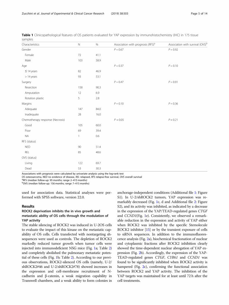
ResultsROCK2 deprivation inhibits the in vivo growth andmetastatic ability of OS cells through the modulation ofYAP activityThe stable silencing of ROCK2 was induced in U-2OS cellsto evaluate the impact of this kinase on the metastatic cap-ability of OS cells. Cells transfected with nontargeting sh-sequences were used as controls. The depletion of ROCK2markedly reduced tumor growth when tumor cells wereinjected into immunodeficient NSG mice (Fig. 1a; Table 2)and completely abolished the pulmonary metastatic poten-tial of these cells (Fig. 1b; Table 2). According to our previ-ous observations, ROCK2-silenced OS cells (namely, U-2/shROCK2#46 and U-2/shROCK2#78) showed increases inthe expression and cell-membrane recruitment of N-cadherin and β-catenin, a weak migration capability inTranswell chambers, and a weak ability to form colonies in
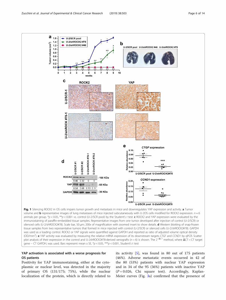
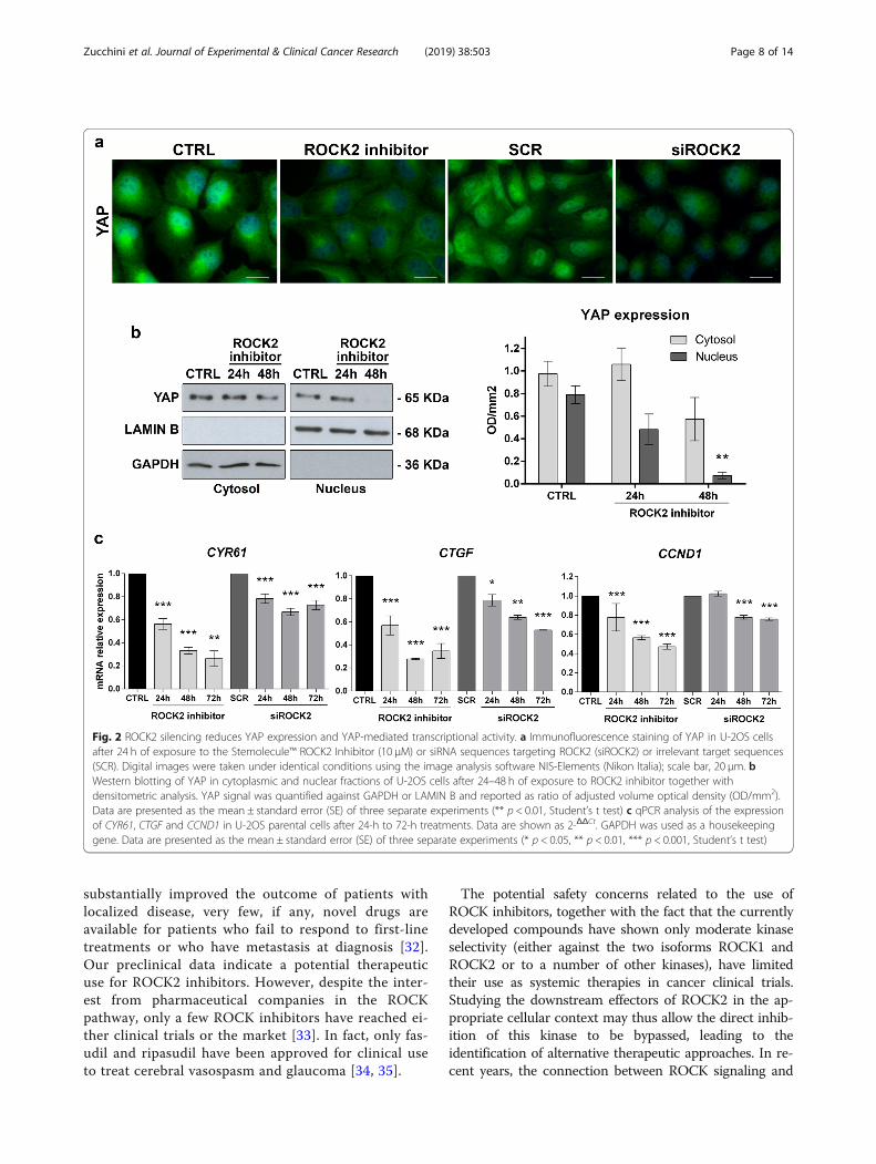
anchorage-independent conditions (Additional file 1: FigureS1). In U-2/shROCK2 tumors, YAP expression was re-markably decreased (Fig. 1c, d and Additional file 2: FigureS2), and its activity was inhibited, as indicated by a decreasein the expression of the YAP/TEAD-regulated genes CTGFand CCND1(Fig. 1e). Consistently, we observed a remark-able reduction in the expression and activity of YAP eitherwhen ROCK2 was inhibited by the specific StemoleculeROCK2 inhibitor [15] or by the transient exposure of cellsto siRNA sequences. In addition to the immunofluores-cence analysis (Fig. 2a), biochemical fractionation of nuclearand cytoplasmic fractions after ROCK2 inhibition clearlyshowed the time-dependent nuclear abrogation of YAP ex-pression (Fig. 2b). Accordingly, the expression of the YAP-TEAD-regulated genes CTGF, CYR61 and CCND1 wasfound to be significantly inhibited when ROCK2 activity ishampered (Fig. 2c), confirming the functional associationbetween ROCK2 and YAP activity. The inhibition of theYAP targets was maintained for at least until 72 h after thecell treatments.
Table 1 Clinicopathological features of OS patients evaluated for YAP expression by immunohistochemistry (IHC) in 175 tissuesamples
Characteristics N % Association with prognosis (RFS)a Association with survival (OVS)b
Gender P = 0.67 P = 0.92
Female 72 41.1
Male 103 58.9
Age P = 0.37 P = 0.10
≤ 14 years 82 46.9
> 14 years 93 53.1
Surgery P = 0.47 P = 0.91
Resection 158 90.3
Amputation 12 6.9
Rotation plastic 5 2.8
Margins P = 0.10 P = 0.36
Adequate 147 84.0
Inadequate 28 16.0
Chemotherapy response (Necrosis) P = 0.05 P = 0.21
Good 105 60.0
Poor 69 39.4
NA 1 0.6
RFS (status)
NED 90 51.4
REL 85 48.6
OVS (status)
Living 122 69.7
Dead 53 30.3
Associations with prognosis were calculated by univariate analysis using the log-rank testOS osteosarcoma, NED no evidence of disease, REL relapsed, RFS relapse-free survival, OVS overall survivalaRFS (median follow-up: 95 months; range 2–415 months)bOVS (median follow-up: 156 months; range 7–415 months)
Zucchini et al. Journal of Experimental & Clinical Cancer Research (2019) 38:503 Page 5 of 14
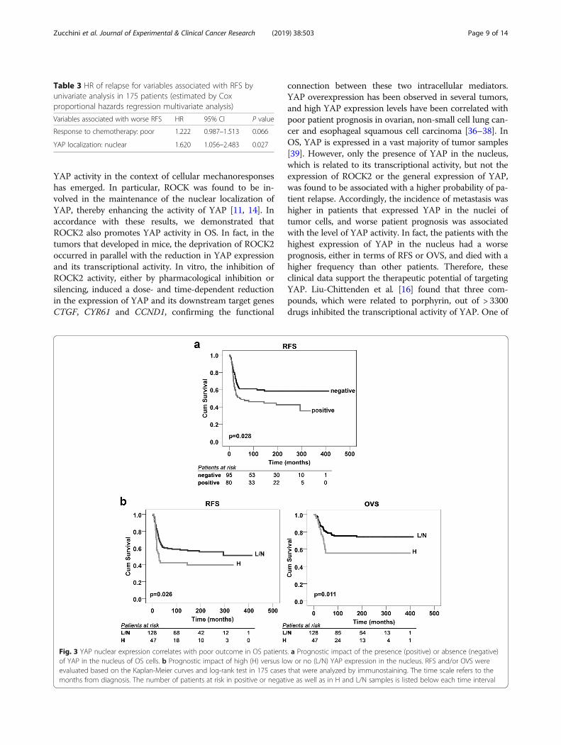
YAP activation is associated with a worse prognosis forOS patientsPositivity for YAP immunostaining, either at the cyto-plasmic or nuclear level, was detected in the majorityof primary OS (131/175; 75%), while the nuclearlocalization of the protein, which is directly related to
its activity [5], was found in 80 out of 175 patients(46%). Adverse metastatic events occurred in 42 ofthe 80 (53%) patients with nuclear YAP expressionand in 34 of the 95 (36%) patients with inactive YAP(P = 0.026, Chi square test). Accordingly, Kaplan-Meier curves (Fig. 3a) confirmed that the presence of
Fig. 1 Silencing ROCK2 in OS cells impairs tumor growth and metastasis in mice and downregulates YAP expression and activity. a Tumorvolume and b representative images of lung metastases of mice injected subcutaneously with U-2OS cells modified for ROCK2 expression. n = 6animals per group. *p < 0.05, **p < 0.001 vs. control (U-2/SCR pool) by the Student’s t test. c ROCK2 and YAP expression were evaluated by theimmunostaining of paraffin-embedded tissue samples. Representative images from one tumor developed after injection of control (U-2/SCR) orsilenced cells (U-2/shROCK2#78). Scale bar, 50 μm; 200x of magnification with zoomed insert to show details. d Western blotting of snap-frozentissue samples from two representative tumors that formed in mice injected with control (U-2/SCR) or silenced cells (U-2/shROCK2#78). GAPDHwas used as a loading control. ROCK2 or YAP signals were quantified against GAPDH and reported as ratio of adjusted volume optical density(OD/mm2). e YAP activity was evaluated by measuring the relative mRNA expression of its downstream targets CTGF and CCND1 by qPCR. Scatterplot analysis of their expression in the control and U-2shROCK2#78-derived xenografts (n = 6) is shown. The 2−ΔCT method, where ΔCT = CT targetgene – CT GAPDH, was used. Bars represent mean ± SE, *p < 0.05, ***p < 0.001, Student’s t test
Zucchini et al. Journal of Experimental & Clinical Cancer Research (2019) 38:503 Page 6 of 14
YAP in the nucleus of OS cells was significantly asso-ciated with a decreased probability of remainingevent-free after diagnosis (P = 0.028, log-rank test).Cox multivariate regression analysis was performedfor the variables that were found to be associatedwith RFS by univariate analysis and showed that thenuclear status of YAP was the only independent riskfactor for poor outcomes (Table 3). To further con-firm this observation, we used the strong expressionof YAP in the nucleus (++/− and +++) to stratify pa-tients as high-expressors (H) or low-expressors/non-expressors (L/N) (47 vs 128 patients). Kaplan-Meiercurves confirmed that very high YAP expression inthe nucleus significantly affected both RFS and OVSin OS patients (Fig. 3b), indicating that the level ofYAP activity is critical for patient outcomes. Consist-ently, the percentage of patients who died from thisdisease was significantly higher in those with highlevels of active YAP (dead patients: 21/47, 45% vs 32/128, 25%, respectively; p = 0.012, Chi square test).
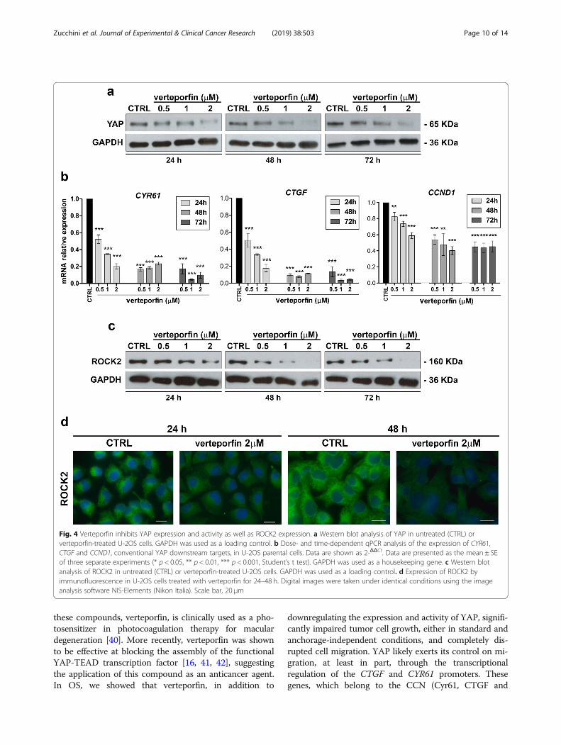
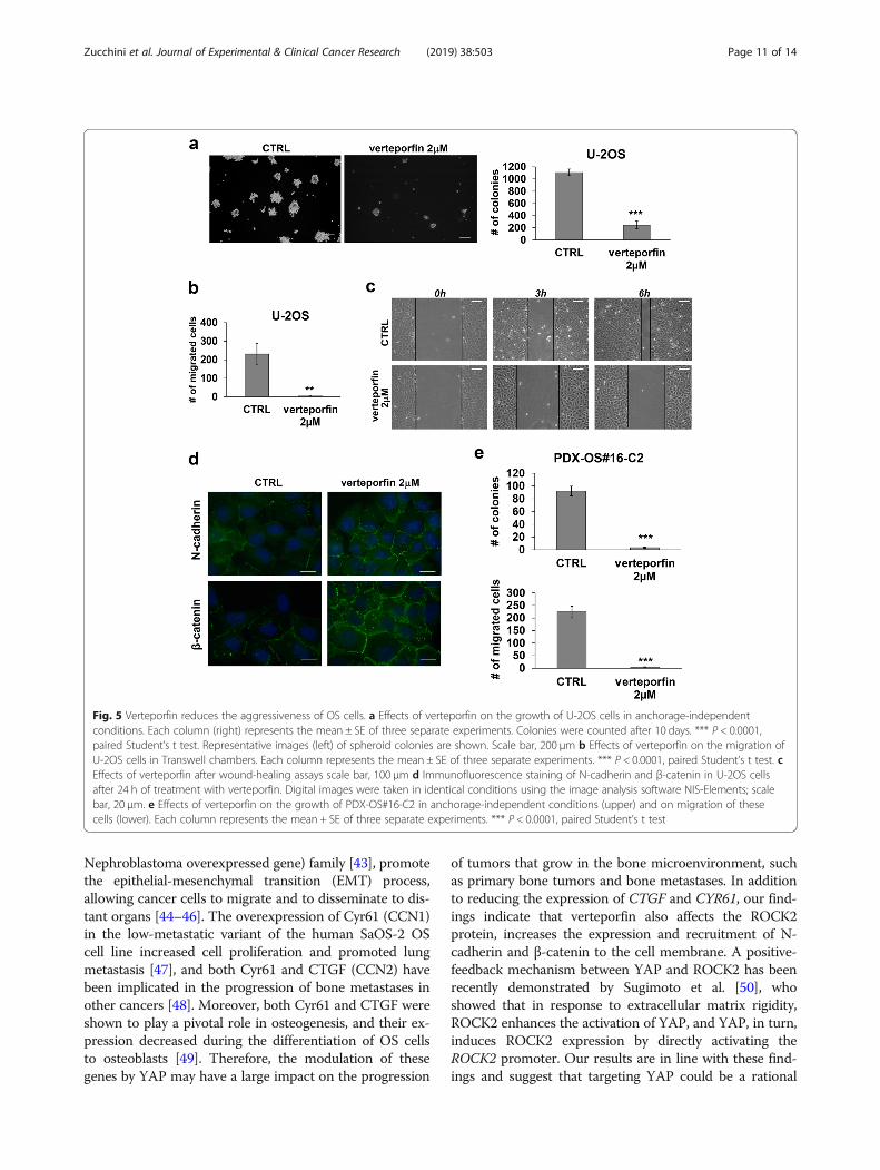
Targeting YAP with verteporfin inhibits the malignancy ofOS cellsTo test the therapeutic potential of YAP inhibition inOS, we used verteporfin, a porphyrin compound thatwas reported to block YAP-TEAD interactions [16].Verteporfin effectively reduced U-2OS cell viability,with an IC50 value of 1.44 ± 0.46 μM. As demonstratedin other tumors, including synovial sarcoma [25], ver-teporfin led to a dose- and time-dependent reductionin the expression (Fig. 4a) and activity (Fig. 4b) ofYAP. Notably, verteporfin was also able to induce adose- and time-dependent decrease in ROCK2 expres-sion both at mRNA (Additional file 3: Figure S3) andprotein levels (Fig. 4c, d), confirming the functionalinterconnection between YAP and ROCK2. Vertepor-fin treatment significantly inhibited the anchorage-independent growth of OS cells (Fig. 5a) andcompletely abrogated the migration of these cells (Fig.5b and c). Cells treated with verteporfin showed
increased expression and cell-membrane recruitmentof N-cadherin and β-catenin (Fig. 5d), thereby display-ing the same phenotype that was previously observedafter ROCK2 depletion (Additional file 1: Figure S1).To build on our observations, we confirmed the
growth-suppressive effects of verteporfin in three celllines derived from PDXs, which have been reported tomodel the genetic features of human tumors, includingbone sarcomas, with a high level of fidelity [17, 26–29].Verteporfin effectively suppressed the cell growth of allthree cell lines under standard conditions, with IC50values ranging from 1 to 2 μM. In addition, in PDX-OS#16-C2, which expressed the highest levels of ROCK2and YAP (Additional file 4: Figure S4), verteporfin com-pletely suppressed the capability of these cells to formcolonies and to migrate (Fig. 5e).
DiscussionROCK2 kinase has been described as a critical medi-ator of biological functions that are implicated in themetastatic processes, including the disruption of adhe-rens junctions, actin cytoskeleton remodeling, the dis-sociation of cell clusters and increased cell motility[30, 31]. In OS, we have previously shown that ROCK2is a crucial intracellular mediator of the CD99-induced suppression of cell migration [15]. The inhib-ition of ROCK2 was shown to impair the migratoryand adhesive behavior of OS cells by decreasing theexpression of ezrin, an actin-binding protein that leadsto cytoskeletal regulation, and by recruiting N-cadherin and β-catenin to the cell membrane. In thisstudy, we expanded these observations and showedthat when ROCK2 expression was stably downregu-lated in OS cells, tumor growth was significantlyinhibited in NSG mice and, notably, tumors com-pletely lost the capability to disseminate and to formspontaneous metastases in the lungs. These resultsstrongly support the idea of specifically targetingROCK2 kinase to prevent the formation of metastasisin OS. Although neoadjuvant chemotherapy has
Table 2 Tumorigenicity and metastatic ability of U-2OS cells after the depletion of ROCK2
Cells Tumor Lung metastases
Incidence Latency (mean days ±SEM)
Volume at 9 weeks(meancm3 ± SEM)
Incidence Mediannumber
Individual values
U-2/SCR pool 6/6 23.2 ± 1.2 1.635 ± 0.051 6/6 > 200 > 200, > 200, > 200, > 200, > 200, >200
U-2/sh ROCK2#78
6/6 37 ± 2.9 0.958 ± 0.233* 0/6† 0‡ 0, 0, 0, 0, 0, 0
U-2/sh ROCK2#46
0/6 NA 0** 0/6† 0‡ 0, 0, 0, 0, 0, 0
†p = 0.01, Fisher’s exact test, vs U-2/SCR pool*p < 0.05, **p <0.0001, Student’s t test, vs. U-2/SCR pool‡p < 0.01, Wilcoxon’s rank sum test, vs. U-2/SCR pool
Zucchini et al. Journal of Experimental & Clinical Cancer Research (2019) 38:503 Page 7 of 14
substantially improved the outcome of patients withlocalized disease, very few, if any, novel drugs areavailable for patients who fail to respond to first-linetreatments or who have metastasis at diagnosis [32].Our preclinical data indicate a potential therapeuticuse for ROCK2 inhibitors. However, despite the inter-est from pharmaceutical companies in the ROCKpathway, only a few ROCK inhibitors have reached ei-ther clinical trials or the market [33]. In fact, only fas-udil and ripasudil have been approved for clinical useto treat cerebral vasospasm and glaucoma [34, 35].
The potential safety concerns related to the use ofROCK inhibitors, together with the fact that the currentlydeveloped compounds have shown only moderate kinaseselectivity (either against the two isoforms ROCK1 andROCK2 or to a number of other kinases), have limitedtheir use as systemic therapies in cancer clinical trials.Studying the downstream effectors of ROCK2 in the ap-propriate cellular context may thus allow the direct inhib-ition of this kinase to be bypassed, leading to theidentification of alternative therapeutic approaches. In re-cent years, the connection between ROCK signaling and
Fig. 2 ROCK2 silencing reduces YAP expression and YAP-mediated transcriptional activity. a Immunofluorescence staining of YAP in U-2OS cellsafter 24 h of exposure to the Stemolecule™ ROCK2 Inhibitor (10 μM) or siRNA sequences targeting ROCK2 (siROCK2) or irrelevant target sequences(SCR). Digital images were taken under identical conditions using the image analysis software NIS-Elements (Nikon Italia); scale bar, 20 μm. bWestern blotting of YAP in cytoplasmic and nuclear fractions of U-2OS cells after 24–48 h of exposure to ROCK2 inhibitor together withdensitometric analysis. YAP signal was quantified against GAPDH or LAMIN B and reported as ratio of adjusted volume optical density (OD/mm2).Data are presented as the mean ± standard error (SE) of three separate experiments (** p < 0.01, Student’s t test) c qPCR analysis of the expressionof CYR61, CTGF and CCND1 in U-2OS parental cells after 24-h to 72-h treatments. Data are shown as 2-ΔΔCt. GAPDH was used as a housekeepinggene. Data are presented as the mean ± standard error (SE) of three separate experiments (* p < 0.05, ** p < 0.01, *** p < 0.001, Student’s t test)
Zucchini et al. Journal of Experimental & Clinical Cancer Research (2019) 38:503 Page 8 of 14
YAP activity in the context of cellular mechanoresponseshas emerged. In particular, ROCK was found to be in-volved in the maintenance of the nuclear localization ofYAP, thereby enhancing the activity of YAP [11, 14]. Inaccordance with these results, we demonstrated thatROCK2 also promotes YAP activity in OS. In fact, in thetumors that developed in mice, the deprivation of ROCK2occurred in parallel with the reduction in YAP expressionand its transcriptional activity. In vitro, the inhibition ofROCK2 activity, either by pharmacological inhibition orsilencing, induced a dose- and time-dependent reductionin the expression of YAP and its downstream target genesCTGF, CYR61 and CCND1, confirming the functional
connection between these two intracellular mediators.YAP overexpression has been observed in several tumors,and high YAP expression levels have been correlated withpoor patient prognosis in ovarian, non-small cell lung can-cer and esophageal squamous cell carcinoma [36–38]. InOS, YAP is expressed in a vast majority of tumor samples[39]. However, only the presence of YAP in the nucleus,which is related to its transcriptional activity, but not theexpression of ROCK2 or the general expression of YAP,was found to be associated with a higher probability of pa-tient relapse. Accordingly, the incidence of metastasis washigher in patients that expressed YAP in the nuclei oftumor cells, and worse patient prognosis was associatedwith the level of YAP activity. In fact, the patients with thehighest expression of YAP in the nucleus had a worseprognosis, either in terms of RFS or OVS, and died with ahigher frequency than other patients. Therefore, theseclinical data support the therapeutic potential of targetingYAP. Liu-Chittenden et al. [16] found that three com-pounds, which were related to porphyrin, out of > 3300drugs inhibited the transcriptional activity of YAP. One of
Fig. 3 YAP nuclear expression correlates with poor outcome in OS patients. a Prognostic impact of the presence (positive) or absence (negative)of YAP in the nucleus of OS cells. b Prognostic impact of high (H) versus low or no (L/N) YAP expression in the nucleus. RFS and/or OVS wereevaluated based on the Kaplan-Meier curves and log-rank test in 175 cases that were analyzed by immunostaining. The time scale refers to themonths from diagnosis. The number of patients at risk in positive or negative as well as in H and L/N samples is listed below each time interval
Table 3 HR of relapse for variables associated with RFS byunivariate analysis in 175 patients (estimated by Coxproportional hazards regression multivariate analysis)
Variables associated with worse RFS HR 95% CI P value
Response to chemotherapy: poor 1.222 0.987–1.513 0.066
YAP localization: nuclear 1.620 1.056–2.483 0.027
Zucchini et al. Journal of Experimental & Clinical Cancer Research (2019) 38:503 Page 9 of 14
these compounds, verteporfin, is clinically used as a pho-tosensitizer in photocoagulation therapy for maculardegeneration [40]. More recently, verteporfin was shownto be effective at blocking the assembly of the functionalYAP-TEAD transcription factor [16, 41, 42], suggestingthe application of this compound as an anticancer agent.In OS, we showed that verteporfin, in addition to
downregulating the expression and activity of YAP, signifi-cantly impaired tumor cell growth, either in standard andanchorage-independent conditions, and completely dis-rupted cell migration. YAP likely exerts its control on mi-gration, at least in part, through the transcriptionalregulation of the CTGF and CYR61 promoters. Thesegenes, which belong to the CCN (Cyr61, CTGF and
Fig. 4 Verteporfin inhibits YAP expression and activity as well as ROCK2 expression. a Western blot analysis of YAP in untreated (CTRL) orverteporfin-treated U-2OS cells. GAPDH was used as a loading control. b Dose- and time-dependent qPCR analysis of the expression of CYR61,CTGF and CCND1, conventional YAP downstream targets, in U-2OS parental cells. Data are shown as 2-ΔΔCt. Data are presented as the mean ± SEof three separate experiments (* p < 0.05, ** p < 0.01, *** p < 0.001, Student’s t test). GAPDH was used as a housekeeping gene. c Western blotanalysis of ROCK2 in untreated (CTRL) or verteporfin-treated U-2OS cells. GAPDH was used as a loading control. d Expression of ROCK2 byimmunofluorescence in U-2OS cells treated with verteporfin for 24–48 h. Digital images were taken under identical conditions using the imageanalysis software NIS-Elements (Nikon Italia). Scale bar, 20 μm
Zucchini et al. Journal of Experimental & Clinical Cancer Research (2019) 38:503 Page 10 of 14
Nephroblastoma overexpressed gene) family [43], promotethe epithelial-mesenchymal transition (EMT) process,allowing cancer cells to migrate and to disseminate to dis-tant organs [44–46]. The overexpression of Cyr61 (CCN1)in the low-metastatic variant of the human SaOS-2 OScell line increased cell proliferation and promoted lungmetastasis [47], and both Cyr61 and CTGF (CCN2) havebeen implicated in the progression of bone metastases inother cancers [48]. Moreover, both Cyr61 and CTGF wereshown to play a pivotal role in osteogenesis, and their ex-pression decreased during the differentiation of OS cellsto osteoblasts [49]. Therefore, the modulation of thesegenes by YAP may have a large impact on the progression
of tumors that grow in the bone microenvironment, suchas primary bone tumors and bone metastases. In additionto reducing the expression of CTGF and CYR61, our find-ings indicate that verteporfin also affects the ROCK2protein, increases the expression and recruitment of N-cadherin and β-catenin to the cell membrane. A positive-feedback mechanism between YAP and ROCK2 has beenrecently demonstrated by Sugimoto et al. [50], whoshowed that in response to extracellular matrix rigidity,ROCK2 enhances the activation of YAP, and YAP, in turn,induces ROCK2 expression by directly activating theROCK2 promoter. Our results are in line with these find-ings and suggest that targeting YAP could be a rational
Fig. 5 Verteporfin reduces the aggressiveness of OS cells. a Effects of verteporfin on the growth of U-2OS cells in anchorage-independentconditions. Each column (right) represents the mean ± SE of three separate experiments. Colonies were counted after 10 days. *** P < 0.0001,paired Student’s t test. Representative images (left) of spheroid colonies are shown. Scale bar, 200 μm b Effects of verteporfin on the migration ofU-2OS cells in Transwell chambers. Each column represents the mean ± SE of three separate experiments. *** P < 0.0001, paired Student’s t test. cEffects of verteporfin after wound-healing assays scale bar, 100 μm d Immunofluorescence staining of N-cadherin and β-catenin in U-2OS cellsafter 24 h of treatment with verteporfin. Digital images were taken in identical conditions using the image analysis software NIS-Elements; scalebar, 20 μm. e Effects of verteporfin on the growth of PDX-OS#16-C2 in anchorage-independent conditions (upper) and on migration of thesecells (lower). Each column represents the mean + SE of three separate experiments. *** P < 0.0001, paired Student’s t test
Zucchini et al. Journal of Experimental & Clinical Cancer Research (2019) 38:503 Page 11 of 14
strategy to inhibit multiple effects of the ROCK2/YAP axisthat affect the invasive phenotype of tumor cells. Interest-ingly, YAP also plays important roles in immune cells andis involved in drug resistance [51–53], further supportingthe systemic use of YAP inhibitors, such as verteporfin, asadjuvant agents to potentiate chemotherapy. AlthoughYAP-independent effects have also been described for ver-teporfin, supporting the view that this compound is a mul-titarget drug that interacts with several proteins involvedin major cellular processes, this apparent lack of specificitydoes not preclude its possible clinical use. This drug stillhas the advantage of being an FDA-approved photo-dynamic therapy, and for rare tumors, such as OS, this ap-proval could make a difference.Some efficacy of agents like pazopanib, which were re-
ported to inhibit multiple targets including YAP [54],have been reported on case reports [55, 56], further sup-porting the investment in this area of research.
ConclusionVery few, if any, effective treatment options exist for OSpatients with metastatic disease. Thus, we desperatelyneed to identify the pathways that promote metastasis andto determine how these pathways act in this specific cellu-lar context. This paper suggests that ROCK2 is an import-ant driver of OS migration and metastasis and providesevidence that the dysregulation of ROCK2 sustains YAPactivity. Patients with the nuclear expression of YAP havea worse prognosis due to a higher incidence of metastasisand may benefit from drugs, such as verteporfin, that in-hibit YAP activity. We showed that this agent inhibitsYAP transcriptional activity and decreases ROCK2 expres-sion, thus activating a positive-feedback loop that remark-ably impacts OS growth and dissemination.
Supplementary informationSupplementary information accompanies this paper at https://doi.org/10.1186/s13046-019-1506-3.
Additional file 1: Figure S1. (a) Evaluation of expression andintracellular localization of ROCK2, N-cadherin and β-catenin in ROCK2 de-pleted U-2OS cell variants by immunoflourescence. Digital images weretaken in identical conditions using the Image Analysis Software Nis Ele-ments. Magnification × 600, scale bar 20 µm (b) Western blotting ofROCK2 in the same cellular variants to confirm ROCK2 depletion. GAPDHwas used as a loading control. (c) Anchorage-independent growth ofcontrols and ROCK2 depleted cells. Each column represents mean ± SE ofat least two separate experiments. ***p < 0.0001, paired Student’s t-test;(d) Migration ability of controls and ROCK2 depleted cells. Each columnrepresents mean ± SE of three separate experiments. * p < 0.05; ** p <0.01, paired Student’s t-test.
Additional file 2: Figure S2. ROCK2 and YAP expression evaluated bythe immunostaining of paraffin-embedded tissue sample. Representativeimages from another tumor developed after injection of control (U-2/SCR) or silenced cells (U-2/shROCK2#78). Scale bar 50 μm; 200x of magni-fication with zoomed insert to show details.
Additional file 3: Figure S3. Dose- and time-dependent qPCR analysisof the expression of ROCK2 in U-2OS parental cells. Data indicate the
percentage of ROCK2 mRNA inhibition with respect to control. Data areshown as mean +/- SE of three separate experiments (* p < 0.05, ** p <0.01, *** p < 0.001, Student's t test). GAPDH was used as a housekeepinggene.
Additional file 4: Figure S4. Western blotting of ROCK2 and YAP inPDX derived cell lines. Equal loading was monitored by anti-GAPDHblotting.
AbbreviationsCCND1: Cyclin D1; CTGF: Connective Tissue Growth Factor; CYR61: CysteineRich Angiogenic Inducer 61; DMSO: Dimetilsulfoxide; FBS: Fetal bovineserum; GAPDH: Glyceraldehyde-3-phosphate dehydrogenase;IHC: Immunohistochemistry; IMDM: Iscove’s modified Dulbecco’s medium;NA: Not available; NED: No evidence of disease; NSG: NOD Scid gamma;OS: Osteosarcoma; OVS: Overall survival; PDX: Patient-derived xenograft; q-PCR: Quantitative real time polymerase chain reaction; REL: Relapsed;RFS: Relapse-free survival; ROCK1: Rho associated coiled-coil containing pro-tein kinases 1; ROCK2: Rho associated coiled-coil containing protein kinases2; YAP: Yes associated protein 1
AcknowledgementsWe strongly acknowledge Cristina Ghinelli for her excellent technicalassistance with the editing of the manuscript. We also would like to thankValentina Chiadini and Paola De Sanctis for her technical help in theachievement of some experimental data.
Authors’ contributionsMCM, CZ and KS were responsible for study design and critical revision ofthe manuscript. CZ and KS wrote the manuscript. MCM, CC, MC, SG, RSPwere responsible for most of the experiments, analyzed the data, andprepared all the figures. MG was responsible for diagnosing osteosarcomasamples; MG and DMD were responsible for collecting osteosarcoma tissuessamples. CF and MP were responsible for updating patient clinicalinformation and follow-up as well as for analysis of clinical data. LL and PLLwere responsible for the experiments in vivo. All authors read and approvedthe final manuscript.
FundingThis work is supported by grants received from: AIRC (IG 2013–14049 to KS);Alleanza Contro il Cancro (ACC-Genomics WG Sarcoma to KS); The EuropeanUnion (ERANET TRANSCAN-2_TORPEDO ER-2015-2360405, to KS). MC re-ceived a fellowship from Horizon2020-IMI2-ITCC-P4 grant agreement n°116064 to KS; SG received a fellowship from (ERANET TRANSCAN-2_TOR-PEDO ER-2015-2360405 to KS); RSP received a fellowship from the Associa-zione Onlus ‘il Pensatore: Matteo Amitrano’ and ‘Liberi di Vivere Luca Righi’.The materials presented and views expressed here are the responsibility ofthe authors only. The sponsor takes no responsibility for any use made ofthe information set out.
Availability of data and materialsFurther information and requests for resources and reagents should bedirected to and will be fulfilled by the Lead Contact, Katia Scotlandi ([email protected]).
Ethics approval and consent to participateThe collection of human tumor tissues was approved by the ethicalcommittee of the IRCCS Istituto Ortopedico Rizzoli (Prot. Gen 0021571 2013/06/28, Prot. Gen 0009323 2016/04/22, Prot. Gen 0009164 2017/09/22 andpatient-informed consent forms were obtained for biobanking and/or for theestablishment of PDX models; all methods were performed in accordancewith institutional guidelines and Italian law.All animal procedures were done in accordance with European directive2010/63/UE and Italian Law (DL 26/2014); experimental protocols werereviewed and approved by the institutional animal care and use committee(“Comitato per il Benessere Animale”) of the University of Bologna and bythe Italian Ministry of Health with notice dated 1/26/2012 and authorizations782/2015-PR, 208/2017-PR and 755/2018-PR.
Consent for publicationNot applicable.
Zucchini et al. Journal of Experimental & Clinical Cancer Research (2019) 38:503 Page 12 of 14
Competing interestsThe authors declare that they have no competing interests.
Author details1Department of Experimental, Diagnostic and Specialty Medicine, (DIMES),University of Bologna, Via Massarenti 9, 40126 Bologna, BO, Italy.2Experimental Oncology Laboratory, IRCCS Istituto Ortopedico Rizzoli, via diBarbiano 1/10, 40136 Bologna, Italy. 3Department of Pathology, IRCCS IstitutoOrtopedico Rizzoli, Bologna, Italy. 4Clinica Ortopedica III, IRCCS IstitutoOrtopedico Rizzoli, Bologna, Italy. 5Department of DIBINEM, University ofBologna, Bologna, Italy.
Received: 23 September 2019 Accepted: 12 December 2019
References1. Harrison DJ, Schwartz CL. Osteogenic sarcoma: systemic chemotherapy
options for localized disease. Curr Treat Options in Oncol. 2017;18:24.2. Luetke A, Meyers PA, Lewis I, Juergens H. Osteosarcoma treatment - where
do we stand? A state of the art review. Cancer Treat Rev. 2014;40:523–32.3. Chou AJ, Geller DS, Gorlick R. Therapy for osteosarcoma: where do we go
from here? Paediatr Drugs. 2008;10:315–27.4. Duchman KR, Gao Y, Miller BJ. Prognostic factors for survival in patients with
high-grade osteosarcoma using the surveillance, epidemiology, and endresults (SEER) program database. Cancer Epidemiol. 2015;39:593–9 Elsevier Ltd.
5. Moon S, Yeon Park S, Woo PH. Regulation of the hippo pathway in cancerbiology. Cell Mol Life Sci. 2018;75:2303–19.
6. Zanconato F, Cordenonsi M, Piccolo S. YAP/TAZ at the roots of Cancer.Cancer Cell. 2016;29:783–803.
7. Basu-Roy U, Bayin NS, Rattanakorn K, Han E, Placantonakis DG, MansukhaniA, et al. Sox2 antagonizes the hippo pathway to maintain stemness incancer cells. Nat Commun. 2015;6:6411.
8. Chan LH, Wang W, Yeung W, Deng Y, Yuan P, Mak KK. Hedgehog signalinginduces osteosarcoma development through Yap1 and H19 overexpression.Oncogene. 2014;33:4857–66.
9. Zhang YH, Li B, Shen L, Shen Y, Chen XD. The role and clinical significanceof YES-associated protein 1 in human osteosarcoma. Int J ImmunopatholPharmacol. 2013;26:157–67.
10. Low BC, Pan CQ, Shivashankar GV, Bershadsky A, Sudol M, Sheetz M. YAP/TAZ as mechanosensors and mechanotransducers in regulating organ sizeand tumor growth. FEBS Lett. 2014;588:2663–70.
11. Dupont S, Morsut L, Aragona M, Enzo E, Giulitti S, Cordenonsi M, et al. Roleof YAP/TAZ in mechanotransduction. Nature. 2011;474:179–83.
12. Kodaka M, Hata Y. The mammalian hippo pathway: regulation and functionof YAP1 and TAZ. Cell Mol Life Sci. 2015;72:285–306.
13. Yu FX, Guan KL. The hippo pathway: regulators and regulations. Genes Dev.2013;27:355–71.
14. Calvo F, Ege N, Grande-Garcia A, Hooper S, Jenkins RP, Chaudhry SI, et al.Mechanotransduction and YAP-dependent matrix remodelling is requiredfor the generation and maintenance of cancer-associated fibroblasts. NatCell Biol. 2013;15:637–46.
15. Zucchini C, Manara MC, Pinca RS, De Sanctis P, Guerzoni C, Sciandra M, etal. CD99 suppresses osteosarcoma cell migration through inhibition ofROCK2 activity. Oncogene. 2014;33:1912–21.
16. Liu-Chittenden Y, Huang B, Shim JS, Chen Q, Lee SJ, Anders RA, et al.Genetic and pharmacological disruption of the TEAD-YAP complexsuppresses the oncogenic activity of YAP. Genes Dev. 2012;26:1300–5.
17. Nanni P, Landuzzi L, Manara MC, Righi A, Nicoletti G, Cristalli C, et al. Bonesarcoma patient-derived xenografts are faithful and stable preclinicalmodels for molecular and therapeutic investigations. Sci Rep. 2019. https://doi.org/10.1038/s41598-019-48634-y.
18. Enneking WF, Spanier SS, Goodman MA. A system for the surgical stagingof musculoskeletal sarcoma. Clin Orthop Relat Res. 1980;153:106-20.
19. Bacci G, Forni C, Longhi A, Ferrari S, Mercuri M, Bertoni F, et al. Local recurrenceand local control of non-metastatic osteosarcoma of the extremities: a 27-yearexperience in a single institution. J Surg Oncol. 2007;96:118–23.
20. Ferrari S, Meazza C, Palmerini E, Tamburini A, Fagioli F, Cozza R, et al.Nonmetastatic osteosarcoma of the extremity. Neoadjuvant chemotherapy withmethotrexate, cisplatin, doxorubicin and ifosfamide. An Italian Sarcoma Groupstudy (ISG/OS-Oss). Tumori. Il Pensiero Scientifico Editore s.r.l. 2014;100:612–9.
21. Ferrari S, Ruggieri P, Cefalo G, Tamburini A, Capanna R, Fagioli F, et al.Neoadjuvant chemotherapy with methotrexate, cisplatin, and doxorubicinwith or without ifosfamide in nonmetastatic osteosarcoma of the extremity:an Italian sarcoma group trial ISG/OS-1. J Clin Oncol. 2012;30:2112–8.
22. Ferrari S, Smeland S, Mercuri M, Bertoni F, Longhi A, Ruggieri P, et al.Neoadjuvant chemotherapy with high-dose ifosfamide, high-dosemethotrexate, cisplatin, and doxorubicin for patients with localizedosteosarcoma of the extremity: a joint study by the italian and Scandinaviansarcoma groups. J Clin Oncol. 2005;23:8845–52.
23. Picci P, Bacci G, Campanacci M, Gasparini M, Pilotti S, Cerasoli S, et al.Histologic evaluation of necrosis in osteosarcoma induced by chemotherapyregional mapping of viable and nonviable tumor. Cancer. 1985;56:1515–21.
24. Bradburn MJ, Clark TG, Love SB, Altman DG. Survival analysis Part III:multivariate data analysis -- choosing a model and assessing its adequacyand fit. Br J Cancer. 2003;89:605-11.
25. Isfort I, Cyra M, Elges S, Kailayangiri S, Altvater B, Rossig C, et al. SS18-SSX-dependentYAP/TAZ signaling in synovial sarcoma. Clin Cancer Res. 2019;25:3718–31.
26. Hidalgo M, Budinska E, Amant F, Maelandsmo GM, Villanueva A, Jonkers J,et al. Patient-derived xenograft models: an emerging platform fortranslational cancer research. Cancer Discov. 2014;4:998–1013.
27. Lu W, Chao T, Ruiqi C, Juan S, Zhihong L. Patient-derived xenograft modelsin musculoskeletal malignancies. J Transl Med. 2018. https://doi.org/10.1186/s12967-018-1487-6.
28. Stewart E, Federico SM, Chen X, Shelat AA, Bradley C, Gordon B, et al.Orthotopic patient-derived xenografts of paediatric solid tumours. Nature.2017;549:96–100 Nature Publishing Group.
29. Rainusso N, Cleveland H, Hernandez JA, Quintanilla NM, Hicks J, VasudevanS, et al. Generation of patient-derived tumor xenografts from percutaneoustumor biopsies in children with bone sarcomas. Pediatr Blood Cancer. 2019.https://doi.org/10.1002/pbc.27579.
30. Croft DR, Sahai E, Mavria G, Li S, Tsai J, Lee WM, et al. Conditional ROCKactivation in vivo induces tumor cell dissemination and angiogenesis.Cancer Res. 2004;64:8994–9001.
31. Lock FE, Ryan KR, Poulter NS, Parsons M, Hotchin NA. Differential regulation ofadhesion complex turnover by ROCK1 and ROCK2. PLoS One. 2012;7:e31423.
32. Meazza C, Scanagatta P. Metastatic osteosarcoma: a challengingmultidisciplinary treatment. Expert Rev Anticancer Ther. 2016;16:543-56.
33. Feng Y, LoGrasso PV, Defert O, Li R. Rho kinase (ROCK) inhibitors and theirtherapeutic potential. J Med Chem. 2016;59:2269–300.
34. Liu GJ, Wang ZJ, Wang YF, Xu LL, Wang XL, Liu Y, et al. Systematicassessment and meta-analysis of the efficacy and safety of fasudil in thetreatment of cerebral vasospasm in patients with subarachnoidhemorrhage. Eur J Clin Pharmacol. 2012;68:131–9.
35. Yamamoto K, Maruyama K, Himori N, Omodaka K, Yokoyama Y, Shiga Y, et al. Thenovel rho kinase (ROCK) inhibitor K-115: a new candidate drug for neuroprotectivetreatment in glaucoma. Invest Ophthalmol Vis Sci. 2014;55:7126–36.
36. Xia Y, Chang T, Wang Y, Liu Y, Li W, Li M, et al. YAP promotes ovariancancer cell tumorigenesis and is indicative of a poor prognosis for ovariancancer patients. PLoS One. 2014;9:e91770 [cited 2019 Sep 5].
37. Cheng H, Zhang Z, Rodriguez-Barrueco RR, Borczuk A, Liu H, Yu J, et al.Functional genomics screen identifies YAP1 as a key determinant toenhance treatment sensitivity in lung cancer cells. Oncotarget. 2016;7:28976–88 Impact Journals LLC.
38. Muramatsu T, Imoto I, Matsui T, Kozaki K-I, Haruki S, Sudol M, et al. YAP is acandidate oncogene for esophageal squamous cell carcinoma.Carcinogenesis. 2011;32:389–98.
39. Bouvier C, Macagno N, Nguyen Q, Loundou A, Jiguet-Jiglaire C, Gentet JC, et al.Prognostic value of the hippo pathway transcriptional coactivators YAP/TAZ andbeta1-integrin in conventional osteosarcoma. Oncotarget. 2016;7:64702–10.
40. Michels S, Schmidt-Erfurth U. Photodynamic therapy with verteporfin: a newtreatment in ophthalmology. Semin Ophthalmol. 2001;16:201–6.
41. Brodowska K, Al-Moujahed A, Marmalidou A, Meyer Zu Horste M, Cichy J,Miller JW, et al. The clinically used photosensitizer Verteporfin (VP) inhibitsYAP-TEAD and human retinoblastoma cell growth in vitro without lightactivation. Exp Eye Res. 2014;124:67–73.
42. Wang C, Zhu X, Feng W, Yu Y, Jeong K, Guo W, et al. Verteporfin inhibitsYAP function through up-regulating 14-3-3sigma sequestering YAP in thecytoplasm. Am J Cancer Res. 2016;6:27–37.
43. Li J, Ye L, Owen S, Weeks HP, Zhang Z, Jiang WG. Emerging role of CCNfamily proteins in tumorigenesis and cancer metastasis. Int J Mol Med. 2015;36:1451–63.
Zucchini et al. Journal of Experimental & Clinical Cancer Research (2019) 38:503 Page 13 of 14
44. Hou CH, Lin FL, Hou SM, Liu JF. Cyr61 promotes epithelial-mesenchymaltransition and tumor metastasis of osteosarcoma by Raf-1/MEK/ERK/Elk-1/TWIST-1 signaling pathway. Mol Cancer. 2014. https://doi.org/10.1186/1476-4598-13-236.
45. Hou CH, Yang RS, Tsao YT. Connective tissue growth factor stimulatesosteosarcoma cell migration and induces osteosarcoma metastasis byupregulating VCAM-1 expression. Biochem Pharmacol. 2018;155:71–81.
46. Habel N, Stefanovska B, Carene D, Patino-Garcia A, Lecanda F, Fromigue O.CYR61 triggers osteosarcoma metastatic spreading via an IGF1Rbeta-dependent EMT-like process. BMC Cancer. 2019;19:62.
47. Sabile AA, Arlt MJE, Muff R, Bode B, Langsam B, Bertz J, et al. Cyr61 expressionin osteosarcoma indicates poor prognosis and promotes intratibial growth andlung metastasis in mice. J Bone Miner Res. 2012;27:58–67.
48. Chen PC, Cheng HC, Yang SF, Lin CW, Tang CH. The CCN family proteins:modulators of bone development and novel targets in bone-associatedtumors. Biomed Res Int. 2014. https://doi.org/10.1155/2014/437096.
49. Perbal B, Zuntini M, Zambelli D, Serra M, Sciandra M, Cantiani L, et al.Prognostic value of CCN3 in osteosarcoma. Clin Cancer Res. 2008;14:701–9.
50. Sugimoto W, Itoh K, Mitsui Y, Ebata T, Fujita H, Hirata H, et al. Substraterigidity-dependent positive feedback regulation between YAP and ROCK2.Cell Adhes Migr. 2018;12:101–8.
51. Zhang Y, Zhang H, Zhao B. Hippo signaling in the immune system. TrendsBiochem Sci. 2018;43:77–80.
52. Zhao Y, Yang X. The hippo pathway in chemotherapeutic drug resistance.Int J Cancer. 2015;137:2767–73.
53. Ferraiuolo M, Pulito C, Finch-Edmondson M, Korita E, Maidecchi A, DonzelliS, et al. Agave negatively regulates YAP and TAZ transcriptionally and post-translationally in osteosarcoma cell lines. Cancer Lett. 2018;433:18–32.
54. Oku Y, Nishiya N, Shito T, Yamamoto R, Yamamoto Y, Oyama C, et al. Smallmolecules inhibiting the nuclear localization of YAP/TAZ forchemotherapeutics and chemosensitizers against breast cancers. FEBS OpenBio. 2015;5:542–9.
55. Umeda K, Kato I, Saida S, Okamoto T, Adachi S. Pazopanib for secondrecurrence of osteosarcoma in pediatric patients. Pediatr Int. 2017;59:937–8.
56. Longhi A, Paioli A, Palmerini E, Cesari M, Abate ME, Setola E, et al.Pazopanib in relapsed osteosarcoma patients: report on 15 cases. ActaOncol. 2019;58:124-28.
Publisher’s NoteSpringer Nature remains neutral with regard to jurisdictional claims inpublished maps and institutional affiliations.
Zucchini et al. Journal of Experimental & Clinical Cancer Research (2019) 38:503 Page 14 of 14