Embed Size (px)
Citation preview
LETTERS
Role for Spi-C in the development of red pulpmacrophages and splenic iron homeostasisMasako Kohyama1,2, Wataru Ise1,2, Brian T. Edelson1, Peter R. Wilker1, Kai Hildner1,2, Carlo Mejia1,2,William A. Frazier3, Theresa L. Murphy1 & Kenneth M. Murphy1,2
Tissue macrophages comprise a heterogeneous group of cell typesdiffering in location, surface markers and function1. Red pulpmacrophages are a distinct splenic subset involved in removingsenescent red blood cells2. Transcription factors such as PU.1 (alsoknown as Sfpi1) and C/EBPa (Cebpa) have general roles in myelo-monocytic development3,4, but the transcriptional basis for pro-ducing tissue macrophage subsets remains unknown. Here weshow that Spi-C (encoded by Spic), a PU.1-related transcriptionfactor, selectively controls the development of red pulp macro-phages. Spi-C is highly expressed in red pulp macrophages, butnot monocytes, dendritic cells or other tissue macrophages.Spic2/2 mice have a cell-autonomous defect in the developmentof red pulp macrophages that is corrected by retroviral Spi-Cexpression in bone marrow cells, but have normal monocyte andother macrophage subsets. Red pulp macrophages highly expressgenes involved in capturing circulating haemoglobin and in ironregulation. Spic2/2 mice show normal trapping of red blood cells inthe spleen, but fail to phagocytose these red blood cells efficiently,and develop an iron overload localized selectively to splenic redpulp. Thus, Spi-C controls development of red pulp macrophagesrequired for red blood cell recycling and iron homeostasis.
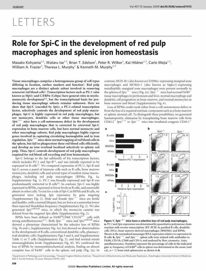
Spi-C belongs to the Spi subfamily of Ets transcription factors,which includes PU.1 and Spi-B5,6, and was initially reported to beexpressed in B cells5,7. We compared expression of PU.1, Spi-B andSpi-C across a panel of immune cells such as B cells, bone marrowmonocytes, dendritic cells and several types of resident tissue macro-phages, including red pulp macrophages (RPMs; Fig. 1a,Supplementary Fig. 1). PU.1 was broadly expressed and Spi-B waspredominantly restricted to B cells8,9. In contrast, Spi-C was highlyexpressed in RPMs, expressed at lower levels in B cells, and essentiallyabsent in other cells. To test for a role of Spi-C in RPMs and B cells, wegenerated mice lacking Spic expression by gene targeting(Supplementary Fig. 2). Male and female Spic2/2 mice are fertileand healthy, with a normal lifespan, but are born at a somewhat lowerthan expected Mendelian frequency (Supplementary Fig. 2). We alsogenerated Spicnull/null mice, in which the neomycin cassette wasdeleted from the targeted Spic allele (Supplementary Fig. 2).
RPMs have been defined as F4/80hiCD681CD11blo/2 cells withstrong autofluorescence10,11. Both Spic2/2 mice and Spicnull/null miceshowed a phenotype characterized by the selective loss of RPMs(Fig. 1b and c, Supplementary Fig. 3a), but showed no abnormalitiesin the development of B cells, conventional dendritic cells, plasmacy-toid dendritic cells (Supplementary Figs 3 and 4, and SupplementaryTable 1) or T cells (Supplementary Fig. 5), and had normal serumimmunoglobulin levels (Supplementary Fig. 4f). We confirmed theloss of RPMs by immunohistochemical analysis, finding an almostcomplete loss of F4/801 cells in the splenic red pulp (Fig. 2a). In
contrast, SIGN-R1 (also known as CD209a)-expressing marginal zonemacrophages and MOMA-1 (also known as Siglec1)-expressingmetallophilic marginal zone macrophages were present normally inthe spleens of Spic2/2 mice (Fig. 2a). Spic2/2 mice had normal F4/801
tissue macrophages in peritoneum and liver, normal macrophage anddendritic cell progenitors in bone marrow, and normal monocytes inbone marrow and blood (Supplementary Fig. 6).
Loss of RPMs could result either from a cell-autonomous defect orfrom the loss of a required extrinsic component such as a bone marrowor splenic stromal cell. To distinguish these possibilities, we generatedhaematopoietic chimaeras by transplanting bone marrow cells fromCD45.21 Spic1/1 or Spic2/2 mice into irradiated congenic CD45.11
1Department of Pathology and Immunology, 2Howard Hughes Medical Institute, 3Department of Biochemistry and Molecular Biophysics, Washington University School of Medicine,660 S. Euclid Avenue, St Louis, Missouri 63110, USA.
a b
c
08/
4F
Auto
CD68
CD11b
Spic−/−
0.06
F4/8
0hig
h (%
)
1.3
CD11c
1.3 0.16
1.3 0.07
1.5 0.15
Spic+/+
Spic+/+
Spic−/−0
0.5
1.0
1.5
Relative Spic expression2 4 6 100 8
1 2 3 40
B cells
DCs
BMDMs
RPMs
B cells
DCs
BMDMs
RPMs
Relative PU.1 expression
Figure 1 | Spic2/2 mice have a selective loss of red pulp macrophages.a, PU.1 and Spic expression was determined by quantitative polymerase chainreaction with reverse transcription (RT–PCR) in purified B cells, dendriticcells (DCs), bone-marrow-derived macrophages (BMDMs) and RPMs.Shown is the normalized messenger RNA expression relative to expression inB cells. b, Spic1/1 and Spic2/2 spleen cells were stained with antibodies toF4/80, CD11b, CD68 and CD11c and analysed by flow cytometry. Auto,autofluorescence. Numbers represent the percentage of cells in the indicatedgate. c, Frequency of F4/80hi cells in spleen was determined as the mean (ands.d., n 5 7) from total splenocytes as shown in b.
Vol 457 | 15 January 2009 | doi:10.1038/nature07472
318 Macmillan Publishers Limited. All rights reserved©2009
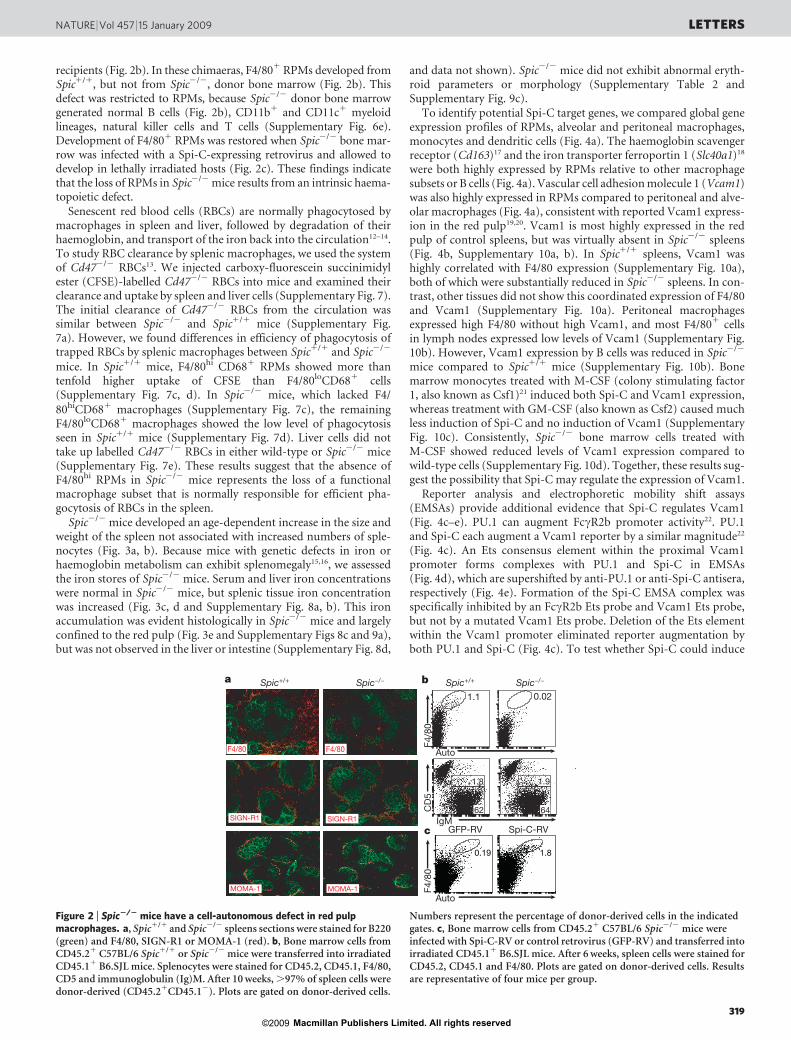
recipients (Fig. 2b). In these chimaeras, F4/801 RPMs developed fromSpic1/1, but not from Spic2/2, donor bone marrow (Fig. 2b). Thisdefect was restricted to RPMs, because Spic2/2 donor bone marrowgenerated normal B cells (Fig. 2b), CD11b1 and CD11c1 myeloidlineages, natural killer cells and T cells (Supplementary Fig. 6e).Development of F4/801 RPMs was restored when Spic2/2 bone mar-row was infected with a Spi-C-expressing retrovirus and allowed todevelop in lethally irradiated hosts (Fig. 2c). These findings indicatethat the loss of RPMs in Spic2/2 mice results from an intrinsic haema-topoietic defect.
Senescent red blood cells (RBCs) are normally phagocytosed bymacrophages in spleen and liver, followed by degradation of theirhaemoglobin, and transport of the iron back into the circulation12–14.To study RBC clearance by splenic macrophages, we used the systemof Cd472/2 RBCs13. We injected carboxy-fluorescein succinimidylester (CFSE)-labelled Cd472/2 RBCs into mice and examined theirclearance and uptake by spleen and liver cells (Supplementary Fig. 7).The initial clearance of Cd472/2 RBCs from the circulation wassimilar between Spic2/2 and Spic1/1 mice (Supplementary Fig.7a). However, we found differences in efficiency of phagocytosis oftrapped RBCs by splenic macrophages between Spic1/1 and Spic2/2
mice. In Spic1/1 mice, F4/80hi CD681 RPMs showed more thantenfold higher uptake of CFSE than F4/80loCD681 cells(Supplementary Fig. 7c, d). In Spic2/2 mice, which lacked F4/
80hiCD681 macrophages (Supplementary Fig. 7c), the remainingF4/80loCD681 macrophages showed the low level of phagocytosisseen in Spic1/1 mice (Supplementary Fig. 7d). Liver cells did nottake up labelled Cd472/2 RBCs in either wild-type or Spic2/2 mice(Supplementary Fig. 7e). These results suggest that the absence ofF4/80hi RPMs in Spic2/2 mice represents the loss of a functionalmacrophage subset that is normally responsible for efficient pha-gocytosis of RBCs in the spleen.
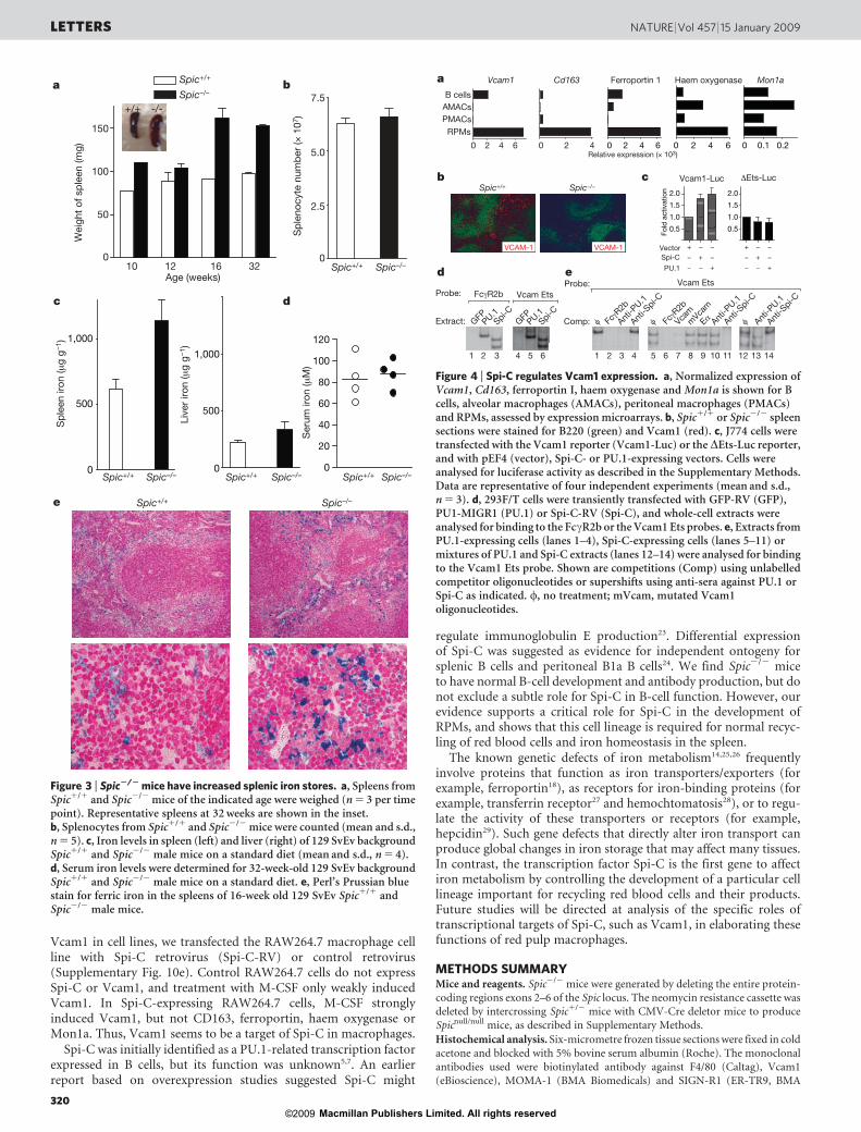
Spic2/2 mice developed an age-dependent increase in the size andweight of the spleen not associated with increased numbers of sple-nocytes (Fig. 3a, b). Because mice with genetic defects in iron orhaemoglobin metabolism can exhibit splenomegaly15,16, we assessedthe iron stores of Spic2/2 mice. Serum and liver iron concentrationswere normal in Spic2/2 mice, but splenic tissue iron concentrationwas increased (Fig. 3c, d and Supplementary Fig. 8a, b). This ironaccumulation was evident histologically in Spic2/2 mice and largelyconfined to the red pulp (Fig. 3e and Supplementary Figs 8c and 9a),but was not observed in the liver or intestine (Supplementary Fig. 8d,
and data not shown). Spic2/2 mice did not exhibit abnormal eryth-roid parameters or morphology (Supplementary Table 2 andSupplementary Fig. 9c).
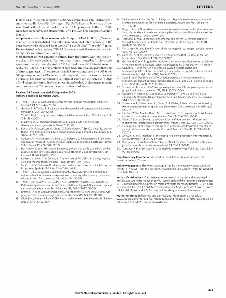
To identify potential Spi-C target genes, we compared global geneexpression profiles of RPMs, alveolar and peritoneal macrophages,monocytes and dendritic cells (Fig. 4a). The haemoglobin scavengerreceptor (Cd163)17 and the iron transporter ferroportin 1 (Slc40a1)18
were both highly expressed by RPMs relative to other macrophagesubsets or B cells (Fig. 4a). Vascular cell adhesion molecule 1 (Vcam1)was also highly expressed in RPMs compared to peritoneal and alve-olar macrophages (Fig. 4a), consistent with reported Vcam1 express-ion in the red pulp19,20. Vcam1 is most highly expressed in the redpulp of control spleens, but was virtually absent in Spic2/2 spleens(Fig. 4b, Supplementary 10a, b). In Spic1/1 spleens, Vcam1 washighly correlated with F4/80 expression (Supplementary Fig. 10a),both of which were substantially reduced in Spic2/2 spleens. In con-trast, other tissues did not show this coordinated expression of F4/80and Vcam1 (Supplementary Fig. 10a). Peritoneal macrophagesexpressed high F4/80 without high Vcam1, and most F4/801 cellsin lymph nodes expressed low levels of Vcam1 (Supplementary Fig.10b). However, Vcam1 expression by B cells was reduced in Spic2/2
mice compared to Spic1/1 mice (Supplementary Fig. 10b). Bonemarrow monocytes treated with M-CSF (colony stimulating factor1, also known as Csf1)21 induced both Spi-C and Vcam1 expression,whereas treatment with GM-CSF (also known as Csf2) caused muchless induction of Spi-C and no induction of Vcam1 (SupplementaryFig. 10c). Consistently, Spic2/2 bone marrow cells treated withM-CSF showed reduced levels of Vcam1 expression compared towild-type cells (Supplementary Fig. 10d). Together, these results sug-gest the possibility that Spi-C may regulate the expression of Vcam1.
Reporter analysis and electrophoretic mobility shift assays(EMSAs) provide additional evidence that Spi-C regulates Vcam1(Fig. 4c–e). PU.1 can augment FccR2b promoter activity22. PU.1and Spi-C each augment a Vcam1 reporter by a similar magnitude22
(Fig. 4c). An Ets consensus element within the proximal Vcam1promoter forms complexes with PU.1 and Spi-C in EMSAs(Fig. 4d), which are supershifted by anti-PU.1 or anti-Spi-C antisera,respectively (Fig. 4e). Formation of the Spi-C EMSA complex wasspecifically inhibited by an FccR2b Ets probe and Vcam1 Ets probe,but not by a mutated Vcam1 Ets probe. Deletion of the Ets elementwithin the Vcam1 promoter eliminated reporter augmentation byboth PU.1 and Spi-C (Fig. 4c). To test whether Spi-C could induce
b
c
1.9 1.81
62.45
1.8
IgM
CD
5 F4
/80
F4/8
0
1.1 0.02
Auto
Auto
1.8 0.19
a
MOMA-1
F4/80
SIGN-R1
F4/80
SIGN-R1
MOMA-1
Spic−/− Spic−/−Spic+/+ Spic+/+
62 64
GFP-RV Spi-C-RV
Figure 2 | Spic2/2 mice have a cell-autonomous defect in red pulpmacrophages. a, Spic1/1 and Spic2/2 spleens sections were stained for B220(green) and F4/80, SIGN-R1 or MOMA-1 (red). b, Bone marrow cells fromCD45.21 C57BL/6 Spic1/1 or Spic2/2 mice were transferred into irradiatedCD45.11 B6.SJL mice. Splenocytes were stained for CD45.2, CD45.1, F4/80,CD5 and immunoglobulin (Ig)M. After 10 weeks, .97% of spleen cells weredonor-derived (CD45.21CD45.12). Plots are gated on donor-derived cells.
Numbers represent the percentage of donor-derived cells in the indicatedgates. c, Bone marrow cells from CD45.21 C57BL/6 Spic2/2 mice wereinfected with Spi-C-RV or control retrovirus (GFP-RV) and transferred intoirradiated CD45.11 B6.SJL mice. After 6 weeks, spleen cells were stained forCD45.2, CD45.1 and F4/80. Plots are gated on donor-derived cells. Resultsare representative of four mice per group.
NATURE | Vol 457 | 15 January 2009 LETTERS
319 Macmillan Publishers Limited. All rights reserved©2009
Vcam1 in cell lines, we transfected the RAW264.7 macrophage cellline with Spi-C retrovirus (Spi-C-RV) or control retrovirus(Supplementary Fig. 10e). Control RAW264.7 cells do not expressSpi-C or Vcam1, and treatment with M-CSF only weakly inducedVcam1. In Spi-C-expressing RAW264.7 cells, M-CSF stronglyinduced Vcam1, but not CD163, ferroportin, haem oxygenase orMon1a. Thus, Vcam1 seems to be a target of Spi-C in macrophages.
Spi-C was initially identified as a PU.1-related transcription factorexpressed in B cells, but its function was unknown5,7. An earlierreport based on overexpression studies suggested Spi-C might
regulate immunoglobulin E production23. Differential expressionof Spi-C was suggested as evidence for independent ontogeny forsplenic B cells and peritoneal B1a B cells24. We find Spic2/2 miceto have normal B-cell development and antibody production, but donot exclude a subtle role for Spi-C in B-cell function. However, ourevidence supports a critical role for Spi-C in the development ofRPMs, and shows that this cell lineage is required for normal recyc-ling of red blood cells and iron homeostasis in the spleen.
The known genetic defects of iron metabolism14,25,26 frequentlyinvolve proteins that function as iron transporters/exporters (forexample, ferroportin18), as receptors for iron-binding proteins (forexample, transferrin receptor27 and hemochtomatosis28), or to regu-late the activity of these transporters or receptors (for example,hepcidin29). Such gene defects that directly alter iron transport canproduce global changes in iron storage that may affect many tissues.In contrast, the transcription factor Spi-C is the first gene to affectiron metabolism by controlling the development of a particular celllineage important for recycling red blood cells and their products.Future studies will be directed at analysis of the specific roles oftranscriptional targets of Spi-C, such as Vcam1, in elaborating thesefunctions of red pulp macrophages.
METHODS SUMMARYMice and reagents. Spic2/2 mice were generated by deleting the entire protein-
coding regions exons 2–6 of the Spic locus. The neomycin resistance cassette was
deleted by intercrossing Spic1/2 mice with CMV-Cre deletor mice to produce
Spicnull/null mice, as described in Supplementary Methods.
Histochemical analysis. Six-micrometre frozen tissue sections were fixed in cold
acetone and blocked with 5% bovine serum albumin (Roche). The monoclonal
antibodies used were biotinylated antibody against F4/80 (Caltag), Vcam1
(eBioscience), MOMA-1 (BMA Biomedicals) and SIGN-R1 (ER-TR9, BMA
bSpic−/−Spic+/+
B cellsAMACsPMACs
RPMs
a
0 2 4
Ferroportin 1Cd163
0 2 4 6
Vcam1
Relative expression (× 103)0 2 4 6
VCAM-1 VCAM-1
c
Fold
act
ivat
ion
+−
−
−
+−
−
−
+
0.5
1.0
1.5
2.0
0.5
1.0
1.5
2.0
+−
−
−
+−
−
−
+
Vcam1-Luc ΔEts-Luc
Probe:Probe:
Vcam Ets
1 2 3 4 5 6
d e
Extract:
Vcam Ets
1211101 2 3 4 5 6 987 13 14
0 2 4 6 0 0.1 0.2
Haem oxygenase Mon1a
Comp:
Vector
PU.1
Spi-C
FcγR2b
FcγR
2b
Anti-P
U.1
Anti-P
U.1
Anti-P
U.1
φ φφ Anti-S
pi-C
Anti-S
pi-C
Anti-S
pi-C
FcγR
2b
Vcamm
Vcam
EαGFPPU.1Spi-C
GFPPU.1Spi-C
Figure 4 | Spi-C regulates Vcam1 expression. a, Normalized expression ofVcam1, Cd163, ferroportin I, haem oxygenase and Mon1a is shown for Bcells, alveolar macrophages (AMACs), peritoneal macrophages (PMACs)and RPMs, assessed by expression microarrays. b, Spic1/1 or Spic2/2 spleensections were stained for B220 (green) and Vcam1 (red). c, J774 cells weretransfected with the Vcam1 reporter (Vcam1-Luc) or the DEts-Luc reporter,and with pEF4 (vector), Spi-C- or PU.1-expressing vectors. Cells wereanalysed for luciferase activity as described in the Supplementary Methods.Data are representative of four independent experiments (mean and s.d.,n 5 3). d, 293F/T cells were transiently transfected with GFP-RV (GFP),PU1-MIGR1 (PU.1) or Spi-C-RV (Spi-C), and whole-cell extracts wereanalysed for binding to the FccR2b or the Vcam1 Ets probes. e, Extracts fromPU.1-expressing cells (lanes 1–4), Spi-C-expressing cells (lanes 5–11) ormixtures of PU.1 and Spi-C extracts (lanes 12–14) were analysed for bindingto the Vcam1 Ets probe. Shown are competitions (Comp) using unlabelledcompetitor oligonucleotides or supershifts using anti-sera against PU.1 orSpi-C as indicated. w, no treatment; mVcam, mutated Vcam1oligonucleotides.
a7.5
5.0
2.5
Sp
leno
cyte
num
ber
(× 1
07)
b
0 0
150
100
50
Wei
ght
of s
ple
en (m
g)
+/+ -/-
10 12 16 32Age (weeks)
d
e
0
20
40
60
80
100
120
Ser
um ir
on (μ
M)
Sp
leen
iron
(μg
g−1 )
Live
r iro
n (μ
g g−
1 )
0
500500
1,000
1,000
0
c
Spic–/–
Spic–/–
Spic–/–
Spic–/–Spic–/–
Spic+/+
Spic+/+
Spic+/+
Spic+/+Spic+/+ Spic–/–Spic+/+
Figure 3 | Spic2/2 mice have increased splenic iron stores. a, Spleens fromSpic1/1 and Spic2/2 mice of the indicated age were weighed (n 5 3 per timepoint). Representative spleens at 32 weeks are shown in the inset.b, Splenocytes from Spic1/1 and Spic2/2 mice were counted (mean and s.d.,n 5 5). c, Iron levels in spleen (left) and liver (right) of 129 SvEv backgroundSpic1/1 and Spic2/2 male mice on a standard diet (mean and s.d., n 5 4).d, Serum iron levels were determined for 32-week-old 129 SvEv backgroundSpic1/1 and Spic2/2 male mice on a standard diet. e, Perl’s Prussian bluestain for ferric iron in the spleens of 16-week old 129 SvEv Spic1/1 andSpic2/2 male mice.
LETTERS NATURE | Vol 457 | 15 January 2009
320 Macmillan Publishers Limited. All rights reserved©2009
Biomedicals), Alexa488-conjugated antibody against B220 (BD PharMingen)
and streptavadin-Alexa555 (Invitrogen). For Perl’s Prussian blue stain, tissues
were fixed with 4% paraformaldehyde in 0.1 M phosphate buffer (pH 7.0),
embedded in paraffin, and stained with Perl’s Prussian blue and pararosaniline
(Sigma).
Adoptive transfer of bone marrow cells. Recipient CD45.11 B6.SJL (Taconic)
mice were lethally irradiated with 1,100 rads and injected intravenously with 107
bone marrow cells obtained from CD45.21 F4 or F5 Spic1/1 or Spic2/2 mice.
Donor-derived cells in spleen (CD45.21) were analysed 10 weeks after transfer
by fluorescence-activated cell sorting.
Measurement of iron content in spleen, liver and serum. Age- and gender-
matched mice were analysed for non-haem iron as described30. Livers and
spleens were weighed and digested in 3 M hydrochloric acid/10% trichloroacetic
acid, at 65 uC for 20 h. Ten microlitres of each acid extract was mixed with 0.5 ml
of chromagen reagent. The absorbance at 535 nm was measured by a DU Series
500 spectrophotometer (Beckman), and compared to an iron standard treated
identically. For serum measurements30, 20 ml of serum was incubated with 20 ml
of acid reagent for 5 min. Supernatant was mixed with 40 ml chromagen reagent,
and absorbance at 535 nm was measured as described above.
Received 26 August; accepted 30 September 2008.Published online 26 November 2008.
1. Taylor, P. R. et al. Macrophage receptors and immune recognition. Annu. Rev.Immunol. 23, 901–944 (2005).
2. Gordon, S. & Taylor, P. R. Monocyte and macrophage heterogeneity. Nature Rev.Immunol. 5, 953–964 (2005).
3. Ye, M. & Graf, T. Early decisions in lymphoid development. Curr. Opin. Immunol. 19,123–128 (2007).
4. Friedman, A. D. Transcriptional control of granulocyte and monocytedevelopment. Oncogene 26, 6816–6828 (2007).
5. Bemark, M., Martensson, A., Liberg, D. & Leanderson, T. Spi-C, a novel Ets proteinthat is temporally regulated during B lymphocyte development. J. Biol. Chem. 274,10259–10267 (1999).
6. Carlsson, R., Hjalmarsson, A., Liberg, D., Persson, C. & Leanderson, T. Genomicstructure of mouse SPI-C and genomic structure and expression pattern of humanSPI-C. Gene 299, 271–278 (2002).
7. Hashimoto, S. et al. Prf, a novel Ets family protein that binds to the PU.1 bindingmotif, is specifically expressed in restricted stages of B cell development. Int.Immunol. 11, 1423–1429 (1999).
8. Lloberas, J., Soler, C. & Celada, A. The key role of PU.1/SPI-1 in B cells, myeloidcells and macrophages. Immunol. Today 20, 184–189 (1999).
9. Su, G. H. et al. Defective B cell receptor-mediated responses in mice lacking theEts protein, Spi-B. EMBO J. 16, 7118–7129 (1997).
10. Taylor, P. R. et al. Dectin-2 is predominantly myeloid restricted and exhibitsunique activation-dependent expression on maturing inflammatory monocyteselicited in vivo. Eur. J. Immunol. 35, 2163–2174 (2005).
11. Taylor, P. R., Brown, G. D., Geldhof, A. B., Martinez-Pomares, L. & Gordon, S.Pattern recognition receptors and differentiation antigens define murine myeloidcell heterogeneity ex vivo. Eur. J. Immunol. 33, 2090–2097 (2003).
12. Bratosin, D. et al. Cellular and molecular mechanisms of senescent erythrocytephagocytosis by macrophages. A review. Biochimie 80, 173–195 (1998).
13. Oldenborg, P. A. et al. Role of CD47 as a marker of self on red blood cells. Science288, 2051–2054 (2000).
14. De Domenico, I., McVey, W. D. & Kaplan, J. Regulation of iron acquisition andstorage: consequences for iron-linked disorders. Nature Rev. Mol. Cell Biol. 9,72–81 (2008).
15. Biggs, T. E. et al. Nramp1 modulates iron homoeostasis in vivo and in vitro: evidencefor a role in cellular iron release involving de-acidification of intracellular vesicles.Eur. J. Immunol. 31, 2060–2070 (2001).
16. Tolosano, E. et al. Enhanced splenomegaly and severe liver inflammation inhaptoglobin/hemopexin double-null mice after acute hemolysis. Blood 100,4201–4208 (2002).
17. Kristiansen, M. et al. Identification of the haemoglobin scavenger receptor. Nature409, 198–201 (2001).
18. Donovan, A. et al. The iron exporter ferroportin/Slc40a1 is essential for ironhomeostasis. Cell Metab. 1, 191–200 (2005).
19. Gurtner, G. C. et al. Targeted disruption of the murine Vcam1 gene — essential roleof Vcam-1 in chorioallantoic fusion and placentation. Genes Dev. 9, 1–14 (1995).
20. Ulyanova, T. et al. VCAM-1 expression in adult hematopoietic andnonhernatopoietic cells is controlled by tissue-inductive signals and reflects theirdevelopmental origin. Blood 106, 86–94 (2005).
21. Leon, B. et al. Dendritic cell differentiation potential of mouse monocytes:monocytes represent immediate precursors of CD82 and CD81 splenic dendriticcells. Blood 103, 2668–2676 (2004).
22. Schweitzer, B. L. et al. Spi-C has opposing effects to PU.1 on gene expression inprogenitor B cells. J. Immunol. 177, 2195–2207 (2006).
23. Carlsson, R., Thorell, K., Liberg, D. & Leanderson, T. SPI-C and STAT6 cancooperate to stimulate IgE germline transcription. Biochem. Biophys. Res. Commun.344, 1155–1160 (2006).
24. Stoermann, B., Kretschmer, K., Duber, S. & Weiss, S. B-1a cells are imprinted bythe microenvironment in spleen and peritoneum. Eur. J. Immunol. 37, 1613–1620(2007).
25. Hentze, M. W., Muckenthaler, M. U. & Andrews, N. C. Balancing acts: Molecularcontrol of mammalian iron metabolism. Cell 117, 285–297 (2004).
26. Wang, F. D. et al. Genetic variation in Mon1a affects protein trafficking andmodifies macrophage iron loading in mice. Nature Genet. 39, 1025–1032 (2007).
27. Fleming, R. E. et al. Targeted mutagenesis of the murine transferrin receptor-2gene produces hemochromatosis. Proc. Natl Acad. Sci. USA 99, 10653–10658(2002).
28. Zhou, X. Y. et al. Knockout of the mouse HFE gene products hemochromatosis.Gastroenterology 114, A1372 (1998).
29. Roetto, A. et al. Mutant antimicrobial peptide hepcidin is associated with severejuvenile hemochromatosis. Nature Genet. 33, 21–22 (2003).
30. Torrance, J. D. & Bothwell, T. D. in Methods in Hematology Vol. 1 (ed. Cook, J. D.)90–115 (1980).
Supplementary Information is linked to the online version of the paper atwww.nature.com/nature.
Acknowledgements This work was supported by the Howard Hughes MedicalInstitute (K.M.M.), and the Burroughs Wellcome Fund Career Award for MedicalScientists (B.T.E.).
Author Contributions M.K. designed experiments, analysed and interpretedresults, and wrote the manuscript; W.I. sorted cells and did retrovirus experiments;B.T.E. contributed gene expression microarray data for mouse tissues; P.R.W. did Bcell analysis; K.H. did T cell differentiation analysis; M.A.F. provided Cd472/2 mice;T.L.M. did EMSA; and K.M.M. directed the study and wrote the manuscript.
Author Information Reprints and permissions information is available atwww.nature.com/reprints. Correspondence and requests for materials should beaddressed to K.M.M. ([email protected]).
NATURE | Vol 457 | 15 January 2009 LETTERS
321 Macmillan Publishers Limited. All rights reserved©2009


![Splenic Red Pulp Macrophages Produce Type I Interferons as …derisilab.ucsf.edu/pdfs/Kim_PLoSOne_2012.pdf · is under way for model organisms such as Listeria [1], relatively little](https://img.pdfslide.net/doc/110x75/5f5e495e53e55028c3484515/splenic-red-pulp-macrophages-produce-type-i-interferons-as-is-under-way-for-model.jpg)