Embed Size (px)
Citation preview
JOURNAL OF BACTERIOLOGY, July 2011, p. 3512–3524 Vol. 193, No. 140021-9193/11/$12.00 doi:10.1128/JB.01410-10Copyright © 2011, American Society for Microbiology. All Rights Reserved.
Role of Dihydrolipoamide Dehydrogenase in Regulation of RaffinoseTransport in Streptococcus pneumoniae�§
Robert E. Tyx,1 Hazeline Roche-Hakansson,1 and Anders P. Hakansson1,2,3*Department of Microbiology and Immunology, University at Buffalo, State University of New York, Buffalo, New York1;
Witebsky Center for Microbial Pathogenesis and Immunology, Buffalo, New York2; and New York State Center ofExcellence in Bioinformatics and Life Sciences, Buffalo, New York3
Received 23 November 2010/Accepted 11 May 2011
Streptococcus pneumoniae strains lacking the enzyme dihydrolipoamide dehydrogenase (DLDH) show mark-edly reduced ability to grow on raffinose and stachyose as sole carbon sources. Import of these sugars occursthrough the previously characterized raffinose ATP-binding cassette (ABC) transport system, encoded by theraf operon, that lacks the necessary ATP-binding protein. In this study, we identified the raffinose ATP-bindingprotein RafK and showed that it was directly involved in raffinose and stachyose import. RafK carries aC-terminal regulatory domain present in a subset of ATP-binding proteins that has been involved in both directregulation of transporter activity (inducer exclusion) and transcription of transporter genes. Pneumococcilacking RafK showed a 50- to 80-fold reduction in expression of the raf operon genes aga (alpha-galactosidase)and rafEFG (raffinose substrate binding and permease genes), and both glucose and sucrose inhibited raffinoseuptake through inducer exclusion. Like RafK, the presence of DLDH also activated the expression of raf operongenes, as DLDH-negative pneumococci showed a significantly decreased expression of aga and rafEFG, butDLDH did not regulate rafK or the putative regulatory genes rafR and rafS. DLDH also bound directly to RafKboth in vitro and in vivo, indicating the possibility that DLDH regulates raffinose transport by a directinteraction with the regulatory domain of the transporter. Finally, although not as attenuated as DLDH-negative bacteria, pneumococci lacking RafK were significantly outcompeted by wild-type bacteria in coloni-zation experiments of murine lung and nasopharynx, indicating a role for raffinose and stachyose transportin vivo.
Dihydrolipoamide dehydrogenases (DLDH) are enzymesclassically involved in the reoxidation of dihydrolipoamide dur-ing conversion of 2-oxo acids (such as pyruvate) in severalmultienzyme complexes in the central metabolism (34, 52).However, previous work from our and another laboratory haveshown that DLDH is not involved in metabolizing 2-oxo acidsin the pneumococcus, indicating that DLDH must serve otherfunctions (41, 42). This is supported by evidence that organ-isms that lack 2-oxo acid dehydrogenases still express a DLDHprotein (9, 10). One such function was suggested by Richarmein the 1980s when he showed that Escherichia coli strains thatfail to make lipoic acid or strains treated with an inhibitor thatmainly inhibits DLDH function resulted in reduced import ofgalactose, maltose, and ribose through ATP-binding cassette(ABC) transporters (35). We then showed that inactivation ofDLDH in the pneumococcus results in an inability of thebacteria to import and utilize galactose and the alpha-galacto-side sugars raffinose and stachyose and that a lack of DLDH isassociated with an almost complete attenuation of this strain inanimal infection experiments (41).
Transport of carbohydrates and other energy sources is im-portant for many aspects of bacterial life and therefore highly
regulated. Fitness in the bacterial host environment is intri-cately tied to accessibility of available energy sources and co-factors. In both Gram-positive and Gram-negative organisms,two types of transport systems are responsible for uptake ofenergy sources. Phosphoenolpyruvate (PEP)-sugar phospho-transferase systems (PTS) are the family of transporters gen-erally responsible for uptake of easily utilizable carbon sourcesand are pivotal in the regulation of other catabolic systems,including ABC transporters, through carbon catabolite repres-sion (CCR) and inducer exclusion (15, 38, 43, 51). ABC trans-porters are involved in importing alternate sources of energyand metal ions but are also involved in protein secretion, cellsignaling, adhesion, and invasion, as well as antibiotic resis-tance, and inactivation of these systems is often associated witha decreased fitness in the host environment (28). This is espe-cially true for the pneumococcus, which relies heavily on ABCtransporters due to the lack of biosynthetic genes in the ge-nome (7, 13, 16, 46).
This study focuses on the effect of DLDH on the raffinosetransport system. DLDH-negative bacteria fail to grow withraffinose as the sole carbon source, but the mechanism ofregulation has not been determined (41). In pneumococci,raffinose is transported through the raffinose ABC transporterencoded mainly by the raf operon. This operon has been char-acterized in some detail previously and contains all the genesnecessary for raffinose transport and utilization except theATP-binding protein of the transporter, which has not beencharacterized (36). The system is similar in function to thewell-characterized multiple-sugar metabolism (MSM) system
* Corresponding author. Mailing address: University at Buffalo,SUNY, Department of Microbiology and Immunology, 145 BRB, 3435Main Street, Buffalo, NY 14214. Phone: (716) 829-6058. Fax: (716)829-2158. E-mail: [email protected].
§ Supplemental material for this article may be found at http://jb.asm.org/.
� Published ahead of print on 20 May 2011.
3512
Dow
nloa
ded
from
http
s://j
ourn
als.
asm
.org
/jour
nal/j
b on
31
Oct
ober
202
1 by
194
.135
.146
.157
.
in Streptococcus mutans (37, 45) but shows a narrower range ofsubstrate transport and transports only raffinose and stachyose(36). The expression of the raf operon is induced by raffinose inthe medium and undergoes carbon catabolite repression in thepresence of sucrose through an unknown mechanism that doesnot involve the catabolite control protein A (CcpA) (36, 49).
In this paper we have identified and characterized RafK, theraffinose transporter ATP-binding protein, located separatelyfrom the raf operon on the chromosome, and have character-ized the interaction of DLDH with RafK and its effect on theexpression and function of the raffinose transporter. RafK car-ries a regulatory domain similar to that of the maltose ATP-binding protein MalK in E. coli that is also regulated by DLDH(5, 25, 35). We show here that DLDH binds to RafK and to itsregulatory domain and suggest that DLDH regulates raffinosetransport both by interfering with expression of the raf operonand by directly interacting with RafK. The mechanism bywhich the DLDH mutant shows such a severe attenuation invivo may make a viable target for future antibacterial drugdevelopment.
MATERIALS AND METHODS
Bacterial strains and growth conditions. Bacterial strains used and producedin this study are presented in Table 1. Streptococcus pneumoniae strain D39 (1)was used throughout the study as the parental strain for all mutations. Pneumo-cocci were routinely grown at 37°C in Todd-Hewitt broth (Difco Laboratories,Detroit, MI) supplemented with 0.5% yeast extract (THY) or on tryptic soy agar(TSA) supplemented with 5% sheep blood, as appropriate. E. coli strain XL-1Blue (Stratagene/Agilent Technologies, Santa Clara, CA) was used for cloning ofrecombinant protein expression and mutation constructs, and M15 (Qiagen,Valencia, CA) was used for protein expression. Frozen stocks of bacterial strainswere made by adding 20% glycerol to bacterial cultures grown to an opticaldensity at 600 nm (OD600) of �0.650, followed by freezing at �80°C. Thefollowing antibiotic concentrations were used for selection: ampicillin (Amp) wasused at 100 �g/ml in E. coli; kanamycin (Kan) was used at 500 and 50 �g/ml forpneumococci and E. coli, respectively; erythromycin (Erm) was used at 0.3 �g/ml;
and streptomycin (Sm) was used at 100 �g/ml for pneumococci. Antibiotics wereobtained from Sigma-Aldrich, St. Louis, MO.
Construction of S. pneumoniae RafK mutants. The Janus cassette was ampli-fied from CP1296 chromosomal DNA (44) using primers Janus-pGEM-F andJanus-pGEM-R (for primer information, see Table S1 in the supplementalmaterial) carrying BglII sites on both ends of the cassette, and the amplicon wasligated into the pGEM-T Easy vector (Promega, Madison, WI), resulting inplasmid pRT-JanusBglII. Approximately 800 base pairs upstream and down-stream of the rafK gene (27, 44) were amplified by PCR using primer pairsRafK-UF-F/RafK-UFD-R2 and RafK-LFD-F2/RafK-LF-R with purified D39chromosomal DNA as a template. The two fragments were fused by overlapPCR, in which a BglII site was engineered between the fragments, and theoverlap product was ligated into the pGEM-T Easy vector (Promega, Madison,WI), resulting in plasmid pRT-�rafK. This vector was linearized with BglII,treated with calf intestinal alkaline phosphatase (Invitrogen, Carlsbad, CA), andligated with the Janus cassette that had been excised from pRT-JanusBglII,resulting in plasmid pRT-�rafK-Janus. pRT-�rafK-Janus was cut with EcoRI torelease the Janus cassette flanked by the up- and downstream fragments, and thelinearized product was purified and transformed into streptomycin-resistant D39bacteria and plated onto selection plates containing 500 �g/ml kanamycin. Thisresulted in strain D39-�RafK-Janus. This strain was used to perform alleleinsertions and a full deletion of the gene.
To delete the gene, D39-�RafK-Janus was transformed with linearized prod-uct from vector pRT-�rafK carrying the flanking sequences fused together. Thetransformed pneumococci were selected on streptomycin plates and named D39-�RafK. To reinsert the original gene, rafK with upper and lower flanking se-quences was amplified using primers RafK-UF-F and RafK-LF-R by PCR fromD39 genomic DNA, the PCR product was used to transform D39-�RafK-Janus,and the transformants were selected on plates containing raffinose as the solecarbon source and named D39-rRafK. Finally, D39-�RafK-Janus was also usedto insert a variant rafK allele lacking the C-terminal regulatory domain. This wasdone by first PCR amplifying a truncated version of RafK (RafK�lip, encodingamino acids 1 to 251) using primers RafK-UF-F and RafK-dlip-UFD-R andfusing it first with an erythromycin cassette (amplified from plasmid pJY4164with primers RafK-dlip-erm-F and RafK-dlip-erm-R) directly downstream of thetruncated gene and then with the downstream flanking sequence amplified withprimers RafK-dlip-LF-F and RafK-LF-R by overlap PCR. This PCR product wascloned into pGEM-T Easy, resulting in plasmid pRT-rRafK�lipErm. The con-struct was released from the plasmid with EcoRI and transformed into D39-�RafK-Janus, and transformants were selected on erythromycin plates. Thesewere named D39-rRafK�lipErm.
TABLE 1. Strains and plasmids used in these studies
Strain or plasmid Characteristics Reference
StrainsD39 Wild-type, type 2 encapsulated 1D39-Smr D39, streptomycin resistant This studyD39-C0832:1 D39 dldh::pSH0832, DLDH negative 41D39-P2A1 D39 dldh-lplA intergenic::pSH08P 41CP1296 Rx derivative, cbp3::kan-rpsL� 44D39-�RafK D39,�rafK::kan-rpsL�, RafK negative This studyD39-rRafK D39, allelic repair rafK, RafK expressing This studyD39-�RafK-Janus D39 �rafK::kan-rpsL�, RafK negative This studyD39-rRafK�lipErm D39 allelic repair rafK�lip, Ermr This studyD39-�RafE D39 rafE::pRT-�rafE, RafE-negative This study
PlasmidspJY4164 Ermr, ori E. coli 54pQE30 Expression vector, Ampr QiagenpAH001 pQE30::dldh (full length) 17pRT-JanusBglII pGEM-T Easy::kan-rpsL� with BglII ends This studypRT-�rafK pGEM-T Easy::rafK deletion overlap PCR This studypRT-�rafK-Janus pGEM-T Easy::rafK deletion with kan-rpsL�, inserted This studypRT-rRafK�lipErm pGEM-T Easy::rafK�lip (amino acids 1 to 251 � Erm cassette amplified from pJY4164) This studypRT-�rafE pJY4164::rafE internal fragment This studypRT-RafK pQE30::rafK (full length) This studypRT-RafK�lip pQE30::rafK�lip (amino acids 1 to 251) This studypRT-RafKLipo pQE30::rafK lipoyl domain only (amino acids 246 to 377) This study
VOL. 193, 2011 DLDH AND RAFFINOSE TRANSPORT IN S. PNEUMONIAE 3513
Dow
nloa
ded
from
http
s://j
ourn
als.
asm
.org
/jour
nal/j
b on
31
Oct
ober
202
1 by
194
.135
.146
.157
.
Mutants carrying the Janus cassette, deletion strains, and strains carryingreinserted allele variants were verified by PCR and by DNA sequencing usingprimers involved in the cloning process as well as primers RafK-UU-F andRafK-LL-R at the Roswell Park Biopolymer Sequencing Facility, Roswell ParkCancer Institute, Buffalo, NY, and further confirmed by separating whole-celllysates on an SDS-PAGE gel, followed by a Western blotting procedure, detect-ing the presence of RafK with anti-RafK antibodies generated as describedbelow. Whole-cell lysates were prepared by growing cells to late log phase(OD600, �0.700), washing them in 1� phosphate-buffered saline (PBS), andresuspending them in 500 �l 100 mM potassium phosphate buffer (pH 7.4)containing 100 �g/ml lysozyme and 20 units/ml DNase I, followed by incubationfor 2 h at 37°C. Insoluble debris was removed by centrifugation at 15,000 � g for10 min. Blot and protein gel pictures were minimally processed with the Auto-Levels adjustment in Adobe Photoshop CS (Adobe Systems Inc., San Jose, CA).
To verify that the deletion of rafK (SPD_1409) did not produce polar effects ondownstream genes, we compared the expression of the downstream geneSPD_1408 by quantitative reverse transcriptase PCR (qRT-PCR) on cDNAproduced from strains D39 and D39-�RafK grown to an OD600 of 0.6 usingprimers SPD_1408-rRT-F and SPD_1408-rRT (for primer information, seeTable S1 in the supplemental material). The SPD_1408 expression was normal-ized to levels of the housekeeping gene cyclophilin D (SPD_1367 in the D39genome) (27) as described in detail below. The expression levels were notsignificantly different between the strains.
Mutational inactivation of the rafE gene. An internal fragment of the rafEopen reading frame (ORF) (nucleotides 6 to 534 of the SPD_1677 open readingframe) was amplified from D39 chromosomal DNA by PCR using the rafE-pJY-F and rafE-pJY-R primer pair with EcoRI and XbaI restriction sites addedto the 5� ends of the forward and reverse primers, respectively (see Table S1 inthe supplemental material). The PCR amplicons were digested with EcoRI andXbaI, ligated into EcoRI–XbaI-digested plasmid pJY4164 (53), and transformedinto E. coli XL1-Blue. Erythromycin-resistant clones were screened by PCR andverified by restriction digest of pure plasmid for insert of the appropriate size.Two independent clones, each harboring each rafE insert, were verified bysequencing, and one of them, designated pRT-�rafE, was used for insertion-duplication mutagenesis.
The resulting strain, D39-�RafE, was verified by sequencing over the insertionpoints in the chromosome. As the mutants carry the complete pJY4164 plasmiddisrupting the rafE gene, we expected this insertion to cause polar effects on thedownstream permease genes rafF and rafG. This was confirmed by comparingthe expression of the rafG gene by qRT-PCR on cDNA produced from strainsD39 and D39-�RafE1 grown to an OD600 of 0.5 using primers RafG-qRT-F andRafG-qRT-F. The rafG expression was normalized to levels of the housekeepinggene cyclophilin D (SPD_1367 in the D39 genome) (27) as described in detailbelow. The expression levels were 287 times lower in the rafE mutant strain,confirming that little to no transporter was expressed in this strain.
Construction of RafK recombinant protein. The full gene sequence for rafK(primer pair RafK-pQE-F and RafK-pQE-R), as well as two truncated variantscovering the N-terminal ATPase domain only (primer pair RafK-pQE-F andRafK-dlip-R) or the C-terminal regulatory lipoyl domain only (primer pairRafK-lipo-F and RafK-pQE-R), was PCR amplified from D39 DNA and in-serted into the pQE30 vector (Qiagen, Valencia, CA), resulting in plasmidspRT-RafK (full length), pRT-RafK�lip (RafK lacking the regulatory domain),and pRT-RafKLipo (regulatory domain alone), respectively. The correct se-quence was verified by DNA sequencing. Each plasmid was transformed into E.coli strain M15. For protein expression and purification, 2 to 400 ml of cells weregrown to an OD600 of 0.650 and induced at 37°C for 4 h with 1 mM IPTG. Cellswere pelleted by centrifugation at 6,000 � g for 15 min and resuspended in 4 mllysis buffer (50 mM NaH2PO4, 300 mM NaCl, 10 mM imidazole, 0.05% Tween20, pH 8.0). Cells were then lysed by sonication and centrifuged at 15,000 � g at4°C for 10 min to remove insoluble debris. Lysates were incubated with Ni-NTAagarose (Invitrogen, Carlsbad, CA). Beads and protein were washed three timeswith 5 ml of wash buffer (50 mM NaH2PO4, 300 mM NaCl, 20 mM imidazole,0.05% Tween 20, pH 8.0), and His-tagged proteins were eluted with elutionbuffer (50 mM NaH2PO4, 300 mM NaCl, 250 mM imidazole, 0.05% Tween 20,pH 8.0). Purified RafK protein were assayed for ATPase activity using the ATPluciferase assay (Invitrogen, Carlsbad, CA) according to the manufacturer’sinstructions and used immediately in binding assays, as ATPase activity andbinding were significantly reduced after 2 days in elution buffer at 4°C. Proteinconcentration was quantified using a BioTek Synergy 2 plate reader with theTake3 microdrop addition (BioTek, Winooski, VT).
The production of an expression vector for DLDH has already been described(17). Induction and expression of DLDH was done as described for RafK, withthe exception that DLDH was precipitated in 100% ammonium sulfate directly
after elution from the Ni-agarose and protein not used immediately was storedat 4°C with little to no loss of enzymatic activity, measured as described inreference 41.
Antibody production. Rabbit polyclonal antibodies to RafK and DLDH weregenerated by Lampire Biological Laboratories, using the Express-Line service(Lampire Biological Laboratories, Pipersville, PA). Recombinant full-lengthRafK and DLDH were used for immunization. Antibody was purified fromantiserum using protein G-Sepharose columns as per the manufacturer’s instruc-tions (GE Healthcare, Piscataway, NJ).
Mouse antibodies against DLDH were generated by injecting 20-week-oldfemale BALB/cByJ mice (Jackson Laboratory, Bar Harbor, ME) subcutaneouslywith 10 �g recombinant DLDH combined with 100 �l Titermax gold adjuvant(Sigma-Aldrich, St. Louis, MO), as per manufacturer’s instructions. After 2weeks, mice were boosted twice with 10 �g recombinant DLDH at 10-dayintervals and sacrificed 1 week after the last booster.
Antibodies used for immunoprecipitations were routinely adsorbed with wholebacterial cells lacking the specific antigen to which antibodies had been raised toremove antibodies that reacted with nonspecific antigens. Polyclonal Anti-RafKor anti-DLDH antiserum (100 �l) was incubated with 100 ml late-log-phaseD39-�RafK or D39-C0832 bacteria, respectively, that were previously washedthree times and resuspended in 10 ml PBS. Bacteria and antibody were incubatedat 37°C for 1 h, and bacteria were removed by centrifugation at 6,000 � g for 10min. This incubation with bacteria was repeated using a fresh 100 ml of bacterialcells washed and resuspended in 10 ml PBS. Postadsorbed and/or purified anti-body concentrations were quantified using a BioTek Synergy 2 plate reader withthe Take3 microdrop addition (BioTek, Winooski, VT). Antibodies were verifiedfor reactivity by Western blotting that detected proteins of interest in cell lysatesfrom S. pneumoniae strain D39.
RNA isolation and quantitative RT-PCR (qRT-PCR). RNA was purified usingQiashredder columns and the RNeasy minikit, according to the manufacturer’sinstructions (Qiagen, Valencia, CA). Briefly, frozen stocks of bacteria were usedto inoculate a 10-ml culture in THY. When the bacteria reached an OD600 of�0.5, 1 ml of bacteria were removed and pelleted by centrifugation at 9,000 � gfor 2 min at 4°C. The pellet was resuspended in 0.5 ml of 0.9% NaCl; 1 ml ofRNAprotect (Qiagen, Valencia, CA) was added, and the mixture was incubatedat room temperature for 5 min. Cells were then pelleted at 9,000 � g for 2 minat room temperature. These cells were frozen at �80°C or used directly for RNAisolation. Cells were resuspended in 500 �l TE buffer (10 mM Tris, 1 mM EDTA,pH 7.5) plus 25% (wt/vol) glucose and treated with 20 �l of a 100-mg/mllysozyme stock and 10 �l of 5,000-U/ml mutanolysin stock at 37°C for 15 min toremove cell wall (lysozyme and mutanolysin were purchased from Sigma-Al-drich, St. Louis, MO). Qiashredder columns were then used to remove unwantedcellular debris, and nucleic acids were isolated on RNeasy columns. Contami-nating DNA was removed on the RNeasy columns using the Bio-Rad RNase-freeDNase kit. Purity and quantitation of RNA were determined using the BioTekSynergy-2 plate reader with the Take3 microdrop addition. cDNA was synthe-sized from 500 ng total RNA using the Bio-Rad iScript cDNA synthesis kit(Bio-Rad, Hercules, CA). cDNA was amplified using SYBR green supermix withgene-specific primers (see Table S1 in the supplemental material) and quantifiedon a Bio-Rad iCycler according to the manufacturer’s instructions. Assays wereperformed in duplicate or triplicate using a 96-well plate in 20-�l reactionvolumes. Expression was analyzed using the standard curve method (32) andnormalized to levels of cyclophilin D (SPD_1367 in the D39 genome) (27).Significance of expression levels was determined by comparison to D39 wild-typeexpression using Student’s t test with significance being defined as P � 0.05.
Carbohydrate uptake assays. Bacteria were grown to mid-log phase (OD600,�0.500), centrifuged at 9,000 � g for 2 min, washed twice in cold PBS, andresuspended in an equal volume of cold PBS. Cells were kept on ice until 1 minprior to addition of radiolabeled carbohydrate. At this time point, cells wereplaced in a 30°C water bath. At time zero, 10 �l diluted radiolabeled carbohy-drate (50 �Ci/ml, diluted in PBS) was added and the cells were gently vortexed.The tube was then placed in a 30°C water bath for incubation. After 2 min(glucose) or 10 min (raffinose) of incubation, cells were captured on a 0.45-�M-pore-size HAWP filter (Millipore, Billerica, MA) and unbound radiolabeledcarbohydrates were washed through the filter with 5 ml PBS. Filters were thenplaced in scintillation vials and radioactivity was counted in a Wallac model 1409liquid scintillation counter. Glucose (D-[1-14C]-glucose; ARC 0120A) and raffin-ose ([galactose-6-3H]; ART 0229) were obtained from American RadiolabeledChemicals, Inc., St. Louis, MO. Significance of uptake was determined by com-parison with D39 wild-type uptake using Student’s t test with significance beingdefined as P � 0.05.
To test for carbon catabolite repression of raffinose transport, bacteria weregrown as described above but washed and resuspended in semidefined SH min-
3514 TYX ET AL. J. BACTERIOL.
Dow
nloa
ded
from
http
s://j
ourn
als.
asm
.org
/jour
nal/j
b on
31
Oct
ober
202
1 by
194
.135
.146
.157
.
imal medium, as previously described (41). Cells (1.5-ml aliquots) were thenexposed to 130 �l of either PBS alone or PBS containing glucose, sucrose, orlactose to a final concentration of 20 mM. Bacteria were incubated for 15 min atroom temperature before raffinose uptake was assessed. Bacteria were also usedto assess alpha-galactosidase activity as described below.
Alpha-galactosidase activity. Bacterial cells (2 ml) incubated in SH mediumwith PBS alone or PBS with glucose, lactose, or sucrose (to a final concentrationof 20 mM) were pelleted by centrifugation and placed at �80°C for 1 h. After thepellet was thawed on ice, the cells were resuspended in 2 ml PBS, and 200 �l ofthe resuspended cells were pelleted at 9,000 � g for 2 min, resuspended in 100�l of the lysis buffer (100 mM NaHPO4-KPO4, pH 7.5, 0.25% Triton X-100), andincubated for 5 min at 37°C to facilitate lysis. A small volume of lysate (2 �l) wasused to determine protein concentration in a BioTek Synergy 2 plate reader withthe Take3 microdrop addition (BioTek, Winooski, VT). Lysate (20 �l) wasadded into 980 �l substrate buffer (1 mM MgCl2, 4.5 mM -mercaptoethanol, 90�g/ml p-nitrophenyl--D-galactopyranoside, 100 mM potassium phosphate, pH7.5), and activity was determined spectrophotometrically at 405 nm over 40 minin the BioTek Synergy 2 plate reader. Activity was determined as U/mg bacteriallysate, where 1 U corresponds to the conversion of 1 mM substrate per minuteat 25°C.
Single-carbon-source growth assays. Cells from a frozen stock were inoculatedinto 10 ml THY, grown to early log phase (OD600, �0.300), washed twice insterile PBS, and resuspended in 4 ml of SH medium containing either nocarbohydrate source or 20 mM (each) glucose, raffinose, stachyose, or isomal-tose. Growth was monitored by measuring the optical density at 600 nm over 48 h.Carbohydrates were purchased from Sigma (Sigma-Aldrich, St. Louis, MO).
Cell fractionation. Pneumococci from a frozen stock were inoculated into 100ml THY, grown to mid-log phase (OD600, �0.500), washed twice in sterile PBS,and resuspended in 5 ml of protoplast buffer (20% [wt/vol] sucrose, 5 mMTris-HCl, 2.5 mM MgCl2, pH 7.4). Then, 250 units of mutanolysin and 100 �g/mllysozyme were added. Cells were incubated overnight at room temperature,pelleted at 5,000 � g for 10 min to remove cell wall supernatant, washed once inprotoplast buffer, and then lysed in 3.5 ml of deionized water. Nonlysed cellswere pelleted at 5,000 � g for 10 min. The lysed cells were centrifuged at 100,000� g for 90 min to pellet membrane. All fractions were then mixed with 200 �lLDS sample buffer and run out on precast 4 to 12% SDS-PAGE gels (Invitrogen,Carlsbad, CA). Fractions were verified using antibodies against proteins associ-ated mainly with one fraction. The cell wall fraction was verified using antibodiesagainst PspA (31), a choline-binding protein that is associated to the cell wall.Antibodies to DLDH were used as the membrane control, as DLDH is mostoften found associated with the membrane (17, 41). Streptavidin-HRP was usedas a cytosol protein control, as it binds to biotin carboxyl carrier protein accB,also known as BCCP (SPD_0386), which is associated mainly with cytosol (17).All antibodies were diluted 1:10,000 and incubated with blots at room temper-ature for 30 min. Goat anti-rabbit-horseradish peroxidase (HRP) (DLDH) andgoat anti-mouse-HRP (PspA) conjugated antibodies were used as secondaryreagents (Invitrogen, Carlsbad, CA), diluted 1:5,000 in 10 ml of PBS and incu-bated with blots at room temperature for 30 min. The blots were detected usingECL detection reagent (GE Healthcare, Piscataway, NJ) after exposure on film.Controls for cell fractions are shown in Fig. S1 in the supplemental material.
Immunoprecipitations. Cell lysates were prepared by growing one liter ofbacteria in THY with or without 20 mM raffinose to an OD600 of 0.8. Cells werecentrifuged at 8,000 � g for 30 min at 4°C and washed once in 25 ml of cold PBS.Cells were centrifuged again at 10,000 � g for 20 min and resuspended in 10 mlof 100 mM potassium phosphate buffer, pH 7.4. To these cells were added 1mg/ml lysozyme, 170 �g/ml phenylmethylsulfonyl fluoride (PMSF), and 100 �Mleupeptin. The cells were incubated for 1.5 h at 37°C and then cooled on icebefore being sonicated eight times for 8 s each time using a Branson Sonifier 450.Insoluble cell debris and any remaining whole cells were pelleted at 15,000 � gfor 15 min at 4°C.
Protein G dynabeads (500 �l [30 mg]) (Invitrogen, Carlsbad, CA) were washedwith PBS plus 0.02% Tween 20 (PBS-T), and �200 �g mouse polyclonal anti-DLDH antibody or rabbit polyclonal anti-RafK antibody was bound as per themanufacturer’s instructions. Cell lysates were incubated with protein G–anti-body-coated beads for at least 15 min at room temperature. Dynabeads andassociated antibodies and proteins were washed three times with 1 ml of PBS-T,and then the antibodies and proteins were eluted off the beads by using 150 �lof 1� LDS sample buffer with reducing agent (Invitrogen, Carlsbad, CA) andboiling for 10 min. Eluates from immunoprecipitations and associated proteinsfrom whole-cell lysates or recombinant proteins were run out on 4 to 12%SDS-PAGE gels and blotted with anti-RafK antibody and anti-DLDH antibody.
Mouse pneumonia and nasopharyngeal colonization model. The animal ex-periments were performed essentially as described earlier (3, 6, 41). Frozen
stocks of bacteria were thawed, washed once, and resuspended in 1.5 ml sterile,cold PBS. Bacteria were plated on TSA blood agar plates to verify quantities instocks. Bacteria (1 � 107 to 2 � 107 CFU) in a 40-�l volume were aspirated intothe nares of isoflurane-anesthetized CBA/CaHN-Btkxid/J mice (Jackson Labo-ratory, Bar Harbor, ME). In competition experiments, wild-type and mutantpneumococci were mixed in a 1:1 ratio. Mice were monitored and sacrificed at24 h postinoculation. Lungs were removed by opening the thoracic cavity andexcising the lung lobes. Nasopharyngeal tissue was harvested by defleshing thenose and skull of the mouse, cutting each maxillary bone and the skull bonebetween the eyes, removing the bone, and removing the tissue present in thenasal conchae with forceps. Harvested tissue was homogenized, and the homog-enate was serially diluted on TSA–5% blood agar plates. For competition ex-periments, an equal volume of homogenate was also plated on TSA–5% bloodagar plates with 500 �g/ml kanamycin to detect the mutants only. Data wereplotted using Prism for Mac OS X version 5.0b (Graphpad Software Inc., LaJolla, CA). Statistical significance for noncompetitive assays was determinedusing Student’s t test, with significance defined as P � 0.05. Statistical significancefor competitive assays were determined by comparing paired groups using theWilcoxon signed-rank test, with significance defined as P � 0.05. The competitiveindex (CI) was calculated using the ratio of mutant to wild-type CFU divided bythe mutant to wild-type input CFU count.
RESULTS
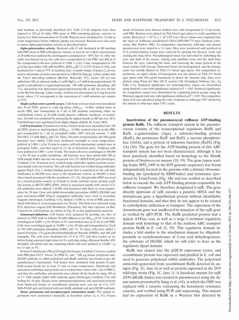
Inactivation of the pneumococcal raffinose ATP-bindingprotein RafK. The raffinose transport system in the pneumo-coccus consists of the transcriptional regulators RafR andRafS, -galactosidase (Aga), a substrate-binding protein(RafE), the permeases RafF and RafG, a sucrose phosphory-lase (GtfA), and a protein of unknown function (RafX) (Fig.1A) (36). The gene for the ATP-binding protein of this ABCtransport system has not been characterized, although it hasbeen putatively identified based on homology to the MsmKprotein of Streptococcus mutans (18, 19). The gene (open read-ing frame SPD_1409 in the D39 genome sequence) was foundindependently located in the genome with a distinct ribosome-binding site (predicted by RBSFinder) and terminator (pre-dicted by TransTerm) (Fig. 1B) and was verified as describedbelow to encode the only ATP-binding protein responsible forraffinose transport. We therefore designated it rafK. The genedirectly upstream of rafK encodes a putative tRNA and thedownstream gene a hypothetical protein without identifiablefunctional domains, and thus they do not appear to be relatedto carbohydrate utilization or transport. The expression of thedownstream gene was unaffected by mutation of the rafK geneas verified by qRT-PCR. The RafK predicted protein had atypical ATPase core as well as a large C-terminal regulatorydomain with homology to that of the maltose ABC transportprotein MalK in E. coli (5, 39). This regulatory domain in-cludes a fold similar to the attachment domain for dihydroli-poamide in acetyltransferases of 2-oxo acid dehydrogenases,the substrate of DLDH, which we will refer to here as theregulatory lipoyl domain.
RafK was cloned into the pQE30 expression vector, andrecombinant protein was expressed and purified in E. coli andused to generate polyclonal rabbit antibodies. The polyclonalantiserum generated from recombinant RafK detected its an-tigen (Fig. 1C, lane 6) as well as protein expressed in the D39wild-type strain (Fig. 1C, lane 1). A knockout mutant for rafK(D39-�RafK-Janus) was created in pneumococci using the Ja-nus system presented by Sung et al. (44), in which the ORF wasreplaced with a cassette containing the kanamycin resistancecassette, and verified using PCR and sequencing. This mutanthad no expression of RafK in a Western blot detected by
VOL. 193, 2011 DLDH AND RAFFINOSE TRANSPORT IN S. PNEUMONIAE 3515
Dow
nloa
ded
from
http
s://j
ourn
als.
asm
.org
/jour
nal/j
b on
31
Oct
ober
202
1 by
194
.135
.146
.157
.
polyclonal antibody produced against the recombinant protein(Fig. 1C, lane 2), similar to the complete deletion mutantD39-�RafK (Fig. 1C, lane 3). Lysate from a strain where thewild-type rafK allele was reinserted back into the same locusexpressed amounts of RafK equal to those of wild-type bacte-ria (Fig. 1C, lane 4). In order to assess whether the regulatorydomain played a critical role in transport of substrate throughthe ABC transporter, a truncated gene product lacking theregulatory domain was reinserted in the original rafK locus,resulting in strain D39-RafK�lipErm. However, the mutantcontaining the gene without the lipoyl domain was found tohave no detectable expression of the truncated protein (Fig.1C, lane 5).
Though the regulatory domain of RafK has a lipoyl-like fold,it does not contain the specific motif associated with an at-tached lipoic acid. This was verified in Western blotting exper-iments with D39 cell lysates using a rabbit -lipoic acid anti-body (Abcam, Cambridge, MA) that failed to detect RafK(Fig. 1C, lane 8).
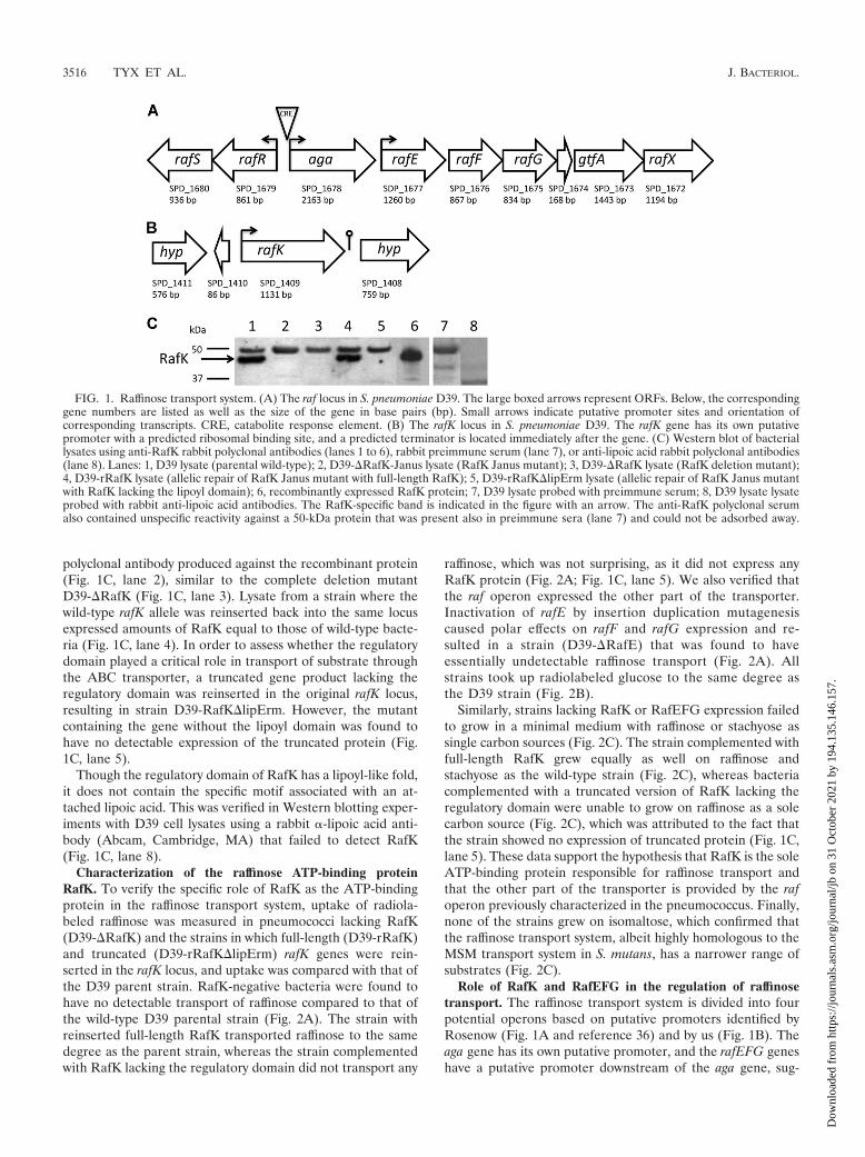
Characterization of the raffinose ATP-binding proteinRafK. To verify the specific role of RafK as the ATP-bindingprotein in the raffinose transport system, uptake of radiola-beled raffinose was measured in pneumococci lacking RafK(D39-�RafK) and the strains in which full-length (D39-rRafK)and truncated (D39-rRafK�lipErm) rafK genes were rein-serted in the rafK locus, and uptake was compared with that ofthe D39 parent strain. RafK-negative bacteria were found tohave no detectable transport of raffinose compared to that ofthe wild-type D39 parental strain (Fig. 2A). The strain withreinserted full-length RafK transported raffinose to the samedegree as the parent strain, whereas the strain complementedwith RafK lacking the regulatory domain did not transport any
raffinose, which was not surprising, as it did not express anyRafK protein (Fig. 2A; Fig. 1C, lane 5). We also verified thatthe raf operon expressed the other part of the transporter.Inactivation of rafE by insertion duplication mutagenesiscaused polar effects on rafF and rafG expression and re-sulted in a strain (D39-�RafE) that was found to haveessentially undetectable raffinose transport (Fig. 2A). Allstrains took up radiolabeled glucose to the same degree asthe D39 strain (Fig. 2B).
Similarly, strains lacking RafK or RafEFG expression failedto grow in a minimal medium with raffinose or stachyose assingle carbon sources (Fig. 2C). The strain complemented withfull-length RafK grew equally as well on raffinose andstachyose as the wild-type strain (Fig. 2C), whereas bacteriacomplemented with a truncated version of RafK lacking theregulatory domain were unable to grow on raffinose as a solecarbon source (Fig. 2C), which was attributed to the fact thatthe strain showed no expression of truncated protein (Fig. 1C,lane 5). These data support the hypothesis that RafK is the soleATP-binding protein responsible for raffinose transport andthat the other part of the transporter is provided by the rafoperon previously characterized in the pneumococcus. Finally,none of the strains grew on isomaltose, which confirmed thatthe raffinose transport system, albeit highly homologous to theMSM transport system in S. mutans, has a narrower range ofsubstrates (Fig. 2C).
Role of RafK and RafEFG in the regulation of raffinosetransport. The raffinose transport system is divided into fourpotential operons based on putative promoters identified byRosenow (Fig. 1A and reference 36) and by us (Fig. 1B). Theaga gene has its own putative promoter, and the rafEFG geneshave a putative promoter downstream of the aga gene, sug-
FIG. 1. Raffinose transport system. (A) The raf locus in S. pneumoniae D39. The large boxed arrows represent ORFs. Below, the correspondinggene numbers are listed as well as the size of the gene in base pairs (bp). Small arrows indicate putative promoter sites and orientation ofcorresponding transcripts. CRE, catabolite response element. (B) The rafK locus in S. pneumoniae D39. The rafK gene has its own putativepromoter with a predicted ribosomal binding site, and a predicted terminator is located immediately after the gene. (C) Western blot of bacteriallysates using anti-RafK rabbit polyclonal antibodies (lanes 1 to 6), rabbit preimmune serum (lane 7), or anti-lipoic acid rabbit polyclonal antibodies(lane 8). Lanes: 1, D39 lysate (parental wild-type); 2, D39-�RafK-Janus lysate (RafK Janus mutant); 3, D39-�RafK lysate (RafK deletion mutant);4, D39-rRafK lysate (allelic repair of RafK Janus mutant with full-length RafK); 5, D39-rRafK�lipErm lysate (allelic repair of RafK Janus mutantwith RafK lacking the lipoyl domain); 6, recombinantly expressed RafK protein; 7, D39 lysate probed with preimmune serum; 8, D39 lysate lysateprobed with rabbit anti-lipoic acid antibodies. The RafK-specific band is indicated in the figure with an arrow. The anti-RafK polyclonal serumalso contained unspecific reactivity against a 50-kDa protein that was present also in preimmune sera (lane 7) and could not be adsorbed away.
3516 TYX ET AL. J. BACTERIOL.
Dow
nloa
ded
from
http
s://j
ourn
als.
asm
.org
/jour
nal/j
b on
31
Oct
ober
202
1 by
194
.135
.146
.157
.
gesting that levels of the raffinose transporter might be regu-lated differentially from those of aga. The rafR and rafS geneshave their own putative promoter and are expressed in theopposite direction from aga. Of these three promoters, the agapromoter contains a catabolite response element (CRE) motif,but aga expression does not involve CcpA, as CcpA-negativebacteria express aga to the same degree as wild-type bacteria(36, 49). aga expression is induced by raffinose and repressedby sucrose as measured by -galactosidase activity (36). Theregulation of the rafK locus has not been characterized.
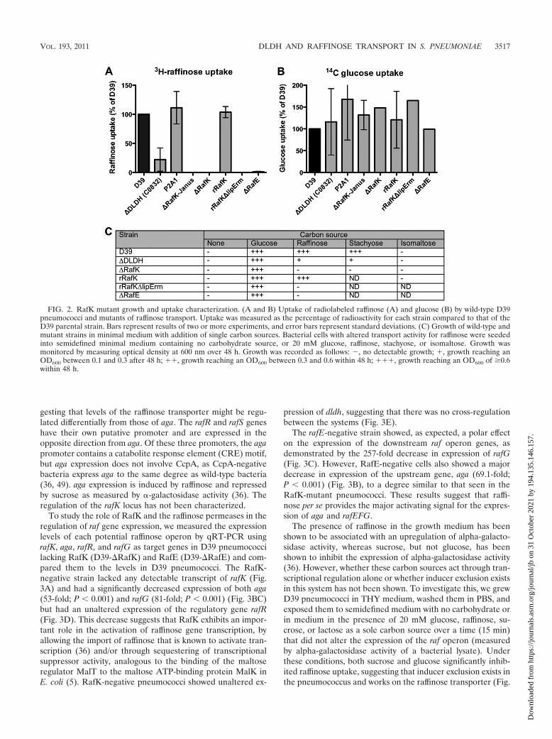
To study the role of RafK and the raffinose permeases in theregulation of raf gene expression, we measured the expressionlevels of each potential raffinose operon by qRT-PCR usingrafK, aga, rafR, and rafG as target genes in D39 pneumococcilacking RafK (D39-�RafK) and RafE (D39-�RafE) and com-pared them to the levels in D39 pneumococci. The RafK-negative strain lacked any detectable transcript of rafK (Fig.3A) and had a significantly decreased expression of both aga(53-fold; P � 0.001) and rafG (81-fold; P � 0.001) (Fig. 3BC)but had an unaltered expression of the regulatory gene rafR(Fig. 3D). This decrease suggests that RafK exhibits an impor-tant role in the activation of raffinose gene transcription, byallowing the import of raffinose that is known to activate tran-scription (36) and/or through sequestering of transcriptionalsuppressor activity, analogous to the binding of the maltoseregulator MalT to the maltose ATP-binding protein MalK inE. coli (5). RafK-negative pneumococci showed unaltered ex-
pression of dldh, suggesting that there was no cross-regulationbetween the systems (Fig. 3E).
The rafE-negative strain showed, as expected, a polar effecton the expression of the downstream raf operon genes, asdemonstrated by the 257-fold decrease in expression of rafG(Fig. 3C). However, RafE-negative cells also showed a majordecrease in expression of the upstream gene, aga (69.1-fold;P � 0.001) (Fig. 3B), to a degree similar to that seen in theRafK-mutant pneumococci. These results suggest that raffi-nose per se provides the major activating signal for the expres-sion of aga and rafEFG.
The presence of raffinose in the growth medium has beenshown to be associated with an upregulation of alpha-galacto-sidase activity, whereas sucrose, but not glucose, has beenshown to inhibit the expression of alpha-galactosidase activity(36). However, whether these carbon sources act through tran-scriptional regulation alone or whether inducer exclusion existsin this system has not been shown. To investigate this, we grewD39 pneumococci in THY medium, washed them in PBS, andexposed them to semidefined medium with no carbohydrate orin medium in the presence of 20 mM glucose, raffinose, su-crose, or lactose as a sole carbon source over a time (15 min)that did not alter the expression of the raf operon (measuredby alpha-galactosidase activity of a bacterial lysate). Underthese conditions, both sucrose and glucose significantly inhib-ited raffinose uptake, suggesting that inducer exclusion exists inthe pneumococcus and works on the raffinose transporter (Fig.
FIG. 2. RafK mutant growth and uptake characterization. (A and B) Uptake of radiolabeled raffinose (A) and glucose (B) by wild-type D39pneumococci and mutants of raffinose transport. Uptake was measured as the percentage of radioactivity for each strain compared to that of theD39 parental strain. Bars represent results of two or more experiments, and error bars represent standard deviations. (C) Growth of wild-type andmutant strains in minimal medium with addition of single carbon sources. Bacterial cells with altered transport activity for raffinose were seededinto semidefined minimal medium containing no carbohydrate source, or 20 mM glucose, raffinose, stachyose, or isomaltose. Growth wasmonitored by measuring optical density at 600 nm over 48 h. Growth was recorded as follows: �, no detectable growth; �, growth reaching anOD600 between 0.1 and 0.3 after 48 h; ��, growth reaching an OD600 between 0.3 and 0.6 within 48 h; ���, growth reaching an OD600 of �0.6within 48 h.
VOL. 193, 2011 DLDH AND RAFFINOSE TRANSPORT IN S. PNEUMONIAE 3517
Dow
nloa
ded
from
http
s://j
ourn
als.
asm
.org
/jour
nal/j
b on
31
Oct
ober
202
1 by
194
.135
.146
.157
.
FIG. 3. Expression of genes involved in raffinose uptake. Levels of gene expression were quantitated by qRT-PCR using the standardcurve method and normalized to levels of cyclophilin D (SPD_1367 in the D39 genome). Genes examined were as follows: (A) rafK: raffinoseABC transporter ATP-binding protein; (B) aga: alpha-galactosidase; (C) rafG: raffinose ABC transporter permease; (D) rafR: potentialraffinose transport transcriptional activator; (E) dldh: dihydrolipoamide dehydrogenase. Strains compared: D39, wild type; DLDH, dldhmutant; P2A1, control mutant that lacks expression of lplA; RafK, rafK deletion with Janus insertion; RafE, rafE interruption resulting inpolar effects on the raf operon. Bars represent starting quantity (SQ) and are the combined data from duplicates of two or more experiments.Error bars represent standard deviations. All statistical tests were Student t tests in comparison to data for D39. *, P � 0.05; **, P � 0.01;***, P � 0.001. ND, not determined. (F) Expression of RafK protein in wild-type and DLDH-negative bacteria (C0832). Cell wall,membrane, and cytosolic fractions from the bacteria were separated by gel electrophoresis, and RafK was detected with anti-RafK antibodiesby Western blotting. Lanes: 1, D39 cell wall fraction; 2, C0832 cell wall fraction; 3, D39 membrane fraction; 4, C0832 membrane fraction;5, D39 cytosolic fraction; 6, C0832 cytosolic fraction.
3518 TYX ET AL. J. BACTERIOL.
Dow
nloa
ded
from
http
s://j
ourn
als.
asm
.org
/jour
nal/j
b on
31
Oct
ober
202
1 by
194
.135
.146
.157
.
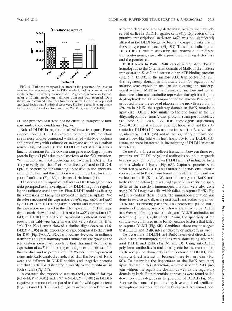
4). The presence of lactose had no effect on transport of raffi-nose under these conditions (Fig. 4).
Role of DLDH in regulation of raffinose transport. Pneu-mococci lacking DLDH displayed a more than 80% reductionin raffinose uptake compared with that of wild-type bacteriaand grew slowly with raffinose or stachyose as the sole carbonsource (Fig. 2A and B). The DLDH mutant strain is also afunctional mutant for the downstream gene encoding a lipoateprotein ligase (LplA) due to polar effects of the dldh mutation.We therefore included LplA-negative bacteria (P2A1) in thisstudy to verify that the effects were directly related to DLDH.LplA is responsible for attaching lipoic acid to the lipoyl do-main of DLDH, and this function was not important for trans-port of raffinose (Fig. 2A) or bacterial virulence (41).
The decreased transport of raffinose in DLDH-negative bac-teria prompted us to investigate how DLDH might be regulat-ing the raffinose uptake system. First, DLDH could be affectingthe expression of the genes involved in raffinose uptake. Wetherefore measured the expression of rafK, aga, rafR, and rafGby qRT-PCR in DLDH-negative bacteria and compared it tothe expression measured in the wild-type strain. DLDH-nega-tive bacteria showed a slight decrease in rafK expression (1.7-fold; P � 0.01) that although significantly different from ex-pression in wild-type bacteria was not very substantial (Fig.3A). The P2A1 strain showed a similar slight decrease (1.6-fold; P � 0.05) in the expression of rafK compared to the resultfor D39 (Fig. 3A). As P2A1 showed no decrease in raffinosetransport and grew normally with raffinose or stachyose as thesole carbon source, we conclude that this small decrease inexpression of rafK is not biologically significant. This was fur-ther verified on the protein level. A Western blot experimentusing anti-RafK antibodies indicated that the levels of RafKwere not different in DLDH-positive and -negative bacteriaand that RafK was distributed primarily to the membrane inboth strains (Fig. 3F).
In contrast, the expression was markedly reduced for aga(6.1-fold, P � 0.001) and rafG (6.6-fold; P � 0.001) in DLDH-negative pneumococci compared to that for wild-type bacteria(Fig. 3B and C). The level of aga expression correlated well
with the decreased alpha-galactosidase activity we have ob-served earlier in DLDH-negative cells (41). Expression of theputative transcriptional activator, rafR, was not significantlyaltered in the DLDH-negative bacteria compared with that inthe wild-type pneumococci (Fig. 3D). These data indicate thatDLDH has a role in activating the expression of raffinosetransporter genes, especially expression of alpha-galactosidaseand the permeases.
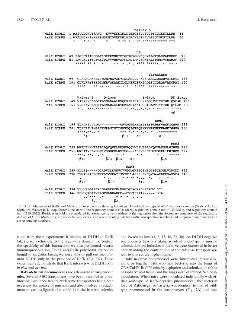
DLDH binds to RafK. RafK carries a regulatory domainhomologous to the C-terminal domain of MalK of the maltosetransporter in E. coli and certain other ATP-binding proteins(Fig. 5; 5, 12, 39). In the maltose ABC transporter in E. coli,this regulatory domain is important both for regulation ofmaltose gene expression through sequestering the transcrip-tional activator MalT in the presence of maltose and for in-ducer exclusion and catabolite repression through binding thedephosphorylated EIIA component of the glucose PTS systemproduced in the presence of glucose in the growth medium (5,39). As in MalK, the regulatory domain in RafK contains alipoyl-like TOBE_2 fold similar to the one found in the E2dihydrolipoamide transferase protein (transport-associatedOB, type 2, PF08402, CATHDB homologous superfamily2.40.50.100), the attachment point for lipoic acid, and the sub-strate for DLDH (41). As maltose transport in E. coli is alsoregulated by DLDH (35) and as the regulatory domains con-tain a lipoyl-like fold with high homology to the DLDH sub-strate, we were interested in investigating if DLDH interactswith RafK.
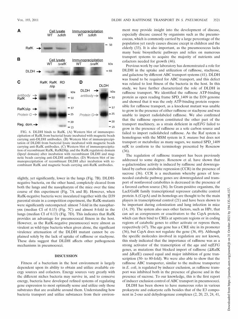
To test for a direct or indirect interaction between these twoproteins, anti-DLDH polyclonal antibodies bound to magneticbeads were used to pull down DLDH and its binding partnersfrom a whole-cell lysate (Fig. 6A). Captured proteins wereseparated by SDS-PAGE, and a number of bands, one of whichcorresponded to RafK, were found in the eluate. This band wasverified to be RafK in a Western blot using anti-RafK anti-bodies for detection (Fig. 6A, right panel). To verify the spec-ificity of the reaction, immunoprecipitations were also doneusing DLDH-negative cells, which failed to capture RafK (Fig.6A). To confirm these results, the immunoprecipitation wasdone in reverse as well, using anti-RafK antibodies to pull outRafK and its binding partners. This procedure pulled out anumber of proteins, one of which was identified to be DLDHin a Western blotting reaction using anti-DLDH antibodies fordetection (Fig. 6B, right panel). Again, the specificity of thecapture was confirmed using RafK-negative bacteria that failedto capture DLDH (Fig. 6B). Combined, these results suggestthat DLDH and RafK interact directly or indirectly in vivo.
To determine if DLDH and RafK interacted directly witheach other, immunoprecipitations were done using recombi-nant DLDH and RafK (Fig. 6C and D). Using anti-DLDHpolyclonal antibodies bound to magnetic beads, recombinantRafK was pulled down only in the presence of DLDH, indi-cating a direct interaction between these two proteins (Fig.6C). To determine the importance of the RafK regulatorylipoyl domain in this interaction, we expressed the RafK pro-tein without the regulatory domain as well as the regulatorydomain by itself. Both recombinant proteins were found pulleddown to various degrees in the presence of DLDH (Fig. 6C).Because the truncated proteins may have contained significanthydrophobic surfaces not normally exposed, we cannot con-
FIG. 4. Raffinose transport is reduced in the presence of glucose orsucrose. Bacteria were grown in THY, washed, and resuspended in SHmedium alone or in the presence of 20 mM glucose, sucrose, or lactose.After a 15-min incubation, raffinose transport was assessed. Datashown are combined data from two experiments. Error bars representstandard deviations. Statistical tests were Student t tests in comparisonto results for PBS-alone treatment. �, P � 0.05; ���, P � 0.001.
VOL. 193, 2011 DLDH AND RAFFINOSE TRANSPORT IN S. PNEUMONIAE 3519
Dow
nloa
ded
from
http
s://j
ourn
als.
asm
.org
/jour
nal/j
b on
31
Oct
ober
202
1 by
194
.135
.146
.157
.
clude from these experiments if binding of DLDH to RafKtakes place exclusively to the regulatory domain. To confirmthe specificity of this interaction, we also performed reverseimmunoprecipitation. Using anti-RafK polyclonal antibodiesbound to magnetic beads, we were able to pull out recombi-nant DLDH only in the presence of RafK (Fig. 6D). Theseexperiments demonstrate that RafK interacts with DLDH bothin vivo and in vitro.
RafK-deficient pneumococcus are attenuated in virulence inmice. Several ABC transporters have been identified as pneu-mococcal virulence factors, with some transporters being bothnecessary for uptake of nutrients and also involved in attach-ment to various ligands that could help the bacteria colonize
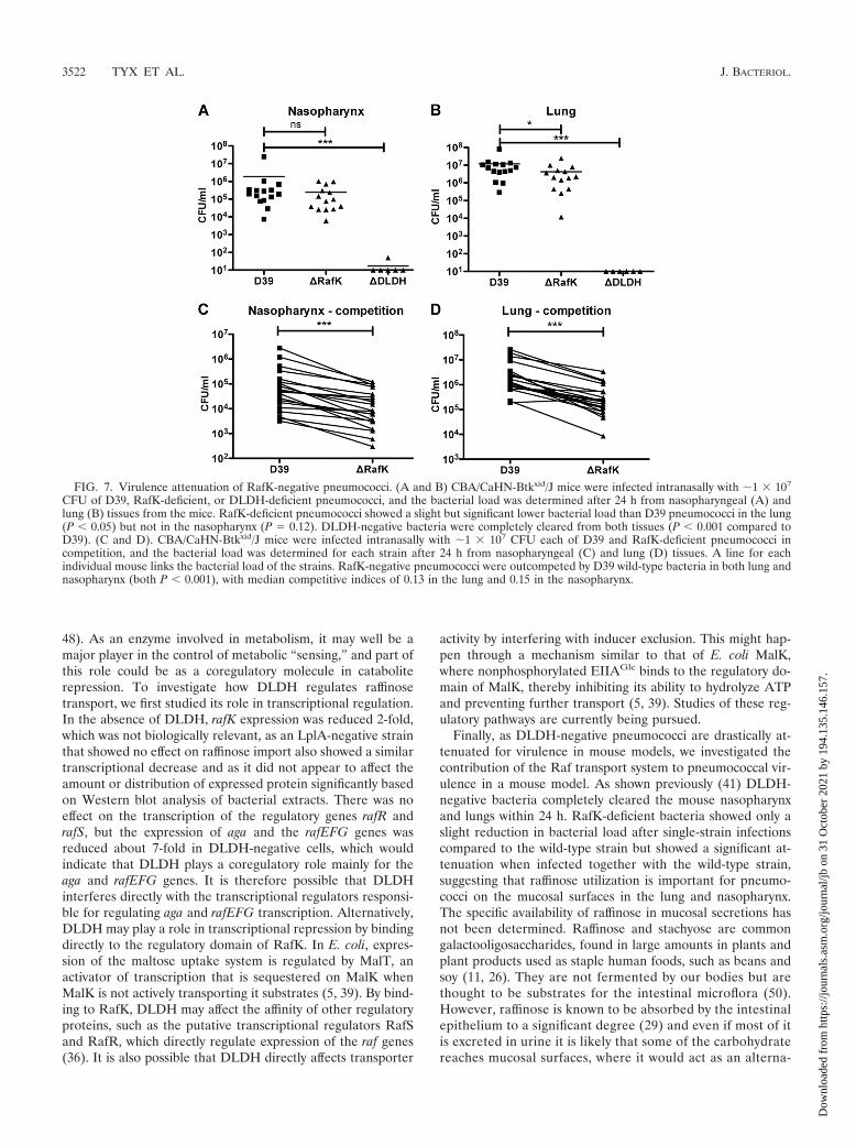
and invade its host (4, 8, 13, 18, 22, 30). As DLDH-negativepneumococci have a striking avirulent phenotype in murinecolonization and infection models, we were interested in betterunderstanding the contribution of the raffinose transport sys-tem to this avirulent phenotype.
RafK-negative pneumococci were introduced intranasally,alone or together with wild-type bacteria, into the lungs ofCBA/CaHN-Btkxid/J mice by aspiration and colonization of thenasopharyngeal tissue, and the lungs were examined 24 h post-inoculation. When mice were inoculated individually with ei-ther wild-type or RafK-negative pneumococci, the bacterialload of RafK-negative bacteria was identical to that of wild-type pneumococci in the nasopharynx (Fig. 7A) and was
FIG. 5. Alignment of RafK and MalK protein sequences showing homology. Annotated are typical ABC transporter motifs (Walker A, Lid,Signature, Walker B, D-loop, Switch), the start of the regulatory domain (RD Start), regulatory domain motif 1 (RDM1), and regulatory domainmotif 2 (RDM2). Residues in bold are considered important conserved residues in the regulatory domain. Secondary structures of the regulatorydomain of E. coli MalK are given under the sequences, with representing -helices with corresponding numbers and representing sheets withcorresponding numbers.
3520 TYX ET AL. J. BACTERIOL.
Dow
nloa
ded
from
http
s://j
ourn
als.
asm
.org
/jour
nal/j
b on
31
Oct
ober
202
1 by
194
.135
.146
.157
.
slightly, yet significantly, lower in the lungs (Fig. 7B). DLDH-negative bacteria, on the other hand, completely cleared fromboth the lungs and the nasopharynx of the mice over the timecourse of this experiment (Fig. 7A and B). However, whenRafK-negative bacteria were inoculated together with the D39parental strain in a competition experiment, the RafK-mutantswere significantly outcompeted: almost 7-fold in the nasophar-ynx (median CI of 0.15) (Fig. 7C) and almost 8-fold in thelungs (median CI of 0.13) (Fig. 7D). This indicates that RafKprovides an advantage for pneumococcal fitness in the host.However, as the RafK-negative pneumococci were almost asvirulent as wild-type bacteria when given alone, the significantvirulence attenuation of the DLDH mutant cannot be ex-plained solely by the lack of uptake of raffinose or stachyose.These data suggest that DLDH affects other pathogenesismechanisms in pneumococci.
DISCUSSION
Fitness of a bacterium in the host environment is largelydependent upon its ability to obtain and utilize available en-ergy sources and cofactors. Energy sources vary greatly withthe different niches bacteria may survive in, and to conserveenergy, bacteria have developed refined systems of regulatinggene expression to most optimally sense and utilize only thosesubstrates that are available around them. Understanding howbacteria transport and utilize substances from their environ-
ment may provide insight into the development of disease,especially disease caused by organisms such as the pneumo-coccus, which is commonly carried by a large percentage of thepopulation yet rarely causes disease except in children and theelderly (33). It is also important, as the pneumococcus lacksmany basic biosynthetic pathways and relies on numeroustransport systems to acquire the majority of nutrients andcofactors needed for growth (46).
Previous work by our laboratory has demonstrated a role forDLDH in the uptake and utilization of raffinose, stachyose,and galactose by different ABC transport systems (41). DLDHwas found to be required for ABC transport, and this defectwas related to lost fitness of the bacteria in the host. In thisstudy, we have further characterized the role of DLDH inraffinose transport. We identified the raffinose ATP-bindingprotein as open reading frame SPD_1409 in the D39 genomeand showed that it was the only ATP-binding protein respon-sible for raffinose transport, as a knockout mutant was unableto grow in the presence of either raffinose or stachyose and wasunable to import radiolabeled raffinose. We also confirmedthat the raffinose operon constituted the other part of thetransport machinery, as a strain deficient in rafEFG failed togrow in the presence of raffinose as a sole carbon source andfailed to import radiolabeled raffinose. As the Raf system ishomologous with the MSM system in S. mutans but does nottransport or metabolize as many sugars, we named SPD_1409rafK to conform to the terminology presented by Rosenow(36).
The regulation of raffinose transport has been previouslyaddressed to some degree. Rosenow et al. have shown that-galactosidase activity is induced by raffinose and downregu-lated by carbon catabolite repression (CCR) in the presence ofsucrose (36). CCR is a mechanism whereby genes of less-needed catabolic pathway genes are downregulated and trans-port of nonfavored catabolites is decreased in the presence ofa favored carbon source (36). In Gram-positive organisms, theLacI/GalR family transcriptional repressor catabolite controlprotein A (CcpA) and its homologs are thought to be the mainplayers in transcriptional control (21) and have been shown tobe important during colonization and lung infection in mice(14, 19). The system also uses other factors, such as HPr, thatcan act as corepressors or coactivators to the CcpA protein,which can then bind to CREs at upstream regions or in codingregions of catabolic genes to either activate or repress them,respectively (47). The aga gene has a CRE site in its promoter(36), but CcpA does not regulate the gene (36, 49). Althoughthe specific molecules involved in regulation are not known,this study indicated that the importance of raffinose was as astrong activator of the transcription of the aga and rafEFGgenes, as mutations that blocked import of raffinose (�RafKand �RafE) caused equal and major inhibition of gene tran-scription (50- to 80-fold). We were also able to show that theraffinose ABC transporter, similar to the maltose transporterin E. coli, is regulated by inducer exclusion, as raffinose trans-port was inhibited both in the presence of glucose and in thepresence of sucrose. To our knowledge, this is the first reportof inducer exclusion control of ABC transport in pneumococci.
DLDH has been shown to have numerous roles in variousprokaryote and eukaryote cells besides that of the E3 compo-nent in 2-oxo acid dehydrogenase complexes (2, 20, 23, 24, 41,
FIG. 6. DLDH binds to RafK. (A) Western blot of immunopre-cipitation of RafK from bacterial lysate incubated with magnetic beadscarrying anti-DLDH antibodies. (B) Western blot of immunoprecipi-tation of DLDH from bacterial lysate incubated with magnetic beadscarrying anti-RafK antibodies. (C) Western blot of immunoprecipita-tion of recombinant RafK, RafKDlip, and the RafK regulatory domain(lipoyl domain) after incubation with recombinant DLDH and mag-netic beads carrying anti-DLDH antibodies. (D) Western blot of im-munoprecipitation of recombinant DLDH after incubation with re-combinant RafK and magnetic beads carrying anti-RafK antibodies.
VOL. 193, 2011 DLDH AND RAFFINOSE TRANSPORT IN S. PNEUMONIAE 3521
Dow
nloa
ded
from
http
s://j
ourn
als.
asm
.org
/jour
nal/j
b on
31
Oct
ober
202
1 by
194
.135
.146
.157
.
48). As an enzyme involved in metabolism, it may well be amajor player in the control of metabolic “sensing,” and part ofthis role could be as a coregulatory molecule in cataboliterepression. To investigate how DLDH regulates raffinosetransport, we first studied its role in transcriptional regulation.In the absence of DLDH, rafK expression was reduced 2-fold,which was not biologically relevant, as an LplA-negative strainthat showed no effect on raffinose import also showed a similartranscriptional decrease and as it did not appear to affect theamount or distribution of expressed protein significantly basedon Western blot analysis of bacterial extracts. There was noeffect on the transcription of the regulatory genes rafR andrafS, but the expression of aga and the rafEFG genes wasreduced about 7-fold in DLDH-negative cells, which wouldindicate that DLDH plays a coregulatory role mainly for theaga and rafEFG genes. It is therefore possible that DLDHinterferes directly with the transcriptional regulators responsi-ble for regulating aga and rafEFG transcription. Alternatively,DLDH may play a role in transcriptional repression by bindingdirectly to the regulatory domain of RafK. In E. coli, expres-sion of the maltose uptake system is regulated by MalT, anactivator of transcription that is sequestered on MalK whenMalK is not actively transporting it substrates (5, 39). By bind-ing to RafK, DLDH may affect the affinity of other regulatoryproteins, such as the putative transcriptional regulators RafSand RafR, which directly regulate expression of the raf genes(36). It is also possible that DLDH directly affects transporter
activity by interfering with inducer exclusion. This might hap-pen through a mechanism similar to that of E. coli MalK,where nonphosphorylated EIIAGlc binds to the regulatory do-main of MalK, thereby inhibiting its ability to hydrolyze ATPand preventing further transport (5, 39). Studies of these reg-ulatory pathways are currently being pursued.
Finally, as DLDH-negative pneumococci are drastically at-tenuated for virulence in mouse models, we investigated thecontribution of the Raf transport system to pneumococcal vir-ulence in a mouse model. As shown previously (41) DLDH-negative bacteria completely cleared the mouse nasopharynxand lungs within 24 h. RafK-deficient bacteria showed only aslight reduction in bacterial load after single-strain infectionscompared to the wild-type strain but showed a significant at-tenuation when infected together with the wild-type strain,suggesting that raffinose utilization is important for pneumo-cocci on the mucosal surfaces in the lung and nasopharynx.The specific availability of raffinose in mucosal secretions hasnot been determined. Raffinose and stachyose are commongalactooligosaccharides, found in large amounts in plants andplant products used as staple human foods, such as beans andsoy (11, 26). They are not fermented by our bodies but arethought to be substrates for the intestinal microflora (50).However, raffinose is known to be absorbed by the intestinalepithelium to a significant degree (29) and even if most of itis excreted in urine it is likely that some of the carbohydratereaches mucosal surfaces, where it would act as an alterna-
FIG. 7. Virulence attenuation of RafK-negative pneumococci. (A and B) CBA/CaHN-Btkxid/J mice were infected intranasally with �1 � 107
CFU of D39, RafK-deficient, or DLDH-deficient pneumococci, and the bacterial load was determined after 24 h from nasopharyngeal (A) andlung (B) tissues from the mice. RafK-deficient pneumococci showed a slight but significant lower bacterial load than D39 pneumococci in the lung(P � 0.05) but not in the nasopharynx (P � 0.12). DLDH-negative bacteria were completely cleared from both tissues (P � 0.001 compared toD39). (C and D). CBA/CaHN-Btkxid/J mice were infected intranasally with �1 � 107 CFU each of D39 and RafK-deficient pneumococci incompetition, and the bacterial load was determined for each strain after 24 h from nasopharyngeal (C) and lung (D) tissues. A line for eachindividual mouse links the bacterial load of the strains. RafK-negative pneumococci were outcompeted by D39 wild-type bacteria in both lung andnasopharynx (both P � 0.001), with median competitive indices of 0.13 in the lung and 0.15 in the nasopharynx.
3522 TYX ET AL. J. BACTERIOL.
Dow
nloa
ded
from
http
s://j
ourn
als.
asm
.org
/jour
nal/j
b on
31
Oct
ober
202
1 by
194
.135
.146
.157
.
tive energy source, available to bacteria colonizing thenasopharynx.
Although RafK-deficient pneumococci were less virulent inmice, they were not attenuated to the degree that was observedin DLDH-negative bacteria, suggesting that attenuation in theDLDH-negative strain was due to additional events beyondjust the repression of the Raf system. Interestingly, three otherATP-binding proteins in the pneumococcal genome (the irontransport protein PitB, the polyamine transport protein PotA,and the ORF SPD_1608 with unknown function [8, 40]) pos-sess a regulatory domain similar to those of RafK and MalK,and initial investigation of the transcriptional levels of thesetransport genes in DLDH-negative bacteria, as well as func-tional transport assays, has shown decreased expression, sug-gesting that they are regulated similarly to the raffinose trans-port system.
To summarize, in this study, we have characterized theraffinose transporter ATP-binding protein RafK. We havedemonstrated that RafK is the sole ATP binding protein thatworks with the raf operon and have shown it to be the soletransporter of the -galactosides raffinose and stachyose. Wehave also shown that DLDH regulation of this transport sys-tem on a transcriptional level may be associated with its abilityto bind directly to RafK and potentially interfere with CCR. Asthe regulatory domain found on RafK is present only on ATP-binding proteins in the archaeal and bacterial kingdoms, abetter understanding of DLDH’s binding to this domain maylead to important breakthroughs that could be utilized forfuture drug development.
REFERENCES
1. Avery, O. T., C. M. MacLeod, and M. McCarty. 1944. Studies on the chem-ical nature of the substance inducing transformation of pneumococcal types.Induction of transformation by a dexoxyribonuceic acid fraction isolatedfrom pneumococcus type III. J. Exp. Med. 79:137–158.
2. Babady, N. E., Y. P. Pang, O. Elpeleg, and G. Isaya. 2007. Cryptic proteolyticactivity of dihydrolipoamide dehydrogenase. Proc. Natl. Acad. Sci. U. S. A.104:6158–6163.
3. Balachandran, P., A. Brooks-Walter, A. Virolainen-Julkunen, S. K. Holling-shead, and D. E. Briles. 2002. Role of pneumococcal surface protein C innasopharyngeal carriage and pneumonia and its ability to elicit protectionagainst carriage of Streptococcus pneumoniae. Infect. Immun. 70:2526–2534.
4. Basavanna, S., et al. 2009. Screening of Streptococcus pneumoniae ABCtransporter mutants demonstrates that LivJHMGF, a branched-chain aminoacid ABC transporter, is necessary for disease pathogenesis. Infect. Immun.77:3412–3423.
5. Bohm, A., J. Diez, K. Diederichs, W. Welte, and W. Boos. 2002. Structuralmodel of MalK, the ABC subunit of the maltose transporter of Escherichiacoli: implications for mal gene regulation, inducer exclusion, and subunitassembly. J. Biol. Chem. 277:3708–3717.
6. Briles, D. E., L. Novak, M. Hotomi, F. W. van Ginkel, and J. King. 2005.Nasal colonization with Streptococcus pneumoniae includes subpopulationsof surface and invasive pneumococci. Infect. Immun. 73:6945–6951.
7. Brown, J. S., S. M. Gilliland, and D. W. Holden. 2001. A Streptococcuspneumoniae pathogenicity island encoding an ABC transporter involved iniron uptake and virulence. Mol. Microbiol. 40:572–585.
8. Brown, J. S., S. M. Gilliland, J. Ruiz-Albert, and D. W. Holden. 2002.Characterization of pit, a Streptococcus pneumoniae iron uptake ABC trans-porter. Infect. Immun. 70:4389–4398.
9. Danson, M. J. 1988. Dihydrolipoamide dehydrogenase: a ‘new’ function foran old enzyme? Biochem. Soc. Trans. 16:87–89.
10. Danson, M. J., K. Conroy, A. McQuattie, and K. J. Stevenson. 1987. Dihy-drolipoamide dehydrogenase from Trypanosoma brucei. Characterizationand cellular location. Biochem. J. 243:661–665.
11. Diaz-Batalla, L., J. M. Widholm, G. C. J. Fahey, E. Castano-Tostado, and O.Paredes-Lopez. 2006. Chemical components with health implications in wildand cultivated Mexican common bean seeds (Phaseolus vulgaris L.). J. Agric.Food Chem. 54:2045–2052.
12. Diederichs, K., et al. 2000. Crystal structure of MalK, the ATPase subunit ofthe trehalose/maltose ABC transporter of the archaeon Thermococcus lito-ralis. EMBO J. 19:5951–5961.
13. Dintilhac, A., and J. P. Claverys. 1997. The adc locus, which affects compe-tence for genetic transformation in Streptococcus pneumoniae, encodes anABC transporter with a putative lipoprotein homologous to a family ofstreptococcal adhesins. Res. Microbiol. 148:119–131.
14. Giammarinaro, P., and J. C. Paton. 2002. Role of RegM, a homologue of thecatabolite repressor protein CcpA, in the virulence of Streptococcus pneu-moniae. Infect. Immun. 70:5454–5461.
15. Gorke, B., and J. Stulke. 2008. Carbon catabolite repression in bacteria:many ways to make the most out of nutrients. Nat. Rev. Microbiol. 6:613–624.
16. Guiral, S., T. J. Mitchell, B. Martin, and J. P. Claverys. 2005. Competence-programmed predation of noncompetent cells in the human pathogen Strep-tococcus pneumoniae: genetic requirements. Proc. Natl. Acad. Sci. U. S. A.102:8710–8715.
17. Hakansson, A. P., and A. W. Smith. 2007. Enzymatic characterization ofdihydrolipoamide dehydrogenase from Streptococcus pneumoniae harbor-ing its own substrate. J. Biol. Chem. 282:29521–29530.
18. Hava, D. L., and A. Camilli. 2002. Large-scale identification of serotype 4Streptococcus pneumoniae virulence factors. Mol. Microbiol. 45:1389–1406.
19. Iyer, R., N. S. Baliga, and A. Camilli. 2005. Catabolite control protein A(CcpA) contributes to virulence and regulation of sugar metabolism in Strep-tococcus pneumoniae. J. Bacteriol. 187:8340–8349.
20. Jang, Y. J., K. S. Chung, C. Park, and H. S. Yoo. 1997. Fission yeastdihydrolipoamide dehydrogenase gene is involved in G1/S cell cycle progres-sion. Biochim. Biophys. Acta 1358:229–239.
21. Kaufman, G. E., and J. Yother. 2007. CcpA-dependent and -independentcontrol of beta-galactosidase expression in Streptococcus pneumoniae oc-curs via regulation of an upstream phosphotransferase system-encodingoperon. J. Bacteriol. 189:5183–5192.
22. Kerr, A. R., et al. 2004. The Ami-AliA/AliB permease of Streptococcuspneumoniae is involved in nasopharyngeal colonization but not in invasivedisease. Infect. Immun. 72:3902–3906.
23. Kim, S. Y., and J. Kim. 2010. Roles of dihydrolipoamide dehydrogenaseLpd1 in Candida albicans filamentation. Fungal Genet. Biol. 47:782–788.
24. Kim, Y., L. O. Ingram, and K. T. Shanmugam. 2008. Dihydrolipoamidedehydrogenase mutation alters the NADH sensitivity of pyruvate dehydro-genase complex of Escherichia coli K-12. J. Bacteriol. 190:3851–3858.
25. Kuhnau, S., M. Reyes, A. Sievertsen, H. A. Shuman, and W. Boos. 1991. Theactivities of the Escherichia coli MalK protein in maltose transport, regula-tion, and inducer exclusion can be separated by mutations. J. Bacteriol.173:2180–2186.
26. Kumar, V., et al. 2010. Sucrose and raffinose family oligosaccharides (RFOs)in soybean seeds as influenced by genotype and growing location. J. Agric.Food Chem. 58:5081–5085.
27. Lanie, J. A., et al. 2007. Genome sequence of Avery’s virulent serotype 2strain D39 of Streptococcus pneumoniae and comparison with that of un-encapsulated laboratory strain R6. J. Bacteriol. 189:38–51.
28. Linton, K. J., and C. F. Higgins. 1998. The Escherichia coli ATP-bindingcassette (ABC) proteins. Mol. Microbiol. 28:5–13.
29. Lobley, R. W., P. C. Burrows, R. Warwick, D. J. Dawson, and R. Holmes.1990. Simultaneous assessment of intestinal permeability and lactose toler-ance with orally administered raffinose, lactose and L-arabinose. Clin. Sci.(Lond.) 79:175–183.
30. Marra, A., S. Lawson, J. S. Asundi, D. Brigham, and A. E. Hromockyj. 2002.In vivo characterization of the psa genes from Streptococcus pneumoniae inmultiple models of infection. Microbiology 148:1483–1491.
31. McDaniel, L. S., G. Scott, J. F. Kearney, and D. E. Briles. 1984. Monoclonalantibodies against protease-sensitive pneumococcal antigens can protectmice from fatal infection with Streptococcus pneumoniae. J. Exp. Med.160:386–397.
32. Morrison, T. B., J. J. Weis, and C. T. Wittwer. 1998. Quantification oflow-copy transcripts by continuous SYBR Green I monitoring during am-plification. Biotechniques 24:954–958, 960, 962.
33. O’Brien, K. L., and M. Santosham. 2004. Potential impact of conjugatepneumococcal vaccines on pediatric pneumococcal diseases. Am. J. Epide-miol. 159:634–644.
34. Perham, R. N., L. C. Packman, and S. E. Radford. 1987. 2-Oxo acid dehy-drogenase multi-enzyme complexes: in the beginning and halfway there.Biochem. Soc Symp. 54:67–81.
35. Richarme, G. 1985. Possible involvement of lipoic acid in binding protein-dependent transport systems in Escherichia coli. J. Bacteriol. 162:286–293.
36. Rosenow, C., M. Maniar, and J. Trias. 1999. Regulation of the alpha-galactosidase activity in Streptococcus pneumoniae: characterization of theraffinose utilization system. Genome Res. 9:1189–1197.
37. Russell, R. R., J. Aduse-Opoku, I. C. Sutcliffe, L. Tao, and J. J. Ferretti.1992. A binding protein-dependent transport system in Streptococcus mu-tans responsible for multiple sugar metabolism. J. Biol. Chem. 267:4631–4637.
38. Saier, M. H. J., et al. 1996. Catabolite repression and inducer control inGram-positive bacteria. Microbiology 142:217–230.
39. Samanta, S., et al. 2003. Disulfide cross-linking reveals a site of stable
VOL. 193, 2011 DLDH AND RAFFINOSE TRANSPORT IN S. PNEUMONIAE 3523
Dow
nloa
ded
from
http
s://j
ourn
als.
asm
.org
/jour
nal/j
b on
31
Oct
ober
202
1 by
194
.135
.146
.157
.
interaction between C-terminal regulatory domains of the two MalK sub-units in the maltose transport complex. J. Biol. Chem. 278:35265–35271.
40. Shah, P., B. Nanduri, E. Swiatlo, Y. Ma, and K. Pendarvis. 2011. Polyaminebiosynthesis and transport mechanisms are crucial for fitness and pathogen-esis of Streptococcus pneumoniae. Microbiology 157:504–515.
41. Smith, A. W., H. Roche, M. C. Trombe, D. E. Briles, and A. Hakansson. 2002.Characterization of the dihydrolipoamide dehydrogenase from Streptococ-cus pneumoniae and its role in pneumococcal infection. Mol. Microbiol.44:431–448.
42. Spellerberg, B., et al. 1996. Pyruvate oxidase, as a determinant of virulencein Streptococcus pneumoniae. Mol. Microbiol. 19:803–813.
43. Stulke, J., and W. Hillen. 1998. Coupling physiology and gene regulation inbacteria: the phosphotransferase sugar uptake system delivers the signals.Naturwissenschaften 85:583–592.
44. Sung, C. K., H. Li, J. P. Claverys, and D. A. Morrison. 2001. An rpsLcassette, Janus, for gene replacement through negative selection in Strepto-coccus pneumoniae. Appl. Environ. Microbiol. 67:5190–5196.
45. Tao, L., I. C. Sutcliffe, R. R. Russell, and J. J. Ferretti. 1993. Transport ofsugars, including sucrose, by the msm transport system of Streptococcusmutans. J. Dent. Res. 72:1386–1390.
46. Tettelin, H., et al. 2001. Complete genome sequence of a virulent isolate ofStreptococcus pneumoniae. Science 293:498–506.
47. Thibault, L., and C. Vadeboncoeur. 1985. Phosphoenolpyruvate-sugar phos-photransferase transport system of Streptococcus mutans: purification of
HPr and enzyme I and determination of their intracellular concentrations byrocket immunoelectrophoresis. Infect. Immun. 50:817–825.
48. Tian, J., et al. 2005. Mycobacterium tuberculosis appears to lack alpha-ketoglutarate dehydrogenase and encodes pyruvate dehydrogenase in widelyseparated genes. Mol. Microbiol. 57:859–868.
49. van Opijnen, T., K. L. Bodi, and A. Camilli. 2009. Tn-seq: high-throughputparallel sequencing for fitness and genetic interaction studies in microorgan-isms. Nat. Methods 6:767–772.
50. Vinjamoori, D. V., J. R. Byrum, T. Hayes, and P. K. Das. 2004. Challengesand opportunities in the analysis of raffinose oligosaccharides, pentosans,phytate, and glucosinolates. J. Anim. Sci. 82:319–328.
51. Warner, J. B., and J. S. Lolkema. 2003. CcpA-dependent carbon cataboliterepression in bacteria. Microbiol. Mol. Biol. Rev. 67:475–490.
52. Williams, C. H. 1992. Lipoamide dehydrogenase, glutathione reductase,thioredoxin reductase, and mercuric reductase—a family of flavoenzymetranshydrogenases, p. 121–211. In F. Muller (ed.), Chemistry and biochem-istry of flavoenzymes, CRC, Boca Raton, FL.
53. Yother, J., and D. E. Briles. 1992. Structural properties and evolutionaryrelationships of PspA, a surface protein of Streptococcus pneumoniae, asrevealed by sequence analysis. J. Bacteriol. 174:601–609.
54. Yother, J., G. L. Handsome, and D. E. Briles. 1992. Truncated forms of PspAthat are secreted from Streptococcus pneumoniae and their use in functionalstudies and cloning of the pspA gene. J. Bacteriol. 174:610–618.
3524 TYX ET AL. J. BACTERIOL.
Dow
nloa
ded
from
http
s://j
ourn
als.
asm
.org
/jour
nal/j
b on
31
Oct
ober
202
1 by
194
.135
.146
.157
.