Embed Size (px)
Citation preview
Medical Journal of Babylon-Vol. 9- No. 3 -2012 1021 -مجلة بابل الطبية- المجلد التاسع- العدد الثالث
Ahmed Turki Obaid and Rafid Fakhir Hussen
629
Abstract Objective: to compare the outcome of extracorporeal shock wave lithotripsy for renal stones with and
without double J stent.
Patients and Methods: A comparative cross sectional study was carried out at the urological
department of Al-Hilla teaching hospital from February 2011 to August 2011. Forty patients with renal
stones measuring from (2cm_2.5cm) were selected for the treatment by extracorporeal shock wave
lithotripsy (ESWL). All of these patients were adults with normal renal function and had unilateral
radiopaque renal stones, no metabolic abnormalities, no symptomatic urinary tract infection. Children,
renal failure and patients with lower caliceal stones were excluded from the study. They were divided
in two groups of 20 each. Group A patients underwent ESWL without a double J stent and in group B a
double J stent was placed before ESWL. Electrohydraulic lithotripter machine (PCK lithotripter,
Turkey), was used to impart shock wave. 2000 shock wave were given in a session. Both the groups
were compared for stone fragmentation, incomplete stone clearance, clinically insignificant stone
fragment, steinstrasse, stone free rate, renal colic, fever, hemmaturia, dysuria, and number of ESWL
session.
Result: Regarding stone fragmentation in group A (stentless), 19 patients had stone fragmentation
from total number of patients of 20 which account 95% while in group B (stented), 18 patients
develops stone fragmentation from total number of patients of 20 which account 90%. Incomplete
stone clearance occurs in 4 (20%) patients without DJS and in 8 (40%) patients with DJS. Clinically
insignificant stone fragments encounter in 3 (15%) patients in stentless group and also in 3 (15%) in
stented group.
Steinstrasse developed in 1 (5%) patient without DJS and none in patients with DJS.
Stone free rate encounter in 11 (55%) patients in stentless group and 7 (35%) patients in stented group.
Renal colic developed in 9 (45%) patients without DJS and in 3 (15%) patients with DJS. Hemmaturia
encounter in 12 (60%) patients without DJS and in 15 (75%) with DJS.
Fever not encountered in patients without DJS and developed in 15 (75%) patients with DJS.
Frequency developed in 2 (10%) patients without DJS and in 10 (50%) patients with DJS, while
dysuria developed in 3 (15%) patients without DJS and in
10 (50%) patients with DJS.
Conclusion: pre ESWL double J stenting for 2-2.5 cm renal stone was not beneficial regarding stone
fragmentation, stone clearance, steinstrasse and stone free rate, however renal colic was significantly
less in stented group. Fever and lower urinary tract symptoms (LUTS) was significantly higher in
stented patients.
الخالصةمقارنة النتائج النهائية لعالج حصاة الكمى بجهاز تفتيت الحصى خارج الجسم مع وضع قسطور الحالب من الغرض من الدراسة:
عدمه.الى اب ۳۱۲۲ دراسة مقارنة مقطعية اجريت في شعبة الجراحة البولية في مستشفى الحمة التعميمي لمفترة من شباط ريقة العمل:ط
۳۱۲۲.
Role of Double J Stent in Patients with Renal Stones Undergo
Extracorporeal Shock wave Lithotripsy
Ahmed Turki Obaid Rafid Fakhir Hussen
Dept. Surgary, College of Medicine, University of Babylon, Hilla, Iraq.
M J B
Medical Journal of Babylon-Vol. 9- No. 3 -2012 1021 -مجلة بابل الطبية- المجلد التاسع- العدد الثالث
Ahmed Turki Obaid and Rafid Fakhir Hussen
630
( سم عولجوا بجهاز تفتيت الحصى خارج الجسم وجميعهم من البالغين مع وظائف ۲.۳-۳أربعون مريضا لديهم حصاة الكمى وبطول )الكمى الوحيدة المشخصة شعاعيا وتم استبعاد جميع المرضى الذين لديهم فعاليات أيضية غير طبيعية والذين كمية طبيعية ولديهم حصاة
لديهم أمراض في المجاري البولية والذين لديهم أعراض فشل كموي والذين لديهم حصى في الفص السفمي لمكمية.كمى بدون قسطور الحالب . المجموعة ب : تم تفتيت حصاة الكمى تم تقسيم المرضى الى مجموعتين, المجموعة أ : تم تفتيت حصاة ال
مع وضع قسطور الحالب. كمتا المجموعتين عولجوا بجهاز تفتيت الحصى خارج الجسم من نوع تركي المنشأ, تم اعطاء المريض عدم التخمص من حصاة ضربة في كل جمسة عالجية. تمت المقارنة بين المجموعتين اعتمادا عمى : تفتيت كامل لمحصى, ۳۱۱۱
الكمة بشكل نهائي , أجزاء من حصاة الكمى غير مهمة سريريا, تكون ظاهرة حصاة الطريق اسفل الحالب, نسبة التخمص من حصاة الكمى, المغص الكموي, التبول الدموي, الحمى, عسرة البول, عدد جمسات العالج وكثرة التبول.
(, أما المجموعة ب: تسعة ۱۹٪كامل المجموعة أ: ثمانية عشر مريضا من أصل عشرين) عتمادا عمى تفتيت حصاة الكمى ال النتائج:(. اما بالنسبة لعدد المرضى الذين لم يتم التخمص من حصاة الكمة بشكل نهائي, بما يخص ۲۹٪عشر مريض من أصل عشرين )
(. وبما يخص ٤۱۹ى من أصل عشرين )(.ا ما المجموعة ب :ثمانية مرض۳۱۹المجموعة أ: أربعة مرضى من أصل عشرين مريضا)(, أما ۲۲۹عدد المرضى الذين لديهم اجزاء من حصاة الكمة الغير مهمة سريريا, المجموعة أ: ثالثة مرضى من أصل عشرين )
المجموعة ب: ثالثة مرضى من أصل عشرين. وبالنسبة لظاهرة حصاة طريق اسفل الحالب, المجموعة أ: مريض واحد فقط من أصل (, اما المجموعة ب فاليوجد مريض تكون لدية ظاهرة حصاة طريق اسفل الحالب. اما بما يخص نسبة التخمص من ۲۹)عشرين
(, المجموعة ب: سبعة مرضى من أصل عشرين ۲۲۹حصاة الكمى المجموعة أ: أحد عشر مريض من أصل عشرين مريض )(, وثالثة مرضى من أصل عشرين في ٤۲۹: )(. المغص الكموي تسعة مرضى من أصل عشرين في المجموعة أ٣۲۹مريض)
(,في ٤۲۹(, وتسعة مرضى من اصل عشرين)٦۱۹(. التبول الدموي خمسة عشر مريض من اصل عشرين)۲۲۹المجموعة ب )المجموعة أوالمجموعة ب عمى التوالي. اما بالنسبة لمحمى فاليوجد اي مريض كانت لديه حمى في المجموعة أ وهناك خمسة عشر
(.واخيرا ۲۱۹( في المجموعة أ, وعشرة مرضى في المجموعة ب )۲۱۹(. كثرة التبول فهناك مريضان)۲۹٪مجموعة ب )مريض في ال ( في المجموعة ب.۲۱۹( وعشرة مرضى من أصل عشرين )۲۲۹عسرة التبول هناك ثالثة مرضى من أصل عشرين في المجموعةأ )
(سم غير 2.٥-2ز تفتيت الحصى الخارجي لحصاة الكمى ذات الطول )وضع قسطور الحالب قبل اجراء التفتيت بجها االستنتاجات:مجدي بما يتعمق بتكسير الحصى , الشفاء من حصاة الكمى ,ظاهرة حصاة الطريق اسفل الحالب, ونسبة التخمص من حصاة الكمى,
ولكن ذا فائدة بمايتعمق بتقميل المغص الكموي. ــــــــــــــــــ ـــــــــــــــــــــــــــــــــــــــــــــــــــــــــــــــــــــــــــــــــــــــــــــــــــــــــــــــــــــــــــــــــــــــــــــــــــــــــــــــ ـــــــــــــــــ ـــــــــــــــــ ـــــــــــــــــ ـــــــــــــــــ ـــــــــــــــــ ـــــــــــــــــــــــــ ــــــــــــــــــــــــــــــــــــــــــــــــــــــــــــــــــــــــــــــــــــــــــ ـــــــــــــــــــــــــــــــــــــــــــــــــــــ ـــــــــــــــــــــــــ ــــــــــــــــــــــــــــــــــــــــــــــــــــــــــــــــــــــــــــــــــــــــــــــــــــ ـــــــ ــــــــــــــــــــ
Introduction rinary stones are common
problem in our country because
of our geographical location
(Iraq lies within stone belt area
extending from Indonesia to Egypt)[1].
Urinary calculi are the third most
common affliction of the urinary tract,
exceeded only by urinary tract
infections and pathological conditions
of the prostate[2]. It is estimated that at
least 10% of the population in the
industrialized part of the world is
afflicted by urinary tract stone disease.
In some geographical area the
prevalence is even greater, for instance,
in the Arabian countries [3].
Environmental or occupational factors,
as well as the presence of any bowel
disease associated diarrhea and/or
malabsorption, each can predispose to
stone disease. In this case, two factor
have been hypothesized: climate
induced perspiration resulting in a more
concentrated urine, and sunlight-induced
vitamin D conversion promoting
calcium absorption from the food
[4].For both sexes, the peak age at first
is between twenty and thirty years.
About three males are afflicted for every
female. However there appears to be a
changing pattern in as much as stone
disease now is becoming more common
in young females because stones in the
urinary tract may be present but
asymptomatic[5,6].
Treatment of ureteric and renal
calculi continuous to be refined and
improved. Majority of patients with
upper urinary calculi are treated with
non invasive or minimally invasive
methods [7]. Current treatment
U
Medical Journal of Babylon-Vol. 9- No. 3 -2012 1021 -مجلة بابل الطبية- المجلد التاسع- العدد الثالث
Ahmed Turki Obaid and Rafid Fakhir Hussen
631
modalities of the upper urinary calculi
are extracorporeal shock wave
lithotripsy (ESWL), various endoscopic
techniques like ureterorenoscopy with
intracorporeal shock wave lithotripsy,
percutaneous nephrolithotomy (PNL),
laparoscopic and open surgery[8].
Advent of ESWL as non invasive
technique revolutionized therapy for
renal tract stones; it is safe and effective
in large number of patients [9].
The introduction of shock wave
lithotripsy (SWL) for the treatment of
renal stones by Chaussy et al. in 1980
has been the revolution of the
century[10]. SWL has been proven to be
an effective noninvasive treatment
modality for most upper urinary tract
calculi. The treatment of renal calculi is
based on various factors such as size,
location, composition of stones, and
associated anatomical abnormalities
[11]. Stone burden (size and number) is
perhaps the single most important factor
in determining the appropriate treatment
modality for a patient with kidney
calculi [12].
ESWL, the least invasive of the
surgical methods of stone removal,
utilizes an underwater energy wave
focused on the stone to shatter it into
passable fragments.
It is especially suitable for stones that
are smaller than 2 cm and lodged in the
upper or middle calyx [13]. It is
contraindicated in pregnancy,
untreatable bleeding disorders, tightly
impacted stones, or in cases of ureteral
obstruction distal to the stone. In
addition, the effectiveness is limited for
very hard stones (which tend to be dense
on CT scan), cystine stones, and in very
large patients [14].
Renal and ureteral calculi at all
locations that require surgical treatment
can be treated with ESWL. The overall
success for stone less than 2cm is 80-
90%[15]. Stone in the lower pole
calyceal location and impacted ureteral
calculi have a 60-70% chance of
successful fragment [16]. Impacted
ureteral calculi may benefit from pre-
ESWL manipulation into nonobstructing
upper tract position. Ureteral stent is
usually placed at the time of
manipulation to help fragment clear. In
general patients require some type of
anesthesia to control pain during ESWL
procedures[17]. There is many
complications of ESWL like failure to
fragment the stone or inadequate
fragmentation that occur in 10-20% of
treatments, multiple or large fragment
that may cause obstruction of the ureter
and pain. Additional treatment will be
requiring such as repeat ESWL or
ureteroscopy [18].
Patients with calculi size between
10 and 20 mm are often treated with
SWL as first-line management.
Percutaneous nephrostolithotomy (PNL)
is often the modality of treatment for
stones of more than 20 mm in diameter
and some prefer PNL for stone larger
than 2.5cm. This is due to the higher
retreatment rates and lower likelihood of
achieving stone-free state with SWL in
comparison of PNL [19]. Some prefer to
do pretreatment prophylactic DJ stenting
when they prefer to treat larger renal
stones (>2 -2.5cm) with SWL due to
fear of having complications. In our
department, as a policy we do not follow
prophylactic DJ stenting even for larger
renal stones since patients are closely
followed during whole treatment
session.
Since its first description in 1967 by
Zimskind, the double-J ureteral stent has
been an indispensable tool in the
urologist’s surgical armament-arium. By
definition, the double-J or pigtail stent is
a catheter or tube placed within the
ureteral lumen in a retrograde or
antegrade fashion in order to maintain
its patency [19]. The pigtail catheter
provides a self-retaining capability due
to a double coil design at proximal and
distal ends that work to securely anchor
the stent in the upper urinary tract (renal
Medical Journal of Babylon-Vol. 9- No. 3 -2012 1021 -مجلة بابل الطبية- المجلد التاسع- العدد الثالث
Ahmed Turki Obaid and Rafid Fakhir Hussen
632
pelvis and upper calyx) and the bladder.
This prevents stent migration
proximally or distally despite urinary
flow, patient movement, and ureteral
peristalsis.
Ureteral stents play a major role in a
wide range of situations where urinary
drainage is needed. Urgent indications
include cases of obstructive
pyelonephritis and intolerable acute
renal colic
[21], safety indications
following endoscopic procedures
include ureteral edema or perforation,
steinstrasse[22], history of renal failure,
and solitary or transplant kidney.
Relative indications would still include
stone burden larger than 2 cm
undergoing extracorporeal shockwave
lithotripsy, pregnancy, long- standing
impacted stone, recent history of urinary
tract infection or sepsis, stent to passive
dilate the ureter and or ureteral orifice,
prolonged endoscopic operative time
(over 45 minutes) and any patient with
imminent post-operative plans such as a
second-look ureteroscopy [23].
Despite these ultimate indications,
ureteral stents are thought to be
overused in contemporary urology
practice[24].
Stent discomfort can vary from one
patient to another in an idiosyncratic
manner, but is believed to affect over
80% of patients[25, 26].
Several studies in literature describe
the symptoms related to ureteral stents
and their respective estimated incidence:
irritative voiding symptoms including
frequency (50-60%), urgency (57-60%),
dysuria (40%), incomplete emptying
(76%), flank (19-32%) and suprapubic
pain (30%), incontinence, and hematuria
(25%) are included [27,28,29,30,31].
This study is aimed at to assess the
efficacy of SWL as monotherapy for
larger renal stones (2-2.5) cm because
this size of stone is controversial
whether to start with SWL or PNL and
also to assess the safety and the efficacy
of the prophylactic DJ stent, after
knowing patients various options
treatment.
Patients and Methods
This study had been performed in
the urological department of AL hilla
teaching hospital between February
2011 to august 2011. It was comparative
cross sectional study. We analyzed the
hospital record of forty patients (27
male, 13 females) with average age of
31 years.
Those patients underwent ESWL as
monotherapy for nonstarghorn radio-
opaque renal stone in which the lowest
diameter 2cm and the greatest diameter
2.5cm, the following inclusion criteria
was used for selecting patients. Adult
patients with unilateral radio-opaque
renal stone apart from lower caliceal
stone in which the greatest diameter
2.5cm and the lowest diameter 2cm, all
patient had no metabolic abnormalities,
normal renal function, no major renal
abnormalities, no previous surgery and
no symptomatic urinary tract infections.
Detailed history and clinical
examination was performed followed by
baseline investigations including serum
urea and creatinine and general urine
examination. Pretreatment kidney,
ureters, and bladder (KUB) plain films,
an intravenous pyelography and an
abdominal ultrasound were performed in
all patients.
Forty patients randomly taken
and divided into 2 groups,1st group
underwent ESWLsession without
insertion of double J stent and the
second group planned for insertion of
double J stent under local or general
anesthesia and then underwent ESWL
sessions. All patients of both groups
were subjected for shockwave via same
electrohydraulic lithotripter machine
(PCK lithotripter,Turkey).
Stone localization performed under
fluoroscope, treatment was started at 3
Kv and energy increased up to 17 Kv,
2000 shock wave was given for each
Medical Journal of Babylon-Vol. 9- No. 3 -2012 1021 -مجلة بابل الطبية- المجلد التاسع- العدد الثالث
Ahmed Turki Obaid and Rafid Fakhir Hussen
633
patient and the shock wave rate of 100
per minute and some of those patient
perform session of ESWL every 2 weeks
for a period of 3 months according to
the response. All patients were advised
to have fluid intake of about 2.5–3 L
day. All were instructed to report even
the minor complications after treatment
and were kept under a close follow-up.
Patients followed after 1week, then
every 2 week for period of 3 month. In
each visit details history and clinical
examination had been taken, asking
about fever, number of colic, severity of
lower urinary tract symptoms.
Ultrasound and KUB had been
performed to show fate of stones, stone
fragmentation, stone clearance,
development of steinstrasse.
So, for each patient in both groups we
evaluated: renal colic, fever, lower
urinary tract symptoms, development of
steinstrasse, stone clearance, number of
ESWL sessions.
A statistical software package (SPSS)
was used for all statistical analysis.
Comparison between groups were done
using Mann U test.
Result
Forty patients complaining of renal
pelvic stone were subjected for treatment
with ESWL divided into 2 groups.
Group A underwent ESWL sessions
without insertion of double J stent and
the group B planned for insertion of
double J stent under local or general
anesthesia and then underwent ESWL
sessions.
Mean age of patients in group A 33.8
years, median age in group A 29.5 years
while in group B mean age of patients
35.5 years, and median age 31 years, so
P value for both mean and median age in
both groups > 0.05.
Mean and median stone size of group
A are 22.15mm, 22mm respectively,
while mean and median stone size in
group B are 22mm, 22mm respectively.
P value is more than 0.05 for both mean
and median stones size. Regarding
number of shock wave, mean shock
wave number in group A are 7575, while
in group B are 8550, P value <0.05.
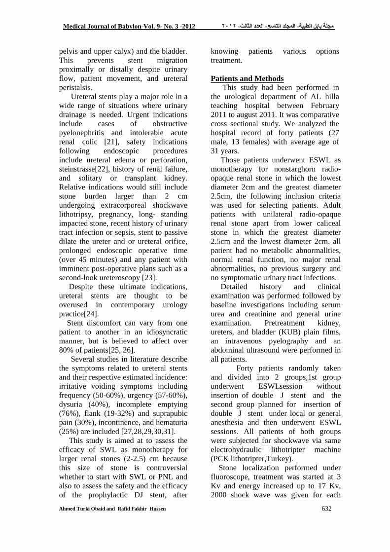
Regarding stone fragmentation in
group A (stentless), 19 patients had stone
fragmentation from total number of
patients of 20 which account 95% while
in group B (stented), 18 patients
develops stone fragmentation from total
number of patients of 20 which account
90% .
Stone fragmentation define as
radiological evidence of stone
fragmentation after extracorporeal
shock wave lithotripsy , so there is
no significant difference in the stone
fragmentation in both groups as it does
not depend on the present or absence of
the double j stent ,P value > 0.05.
Table 1 show stone fragmentation
Group Number of patients Percent %
Group A 19 95%
Group B 18 90%
Medical Journal of Babylon-Vol. 9- No. 3 -2012 1021 -مجلة بابل الطبية- المجلد التاسع- العدد الثالث
Ahmed Turki Obaid and Rafid Fakhir Hussen
634
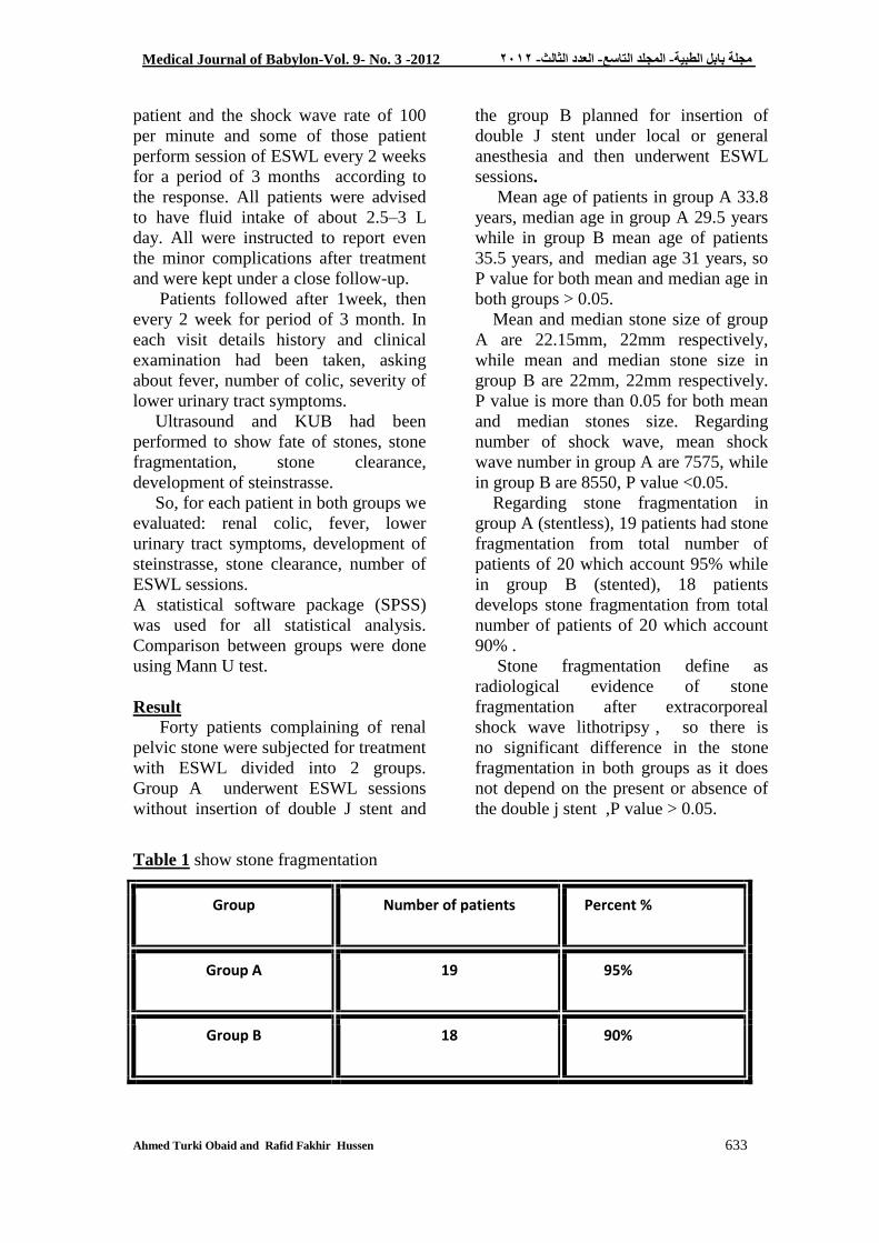
Incomplete stone clearance which
define as radiological evidence of
stone fragmentation of more than 5
mm. In group A ,4 patients had
incomplete stone clearance which
account 20% of total number of
patients in group A , while in group B,
8 patient had incomplete stone
clearance which account 40% of total
number of patient in group B(20). P
value < 0.05.
Table 2 show incomplete stone clearance
Group Number of patients Percent %
Group A 4 20%
Group B 8 40%
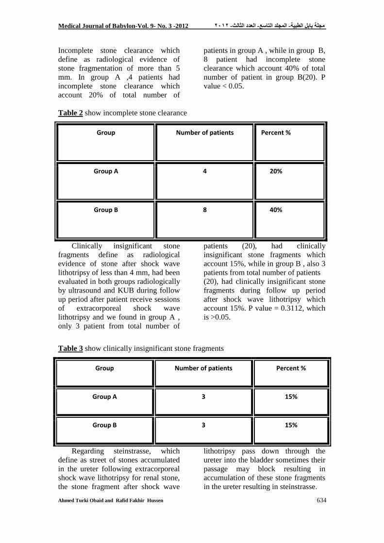
Clinically insignificant stone
fragments define as radiological
evidence of stone after shock wave
lithotripsy of less than 4 mm, had been
evaluated in both groups radiologically
by ultrasound and KUB during follow
up period after patient receive sessions
of extracorporeal shock wave
lithotripsy and we found in group A ,
only 3 patient from total number of
patients (20), had clinically
insignificant stone fragments which
account 15%, while in group B , also 3
patients from total number of patients
(20), had clinically insignificant stone
fragments during follow up period
after shock wave lithotripsy which
account 15%. P value = 0.3112, which
is >0.05.
Table 3 show clinically insignificant stone fragments
Group Number of patients Percent %
Group A 3 15%
Group B 3 15%
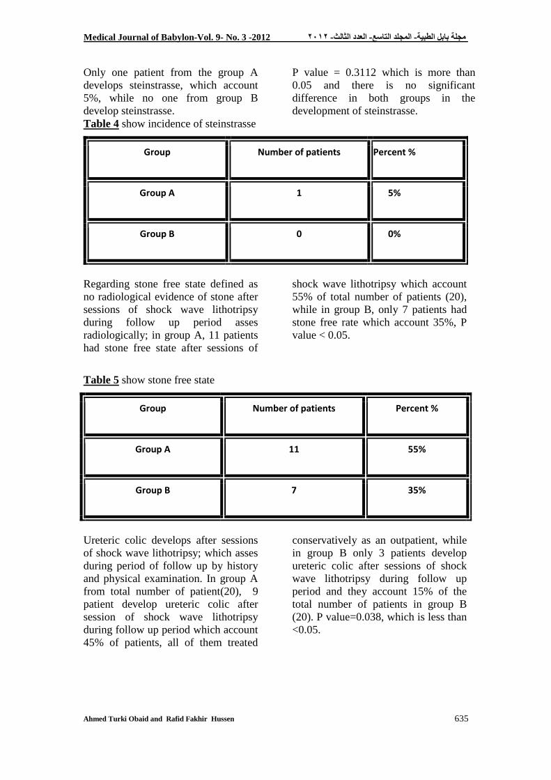
Regarding steinstrasse, which
define as street of stones accumulated
in the ureter following extracorporeal
shock wave lithotripsy for renal stone,
the stone fragment after shock wave
lithotripsy pass down through the
ureter into the bladder sometimes their
passage may block resulting in
accumulation of these stone fragments
in the ureter resulting in steinstrasse.
Medical Journal of Babylon-Vol. 9- No. 3 -2012 1021 -مجلة بابل الطبية- المجلد التاسع- العدد الثالث
Ahmed Turki Obaid and Rafid Fakhir Hussen
635
Only one patient from the group A
develops steinstrasse, which account
5%, while no one from group B
develop steinstrasse.
P value = 0.3112 which is more than
0.05 and there is no significant
difference in both groups in the
development of steinstrasse.
Table 4 show incidence of steinstrasse
Group Number of patients Percent %
Group A 1 5%
Group B 0 0%
Regarding stone free state defined as
no radiological evidence of stone after
sessions of shock wave lithotripsy
during follow up period asses
radiologically; in group A, 11 patients
had stone free state after sessions of
shock wave lithotripsy which account
55% of total number of patients (20),
while in group B, only 7 patients had
stone free rate which account 35%, P
value < 0.05.
Table 5 show stone free state
Group Number of patients Percent %
Group A 11 55%
Group B 7 35%
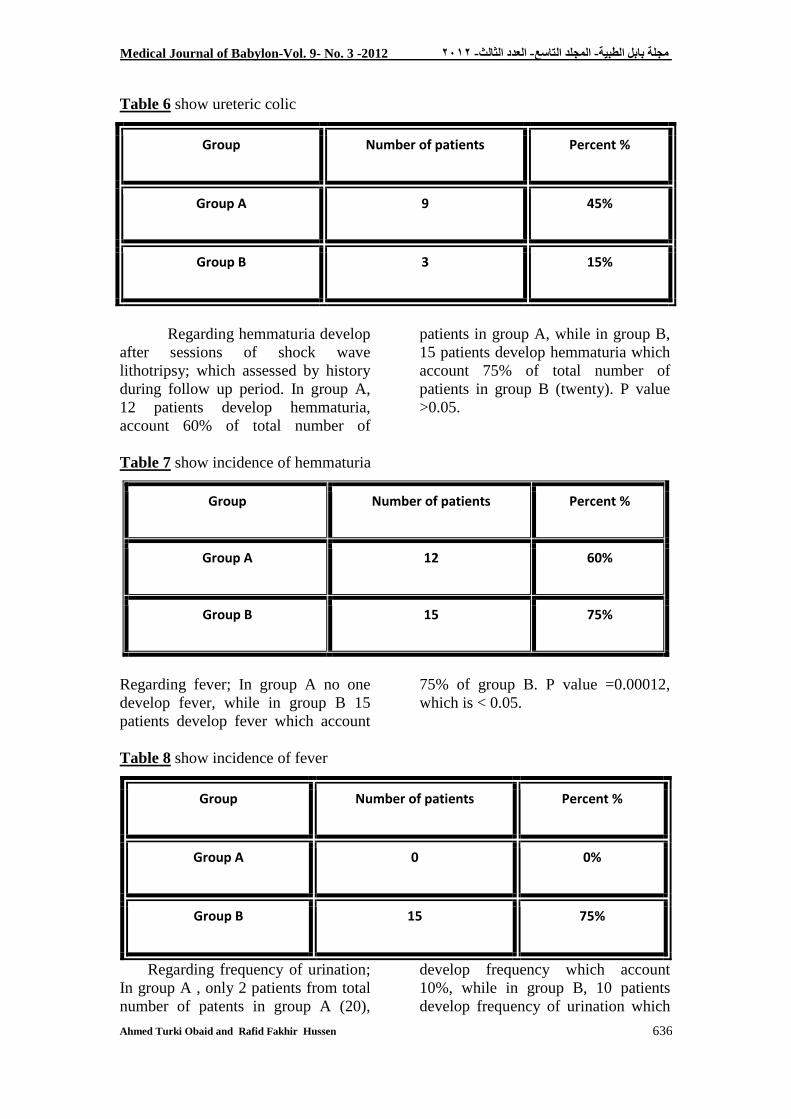
Ureteric colic develops after sessions
of shock wave lithotripsy; which asses
during period of follow up by history
and physical examination. In group A
from total number of patient(20), 9
patient develop ureteric colic after
session of shock wave lithotripsy
during follow up period which account
45% of patients, all of them treated
conservatively as an outpatient, while
in group B only 3 patients develop
ureteric colic after sessions of shock
wave lithotripsy during follow up
period and they account 15% of the
total number of patients in group B
(20). P value=0.038, which is less than
<0.05.
Medical Journal of Babylon-Vol. 9- No. 3 -2012 1021 -مجلة بابل الطبية- المجلد التاسع- العدد الثالث
Ahmed Turki Obaid and Rafid Fakhir Hussen
636
Table 6 show ureteric colic
Group Number of patients Percent %
Group A 9 45%
Group B 3 15%
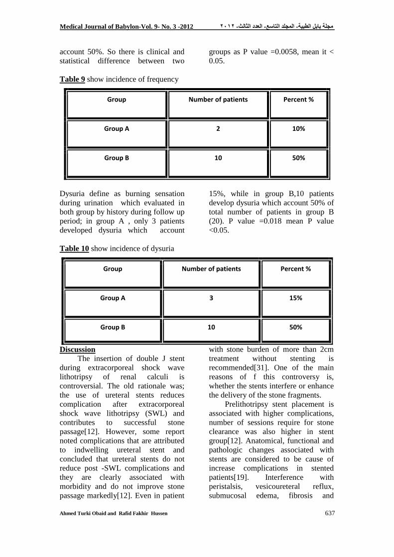
Regarding hemmaturia develop
after sessions of shock wave
lithotripsy; which assessed by history
during follow up period. In group A,
12 patients develop hemmaturia,
account 60% of total number of
patients in group A, while in group B,
15 patients develop hemmaturia which
account 75% of total number of
patients in group B (twenty). P value
>0.05.
Table 7 show incidence of hemmaturia
Group Number of patients Percent %
Group A 12 60%
Group B 15 75%
Regarding fever; In group A no one
develop fever, while in group B 15
patients develop fever which account
75% of group B. P value =0.00012,
which is < 0.05.
Table 8 show incidence of fever
Group Number of patients Percent %
Group A 0 0%
Group B 15 75%
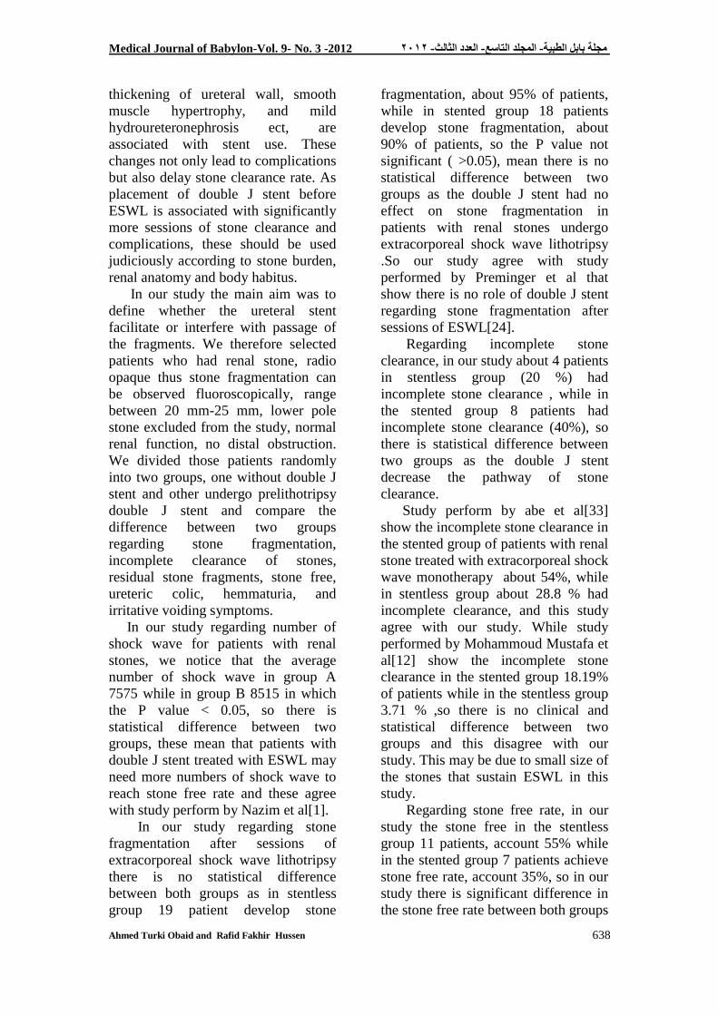
Regarding frequency of urination;
In group A , only 2 patients from total
number of patents in group A (20),
develop frequency which account
10%, while in group B, 10 patients
develop frequency of urination which
Medical Journal of Babylon-Vol. 9- No. 3 -2012 1021 -مجلة بابل الطبية- المجلد التاسع- العدد الثالث
Ahmed Turki Obaid and Rafid Fakhir Hussen
637
account 50%. So there is clinical and
statistical difference between two
groups as P value =0.0058, mean it <
0.05.
Table 9 show incidence of frequency
Group Number of patients Percent %
Group A 2 10%
Group B 10 50%
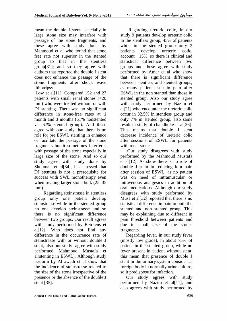
Dysuria define as burning sensation
during urination which evaluated in
both group by history during follow up
period; in group A , only 3 patients
developed dysuria which account
15%, while in group B,10 patients
develop dysuria which account 50% of
total number of patients in group B
(20). P value =0.018 mean P value
<0.05.
Table 10 show incidence of dysuria
Group Number of patients Percent %
Group A 3 15%
Group B 10 50%
Discussion
The insertion of double J stent
during extracorporeal shock wave
lithotripsy of renal calculi is
controversial. The old rationale was;
the use of ureteral stents reduces
complication after extracorporeal
shock wave lithotripsy (SWL) and
contributes to successful stone
passage[12]. However, some report
noted complications that are attributed
to indwelling ureteral stent and
concluded that ureteral stents do not
reduce post -SWL complications and
they are clearly associated with
morbidity and do not improve stone
passage markedly[12]. Even in patient
with stone burden of more than 2cm
treatment without stenting is
recommended[31]. One of the main
reasons of f this controversy is,
whether the stents interfere or enhance
the delivery of the stone fragments.
Prelithotripsy stent placement is
associated with higher complications,
number of sessions require for stone
clearance was also higher in stent
group[12]. Anatomical, functional and
pathologic changes associated with
stents are considered to be cause of
increase complications in stented
patients[19]. Interference with
peristalsis, vesicoureteral reflux,
submucosal edema, fibrosis and
Medical Journal of Babylon-Vol. 9- No. 3 -2012 1021 -مجلة بابل الطبية- المجلد التاسع- العدد الثالث
Ahmed Turki Obaid and Rafid Fakhir Hussen
638
thickening of ureteral wall, smooth
muscle hypertrophy, and mild
hydroureteronephrosis ect, are
associated with stent use. These
changes not only lead to complications
but also delay stone clearance rate. As
placement of double J stent before
ESWL is associated with significantly
more sessions of stone clearance and
complications, these should be used
judiciously according to stone burden,
renal anatomy and body habitus.
In our study the main aim was to
define whether the ureteral stent
facilitate or interfere with passage of
the fragments. We therefore selected
patients who had renal stone, radio
opaque thus stone fragmentation can
be observed fluoroscopically, range
between 20 mm-25 mm, lower pole
stone excluded from the study, normal
renal function, no distal obstruction.
We divided those patients randomly
into two groups, one without double J
stent and other undergo prelithotripsy
double J stent and compare the
difference between two groups
regarding stone fragmentation,
incomplete clearance of stones,
residual stone fragments, stone free,
ureteric colic, hemmaturia, and
irritative voiding symptoms.
In our study regarding number of
shock wave for patients with renal
stones, we notice that the average
number of shock wave in group A
7575 while in group B 8515 in which
the P value < 0.05, so there is
statistical difference between two
groups, these mean that patients with
double J stent treated with ESWL may
need more numbers of shock wave to
reach stone free rate and these agree
with study perform by Nazim et al[1].
In our study regarding stone
fragmentation after sessions of
extracorporeal shock wave lithotripsy
there is no statistical difference
between both groups as in stentless
group 19 patient develop stone
fragmentation, about 95% of patients,
while in stented group 18 patients
develop stone fragmentation, about
90% of patients, so the P value not
significant ( >0.05), mean there is no
statistical difference between two
groups as the double J stent had no
effect on stone fragmentation in
patients with renal stones undergo
extracorporeal shock wave lithotripsy
.So our study agree with study
performed by Preminger et al that
show there is no role of double J stent
regarding stone fragmentation after
sessions of ESWL[24].
Regarding incomplete stone
clearance, in our study about 4 patients
in stentless group (20 %) had
incomplete stone clearance , while in
the stented group 8 patients had
incomplete stone clearance (40%), so
there is statistical difference between
two groups as the double J stent
decrease the pathway of stone
clearance.
Study perform by abe et al[33]
show the incomplete stone clearance in
the stented group of patients with renal
stone treated with extracorporeal shock
wave monotherapy about 54%, while
in stentless group about 28.8 % had
incomplete clearance, and this study
agree with our study. While study
performed by Mohammoud Mustafa et
al[12] show the incomplete stone
clearance in the stented group 18.19%
of patients while in the stentless group
3.71 % ,so there is no clinical and
statistical difference between two
groups and this disagree with our
study. This may be due to small size of
the stones that sustain ESWL in this
study.
Regarding stone free rate, in our
study the stone free in the stentless
group 11 patients, account 55% while
in the stented group 7 patients achieve
stone free rate, account 35%, so in our
study there is significant difference in
the stone free rate between both groups
Medical Journal of Babylon-Vol. 9- No. 3 -2012 1021 -مجلة بابل الطبية- المجلد التاسع- العدد الثالث
Ahmed Turki Obaid and Rafid Fakhir Hussen
639
mean the double J stent especially in
large stone size may interfere with
passage of the stone fragments, and
these agree with study done by
Mahmoud et al who found that stone
free rate not superior in the stented
group to that in the stentless
group[31]; and so they agree with
authors that reported the double J stent
does not enhance the passage of the
stone fragments after shock wave
lithotripsy.
Low et al[11]. Compared 152 and 27
patients with small renal stones (<20
mm) who were treated without or with
DJ stenting. There was no significant
difference in stone-free rates at 1
month and 3 months (61% nonstented
vs. 67% stented group). And these
agree with our study that there is no
role for pre ESWL stenting in enhance
or facilitate the passage of the stone
fragments but it sometimes interferes
with passage of the stone especially in
large size of the stone. And so our
study agree with study done by
Shouman et al[34], has stressed that
DJ stenting is not a prerequisite for
success with SWL monotherapy even
when treating larger stone bulk (25–35
mm).
Regarding steinstrasse in stentless
group only one patient develop
steinstrasse while in the stented group
no one develop steinstrasse and so
there is no significant difference
between two groups. Our result agrees
with study performed by Beirkens et
al[12]. Who does not find any
difference in the occurrence rate of
steinstrasse with or without double J
stent, also our study agree with study
performed Mahmoud Mustafa et
al(stenting in ESWL). Although study
perform by Al awadi et al show that
the incidence of steinstrasse related to
the size of the stone irrespective of the
presence or the absence of the double J
stent [35].
Regarding ureteric colic, in our
study 9 patients develop ureteric colic
in the stentless group, 45% of patients
while in the stented group only 3
patients develop ureteric colic,
account 15%, so there is clinical and
statistical difference between two
groups and these agree with study
performed by Amar et al who show
that there is significant difference
between stentless and stented groups,
as many patients sustain pain after
ESWL in the non stented than those in
stented group. Also our study agree
with study performed by Nazim et
al[21] who encounter the ureteric colic
occur in 32.5% in stentless group and
only 7% in stented group, also same
result in study of chandhoke et al[36].
This means that double J stent
decrease incidence of ureteric colic
after sessions of ESWL for patients
with renal stones.
Our study disagrees with study
performed by the Mahmoud Mustafa
et al[12]. As show there is no role of
double J stent in reducing loin pain
after session of ESWL, as no patient
was on need of intramuscular or
intravenous analgesics in addition of
oral medications. Although our study
disagrees with study performed by
Musa et al[32] reported that there is no
statistical difference in pain in both the
stented and non stented group. This
may be explaining due to different in
pain threshold between patients and
due to small size of the stones
fragments.
Regarding fever, in our study fever
(mostly low grade), in about 75% of
patient in the stented group, while no
fever present in patient without stent,
this mean that presence of double J
stent in the urinary system consider as
foreign body in normally urine culture,
so it predispose for infection.
Our study agrees with study
performed by Nazim et al[11], and
also agrees with study performed by
Medical Journal of Babylon-Vol. 9- No. 3 -2012 1021 -مجلة بابل الطبية- المجلد التاسع- العدد الثالث
Ahmed Turki Obaid and Rafid Fakhir Hussen
640
Musa et al (32)
. Also agree with study
performed Amar et al.as all these study
show slightly increase in the incidence
of fever in the stented group.
Regarding hemmaturia , 12 patients
in the stentless group which account
60% of the patients develop
macroscopic hemmaturia while 15
patients in the stented group develop
hemmaturia, which account 75%
develop macroscopic hemmaturia, so
there is no significant difference
between two group, so there is clinical
but not statistical difference between
two groups, as hemmaturia occur more
in the stented group, and our study
agree with study performed by the
study done by Nazim et al as he show
the incidence of hammaturia is
statistically and clinically more in the
stented group than that of the stentless
group (92.5%, 67.5%), respectively,
and also our study agree with study
performed by El- Assmy et al[37] who
show that hemmaturia is more in the
stented than those in the non stented
group. although our study agree with
study performed Perminger et al who
show increase incidence of the
hematuria in the stented group[38].
lower urinary tract symptoms
frequency and dysuria more common
in the stented group 10%,10%
respectively which account 50%,50%
respectively while those in the non
stented group it occur in 10%,15%
respectively, so there is statistical
clinical significance between two
groups and this due to the irritative
effect of the double J stent , and these
agree with study performed
Chandhoke et al[36], and the study
done by El- Assmy et al[37], as they
show increase incidence of irritative
symptoms in the stented group.
Conclusion
Double J stent neither enhance
the passage of stone fragments nor
reduce the complications following
shock wave lithotripsy. Stenting is
unnecessary during ESWL in renal
stones with diameters from 2 cm to 2.5
cm. This comparative study supports
the fact that the use of double J stent
does not alter the outcome of treatment
of patients with (2 - 2.5) cm stone with
ESWL with and without double J
stent. Further prospective trials should
be designed to define the criteria for
stented ESWL treatment.
References
1. Hussain M, Lal M, Ali B, S A
Naqvi, S.A.H Rizvi: Urolithiasis in
Sindh: a single centre experience with
a review of 10,000 cases. J Nephrol
Urol Transplant 1998; 1: 10-3.
2. Heptinstall RH. Pathology of the
kidney. 4th ed. Little Brown. 1992. P:
1572-1581.
3. Edward M. and William C.
CmpBells urology. 7th ed. WB
Sunders.1997.p:2662.
4. Litwin MS and Saigal CS.
Urological disease in American.
Washington, DC. US government
printing office.2007.p:283-287.
5. Brenner, BM, ed (2007). 8th ed.);
p:515-517.
6. Committe to review dietary
reference intakes for vitamin D and
calcium, institute of medicine of the
national academies (2011). Ross, AC;
Taylor, CL; Yaktine, AL et al.p:980-
982.
7. Khan IU, Butt MK, Kalim M,
Sarwar Q, Khan FA. Efficiency of
extracorporeal shock wave
lithotripsy in upper urinary tract
calculi with reference to stone size,
radiodensity, shape, and size.
Experience at Mayo Hospital, Lahore.
pak J Surg 1996; 12: 192-7.
8. Marcovish R, Smith AD. Renal
pelvic stones: choosing shock wave
lithotripsy or percutaneous
nephrolithotomy. Int Braz J Urol 2003;
29: 192-7.
Medical Journal of Babylon-Vol. 9- No. 3 -2012 1021 -مجلة بابل الطبية- المجلد التاسع- العدد الثالث
Ahmed Turki Obaid and Rafid Fakhir Hussen
641
9. Tombolini P, Ruoppolo M,
Bellorofonte C, Zaatar C, Follini M.
Lithotripsy in the treatment of urinary
lithiasis. J Nephro 2000; 13:s71-s82.
10. Chaussy C, Brendel W,
Schmiedt E. Extracorporeally induced
destruction of kidney stones by shock
waves. Lancet 1980; 2:1265-8.
11. Motola JA, Smith AD.
Therapeutic options for the
management of upper tract calculi.
Urol Clin North Am 1990; 17:191-
206.
12. Bierkens AF, Hendrikx AJ,
Lemmens WA, Debruyne FM.
Extracorporeal shock wave lithotripsy
for large renal calculi: the role of
ureteral stent: a randomized trial. J
Urol 1991; 145:699-702.
13. Stoller ML, Balton DM. Urinary
stone disease. In: Tanogho EA,
McAninch JW, (edi). Smiths general
urology. 15th ed. San Francisco:
McGrawHill, 2000: 291-320.
14. Borghi L, Schianchi T, Meschi
T, et al: Comparison of two diets for
the prevention of recurrent stones in
idiopathic hypercalciuria. N Engl J
Med 346:77-84, 2002.
15. Coe FL, Evan A, Worcester E:
Kidney stone disease. J Clin Invest
115:2598-2608,2005.
16. Evan AP, Coe FL, Lingeman JE,
Worcester E: Insights on the pathology
of kidney stone formation. Urol Res
33:383-389, 2005.
17. Lingeman JE, Kim SC, Kuo RL,
et al: Shock wave lithotripsy:anecdotes
and insights. J Endourol 17:687-693,
2003.
18. Lingeman JE, Coury TA,
Newman DM, Kahnoski RJ, Mertz JH,
Mosbaugh PG, et al. Comparison of
results and morbidity of percutaneous
nephrostolithotomy and extracorporeal
shock wave lithotripsy. J Urol
1987;138:485-90.
19. Zimskind PD, Fetter TR,
Wilkerson JL. Clinical use of long
term indwelling silicone rubber
ureteral splints inserted
cistoscopically. J Urol 1967;97:840-4.
20. Monga M. Ureteral Stents: New
materials and designs. In: Williams JC,
Evans A, Lingeman J, editors. Renal
Stone Disease. 2nd ed. Melville NY,
American Institute of Physics; 2008. p.
173-81.
21. Chew BH, Knudsen BH and
Denstedt D. The use of stents in
contemporary urology.Curr Opin Urol
2004;14:111-5.
22. Jeong H, Hwak C, Lee SE.
Ureteric stenting after ureteroscopy for
ureteric stones: a prospective
randomized study assessing symptoms
and complications. BJU Int 2004;
93:1032-5.
23. Knudsen BE, Beiko DT,
Denstedt JD. Stenting after
ureteroscopy: pros and cons. Urol Clin
N Am 2004;31:173-80.
24. Auge BK, SarvisJA,
L’Esperance JO, Preminger G.
Practice Patterns of Ureteral Stenting
after Routine Ureteroscopic Stone
Surgery: A Survey of Practicing
Urologists. J Endourol 2007;21:1287–
91.
25. Haleblian G, Kijvikain K, de la
Rosette J, Preminger G. Ureteral
stenting and urinary stone
management: a systematic review. J
Urol 2008;179:424-30.
26. Joshi HB, Okeke A, Newns N,
Keeley FX Jr, Timoney AG.
Characterization of urinary symptoms
in patients with ureteral stents.
Urology 2002;59:511-9.
27. Byrne RR, Auge BK,
Kourambas J, et al. Routine ureteral
stenting is not necessary after
ureteroscopy and ureteropyeloscopy: a
randomized trial. J Endourol
2002;16:9-13.
28. Hao P, Li W, Song C, Yan J,
Song B, Li L. Clinical Evaluation Of
Double- Pigtail in Patients with Upper
Urinary Tract Diseases: Report of
2685 cases. J Endourol 2008;22:65-70.
Medical Journal of Babylon-Vol. 9- No. 3 -2012 1021 -مجلة بابل الطبية- المجلد التاسع- العدد الثالث
Ahmed Turki Obaid and Rafid Fakhir Hussen
642
29. Thomas R. Indwelling ureteral
stents: Impact of material and shape on
patient comfort. J Endourol
1993;7:137-40.
30. Smedley FH, Rimmer J, Taube
M, et al. 168 Double J (pigtail) ureteric
catheter insertions: A retrospective
review. Ann R Coll Surg Engl
1988;70:377–9.
31. Damiano R, Oliva R, Esposito C,
De Sio M, Autorino R, D Armiento M.
Early and late complications of double
pigtail ureteral stent Urol Int 2002;
69:136-40.
32. Musa AA. Use of double J stent
prior to extra corporeal shock wave
lithotripsy is not beneficial: result of a
prospective randomized study. Int Urol
Nephrol 2007;40:19-22.
33. Abe T, Akakura K, Kawaguchi
M, Ueda T, lto H, et al. Outcomes of
shock lithotripsy for upper urinary-
tract stones: A large-scale study at a
single institution. J Endourol
2005;19:768-73.
34. Shouman AM, Ziada AM,
Ghoneim IA. Extracorporeal shock
wave lithotripsy monotherapy for renal
stones >25mm in children. Urology
2009;74:109-11.
35. Al Awadi K, Abdul Haleem H,
Kehinde EO, Al-Taweed A, Stein
strasse: a comparision of incidence
with and without J stenting and the
effect of J stenting on subsequent
management. Br J Urol Int
1999;84:618-21.
36. Chandhoke PS, Werencke C,
Chee-Awai RA, A randomized
outcomes trial of ureteral stents for
extracorporeal shock wave lithotripsy
of solitary kidney or proximal ureteral
sones. J Urol 2002;167:1981-3.
37. EL-Assmy A, El-Nahas AR,
SheirKZ. Is pre-shock wave lithotripsy
stenting necessary for ureteral stones
with moderate or severe
hydronephrosis? J Urol 2006; 176
:2059-62.
38. Preminger GM, Kettelhut MC,
Elkins SL, Seger J, Fetner CD.
Ureteral stenting during extracorporeal
shock wave lithotripsy: help or
inheritance? J Urol 1989; 142: 32-26.