Embed Size (px)
Citation preview

Role of FAST and CT scan in abdominal trauma
International Archives of Integrated Medicine, Vol.
Copy right © 2015, IAIM, All Rights Reserved.
Original Research Article
Role of Focused Assessment with
Sonography for Trauma (FAST) and CT scan
in abdominal trauma: Radiologist’s
perspective
Nirav Patel1*
, Niket Domadia 12
nd year Resident,
21
st year Resident,
Department of Radiology, SBKS Medical Institute and Research Centre, Sumandeep Vidyapeeth,
Vadodara, Gujarat, India *Corresponding author email: [email protected]
How to cite this article: Nirav Patel, Niket Domadia, Konark
Assessment with Sonography for Trauma (FAST) and CT scan in abdominal trauma: Radiologist’s
perspective. IAIM, 2015; 2(6): 123
Available online at
Received on: 08-06-2015
Abstract
Background: Diagnosis of abdominal trauma is a real. Diagnostic tools that help the treating doctor
in optimum management of abdominal trauma include; Focused Assessment
Trauma (FAST), Diagnostic peritoneal lavage (DPL) and CT scan.
Objectives: The aim of this communication wa
abdomen in the diagnosis of abdominal trauma.
Material and methods: This study aimed at
Department of Dhiraj General Hospita
patients.
Results: Out of 50 patients of abdominal trauma, 12 patients (24%) were in age group 21
with male to female ratio of approximately 5.2:
accident according for 54% of total cases.
followed by kidney. USG showed overall sensitivity 57.48%, specificity 97.77%, positive predictive
value 88.9125, negative predictive value 97.185 and
sensitivity of 95.35%, specificity of 100%, positive predictive value of 100%, negative predictive value
77.78% and accuracy 96%.
Conclusion: FAST is useful as the initial diagnostic tool for abdominal trauma to d
abdominal fluid. With proper training and understanding the limitations of ultrasound, the results of
FAST can be optimized. DPL is indicated to diagnose suspected internal abdominal injury when
Role of FAST and CT scan in abdominal trauma
International Archives of Integrated Medicine, Vol. 2, Issue 6, June, 2015.
Rights Reserved.
Role of Focused Assessment with
Sonography for Trauma (FAST) and CT scan
in abdominal trauma: Radiologist’s
, Niket Domadia2, Konark Sarvaiya
1, Anil Rathwa
year Resident, 3Assisstant Professor
Department of Radiology, SBKS Medical Institute and Research Centre, Sumandeep Vidyapeeth,
Nirav Patel, Niket Domadia, Konark Sarvaiya, Anil Rathwa
Assessment with Sonography for Trauma (FAST) and CT scan in abdominal trauma: Radiologist’s
123-135.
Available online at www.iaimjournal.com
Accepted on:
: Diagnosis of abdominal trauma is a real. Diagnostic tools that help the treating doctor
in optimum management of abdominal trauma include; Focused Assessment w
Trauma (FAST), Diagnostic peritoneal lavage (DPL) and CT scan.
he aim of this communication was to define the recent role of FAST and CT scan of the
abdomen in the diagnosis of abdominal trauma.
This study aimed at evaluating patients who came to the Radiology
General Hospital, by using USG and CT scan. This study comprised of 50
50 patients of abdominal trauma, 12 patients (24%) were in age group 21
male to female ratio of approximately 5.2: 1. The commonest mode of trauma was road
accident according for 54% of total cases. Spleen and liver were the most common organs injured,
USG showed overall sensitivity 57.48%, specificity 97.77%, positive predictive
value 88.9125, negative predictive value 97.185 and accuracy of 90.75%. CT scan showed highest
sensitivity of 95.35%, specificity of 100%, positive predictive value of 100%, negative predictive value
: FAST is useful as the initial diagnostic tool for abdominal trauma to d
abdominal fluid. With proper training and understanding the limitations of ultrasound, the results of
FAST can be optimized. DPL is indicated to diagnose suspected internal abdominal injury when
ISSN: 2394-0026 (P)
ISSN: 2394-0034 (O)
Page 123
Role of Focused Assessment with
Sonography for Trauma (FAST) and CT scan
in abdominal trauma: Radiologist’s
, Anil Rathwa3
Department of Radiology, SBKS Medical Institute and Research Centre, Sumandeep Vidyapeeth,
Sarvaiya, Anil Rathwa. Role of Focused
Assessment with Sonography for Trauma (FAST) and CT scan in abdominal trauma: Radiologist’s
Accepted on: 14-06-2015
: Diagnosis of abdominal trauma is a real. Diagnostic tools that help the treating doctor
with Sonography for
s to define the recent role of FAST and CT scan of the
who came to the Radiology
USG and CT scan. This study comprised of 50
50 patients of abdominal trauma, 12 patients (24%) were in age group 21-30 years
mode of trauma was road traffic
pleen and liver were the most common organs injured,
USG showed overall sensitivity 57.48%, specificity 97.77%, positive predictive
CT scan showed highest
sensitivity of 95.35%, specificity of 100%, positive predictive value of 100%, negative predictive value
: FAST is useful as the initial diagnostic tool for abdominal trauma to detect intra
abdominal fluid. With proper training and understanding the limitations of ultrasound, the results of
FAST can be optimized. DPL is indicated to diagnose suspected internal abdominal injury when

Role of FAST and CT scan in abdominal trauma
International Archives of Integrated Medicine, Vol.
Copy right © 2015, IAIM, All Rights Reserved.
ultrasound machine is not available, there is no
FAST are equivocal or difficult to interpret in a hemodynamically unstable patient. In contrast, in
hemodynamically stable patients the diagnostic modality of choice is CT with intravenous contrast. It
is useful to detect free air and intra peritoneal fluid, delineate the extent of solid organ injury, detect
retroperitoneal injuries, and help in the decision for conservative treatment. Helical CT is done
rapidly which reduces the time the patient stays in th
sagittal and coronal reconstruction images which are useful for detecting ruptured diaphragm.
Key words
FAST, CT, Hematoma, Tear, Injury
Introduction
Diagnosis of abdominal trauma is a real
challenge. The clinical findings are usually not
reliable. Abdominal examination
by different factors like fractures of
ribs, contusion and abrasions of the abdominal
wall, presence of fractured lumbar vertebrae
with retroperitoneal hematoma, and reduced
level of consciousness. Diagnostic tools that help
the treating doctor to take critical decisions like
the need for laparotomy or conservative
treatment are mandatory if we aim for a
favorable outcome. Diagnostic peritoneal lavage
(DPL) had been the gold standard to detect
intra-peritoneal fluid since the sixties. Use of
Focused Assessment with Sonography for
Trauma (FAST) and helical CT scan have
dramatically changed our methods for
diagnosing blunt abdominal trauma, refined our
decisions, and enabled us to select patients for
conservative treatment. The choice of a
particular modality depends on the
hemodynamic stability of the patient, the
reliability of physical examination, the severity
of associated injuries, and the availability of a
particular diagnostic modality. The aim of this
communication is to define the recent role of
FAST and CT scan of the abdomen
diagnosis of blunt abdominal trauma.
The evaluation of the patient with abdominal
trauma is done by following steps.
Role of FAST and CT scan in abdominal trauma
International Archives of Integrated Medicine, Vol. 2, Issue 6, June, 2015.
Rights Reserved.
ultrasound machine is not available, there is no trained person to perform FAST, or the results of
FAST are equivocal or difficult to interpret in a hemodynamically unstable patient. In contrast, in
hemodynamically stable patients the diagnostic modality of choice is CT with intravenous contrast. It
seful to detect free air and intra peritoneal fluid, delineate the extent of solid organ injury, detect
retroperitoneal injuries, and help in the decision for conservative treatment. Helical CT is done
rapidly which reduces the time the patient stays in the CT scan room. Furthermore, this improves
sagittal and coronal reconstruction images which are useful for detecting ruptured diaphragm.
, Tear, Injury, Collection, Trauma, Hemoperitoneum.
inal trauma is a real
dings are usually not
Abdominal examination is compounded
by different factors like fractures of lower chest
ribs, contusion and abrasions of the abdominal
wall, presence of fractured lumbar vertebrae
ematoma, and reduced
level of consciousness. Diagnostic tools that help
the treating doctor to take critical decisions like
ed for laparotomy or conservative
treatment are mandatory if we aim for a
favorable outcome. Diagnostic peritoneal lavage
(DPL) had been the gold standard to detect
since the sixties. Use of
Sonography for
uma (FAST) and helical CT scan have
dramatically changed our methods for
diagnosing blunt abdominal trauma, refined our
decisions, and enabled us to select patients for
conservative treatment. The choice of a
particular modality depends on the
stability of the patient, the
reliability of physical examination, the severity
of associated injuries, and the availability of a
particular diagnostic modality. The aim of this
communication is to define the recent role of
FAST and CT scan of the abdomen in the
diagnosis of blunt abdominal trauma.
The evaluation of the patient with abdominal
trauma is done by following steps.
FAST - Focused abdominal sonography for
trauma
FAST is a rapid screen for intra
and can be performed in less than
FAST is non-invasive, may be easily performed
and can be done concurrently with resuscitation.
In addition, the technology is portable and may
be easily repeated if necessary. Like DPL, it can
determine the presence of hemoperitoneum but
can make no determination as to the etiology of
the hemoperitoneum [1]
operator-dependent and requires true expertise
for reliable use. Like DPL, FAST is ineffective for
imaging the retroperitoneum. The amount of
fluid necessary for a positive FAST remains
unclear. In general, several hundred cubic
centimeters of fluid/blood are necessary to be
clearly visible using FAST, but FAST cannot tell
whether fluid is blood, bile or clear fluid
FAST examination cannot be used to reliably
grade solid organ injuries. FAST is generally
performed in four areas: The u
are placed in four locations.
• Right upper quadrant
• Epigastric area (pericardial)
• Left upper quadrant (perisplenic)
• Suprapubic area—pouch of Douglas
No matter which organ is injured, the
perihepatic view is most commonly p
Blood pools in Morison pouch, the most
dependent portion of the abdomen. The
ISSN: 2394-0026 (P)
ISSN: 2394-0034 (O)
Page 124
trained person to perform FAST, or the results of
FAST are equivocal or difficult to interpret in a hemodynamically unstable patient. In contrast, in
hemodynamically stable patients the diagnostic modality of choice is CT with intravenous contrast. It
seful to detect free air and intra peritoneal fluid, delineate the extent of solid organ injury, detect
retroperitoneal injuries, and help in the decision for conservative treatment. Helical CT is done
e CT scan room. Furthermore, this improves
sagittal and coronal reconstruction images which are useful for detecting ruptured diaphragm.
Focused abdominal sonography for
FAST is a rapid screen for intra-abdominal injury
and can be performed in less than 3 minutes.
invasive, may be easily performed
and can be done concurrently with resuscitation.
In addition, the technology is portable and may
be easily repeated if necessary. Like DPL, it can
determine the presence of hemoperitoneum but
e no determination as to the etiology of
]. FAST is clearly
dependent and requires true expertise
for reliable use. Like DPL, FAST is ineffective for
imaging the retroperitoneum. The amount of
fluid necessary for a positive FAST remains
unclear. In general, several hundred cubic
rs of fluid/blood are necessary to be
clearly visible using FAST, but FAST cannot tell
whether fluid is blood, bile or clear fluid [2].
FAST examination cannot be used to reliably
grade solid organ injuries. FAST is generally
performed in four areas: The ultrasound probes
Right upper quadrant—Morison's pouch
Epigastric area (pericardial)
Left upper quadrant (perisplenic)
pouch of Douglas
No matter which organ is injured, the
perihepatic view is most commonly positive.
Blood pools in Morison pouch, the most
dependent portion of the abdomen. The

Role of FAST and CT scan in abdominal trauma
International Archives of Integrated Medicine, Vol.
Copy right © 2015, IAIM, All Rights Reserved.
pericardial views can be extremely helpful,
although pericardial tamponade is rare after
blunt abdominal injury. The ability of FAST to
determine the need for laparotomy
questionable. McKinney, et al. had
data that suggest that their scoring system can
predict the need for laparotomy
hemodynamically stable patient, a follow
scan should be obtained if non
management is contemplated. Clearly, FAST has
limitations. Its ability to detect small amounts of
fluid is questionable, even in skilled hands. In
addition, a single FAST cannot absolutely
exclude intra-abdominal injury. A recent
international consensus conference concluded
that prudent evaluation would involve two FAST
exams performed at least 6 h
supplemented with serial physical exams to
avoid missing an injury.
E-FAST (Extended FAST) includes two mo
for better assessment.
• Right paracolic gutter.
• Left paracolic gutter.
CT scanning
With the marked decrease in the use of
diagnostic peritoneal lavage [4, 5
abdominal injuries now relies almost exclusively
on the accurate interpretation of findings from
adequately performed CT examinations acquired
in a timely fashion. In patients with multiple
traumas, the “panscan” (CT of the head, neck,
chest, abdomen, and pelvis) has become the
necessary step to enable physicians to diagnose
and ascertain the severity of the injuries and to
determine the order in which these should be
treated. CT is superior to clinical evaluation and
diagnostic peritoneal lavage for diagnosing
important abdominal injuries [6, 7
Shortly after its introduction into clinical practice
nearly 3 decades ago, CT scan redefined our
understanding of the appearance and
Role of FAST and CT scan in abdominal trauma
International Archives of Integrated Medicine, Vol. 2, Issue 6, June, 2015.
Rights Reserved.
pericardial views can be extremely helpful,
although pericardial tamponade is rare after
blunt abdominal injury. The ability of FAST to
determine the need for laparotomy is
, et al. had encouraging
data that suggest that their scoring system can
predict the need for laparotomy [3]. In the
hemodynamically stable patient, a follow-up CT
scan should be obtained if non-operative
Clearly, FAST has
limitations. Its ability to detect small amounts of
fluid is questionable, even in skilled hands. In
addition, a single FAST cannot absolutely
abdominal injury. A recent
international consensus conference concluded
udent evaluation would involve two FAST
exams performed at least 6 hour apart
supplemented with serial physical exams to
(Extended FAST) includes two more scans
With the marked decrease in the use of
4, 5], diagnosis of
abdominal injuries now relies almost exclusively
on the accurate interpretation of findings from
adequately performed CT examinations acquired
a timely fashion. In patients with multiple
traumas, the “panscan” (CT of the head, neck,
chest, abdomen, and pelvis) has become the
necessary step to enable physicians to diagnose
and ascertain the severity of the injuries and to
hich these should be
treated. CT is superior to clinical evaluation and
diagnostic peritoneal lavage for diagnosing
6, 7].
Shortly after its introduction into clinical practice
nearly 3 decades ago, CT scan redefined our
erstanding of the appearance and
importance of abdominal organ injuries
Subsequently, helical CT technology improved
the accuracy and expanded the applications of
CT imaging [9, 10]. Recent hardware and
software developments, especially multi
detector technology [11, 12
potentiated the methods used to evaluate the
poly trauma patient in multiple facets:
diagnostic capability, speed, and patient safety.
CT scan often provides the most detailed images
of traumatic pathology and may
determination of operative intervention
Only CT scanning can make the diagnosis of
organ-specific abdominal injury. CT scanning
images both the abdomen and the
retroperitoneum. CT scan quantitate
amount of blood in the abdomen
Drawbacks of CT scanning relate to the need to
transport the patient from the trauma
resuscitation area, the additional time required
to perform CT scanning compared to FAST or
DPL and it is more expensive. The best CT scan
imagery requires both oral and i
contrast. Some controversy has arisen over the
use of oral contrast and whether the additional
information it provides negates the drawbacks
of increased time to administration and risk of
aspiration [15]. The oral contrast material often
produces nausea and vomiting and must be
administered while the spine remains
immobilized. The intravenous contrast material
has a small incidence of allergic reactions. Some
advocate that oral contrast is unnecessary for
abdominal CT during the initial assessme
requires further study before it is likely to gain
widespread acceptance. No definitive answer
exists at this time to the value of oral contrast in
diagnosing bowel injury.
There are some patients who require CT
scanning despite a normal FAST. Ch
shown that 28 percent of selected patients may
have intra-abdominal solid visceral injury
ISSN: 2394-0026 (P)
ISSN: 2394-0034 (O)
Page 125
importance of abdominal organ injuries [8].
Subsequently, helical CT technology improved
the accuracy and expanded the applications of
. Recent hardware and
software developments, especially multi-
11, 12]
have further
potentiated the methods used to evaluate the
trauma patient in multiple facets:
diagnostic capability, speed, and patient safety.
CT scan often provides the most detailed images
of traumatic pathology and may assist in
determination of operative intervention [13].
Only CT scanning can make the diagnosis of
specific abdominal injury. CT scanning
images both the abdomen and the
can quantitates the
amount of blood in the abdomen [14].
rawbacks of CT scanning relate to the need to
transport the patient from the trauma
resuscitation area, the additional time required
to perform CT scanning compared to FAST or
DPL and it is more expensive. The best CT scan
imagery requires both oral and intravenous
contrast. Some controversy has arisen over the
use of oral contrast and whether the additional
information it provides negates the drawbacks
of increased time to administration and risk of
The oral contrast material often
es nausea and vomiting and must be
administered while the spine remains
immobilized. The intravenous contrast material
has a small incidence of allergic reactions. Some
advocate that oral contrast is unnecessary for
abdominal CT during the initial assessment. This
requires further study before it is likely to gain
widespread acceptance. No definitive answer
exists at this time to the value of oral contrast in
There are some patients who require CT
scanning despite a normal FAST. Chiu, et al. had
shown that 28 percent of selected patients may
abdominal solid visceral injury

Role of FAST and CT scan in abdominal trauma
International Archives of Integrated Medicine, Vol.
Copy right © 2015, IAIM, All Rights Reserved.
without hemoperitoneum. These include those
with abrasions or tenderness in the lower chest,
abdomen, or pelvis. Other findings mandating
CT are pelvic fractures or thoraco
fractures.
DPL – Diagnostic Peritoneal Lavage
DPL was introduced by Root in 1965 as a rapid
and accurate method to identify
intra-abdominal hemorrhage following trauma.
Subsequent studies have confirmed t
of DPL in diagnosing abdominal hemorrhage as
well as its superiority over physical examination
alone. DPL is rapid, safe, and inexpensive. There
is approximately a 1 percent incidence of major
complication.
A positive DPL, based on microscopi
lavage fluid, has been defined as >
A positive DPL does not necessarily mandate
immediate laparotomy in the hemodynamically
stable patient. Laparotomy based solely on a
positive DPL for red cells results in a non
therapeutic procedure approximately 30 percent
of the time. It has been recommended that
patients with RBC counts in the equivocal range
(i.e., 25,000–75,000 RBC/mm
additional diagnostic testing, such as CT
scanning. It is more accurate than CT for the
early diagnosis of hollow visceral and mesenteric
injuries, but it does not reliably exclude
significant injuries to retroperitoneal structures.
False positive results may occur in the presence
of pelvis fractures. Hemodynamically stable
patients with equivocal results are best
managed by additional diagnostic testing to
avoid unnecessary laparotomies.
diagnostic accuracy generally is equal to that of
diagnostic peritoneal lavage (DPL).
Materials and methods
This study aimed at evaluating patients
came to the Radiology Department of Dhiraj
Role of FAST and CT scan in abdominal trauma
International Archives of Integrated Medicine, Vol. 2, Issue 6, June, 2015.
Rights Reserved.
without hemoperitoneum. These include those
with abrasions or tenderness in the lower chest,
abdomen, or pelvis. Other findings mandating
fractures or thoraco-lumbar spine
Diagnostic Peritoneal Lavage
DPL was introduced by Root in 1965 as a rapid
and accurate method to identify the presence of
abdominal hemorrhage following trauma.
Subsequent studies have confirmed the efficacy
of DPL in diagnosing abdominal hemorrhage as
well as its superiority over physical examination
alone. DPL is rapid, safe, and inexpensive. There
is approximately a 1 percent incidence of major
A positive DPL, based on microscopic analysis of
luid, has been defined as >105
RBC/mm3.
A positive DPL does not necessarily mandate
immediate laparotomy in the hemodynamically
stable patient. Laparotomy based solely on a
positive DPL for red cells results in a non-
procedure approximately 30 percent
of the time. It has been recommended that
e equivocal range
75,000 RBC/mm3) undergo
additional diagnostic testing, such as CT
scanning. It is more accurate than CT for the
diagnosis of hollow visceral and mesenteric
injuries, but it does not reliably exclude
significant injuries to retroperitoneal structures.
False positive results may occur in the presence
of pelvis fractures. Hemodynamically stable
results are best
managed by additional diagnostic testing to
avoid unnecessary laparotomies. FAST's
diagnostic accuracy generally is equal to that of
diagnostic peritoneal lavage (DPL).
evaluating patients who
epartment of Dhiraj
General Hospital, by using USG and CT scan. This
study comprised of 50 patients.
Inclusion criteria
• Only those patients who
participate in study were
• Patients referred to the Radiology
Department for X-ray, USG and CT
Already diagnosed cases of abdominal
trauma, which need follow up
radiological investigations and were
referred to Radiology Department, were
included in study.
• Patients came for X
scan for other diseases, an
accidentally found to have abdomin
lesion due to trauma, were
this study.
• Sample size was total
Exclusion criteria
• Patients presented
Department having abdominal lesions
due to trauma in the past and were
cured completely we
the study.
• Patients not consented
investigation.
• Patients who were unable to cooperate
for the procedure.
Description of tools:
• X-Ray machine: 600 mA Siemens, 500
mA Siemens, 300 mA Siemens
• CR system: Kodak/AGFA
• USG: Philips HD 7 and Philips HD 9
• CT scan machine: Siemens emotion 16
slice MDCT
Results
Of these 50 patients of abdominal trauma, 12
patients (24%) were in age group 21
followed by 31-40 year age
ISSN: 2394-0026 (P)
ISSN: 2394-0034 (O)
Page 126
USG and CT scan. This
study comprised of 50 patients.
Only those patients who wanted to
participate in study were included.
Patients referred to the Radiology
ray, USG and CT scan.
Already diagnosed cases of abdominal
trauma, which need follow up
adiological investigations and were
referred to Radiology Department, were
Patients came for X-ray, USG and CT
scan for other diseases, and were
accidentally found to have abdominal
lesion due to trauma, were included in
Sample size was total 50 patients.
Patients presented to Radiology
Department having abdominal lesions
due to trauma in the past and were
cured completely were excluded from
Patients not consented for the
re unable to cooperate
600 mA Siemens, 500
Siemens, 300 mA Siemens
Kodak/AGFA
Philips HD 7 and Philips HD 9
Siemens emotion 16
Of these 50 patients of abdominal trauma, 12
patients (24%) were in age group 21-30 years,
40 year age group 22% and 41-

Role of FAST and CT scan in abdominal trauma
International Archives of Integrated Medicine, Vol.
Copy right © 2015, IAIM, All Rights Reserved.
50 years 20%. In the present study abdominal
trauma was most common among males, 42 out
of 50 patients (84%) with male to female ratio of
approximately 5.2: 1. (Table – 1)
The commonest mode of trauma was road
traffic accident according for 54% of total cases.
(Table – 2) This was similar to findings by other
studies [3].
In the present study, spleen and liver were the
most common organs injured, followed by
kidney. Of 17 Patients with splenic injury, USG
correctly diagnosed splenic injury in 12 patients
and missed in 5 patients. Of the 5 patients, USG
showed perisplenic collection without
identifiable injury in 2 patients and in the
remaining 3 patients only free fluid in abdomen
was identified. CT correctly diagnosed
injury in all 17 patients. (Table –
Of 17 patients with liver injury, USG correctly
diagnosed liver injury in 11 patients and missed
in 6 patients. Of the 6 patients, 5 patients
showed free fluid in abdomen on USG and 1
patient showed no signs of liver injury on USG.
CT showed subcapsular hematoma in 2 patients
which was missed on USG. Four patients had
lacerations of liver which was missed on USG.
patients showed parenchymal contusion with or
without intraparenchymal hematoma on USG, 2
patients also revealed associated laceration on
CT. CT correctly diagnosed liver injury in all 17
patients.
Of 13 patients with renal injury, USG correctly
diagnosed renal injury in 8 patients and no signs
of renal injury in 3 patients. Of the 3 patients
with no renal injury on USG, 3 showed only
perirenal collection.
CT correctly diagnosed renal injury in all 11
patients.
Role of FAST and CT scan in abdominal trauma
International Archives of Integrated Medicine, Vol. 2, Issue 6, June, 2015.
Rights Reserved.
50 years 20%. In the present study abdominal
trauma was most common among males, 42 out
of 50 patients (84%) with male to female ratio of
)
mode of trauma was road
accident according for 54% of total cases.
s similar to findings by other
In the present study, spleen and liver were the
most common organs injured, followed by
Of 17 Patients with splenic injury, USG
agnosed splenic injury in 12 patients
and missed in 5 patients. Of the 5 patients, USG
showed perisplenic collection without
identifiable injury in 2 patients and in the
remaining 3 patients only free fluid in abdomen
CT correctly diagnosed splenic
– 3)
Of 17 patients with liver injury, USG correctly
diagnosed liver injury in 11 patients and missed
in 6 patients. Of the 6 patients, 5 patients
showed free fluid in abdomen on USG and 1
of liver injury on USG.
CT showed subcapsular hematoma in 2 patients
which was missed on USG. Four patients had
lacerations of liver which was missed on USG. 5
patients showed parenchymal contusion with or
without intraparenchymal hematoma on USG, 2
nts also revealed associated laceration on
CT correctly diagnosed liver injury in all 17
Of 13 patients with renal injury, USG correctly
diagnosed renal injury in 8 patients and no signs
of renal injury in 3 patients. Of the 3 patients
no renal injury on USG, 3 showed only
CT correctly diagnosed renal injury in all 11
Out of total 4 patients with bladder injury, only 1
was detected on USG, rest 3 was not been able
to detect on USG.
Of 3 cases of pancreatic injury, USG diagnosed
pancreatic injury in 2 cases and missed in 1 case.
CT correctly diagnosed in all 3 cases.
which was missed on USG, findings showed free
fluid around pancreas but could not identify
pancreatic injury. On CT pancrea
found in the head. In 2 cases USG showed
pancreatic contusion, of which CT showed
laceration of pancreas in one patient and other
only contusion was found.
In our study, there were no cases of bowel wall
contusion. There were 3 cases of b
perforation , out of which 2 patients showed
comet tail artifact in free fluid on USG which
gave us a suspicion about bowel perforation ,
but CT scan diagnosed bowel perforation in all
the 3 patients correctly.
Out of 3 total patients, USG just showe
fluid in abdomen and no specific signs of
mesenteric injury, CT scan showed confirmed
mesenteric injury in 1 patient and query
mesenteric injury in 1 patient which was later
confirmed by Exploratory laprotomy.
patient with positive ileal perfor
scan, mesenteric injury was found on
exploratory laprotomy. Of these 50 patients
with blunt abdominal trauma, 42 patients
showed free fluid in abdomen
hemoperitoneum on USG.
hemoperitoneum in 45 patients.
didn’t have hemoperitoneum out of 50 patients
USG showed free fluid in 42 patients and had 3
false negative results. Out of 5 patients having
organ injury, not associated with
hemoperitoneum were splenic injury in which
subcapsular hematoma was present,
in which renal contusion and perinephric
ISSN: 2394-0026 (P)
ISSN: 2394-0034 (O)
Page 127
Out of total 4 patients with bladder injury, only 1
was detected on USG, rest 3 was not been able
pancreatic injury, USG diagnosed
pancreatic injury in 2 cases and missed in 1 case.
CT correctly diagnosed in all 3 cases. In 1 case
which was missed on USG, findings showed free
fluid around pancreas but could not identify
pancreatic injury. On CT pancreatic fracture was
In 2 cases USG showed
pancreatic contusion, of which CT showed
laceration of pancreas in one patient and other
, there were no cases of bowel wall
There were 3 cases of bowel
perforation , out of which 2 patients showed
comet tail artifact in free fluid on USG which
gave us a suspicion about bowel perforation ,
but CT scan diagnosed bowel perforation in all
, USG just showed free
fluid in abdomen and no specific signs of
mesenteric injury, CT scan showed confirmed
mesenteric injury in 1 patient and query
mesenteric injury in 1 patient which was later
confirmed by Exploratory laprotomy. In 1
ve ileal perforation on CT
, mesenteric injury was found on
Of these 50 patients
with blunt abdominal trauma, 42 patients
ee fluid in abdomen/
hemoperitoneum on USG. CT scan showed
hemoperitoneum in 45 patients. 5 patients
hemoperitoneum out of 50 patients.
fluid in 42 patients and had 3
alse negative results. Out of 5 patients having
organ injury, not associated with
hemoperitoneum were splenic injury in which
subcapsular hematoma was present, renal injury
in which renal contusion and perinephric

Role of FAST and CT scan in abdominal trauma
International Archives of Integrated Medicine, Vol.
Copy right © 2015, IAIM, All Rights Reserved.
hematoma were present, urteric inju
urinoma was present, bladder injury in which
vesico-cutaneous fistula was present,
testicular injury in which intra
rupture of the testis was seen along with
hematocoele was present.
There were total 8 patients out of 50 patients
with pleural injury in the form of pleural
effusion, hemothorax, and pneumothorax seen.
Out of 8 patients, 7 patients were positive on
USG, and CT scan detected all 8 cases.
USG showed 1 false negative value.
When p-value was found to be less than 0.05,
then the result would be considered statistically
significant. (Photo – 1 to 13)
Photo - 1: USG of patient showing splenic
contusion at the inferior pole of
Discussion
The challenge in the imaging of abdominal
trauma is to accurately identify injuries that
require early exploration and at the same time
avoid unnecessary operative intervention in
cases that can be managed conservatively.
In recent years, CT and USG have to a great
extent replaced all other modalities of
investigation. But both have their limitations. In
Role of FAST and CT scan in abdominal trauma
International Archives of Integrated Medicine, Vol. 2, Issue 6, June, 2015.
Rights Reserved.
urteric injury in which
bladder injury in which
cutaneous fistula was present, and
testicular injury in which intra-parenchymal
as seen along with
There were total 8 patients out of 50 patients
with pleural injury in the form of pleural
and pneumothorax seen.
7 patients were positive on
l 8 cases. Hence
USG showed 1 false negative value.
value was found to be less than 0.05,
then the result would be considered statistically
USG of patient showing splenic
contusion at the inferior pole of the spleen.
The challenge in the imaging of abdominal
trauma is to accurately identify injuries that
require early exploration and at the same time
avoid unnecessary operative intervention in
cases that can be managed conservatively.
have to a great
extent replaced all other modalities of
investigation. But both have their limitations. In
spite of diagnostic superiority, availability of CT
is still limited and it also requires stable patients.
On the other hand, inability to consistently
detect pancreatic, bowel and mesenteric injuries
and inability to functionally assess the kidneys
and frequent interference by gaseous distension
and associated bone or soft tissue injuries are
major limitations of USG.
Photo – 2A, 2B: CT scan (axial and coronal) of
showing grade III splenic injury.
This prospective study of fifty cases of blunt
abdominal trauma was carried out by,
USG (real time ultrasound) and CT (
tomography).
A
B
ISSN: 2394-0026 (P)
ISSN: 2394-0034 (O)
Page 128
spite of diagnostic superiority, availability of CT
is still limited and it also requires stable patients.
, inability to consistently
detect pancreatic, bowel and mesenteric injuries
and inability to functionally assess the kidneys
and frequent interference by gaseous distension
and associated bone or soft tissue injuries are
(axial and coronal) of
showing grade III splenic injury.
This prospective study of fifty cases of blunt
abdominal trauma was carried out by,
real time ultrasound) and CT (computed

Role of FAST and CT scan in abdominal trauma
International Archives of Integrated Medicine, Vol.
Copy right © 2015, IAIM, All Rights Reserved.
Photo – 3A: USG of the patient showing acute
hematoma in the right lobe of the liver.
Photo – 3B: Doppler USG of patient
hematoma extending to middle hepatic veins.
Photo – 4: CT scan of patient showing grade V
liver injury and hemoperitoneum.
Forty six (46%) patient were in the age group of
21-40 years, which is the most active span of
life. The incidence of trauma was much among
Role of FAST and CT scan in abdominal trauma
International Archives of Integrated Medicine, Vol. 2, Issue 6, June, 2015.
Rights Reserved.
USG of the patient showing acute
in the right lobe of the liver.
Doppler USG of patient showing
extending to middle hepatic veins.
patient showing grade V
liver injury and hemoperitoneum.
six (46%) patient were in the age group of
40 years, which is the most active span of
The incidence of trauma was much among
and most common made of trauma was road
traffic accident, followed by fall from height.
of all the patients, 45 had
injury and hemoperitoneum.
organ injured were spleen and liver followed by
kidney.
Photo – 5: USG of this patient shows co
of fluid in the pancreatic head region.
Photo – 6: CT scan of patient show
head of pancreas grade V pancreatic injury.
X-rays raised suspicion about left upper
abdominal injury, probably spleen or left kidney,
but were not accurate in diagnosing splenic
injury. Splenic injuries can be detected on
ultrasound, While CT remains an
method of identifying, classifying and
quantifying it, but decision of laparotomy
remains on hemodynamic stability of patients.
USG showed sensitivity of 64.7% and specificity
of 97% in diagnosing splenic injury. CT scan
ISSN: 2394-0026 (P)
ISSN: 2394-0034 (O)
Page 129
and most common made of trauma was road
traffic accident, followed by fall from height. Out
, 45 had abdominal organ
injury and hemoperitoneum. The most common
organ injured were spleen and liver followed by
USG of this patient shows collection
the pancreatic head region.
CT scan of patient shows fracture of
grade V pancreatic injury.
rays raised suspicion about left upper
abdominal injury, probably spleen or left kidney,
but were not accurate in diagnosing splenic
injury. Splenic injuries can be detected on
ultrasound, While CT remains an accurate
method of identifying, classifying and
quantifying it, but decision of laparotomy
remains on hemodynamic stability of patients.
USG showed sensitivity of 64.7% and specificity
of 97% in diagnosing splenic injury. CT scan
Role of FAST and CT scan in abdominal trauma
International Archives of Integrated Medicine, Vol.
Copy right © 2015, IAIM, All Rights Reserved.
remained the gold standard modality for
diagnosing the splenic injury.
Photo - 7: USG of patient showing
collection, compressing the kidney causing page
kidney.
Photo – 8A, 8B: CT scan (axial and coronal) of
patient showing grade II renal injury
subcapsular and perirenal hematoma)
to left psoas muscle and causing compression of
the left kidney. X-rays of patient were under
normal limits.
A
B
Role of FAST and CT scan in abdominal trauma
International Archives of Integrated Medicine, Vol. 2, Issue 6, June, 2015.
Rights Reserved.
d modality for
USG of patient showing subcapsular
essing the kidney causing page
(axial and coronal) of
patient showing grade II renal injury (extensive
lar and perirenal hematoma) extending
left psoas muscle and causing compression of
rays of patient were under
Photo - 9: USG showing retroperitoneal
hematoma.
Photo – 10: CT scan (Axial cuts) of patient
showing retroperitoneal
perinephric fat stranding.
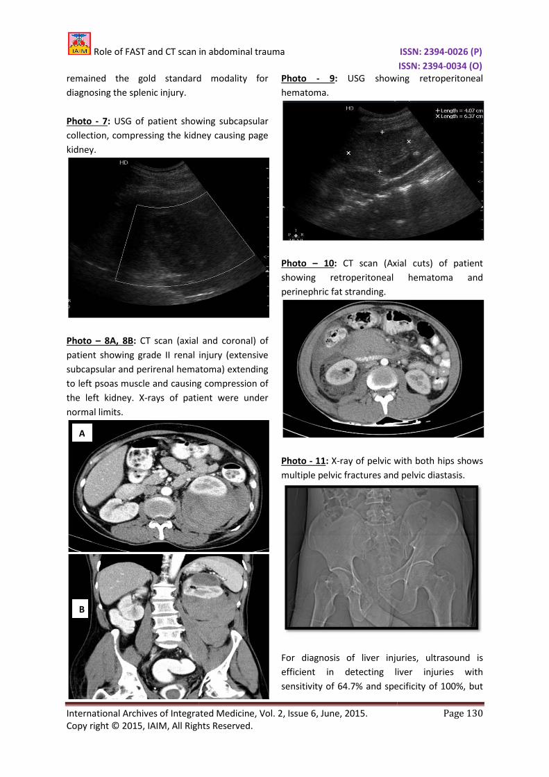
Photo - 11: X-ray of pelvic with both hips shows
multiple pelvic fractures and pelvic diastasis.
For diagnosis of liver injuries, ultrasound is
efficient in detecting liver injurie
sensitivity of 64.7% and specificity of 100%, but
ISSN: 2394-0026 (P)
ISSN: 2394-0034 (O)
Page 130
USG showing retroperitoneal
(Axial cuts) of patient
retroperitoneal hematoma and
with both hips shows
pelvic fractures and pelvic diastasis.
For diagnosis of liver injuries, ultrasound is
efficient in detecting liver injuries with
and specificity of 100%, but
Role of FAST and CT scan in abdominal trauma
International Archives of Integrated Medicine, Vol.
Copy right © 2015, IAIM, All Rights Reserved.
CT plays important role in detecting organ
injury, characterize its type, location and extent
of injury, which influences treatment plan. For
patient treated conservatively, ultrasound is
valuable in follow up studies.
Photo – 12: USG of patient showed free fluid in
the pelvis.
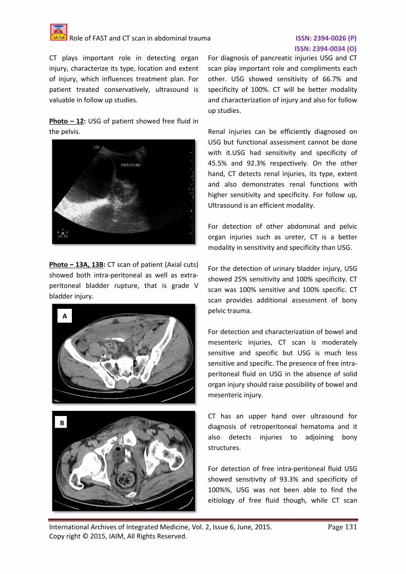
Photo – 13A, 13B: CT scan of patient (Axial cuts)
showed both intra-peritoneal as well as extra
peritoneal bladder rupture, that is grade V
bladder injury.
A
B
Role of FAST and CT scan in abdominal trauma
International Archives of Integrated Medicine, Vol. 2, Issue 6, June, 2015.
Rights Reserved.
CT plays important role in detecting organ
injury, characterize its type, location and extent
of injury, which influences treatment plan. For
patient treated conservatively, ultrasound is
USG of patient showed free fluid in
CT scan of patient (Axial cuts)
peritoneal as well as extra-
, that is grade V
For diagnosis of pancreatic injuries USG and CT
scan play important role and compliments each
other. USG showed sensitivity of 66.7% and
specificity of 100%. CT will be better modality
and characterization of injury and also for follow
up studies.
Renal injuries can be efficiently diagnosed on
USG but functional assessment cannot be done
with it.USG had sensitivity and specificity
45.5% and 92.3% respectively
hand, CT detects renal injuries, its type, extent
and also demonstrates renal fun
higher sensitivity and specificity. For follow up,
Ultrasound is an efficient modality.
For detection of other abdominal and pelvic
organ injuries such as ureter, CT is a better
modality in sensitivity and specificity than USG.
For the detection of urinary bladder injury, USG
showed 25% sensitivity and 100% specificity. CT
scan was 100% sensitive and 100% specific. CT
scan provides additional assessment of bony
pelvic trauma.
For detection and characterization of bowel and
mesenteric injuries, CT scan is moderately
sensitive and specific but USG is much less
sensitive and specific. The presence of free intra
peritoneal fluid on USG in the absence of solid
organ injury should raise possibility of bowel and
mesenteric injury.
CT has an upper hand over ultrasound for
diagnosis of retroperitoneal hematoma and it
also detects injuries to adjoining bony
structures.
For detection of free intra-peritoneal fluid USG
showed sensitivity of 93.3% and specificity of
100%%, USG was not been able
eitiology of free fluid though, while CT scan
ISSN: 2394-0026 (P)
ISSN: 2394-0034 (O)
Page 131
For diagnosis of pancreatic injuries USG and CT
scan play important role and compliments each
other. USG showed sensitivity of 66.7% and
specificity of 100%. CT will be better modality
and characterization of injury and also for follow
njuries can be efficiently diagnosed on
USG but functional assessment cannot be done
with it.USG had sensitivity and specificity of
45.5% and 92.3% respectively. On the other
hand, CT detects renal injuries, its type, extent
and also demonstrates renal functions with
higher sensitivity and specificity. For follow up,
Ultrasound is an efficient modality.
For detection of other abdominal and pelvic
organ injuries such as ureter, CT is a better
modality in sensitivity and specificity than USG.
ion of urinary bladder injury, USG
showed 25% sensitivity and 100% specificity. CT
scan was 100% sensitive and 100% specific. CT
scan provides additional assessment of bony
For detection and characterization of bowel and
injuries, CT scan is moderately
sensitive and specific but USG is much less
sensitive and specific. The presence of free intra-
peritoneal fluid on USG in the absence of solid
organ injury should raise possibility of bowel and
upper hand over ultrasound for
diagnosis of retroperitoneal hematoma and it
also detects injuries to adjoining bony
peritoneal fluid USG
showed sensitivity of 93.3% and specificity of
100%%, USG was not been able to find the
eitiology of free fluid though, while CT scan

Role of FAST and CT scan in abdominal trauma
International Archives of Integrated Medicine, Vol.
Copy right © 2015, IAIM, All Rights Reserved.
showed 100% sensitivity and 100% specificity in
detecting intra-peritoneal free fluid
For detection of pleural injury, CT scan showed
100% sensitivity and 100% specificity in
diagnosing pleural injuries.
In this study, USG showed sensitivity of 100%,
specificity of 62.5% and overall accuracy of 94%
as compared to that of CT, which showed
sensitivity of 100%, specificity of 100% and
accuracy of 100% in detection of free intra
peritoneal fluid.
CT scan showed an upper hand in diagnosing
retroperitoneal hematoma and psoas
hematoma.
CT scan diagnosed all the solid organ injuries
except in 1 patient of diaphragmatic injury, CT
raised a suspicion of diaphragmatic injury in the
form of detecting subphrenic collection and
raised dome of the left diaphragm and in
another patient CT scan missed mesenteric tear
but had diagnosed ileal perforation in the same
patients. CT scan also gave details about basal
consolidation and lung window better than X
or USG.CT scan gave details about vertebral
fractures which were missed on X
Hence, it was calculated from our
showed overall sensitivity 57.48%, specificity
97.77%, positive predictive value 88.9125,
negative predictive value 97.185 and acc
90.75%. CT scan showed highest sensitivity of
95.35%, specificity of 100%, positive predictive
value of 100%, negative predictive value 77.78%
and accuracy 96%. (Table – 4)
We conclude that X-rays findings if negative do
not suggest that there is no abdominal trauma
and could be replaced by CT scanning of
abdomen and lower thorax , but cost
effectiveness remains the main issue, USG gave
Role of FAST and CT scan in abdominal trauma
International Archives of Integrated Medicine, Vol. 2, Issue 6, June, 2015.
Rights Reserved.
showed 100% sensitivity and 100% specificity in
peritoneal free fluid.
pleural injury, CT scan showed
100% sensitivity and 100% specificity in
In this study, USG showed sensitivity of 100%,
specificity of 62.5% and overall accuracy of 94%
as compared to that of CT, which showed
sensitivity of 100%, specificity of 100% and
accuracy of 100% in detection of free intra-
CT scan showed an upper hand in diagnosing
retroperitoneal hematoma and psoas
CT scan diagnosed all the solid organ injuries
except in 1 patient of diaphragmatic injury, CT
raised a suspicion of diaphragmatic injury in the
hrenic collection and
raised dome of the left diaphragm and in
another patient CT scan missed mesenteric tear
but had diagnosed ileal perforation in the same
patients. CT scan also gave details about basal
consolidation and lung window better than X-ray
USG.CT scan gave details about vertebral
fractures which were missed on X-ray.
Hence, it was calculated from our study, USG
showed overall sensitivity 57.48%, specificity
97.77%, positive predictive value 88.9125,
negative predictive value 97.185 and accuracy of
CT scan showed highest sensitivity of
95.35%, specificity of 100%, positive predictive
value of 100%, negative predictive value 77.78%
rays findings if negative do
not suggest that there is no abdominal trauma
and could be replaced by CT scanning of
abdomen and lower thorax , but cost-
effectiveness remains the main issue, USG gave
us an fair information about most of th
organ injuries and could be easily used in
pregnant females and is cost
could not give the extent of the injury in lot of
patients, USG also could not give any
information about the bony injury cuts or lower
lung injury and could not help classify the injury.
CT scan was the most sensitive and most specific
modality in the patients with abdominal trauma
patients. CT scan helped in classifying the injury
and thus helped in management of these
patients, prevented lot of unwanted l
and saved lot of lives by giving a correct
classification and thus timely laprotomies.
Conclusion
FAST is useful as the initial diagnostic tool for
abdominal trauma to detect intra
fluid. Indications for diagnostic peritoneal lavage
are becoming more restricted. In
hemodynamically stable patients, the diagnostic
modality of choice is CT scanning. These three
modalities are complementary and not
competitive. Their usefulness is maximized when
they are applied properly within defined cli
algorithms.
References
1. Scalea TM, Rodriguez A, Chiu WC,
Brenneman FD, Fallon WF, Kato K, et al.
Focused assessment with sonography
for trauma (FAST): Results from an
international consensus conference.
Trauma, 1999; 46: 466
2. Wherrett LJ, Boulanger BR, McLellan BA,
Brenneman FD, Rizoli SB, Culhane J, et
al. Hypotension after blunt abdominal
trauma: The role of emergent
abdominal sonography in surgical triage.
J Trauma, 1996; 41: 815
3. McKenney M, Lentz K, Nunez D, et al.
Can ultrasound replace diagnostic
ISSN: 2394-0026 (P)
ISSN: 2394-0034 (O)
Page 132
us an fair information about most of the solid
organ injuries and could be easily used in
pregnant females and is cost-effective , but USG
could not give the extent of the injury in lot of
patients, USG also could not give any
information about the bony injury cuts or lower
d not help classify the injury.
CT scan was the most sensitive and most specific
modality in the patients with abdominal trauma
patients. CT scan helped in classifying the injury
and thus helped in management of these
patients, prevented lot of unwanted laprotomies
and saved lot of lives by giving a correct
classification and thus timely laprotomies.
FAST is useful as the initial diagnostic tool for
abdominal trauma to detect intra-abdominal
fluid. Indications for diagnostic peritoneal lavage
becoming more restricted. In
emodynamically stable patients, the diagnostic
modality of choice is CT scanning. These three
modalities are complementary and not
competitive. Their usefulness is maximized when
they are applied properly within defined clinical
Scalea TM, Rodriguez A, Chiu WC,
Brenneman FD, Fallon WF, Kato K, et al.
Focused assessment with sonography
for trauma (FAST): Results from an
international consensus conference. J
1999; 46: 466-72.
LJ, Boulanger BR, McLellan BA,
Brenneman FD, Rizoli SB, Culhane J, et
al. Hypotension after blunt abdominal
trauma: The role of emergent
abdominal sonography in surgical triage.
1996; 41: 815-20.
McKenney M, Lentz K, Nunez D, et al.
nd replace diagnostic

Role of FAST and CT scan in abdominal trauma
International Archives of Integrated Medicine, Vol.
Copy right © 2015, IAIM, All Rights Reserved.
peritoneal lavage in the assessment of
blunt trauma? J Trauma
441.
4. Drost TF, Rosemurgy AS, Kearney RE,
Roberts P. Diagnostic peritoneal lavage.
Limited indications due to evolving
concepts in trauma care.
57(2): 126–128.
5. Catre MG. Diagnostic peritoneal lavage
versus abdominal computed
tomography in blunt abdominal trauma:
A review of prospective studies.
Surg, 1995; 38(2): 117–122.
6. Gonzalez RP, Ickler J, Gachassin P.
Complementary roles of diagnosti
peritoneal lavage and computed
tomography in the evaluation of blunt
abdominal trauma. J Trauma
51(6): 1128–1134; discussion 1134
1136.
7. Federle MP. Computed tomography of
blunt abdominal trauma.
North Am, 1983; 21(3): 461
8. Fishman EK. Spiral CT. Applications in
the emergency patient.
1996; 16(4): 943–948.
9. Novelline RA, Rhea JT, Rao PM, Stuk JL.
Helical CT in emergency radiology.
Radiology, 1999; 213(2): 321
10. Fang JF, Wong YC, Lin BC, Hsu YP, Chen
MF. Usefulness of multi
Source of support: Nil
Role of FAST and CT scan in abdominal trauma
International Archives of Integrated Medicine, Vol. 2, Issue 6, June, 2015.
Rights Reserved.
peritoneal lavage in the assessment of
J Trauma, 1994; 37: 439–
Drost TF, Rosemurgy AS, Kearney RE,
Roberts P. Diagnostic peritoneal lavage.
Limited indications due to evolving
concepts in trauma care. Am Surg, 1991;
Catre MG. Diagnostic peritoneal lavage
versus abdominal computed
tomography in blunt abdominal trauma:
A review of prospective studies. Can J
122.
Gonzalez RP, Ickler J, Gachassin P.
Complementary roles of diagnostic
peritoneal lavage and computed
tomography in the evaluation of blunt
J Trauma, 2001;
1134; discussion 1134–
Federle MP. Computed tomography of
blunt abdominal trauma. Radiol Clin
, 1983; 21(3): 461–475.
n EK. Spiral CT. Applications in
the emergency patient. Radio Graphics,
Novelline RA, Rhea JT, Rao PM, Stuk JL.
Helical CT in emergency radiology.
1999; 213(2): 321–339.
Fang JF, Wong YC, Lin BC, Hsu YP, Chen
MF. Usefulness of multi-detector
computed tomography for the initial
assessment of blunt abdominal trauma
patients. World J Surg
182.
11. Milia DJ, Brasel K. Current use of CT in
the evaluation and management of
injured patients. Surg Clin North Am
2011; 91(1): 233–248.
12. Peitzman AB, Makaroun MS, Slasky BS,
Ritter P. Prospective study of computed
tomography in initial management of
blunt abdominal trauma.
1986; 26: 585–592.
13. Rodriguez C, Barone JE, Wilbanks TO,
Rha CK, Miller K. Isolated free fluid on
computed tomographic scan in blunt
abdominal trauma: A systematic review
of incidence and management.
Trauma, 2002; 53: 79
14. Federle MP, Peitzma
oral contrast material in abdominal
trauma CT scans: Is it dangerous?
Trauma, 1995; 38: 51
15. Chiu WC, Cushing BM, Rodriguez A, Ho
SM, Mirvis SE, Shanmuganathan K, et al.
Abdominal injuries without
hemoperitoneum: A potential limita
of focused abdominal sonography for
trauma (FAST). J Trauma, 1997; 42(4):
617-23.
Nil Conflict of interest:
ISSN: 2394-0026 (P)
ISSN: 2394-0034 (O)
Page 133
computed tomography for the initial
assessment of blunt abdominal trauma
World J Surg, 2006; 30(2): 176–
K. Current use of CT in
the evaluation and management of
Surg Clin North Am,
248.
Peitzman AB, Makaroun MS, Slasky BS,
Ritter P. Prospective study of computed
tomography in initial management of
blunt abdominal trauma. J Trauma,
Rodriguez C, Barone JE, Wilbanks TO,
Rha CK, Miller K. Isolated free fluid on
computed tomographic scan in blunt
abdominal trauma: A systematic review
of incidence and management. J
: 79–85.
Federle MP, Peitzman A, Krugh J. Use of
oral contrast material in abdominal
trauma CT scans: Is it dangerous? J
: 51–53.
Chiu WC, Cushing BM, Rodriguez A, Ho
SM, Mirvis SE, Shanmuganathan K, et al.
Abdominal injuries without
hemoperitoneum: A potential limitation
of focused abdominal sonography for
trauma (FAST). J Trauma, 1997; 42(4):
Conflict of interest: None declared.

Role of FAST and CT scan in abdominal trauma
International Archives of Integrated Medicine, Vol.
Copy right © 2015, IAIM, All Rights Reserved.
Table - 1: Age and Sex distribution (n=50).
Age group (years) Male
0-10 5
11-20 6
21-30 10
31-40 9
41-50 9
51-60 2
61+ 1
Total 42
Table - 2: Distribution of patients according to mechanism of injury (n=50).
Mode of trauma
RTA
FH
Penetrating abdominal trauma
Fall of wall on abdomen.
Iatrogenic trauma
Injury to the groin by cricket ball.
Total
Table – 3: Distribution of patients according to organ injury on USG and CT scan and no.
confirmed (n=50).
Organ USG
Spleen 12
Liver 11
Pancreas 2
Kidney 8
Ureter 0
Urinary bladder 1
Bowel 2
Mesentery 0
Retroperitoneal hematoma 1
Testis 1
Diaphramatic injury 0
Psoas hematoma 0
Role of FAST and CT scan in abdominal trauma
International Archives of Integrated Medicine, Vol. 2, Issue 6, June, 2015.
Rights Reserved.
Age and Sex distribution (n=50).
Female Total Total (%)
1 6 12
1 7 14
2 12 24
2 11 22
1 10 20
1 3 6
0 1 2
8 50 100
patients according to mechanism of injury (n=50).
No. of patients Percentage of t
27 54%
18 36%
1 2%
2 4%
1 2%
Injury to the groin by cricket ball. 1 2%
50 100%
Distribution of patients according to organ injury on USG and CT scan and no.
USG CT scan No. of cases confirmed
12 17 17
11 17 17
2 3 3
8 11 11
0 1 1
1 4 4
2 3 3
0 2 3
1 2 2
1 1 1
0 0 1
0 2 2
ISSN: 2394-0026 (P)
ISSN: 2394-0034 (O)
Page 134
Percentage of total (%)
Distribution of patients according to organ injury on USG and CT scan and no. of cases
cases confirmed

Role of FAST and CT scan in abdominal trauma
International Archives of Integrated Medicine, Vol.
Copy right © 2015, IAIM, All Rights Reserved.
Table - 4: Organ wise sensitivity P
Organ USG
Spleen 12 (24.0)
Liver 11 (22.0)
Pancreas 2 (4.0)
Kidney 8 (16.0)
Ureter 0 (0.0)
Urinary Bladder 1 (2)
Bowel 2 (4.0)
Mesentry 0 (0)
Retroperitoneal
Hematoma
1 (2.0)
Testis 1 (2)
Diaphragmatic
injury
0 (0)
Psoas Hematoma 0 (0)
PE 7 (14)
FF/HP 42 (84.0)
Role of FAST and CT scan in abdominal trauma
International Archives of Integrated Medicine, Vol. 2, Issue 6, June, 2015.
Rights Reserved.
Organ wise sensitivity P-value and Z-value.
CT No. of confirmed cases
17 (34.0) 17
17 (34.0) 17
3 (6.0) 3
11 (22.0) 11
1 (2.0) 1
4 (8) 4
3 (6.0) 3
2 (4.0) 3
2 (4.0) 2
1 (2) 1
0 (0) 1
2 (4) 2
8 (16) 8
45 (90.0) 45
ISSN: 2394-0026 (P)
ISSN: 2394-0034 (O)
Page 135
Z-value p-value
1.109 0.268
1.348 0.178
0.456 0.646
0.767 0.443
1.01 0.312
1.389 0.165
0.459 0.646
1.44 0.149
0.587 0.557
0 1.00
- -
1.44 0.149
0.280 0.779
0.896 0.371