Embed Size (px)
Citation preview

Role of imaging in Heart Failure:
Heart failure in corrected
congenital heart disease
Sotiria C. Apostolopoulou
Dept of Pediatric Cardiology & Adult Congenital Heart Disease
Onassis Cardiac Surgery Center

Imaging modalities
⚫ Chest X-Ray
⚫ Echocardiography
⚫ Cardiovascular Magnetic Resonance
⚫ Cardiovascular Computed Tomography
⚫ Nuclear Scintigraphy
⚫ Barium Swallow
⚫ X-ray Angiography

Tetralogy of Fallot - Repair
Repaired TOF: RV - PI - TR

Repaired TOF: PI - TR
LPA PW showing degrees of diastolic flow reversal
TR morphology
RVp estimate = 4v2 + RAP
Mild PI: persistent flow gradient at end-diastole
Moderate PI: equilibration of MPA and RV
pressures only at end-diastole
Severe PI: early diastolic pressure equilibration
J Am Soc Echocardiogr 2014;27:111-41

Repaired TOF: RV
TAPSE(weak correlation with
CMR in TOF)
RV fractional
area change (%)
(modest correlation with
CMR in TOF)
J Am Soc Echocardiogr
2014;27:111-41

Repaired TOF: Newer techniques
2D Speckle
tracking(no available guidelines
in CHD)
TDI(no available guidelines
in CHD)
Nl RV fxn (S′ 14 cm/sec) ↓ RV fxn (S′ 7 cm/sec)
Nl RV fxn TOF with ↓ RV fxn Echocardiography. 2014;31(4)
J Am Soc Echocardiogr 2014;27
RV MPIRV MPI correlates modestly with CMR RV EFCirc Cardiovasc Imaging . 2012;5(5)

TOF – TDI - Dobutamine
Apostolopoulou et al. Int J Cardiovasc Imaging (2007) 23:25–31
25 patients
11±6 (6–27) yo
Dobutamine

TOF
Bike
Roche et al. J Am Soc Echocardiogr 2015;28:294-301
29 TOF patients
Bike

CMR in CHD
⚫ Anatomical delineation
⚫ Static and Cine imaging
⚫ High–spatial resolution 3D reconstruction
⚫ Phase-contrast (PC) for blood flow measurements
⚫ Late gadolinium enhancement
⚫ Accurate, objective, reproducible and quantitative measurements
- biventricular size and function
- pulmonary and systemic blood flow measurements
- differential PA flow
- valve regurgitant volumes (e.g., PR, AR)
- myocardial viability and presence of scarring
- origin & proximal course of coronaries

CMR – Anatomy – Cine – PS/PI

39.55ml
68.69ml
37.8ml
68.5ml
36.9ml
CMR – Measurements – RV/LV/PI

CMR - Myocardial Enhancement
Gadolinium uptake in areas of fibrosis 10 to 20 min after contrast injection

Criteria for PVR - Role of CMR
⚫ Asymptomatic patients with ≥ 2 of:
1. RVEDV index>150 ml/m2 or Z-score>4 (large patients RV/LV EDV >2)
2. RV end-systolic volume index > 80 ml/m2
3. RV EF < 47%
4. LV EF < 55%
e. Large RVOT aneurysm
5. QRS duration >140 ms
6. Sustained tachyarrhythmia related to right heart volume load
7. Other:
- RVOT obstruction with RVp ≥ 2/3 systemic
- Branch PA stenosis (<30% flow to affected lung) not amenable to stenting
- ≥ Moderate TR
- Residual ASD or VSD with Qp/Qs ≥ 1.5
- Severe aortic regurgitation
- Severe aortic dilatation (diameter ≥ 5 cm)
⚫ Symptomatic patients (exercise intolerance or heart failure) and ≥ 1 of the above
Journal of Cardiovascular Magnetic Resonance 2011

TOF - Cardiac CT
⚫Anatomical delineation, excellent spatial
resolution
⚫Static and Cine imaging
⚫ 3D reconstruction
⚫ ↑ delineation of small vessels (coronaries,
distal PA branches)
⚫Compatible with pacemakers and
defibrillators
⚫Less artifacts by stainless-steel metallic
artifacts
⚫Radiation, No hemodynamic information

TOF Nuclear Scintigraphy
⚫ Ventricular size & function
⚫ Pulmonary perfusion
⚫ Quantification of cardiac shunts
⚫ Quantification of differential PA blood flow
⚫ V-scan to assess ventilation-perfusion mismatch
⚫ Myocardial perfusion and viability
⚫ Radiation
⚫ Only used in contraindications to CMR and cardiac CT

TOF X-ray Angiography
⚫ Imaging RVOT/PAs/Aorta
⚫ RV size & function / PI qualitatively
⚫ Hemodynamics
⚫ Coronaries (adults, abnormal course, Melody implantation)
⚫ Catheter interventions:
- PA balloon dilation and stenting
- percutaneous PV implantation (Melody/Sapien)
- occlusion of aortopulmonary collaterals
- closure of residual septal defects
- coronary artery interventions
⚫ Critically ill patient
⚫ Invasive, radiation, cost

Repaired TOF – Angiography

Systemic RV - CCTGA
Double
switch

CCTGA – Adult algorithm
Circulation. 2016;134:1293-1302

CCTGA - Exercise
Lancellotti et al. J Am Soc Echocardiogr 2017;30:101-38
Situs inversus and CCTGA: Increased global myocardial thickening and base-
to-apex shortening with significant decrease in RV end-systolic volume

Transposition of the Great Arteries
Rastelli

TGA s/p Mustard/Senning - Dobut
Wi et al. Circulation. 2004; 110:1380-1386
•27 patients
•Age 29±7 yo
•Dobutamine
•Positive
correlation of rest
RV free wall long-
axis excursion at
rest with CPET

Univentricular Heart - Fontan
• Passive flow of blood
through connection of
SVC and IVC directly
to the pulmonary
arteries
• Use of the normal size
ventricle (right or left)
as systemic pump

Fontan - Dobutamine
Brili et al. Hellenic J Cardiol(2007) 48:252-7
10 patients
Age: 28 ±5 yo

Fontan - Dobutamine
Schlangen et al. Circ Cardiovasc Imaging. 2014;7:880-886
52 patients
Median age:
6.6 (2.9–22) yrs

Surveillance frequency in repaired CHD
J Am Soc Echocardiogr 2014;27:111-41

Comparison of imaging modalities
J Am Soc Echocardiogr 2014;27:111-41

Conclusions
⚫ Multimodality approach in imaging in repaired CHD
⚫ No single test gives all necessary information
⚫ Choice influenced by:- age, need for sedation, acoustic windows
- clinical question
- local availability and expertise
- cost
- radiation exposure
- pacemakers/defibrillators
⚫ Research to identify predictors of deterioration vs stable
course in this growing population


TOF CMR
⚫ Steady-state free precession (SSFP) → , which is a type of gradient-echo technique characterized by high
signal-to-noise ratio, high T2/T1 contrast ratio, and sharp borders between the blood pool and the
myocardium
⚫ Electrocardiographically gated SSFP can be used as a cine magnetic resonance sequence, which is
typically used for assessment of ventricular size and function, valve function, and intracardiac and
extracardiac anatomy
⚫ electrocardiographically gated, respiratory-navigated SSFP sequence can yield a high–spatial resolution
static 3D data set, which is often used for detailed assessment of intracardiac anatomy and/or coronary
artery anatomy.
⚫ Electrocardiographically gated turbo (fast) spin-echo (TSE) imaging offers high spatial resolution
(submillimeter in-plane), excellent contrast between elements of soft tissue, and decreased sensitivity to
metallic artifacts compared with gradient-echo sequences, although it provides only static images.
⚫ Contrast-enhanced magnetic resonance angiography (MRA) represents a robust 3D technique
⚫ Electrocardiographically gated, phase-contrast (PC) flow measurements are used for measurements of
blood flow, including flow rates in the great arteries and veins, differential PA flow, and regurgitant
volumes (e.g., PR, AR


CMR - Myocardial Enhancement
Myocardial disarray and plexiform fibrosis

CMR - Myocardial Enhancement
Myocardial disarray and plexiform fibrosis

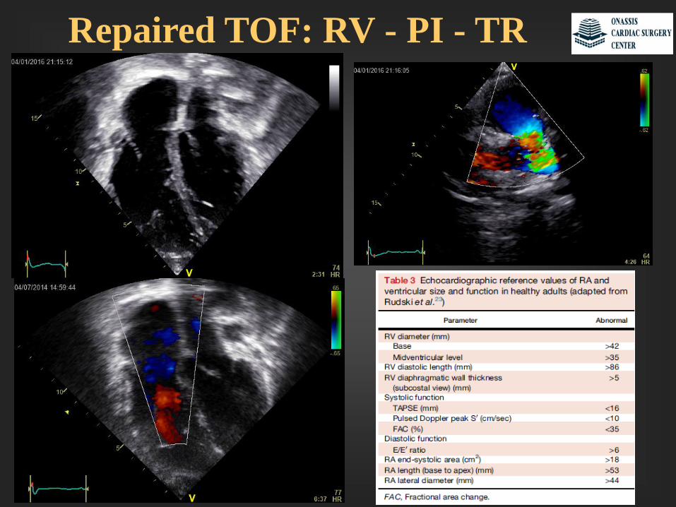
Comprehensive ECHO views
Subxiphoid
coronal
4 chamber
Subxiphoid
saggital
Parasternal
long axis
Parasternal
short axis
Suprasternal
short axis
Suprasternal
long axis
Suprasternal
right
J Am Soc Echocardiogr 2006;19:1413-30


Ventricular Septal Defects (VSD)

Ventricular Septal Defects (VSD)
Muscular
5-20%
Trabecular
Perimembranous
80%Supracristal or conal (outlet)
5-7%
Inlet
8%
Apical
Marginal
(swiss cheese)

Guidelines Repaired TOF

Imaging surveillance frequency in repaired TOF
J Am Soc Echocardiogr 2014;27:111-41

Goals of Imaging⚫ Intracardiac and extracardiac shunts
⚫ TR (degree and mechanism), estimated RVp
⚫ RV evaluation - size and function- regional RV wall motion abnormalities- RVOT (obstruction and/or aneurysm)
⚫ Degree of PR
⚫ Assessment of the main and branch PAs
⚫ LV size and function
⚫ Aorta - size of the aortic root and ascending aorta- degree of AR- aortic arch sidedness
⚫ Origin and proximal course of LCA/RCA
⚫ Systemic-to-pulmonary collateral vessels
⚫ Assessment of myocardial viability

Tetralogy of Fallot
= anterior malalignment of the outlet septum
1. Ventricular septal defect
2. Narrow RV outflow tract
3. Aorta overriding interventricular septum
4. Right ventricular hypertrophy

Conduit stenosis and RV fxn -
Hasan et al. Am J Cardiol 2012;110:1527–1533
• 35 patients with conduits
• Median age 17 (6-56) yo
• - 25 Fallot
- 9 truncus arteriosus
- 6 TGA

TOF – X-Ray Angiography

TOF – Angio – PAs – MAPCAs

Unrepaired TOF (from ECHO) Repaired TOF (from CMR)
May be useful in organizing interventions, PV implantation, construction of impantable materials that fit in the patient’s RVOT
TOF - 3D printing

TOF – Chest X-ray
• Boot-shaped heart (not common)
• ↓ pulmonary markings in ↓ pulmonary blood flow

CMR – Anatomy – Angio
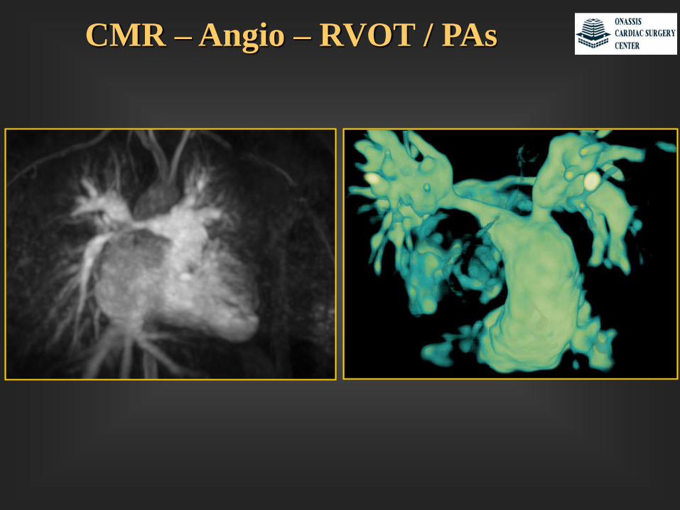
CMR – Angio – RVOT / PAs

CMR – Anatomy – Cine – PS/PI

CMR – Cine – RVOT / Flows

CMR – Anatomy - Cine – Tagging

Unrepaired TOF – Angio

ECHO: 3D / TEE / Stress
⚫3D ECHO:
- anatomy
- RV & LV size/fxn (underestimate vs CMR)
⚫TEE:
- PFO/ASD
- TTE challenging
- infective endocarditis
- guide to interventional procedures
⚫Stress ECHO:
* Stress Echo 2020 study with TOF branch (recruits since 2016)
- assess RV contractile reserve
- correlation with severity (NYHA, BNP, peak VO2, 6MWD)
- assess medium and long-term prognostic value of SE

Tetralogy of Fallot
Brili et al. J Am Soc Echocardiogr 2008;21:1093-1098
21 patients (median age 27.7 (19-48) years, Dobutamine

TGA s/p Mustard/Senning - Dobut
Vogt et al. European Journal of Echocardiography (2009) 10, 691–694
•16 patients (25.6±3.7yo)
• Dobutamine
•At rest, all patients had reduced:
- IVA
- s-velocities
- e-velocities
compared with normal subjects
with systemic LV

Pulmonary Hypertension – Master 2 step
Van Riel et al. IJC 197 (2015) 312–314
• 78 patients (43.2 ± 14.5 yo)
• Symptom-limited Master two-step test
• Systolic PAP: TR CW + RAP (from IVC size
and collapsibility)
• Mean PAP = 0.6× systolic PAP + 2
• Suspected early PVD: - mean PAP>34 mmHg
16 patients (21%) - CO>10 L/min
- mean PAP/CO > 3

Pulmonary Hypertension - Bike
Van De Bruaene et al. Eur J Prev Cardiol. 2013 Aug;20(4):597-604
• 20 open ASDs39.3±17.5 yo
• 30 closed ASDs 42.4±16.8 yo

Conclusions
⚫ Indications for Stress ECHO in pediatric and CHD are evolving
⚫ Serial testing may provide help in
- diagnosis
- risk stratification
- follow-up
- evaluation of treatment
⚫ The impact of SE on patient outcomes is under research
⚫ Efficiency may be increased with comprehensive TTE
⚫ Deformation and multidimensional imaging may utility of SE
⚫ SE has great versatility and its use is likely to expand