Embed Size (px)
Citation preview
ORIGINAL ARTICLE
495
Role of native Thiol, total Thiol and dynamic Disulphide in diagnosis of patient with prostate cancer and prostatitis_______________________________________________Mehmet Solakhan 1, Hülya Çiçek 2, Nuri Orhan 2, Mustafa Yildirim 3, 4
1 Department of Urology, Medicalpark Gaziantep Hospital, Bahçeşehir University School of Medicine, Gaziantep, Turkey; 2 Department of Medical Biochemistry, Medicalpark Gaziantep Hospital, Gaziantep, Turkey; 3 Department of Internal Medicine, Medicalpark Gaziantep Hospital, Bahçeşehir University School of Medicine, Gaziantep, Turkey; 4 Department Medical Oncology, Medicalpark Gaziantep Hospital, Bahçeşehir University School of Medicine, Gaziantep, Turkey
Vol. 45 (3): 495-502, May - June, 2019
doi: 10.1590/S1677-5538.IBJU.2018.0469
ABSTRACT
Background: Our study investigates whether Native Thiol, Total Thiol and disulphide levels measured in serum of patients with prostate cancer and prostatitis and of healthy subjects, have any role in differential diagnosis.Materials and Methods: Patients followed up for histopathologically verified diagnosis of prostate cancer and prostatitis in 2016-2017 at the Medicalpark Gaziantep Hospital Urology Clinic were included in the study. Native Thiol (NT), Total Thiol (TT), Dynamic Disulphide (DD) levels in serum were measured by a novel automated method.Results: NT, TT, DD, NT / TT ratios, DD / TT ratio and DD / NT ratio were measured as 118.4 ± 36.8μmoL / L, 150.3 ± 45.3μmoL / L, 15.9 ± 7μmoL / L, 78.8 ± 7μmoL / L, 10.5 ± 3.5μmoL / L, 13.8 ± 5.8μmoL / L respectively in patients with prostate cancer; as 116.4 ± 40.5μmoL / L, 147.5 ± 50.1μmoL / L, 15.5 ± 8.7μmoL / L, 79.7 ± 9μmoL / L, 10.1 ± 4.5μmoL / L, 13.5 ± 7.2μmoL / L in patients with prostatitis and as 144.1 ± 21.2μmoL / L, 191 ± 32.3μmoL / L, 23.4 ± 10.1μmoL / L, 76.1 ± 98.3μmoL / L, 11.9 ± 4.1μmoL / L, 16.4 ± 6.9μmoL / L in healthy subjects. Significant difference was detected between groups of NT, TT and DD levels (p = 0.008, p = 0.001, p = 0.002). No significant dif-ference was detected in terms of the NT / TT, DD / TT and DD / NT rates (p = 0.222, p = 0.222, p = 0.222).Conclusions: Serum NT, TT, DD levels in patients with prostatitis and prostate cancer were found significantly lower compared to the control group. This indicates that just as inflammation, prostate cancer also increases oxidative stress on tissues.
ARTICLE INFO
Keywords:Prostatic Neoplasms; Oxidative Stress
Int Braz J Urol. 2019; 45: 495-502
_____________________Submitted for publication:July 17, 2018_____________________Accepted after revision:November 03, 2018_____________________Published as Ahead of Print:January 05, 2019
INTRODUCTION
Prostate cancer is the second most preva-lent type of cancer in men and is the second most prevalent cause of death from cancer in men. Past studies have shown the important role of age, ge-
netic predisposition, androgen hormones, diet-re-lated factors, inflammation and oxidative stress in the development of this disease (1). It was reported that oxidative stress can play a significant role in the development of prostate cancer through lipid per-oxidation and similar mechanisms (2).
ibju | Role of oxidative stRess in pRostate diseases
496
It was reported that oxidative stress resul-ted from the disruption of the balance of antioxi-dants and reactive oxygen radicals and that this caused various systemic diseases. Over-production of reactive oxygen types (ROT) causes damage in proteins and lipids. Oxidative damage is irrever-sible in serum and tissue proteins and significant changes occur in the structure and activity of pro-teins and biomolecules. Oxidative modifications in DNA and proteins can impact certain cellular functions, resulting in cell damage, death or mu-tation and carcinogenesis formation (2).
Thiols are essential and strong anti-oxi-dant molecules in the sulfhydryl group, consis-ting of hydrogen atom and sulfur atom bonded to a carbon atom (3). The disulphide bond in their structure is a covalent bond and is also na-med as SS-bond or disulphide bridge. They play an important role in protecting oxidant stress from harmful effects. The leading thiols found in plasma are low molecule-weight thiols inclu-ding albumin thiols, protein thiols and cystei-ne, cysteinylglycine, glutathione, homocysteine and γ-glutamylcysteine. The thiol groups are oxidized with disulphide bonds getting rever-sibly oxidized by ROTs. This mechanism me-diates their anti-oxidant effects (4). The created disulphide bonds can again be reduced to thiol groups. Dynamic thiol-disulphide homeostasis plays an important role in anti-oxidant defense, detoxification, apoptosis, arranging enzymatic activity and cellular signal transmission (5).
When oxidative stress occurs, it has been noted than reduced thiol concentration increases and disulphide values increase in correlation (6). Studies have reported this deterioration of home-ostasis leads to chronic kidney deficiency, diabetes mellitus, cardiovascular diseases, cancer, chronic inflammatory diseases and various neuro-degene-rative diseases (4).
Prostate Specific Antigen (PSA) is a cri-tical marker in prostate cancer diagnosis. Serum total prostate specific antigen (t PSA) levels to-gether with abnormal digital rectal examination were the most prevalent methods used in prostate biopsy indication in recent years (7). Increased le-vels of serum PSA are associated not only with cancer but also with bacterial prostatitis, prostatic
inflammation, benign prostate hypertrophy and urinary system infection (8).
Prostatitis is a disease that is observed at a rate of 8.2% (2.2-9.7%) in men. Acute bacte-rial prostatitis (ABP) is a pyogenic urinary system infection of the urinary system. It is observed at a rate of 5% among general prostatitis (8). Es-cherichia coli is the most frequent cause of acute bacterial prostatitis. Enterococcus, Proteus, Pseu-domonas, Klebsiella and Serratia are factors less frequently responsible. ABP can cause urinary re-tention causing edema in the prostate. It can also cause serious complications from prostate abscess to urosepsis (8). Its treatment is usually performed according to clinical symptoms. Parenteral anti-biotics and hydration are performed in the ear-ly stage. Catheter and drainage are implemented if unable to urinate. Serum PSA values are usu-ally high in ABP (9). Additionally, high levels of erythrocyte sedimentation rate (ESR), C-reactive protein (CRP) and white blood cell count in full blood count accompany the situation.
In our study, serum t PSA level, NT, TT, DD levels were measured in patients diagnosed with prostate cancer and in healthy subjects at the Me-dicalpark Gaziantep Hospital Urology Clinic and Disulphide / NT, Disulphide / TT, NT / TT ratios were calculated to find Dynamic Thiol / Disulphide homeostasis levels for the purpose of investigating whether it has predictive value in differentiating Prostatitis-Prostate Cancer and diagnosing Pros-tate Cancer and its value in predicting prognosis.
MATERIAL AND METHODS
Patient Selection The study was conducted on patients his-
topathologically diagnosed with prostate cancer or prostatitis and followed up by the Gaziantep Medicalpark Hospital Urology Clinic in 2014-2017 and on healthy subjects compatible with the pa-tients in terms of age. Patients previously treated and with metastasis, persons who were smoking and / or using alcohol, had chronic disease, acute or chronic infection or using antihyperlipidemic, antibiotic or anti-oxidant drugs were excluded from the study. We have taken informed con-sent form from the patients and healthy subjects
ibju | Role of oxidative stRess in pRostate diseases
497
to participate on the study. Socio-demographic characteristics, known diseases, medical history information such as personal and family history characteristics, used drugs etc., data such as rou-tine laboratory tests were obtained retrospectively and recorded.
Sampling and Measurement of NT, TT, DD Blood samples of the patients were collected
prior to starting the medication. Non-anticoagulant venous bloods of the subjects taken into tubes after 12 hours of fasting were centrifuged at 3500rpm for 10 minutes during the first 2 hours and then split into Eppendorf Microtubes and stored at -80ºC. The-se samples were kept at 4°C temperature one night before measurement and put into room temperature 2 hours prior to the study and then the samples were mixed with a vortex and measurement was perfor-med twice for each sample.
A novel automated assay method was used to measure dynamic thiol / disulphide homeostasis. The principle of this method is based on reducing the disulfide bonds of proteins to compose disulphides in oxidative medium. Sodium borohydride (NaBH4) is added for reduction of disulphide bonds into thiol groups again. The sum of residual thiol and reducted thiol groups encloses the total thiol. The remaining NaBH4 and DTNB (5,5’-dithio-bis- (2-nitrobenzoic acid)) are removed by formaldehyde. In this way, the amount of native and reduced thiol groups are de-fined separately. The difference between the TT and the NT is divided by two to calculate the quantity of the DD bonds. Also, we calculate the NT, TT, DD / TT, NT / TT and DD / NT percent ratio (10).
Measurement of serum t PSA levels were conducted automatically with the electro-che-miluminescent method using the Hitachi Modu-lar Analytics E 170 device (Roche Diagnostics GmbH, Germany).
The patients were classified into risk groups according to Gleason score and t PSA values. Split into risk groups was as follows: low risk prostate cancer: T1-T2a stage and Gle-ason score ≤ 6 and t PSA ≤ 10, moderate risk prostate cancer: T2b stage and / or Gleason sco-re = ≤ 7 and 10 ≤ t PSA ≤ 20 and high-risk pros-tate cancer: ≥ T2c stage or Gleason score 8-10 or t PSA > 20 (11).
Statistical analysis
Statistical analyses were performed using the SPSS for Windows 15.0 package software. Compliance of the variables to normal distribu-tion was examined using visual (histogram and probability graphs) and analytic methods (Kolmo-gorov-Smirnov / Shapiro-Wilk tests). In the Kol-mogorov-Smirnov test, cases with p value greater than 0.05 were accepted as normal distribution. Differences between prostate cancer, prostatitis and the control groups in terms of NT, TT and DD were compared using the unilateral ANOVA test, as these variables showed normal distribution. The homogeneity of the variations was evaluated using the Levene test. Cases where the p value was lower than 0.05 were evaluated as statistically sig-nificant results. In cases with significant differen-ce between the groups, post-hoc pair comparisons were performed using the Tukey’s Test.
NT / TT, DD / TT and DD / NT ratios were detected to not show normal distribution. The di-fferences between these variables between pros-tate cancer, prostatitis and control groups were compared using the Kruskal-Wallis test. Pair com-parisons were performed using the Mann-Whitney U test and evaluated using the Bonferroni correc-tion. Total type-1 error level was used as 5% for statistical significance.
RESULTS
A total of 80 subjects were included in the study, consisting of 30 (37.5%) prostatitis patients, 25 (31.3%) prostate cancer patients and 25 (31.3%) healthy subjects. Patients diagnosed with prostatitis had a mean age of 60.5 ± 12.8 (range 31-83). Pa-tients diagnosed with prostate cancer had a mean age of 70.6 ± 6 (range 58-82). The age difference between the two groups was evaluated using the Student t test as they had normal distribution. Sta-tistically significant difference was detected between the two groups in terms of age (p = 0.001). Patients diagnosed with prostatitis consisted of younger pa-tients.
Total Prostate Specific Antigen was detected as 139 ± 257.8 (range 4-1200) in patients with pros-tate cancer and as 51.3 ± 112 (range 3.9-405) in pa-
ibju | Role of oxidative stRess in pRostate diseases
498
tients with prostatitis. Statistically significant diffe-rence was detected (p = 0.020) between patients with prostate cancer and prostatitis and tPSA value was detected to be higher in prostate cancer (Table-1).
Prostate cancer patient group NT was de-tected as 118.4 ± 36.8 (range 68.1-201.3) μmoL / L, TT 150.3 ± 45.3 (range 87.2-243.9) μmoL / L, DD 15.9 ±7 (range 4.4-31) μmoL / L, NT / TT rate 78.8 ± 7 (range 67.1-91), DD / TT rate 10.5 ± 3.5 (range 4.5-16.4) and DD / NT rate 13.8 ± 5.8 (range 4.9-24.4).
In the prostatitis patient group NT was de-tected as 116.4 ± 40.5 (range 53.5-281.3) μmoL / L, TT 147.5 ± 50.1 (range 61-326.8) μmoL / L, DD 15.5 ± 8.7 (range 2.7-32.3) μmoL / L, NT / TT rate 79.7 ± 9 (range 66.7-94.7), DD / TT rate 10.1 ± 4 .5 (range 2.6-16.6) and DD /NT rate 13.5 ± 7.2 (range 2.7-24.9).
In the control group NT was detected as 144.1 ± 21.2 (range 106.9-194.2) μmoL / L, TT 191 ± 32.3 (range 133.3-280.4) μmoL / L, DD 23.4 ± 10.1 (range 5-43.1) μmoL / L, NT / TT rate 76.1 ± 98.3 (range 66.2-92.8), DD / TT rate 11.9 ± 4.1 (range 3.5-16.8) and DD / NT rate 16.4 ± 6.9 (ran-ge 3.8-25.5).
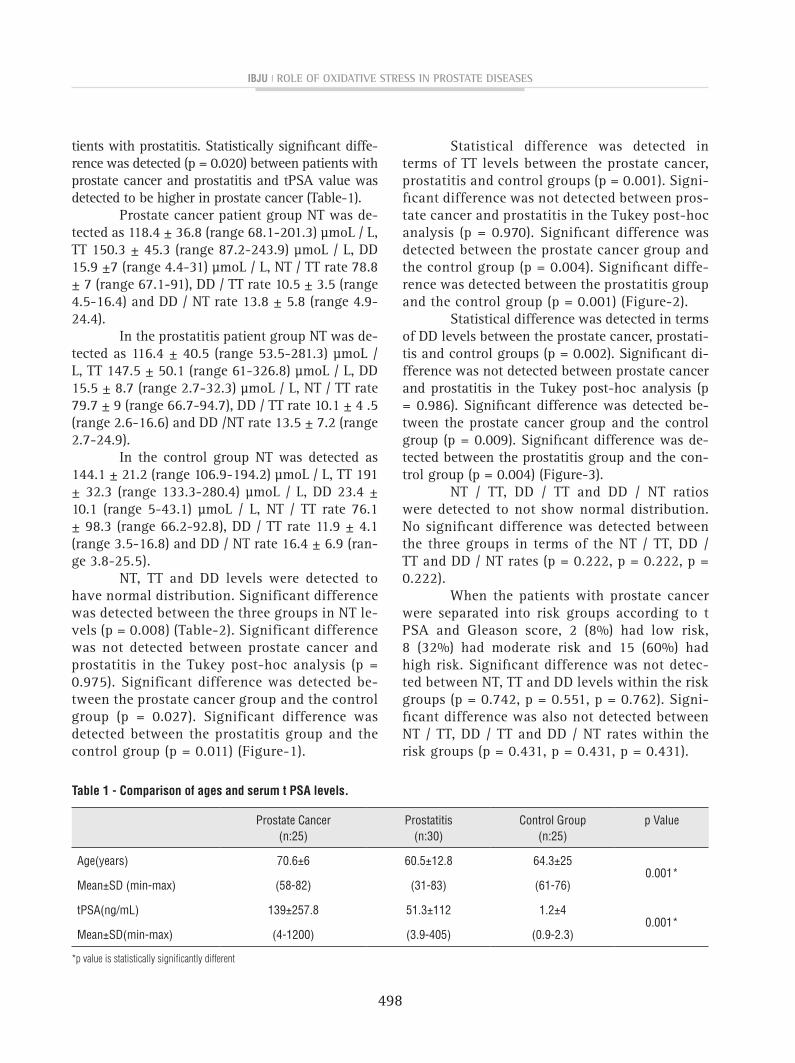
NT, TT and DD levels were detected to have normal distribution. Significant difference was detected between the three groups in NT le-vels (p = 0.008) (Table-2). Significant difference was not detected between prostate cancer and prostatitis in the Tukey post-hoc analysis (p = 0.975). Significant difference was detected be-tween the prostate cancer group and the control group (p = 0.027). Significant difference was detected between the prostatitis group and the control group (p = 0.011) (Figure-1).
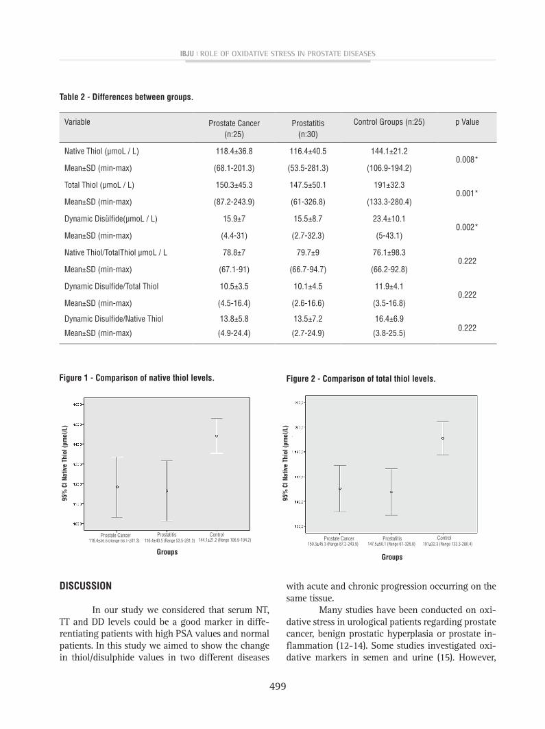
Statistical difference was detected in terms of TT levels between the prostate cancer, prostatitis and control groups (p = 0.001). Signi-ficant difference was not detected between pros-tate cancer and prostatitis in the Tukey post-hoc analysis (p = 0.970). Significant difference was detected between the prostate cancer group and the control group (p = 0.004). Significant diffe-rence was detected between the prostatitis group and the control group (p = 0.001) (Figure-2).
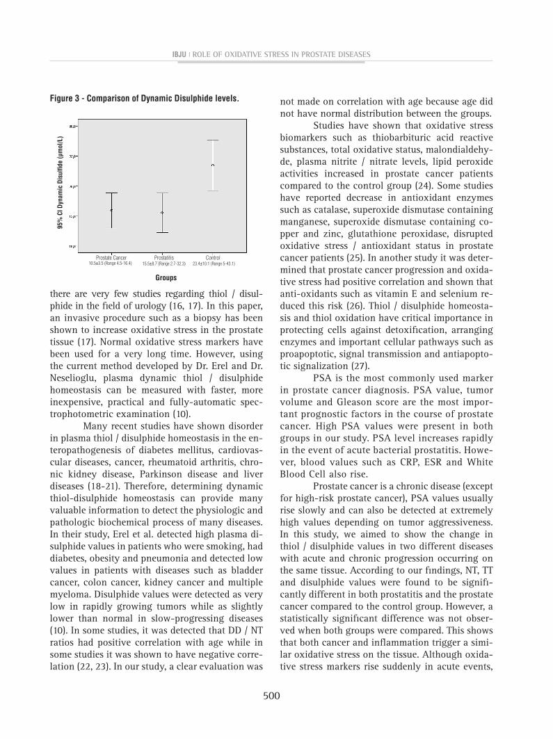
Statistical difference was detected in terms of DD levels between the prostate cancer, prostati-tis and control groups (p = 0.002). Significant di-fference was not detected between prostate cancer and prostatitis in the Tukey post-hoc analysis (p = 0.986). Significant difference was detected be-tween the prostate cancer group and the control group (p = 0.009). Significant difference was de-tected between the prostatitis group and the con-trol group (p = 0.004) (Figure-3).
NT / TT, DD / TT and DD / NT ratios were detected to not show normal distribution. No significant difference was detected between the three groups in terms of the NT / TT, DD / TT and DD / NT rates (p = 0.222, p = 0.222, p = 0.222).
When the patients with prostate cancer were separated into risk groups according to t PSA and Gleason score, 2 (8%) had low risk, 8 (32%) had moderate risk and 15 (60%) had high risk. Significant difference was not detec-ted between NT, TT and DD levels within the risk groups (p = 0.742, p = 0.551, p = 0.762). Signi-ficant difference was also not detected between NT / TT, DD / TT and DD / NT rates within the risk groups (p = 0.431, p = 0.431, p = 0.431).
Table 1 - Comparison of ages and serum t PSA levels.
Prostate Cancer(n:25)
Prostatitis(n:30)
Control Group(n:25)
p Value
Age(years) 70.6±6 60.5±12.8 64.3±250.001*
Mean±SD (min-max) (58-82) (31-83) (61-76)
tPSA(ng/mL) 139±257.8 51.3±112 1.2±40.001*
Mean±SD(min-max) (4-1200) (3.9-405) (0.9-2.3)
*p value is statistically significantly different
ibju | Role of oxidative stRess in pRostate diseases
499
DISCUSSION
In our study we considered that serum NT, TT and DD levels could be a good marker in diffe-rentiating patients with high PSA values and normal patients. In this study we aimed to show the change in thiol/disulphide values in two different diseases
with acute and chronic progression occurring on the same tissue.
Many studies have been conducted on oxi-dative stress in urological patients regarding prostate cancer, benign prostatic hyperplasia or prostate in-flammation (12-14). Some studies investigated oxi-dative markers in semen and urine (15). However,
Table 2 - Differences between groups.
Variable Prostate Cancer(n:25)
Prostatitis(n:30)
Control Groups (n:25) p Value
Native Thiol (μmoL / L) 118.4±36.8 116.4±40.5 144.1±21.20.008*
Mean±SD (min-max) (68.1-201.3) (53.5-281.3) (106.9-194.2)
Total Thiol (μmoL / L) 150.3±45.3 147.5±50.1 191±32.30.001*
Mean±SD (min-max) (87.2-243.9) (61-326.8) (133.3-280.4)
Dynamic Disülfide(μmoL / L) 15.9±7 15.5±8.7 23.4±10.10.002*
Mean±SD (min-max) (4.4-31) (2.7-32.3) (5-43.1)
Native Thiol/TotalThiol μmoL / L 78.8±7 79.7±9 76.1±98.30.222
Mean±SD (min-max) (67.1-91) (66.7-94.7) (66.2-92.8)
Dynamic Disulfide/Total Thiol 10.5±3.5 10.1±4.5 11.9±4.10.222
Mean±SD (min-max) (4.5-16.4) (2.6-16.6) (3.5-16.8)
Dynamic Disulfide/Native Thiol 13.8±5.8 13.5±7.2 16.4±6.90.222
Mean±SD (min-max) (4.9-24.4) (2.7-24.9) (3.8-25.5)
Figure 1 - Comparison of native thiol levels. Figure 2 - Comparison of total thiol levels.
118.4±36.8 (Range 68.1-201.3)
Groups
95%
CI N
ativ
e Th
iol (
µmol
/L)
Prostate Cancer Prostatitis Control116.4±40.5 (Range 53.5-281.3) 144.1±21.2 (Range 106.9-194.2)
95%
CI N
ativ
e Th
iol (
µmol
/L)
Prostate Cancer
Groups
Prostatitis147.5±50.1 (Range 61-326.8)
Control191±32.3 (Range 133.3-280.4)150.3±45.3 (Range 87.2-243.9)
ibju | Role of oxidative stRess in pRostate diseases
500
there are very few studies regarding thiol / disul-phide in the field of urology (16, 17). In this paper, an invasive procedure such as a biopsy has been shown to increase oxidative stress in the prostate tissue (17). Normal oxidative stress markers have been used for a very long time. However, using the current method developed by Dr. Erel and Dr. Neselioglu, plasma dynamic thiol / disulphide homeostasis can be measured with faster, more inexpensive, practical and fully-automatic spec-trophotometric examination (10).
Many recent studies have shown disorder in plasma thiol / disulphide homeostasis in the en-teropathogenesis of diabetes mellitus, cardiovas-cular diseases, cancer, rheumatoid arthritis, chro-nic kidney disease, Parkinson disease and liver diseases (18-21). Therefore, determining dynamic thiol-disulphide homeostasis can provide many valuable information to detect the physiologic and pathologic biochemical process of many diseases. In their study, Erel et al. detected high plasma di-sulphide values in patients who were smoking, had diabetes, obesity and pneumonia and detected low values in patients with diseases such as bladder cancer, colon cancer, kidney cancer and multiple myeloma. Disulphide values were detected as very low in rapidly growing tumors while as slightly lower than normal in slow-progressing diseases (10). In some studies, it was detected that DD / NT ratios had positive correlation with age while in some studies it was shown to have negative corre-lation (22, 23). In our study, a clear evaluation was
Figure 3 - Comparison of Dynamic Disulphide levels. not made on correlation with age because age did not have normal distribution between the groups.
Studies have shown that oxidative stress biomarkers such as thiobarbituric acid reactive substances, total oxidative status, malondialdehy-de, plasma nitrite / nitrate levels, lipid peroxide activities increased in prostate cancer patients compared to the control group (24). Some studies have reported decrease in antioxidant enzymes such as catalase, superoxide dismutase containing manganese, superoxide dismutase containing co-pper and zinc, glutathione peroxidase, disrupted oxidative stress / antioxidant status in prostate cancer patients (25). In another study it was deter-mined that prostate cancer progression and oxida-tive stress had positive correlation and shown that anti-oxidants such as vitamin E and selenium re-duced this risk (26). Thiol / disulphide homeosta-sis and thiol oxidation have critical importance in protecting cells against detoxification, arranging enzymes and important cellular pathways such as proapoptotic, signal transmission and antiapopto-tic signalization (27).
PSA is the most commonly used marker in prostate cancer diagnosis. PSA value, tumor volume and Gleason score are the most impor-tant prognostic factors in the course of prostate cancer. High PSA values were present in both groups in our study. PSA level increases rapidly in the event of acute bacterial prostatitis. Howe-ver, blood values such as CRP, ESR and White Blood Cell also rise.
Prostate cancer is a chronic disease (except for high-risk prostate cancer), PSA values usually rise slowly and can also be detected at extremely high values depending on tumor aggressiveness. In this study, we aimed to show the change in thiol / disulphide values in two different diseases with acute and chronic progression occurring on the same tissue. According to our findings, NT, TT and disulphide values were found to be signifi-cantly different in both prostatitis and the prostate cancer compared to the control group. However, a statistically significant difference was not obser-ved when both groups were compared. This shows that both cancer and inflammation trigger a simi-lar oxidative stress on the tissue. Although oxida-tive stress markers rise suddenly in acute events,
Groups
95%
CI D
ynam
ic D
isul
fide
(µm
ol/L
)
Prostate Cancer Prostatitis15.5±8.7 (Range 2.7-32.3)
Control23.4±10.1 (Range 5-43.1)10.5±3.5 (Range 4.5-16.4)
ibju | Role of oxidative stRess in pRostate diseases
501
this increase takes place gradually in chronic pro-cesses. However, this process continues in cancer patients unless treated. In inflammation, the oxi-dative process returns to normal after the causati-ve infectious event is remedied. Thiols, which are an anti-oxidant structure in the serum, may have decreased upon exposure to severe oxidation be-cause anti-oxidant defense weakens or oxidation increases in prostate cancer or prostatitis patients. Oxidation products more progressed than disul-phides were formed as thiols were subject to se-vere oxidation. As these are usually products of irreversible thiol oxidation, it is considered that disulphides are also low (10).
Earlier studies have not shown the relation between tPSA level and thiol / disulphide values. In our study tPSA values and thiol/disulphide va-lues were compared in both groups. However, al-though tPSA values were high in both groups, it was detected to be statistically higher in the pros-tate cancer group. However, no difference was de-tected between both groups in Thiol / Disulphide ratios. High tPSA values are a direct indicator of prostate tissue damage.
The main limitations of the present study are its retrospective and non-randomized nature. In addition, the number of patients involved is small. These results need to be supported by pros-pective, randomized studies, and comprehensive patient series.
CONCLUSIONS
Calculating thiol / disulphide values, whi-ch is a new marker, is an easy, inexpensive and reliable method. Serum NT, TT, DD levels in pa-tients with prostatitis and prostate cancer were found significantly lower compared to the control group. This shows that just as inflammation, pros-tate cancer also increases oxidative stress on tis-sues. We consider that NT, TT, DD levels measures in serum can be used in the differential diagnosis of these pathologies.
ABBREVIATIONS
NT = Native ThiolTT = Total Thiol
DD = Dynamic DisulphideROT = Reactive Oxygen TypesPSA = Prostate Specific AntigentPSA = Total Prostate Specific AntigenABP = Acute Bacterial ProstatitisESR = Erythrocyte Sedimentation RateCRP = C-Reactive ProteinNaBH4 = Sodium BorohydrideDTNB = 5,5’-dithio-bis- (2-nitrobenzoic acid))
Declarations
Approval was obtained for this research with the decision of the SANKO University Fa-culty of Medical Local Ethics Committee dated 29.03.2018 / numbered 2018 / 04 and the study was conducted in compliance with the Helsinki Declaration Rules. All patients participating in the study were informed about the study and their written consent was obtained. The data supporting the results reported in the article are applicable.
The authors declare that they have no competing interests.
We have no funding body in this study. MS did physical examinations and ac-
quired the data of patients. HÇ studied on de-sign and biochemical parameters. MY performed the statistical analysis and interpretation of data drafting of the manuscript. NO worked on critical revision of the manuscript for important intellec-tual content. All authors read and approved the final manuscript.
CONFLICT OF INTEREST
None declared. REFERENCES
1. Aus G, Abbou CC, Bolla M, Heidenreich A, Schmid HP, van Poppel H, et al. EAU guidelines on prostate cancer. Eur Urol. 2005;48:546-51.
2. Zhang W, Zheng X, Wang X. Oxidative stress measured by thioredoxin reductase level as potential biomarker for prostate cancer. Am J Cancer Res. 2015;5:2788-98.
3. Eren Y, Dirik E, Neşelioğlu S, Erel Ö. Oxidative stress and decreased thiol level in patients with migraine: cross-sectional study. Acta Neurol Belg. 2015;115:643-9.
ibju | Role of oxidative stRess in pRostate diseases
502
4. Turell L, Radi R, Alvarez B. The thiol pool in human plasma: the central contribution of albumin to redox processes. Free Radic Biol Med. 2013;65:244-53.
5. Liebler CD. Antioxidant Reactions of Carotenoids. New York Academy of Sciences, 1994; pp. 20-30.
6. Jones DP, Liang Y. Measuring the poise of thiol/disulfide couples in vivo. Free Radic Biol Med. 2009;47:1329-38.
7. Roberts RO, Bergstralh EJ, Lieber MM, Jacobsen SJ. Digital rectal examination and prostate-specific antigen abnormalities at the time of prostate biopsy and biopsy outcomes, 1980 to 1997. Urology. 2000;56:817-22.
8. Krieger JN, Lee SW, Jeon J, Cheah PY, Liong ML, Riley DE. Epidemiology of prostatitis. Int J Antimicrob Agents. 2008;31(Suppl 1):S85-90.
9. Milbrandt M, Winter AC, Nevin RL, Pakpahan R, Bradwin G, De Marzo AM, et al. Insight into infection-mediated prostate damage: Contrasting patterns of C-reactive protein and prostate-specific antigen levels during infection. Prostate. 2017;77:1325-34.
10. Erel O, Neselioglu S. A novel and automated assay for thiol/disulphide homeostasis. Clin Biochem. 2014;47:326-32.
11. Neupane S, Steyerberg E, Raitanen J, Talala K, Pylväläinen J, Taari K, et al. Prognostic factors of prostate cancer mortality in a Finnish randomized screening trial. Int J Urol. 2018;25:270-6.
12. Minciullo PL, Inferrera A, Navarra M, Calapai G, Magno C, Gangemi S. Oxidative stress in benign prostatic hyperplasia: a systematic review. Urol Int. 2015;94:249-54.
13. Ripple MO, Henry WF, Rago RP, Wilding G. Prooxidant-antioxidant shift induced by androgen treatment of human prostate carcinoma cells. J Natl Cancer Inst. 1997;89:40-8.
14. Chaiswing L, Zhong W, Oberley TD. Increasing discordant antioxidant protein levels and enzymatic activities contribute to increasing redox imbalance observed during human prostate cancer progression. Free Radic Biol Med. 2014;67:342-52.
15. Hagan S, Khurana N, Chandra S, Abdel-Mageed AB, Mondal D, Hellstrom WJ, et al. Differential expression of novel biomarkers (TLR-2, TLR-4, COX-2, and Nrf-2) of inflammation and oxidative stress in semen of leukocytospermia patients. Andrology. 2015;3:848-55.
16. Hanikoglu F, Hanikoglu A, Kucuksayan E, Alisik M, Gocener AA, Erel O, et al. Dynamic thiol/disulphide homeostasis before and after radical prostatectomy in patients with prostate cancer. Free Radic Res. 2016;50(Supp1):S79-S84.
17. Tokgöz H, Taş S, Giray Ö, Yalçinkaya S, Tokgöz Ö, Koca C, et al. The change in serum Thiol/Disulphide homeostasis after transrectal ultrasound guided prostate biopsy. Int Braz J Urol. 2017;43:455-61.
18. Matteucci E, Giampietro O. Thiol signalling network with an eye to diabetes. Molecules. 2010;15:8890-903.
19. Go YM, Jones DP. Cysteine/cystine redox signaling in cardiovascular disease. Free Radic Biol Med. 2011;50:495-509.
20. Rodrigues SD, Batista GB, Ingberman M, Pecoits-Filho R, Nakao LS. Plasma cysteine/cystine reduction potential correlates with plasma creatinine levels in chronic kidney disease. Blood Purif. 2012;34:231-7.
21. Kuo LM, Kuo CY, Lin CY, Hung MF, Shen JJ, Hwang TL. Intracellular glutathione depletion by oridonin leads to apoptosis in hepatic stellate cells. Molecules. 2014;19:3327-44.
22. Ates I, Ozkayar N, Inan B, Yilmaz FM, Topcuoglu C, Neselioglu S, et al. Dynamic thiol/disulphide homeostasis in patients with newly diagnosed primary hypertension. J Am Soc Hypertens. 2016;10:159-66.
23. Kundi H, Ates I, Kiziltunc E, Cetin M, Cicekcioglu H, Neselioglu S, et al. A novel oxidative stress marker in acute myocardial infarction; thiol/disulphide homeostasis. Am J Emerg Med. 2015;33:1567-71.
24. Aydin A, Arsova-Sarafinovska Z, Sayal A, Eken A, Erdem O, Erten K, et al. Oxidative stress and antioxidant status in non-metastatic prostate cancer and benign prostatic hyperplasia. Clin Biochem. 2006;39:176-9.
25. Arsova-Sarafinovska Z, Eken A, Matevska N, Erdem O, Sayal A, Savaser A, et al. Increased oxidative/nitrosative stress and decreased antioxidant enzyme activities in prostate cancer. Clin Biochem. 2009;42:1228-35.
26. Ramamoorthy V, Rubens M, Saxena A, Shehadeh N. Selenium and vitamin E for prostate cancer--justifications for the SELECT study. Asian Pac J Cancer Prev. 2015;16:2619-27.
27. Allen EM, Mieyal JJ. Protein-thiol oxidation and cell death: regulatory role of glutaredoxins. Antioxid Redox Signal. 2012;17:1748-63.
_______________________Correspondence address:
Mehmet Solakhan, MDDepartment of Urology,
Medicalpark Gaziantep HospitalBahcesehir University School of Medicine,
Istanbul, TurkeyMücahitler Neigborhood, 52063
Şehitkamil-Gaziantep, 27090, TurkeyFax: + 90 342 324-8860
E-mail: [email protected]