Embed Size (px)
Citation preview

Original Article
Role of PET-CT in the assessment of myocardialviability in patients with left ventricular dysfunction
Madhur Kumar Srivatsava a,1, M. Indirani b, I. Sathyamurthy c,*,G. Sengottuvelu c, Avani S. Jain b, S. Shelley b
aConsultant, Nuclear Medicine and PET-CT, Yashoda Hospital, Hyderabad, IndiabDepartment of Nuclear Medicine and PET-CT, Apollo Main Hospital, Chennai, Indiac Interventional Cardiologist, Dept of Cardiology, Apollo Main Hospital, 21, Greams Lane, Chennai 600006, India
i n d i a n h e a r t j o u r n a l 6 8 ( 2 0 1 6 ) 6 9 3 – 6 9 9
a r t i c l e i n f o
Article history:
Received 29 June 2015
Accepted 10 November 2015
Available online 11 January 2016
Keywords:
LV dysfunction
Myocardial viability
F-18 FDG cardiac PET-CT
a b s t r a c t
Aim: Role of PET-CT in assessment of myocardial viability in patients with LV dysfunction.
Methods: This prospective study included 120 patients with LV dysfunction who underwent
99mTechnetium-Sestamibi myocardial perfusion SPECT-CT and 18FFDG cardiac PET-CT.
They also underwent serial echocardiography and coronary angiography along with myo-
cardial perfusion and FDG PET study.
Results: Thirty-three patients had single vessel disease, 48 had triple vessel disease, and rest
had double vessel disease. Among 786 segments, matched defects were seen in 432 (55%)
and mismatched defects in 354 (45%) segments. 78 patients were surgically managed, and 42
were medically managed. The change in LVEF after surgical management was statistically
significant compared to medical management.
Conclusion: Viability assessment should be performed in patients who present after 12 h of
acute myocardial infarction or with LV dysfunction due to ischemic heart disease to decide
upon appropriate surgical management.
# 2015 Cardiological Society of India. Published by Elsevier B.V. This is an open access
article under the CC BY-NC-ND license (http://creativecommons.org/licenses/by-nc-nd/4.0/).
Available online at www.sciencedirect.com
ScienceDirect
journal homepage: www.elsevier.com/locate/ihj
1. Introduction
Coronary artery disease (CAD) is one of the leading causes ofmorbidity and mortality in India and worldwide. In India, CADoccurs 5–10 years earlier than in western countries affectingthe working population mainly between 35 and 65 years ofage.1 Various factors such as sedentary lifestyle, dietaryindiscipline, and increase in prevalence of diabetes mellitus
* Corresponding author.E-mail address: [email protected] (I. Sathyamurthy).
1 Formerly fellow in Nuclear Medicine at Apollo Hospital, Chennai,
http://dx.doi.org/10.1016/j.ihj.2015.11.0170019-4832/# 2015 Cardiological Society of India. Published by Elsevier
(http://creativecommons.org/licenses/by-nc-nd/4.0/).
(DM) have worsened the situation. Majority of the patients,who present with features of left ventricular (LV) dysfunctionhave ischemic CAD.2 Though our knowledge about thepathophysiology of CAD has tremendously improved in pastyears, still the prognosis remains grave with annual mortalityaround 10–15%.3 Since the time Braunwald4 described themyocardial ischemic process after brief coronary occlusion as‘‘hit, run and stun’’ and Rahimtoola5 described hibernatingmyocardium, the viability assessment plays an important role
India.
B.V. This is an open access article under the CC BY-NC-ND license
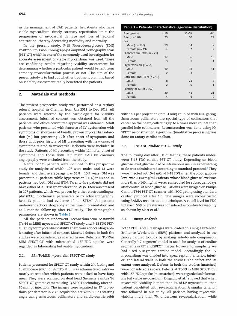
Table 1 – Patients characteristics (age-wise distribution).
Age (years) <50 51–65 >66Age (n = 120) 33 60 27SexMale (n = 107) 29 54 2Female (n = 13) 4 7 2
Diabetes mellitus (n = 71)Male 17 36 13Female 3 1 1
Hypertension (n = 64)Male 12 33 11Female 2 4 2
Both DM and HTN (n = 40)Male 8 24 5Female 2 0 1
History of MI (n = 107)Male 30 44 21Female 4 6 2
i n d i a n h e a r t j o u r n a l 6 8 ( 2 0 1 6 ) 6 9 3 – 6 9 9694
in the management of CAD patients. In patients who haveviable myocardium, timely coronary reperfusion limits theprogression of myocardial damage and loss of regionalcontraction, thereby decreasing morbidity and mortality.
In the present study, F-18 Fluorodeoxyglucose (FDG)Positron Emission Tomography-Computed Tomography scan(PET-CT) which is one of the commonly used investigation foraccurate assessment of viable myocardium was used. Thereare conflicting results regarding viability assessment fordetermining whether a particular patient would benefit fromcoronary revascularization process or not. The aim of thepresent study is to find out whether treatment planning basedon viability assessment really benefitted the patients.
2. Materials and methods
The present prospective study was performed at a tertiaryreferral hospital in Chennai from Jan 2011 to Dec 2013. Allpatients were referred by the cardiologists for viabilityassessment. Informed consent was obtained from all thepatients, and ethics committee approval was obtained. Adultpatients, who presented with features of LV dysfunction withsymptoms of shortness of breath, proven myocardial infarc-tion (MI) but presenting 12 h after onset of symptoms andthose with prior history of MI presenting with new onset ofsymptoms related to myocardial ischemia were included inthe study. Patients of MI presenting within 12 h after onset ofsymptoms and those with left main CAD by coronaryangiography were excluded from the study.
A total of 120 patients were included in this prospectivestudy for analysis; of which, 107 were males and 13 werefemale, and their average age was 56.8 � 10.9 years. DM waspresent in 71 patients, while hypertension (HTN) in 64 and 40patients had both DM and HTN. Twenty-five patients did nothave either of it. ST segment elevation MI (STEMI) was presentin 107 patients, which was proven by either electrocardiogra-phy (ECG), biochemical parameters or by echocardiography.Rest 13 patients had evidence of non-STEMI. All patientsunderwent echocardiography at the time of presentation andat 3 months follow-up after PET study. The demographicparameters are shown in Table 1.
All the patients underwent Technetium-99m Sestamibi(Tc-99 m MIBI) myocardial SPECT-CT study and F-18 FDG PET-CT study for myocardial viability apart from echocardiograph-ic testing after informed consent. Matched defects in both thestudies were considered as scarred tissue. Defects in Tc-99mMIBI SPECT-CT with mismatched 18F-FDG uptake wereregarded as hibernating but viable myocardium.
2.1. 99mTc-MIBI myocardial SPECT-CT study
Patients presented for SPECT CT study within 2 h fasting and10 millicurie (mCi) of 99mTc-MIBI was administered intrave-nously at rest after which patients were asked to have fattymeal. They were scanned on dual head Siemens Symbia T6SPECT-CT gamma camera using IQ.SPECT technology after 45–60 min of injection. The images were acquired in 17 projec-tions per detector in 2088 acquisition arc with 598 as startingangle using smartzoom collimators and cardio-centric orbit
with 14 s per projection (total 4 min) coupled with ECG gating.Smartzoom collimators are special type of collimators thatcenter on the heart, collecting up to 4 times more counts thanparallel hole collimators. Reconstruction was done using IQ.SPECT reconstruction algorithm. Quantitative processing wasdone on Emory cardiac toolbox.
2.2. 18F-FDG cardiac PET-CT study
The following day after 6 h of fasting, these patients under-went F-18 FDG cardiac PET-CT study. Depending on bloodglucose level, glucose load or intravenous insulin as per slidingscale was administered according to standard protocol.6 Theywere injected with 5–8 mCi of F-18 FDG when the blood glucoselevel was <140 mg/ml. Patients, whose blood glucose level wasmore than >140 mg/ml, were rescheduled for subsequent daysafter control of blood glucose. Patients were imaged on PhilipsGemini TF64 PET-CT scanner with ECG gating using standardcardiac protocol after 1 h. The images were reconstructedusing RAMLA reconstruction technique. A cutoff level for FDGuptake of 50% or greater was considered as positive for viabilityas shown by Slart et al.7
2.3. Image analysis
Both SPECT and PET images were loaded on a single ExtendedBrilliance Workstation (EBW) platform and analyzed in theEmory cardiac toolbox by making side-to-side comparison.Generally '17-segment' model is used for analysis of cardiacsegments in PET and SPECT images. However for simplicity, wehad used 5-segment cardiac model. Accordingly the LVmyocardium was divided into apex, septum, anterior, inferi-or, and lateral walls in both the studies. The defect and itsextent were analyzed. Defects in both the studies (matched)were considered as scars. Defects at Tc-99 m MIBI SPECT, butwith 18F-FDG uptake (mismatched), were regarded as hibernat-ing but viable myocardium. D'Egadio et al.8 showed that whenmyocardial viability is more than 7% of LV myocardium, thenpatient benefitted with revascularization. A similar criterionwas followed in our study. All patients having myocardialviability more than 7% underwent revascularization, while

Table 2 – Region wise distribution of matched andmismatched segments.
Walls Study
99mTcMIBI Study
F-18FDG PET Study
Mismatched Matched
Apex 224 99 125Anterior 179 74 105Septum 207 108 99Lateral 82 39 43Inferior 94 34 60Total 786 354 432
Table 3 – Segmental involvement and comorbidities.
Matchedsegments
Mismatchedsegments
p value
Diabetes mellitus(n = 71)
262 210 0.0685
Hypertension (n = 64) 270 162 0.0005Both (n = 40) 171 101 0.0043None (n = 25) 70 84 0.2590
Table depicts distribution of matched and mismatched segmentsin patients with diabetes mellitus and hypertension, having bothco-morbidities or having none of it. As depicted, patients withhypertension or having both co-morbidities, had statisticallysignificantly more matched segments than mismatched segments.
i n d i a n h e a r t j o u r n a l 6 8 ( 2 0 1 6 ) 6 9 3 – 6 9 9 695
those having less than 7% viability or nonviable myocardiumunderwent medical management.
2.4. Echocardiography
Patients underwent echocardiography at the time of PET scansand 3 months after starting treatment during follow-up,whether medical or surgical. Echocardiographic images wereobtained in the standard parasternal long, short axes andapical 4- and 2-chamber views utilizing digital Vivid 7ultrasound equipment with a combined tissue imaging 2.5–4.0 MHz transducer. At least three cardiac cycles weremonitored at the LV base, midpapillary muscle level, andapex for wall motion assessment. Two-dimensional (2D)ventricular volumes and LV ejection fraction (LVEF) weremeasured from the 4- and 2-chamber views using the modifiedSimpson's formula.9 Regional wall motion abnormality(RWMA) was recorded as normokinesia, hypokinesia, akinesiaor dyskinesia. Patients were considered responding to treat-ment if there was either increase or no change in LVEF onfollow-up echocardiograms as reported earlier.10,11
2.5. Coronary angiography
All patients had undergone invasive coronary angiography.Twenty-nine patients had angiography done elsewhere andwere referred for viability study to our hospital. Rest 91underwent coronary angiography either before or within oneweek of myocardial perfusion and FDG PET study. The stenosisin left anterior descending (LAD), left circumflex (LCx), andright coronary artery (RCA) were noted. The stenosis in theirrespective branches were categorized under main artery foranalysis purpose.
2.6. Statistical analysis
Continuous variables were expressed as mean � SD, and allcategorical variables were expressed as percentages. A paired 't' test was used for intra group comparison and unpaired 't' testfor comparison between two groups. Differences were consid-ered significant at p value <0.05. Pearson's correlation wasused to find the relationship between two variables to assesshow strongly they were related to each other. Confidenceintervals (C.I.) were calculated at 95% interval levels.
3. Results
On analysis of coronary artery stenosis distribution, 33 patientshad single vessel disease (SVD) and 48 patients had triple vesseldisease (TVD). Rest of the patients had double vessel disease(DVD). Most commonly involved artery and its branches in ourstudy was LAD in 113 patients (94%) followed by RCA in 71patients (59%) while LCx was involved in 72 patients (60%).
A total of 786 segments of LV myocardium showed reducedperfusion on 99mTc-MIBI study, most commonly in apex (224),followed by septum (207), anterior (179), inferior (94) and lateralwalls (82). Matched perfusion defects were seen in 432 segments(55%) and mismatched perfusion defects were noted in 354 (45%)segments (Table 2). Patients having DM, HTN or both showed
more number of matched segments (scarred myocardium) thanmismatched segments (viable myocardium); however, thisdifference was statistically significant in hypertensive patients(p = 0.0005) and also in patients having both co-morbidities(p = 0.0043) but not in those with diabetes alone (p = 0.0685).Patients who did not have any co-morbidities showed morenumber of mismatched segments than matched segments,however this observation was not statistically significant(p = 0.2590) (Table 3).
Echocardiographically, there were 376 hypokinetic, 343akinetic and 5 dyskinetic segments. In some segments theechocardiography showed RWMA but the perfusion wasnormal and vice versa was also observed. In 33 patients PETand SPECT study showed myocardial changes in 62 wallsegments whereas echocardiography showed no RWMA. Ofthese, 19 patients showed mismatched defects in 29 wallsegments and 14 patients showed matched defects in 33 wallsegments. Similarly in 50 patients, echocardiography showedRWMA in 67 wall segments with normal perfusion and thedetails are as shown in Table 4.
These patients underwent either revascularization ormedical management depending on the findings on viabilityassessment as described previously. Revascularization includ-ed either stenting or coronary artery bypass graft (CABG)surgery. Seventy-eight patients with myocardial viability ofmore than 7% underwent revascularization and 42 patientswere managed medically as they showed either less than 7%myocardial viability or non-viable segments.
There was either increase or no change in LVEF in 106patients (88%) after treatment; of which 73 patients managed
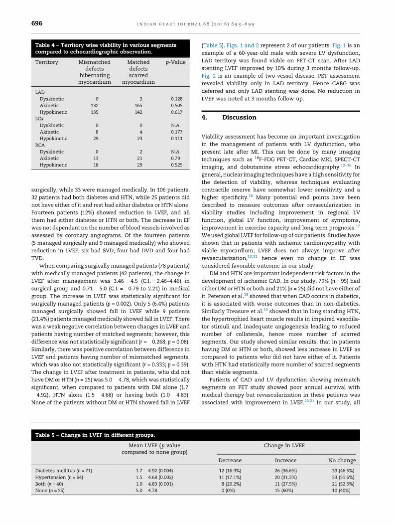
Table 4 – Territory wise viability in various segmentscompared to echocardiographic observation.
Territory Mismatcheddefects
hibernatingmyocardium
Matcheddefectsscarred
myocardium
p-Value
LADDyskinetic 0 3 0.128Akinetic 132 165 0.505Hypokinetic 135 142 0.617
LCxDyskinetic 0 0 N.A.Akinetic 8 4 0.177Hypokinetic 29 23 0.111
RCADyskinetic 0 2 N.A.Akinetic 13 21 0.79Hypokinetic 18 29 0.525
i n d i a n h e a r t j o u r n a l 6 8 ( 2 0 1 6 ) 6 9 3 – 6 9 9696
surgically, while 33 were managed medically. In 106 patients,32 patients had both diabetes and HTN, while 25 patients didnot have either of it and rest had either diabetes or HTN alone.Fourteen patients (12%) showed reduction in LVEF, and allthem had either diabetes or HTN or both. The decrease in EFwas not dependant on the number of blood vessels involved asassessed by coronary angiograms. Of the fourteen patients(5 managed surgically and 9 managed medically) who showedreduction in LVEF, six had SVD, four had DVD and four hadTVD.
When comparing surgically managed patients (78 patients)with medically managed patients (42 patients), the change inLVEF after management was 3.46 � 4.5 (C.I. = 2.46–4.46) insurgical group and 0.71 � 5.0 (C.I. = �0.79 to 2.21) in medicalgroup. The increase in LVEF was statistically significant forsurgically managed patients (p = 0.002). Only 5 (6.4%) patientsmanaged surgically showed fall in LVEF while 9 patients(21.4%) patients managed medically showed fall in LVEF. Therewas a weak negative correlation between changes in LVEF andpatients having number of matched segments; however, thisdifference was not statistically significant (r = �0.268; p = 0.08).Similarly, there was positive correlation between difference inLVEF and patients having number of mismatched segments,which was also not statistically significant (r = 0.333; p = 0.39).The change in LVEF after treatment in patients, who did nothave DM or HTN (n = 25) was 5.0 � 4.78, which was statisticallysignificant, when compared to patients with DM alone (1.7� 4.92), HTN alone (1.5 � 4.68) or having both (1.0 � 4.83).None of the patients without DM or HTN showed fall in LVEF
Table 5 – Change in LVEF in different groups.
Mean LVEF ( p valuecompared to none group)
Diabetes mellitus (n = 71) 1.7 � 4.92 (0.004)
Hypertension (n = 64) 1.5 � 4.68 (0.002)
Both (n = 40) 1.0 � 4.83 (0.001)
None (n = 25) 5.0 � 4.78
(Table 5). Figs. 1 and 2 represent 2 of our patients. Fig. 1 is anexample of a 60-year-old male with severe LV dysfunction,LAD territory was found viable on PET-CT scan. After LADstenting LVEF improved by 10% during 3 months follow-up.Fig. 2 is an example of two-vessel disease. PET assessmentrevealed viability only in LAD territory. Hence CABG wasdeferred and only LAD stenting was done. No reduction inLVEF was noted at 3 months follow-up.
4. Discussion
Viability assessment has become an important investigationin the management of patients with LV dysfunction, whopresent late after MI. This can be done by many imagingtechniques such as 18F-FDG PET-CT, Cardiac MRI, SPECT-CTimaging, and dobutamine stress echocardiography.12–16 Ingeneral, nuclear imaging techniques have a high sensitivity forthe detection of viability, whereas techniques evaluatingcontractile reserve have somewhat lower sensitivity and ahigher specificity.16 Many potential end points have beendescribed to measure outcomes after revascularization inviability studies including improvement in regional LVfunction, global LV function, improvement of symptoms,improvement in exercise capacity and long term prognosis.17
We used global LVEF for follow-up of our patients. Studies haveshown that in patients with ischemic cardiomyopathy withviable myocardium, LVEF does not always improve afterrevascularization,10,11 hence even no change in EF wasconsidered favorable outcome in our study.
DM and HTN are important independent risk factors in thedevelopment of ischemic CAD. In our study, 79% (n = 95) hadeither DM or HTN or both and 21% (n = 25) did not have either ofit. Peterson et al.18 showed that when CAD occurs in diabetics,it is associated with worse outcomes than in non-diabetics.Similarly Treasure et al.19 showed that in long standing HTN,the hypertrophied heart muscle results in impaired vasodila-tor stimuli and inadequate angiogenesis leading to reducednumber of collaterals, hence more number of scarredsegments. Our study showed similar results, that in patientshaving DM or HTN or both, showed less increase in LVEF ascompared to patients who did not have either of it. Patientswith HTN had statistically more number of scarred segmentsthan viable segments.
Patients of CAD and LV dysfunction showing mismatchsegments on PET study showed poor annual survival withmedical therapy but revascularization in these patients wasassociated with improvement in LVEF.20,21 In our study, all
Change in LVEF
Decrease Increase No change
12 (16.9%) 26 (36.6%) 33 (46.5%)11 (17.1%) 20 (31.3%) 33 (51.6%)8 (20.2%) 11 (27.5%) 21 (52.5%)0 (0%) 15 (60%) 10 (40%)

Fig. 1 – A 60-year hypertensive male, LVEF 35%. (a) Coronary angiography shows 80% stenosis in proximal LAD. (b) Thrombusin the distal segment of LAD. (c) Viability assessment showed >95% viability in LAD territory (White arrows). The patientunderwent LAD stenting. His LVEF increased by 10% at 3 months follow-up.
Fig. 2 – A 57-year diabetic and hypertensive male. (a) Coronary angiography shows 80% stenosis in proximal LAD beforebifurcation and 80% stenosis after bifurcation. (b) A long diseased segment in the LCX. (c) Viability assessment showed viablemyocardium in LAD territory (White arrows) while distal LCX showed scarred tissue (White arrowhead). Rest of the LCXterritory showed normal perfusion with no mismatch viability. Hence CABG was deferred and patient underwent LADstenting alone and no reduction in LVEF was noted at 3 months follow-up.
i n d i a n h e a r t j o u r n a l 6 8 ( 2 0 1 6 ) 6 9 3 – 6 9 9 697

i n d i a n h e a r t j o u r n a l 6 8 ( 2 0 1 6 ) 6 9 3 – 6 9 9698
patients who had myocardial viability on PET underwentsurgical management and of these only 6.4% of patientsshowed fall in LVEF as compared to 21.8% of patients who weremanaged medically.
The STICH trial22 showed that patients with viablemyocardium had lower overall rates of death than thosewithout viable myocardium (p = 0.003), however after adjust-ment for other baseline prognostic variables in a multivariatemodel, the pre-specified viability status was not statisticallysignificant (p = 0.21), thereby concluding that as regardsmortality, viability assessment did not have survival advan-tage in patients undergoing CABG surgery compared tomedical therapy. However, this study had many drawbackssuch as using SPECT alone for viability assessment, asymp-tomatic subjects accounted for 40% of patients enrolled andonly 49% of patients underwent careful functional evaluationpre-randomization. Our study differs from STICH trial inmethodology, use of 18F-FDG PET-CT in viability assessmentand use of LVEF as end point assessment. In our study, we didnot take symptoms or survival as endpoints, and this explainswhy our study found viability assessment useful in decidingmanagement in 40% of our patients.
Haas et al.23 showed that not performing a viability studybefore surgical management, resulted in too many high-riskpatients without viability being subjected to surgery resultingin worse outcomes. To some extent we avoided this situationas only those patients who showed myocardial viabilityunderwent revascularization and our result showed that93.6% showed no decrease in post-revascularization LVEF.Similarly Dreyfus et al.24 reported that viability assessmentshould be part of selection process in patients with low LVEFfor surgical revascularization. We are in agreement with theirviews that proper selection of patients based on viabilityassessment helps in reducing peri-operative and post-opera-tive mortality and also improves outcomes.
5. Conclusion
Our study has shown that evidence based viability study canbe used to individualize the management. It helped in deciding, whether patient should receive surgical or medical treatmentbased on viability assessment. This also helped in preventingunnecessary economic loss to the patient apart from reducingmorbidity. In our opinion the viability study should beperformed in all patients who present 12 h after acute MIand also in those who present with LV dysfunction due toischemic heart disease.
Conflicts of interest
The authors have none to declare.
r e f e r e n c e s
1. Sharma M, Ganguly NK. Premature coronary artery diseasein Indians and its associated risk factors. Vasc Health RiskManag. 2005;1:217–225.
2. Gheorgiade M, Bonow RO. Chronic heart failure in the UnitedStates: a manifestation of coronary heart disease. Circulation.1988;97:28–29.
3. Travin MI, Bergmann SR. Assessment of myocardialviability. Semin Nucl Med. 2005;2:2–19.
4. Braunwald E, Kloner RA. The stunned myocardium:prolonged, post ischemic ventricular dysfunction.Circulation. 1982;66:1146–1149.
5. Rahimtoola SH. The hibernating myocardium. Am Heart J.1989;117:211–221.
6. Ziessman HA, O'Malley JP. Cardiac system. In: Thrall JH, ed.In: Nuclear Medicine: The Requisites in Radiology 3rd ed.Philadelphia: Elseviers Saunders; 2006:489.
7. Slart RH, Bax JJ, van Veldhuisen DJ, et al. Prediction offunctional recovery after revascularization in patients withcoronary artery disease and left ventricular dysfunction bygated FDG-PET. J Nucl Cardiol. 2006;13:210–219.
8. D'Egidio G, Nichol G, Williams K, et al. Identification of high-risk patients with increasing ischemic cardiomyopathy.JACC Cardiovasc Imaging. 2009;2:1060–1068.
9. Schiller NB, Shah PM, Crawford M, et al. Recommendationsfor quantitation of the left ventricle by two-dimensionalechocardiography cardiography. American Society ofEchocardiography Committee on Standards, Subcommitteeon Quantitation of Two-Dimensional Echocardiograms.J Am Soc Echocardiogr. 1989;2:358–367.
10. Rizzello V, Poldermans D, Biagini E, et al. Prognosis ofpatients with ischaemic cardiomyopathy after coronaryrevascularisation: relation to viability and improvementin left ventricular ejection fraction. Heart. 2009;95:1273–1277.
11. Acampa W, Petretta M, Spinelli L, Salvatore M, Cuocolo A.Survival benefit after revascularization is independent ofleft ventricular ejection fraction improvement in patientswith previous myocardial infarction and viablemyocardium. Eur J Nucl Med Mol Imaging. 2005;32:430–437.
12. Ichiro M, Junichi T, Kenichi N, Norihisa T, Kinichi H.Myocardial viability assessment using nuclear imaging.Ann Nucl Med. 2003;17:169–179.
13. Ramos M, DePasquale E, Coplan NL. Assessment ofmyocardial viability: review of the clinical significance.Rev Cardiovasc Med. 2008;9:225–231.
14. Partington SL, Kwong RY, Dorbala S. Multimodality imagingin the assessment of myocardial viability. Heart Fail Rev.2011;16:381–395.
15. Chopra HK, Sambi RS, Krishna CK, Parashar SK, Gupta R.Techniques to assess myocardial viability. assessmyocardial viability. Indian Heart J. 2011;63:39–44.
16. Schinkel AF, Poldermans D, Elhendy A, Bax JJ. Assessment ofmyocardial viability in patients with heart failure. J Nucl Med.2007;48:1135–1146.
17. Bax JJ, Poldermans D. Clinical value of assessment ofperfusion and function for the evaluation of myocardialviability in patients with ischemic left ventriculardysfunction. In: Germano G, Berman DS, eds. In: ClinicalGated Cardiac SPECT 2nd ed. Massachusetts: BlackwellPublishing Ltd; 2006:260.
18. Peterson LR, McKenzie C, Schaffer JE. Diabeticcardiovascular disease: getting to the heart of the matter.J Cardiovasc Transl Res. 2012;5:436–445.
19. Treasure CB, Klein JL, Vita JA, et al. Hypertension and leftventricular hypertrophy are associated with impairedendothelium-mediated relaxation in human coronaryresistance vessels. Circulation. 1993;87:86–93.
20. Di Carli MF, Davidson M, Little R, Khanna S, Mody FV,Brunken RC. Value of metabolic imaging with positionemission tomography for evaluating prognosis in patientswith coronary artery disease and left ventriculardysfunction. Am J Cardiol. 1994;73:527–533.

i n d i a n h e a r t j o u r n a l 6 8 ( 2 0 1 6 ) 6 9 3 – 6 9 9 699
21. Allman KC, Shaw LJ, Hachamovitch R, Udelson JE.Myocardial viability testing and impact of revascularizationon prognosis in patients with coronary artery disease andleft ventricular dysfunction: a meta-analysis. J Am CollCardiol. 2002;39:1151–1158.
22. Bonow RO, Maurer G, Lee KL, et al. Myocardial viability andsurvival in ischemic left ventricular dysfunction. N Engl JMed. 2011;364:1617–1625.
23. Haas F, Haehnel CJ, Picker W, et al. Preoperative positronemission tomographic viability assessment andperioperative and postoperative risk in patients withadvanced ischemic heart disease. J Am Coll Cardiol. 1997;30:1693–1700.
24. Dreyfus GD, Duboc D, Blasco A, et al. Myocardial viabilityassessment in ischemic cardiomyopathy: benefits of coronaryrevascularization. Ann Thorac Surg. 1994;57:1402–1407.