Embed Size (px)
Citation preview

Role of soluble guanylate cyclase isoforms and of hydrogen sulfide in gastrointestinal motility
Ingeborg Dhaese
Promotor: Prof. Dr. R.A. Lefebvre
2010
Thesis submitted as partial fulfillment of the requirements for the degree of Doctor in Medical Sciences
Vakgroep Farmacologie – Heymans Instituut

Promotor
Prof. Dr. R.A. Lefebvre Universiteit Gent, Belgium
The studies described in this thesis were supported by the Special Investigation Fund of
Ghent University (GOA 1251004), the fund for Scientific Research Flanders (G.0053.02) and
by Interuniversity Attraction Pole Programme P5/20.

List of Abbreviations
2-APB 2-aminoethyl diphenylborinate 4-AP 4-aminopyridine 8-br-cGMP 8-bromo-cGMP AC adenylate cyclase ANOVA analysis of variance ATP adenosine triphosphate AUC area under the curve BH4 tetrahydrobiopterin BKCa channel large-conductance Ca2+-dependent K+ channel cAMP adenosine 3’,5’-cyclic monophosphate CBS cystathionine β-synthase cDNA complementary deoxyribonucleic acid cGK cGMP-dependent protein kinase cGMP guanosine 3’, 5’-cyclic monophosphate CO carbon monoxide CSE cystathionine γ-lyase Ct threshold cycle DETA-NO diethylenetriamine NONOate DMSO dimethyl sulfoxide DNA deoxyribonucleic acid DSS dextrane sulphate sodium EAS external anal sphincter EC50 half maximal effective concentration EDRF endothelial-derived relaxant factor EFS electrical field stimulation EIA enzyme immunoassay Emax maximum effect eNOS endothelial NOS ENS enteric nervous system FAD flavin adenine dinucleotide FAM 6-carboxylfluorescein FD70 fluorescein-labelled dextran (70 kDa) FMN flavin mononucleotide GC geometric center GI gastrointestinal GMCs giant migrating contractions GTP guanosine triphosphate HO heme oxygenase HPRT hypoxanthine-guanine phosphoribosyltransferase HRP horseradish peroxidase H2S hydrogen sulfide IAS internal anal sphincter
IC50 half maximal inhibition concentration ICC interstitial cells of Cajal ICS ileocaecal sphincter IKCa channel intermediate conductance Ca2+-dependent K+ channel iNOS inducible NOS IP3 inositol triphosphate IPAN intrinsic primary afferent neuron IRAG Ins(1,4,5)P3-receptor associated cGMP kinase substrate KATP channel ATP-dependent K+ channel KI knock-in KO knock-out L0 optimal load LES lower esophagal sphincter L-NAME Nω-nitro-L-arginine methyl ester MLC myosin light chain MLCK myosin light chain kinase MLCP myosin light chain phosphatase MMC migrating motor complex mRNA messenger ribonucleic acid NADPH nicotinamide adenine dinucleotide phosphate NaHS sodium hydrogen sulfide NANC non-adrenergic non-cholinergic nNOS neuronal NOS NO nitric oxide NOS nitric oxide synthase OD optical density ODQ 1H-[1,2,4]-oxadiazolo-[4,3-a]-quinoxalin-1-one PACAP pituitary adenylate cyclase activating peptide PBS phosphate buffered saline PDE phosphodiesterase pGC particulate guanylate cyclase PGF2α prostaglandin F2α PKC protein kinase C PKG protein kinase G PLC phospholipase C PSD-95 postsynaptic density 95 RNA ribonucleic acid RNS reactive nitrogen species RT-PCR reverse transcriptase –polymerase chain reaction S.E.M. standard error of the mean SERCA sarcoendoplasmic reticulum Ca2+ ATPase sGC soluble guanylate cyclase SKCa channel small conductance Ca2+-dependent K+ channel SMC smooth muscle cell
SNP sodium nitroprusside SQ 22536 9-(tetrahydro-2-furanyl)-9H-purin-6-amine SR sarcoplasmatic reticulum TRPV1 transient receptor potential vanilloid type 1 UES upper esophagal sphincter VIP vasoactive intestinal peptide WT wild-type Y-2762 (R)-(+)-trans-4-(1-aminoethyl)-N-(4-pyridyl)cyclohexanecarboxamide


Table of Contents
Chapter I Literature Survey ........................................................................... 17
I.1 General aspects of the gastrointestinal tract.................................................................. 17 I.1.1 Function ........................................................................................................................... 17 I.1.2 Motility along the gastrointestinal canal........................................................................... 17
I.1.2.1 The esophagus ....................................................................................................... 18 I.1.2.2 The stomach ........................................................................................................... 18 I.1.2.3 The small intestine .................................................................................................. 19 I.1.2.4 The large intestine................................................................................................... 19
I.2 Control of GI motility ......................................................................................................... 20 I.2.1 Extrinsic innervation......................................................................................................... 21 I.2.2 Intrinsic innervation.......................................................................................................... 23 I.2.3 Inhibitory NANC neurotransmission................................................................................. 25
I.2.3.1 ATP, VIP/PACAP .................................................................................................... 25 I.2.3.2 NO........................................................................................................................... 27 I.2.3.3 CO........................................................................................................................... 31 I.2.3.4 H2S .......................................................................................................................... 32
I.3 Soluble guanylate cyclase ................................................................................................ 32 I.3.1 Structure .......................................................................................................................... 33 I.3.2 Expression ....................................................................................................................... 34 I.3.3 Smooth muscle relaxation by sGC .................................................................................. 35
I.3.3.1 Activation of sGC .................................................................................................... 35 I.3.3.2 Mechanisms downstream of cGMP ........................................................................ 37
I.3.4 Role of sGC in GI motility ................................................................................................ 39 I.4 Hydrogen sulfide................................................................................................................ 40
I.4.1 Identification of H2S as neurotransmitter ......................................................................... 40 I.4.2 Influence of H2S on smooth muscle contractility ............................................................. 41
I.5 References.......................................................................................................................... 42
Chapter II Aims................................................................................................ 57
II.1 References.......................................................................................................................... 58
Chapter III Small intestinal motility in soluble guanylate cyclase α1
knockout mice ........................................................................................................ 61
III.1 Abstract............................................................................................................................... 61
III.2 Introduction ........................................................................................................................ 62
III.3 Materials and Methods ...................................................................................................... 63 III.3.1 Animals........................................................................................................................ 63 III.3.2 Real-time quantitative reverse transcriptase-polymerase chain reaction (RT-PCR) .. 63 III.3.3 Muscle tension experiments........................................................................................ 64
III.3.3.1 Tissue preparation .................................................................................................. 64 III.3.3.2 Isometric tension recording..................................................................................... 64 III.3.3.3 Protocol ................................................................................................................... 64 III.3.3.4 Data analysis........................................................................................................... 65
III.3.4 cGMP analysis............................................................................................................. 66 III.3.5 Transit.......................................................................................................................... 66
III.3.5.1 Small intestinal transit (phenol red method)............................................................ 66 III.3.5.2 Intestinal transit (fluorescein-labelled dextran method) .......................................... 67 III.3.5.3 Whole gut transit (carmine method)........................................................................ 67
III.3.6 Statistics ...................................................................................................................... 67 III.3.7 Drugs Used.................................................................................................................. 68
III.4 Results ................................................................................................................................ 68 III.4.1 Real-time quantitative RT-PCR................................................................................... 68 III.4.2 Muscle tension experiments........................................................................................ 69
III.4.2.1 General observations in WT and KO mice.............................................................. 69 III.4.2.2 Contractile responses to carbachol and PGF2α in WT and KO mice .................... 69 III.4.2.3 Contractile responses to electrical field stimulation in WT and KO mice................ 70 III.4.2.4 Inhibitory responses to electrical field stimulation in WT and KO mice .................. 70 III.4.2.5 Inhibitory responses to exogenously applied NO in WT and KO mice ................... 73 III.4.2.6 Inhibitory responses to exogenously applied VIP in WT and KO mice................... 76 III.4.2.7 Inhibitory responses to electrical field stimulation and exogenously applied NO in male heterozygous mice........................................................................................................... 76
III.4.3 cGMP analysis............................................................................................................. 76 III.4.4 Transit.......................................................................................................................... 77
III.4.4.1 Small intestinal transit ............................................................................................. 77 III.4.4.2 Intestinal transit ....................................................................................................... 78 III.4.4.3 Whole gut transit time ............................................................................................. 78
III.5 Discussion .......................................................................................................................... 79 III.5.1 Role of sGC in male mice............................................................................................ 80 III.5.2 Role of sGC in female mice......................................................................................... 81 III.5.3 The sGCα1 KO model.................................................................................................. 81 III.5.4 Implication on intestinal transit .................................................................................... 82
III.6 References.......................................................................................................................... 83
Chapter IV Involvement of soluble guanylate cyclase α1 and α2, and SKCa
channels in NANC relaxation of mouse distal colon........................................... 89
IV.1 Abstract............................................................................................................................... 89
IV.2 Introduction ........................................................................................................................ 90
IV.3 Materials and Methods ...................................................................................................... 91 IV.3.1 Animals........................................................................................................................ 91 IV.3.2 Muscle tension experiments........................................................................................ 91
IV.3.2.1 Tissue preparation and tension recording............................................................... 91 IV.3.2.2 Protocol ................................................................................................................... 92 IV.3.2.3 Data analysis........................................................................................................... 93
IV.3.3 cGMP analysis............................................................................................................. 93 IV.3.4 Western blot ................................................................................................................ 94 IV.3.5 Statistics ...................................................................................................................... 94 IV.3.6 Drugs used .................................................................................................................. 94
IV.4 Results ................................................................................................................................ 95 IV.4.1 Western blot ................................................................................................................ 95 IV.4.2 General observations of the distal colon muscle strips ............................................... 95 IV.4.3 Inhibitory responses .................................................................................................... 97
IV.4.3.1 Inhibitory responses to EFS and NO ...................................................................... 97 IV.4.3.2 Influence of ODQ and L-NAME............................................................................. 100 IV.4.3.3 Influence of apamin and apamin plus ODQ or L-NAME....................................... 101
IV.4.4 Inhibitory responses to VIP and 8-Br-cGMP ............................................................. 103 IV.4.5 cGMP measurements................................................................................................ 104
IV.5 Discussion ........................................................................................................................ 105
IV.5.1 Inhibitory responses to 8-Br-cGMP and VIP ............................................................. 105 IV.5.2 Inhibitory responses to exogenous NO ..................................................................... 106 IV.5.3 Inhibitory responses to EFS ...................................................................................... 107 IV.5.4 Basal and PGF2α-induced contractile activity............................................................ 108 IV.5.5 Conclusion................................................................................................................. 109
IV.6 References........................................................................................................................ 109
Chapter V Role of soluble guanylate cyclase in gastrointestinal motility:
Gastrointestinal phenotyping of NO resistant sGCbeta1his105phe knock in
mice ...................................................................................................... 115
V.1 Abstract............................................................................................................................. 115
V.2 Introduction ...................................................................................................................... 116
V.3 Methods............................................................................................................................. 117 V.3.1 Ethical approval ............................................................................................................. 117 V.3.2 Animals .......................................................................................................................... 117 V.3.3 Muscle tension experiments .......................................................................................... 117
V.3.3.1 Tissue preparation ................................................................................................ 117 V.3.3.2 Isometric tension recording................................................................................... 117 V.3.3.3 Protocols ............................................................................................................... 118 V.3.3.4 Data analysis......................................................................................................... 119
V.3.4 Gastric emptying............................................................................................................ 119 V.3.5 Transit ............................................................................................................................ 120
V.3.5.1 Intestinal transit (fluorescein-labelled dextran method) ........................................ 120 V.3.5.2 Whole gut transit time (carmine method) .............................................................. 120
V.3.6 Small intestinal contractility............................................................................................ 121 V.3.7 Histology ........................................................................................................................ 121 V.3.8 sGC enzyme activity ...................................................................................................... 121 V.3.9 Drugs used..................................................................................................................... 122 V.3.10 Statistics .................................................................................................................... 122
V.4 Results .............................................................................................................................. 122 V.4.1 General observations..................................................................................................... 122 V.4.2 Gastric emptying, small intestinal transit and whole gut transit time............................. 123 V.4.3 Histology ........................................................................................................................ 125 V.4.4 Muscle tension experiments .......................................................................................... 125
V.4.4.1 Tissue weight ........................................................................................................ 125 V.4.4.2 Contractile responses to carbachol and PGF2α................................................... 125 V.4.4.3 Relaxant responses to EFS .................................................................................. 126 V.4.4.4 Relaxant responses to NO.................................................................................... 130 V.4.4.5 Inhibitory responses to 8-Br-cGMP and VIP......................................................... 130
V.4.5 sGC enzyme activity ...................................................................................................... 131 V.5 Discussion ........................................................................................................................ 133
V.6 References........................................................................................................................ 135
Chapter VI Myosin light chain phosphatase activation is involved in the
hydrogen sulfide-induced relaxation in mouse gastric fundus........................ 141
VI.1 Abstract............................................................................................................................. 141
VI.2 Introduction ...................................................................................................................... 142
VI.3 Methods............................................................................................................................. 143 VI.3.1 Animals...................................................................................................................... 143 VI.3.2 Tissue preparation..................................................................................................... 143 VI.3.3 Protocols.................................................................................................................... 143 VI.3.4 Data analysis ............................................................................................................. 144 VI.3.5 Statistics .................................................................................................................... 144 VI.3.6 Drugs used ................................................................................................................ 144
VI.4 Results .............................................................................................................................. 145 VI.4.1 Basic observations .................................................................................................... 145 VI.4.2 Influence of glibenclamide, ODQ plus SQ 22536, L-NAME, tetrodotoxin and ω-conotoxin ................................................................................................................................... 147 VI.4.3 Influence of channel and receptor blockers .............................................................. 148 VI.4.4 Influence of calyculin-A and Y-27632........................................................................ 149
VI.5 Discussion ........................................................................................................................ 151
VI.6 References........................................................................................................................ 155
Chapter VII Mechanisms of action of hydrogen sulfide in relaxation of
mouse distal colonic smooth muscle................................................................. 161
VII.1 Abstract............................................................................................................................. 161
VII.2 Introduction ...................................................................................................................... 162
VII.3 Material and Methods ...................................................................................................... 163 VII.3.1 Animals...................................................................................................................... 163 VII.3.2 Tissue preparation..................................................................................................... 163 VII.3.3 Measurement of contractile tension .......................................................................... 163 VII.3.4 Simultaneous measurement of contractile tension and cytosolic calcium concentration ................................................................................................................................... 164 VII.3.5 Data analysis and statistics ....................................................................................... 165 VII.3.6 Drugs used ................................................................................................................ 166
VII.4 Results .............................................................................................................................. 167 VII.4.1 Effect of NaHS on contractility................................................................................... 167 VII.4.2 Influence of channel, receptor, nerve and enzyme blockers on NaHS-induced relaxation ................................................................................................................................... 169 VII.4.3 Effect of NaHS on cytosolic calcium concentration................................................... 173
VII.5 Discussion ........................................................................................................................ 176
VII.6 References........................................................................................................................ 179
Chapter VIII General Discussion and Conclusion ......................................... 185
VIII.1 The relative importance of the sGC isoforms α1β1 and α2β1 in the nitrergic relaxation of gastrointestinal smooth muscle ............................................................................................. 185
VIII.2 sGC-dependency of the nitrergic component of GI smooth muscle relaxation........ 187
VIII.3 Role of the nitrergic activation of sGC in the regulation of GI motility ...................... 189
VIII.4 Influence of H2S on GI contractility ................................................................................ 191
VIII.5 Future perspectives ......................................................................................................... 194

VIII.6 References........................................................................................................................ 195
Chapter IX Summary ...................................................................................... 201
Chapter X Samenvatting ............................................................................... 207
Dankwoord ...……………………………………………………………………….........213


Chapter I
LITERATURE SURVEY

Literature Survey
17
Chapter I Literature Survey
I.1 General aspects of the gastrointestinal tract
I.1.1 Function
The human gastrointestinal (GI) tract consists of the alimentary canal from the mouth to the
anus and the associated glandular organs that empty their contents in the canal. The overall
function of the GI tract is to take in nutrients and eliminate wastes. To fulfil this role, the major
physiological processes that occur in the GI tract are motility, secretion, digestion, absorption
and elimination. Food is taken into the mouth as large particles containing macromolecules
that are not absorbable. The breaking down of food into absorbable material occurs by
grinding and mixing the food (motility) with various secretions containing enzymes, ions and
water that enter the GI canal. The enzymes convert the macromolecules into absorbable
molecules in a process termed digestion. The products of digestion are then transported
across the epithelium to enter the blood or lymph by a process termed absorption. Also most
of the added secretions are absorbed for reuse. Finally, undigested waste products are
eliminated by GI motility (Raybould et al., 2003).
I.1.2 Motility along the gastrointestinal canal
The GI canal, while functioning as whole in the transport of ingested material, is
physiologically partitioned into subdivisions by a series of sphincters. The esophagus begins
at the upper esophagal sphincter (UES) and extends to the lower esophagal sphincter (LES),
while the stomach is defined proximally by the LES and distally by the pylorus. The small
intestine is bordered by the pylorus and the ileocaecal sphincter (ICS). Finally, the most
distal portion of the GI canal, the large intestine, begins at the ICS and continues to the exit
of the GI canal at the internal (IAS) and external anal sphincter (EAS). The structure of the
gastrointestinal wall is similar throughout the canal, consisting of the inner mucosal layer, the
submucosa, the muscle layer and the serosa. GI motility is achieved by coordinated activity
of the smooth muscle cells of the muscle layer. The muscle layer is organized in a thick inner
circular and a thinner outer longitudinal smooth muscle layer. In the large intestine, the latter
consists of three separate longitudinal ribbons of smooth muscle, the taenia coli. Sphincters

Literature Survey
18
are thickened extensions of the circular smooth muscle layer, except for the UES and the
EAS which are striated muscle.
I.1.2.1 The esophagus
The upper one third of the esophagus also makes up an exception in that it contains striated
muscle instead of smooth muscle, whereas the lower two thirds of the esophagus contain
smooth muscle, just as the rest of the GI canal. When food is taken in, relaxation of the UES
allows the food bolus to enter from the pharynx into the esophagus. This produces
esophagal distension, which initiates a peristaltic wave i.e. a ring of contraction that moves
aborally over variable distances and thereby transports the food bolus further towards the
stomach. Finally, relaxation of the LES allows the food bolus to pass into the stomach
(Raybould et al., 2003).
I.1.2.2 The stomach
In a consideration of its motor functions, the stomach can be divided into three functional
regions: the proximal stomach (cardia, fundus, proximal body), the distal stomach (distal
body and antrum) and the pylorus. Coordinated actions of these regions regulate the
emptying of gastric contents. Upon arrival of the ingested food bolus in the stomach, the proximal stomach serves to
accommodate and store the ingested food without a significant increase in intragastric
pressure. Maintenance of intragastric pressure is controlled by two proximal gastric reflexes
(receptive relaxation and gastric accommodation). Receptive relaxation is the reduction in
proximal gastric tone that is initiated by the act of swallowing. Gastric accommodation or
adaptive relaxation is the relaxation of the proximal stomach in response to gastric
distension. Finally, the proximal stomach propels the chyme into the distal stomach by
generating a compressive tonic force. The distal stomach exhibits phasic contractions of the
antrum that serve to grind solid food and to regulate gastric emptying of solid and, to a lesser
extent, liquid meals. Finally, in order to enter into the duodenum, the digesting food has to
pass the pyloric sphincter. Prolonged periods of closure of the pylorus are interrupted by brief
intervals during which the antral contents can pass into the intestine. The resistance to flow
is provided by tonic and phasic pyloric motor activity. During the fed state, the pylorus acts as
a sieve, not allowing large particles to traverse to the intestine. Instead, the latter are
propelled back into the stomach for further grinding. Taken together, the tonic contractions of
the proximal stomach, the propulsive contractions of the distal stomach and the pyloric
activity are thus three important mechanisms for the regulation of gastric emptying.
Under fasting conditions, the distal stomach exhibits distinct phasic motor patterns than
under postprandial conditions. The migrating motor complex (MMC) is a stereotypical pattern
Literature Survey
19
that clears the stomach of undigested debris (food particles, mucus and sloughed epithelial
cells) during fasting and has been termed the GI housekeeper. The MMC consists of four
phases: Phase I is a period of motor quiescence, phase II exhibits irregular contractions and
phase III is a period of intense and rhythmic contractions that begin in the gastric body and
propagate to the pylorus. Phase IV is a transition period of irregular contractions between
between phase III and phase I. Phase II is believed to represent a period in which fasting
gastric contents are mixed, phase III contractions are highly propagative. During phase III,
the pylorus is open and allows intestinal delivery of large particles (Hasler, 2003b).
I.1.2.3 The small intestine
The small intestine is divided into three functional regions: duodenum, jejunum and ileum.
Through relaxation and thus opening of the sphincter of Oddi, bile, produced in the liver and
stored in the gallbladder, and digestive enzymes, produced and stored in the pancreas, enter
the GI canal at the level of the duodenum. The jejunum is the major organ for digestion and
absorption of meal nutrients. The ileum has a specific role in the absorption of cobalamin
(vitamin B12). Following ingestion of a meal, the small intestine exhibits two motor patterns:
segmentation and peristalsis. Segmentation is the process by which rings of contraction
develop at uniform intervals, dividing the lumen into segments. Segmentation is the primary
mechanism by which contents of the intestine are mixed with secretions and moved across
the mucosa to enhance absorption. Peristalsis in the intestine consists of two phases: an
excitatory response proximal to the food bolus (ascending contraction) and a distal inhibition
(descending relaxation). The ascending contraction is characterized by simultaneous circular
muscle shortening and longitudinal muscle relaxation, whereas the descending relaxation
involves simultaneous longitudinal muscle contraction and circular muscle relaxation.
Peristalsis is the primary mechanism by which contents are moved along the intestine in the
digestive period. Under fasting conditions, the small intestine exhibits the same motor pattern
as the stomach, called the MMC or intestinal housekeeper. The small intestinal MMC consist
of the same four phases as the gastric MMC. As such, MMCs start in the stomach and pass
along the intestine to the ileum, thereby propelling undigested food residue and sloughed
enterocytes from the proximal gut into the large intestine. Every MMC cycle lasts about
ninety minutes (Hasler, 2003a; Raybould et al., 2003).
I.1.2.4 The large intestine
The large intestine consists of the caecum, colon and rectum. The caecum receives the
chyme from the small intestine, through the ICS. The caecum and ascending colon receive
about 2 liters of ileal affluent daily. Further re-absorption of water and ions in the colon
reduce the volume to about 200 milliliters per day. Prominent mixing patterns, such as short

Literature Survey
20
and long duration contractions that are stationary or that propagate only for short distances in
oral or aboral direction, promote the process of faecal desiccation. Due to the dehydration
process, the faecal material is semisolid to solid after passage through the colon. The
transverse, descending and sigmoid portions of the colon store faecal material and propel
the material to the rectum. To this purpose, the colon exhibits both storage motor patterns
(i.e. changes in tone) and propulsive motor patterns (i.e. phasic contractions). The latter
include peristaltic motor patterns and giant migrating contractions (GMCs). GMCs, also
termed high-amplitude propagated contractions, propagate aborally over extended distances
and evoke mass movements of faeces. GMCs are most prominently after awakening, after
eating, or in association with defecation. In the colon, an organized fasting motor pattern
such as the MMC is not observed in humans (Hasler et al., 2003a).
The rectum exhibits a compliant wall that allows it to serve as a reservoir for faecal material
until it can be expelled through the anal canal. Rectal motor complexes that do not propagate
orally or aborally facilitate the storage function of the rectum. The anal canal is surrounded
by two sphincters: the internal anal sphincter is a modified extension of the circular muscle of
the rectum; the external anal sphincter is striated muscle. Distension of the rectum activates
the rectoanal reflex, which relaxes the internal anal sphincter and simultaneously contracts
the external anal sphincter. The latter preserves continence. When rectal pressure further
increases, both the internal and the external anal sphincter will relax, resulting in a reflex
expulsion of the faecal material in the rectum. Defecation can however be voluntarily
inhibited by keeping the external anal sphincter contracted. In order to defecate, the
individual allows the external anal sphincter to relax (Grundy et al., 2006; Hasler et al.,
2003a).
I.2 Control of GI motility
The different functions of the GI tract, including the motor patterns that are implicated in the
generation of GI motility, are regulated by hormonal and neuronal control mechanisms.
Hormonal control occurs via paracrine and endocrine regulation mechanisms. Paracrine
regulation describes the process whereby a chemical released from a sensing cell diffuses
through the interstitial space to influence the function of neighbouring cells. An example of a
paracrine mechanism is the inhibition of gastrin release by somatostatin. Somatostatin is
released from cells in the gastric antral mucosa in response to low intra-gastric pH.
Endocrine regulation describes the process whereby the sensing cells respond to a stimulus
by releasing their contents into the circulation to act on distant target cells. A particular target
cell responds because it possesses high-affinity receptors specific for the hormone
(Raybould et al., 2003). An example of endocrine regulation is the release of motilin from the
Literature Survey
21
endocrine cells of the duodenal mucosa during fasting, which initiates MMC phase III
contractions (De Smet et al., 2009). As in this thesis the role of the neurotransmitter nitric
oxide (NO) in GI motility is investigated, the next sections of the introduction focus mainly on
the neuronal control of GI motility.
Neuronal control is mediated via the extrinsic nervous system and the intrinsic nervous
system (enteric nervous system; ENS). The presence of extensive intrinsic neuronal circuits
make the GI tract unique among mammalian organs. The ENS contains reflex pathways
capable of functioning independently of central control, although the extrinsic nervous system
modifies activity within the gut wall. The ENS is most commonly the medium through which
extrinsic neurons control GI function (Furness et al., 2003).
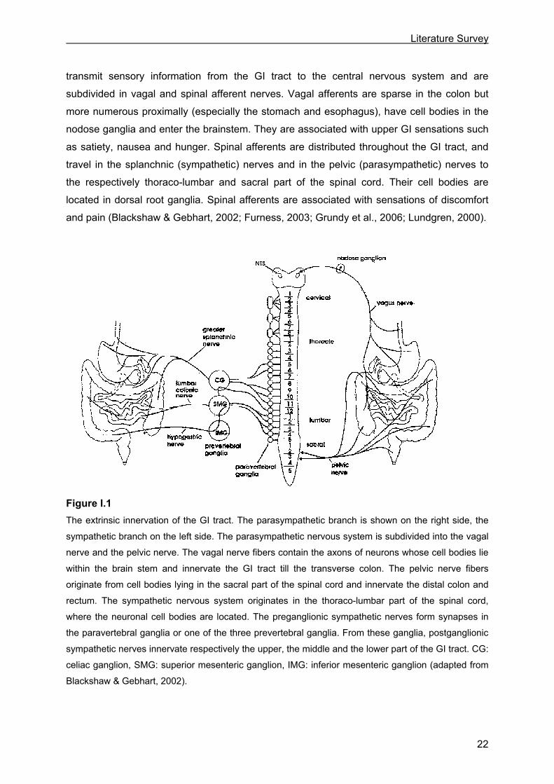
I.2.1 Extrinsic innervation
The extrinsic nervous system consists of a parasympathetic and a sympathetic component
(Fig. I.1). The parasympathetic nervous system can be anatomically subdivided into two
nerves: the vagal nerve and the pelvic nerve. The vagal nerve fibers contain the axons of
neurons whose cell bodies lie within the brain stem and innervate the upper GI tract. The
pelvic nerve fibers originate from cell bodies lying in the sacral part of the spinal cord and
innervate the distal colon and rectum. The vagal and pelvic neurons do not act directly on GI
smooth muscle, but form synaptic connections with enteric neurons; the transmission of the
neuronal input is mediated by acetylcholine acting on nicotinic receptors. The primary
functions of the efferent vagal and pelvic nerves are stimulation of motility, secretion and
blood flow.
The sympathetic nervous system originates in the thoraco-lumbar part of the spinal cord,
where the neuronal cell bodies are located. The preganglionic sympathetic nerves release
acetylcholine as neurotransmitter and form nicotinic synapses in the paravertebral ganglia or
one of the three prevertebral ganglia, named the celiac ganglion, the superior mesenteric
ganglion and the inferior mesenteric ganglion. From these ganglia, postganglionic
sympathetic nerves, that release noradrenaline as neurotransmitter, originate and innervate
respectively the upper, the middle and the lower part of the GI tract. The primary functions of
the efferent sympathetic nerves are regulation of blood flow by constricting arterioles,
reduction of water and electrolyte secretion and inhibition of motility. The regulation of motility
is not mediated via a direct action of the adrenergic postganglionic nerves on the GI smooth
muscle, except for sphincter regions, but via inhibition of the release of excitatory
neurotransmitters from parasympathetic neurons or enteric neurons.
Besides the above mentioned efferent parasympathetic and sympathetic nerve fibers that
mediate the control of the effector systems of the GI tract, also afferent or sensory nerve
fibers run along both subdivisions of the extrinsic nervous system. The afferent neurons
Literature Survey
22
transmit sensory information from the GI tract to the central nervous system and are
subdivided in vagal and spinal afferent nerves. Vagal afferents are sparse in the colon but
more numerous proximally (especially the stomach and esophagus), have cell bodies in the
nodose ganglia and enter the brainstem. They are associated with upper GI sensations such
as satiety, nausea and hunger. Spinal afferents are distributed throughout the GI tract, and
travel in the splanchnic (sympathetic) nerves and in the pelvic (parasympathetic) nerves to
the respectively thoraco-lumbar and sacral part of the spinal cord. Their cell bodies are
located in dorsal root ganglia. Spinal afferents are associated with sensations of discomfort
and pain (Blackshaw & Gebhart, 2002; Furness, 2003; Grundy et al., 2006; Lundgren, 2000).
Figure I.1 The extrinsic innervation of the GI tract. The parasympathetic branch is shown on the right side, the
sympathetic branch on the left side. The parasympathetic nervous system is subdivided into the vagal
nerve and the pelvic nerve. The vagal nerve fibers contain the axons of neurons whose cell bodies lie
within the brain stem and innervate the GI tract till the transverse colon. The pelvic nerve fibers
originate from cell bodies lying in the sacral part of the spinal cord and innervate the distal colon and
rectum. The sympathetic nervous system originates in the thoraco-lumbar part of the spinal cord,
where the neuronal cell bodies are located. The preganglionic sympathetic nerves form synapses in
the paravertebral ganglia or one of the three prevertebral ganglia. From these ganglia, postganglionic
sympathetic nerves innervate respectively the upper, the middle and the lower part of the GI tract. CG:
celiac ganglion, SMG: superior mesenteric ganglion, IMG: inferior mesenteric ganglion (adapted from
Blackshaw & Gebhart, 2002).
Literature Survey
23
I.2.2 Intrinsic innervation
The enteric nervous system (ENS) consists of nerve cell bodies embedded in the wall of the
gut. The nerve cell bodies are grouped in small aggregates, the enteric ganglia, which are
connected to form two major ganglionated plexuses in the GI tract: the myenteric plexus,
also called the Auerbach plexus, and the submucosal plexus, which is often referred to as
the Meissner plexus. The myenteric plexus lies between the longitudinal and circular smooth
muscle layers and forms a continuous network around the circumference of the GI tract from
the upper esophagus to the IAS. In the parts of the large intestine where the longitudinal
muscle is gathered into taenia, the myenteric plexus is prominent underneath the taenia and
is sparser over the rest of the colonic surface. The submucosal plexus of ganglia is
significant only in the small and large intestines; extensive networks of linked ganglia are not
found in the submucosa of the esophagus and stomach, although isolated ganglia are
sometimes encountered in these regions. In humans, an additional deep muscular plexus
innervates the interface of the inner and outer circular muscle layers of the small intestine
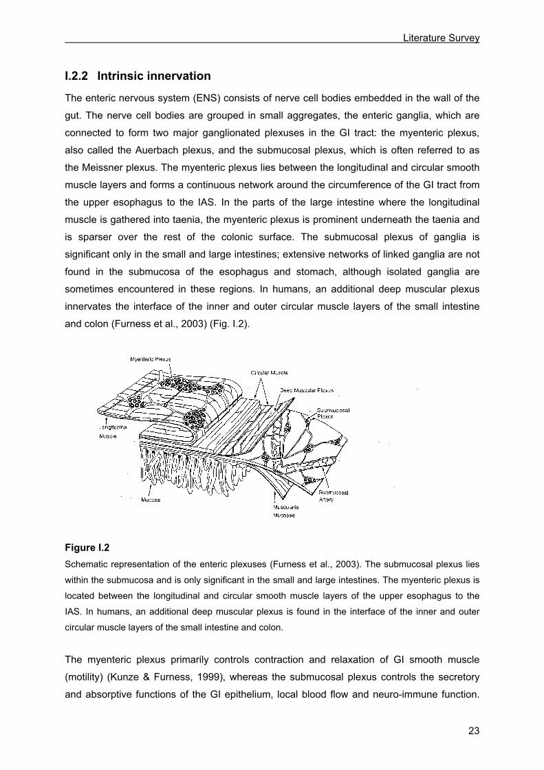
and colon (Furness et al., 2003) (Fig. I.2).
Figure I.2 Schematic representation of the enteric plexuses (Furness et al., 2003). The submucosal plexus lies
within the submucosa and is only significant in the small and large intestines. The myenteric plexus is
located between the longitudinal and circular smooth muscle layers of the upper esophagus to the
IAS. In humans, an additional deep muscular plexus is found in the interface of the inner and outer
circular muscle layers of the small intestine and colon.
The myenteric plexus primarily controls contraction and relaxation of GI smooth muscle
(motility) (Kunze & Furness, 1999), whereas the submucosal plexus controls the secretory
and absorptive functions of the GI epithelium, local blood flow and neuro-immune function.
Literature Survey
24
Both plexuses receive synaptic inputs from efferent and afferent nerve fibers of the extrinsic
nervous system. However, most of the synaptic inputs to enteric neurons come from other
enteric neurons (Galligan, 2002).
The neurons in both plexuses can be classified as: 1) sensory neurons, 2) interneurons and
3) motor neurons. The sensory neurons, also called “intrinsic primary afferent neurons”
(IPANs) become activated by three types of stimuli (distension, mechanical distortion of the
mucosa and change in luminal chemistry), and initiate motility changes as well as secretory
and blood flow changes. IPANs have their sensory endings in the mucosa and their cell
bodies in the myenteric or submucosal plexus. The IPANs synapse with interneurons, which
in turn make connections with other orally (ascending) or anally (descending) directed
interneurons and finally with motor neurons. The motor neurons are the final effectors and
are involved in the regulation of secretion (secretoneurons) or motility. The motor neurons
that innervate the longitudinal and circular smooth muscle layers can be classified into
excitatory and inhibitory neurons. The primary neurotransmitter released by activation of the
excitatory motor neurons is acetylcholine. Also tachykinins, which represent excitatory non-
cholinergic non-adrenergic or NANC neurotransmitters, are released albeit playing a lesser
role in the contraction of smooth muscle cells than acetylcholine. The activation of the
inhibitory motor neurons results in the release of inhibitory NANC neurotransmitters (see
I.2.3) causing relaxation of the smooth muscle cells (Furness, 2003; Kunze & Furness,
1999).
To study which transmitters mediate the contractile and relaxing responses of GI smooth
muscle, experiments are designed using an organ bath equipped with two platinum
electrodes and force transducers to record contraction/relaxation of GI smooth muscle
preparations. The latter contain both muscle layers and the myenteric plexus which is lying
between the circular and longitudinal muscle. The neurons of the myenteric plexus can be
activated by electrical field stimulation. Electrical field stimulation is achieved by applying
current through two platinum electrodes which are positioned close to the GI smooth muscle
preparation. By use of particular stimulation parameters the applied electrical pulses
depolarize the cell membranes of the enteric neurons and generate action potentials. These
action potentials activate enteric synapses to release their inhibitory and/or excitatory
neurotransmitters. Thereby, a cocktail of differently acting transmitters is released and
causes relaxation as well as contraction of the GI smooth muscle preparation. The
transmitters involved can be identified by adding antagonists to the organ bath. For example,
as the excitatory neurotransmitter acetylcholine acts on muscarinic receptors on the smooth
muscle, it is possible to block acetylcholine-induced contraction by using the muscarinic
antagonist atropine.
Literature Survey
25
In between the motor neurons of the ENS and the smooth muscle cells, a network of
interstitial cells of Cajal (ICC) is located. The ICCs are referred to as the pacemaking cells of
the gut, generating and propagating slow wave activity and potential oscillations. The ICCs
are closely associated with the varicose nerve terminals via synaptic-like structures and
express receptors for the neurotransmitters utilized by the enteric motor neurons. This
enables the ICCs to receive neuronal input, which leads to the generation of electrical
signals. The electrical responses elicited in the ICCs are conducted to the smooth muscle
cells via gap junctions, which lead to excitatory depolarization or inhibitory hyperpolarization
responses and thus respectively contraction or relaxation of the smooth muscle cells. The
ICCs are as such suggested to play an important role in the cholinergic excitatory and
nitrergic inhibitory neurotransmission (Sanders, 1996; Ward & Sanders, 2001).
I.2.3 Inhibitory NANC neurotransmission
The first functional evidence for the presence of non-cholinergic non-adrenergic or NANC
neurotransmitters in the GI tract was provided by Burnstock et al. (1963), based on the
observation that stimulation of intrinsic nerves in guinea-pig taenia coli resulted in a smooth
muscle inhibitory junction potential that was not influenced by the muscarinic receptor
antagonist atropine, with muscarinic receptors being the effectors of acetylcholine, and the
adrenergic neuron blocker bretylium.
NANC neurotransmitters can either be excitatory or inhibitory. Tachykinins elicit excitatory
smooth muscle responses, whereas ATP, vasoactive intestinal peptide (VIP), nitric oxide
(NO), carbon monoxide (CO) and recently hydrogen sulfide (H2S) are reported to elicit
inhibitory smooth muscle responses. This section focuses on the inhibitory NANC
neurotransmitters. ATP and VIP represent “classical” or “typical” neurotransmitters in the
sense that they are synthesized and stored in vesicles in the neurons, released by
exocytosis into the synaptic cleft, and act upon receptors in the plasma membrane of the
adjacent target cell. NO, CO and H2S are however atypical neural messengers, being small
gaseous molecules that are enzymatically synthesized only on demand, capable of diffusing
freely across the plasma membrane and acting directly on (intra)cellular targets in the
adjacent target cells (Barañano et al., 2001; Burnstock, 2006; Kasparek et al., 2008; Wang,
2002). The term “gasotransmitter” was introduced to characterize these three gases (Wang,
2002). The evidence that these three substances act as a GI neurotransmitter is very strong
for NO, much weaker for CO and weakest for H2S.
I.2.3.1 ATP, VIP/PACAP
The role of the purine nucleotide ATP as a NANC neurotransmitter was proposed for the first
time by Burnstock and colleagues and is now generally accepted. In addition to the presence
Literature Survey
26
of high concentrations of ATP in subpopulations of myenteric neurons in different regions of
the gut (Belai & Burnstock, 1994; Crowe & Burnstock, 1981) also the release of ATP was
reported (Burnstock et al., 1978), providing evidence for the purinergic neurotransmission.
Moreover, the role of ATP as a co-neurotransmitter in NANC relaxation was demonstrated or
suggested for different regions of the GI tract in several species, such as the mouse stomach
(Mulè & Serio, 2003), the rat pyloric sphincter (Soediono & Burnstock, 1994), the mouse (De
Man et al., 2003) and human (Xue et al., 1999) jejunum, the rat ileum (Benko et al., 2006),
the mouse (Serio et al., 2003a), rat (Pluja et al., 1999; Van Crombruggen & Lefebvre, 2004),
hamster (El Mahmoudy et al., 2006) and human (Benko et al., 2007; Boeckxstaens et al.,
1993; Keef et al., 1993) colon and the guinea pig IAS (Rae & Muir, 1996).
ATP is a ligand for P2 purinoceptors existing in two main subtypes: (1) the P2X receptors
that are ligand-gated ion channels and (2) the P2Y receptors that are coupled to G proteins
(Burnstock, 2006). Inhibitory responses are assumed to be mediated mainly by P2Y
receptors although some reports suggest the involvement of P2X receptor subtypes in
relaxant responses (De Man et al., 2003; Van Crombruggen et al., 2007). The main
transduction pathway activated by binding of ATP to P2Y receptors involves the activation of
phospholipase C (PLC), increased production of inositol triphosphate (IP3) (Boyer et al.,
1989) and release of Ca2+ from internal stores via IP3 receptors (Ca2+ puffs) and to a lesser
extent ryanodine receptors (Ca2+ sparks). It is suggested that this Ca2+ release is highly
directional and causes local Ca2+ transients near the plasma membrane without significant
changes in global cytoplasmatic Ca2+ concentration, leading to the activation of small
conductance Ca2+-dependent K+ channels (SKCa channels), hyperpolarization and
subsequently relaxation of the smooth muscle cell (Bayguinov et al., 2000; Koh et al., 1997;
Kong et al., 2000). Also other, PLC-independent, pathways activated by P2Y receptors have
been proposed (von Kügelgen & Wetter, 2000), including the activation of adenylate cyclase
(Qi et al., 2001; Zizzo et al., 2006). In addition, ATP was suggested to induce relaxation via
the production of NO (Giaroni et al., 2002; Xue et al., 2000b).
A role of neurotransmitter in NANC relaxation was also proposed for the peptide VIP and the
closely related pituitary adenylate cyclase activating peptide (PACAP) as both were found to
be localized in the myenteric plexus and in varicose nerve terminals running along the
smooth muscle layers (McConalogue et al., 1995; Portbury et al., 1995; Sundler et al., 1992;
Suzuki et al., 1996). An involvement of VIP in NANC relaxation was suggested in different GI
regions such as the mouse stomach (Mulè & Serio, 2003), the human (Tonini et al., 2000),
rat (D’Amato et al., 1992; Li & Rand, 1990), cat (Barbier & Lefebvre, 1993) and pig (Lefebvre
et al., 1995) gastric fundus, the mouse jejunum (Satoh et al., 1999), the hamster (El-
Mahmoudy et al., 2006) and mouse (Satoh et al., 1999) colon and the mouse IAS (Rattan et
Literature Survey
27
al., 2005). In the above mentioned gastric fundus tissues, the authors proposed a joint role
for NO and VIP in the NANC relaxation with NO being released during the initial fast but also
during sustained relaxation, whereas VIP was only released during the sustained relaxation;
an exception is Tonini et al. (2000) who reported the release of VIP during short-lasting
electrical stimulation. PACAP was reported to be involved in the NANC relaxation in for
example the mouse colon (Satoh et al., 1999) and the guinea pig caecum (McConalogue et
al., 1995).
Three G protein-coupled receptors for VIP/PACAP have been identified: VPAC1 and VPAC2
receptors with a similar affinity for both VIP and PACAP and the PAC1 receptors with a much
higher affinity for PACAP than for VIP (Harmar et al., 1998). The principal transduction
pathway following the peptidergic activation of these receptors starts with the activation of
adenylate cyclase and subsequent stimulation of cAMP, although also cAMP-independent
such as the activation of phospholipase C or the stimulation of tyrosine kinase, the latter
leading to the activation of SKCa channels, have been reported (Kishi et al., 2000; Laburthe &
Couvineau, 2002; MacKenzie et al., 2001; Takeuchi et al., 1999).
I.2.3.2 NO
In 1980, Furchgott & Zawadski observed that a substance released by vascular endothelial
cells induced relaxation of the smooth muscle cells beneath. The substance was called the
endothelium-derived relaxing factor (EDRF). It was only at the end of the 1980s that the
identity of EDRF was unravelled and was reported to be NO by the work of two groups, one
led by Ignarro (Ignarro et al., 1987) and one by Moncada (Palmer et al., 1987).
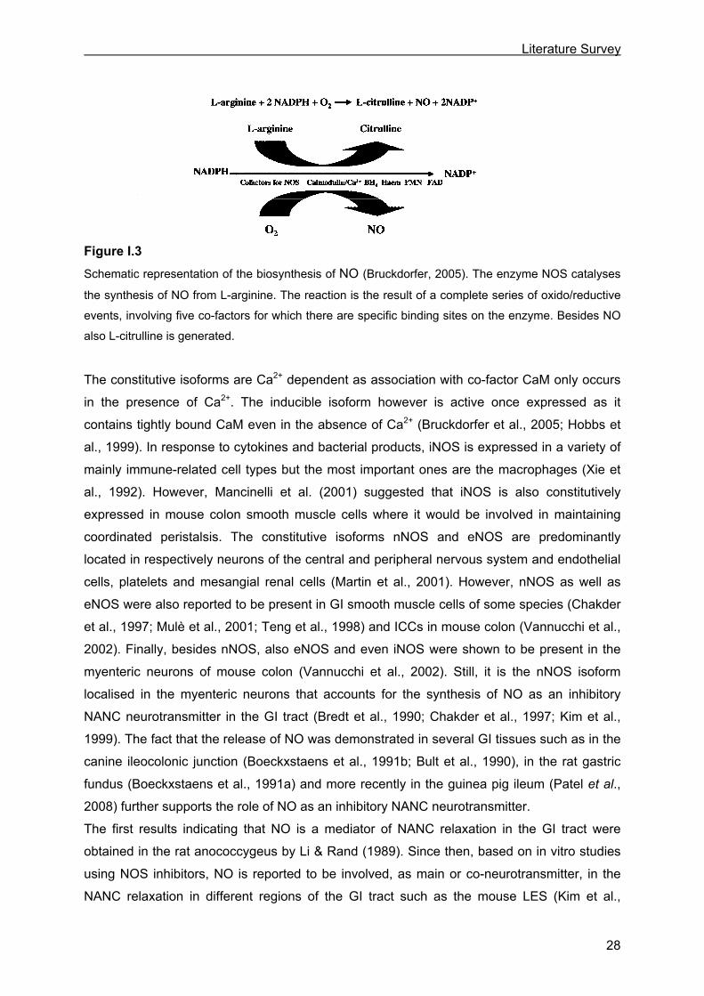
NO is a small gaseous molecule that is synthesized from L-arginine by the enzyme nitric
oxide synthase (NOS). The synthesis of NO involves the formation of the intermediate N-
hydroxy-L-arginine after which further oxidation leads to the production of NO and L-citrulline.
L-citrulline can then be reconverted to L-arginine. In order to exert its action, NOS requires
the presence of two co-substrates i.e. adenine dinucleotide phosphate (NADPH) and O2 and
several co-factors such as flavin mononucleotide (FMN), flavin adenine dinucleotide (FAD),
tetrahydrobiopterin (BH4), heme protoporphyrin IX and calmodulin (CaM) (Fig. I.3).
NOS exists in three isoforms that are all active as homo-dimers. Two isoforms can be
classified as constitutive enzymes i.e. neuronal NOS (nNOS or NOS-1) and endothelial NOS
(eNOS or NOS-3), and one as an inducible enzyme i.e. inducible NOS (iNOS or NOS-2).
Literature Survey
28
Figure I.3
Schematic representation of the biosynthesis of NO (Bruckdorfer, 2005). The enzyme NOS catalyses
the synthesis of NO from L-arginine. The reaction is the result of a complete series of oxido/reductive
events, involving five co-factors for which there are specific binding sites on the enzyme. Besides NO
also L-citrulline is generated.
The constitutive isoforms are Ca2+ dependent as association with co-factor CaM only occurs
in the presence of Ca2+. The inducible isoform however is active once expressed as it
contains tightly bound CaM even in the absence of Ca2+ (Bruckdorfer et al., 2005; Hobbs et
al., 1999). In response to cytokines and bacterial products, iNOS is expressed in a variety of
mainly immune-related cell types but the most important ones are the macrophages (Xie et
al., 1992). However, Mancinelli et al. (2001) suggested that iNOS is also constitutively
expressed in mouse colon smooth muscle cells where it would be involved in maintaining
coordinated peristalsis. The constitutive isoforms nNOS and eNOS are predominantly
located in respectively neurons of the central and peripheral nervous system and endothelial
cells, platelets and mesangial renal cells (Martin et al., 2001). However, nNOS as well as
eNOS were also reported to be present in GI smooth muscle cells of some species (Chakder
et al., 1997; Mulè et al., 2001; Teng et al., 1998) and ICCs in mouse colon (Vannucchi et al.,
2002). Finally, besides nNOS, also eNOS and even iNOS were shown to be present in the
myenteric neurons of mouse colon (Vannucchi et al., 2002). Still, it is the nNOS isoform
localised in the myenteric neurons that accounts for the synthesis of NO as an inhibitory
NANC neurotransmitter in the GI tract (Bredt et al., 1990; Chakder et al., 1997; Kim et al.,
1999). The fact that the release of NO was demonstrated in several GI tissues such as in the
canine ileocolonic junction (Boeckxstaens et al., 1991b; Bult et al., 1990), in the rat gastric
fundus (Boeckxstaens et al., 1991a) and more recently in the guinea pig ileum (Patel et al.,
2008) further supports the role of NO as an inhibitory NANC neurotransmitter.
The first results indicating that NO is a mediator of NANC relaxation in the GI tract were
obtained in the rat anococcygeus by Li & Rand (1989). Since then, based on in vitro studies
using NOS inhibitors, NO is reported to be involved, as main or co-neurotransmitter, in the
NANC relaxation in different regions of the GI tract such as the mouse LES (Kim et al.,
Literature Survey
29
1999), the guinea pig (Lefebvre et al., 1992) and pig (Lefebvre et al., 1995) gastric fundus,
the rat pyloric sphincter (Soediono et al., 1994), the mouse duodenum (Serio et al., 2003b),
the rat (Vanneste et al., 2004), equine (Rakestraw et al., 1996) and human (Murr et al., 1999)
jejunum, the rat ileum (Kanada et al., 1992), the guinea pig (Shuttleworth et al., 1999), rat
(Van Crombruggen & Lefebvre, 2004) and human (Boeckxstaens et al., 1993; Keef et al.,
1997) colon, the rat rectum (Takeuchi et al., 1998) and the mouse (Rattan et al., 2005) and
human (O’Kelly et al., 1993) IAS. The important role of NO in NANC relaxation and hence GI
motility becomes also evident from the study of nNOS knock-out (KO) mice and in vivo
studies in various species. nNOS KO mice, who lack the nNOS gene and are therefore
chronically deprived of nNOS generated NO, show an enlarged stomach with hypertrophy of
the gastric circular muscle layer and the pyloric sphincter (Huang et al., 1993; Mashimo et
al., 2000). The NANC relaxation is reduced in the gastric fundus strips of these mice (Dick et
al., 2002) and gastric emptying of solids and liquids is delayed (Mashimo et al., 2000), all
pointing to an essential role for NO in gastric motility.
An influence of NO on gastric emptying is reported in various studies, however, conflicting
results are obtained. Inhibition of NOS was reported to delay (semi-)solid gastric emptying in
dogs (Orihata & Sarna, 1994) and pigs (Lefebvre et al., 2005), to increase semi-liquid gastric
emptying in humans (Konturek et al., 1999), while having no effect on solid gastric emptying
in humans (Hirsch et al., 2000). These discrepancies might be related to the fact that NO
was reported to not only influence the gastric accommodation of the proximal stomach
(Desai et al., 1991; Tack et al., 2002), but also the antral motor activity (Konturek et al.,
1999) and the pyloric sphincter relaxation (Sivarao et al., 2008). The consequences of NOS
inhibition on these three important mechanisms in the regulation of gastric emptying (see
I.1.2.2) might inter- and counteract with each other leading to the various implications on
gastric emptying. In this regard, the delayed gastric emptying observed in the nNOS KO mice
by Mashimo et al. (2000), despite the accelerating effect due to decreased gastric fundus
relaxation (Dick et al., 2002), could be explained by a counteraction resulting from the
impairment of the pyloric sphincter relaxation (Sivarao et al., 2008).
The nNOS KO mice not only revealed a role of NO in the gastric motility, but also in the
jejunal inhibitory neurotransmission as NANC relaxations were found to be reduced in the
jejunal strips of these mice (Xue et al., 2000a). Furthermore, the use of NOS inhibitors
resulted in a delayed small intestinal transit in rats (Karmeli et al., 1997), dogs (Chiba et al.,
2002) and humans (Fraser et al., 2005), which might be related to the conversion of regular
peristalsis to irregular non-propulsive contractions when NOS is inhibited (Bogeski et al.,
2005). NO was also reported to play a role in the interdigestive MMC which migrates along

Literature Survey
30
the small intestine, as NOS inhibition was associated with stimulation of phase III activity
(Russo et al., 1999).
Finally, studies using NOS inhibitors showed a role for NO in the descending relaxation in rat
colon (Hata et al., 1990) and the colonic propulsive activity in guinea pigs (Foxx-Orenstein &
Grider, 1996). NOS inhibitors delayed the colonic transit in rats, suggesting that NO
enhances transit in the rat colon by mediating descending relaxation which, in turn, facilitates
the propulsion of the colonic contents (Mizuta et al., 1999). NO was also reported to
modulate the direction and frequency of the spontaneous colonic MMCs which were
recorded along the mouse colon (Powel & Bywater, 2001). The principal intracellular target of NO is the heme–containing protein soluble guanylate
cyclase (sGC). By binding to the prosthetic heme group of sGC, NO activates sGC, which
leads to the production of the second messenger cyclic guanosine 3’-5’-monophosphate
(cGMP) (Lucas et al., 2000). The mechanisms downstream of cGMP leading to smooth
muscle relaxation are discussed in I.3.3.2. However, effects of NO not involving the sGC-
cGMP pathway have also been reported. Bolotina et al. (1994) provided evidence that NO is
able to directly activate Ca2+-dependent K+ channels in vascular smooth muscle, whereas
Koh et al. (1995) and Lang & Watson (1998) proposed a direct activation by NO of
respectively voltage-activated and Ca2+-dependent K+ channels in colonic smooth muscle.
The direct (i.e. sGC-independent) action of NO on these channels is suggested to involve
chemical modification of the sulfhydryl groups present on the channels (Bolotina et al., 1994).
The latter is called S-nitrosylation and results from the interaction of NO with O2 or O2.-
leading to the formation of reactive nitrogen species (RNS) such as NO+, which is then added
to the sulfhydryl group of a cysteine residue. RNS can also lead to oxidation (when one or
two electrons are removed) or nitration (when NO2+ is added) of substrates. These RNS-
mediated effects of NO were initially proposed to be mainly involved in pathological
conditions, as they only prevail at higher concentrations of NO (> 1 µM), such as these
produced when iNOS is induced. In contrast, the direct effects of NO, such as the
physiologically relevant interaction with heme-containing proteins as sGC and also
cytochrome P450, predominate at low concentrations of NO (< 1 µM), which are produced by
the constitutive isoforms. However, there is increasing evidence that nitration of proteins may
also occur in normal cells, albeit at a much lower level. It is possible that these nitrations may
play a role in signal transduction processes but this is still a matter for speculation (Davis et
al., 2001; Bruckdorfer et al., 2005). The identification of a population of endogenously S-
nitrosylated proteins, including metabolic, structural and signalling proteins that may be
effectors for nNOS-generated NO points to protein S-nitrosylation as a physiological
signalling mechanism for NO (Ahern et al., 2002; Jaffrey et al., 2001).
Literature Survey
31
I.2.3.3 CO
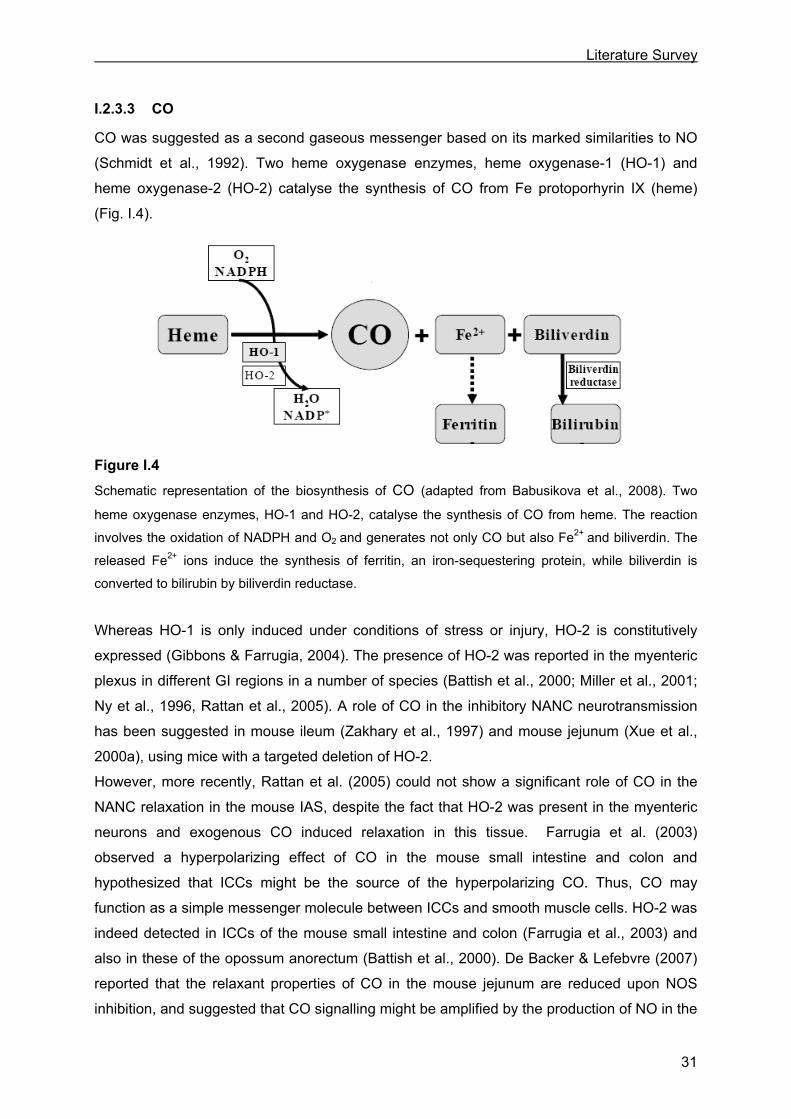
CO was suggested as a second gaseous messenger based on its marked similarities to NO
(Schmidt et al., 1992). Two heme oxygenase enzymes, heme oxygenase-1 (HO-1) and
heme oxygenase-2 (HO-2) catalyse the synthesis of CO from Fe protoporhyrin IX (heme)
(Fig. I.4).
Figure I.4
Schematic representation of the biosynthesis of CO (adapted from Babusikova et al., 2008). Two
heme oxygenase enzymes, HO-1 and HO-2, catalyse the synthesis of CO from heme. The reaction
involves the oxidation of NADPH and O2 and generates not only CO but also Fe2+ and biliverdin. The
released Fe2+ ions induce the synthesis of ferritin, an iron-sequestering protein, while biliverdin is
converted to bilirubin by biliverdin reductase.
Whereas HO-1 is only induced under conditions of stress or injury, HO-2 is constitutively
expressed (Gibbons & Farrugia, 2004). The presence of HO-2 was reported in the myenteric
plexus in different GI regions in a number of species (Battish et al., 2000; Miller et al., 2001;
Ny et al., 1996, Rattan et al., 2005). A role of CO in the inhibitory NANC neurotransmission
has been suggested in mouse ileum (Zakhary et al., 1997) and mouse jejunum (Xue et al.,
2000a), using mice with a targeted deletion of HO-2.
However, more recently, Rattan et al. (2005) could not show a significant role of CO in the
NANC relaxation in the mouse IAS, despite the fact that HO-2 was present in the myenteric
neurons and exogenous CO induced relaxation in this tissue. Farrugia et al. (2003)
observed a hyperpolarizing effect of CO in the mouse small intestine and colon and
hypothesized that ICCs might be the source of the hyperpolarizing CO. Thus, CO may
function as a simple messenger molecule between ICCs and smooth muscle cells. HO-2 was
indeed detected in ICCs of the mouse small intestine and colon (Farrugia et al., 2003) and
also in these of the opossum anorectum (Battish et al., 2000). De Backer & Lefebvre (2007)
reported that the relaxant properties of CO in the mouse jejunum are reduced upon NOS
inhibition, and suggested that CO signalling might be amplified by the production of NO in the
Literature Survey
32
ICCs. As a result of these observations, the role for CO as a neurotransmitter is still under
debate.
The most important target of CO is sGC. Indeed, CO can activate sGC (Stone & Marletta,
1994), resulting in an increased production of second messenger cGMP. The mechanisms
downstream of cGMP leading to smooth muscle relaxation are discussed in I.3.3.2. The
involvement of sGC in CO-induced relaxation was shown by Rattan et al. (2004) in the
internal anal sphincter. CO was also reported to directly activate large conductance Ca2+-
dependent K+ channels in vascular smooth muscle (Wang & Wu, 1997).
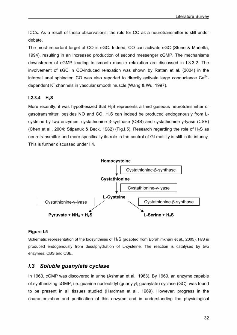
I.2.3.4 H2S
More recently, it was hypothesized that H2S represents a third gaseous neurotransmitter or
gasotransmitter, besides NO and CO. H2S can indeed be produced endogenously from L-
cysteine by two enzymes, cystathionine β-synthase (CBS) and cystathionine γ-lyase (CSE)
(Chen et al., 2004; Stipanuk & Beck, 1982) (Fig.I.5). Research regarding the role of H2S as
neurotransmitter and more specifically its role in the control of GI motility is still in its infancy.
This is further discussed under I.4.
Figure I.5
Schematic representation of the biosynthesis of H2S (adapted from Ebrahimkhani et al., 2005). H2S is
produced endogenously from desulphydration of L-cysteine. The reaction is catalysed by two
enzymes, CBS and CSE.
I.3 Soluble guanylate cyclase
In 1963, cGMP was discovered in urine (Ashman et al., 1963). By 1969, an enzyme capable
of synthesizing cGMP, i.e. guanine nucleotidyl (guanylyl; guanylate) cyclase (GC), was found
to be present in all tissues studied (Hardman et al., 1969). However, progress in the
characterization and purification of this enzyme and in understanding the physiological
Homocysteine
Cystathionine
L-Cysteine
Pyruvate + NH3 + H2S L-Serine + H2S
Cystathionine-γ-lyase
Cystathionine-β-synthase
Cystathionine-β-synthase Cystathionine-γ-lyase
Literature Survey
33
significance of cGMP was delayed until the 1980s when two important discoveries were
made. The first was the discovery that a peptide made in the heart, atrial natriuretic peptide
(ANP), could increase cGMP by binding to the transmembrane form of GC (particulate
guanylate cyclase; pGC). The second was the discovery that the endogenous molecule
EDRF could activate the soluble form of guanylate cyclase (soluble guanylate cyclase; sGC).
EDRF was later identified as NO, which established NO as an endogenous activator of sGC.
Hence, the NO-sGC-cGMP transduction pathway was born. Extensive research has shown
this signalling pathway to be widespread in mammalian tissues and important in mediating
numerous physiological processes including peripheral and central neurotransmission and
smooth muscle relaxation (Hobbs, 1997; Beavo & Brunton, 2002; Pyriochou &
Papapetropoulos, 2005).
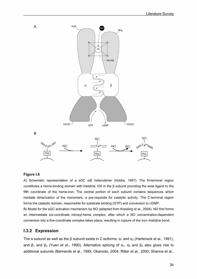
I.3.1 Structure
sGC is a heterodimeric heme-containing protein that is composed of a larger α subunit and a
smaller β subunit (Harteneck et al., 1991). Each subunit can be divided into three functional
domains: an N-terminal regulatory heme-binding domain, a central dimerization domain and
a C-terminal catalytic domain (Lucas et al., 2000) (Fig. I.6A).
The N-terminal domain of the β subunit is the most important for the heme-binding, with
histidine at position 105 as the essential amino acid required for the binding of the heme
moiety (Wedel et al., 1994; Zhao et al., 1998). The prosthetic heme group is as such a five-
membered ring wherein four nitrogen atoms are coordinated with a central iron (Fe2+;
reduced or ferrous form) and with as fifth member of the ring histidine 105 as axial ligand
(Lucas et al., 2000). More recently, other residues from the β subunit, i.e. tyrosine 135,
arginine 139 and serine 137, were reported to be involved in stabilizing the binding of the
heme moiety and to form a unique heme binding motif (Schmidt et al., 2005).
The central dimerization domain is involved in the formation of heterodimers, which is a pre-
requisite for sGC to exhibit catalytic activity (Pyriochou & Papapetropoulos, 2005). Indeed,
both subunits are necessary for catalytic activity (Harteneck et al., 1990).
The C-terminal catalytic domains of the sGC subunits are the most conserved regions that
also exhibit a substantial degree of similarity with adenylyl cyclase. sGC converts guanosine-
5’-triphosphate (GTP) to cGMP (Pyriochou & Papapetropoulos, 2005).
Literature Survey
34
Figure I.6
A) Schematic representation of a sGC α/β heterodimer (Hobbs, 1997). The N-terminal region
constitutes a heme-binding domain with histidine 105 in the β subunit providing the axial ligand to the
fifth coordinate of the heme-iron. The central portion of each subunit contains sequences which
mediate dimerization of the monomers, a pre-requisite for catalytic activity. The C-terminal region
forms the catalytic domain, responsible for substrate binding (GTP) and conversion to cGMP.
B) Model for the sGC activation mechanism by NO (adapted from Koesling et al., 2004). NO first forms
an intermediate six-coordinate nitrosyl-heme complex, after which a NO concentration-dependent
conversion into a five-coordinate complex takes place, resulting in rupture of the iron–histidine bond.
I.3.2 Expression
The α subunit as well as the β subunit exists in 2 isoforms: α1 and α2 (Harteneck et al., 1991),
and β1 and β2 (Yuen et al., 1990). Alternative splicing of α1, α2 and β2 also gives rise to
additional subunits (Behrends et al., 1995; Okamoto, 2004; Ritter et al., 2000; Sharina et al.,
A
B
Literature Survey
35
2008). However, these additional subunits as well as the β2 subunit are generally deemed
not active or even dominant negative (Behrends et al., 1995; Sharina et al., 2008), although
the formation of an active β2 homodimer (Koglin et al., 2001) and an α1β2 heterodimer with
reduced sensitivity to NO (Gupta et al., 1997) have been reported. But the fact that the β2
subunit was almost undetectable in tissue argues against a relevant physiological role of this
subunit (Mergia et al., 2003). This leaves the α1β1 and the α2β1 as the only physiological
active isoforms, with no differences in kinetic properties and sensitivity towards NO between
the two isoforms (Russwurm et al., 1998). The α1 and the β1 subunit were detected in all
tested human (Budworth et al., 1999) and mouse (Mergia et al., 2003) tissues whereas the
α2 subunit showed a more restricted expression pattern with high levels in brain, placenta,
spleen and uterus only (Budworth et al., 1999). In brain, the amount of α1β1 was
quantitatively similar to that of α2β1; in all other tissues however, including ileum and colon,
α1β1 was the predominant isoform (Mergia et al., 2003).
Although the enzyme is originally called ‘soluble’ guanylate cyclase, it was demonstrated in
brain that the α2β1 isoform is associated with the PDZ-containing post-synaptic density
protein-95 (PSD-95), this via the C-terminal PDZ domain of the α2 subunit. As a
consequence of this interaction, the sGCα2β1 isoform is recruited to the membrane of
synaptosomes, where it would then be co-localized with the PSD-95 interacting neuronal NO
synthase (Nedvetsky et al., 2002; Russwurm et al., 2001). Also the translocation of the α1β1
isoform to the plasma membrane in response to elevated Ca2+ concentrations has been
reported, rendering the enzyme more sensitive to NO (Zabel et al., 2002).
I.3.3 Smooth muscle relaxation by sGC
I.3.3.1 Activation of sGC
To induce relaxation via sGC, NO and CO first need to activate sGC by binding to the
enzyme. NO binds hereby to the heme moiety of sGC, following a two-step mechanism (Fig.
I.6B). First, a six-coordinate intermediate NO-Fe2+-histidine complex is formed. Next, the
breakage of the Fe2+-histidine bond leads to the formation of a five-coordinate nitrosyl-heme
complex and concomitant conformational changes of the enzyme. This results in activation of
sGC, as these rearrangements move the C-terminal domain of the β subunit closer to the C-
terminal domain of the α subunit, thus forming a more closed, circular catalytic domain
(Kosarikov et al., 2001). Besides the first step, also the second step was reported to be
dependent of the NO concentration (Zhao et al., 1999). This implies that this second step
involved additional reactions of NO with sGC. The requirement of the binding of a second NO
molecule to the proximal face of the heme, thereby effectively displacing the histidine was
suggested by Lawson et al. (2003). The second NO than repels the first, distally bound, NO
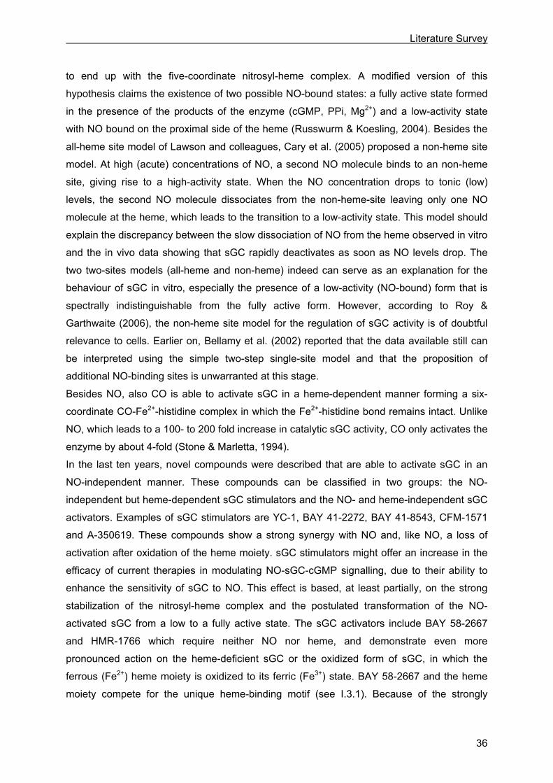
Literature Survey
36
to end up with the five-coordinate nitrosyl-heme complex. A modified version of this
hypothesis claims the existence of two possible NO-bound states: a fully active state formed
in the presence of the products of the enzyme (cGMP, PPi, Mg2+) and a low-activity state
with NO bound on the proximal side of the heme (Russwurm & Koesling, 2004). Besides the
all-heme site model of Lawson and colleagues, Cary et al. (2005) proposed a non-heme site
model. At high (acute) concentrations of NO, a second NO molecule binds to an non-heme
site, giving rise to a high-activity state. When the NO concentration drops to tonic (low)
levels, the second NO molecule dissociates from the non-heme-site leaving only one NO
molecule at the heme, which leads to the transition to a low-activity state. This model should
explain the discrepancy between the slow dissociation of NO from the heme observed in vitro
and the in vivo data showing that sGC rapidly deactivates as soon as NO levels drop. The
two two-sites models (all-heme and non-heme) indeed can serve as an explanation for the
behaviour of sGC in vitro, especially the presence of a low-activity (NO-bound) form that is
spectrally indistinguishable from the fully active form. However, according to Roy &
Garthwaite (2006), the non-heme site model for the regulation of sGC activity is of doubtful
relevance to cells. Earlier on, Bellamy et al. (2002) reported that the data available still can
be interpreted using the simple two-step single-site model and that the proposition of
additional NO-binding sites is unwarranted at this stage.
Besides NO, also CO is able to activate sGC in a heme-dependent manner forming a six-
coordinate CO-Fe2+-histidine complex in which the Fe2+-histidine bond remains intact. Unlike
NO, which leads to a 100- to 200 fold increase in catalytic sGC activity, CO only activates the
enzyme by about 4-fold (Stone & Marletta, 1994).
In the last ten years, novel compounds were described that are able to activate sGC in an
NO-independent manner. These compounds can be classified in two groups: the NO-
independent but heme-dependent sGC stimulators and the NO- and heme-independent sGC
activators. Examples of sGC stimulators are YC-1, BAY 41-2272, BAY 41-8543, CFM-1571
and A-350619. These compounds show a strong synergy with NO and, like NO, a loss of
activation after oxidation of the heme moiety. sGC stimulators might offer an increase in the
efficacy of current therapies in modulating NO-sGC-cGMP signalling, due to their ability to
enhance the sensitivity of sGC to NO. This effect is based, at least partially, on the strong
stabilization of the nitrosyl-heme complex and the postulated transformation of the NO-
activated sGC from a low to a fully active state. The sGC activators include BAY 58-2667
and HMR-1766 which require neither NO nor heme, and demonstrate even more
pronounced action on the heme-deficient sGC or the oxidized form of sGC, in which the
ferrous (Fe2+) heme moiety is oxidized to its ferric (Fe3+) state. BAY 58-2667 and the heme
moiety compete for the unique heme-binding motif (see I.3.1). Because of the strongly
Literature Survey
37
reduced affinity of the oxidized heme for the sGC heme-binding pocket in comparison to the
reduced heme, only the oxidized heme can be effectively replaced by BAY 58-2267. sGC
activators might offer an advantage over current therapies using NO-donors in the treatment
of various pathophysiological conditions associated with oxidative stress. Indeed, oxidative
stress leads to a disequilibrium between the oxidized and the reduced form of sGC, in favour
of oxidized form. sGC activators, unlike NO, are able to activate oxidized sGC (Evgenov et
al., 2006).
I.3.3.2 Mechanisms downstream of cGMP
Activation of sGC results in increased intracellular cGMP levels. The resultant accumulated
cGMP exerts its physiological effects through three intracellular effectors: cGMP-dependent
protein kinases (cGKs or PKGs), cyclic nucleotide-gated ion channels (CNGs) and cGMP-
regulated phosphodiesterases (PDEs). The most important mediators of GI signalling are
PKGs and PDEs. The PKGs belong to the serine/threonine kinase family and exist in two
subtypes: the cytosolic PKG I, which is present in high concentrations in smooth muscle, and
the membrane-bound PKG II, which is expressed in the intestinal mucosa. The hypothesis is
that PKG I plays an important role in intestinal smooth muscle relaxation, whereas PKG II
regulates intestinal secretion. Due to alternative splicing, PKG I has two isoforms i.e. PKG Iα
and PKG Iβ (Hofmann et al., 2000; Lucas et al., 2000).
PKG I leads to smooth muscle relaxation by lowering the intracellular Ca2+ concentration
([Ca2+]i) and/or by desensitization of the contractile apparatus to [Ca2+]i (Lucas et al., 2000;
Toda & Herman, 2005) (Fig. I.7).
PKG I can reduce [Ca2+]i by several mechanisms. One mechanism is the inhibition of the
release of Ca2+ from the sarcoplasmatic reticulum (SR), either by inhibition of agonist-induced
inositol 1,4,5-triphosphate (IP3)-generation and/or by inhibition of the IP3-receptor. The first
likely involves phosphorylation of a member of the G protein family or an isozyme of the
phospholipase C family (Ruth et al., 1993); the latter is dependent of the interaction of PKG
Iβ with the IP3 receptor-associated cGMP kinase substrate (IRAG) (Ammendola et al., 2001).
Secondly, PKG I can promote Ca2+ sequestration through activation of the sarcoplasmatic
reticulum Ca2+ pumping ATP-ase (SERCA) by phosphorylating and thus inactivating
phospholamban and thereby relieving SERCA from its inhibitory effect (Cornwell et al.,
1991). A third mechanism to reduce [Ca2+]i is inhibiting Ca2+ influx through L-type Ca2+
channels by both direct impairment of the channel activity (Liu et al., 1997) and indirect
hyperpolarization via an increase in the open probability of the Ca2+-dependent K+ channels
(Schubert & Nelson, 2001).

Literature Survey
38
Figure I.7 Mechanisms underlying smooth muscle relaxation mediated by cGMP (adapted from Lucas et al.,
2000). cGMP induces smooth muscle relaxation by lowering ([Ca2+]i) and/or by desensitization of the
contractile apparatus to [Ca2+]i. cGMP can reduce [Ca2+]i by (1) inhibiting Ca2+ influx through L-type
Ca2+ channels; (2) increasing Ca2+ efflux through activation of (2d) the Ca2+-pumping ATPase and (2b)
the Na+/Ca2+ exchanger; also, cGMP may produce membrane hyperpolarization through activation of
(2c) the Na+/K+ ATPase and (2a) K+ channels, thereby increasing Ca2+ extrusion by the Na+/Ca2+
exchanger; (3) increasing of Ca2+ sequestration through activation of the sarcoplasmatic reticulum
Ca2+-pumping ATPase by inactivating phospholamban [Ph] and thereby relieving the sarcoplasmatic
reticulum Ca2+-pumping ATPase from its inhibitory effect; (4) decreasing of Ca2+ mobilization through
inhibition of agonist-induced IP3 formation or inhibition of the IP3 receptor in the sarcoplasmatic
reticulum. cGMP desensitizes the contractile apparatus to Ca2+ (5) probably by activating the myosin
light chain phosphatase, resulting in dephoshorylation of the myosin light chain. : inhibitory effect;
: stimulatory effect. R, receptor; G, G protein; PLC, phospholipase C.
Finally, PKG I can increase the Ca2+ efflux through activation of the Ca2+-pumping ATPase,
the Na+/Ca2+ exchanger or induce hyperpolarization via activation of the Na+/K+ ATPase,
thereby again increasing Ca2+ extrusion by the Na+/Ca2+ exchanger (Lucas et al., 2000).
+
_ +
Literature Survey
39
Taken together, the resulting decrease in [Ca2+]i will attenuate the activity of the
Ca2+/calmodulin-dependent myosin light chain kinase (MLCK), leading to a reduced
phosphorylation of the myosin light chain (MLC). This will subsequently reduce myosin’s
ATPase activity and cross-bridge cycling with relaxation of the smooth muscle cell as a
result. In contrast to the activation by MLCK, myosin’s ATPase activity can be inhibited by
the myosin light chain phosphatase (MLCP), which dephosphorylates the MLC. It is in fact
the MLCK to MLCP activity ratio which determines the phosphorylation state of the MLC and
thus the myosin’s ATPase activity and the contraction level of the smooth muscle.
PKG I can activate MLCP without inducing a decrease in [Ca2+]i; this mechanism of action is
called Ca2+ desensitization as relaxation of the contractile apparatus then occurs in the
presence of an unaltered/high [Ca2+]i (Somlyo & Somlyo, 2003). A direct stimulation of MLCP
through interaction with PKG Iα was reported by Surks et al. (1999). In addition, PKG I was
shown to relieve MLCP from the inhibitory actions of the Rho-A/Rho-kinase pathway
(Sauzeau et al., 2000) or protein CPI-17 (Bonnevier & Arner, 2004). Finally, PKG I was
reported to phosphorylate telokin, thereby up-regulating its enhancing effect on MLCP (Wu et
al., 1998).
PDEs cleave the phosphodiester bond of cGMP and adenosine 3’-5’ cyclic monophosphate
(cAMP), hydrolysing the second messengers to their corresponding nucleotide, 5’
monophosphate (Lucas et al., 2000). The most important cGMP-regulated PDE in smooth
muscle is PDE-5 and inhibition of this PDE induces relaxation by means of cGMP
accumulation (Rybalkin et al., 2003).
I.3.4 Role of sGC in GI motility
Up to date, investigators approached the study of the role of sGC in GI motility mainly by
inhibiting sGC by means of the sGC inhibitor 1H[1,2,4,]oxadiazolo [4,3-a]quinoxalin-1-one
(ODQ; Schrammel et al., 1996). In vitro experiments with ODQ revealed for example a role
for sGC in the NANC relaxation of rat gastric fundus (Lefebvre, 1998), rat (Vanneste et al.,
2004) and human (Zyromski et al., 2001) jejunum and rat distal colon (Van Crombruggen &
Lefebvre, 2004). Similar findings were reported for mouse duodenum (Serio et al., 2003b),
jejunum and ileum (Ueno et al., 2004).
Knocking out cGMP’s effector PKG I induced dilation of the stomach and signs of pyloric
stenosis. The electrically induced relaxation of gastric fundus strips was impaired (Ny et al.,
2000) as well as gastric emptying of a barium suspension (Pfeifer et al., 1998). The
interruption of the NO-sGC-cGMP-PKG I pathway also delayed small intestinal transit of a
barium suspension. Instead of regular peristalsis, spastic contractions of long intestinal
segments followed by scarce and slow relaxations were observed in the PKG I KO mice
Literature Survey
40
(Pfeifer et al., 1998). Taken together, these findings suggest an important role for sGC in the
GI tract.
I.4 Hydrogen sulfide
H2S, just as NO and CO, was formerly considered solely as an environmental toxicant and
metabolic poison. Indeed, all of these gases potently interact with metals/metalloproteins,
notably the heme moiety. For example, all are potent inhibitors of mitochondrial cytochrome c
oxidase, thereby inhibiting the respiratory system (Li & Moore, 2007). The turning point in
‘gas biology’ came with the realization that these gases are generated naturally within
mammalian cells (Li & Moore, 2007).
I.4.1 Identification of H2S as neurotransmitter
After NO and CO, H2S became the third possible gaseous neurotransmitter, based on its
endogenous production by CSE/CBS (see I.2.3.4) and its biological effects such as smooth
muscle relaxation. Both H2S-synthetizing enzymes were shown in human and guinea-pig
myenteric neurons (Schicho et al., 2006). CSE but not CBS was shown to be present in
mouse colon myenteric neurons, and the intact colonic muscle layer containing the myenteric
plexus generated detectable levels of H2S (Linden et al., 2008). Also, H2S was shown to
inhibit spontaneous or induced contractions in the guinea-pig ileum (Hosoki et al., 1997;
Teague et al., 2002), the rat and rabbit ileum (Teague et al., 2002), the human and rat colon
and the mouse colon and jejunum (Gallego et al., 2008). Moreover, brain CBS (like NOS)
activity was shown to be both Ca2+- and calmodulin-dependent (Eto et al., 2002), suggesting
that ‘short-term’ control of neuronal H2S (such as NO) production might be achieved by influx
of Ca2+ into neurons following depolarization (Li & Moore, 2007). Designating a molecule as
a neurotransmitter presumes however also that it possesses a mechanism of inactivation.
Indeed, H2S is metabolized to sulfate and thiosulfate via the oxidation metabolism in
mitochondria or is scavenged by methemoglobin or metallo- or disulfide-containing
molecules such as oxidized glutathione. H2S can also be converted into lower toxic
compounds of methylmercaptan and dimethylsulfate via the methylation metabolism in
cytosol. The metabolic product exhausts from the kidney and intestinal tract and lungs within
24 hours, so the endogenously-generated H2S under physiological condition is hardly
accumulated or toxic to cells due to the balanced metabolism of the gas (Chen et al., 2007;
Wang, 2002).

Literature Survey
41
I.4.2 Influence of H2S on smooth muscle contractility
H2S-induced smooth muscle relaxation was observed not only in the GI tract (Gallego et al.,
2008; Hosoki et al., 1997; Teague et al., 2002) but also in the vascular system (Cheng et al.,
2004; Webb et al., 2008; Zhao et al., 2001), the respiratory system (Kubo et al., 2007) and
the reproductive system (d’Emmanuele di Villa Bianca et al., 2009; Sidhu et al., 2001;
Srilatha et al., 2007).
The mechanism by which H2S brings about smooth muscle relaxation is however not fully
understood. The most complete body of evidence comes from work based on vascular
smooth muscle. According to studies in this tissue, the H2S-induced smooth muscle
relaxation appears to be mediated in large part by the opening of K+ channels on the surface
of smooth muscle cells. The most important participant in H2S-induced vasodilatation seems
to be the ATP-dependent K+ channel (KATP channel), this based on studies where the effect
of H2S was shown to be mimicked by the KATP channel opener pinacidil and inhibited by the
KATP channel blocker glibenclamide (Cheng et al., 2004; Webb et al., 2008; Zhao et al.,
2001). The stimulatory effect of H2S on KATP channel was further demonstrated by directly
measuring the KATP channel current (Cheng et al., 2004; Tang et al., 2005; Zhao et al., 2001).
The activation of KATP channels by H2S leads to membrane hyperpolarization, which in turn
closes the voltage-dependent Ca2+ channels with relaxation of the smooth muscle as a
result.
Although most reports indicate KATP channels as mediator of H2S-induced vasorelaxation,
there are some reports that suggest alternative mechanisms for H2S to induce relaxation.
H2S may directly inhibit the voltage-dependent Ca2+ channels as suggested by Zhao & Wang
(2002). A study by Kiss et al. (2008) hypothesize that H2S-induced vasorelaxation is induced
by the inhibition of cytochrome c oxidase, leading to loss of mitochondrial ATP generation
and thus energy deficit and intracellular acidosis, which in turn activates the Cl-/HCO3-
exchanger. Similarly, it is shown that H2S regulates intracellular pH in vascular smooth
muscle cells via activation of the Cl-/HCO3- exchanger and that this is, at least partially,
responsible for H2S-mediated vasorelaxation (Lee et al., 2007).
Instead of a direct effect on the smooth muscle, Zhao & Wang (2002) suggested that H2S
may also affect vasocontractility by facilitating the release of endothelium-derived relaxant
factors such as NO. However, Kiss et al. (2008) could not show a role of NO in the H2S-
induced vasorelaxation. Also, as opposed to NO and CO, H2S-induced relaxation seems not
to be mediated by the sGC-cGMP signalling pathway, as the sGC inhibitor ODQ did not
reduce the H2S-mediated relaxation in rat aortic rings (Zhao & Wang, 2002) and mouse
bronchial rings (Kubo et al., 2007).

Literature Survey
42
Similar to results in the vascular system, K+ channels were also reported to be possibly
involved in the H2S-induced relaxation of GI smooth muscle. Indeed, Gallego et al. (2008)
showed that the H2S-induced inhibition of spontaneous motor complexes in mouse colon and
jejunum was mediated by SKCa channels, whereas both SKCa channels and KATP channels
seemed to be involved in the H2S-induced inhibition of spontaneous motility in rat and human
colon. Also in a rat model using colorectal distension, the H2S-induced antinociceptive and
relaxant effects were mediated by KATP channels (Distrutti et al., 2006). However, Teague et
al. (2002) could not show a role for KATP channels in the H2S-induced relaxation in the
guinea-pig ileum. Also about the possible role of NO in the H2S-induced relaxation,
conflicting data are reported: Teague et al. (2002) and Gallego et al. (2008) showed no
influence of NOS-inhibitors on the H2S-induced relaxation in the guinea pig ileum and the
mouse colon and jejunum respectively, whereas Distrutti et al. (2006) observed that NOS
inhibition almost completely reversed the antinociceptive and relaxant effects of H2S in rat
colon. These discrepant results together with the to date only limited number of reports
concerning the H2S-induced smooth muscle relaxation in the GI tract make the latter an
interesting territory for future experiments.
There should also be mentioned that another potential source of H2S, besides neuronal
endogenous production, is H2S produced by bacteria within the bowel lumen. However, a
physiological role of H2S originating from this source has not yet been described (Kasparek
et al., 2008). Colonic mucosa possesses specialized detoxification systems involving the
enzymes thiol methyltransferase (Weisiger et al., 1980) or rhodanese (Picton et al., 2002),
protecting the colon from what otherwise might be injurious concentrations of H2S.
I.5 References AHERN, G.P., KLYACHKO, V.A. & JACKSON, M.B. (2002). cGMP and S-nitrosylation : two routes for modulation of neuronal excitability by NO. Trends Neurosci., 25, 510-517.
AMMENDOLA, A., GEISELHÖRINGER, A., HOFMANN, F. & SCHLOSSMANN, J. (2001). Molecular determinants of the interaction between the inositol 1,4,5-triphosphate receptor-associated cMGP kinase substrate (IRAG) and cGMP kinase Iβ. J. Biol. Chem., 276, 24153-24159.
ASHMAN, D.F., LIPTON, R., MELICOW, M.M. & PRICE, T.D. (1963). Isolation of adenosine 3’-5’-monophosphate and guanosine 3’-5’-monophosphate from rat urine. Biochem. Biophys. Res. Commun., 11, 330-334.
BABUSIKOVA, E., JESENAK, M., DURDIK, P., DOBROTA, D. & BANOVCIN, P. (2008). Exhaled carbon monoxide as a new marker of respiratory diseases in children. J. Physiol. Pharmacol., 59, 9-17.
BARANANO, D.E., FERRIS, C.D. & SNYDER, S.H. (2001). Atypical neural messengers. Trends Neurosci., 24, 99-106.
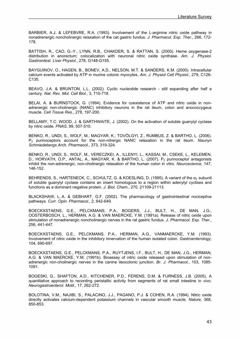
Literature Survey
43
BARBIER, A.J. & LEFEBVRE, R.A. (1993). Involvement of the L-arginine :nitric oxide pathway in nonadrenergic noncholinergic relaxation of the cat gastric fundus. J. Pharmacol. Exp. Ther., 266, 172-178.
BATTISH, R., CAO, G.-Y., LYNN, R.B., CHAKDER, S. & RATTAN, S. (2000). Heme oxygenase-2 distribution in anorectum: colocalization with neuronal nitric oxide synthase. Am. J. Physiol. Gastrointest. Liver Physiol., 278, G148-G155.
BAYGUINOV, O., HAGEN, B., BONEV, A.D., NELSON, M.T. & SANDERS, K.M. (2000). Intracellular calcium events activated by ATP in murine colonic myocytes. Am. J. Physiol Cell Physiol., 279, C126-C135.
BEAVO, J.A. & BRUNTON, L.L. (2002). Cyclic nucleotide research - still expanding after half a century. Nat. Rev. Mol. Cell Biol., 3, 710-718.
BELAI, A. & BURNSTOCK, G. (1994). Evidence for coexistence of ATP and nitric oxide in non-adrenergic non-cholinergic (NANC) inhibitory neurons in the rat ileum, colon and anococcygeus muscle. Cell Tissue Res., 278, 197-200.
BELLAMY, T.C. WOOD, J. & GARTHWAITE, J. (2002). On the activation of soluble guanylyl cyclase by nitric oxide. PNAS, 99, 507-510.
BENKO, R., UNDI, S., WOLF, M., MAGYAR, K., TOVÖLGYI, Z., RUMBUS, Z. & BARTHO, L. (2006). P2 purinoceptors account for the non-nitrergic NANC relaxation in the rat ileum. Naunyn Schmiedebergs Arch. Pharmacol., 373, 319-324.
BENKO, R., UNDI, S., WOLF, M., VERECZKEI, A., ILLENYI, L., KASSAI, M., CSEKE, L., KELEMEN, D., HORVATH, O.P., ANTAL, A., MAGYAR, K. & BARTHO, L. (2007). P2 purinoceptor antagonists inhibit the non-adrenergic, non-cholinergic relaxation of the human colon in vitro. Neuroscience, 147, 146-152.
BEHRENDS, S., HARTENECK, C., SCHULTZ, G. & KOESLING, D. (1995). A variant of the α2 subunit of soluble guanylyl cyclase contains an insert homologous to a region within adenylyl cyclises and functions as a dominant negative protein. J. Biol. Chem., 270, 21109-21113.
BLACKSHAW, L..A. & GEBHART, G.F. (2002). The pharmacology of gastrointestinal nociceptive pathways. Curr. Opin. Pharmacol., 2, 642-649.
BOECKXSTAENS, G.E., PELCKMANS, P.A., BOGERS, J.J., BULT, H., DE MAN, J.G., OOSTERBOSCH, L., HERMAN, A.G. & VAN MAERCKE, Y.M. (1991a). Release of nitric oxide upon stimulation of nonadrenergic noncholinergic nerves in the rat gastric fundus. J. Pharmacol. Exp. Ther., 256, 441-447.
BOECKXSTAENS, G.E., PELCKMANS, P.A., HERMAN, A.G., VANMAERCKE, Y.M. (1993). Involvement of nitric oxide in the inhibitory innervation of the human isolated colon. Gastroenterology, 104, 690-697.
BOECKXSTAENS, G.E., PELCKMANS, P.A., RUYTJENS, I.F., BULT, H., DE MAN, J.G., HERMAN, A.G. & VAN MAERCKE, Y.M. (1991b). Bioassay of nitric oxide released upon stimulation of non-adrenergic non-cholinergic nerves in the canine ileocolonic junction. Br. J. Pharmacol., 103, 1085-1091.
BOGESKI, G., SHAFTON, A.D., KITCHENER, P.D., FERENS, D.M. & FURNESS, J.B. (2005). A quantitative approach to recording peristaltic activity from segments of rat small intestine in vivo. Neurogastroenterol. Motil., 17, 262-272.
BOLOTINA, V.M., NAJIBI, S., PALACINO, J.J., PAGANO, P.J. & COHEN, R.A. (1994). Nitric oxide directly activates calcium-dependent potassium channels in vascular smooth muscle. Nature, 368, 850-853.

Literature Survey
44
BONNEVIER, J. & ARNER, A. (2004). Actions downstream of cyclic GMP-protein kinase G can reverse protein kinase C-mediated phosphorylation of CPI-17 and Ca2+ sensitization in smooth muscle. J. Biol. Chem., 279, 28998-29003.
BOYER, J.L., DOWNES, C.P. & HARDEN, T.K. (1989). Kinetics of activation of phospholipase C by P2Y purinergic receptor agonists and guanine nucleotides. J. Biol. Chem., 264, 884-890.
BREDT, D.S., HWANG, P.M. & SNYDER, S.H. (1990). Localization of nitric oxide synthase indicating a neural role for nitric oxide. Nature, 347, 768-770.
BRUCKDORFER, R. (2005). The basics about nitric oxide. Mol. Aspects Med., 26, 3-31.
BUDWORTH, J., MEILLERAIS, S., CHARLES, I. & POWELL, K. (1999). Tissue distribution of the human soluble guanylate cyclases. Biochem. Biophys. Res. Commun., 263, 696-701.
BULT, H., BOECKXSTAENS, G.E., PELCKMANS, P.A., JORDAENS, F.H., VAN MAERCKE, Y.M. & HERMAN, A.G. (1990). Nitric oxide as an inhibitory non-adrenergic non-cholinergic neurotransmitter. Nature, 345, 346-347.
BURNSTOCK, G., CAMPBELL, G., BENNETT, M. & HOLMAN, M.E. (1963). Inhibition of smooth muscle of the taenia coli. Nature, 200, 581-582.
BURNSTOCK, G. (2006). Purinergic signalling. Br. J. Pharmacol., 147, S172-S181.
BURNSTOCK, G., COCKS, T., KASAKOV, L. & WONG, H.K. (1978). Direct evidence for ATP release from non-adrenergic non-cholinergic (“purinergic”) nerves in the guinea-pig taenia coli and bladder. Eur. J. Pharmacol., 49, 145-149.
CARY, S.P., WINGER, J.A. & MARLETTA, M.A. (2005). Tonic and acute nitric oxide signalling through soluble guanylate cyclase is mediated by nonheme nitric oxde, ATP and GTP. PNAS, 102, 13064-13069;
CHAKDER, S., BANDYOPADHYAY, A. & RATTAN, S. (1997). Neuronal NOS gene expression in gastrointestinal myenteric neurons and smooth muscle cells. Am. J. Physiol. Cell Physiol., 273, C1868-C1875.
CHEN, C.-Q., XIN, H. & ZHU, Y.-Z. (2007). Hydrogen sulfide: third gaseous transmitter, but with great pharmacological potential. Acta Pharmacol. Sin., 28, 1709-1716.
CHEN, X., JHEE, K.-H. & KRUGER, W.D. (2004). Production of the neuromodulator H2S by cystathionine β-synthase via the condensation of cysteine and homocysteine. J. Biol. Chem., 279, 52082-52086.
CHENG, Y., NDISANG, J.F., TANG, G., CAO, K. & WANG, R. (2004). Hydrogen sulfide-induced relaxation of resistance mesenteric artery beds of rats. Am. J. Physiol. Heart Circ. Physiol., 287, H2316-H2323.
CHIBA, T., BHARUCHA, A.E., THOMFORDE, G.M., KOST, L.J. & PHILLIPS, S.F. (2002). Model of rapid gastrointestinal transit in dogs: effects of muscarinic antagonists and a nitric oxide synthase inhibitor. Neurogastroenterol Motil., 14, 535-541.
CORNWELL, T.L., PRYZWANSKY, K.B., WYATT, T.A. & LINCOLN, T.M. (1991). Regulation of sarcoplasmatic reticulum protein phosphorylation by localized cyclic GMP-dependent protein kinase in vascular smooth muscle cells. Mol. Pharmacol., 40, 923-931.
CROWE, R. & BURNSTOCK, G. (1981). Comparative studies of quinacrine-positive neurons in the myenteric plexus of stomach and intestine of guinea-pig, rabbit and rat. Cell Tissue Res., 221, 93-107.
Literature Survey
45
D’AMATO, M., CURRO, D. & MONTUSCHI, P. (1992). Evidence for dual components in the non-adrenergic non-cholinergic relaxation in the rat gastric fundus: role of endogenous nitric oxide and vasoactive intestinal polypeptide. J. Auton. Nerv. Syst., 37, 175-186.
DAVIS, K.L., MARTIN, E., TURKO, I.V. & MURAD, F. (2001). Novel effects of nitric oxide. Annu. Rev. Pharmacol. Toxicol., 41, 203-236.
DE BACKER, O. & LEFEBVRE, R.A. (2007). Mechanisms of relaxation by carbon monoxide-releasing molecule-2 in murine gastric fundus and jejunum. Eur. J. Pharmacol., 572, 197-206.
DE MAN, J.G., DE WINTER, B.Y., SEERDEN, T.C., DE SCHEPPER, H.U., HERMAN, A.G. & PELCKMANS, P.A. (2003). Functional evidence that ATP or a related purine is an inhibitory NANC neurotransmitter in the mouse jejunum: study on the identity of P2X and P2Y purinoceptors involved. Br. J. Pharmacol., 140, 1108-1116.
D’EMMANUELE DI VILLA BIANCA, R., SORRENTINO, R., MAFFIA, P., MIRONE, V., IMBIMBO, C., FUSCO, F., DE PALMA, R., IGNARRO, L.J. & CIRINO, G. (2009). Hydrogen sulfide as a mediator of human corpus cavernosum smooth-muscle relaxation. Proc. Natl. Acad. Sci. USA, 106, 4513-4518.
DESAI, K.M., SESSA, W.C. & VANE, J.R. (1991). Involvement of nitric oxide in the reflex relaxation of the stomach to accommodate food or fluid. Nature, 351, 477-479.
DE SMET, B., MITSELOS, A. & DEPOORTERE, I. (2009). Motilin and ghrelin as prokinetic drug targets. Pharmacol. Ther., 123, 207-223.
DICK, J.M.C., VAN MOLLE, W., BROUCKAERT, P. & LEFEBVRE, R.A. (2002). Relaxation by vasoactive intestinal polypeptide in the gastric fundus of nitric oxide synthase-deficient mice. J. Physiol., 538, 133-143.
DISTRUTTI, E., SEDIARI, L., MENCARELLI, A., RENGA, B., ORLANDI, S., ANTONELLI, E., ROVIEZZO, F., MORELLI, A ., CIRINO, G., WALLACE, J.L. & FIORUCCI, S. (2006). Evidence that hydrogen sulfide exerts antinociceptive effects in the gastrointestinal tract by activating KATP channels. J. Pharmacol. Exp. Ther., 316, 325-335.
EBRAHIMKHANI, M.R., MANI, A.R. & MOORE, K. (2005). Hydrogen sulphide and the hyperdynamic circulation in cirrhosis: a hypothesis. Gut, 54, 1668-1671.
EL MAHMOUDY, A., KHALIFA, M., DRAID, M., SHIINA, T., SHIMIZU, Y., EL SAYED, M. & TAKEWAKI, T. (2006). NANC inhibitory neuromuscular transmission in the hamster distal colon. Pharmacol. Res., 54, 452-460.
ETO, K., OGASAWARA, M., UMEMURA, K., NAGAI, Y. & KIMURA, H. (2002). Hydrogen sulfide is produced in response to neuronal excitation. J. Neurosci., 22, 3386-3391.
EVGENOV, O.V., PACHER, P., SCHMIDT, P.M., HASKO, G., SCHMIDT, H.H.W & STASCH, J.P. (2006). NO-independent stimulators and activators of soluble guanylate cyclase: discovery and therapeutic potential. Nat. Rev. Drug Discov., 5, 755-768.
FARRUGIA, G., LEI, S., LIN, X., MILLER, S.M., NATH, K.A., FERRIS, C.D., LEVITT, M. & SZURSZEWSKI, J.H. (2003). A major role for carbon monoxide as an endogenous hyperpolarizing factor in the gastrointestinal tract. PNAS, 100, 8567-8570.
FOXX-ORENSTEIN, A.E. & GRIDER, J.R. (1996). Regulation of colonic propulsion by enteric excitatory and inhibitory neurotransmitters. Am. J. Physiol. Gastrointest. Liver Physiol., 271, G433-G437.
FRASER, R., VOZZO, R., DI MATTEO, A.C., BOECKXSTAENS, G., ADACHI, K., DENT, J. & TOURNADRE, J.P. (2005). Endogenous nitric oxide modulates small intestinal nutrient transit and activity in healthy adult humans. Scand. J. Gastroenterol., 40, 1290-1295.

Literature Survey
46
FURCHGOTT, R.F. & ZAWADZKI, J.V. (1980). The obligatory role of endothelial cells in the relaxation of arterial smooth muscle by acetylcholine. Nature, 288, 373-376.
FURNESS, J.B., CLERC, N., VOGALIS, F. & STEBBING, M. (2003). The enteric nervous system and its extrinsic connections. In Textbook of gastroenterology. ed. Yamada, T. & Alpers, D.H. pp. 12-34. Philadelphia: Lippincott Williams and Wilkins.
GALLEGO, D., CLAVE, P., DONOVAN, J., RAHMATI, R., GRUNDY, D., JIMENEZ, M. & BEYAK, M.J. (2008). The gaseous mediator, hydrogen sulphide, inhibits in vitro motor patterns in the human, rat and mouse colon and jejunum. Neurogastroenterol. Motil., 20, 1306-1316.
GALLIGAN, J.J. (2002). Pharmacology of synaptic transmission in the enteric nervous system. Curr. Opin. Pharmacol., 2, 623-629.
GIARONI, C., KNIGHT, G.E., RUAN, H.-Z., GLASS, R., BARDINI, M., LECCHINI, S., FRIGO, G. & BURNSTOCK, G. (2002). P2 receptors in the murine gastrointestinal tract. Neuropharmacology, 43, 1313-1323.
GIBBONS, S.J. & FARRUGIA, G. (2004). The role of carbon monoxide in the gastrointestinal tract. J. Physiol., 556, 325-336.
GUPTA, G., AZAM, M., YANG, L. & DANZIGER, R.S. (1997). The β2 subunit inhibits stimulation of the α1 β1 form of soluble guanylyl cyclase by nitric oxide. Potential relevance to regulation of blood pressure. J. Clin. Invest., 100, 1488-1492.
GRUNDY D., AL-CHAER, E.D., AZIZ, Q., COLLINS, S.M., KE, M., TACHÉ, Y. & WOOD., J.D. (2006). Fundamentals of neurogastroenterology: basic science. Gastroenterology, 130, 1391-1411.
HARDMAN, J.G. & SUTHERLAND, E.W. (1969). Guanyl cyclase, an enzyme catalyzing the formation of guanosine 3’-5’-monophosphate from guanosine triphosphate. J. Biol. Chem., 244, 6363-6370.
HARMAR, A., ARIMURA, A., GOZES, I., JOURNOT, L., LABURTHE, M., PISEGNA, J.R., RAWLINGS, S.R., ROBBERECHT, P., SAID, S.I., SREEDHARAN, S.P., WANK, S.A. & WASCHEK, J.A. (1998). International union of pharmacology. VII. Nomenclature of receptors for vasoactive intestinal peptide and pituitary cyclase activating polypeptide. Pharmacol. Rev., 50, 265-270.
HARTENECK, C., KOESLING, D., SÖLING, A., SCHULTZ, G. & BOHME, E. (1990). Expression of soluble guanylyl cyclase: catalytic activity requires two enzyme subunits. FEBS Lett., 272, 221-223.
HARTENECK, C., WEDEL, B., KOESLING, D., MALKEWITZ, J., BOHME, E. & SCHULTZ,G. (1991). Molecular cloning and expression of a new alpha-subunit of soluble guanylyl cyclase. Interchangeability of the alpha-subunits of the enzyme. FEBS Lett., 292, 217-222.
HASLER, W. (2003a). Motility of the small intestine and colon. In Textbook of gastroenterology. ed. Yamada, T. & Alpers, D.H. pp. 220-247. Philadelphia: Lippincott Williams and Wilkins.
HASLER, W. (2003b). Physiology of gastric motility and gastric emptying. In Textbook of gastroenterology. ed. Yamada, T. & Alpers, D.H. pp. 195-219. Philadelphia: Lippincott Williams and Wilkins.
HATA, F., ISHII, T., KANADA, A., YAMANO, N., KATAOKA, T., TAKEUCHI, T. & YAGASAKI, O. (1990). Essential role of nitric oxide in descending inhibition in the rat proximal colon. Biochem. Biophys. Res. Commun., 172, 1400-1406.
HIRSCH, D.P., TIEL-VAN BUUL, M.M., TYTGAT, G.N. & BOECKXSTAENS, G.E. (2000). Effect of L-NMMA on postprandial transient lower esophageal sphincter relaxations in healthy volunteers. Dig. Dis. Sci., 45, 2069-2075.
Literature Survey
47
HOBBS, A.J. (1997). Soluble guanylate cyclase: the forgotten sibling. Trends Pharmacol. Sci., 18, 484-491.
HOBBS, A.J., HIGGS, A. & MONCADA, S. (1999). Inhibition of nitric oxide synthase as a potential therapeutic target. Annu. Rev. Pharmacol. Toxicol., 39, 191-220.
HOFMANN, F., AMMENDOLA, A. & SCHLOSSMANN, J. (2000). Rising behind NO: cGMP-dependent protein kinases. J. Cell Sci., 113, 1671-1676.
HOSOKI, R., MATSUKI, N. & KIMURA, H. (1997). The possible role of hydrogen sulfide as an endogenous smooth muscle relaxant in synergy with nitric oxide. Biochem. Biophys. Res. Commun., 237, 527-531.
HUANG, P.L., DAWSON, T.M., BREDT, D.S., SNYDER, S.H. & FISHMAN, M.C. (1993). Targeted disruption of the neuronal nitric oxide synthase gene. Cell, 75, 1273-1286.
IGNARRO, L.J., BUGA, G.M., WOOD, K.S., BYRNS, R.E. & CHAUDHURI, G. (1987). Endothelium-derived relaxing factor produced and released from artery and vein is nitric oxide. Proc. Natl. Acad. Sci. USA, 84, 9265-9269.
JAFFREY, S.R., ERDJUMENT-BROMAGE, H., FERRIS, C.D., TEMPST, P. & SNYDER, S.H. (2001). Protein S-nitrosylation: a physiological signal for neuronal nitric oxide. Nat. Cell Biol., 3,193-197.
KANADA, A., HATA, F., SUTHAMNATPONG, N., MAEHARA, T., ISHII, T., TAKEUCHI, T. & YAGASAKI, O. (1992). Key roles of nitric oxide and cyclic GMP in nonadrenergic and noncholinergic inhibition in rat ileum. Eur. J. Pharmacol., 216, 287-292.
KARMELI, F., STALNIKOWICZ, R. & RACHMILEWITZ, D. (1997). Effect of colchicine and bisacodyl on rat intestinal transit and nitric oxide synthase activity. Scand. J. Gastroenterol., 32, 791-796.
KASPAREK, M.S., LINDEN, D.R., KREIS, M.E., SARR, M.G. (2008). Gasotransmitters in the gastrointestinal tract. Surgery, 143, 455-459.
KEEF, K.D., DU, C., WARD, S.M., MCGREGOR, B. & SANDERS, K.M. (1993). Enteric inhibitory neural regulation of human colonic circular muscle: role of nitric oxide. Gastroenterology, 105, 1009-1016.
KIM, C.D., GOYAL, R.K. & MASHIMO, H. (1999). Neuronal NOS provides nitrergic inhibitory neurotransmitter in mouse lower esophageal sphincter. Am. J. Physiol. Gastrointest. Liver Physiol., 277, G280-G284.
KISS, L., DEITCH, E.A. & SZABO, C. (2008). Hydrogen sulfide decreases adenosine triphosphate levels in aortic rings and leads to vasorelaxation via metabolic inhibition. Life Sci., 83, 589-594.
KISHI, M., TAKEUCHI, T., KATAYAMA, H., YAMAZAKI, Y., NISHIO, H., HATA, F. & TAKEWAKI, T. (2000). Involvement of cyclic AMP-PKA pathway in VIP-induced, charybdotoxin-sensitive relaxation of longitudinal muscle of the distal colon of Wistar-ST rats. Br. J. Pharmacol., 129, 140-146.
KOGLIN, M., VEHSE, K., BUDAEUS, L., SCHOLZ, H. & BEHRENDS, S. (2001). Nitric oxide activates the β2 subunit of soluble guanylyl cyclase in the absence of a second subunit. J. Biol. Chem., 276, 30737-30743.
KOH, S.D., CAMPBELL, J.D., CARL, A. & SANDERS, K.M. (1995). Nitric oxide activates multiple potassium channels in canine colonic smooth muscle. J. Physiol., 489, 735-743.
KOH, S.D., DICK, G.M. & SANDERS, K.M. (1997). Small-conductance Ca2+-dependent K+ channels activated by ATP in murine colonic smooth muscle. Am. J. Physiol. Cell Physiol., 273, C2010-C2021.

Literature Survey
48
KONG, I.D., KOH, S.D. & SANDERS K.M. (2000). Purinergic activation of spontaneous transient outward currents in guinea pig taenia colonic myocytes. Am. J. Physiol. Cell Physiol., 278, C352-C362.
KONTUREK, J.W., FISCHER, H., GROMOTKA, P.M., KONTUREK, S.J. & DOMSCHKE, W. (1999). Endogenous nitric oxide in the regulation of gastric secretory and motor activity in humans. Aliment. Pharmacol. Ther., 13, 1683-1691.
KOSARIKOV, D.N., LEE, J.M., UVERSKY, V.N. & GERBER, N.C. (2001). Role of conformational changes in the heme-dependent regulation of human soluble guanylate cyclase. J. Inorg. Biochem., 87, 267-276.
KUBO, S., DOE, I., KUROKAWA, Y. & KAWABATA, A. (2007). Hydrogen sulfide causes relaxation in mouse bronchial smooth muscle. J. Pharmacol. Sci., 104, 392-396.
KUNZE, W.A.A. & FURNESS, J.B. (1999). The enteric nervous system and regulation of intestinal motility. Annu. Rev. Physiol., 61, 117-142.
LABURTHE, M. & COUVINEAU, A. (2002). Molecular pharmacology and structure of VPAC receptors for VIP and PACAP. Receptors. Regul. Pept., 108, 165-173.
LANG, R.J. & WATSON, M.J. (1998). Effects of nitric oxide donors, S-nitroso-L-cysteine and sodium nitroprusside, on the whole-cell and single channel currents in single myocytes of the guinea-pig proximal colon. Br. J. Pharmacol., 123, 505-517.
LAWSON, D.M., STEVENSON, C.E.M., ANDREW, C.R., GEORGE, S.J. & EADY, RR. (2003). A two-faced molecule offers NO explanation: the proximal binding of nitric oxide to haem. Biochem. Soc. Trans., 31, 553-557.
LEE, S.W., CHENG, Y., MOORE, P.K. & BIAN, J.-S. (2007). Hydrogen sulfide regulates intracellular pH in vascular smooth muscle cells. Biochem. Biophys. Res. Commun., 358, 1142-1147.
LEFEBVRE, R.A. (1998). Influence of a selective guanylate cyclase inhibitor, and of the contraction level, on nitrergic relaxations in the gastric fundus. Br. J. Pharmacol., 124, 1439-1448.
LEFEBVRE, R.A., BAERT, E. & BARBIER, A.J. (1992). Influence of NG-nitro-L-arginine on non-adrenergic non-cholinergic relaxation in the guinea pig gastric fundus. Br. J. Pharmacol., 106, 173-179.
LEFEBVRE, R.A., DICK, J.M.C., GUERIN, S. & MALBERT, C.H. (2005). Involvement of NO in gastric emptying of semi-solid meal in conscious pigs. Neurogastroenterol. Motil., 17, 229-235.
LEFEBVRE, R.A., SMITS, G.J.M. & TIMMERMANS, J.-P. (1995). Study of NO and VIP as non-adrenergic non-cholinergic neurotransmitters in the pig gastric fundus. Br. J. Pharmacol., 116, 2017-2026.
LI, C.G. & RAND, M.J. (1989). Evidence for a role of nitric oxide in the neurotransmitter system mediating relaxation of the rat anococcygeus muscle. Clin. Exp. Pharmacol. Physiol, 16, 933-938.
LI, C.G. & RAND, M.J. (1990). Nitric oxide and vasoactive intestinal polypeptide mediate non-adrenergic, non-cholinergic inhibitory transmission to smooth muscle of the rat gastric fundus. Eur. J. Pharmacol., 191, 303-309.
LI, L. & MOORE, P.K. (2007). An overview of the biological significance of endogenous gases: new roles for old molecules. Biochem. Soc. Trans., 35, 1138-1141.
LINDEN, D.R., SHA, L., MAZZONE, A., STOLTZ, G.J., BERNARD, C.E., FURNE, J.K., LEVITT, M.D., FARRUGIA, G. & SZURSZEWSKI, J.H. (2008). Production of the gaseous signal molecule hydrogen sulfide in mouse tissues. J. Neurochem., 106, 1577-1585.
Literature Survey
49
LIU, H., XIONG, Z. & SPERELAKIS, N. (1997). Cyclic nucleotides regulate the activity of L-type calcium channels in smooth muscle cells from rat portal vein. J. Mol. Cell Cardiol., 29, 1411-1421.
LUCAS, K.A., PITARI, G.M., KAZEROUNIAN, S., RUIZ-STEWART, I., PARK, J., SCHULZ, S., CHEPENIK, K.P. & WALDMAN, S.A. (2000). Guanylyl cyclases and signaling by cyclic GMP. Pharmacol. Rev., 52, 375-414.
LUNDGREN, O. (2000). Sympathetic input into the enteric nervous system. Gut, 47, iv33-iv35.
MACKENZIE, C.J., LUTZ, E.M., JOHNSON, M.S., ROBERTSON, D.N., HOLLAND, P.J. & MITCHELL, R. (2001). Mechanisms of phospholipase C activation by the vasoactive intestinal polypeptide/ pituitary cyclase activating polypeptide type 2 receptor. Endocrinology, 142, 1209-1217.
MANCINELLI, R., FABRIZI, A., VARGIU, R., MORRONE, L., BAGETTA, . & AZZENA, G.B. (2001). Functional role of inducible nitric oxide synthase on mouse colonic motility. Neurosci. Lett., 311, 101-104.
MARTIN,M. J., JIMENEZ, M.D. & MOTILVA, V. (2001). New issues about nitric oxide and its effects on the gastrointestinal tract. Curr. Pharm. Des, 7, 881-908.
MASHIMO, H., KJELLIN, A. & GOYAL, R.K. (2000). Gastric stasis in neuronal nitric oxide synthase-deficient knockout mice. Gastroenterology, 119, 766-773.
MCCONALOGUE, K., FURNESS, J.B, VREMEC, M.A., HOLST, J.J., TORNØE, K. & MARLEY, P.D. (1995). Histochemical, pharmacological, biochemical and chromatographic evidence that pituitary cyclase activating peptide is involved in inhibitory neurotransmission in the taenia of the guinea-pig caecum. J. Auton. Nerv. Syst., 50, 311-322.
MERGIA, E., RUSSWURM, M., ZOIDL, G. & KOESLING, D. (2003). Major occurrence of the new alpha2beta1 isoform of NO-sensitive guanylyl cyclase in brain. Cell Signal., 15, 189-195.
MILLER, S.M., REED, D., SARR, M.G., FARRUGIA, G. & SZURSZEWSKI, J.H. (2001). Neurogastroenterol. Motil., 13, 121-131.
MIZUTA, Y., TAKAHASHI, T. & OWYANG, C. (1999). Nitrergic regulation of colonic transit in rats. Am. J. Physiol. Gastrointest. Liver Physiol., 277, G275-G279.
MULÈ, F. & SERIO, R. (2003). NANC inhibitory neurotransmission in mouse isolated stomach: involvement of nitric oxide, ATP and vasoactive intestinal polypeptide. Br. J. Pharmacol., 140, 431-437.
MULE, F., VANNUCCHI, M.G., CORSANI, L., SERIO,R. & FAUSSONE-PELLEGRINI, M.S. (2001). Myogenic NOS and endogenous NO production are defective in colon from dystrophic (mdx) mice. Am. J. Physiol. Gastrointest. Liver Physiol., 281, G1264-G1270.
MUR, M.M., BALSIGER, B.M., FARRUGIA, G. & SARR, M.G. (1999). Role of nitric oxide, vasoactive intestinal polypeptide, and ATP in inhibitory neurotransmission in human jejunum. J. Surg. Res., 84, 8-12.
NEDVETSKY, P.I., SESSA, W.C. & SCHMIDT, H.H.H.W. (2002). There’s NO binding like NOS binding: Protein-protein interactions in NO-cGMP-signaling. PNAS, 99, 16510-16512.
NY, L., ALM, P., EKSTROM, P., LARSSON, B., GRUNDEMAR, L.. & ANDERSSON, K.-E. (1996). Localization and activity of haem oxygenase and functional effects of carbon monoxide in the feline lower oesophageal sphincter. Br. J. Pharmacol., 118, 392-399.
NY, L., PFEIFER, A., ASZODI, A., AHMAD, M., ALM, P., HEDLUND, P., FASSLER, R. & ANDERSSON, K.-E. (2000). Impaired relaxation of stomach smooth muscle in mice lacking cyclic GMP-dependent protein kinase I. Br. J. Pharmacol., 129, 395-401.
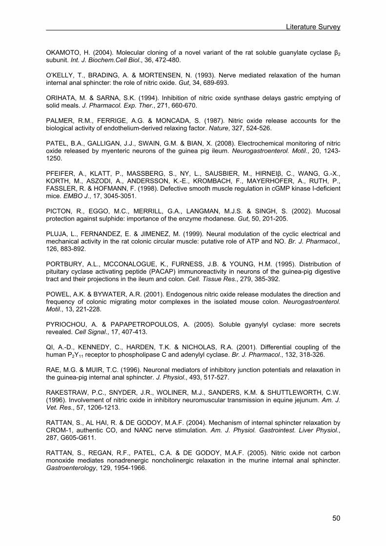
Literature Survey
50
OKAMOTO, H. (2004). Molecular cloning of a novel variant of the rat soluble guanylate cyclase β2 subunit. Int. J. Biochem.Cell Biol., 36, 472-480.
O’KELLY, T., BRADING, A. & MORTENSEN, N. (1993). Nerve mediated relaxation of the human internal anal sphincter: the role of nitric oxide. Gut, 34, 689-693.
ORIHATA, M. & SARNA, S.K. (1994). Inhibition of nitric oxide synthase delays gastric emptying of solid meals. J. Pharmacol. Exp. Ther., 271, 660-670.
PALMER, R.M., FERRIGE, A.G. & MONCADA, S. (1987). Nitric oxide release accounts for the biological activity of endothelium-derived relaxing factor. Nature, 327, 524-526.
PATEL, B.A., GALLIGAN, J.J., SWAIN, G.M. & BIAN, X. (2008). Electrochemical monitoring of nitric oxide released by myenteric neurons of the guinea pig ileum. Neurogastroenterol. Motil., 20, 1243-1250.
PFEIFER, A., KLATT, P., MASSBERG, S., NY, L., SAUSBIER, M., HIRNEIβ, C., WANG, G.-X., KORTH, M., ASZODI, A., ANDERSSON, K.-E., KROMBACH, F., MAYERHOFER, A., RUTH, P., FASSLER, R. & HOFMANN, F. (1998). Defective smooth muscle regulation in cGMP kinase I-deficient mice. EMBO J., 17, 3045-3051.
PICTON, R., EGGO, M.C., MERRILL, G.A., LANGMAN, M.J.S. & SINGH, S. (2002). Mucosal protection against sulphide: importance of the enzyme rhodanese. Gut, 50, 201-205.
PLUJA, L., FERNANDEZ, E. & JIMENEZ, M. (1999). Neural modulation of the cyclic electrical and mechanical activity in the rat colonic circular muscle: putative role of ATP and NO. Br. J. Pharmacol., 126, 883-892.
PORTBURY, A.L., MCCONALOGUE, K., FURNESS, J.B. & YOUNG, H.M. (1995). Distribution of pituitary cyclase activating peptide (PACAP) immunoreactivity in neurons of the guinea-pig digestive tract and their projections in the ileum and colon. Cell. Tissue Res., 279, 385-392.
POWEL, A.K. & BYWATER, A.R. (2001). Endogenous nitric oxide release modulates the direction and frequency of colonic migrating motor complexes in the isolated mouse colon. Neurogastroenterol. Motil., 13, 221-228.
PYRIOCHOU, A. & PAPAPETROPOULOS, A. (2005). Soluble gyanylyl cyclase: more secrets revealed. Cell Signal., 17, 407-413.
QI, A.-D., KENNEDY, C., HARDEN, T.K. & NICHOLAS, R.A. (2001). Differential coupling of the human P2Y11 receptor to phospholipase C and adenylyl cyclase. Br. J. Pharmacol., 132, 318-326.
RAE, M.G. & MUIR, T.C. (1996). Neuronal mediators of inhibitory junction potentials and relaxation in the guinea-pig internal anal sphincter. J. Physiol., 493, 517-527.
RAKESTRAW, P.C., SNYDER, J.R., WOLINER, M.J., SANDERS, K.M. & SHUTTLEWORTH, C.W. (1996). Involvement of nitric oxide in inhibitory neuromuscular transmission in equine jejunum. Am. J. Vet. Res., 57, 1206-1213.
RATTAN, S., AL HAI, R. & DE GODOY, M.A.F. (2004). Mechanism of internal sphincter relaxation by CROM-1, authentic CO, and NANC nerve stimulation. Am. J. Physiol. Gastrointest. Liver Physiol., 287, G605-G611.
RATTAN, S., REGAN, R.F., PATEL, C.A. & DE GODOY, M.A.F. (2005). Nitric oxide not carbon monoxide mediates nonadrenergic noncholinergic relaxation in the murine internal anal sphincter. Gastroenterology, 129, 1954-1966.
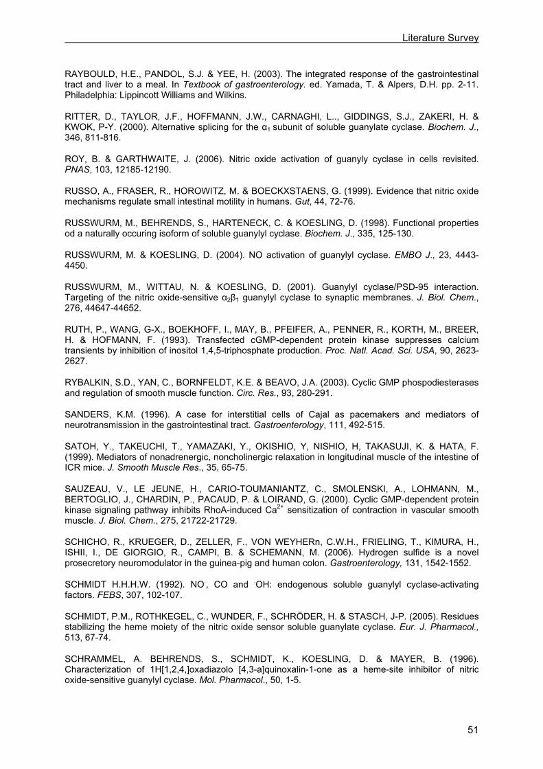
Literature Survey
51
RAYBOULD, H.E., PANDOL, S.J. & YEE, H. (2003). The integrated response of the gastrointestinal tract and liver to a meal. In Textbook of gastroenterology. ed. Yamada, T. & Alpers, D.H. pp. 2-11. Philadelphia: Lippincott Williams and Wilkins.
RITTER, D., TAYLOR, J.F., HOFFMANN, J.W., CARNAGHI, L.., GIDDINGS, S.J., ZAKERI, H. & KWOK, P-Y. (2000). Alternative splicing for the α1 subunit of soluble guanylate cyclase. Biochem. J., 346, 811-816.
ROY, B. & GARTHWAITE, J. (2006). Nitric oxide activation of guanyly cyclase in cells revisited. PNAS, 103, 12185-12190.
RUSSO, A., FRASER, R., HOROWITZ, M. & BOECKXSTAENS, G. (1999). Evidence that nitric oxide mechanisms regulate small intestinal motility in humans. Gut, 44, 72-76.
RUSSWURM, M., BEHRENDS, S., HARTENECK, C. & KOESLING, D. (1998). Functional properties od a naturally occuring isoform of soluble guanylyl cyclase. Biochem. J., 335, 125-130.
RUSSWURM, M. & KOESLING, D. (2004). NO activation of guanylyl cyclase. EMBO J., 23, 4443-4450.
RUSSWURM, M., WITTAU, N. & KOESLING, D. (2001). Guanylyl cyclase/PSD-95 interaction. Targeting of the nitric oxide-sensitive α2β1 guanylyl cyclase to synaptic membranes. J. Biol. Chem., 276, 44647-44652.
RUTH, P., WANG, G-X., BOEKHOFF, I., MAY, B., PFEIFER, A., PENNER, R., KORTH, M., BREER, H. & HOFMANN, F. (1993). Transfected cGMP-dependent protein kinase suppresses calcium transients by inhibition of inositol 1,4,5-triphosphate production. Proc. Natl. Acad. Sci. USA, 90, 2623-2627.
RYBALKIN, S.D., YAN, C., BORNFELDT, K.E. & BEAVO, J.A. (2003). Cyclic GMP phospodiesterases and regulation of smooth muscle function. Circ. Res., 93, 280-291.
SANDERS, K.M. (1996). A case for interstitial cells of Cajal as pacemakers and mediators of neurotransmission in the gastrointestinal tract. Gastroenterology, 111, 492-515.
SATOH, Y., TAKEUCHI, T., YAMAZAKI, Y., OKISHIO, Y, NISHIO, H, TAKASUJI, K. & HATA, F. (1999). Mediators of nonadrenergic, noncholinergic relaxation in longitudinal muscle of the intestine of ICR mice. J. Smooth Muscle Res., 35, 65-75.
SAUZEAU, V., LE JEUNE, H., CARIO-TOUMANIANTZ, C., SMOLENSKI, A., LOHMANN, M., BERTOGLIO, J., CHARDIN, P., PACAUD, P. & LOIRAND, G. (2000). Cyclic GMP-dependent protein kinase signaling pathway inhibits RhoA-induced Ca2+ sensitization of contraction in vascular smooth muscle. J. Biol. Chem., 275, 21722-21729.
SCHICHO, R., KRUEGER, D., ZELLER, F., VON WEYHERn, C.W.H., FRIELING, T., KIMURA, H., ISHII, I., DE GIORGIO, R., CAMPI, B. & SCHEMANN, M. (2006). Hydrogen sulfide is a novel prosecretory neuromodulator in the guinea-pig and human colon. Gastroenterology, 131, 1542-1552.
SCHMIDT H.H.H.W. (1992). NO., CO and .OH: endogenous soluble guanylyl cyclase-activating factors. FEBS, 307, 102-107.
SCHMIDT, P.M., ROTHKEGEL, C., WUNDER, F., SCHRÖDER, H. & STASCH, J-P. (2005). Residues stabilizing the heme moiety of the nitric oxide sensor soluble guanylate cyclase. Eur. J. Pharmacol., 513, 67-74.
SCHRAMMEL, A. BEHRENDS, S., SCHMIDT, K., KOESLING, D. & MAYER, B. (1996). Characterization of 1H[1,2,4,]oxadiazolo [4,3-a]quinoxalin-1-one as a heme-site inhibitor of nitric oxide-sensitive guanylyl cyclase. Mol. Pharmacol., 50, 1-5.
Literature Survey
52
SCHUBERT, R. & NELSON, M.T. (2001). Protein kinases: tuners of the BKCa channels in smooth muscle. Trends Pharmacol. Sci., 22, 505-512.
SERIO, R., ALESSANDRO, M., ZIZZO, M.G., TAMBURELLO, M.P. & MULE, F. (2003a). Neurotransmitters involved in the fast inhibitory junction potentials in mouse distal colon. Eur. J. Pharmacol., 460, 183-190.
SERIO, R., ZIZZO, M.G. & MULE, F. (2003b). Nitric oxide induces muscular relaxation via cyclic GMP-dependent and -independent mechanisms in the longitudinal muscle of the mouse duodenum. Nitric Oxide, 8, 48-52.
SHARINA, I.G., JELEN, F., BOGATENKOVA, E.P., THOMAS, A., MARTIN, E. & MURAD, F. (2008). α1 soluble guanylyl cyclase (sGC) splice forms as potential regulators of human sGC activity. J. Biol. Chem., 283, 15104-15113.
SHUTTLEWORTH, C.W.R., SWEENEY, K.M. & SANDERS, K.M. (1999). Evidence that nitric oxide acts as an neurotransmitter supplying taenia from the guinea-pig caecum. Br. J. Pharmacol., 127, 1495-1501.
SIDHU, R., SINGH, M., SAMIR, G. & CARSON, R.J. (2001). L-cysteine and sodium hydrosulphide inhibit spontaneous contractility in isolated pregnant rat uterine strips in vitro. Pharmacol. Toxicol., 88, 198-203.
SIVARAO, D.V., MASHIMO, H. & GOYAL, R.K. (2008). Pyloric sphincter dysfunction in nNOS-/- and W/WV mutant mice: animal models of gastroparesis and duodenogastric reflux. Gastroenterology, 135, 1258-1266.
SOEDIONO, P. & BURNSTOCK, G. (1994). Contribution of ATP and nitric oxide to NANC inhibitory transmission in rat pyloric sphincter. Br. J. Pharmacol., 113, 681-686.
SOMLYO, A.P. & SOMLYO, A. (2003). Ca2+ sensitivity of smooth muscle and nonmuscle myosin II: modulated by G proteins, kinases, and myosine phosphatase. Physiol.Rev., 83, 1325-1358.
SRILATHA, B., ADAIKAN, P.G. & MOORE, P.K. (2007). Hydrogen sulphide: a novel endogenous neurotransmitter facilitates erectile function. J. Sex. Med., 4, 1304-1311.
STIPANUK, M.H., & BECK, P.W. (1982). Characterization of the enzymic capacity for cysteine desulphhydration in liver and kidney of the rat. Biochem. J., 206, 267-277.
STONE, J.R. & MARLETTA, M.A. (1994). Soluble guanylate cyclase from bovine lung: activation with nitric oxide and carbon monoxide and spectral characterization of the ferrous and ferric states. Biochem., 33, 5636-5640.
SUNDLER, F., EKBLAD, E., ABSOOD, A., HAKANSON, R., KOVES, K. & ARIMURA, A. (1992). Pituitary adenylate cyclase activating peptide: a novel vasoactive intestinal peptide-like neuropeptide in the gut. Neuroscience, 46, 439-454.
SURKS, H.K., MOCHIZUKI, N., KASAI, Y., GEORGESCU, S.P., TANG, K.M., ITO, M., LINCOLN, T.M. & MENDELSOHN, M.E. (1999). Regulation of myosin phosphatase by a specific interaction with cGMP-dependent protein kinase Iα. Science, 286, 1583-1587.
SUZUKI, M., NOKIHARA, K., ANDO, E. & NARUSE, S. (1996). Immunohistochemical comparison of localization of pituitary adenylate cyclase activating polypeptide (PACAP) and vasoactive intestinal polypeptide (VIP) in the enteric nerve plexus of the guinea pig jejunum. Biomed. Pept. Proteins Nucleic. Acids., 2, 19-22.
TACK, J., DEMEDTS, I., MEULEMANS, A., SCHUURKENS, J. JANSSENS, J. (2002). Role of nitric oxide in the gastric accommodation reflex and in meal induced satiety in humans. Gut, 51, 219-224.

Literature Survey
53
TAKEUCHI, T., KISHI, M., HIRAYAMA, N., YAMAJI, M., ISHII, T., NISHIO, H., HATA, F. & TAKEWAKI, T. (1999). Tyrosine kinase involvement in apamin-sensitive inhibitory responses of rat distal colon. J. Physiol., 514, 177-188.
TAKEUCHI, T., NIIOKA, S., KISHI, M., ISHII, T., NISHIO, H., HATA, F., TAKEWAKI, T. & TAKASUJI, K. (1998). Nonadrenrgic noncholinergic relaxation mediated by nitric oxide with concomitant change in Ca2+ level in rectal circular muscle of rats. Eur. J. Pharmacol., 353, 67-74.
TANG, G., WU, L., LIANG, W. & WANG, R. (2005). Direct stimulation of KATP channels by exogenous and endogenous hydrogen sulfide in vascular smooth muscle cells. Mol. Pharmacol., 68, 1757-1764.
TEAGUE, B., ASIEDU, S. & MOORE, P.K. (2002). The smooth muscle relaxant effect of hydrogen sulphide in vitro: evidence for a physiological role to control intestinal contractility. Br. J. Pharmacol., 137, 139-145.
TENG, B., MURTHY, K.S., KUEMMERLE, J.F., GRIDER, J.R., SASE, K., MICHEL, T. & MAKHLOUF, G.M. (1998). Expression of endothelial nitric oxide synthase in human and rabbit gastrointestinal smooth muscle cells. Am. J. Physiol. Gastrointest. Liver Physiol., 275, G342-G351.
TODA, N. & HERMAN, A.G. (2005). Gastrointestinal function regulation by nitrergic efferent nerves. Pharmacol. Rev., 57, 315-338.
TONINI, M., DE GIORGIO, R., DE PONTI, F., STERNINI, C., SPELTA, V., DIONIGI, P., BARBARA, G., STANGHELLINI, V. & CORINALDESI, R. (2000). Role of nitric-oxide- and vasoactive intestinal polypeptide-containing neurons in human gastric fundus strip relaxations.Br. J. Pharmacol., 129, 12-20.
UENO, T., DUENES, J.A., ZARROUG, A.E. & SARR, M.G. (2004). Nitrergic mechanisms mediating inhibitory control of longitudinal smooth muscle contraction in mouse small intestine. J. Gastrointest. Surg., 8, 831-841.
VAN CROMBRUGGEN, K. & LEFEBVRE, R.A. (2004). Nitrergic-purinergic interactions in rat distal colon motility. Neurogastroenterol. Motil., 16, 81-98.
VAN CROMBRUGGEN, K., VAN NASSAUW, L., TIMMERMANS, J.-P. & LEFEBVRE, R.A. (2007). Inhibitory purinergic P2 receptor characterisation in rat distal colon. Neuropharmacology, 53, 257-271.
VANNUCCHI, M.-G., CORSANI, L., BANI, D. & FAUSSONE –PELLEGRINI, M.-S. (2002). Myenteric neurons and interstitial cells of Cajal of mouse colon express several nitric oxide synthase isoforms. Neurosci. Lett., 326, 191-195.
VON KÜGELGEN, I. & WETTER, A. (2000). Molecular pharmacology of P2Y-receptors. Naunyn Schmiedebergs Arch. Pharmacol., 362, 310-323.
VANNESTE, G., ROBBERECHT, P. & LEFEBVRE, R.A. (2004). Inhibitory pathways in the circular muscle of rat jejunum. Br J Pharmacol., 143, 107-118.
WANG, R. (2002). Two’s company, three’s a crowd: can H2S be the third endogenous gaseous transmitter? FASEB J., 16, 1792-1798.
WANG, R. & WU L. (1997). The chemical modification of KCa channels by carbon monoxide in vascular smooth muscle cells. J. Biol. Chem., 272, 8222-8226.
WARD, S.M. & SANDERS, K.M. (2001). Interstitial cells of Cajal: primary targets of enteric motor innervation. Anat. Rec., 262, 125-135.
WEBB, G.D., LIM, L.H., OH, V.M., YEO, S.B., CHEONG, Y.P., ALI, M., OAKLEY, R.E., LEE, C.N., WONG, P.S., CALEB, M.G., SALTO-TELLEZ, M., BHATIA, M., CHAN, E.S.Y., TAYLOR, E.A. &
Literature Survey
54
MOORE, P.K. (2008). Contractile and vasorelaxant effects of hydrogen sulfide and its biosynthesis in the human internal mammary artery. J. Pharmacol. Exp. Ther., 324, 876-882.
WEDEL, B., HUMBERT, P., HARTENECK, C., FOERSTER, J., MALKEWITZ, J., BÖHME, E., SCHULTZ, G., & KOESLING, D. (1994). Mutation of His-15 in the beta1 subunit yields a nitric oxide-insensitive form of soluble guanylate cyclase. Proc Natl Acad Sci USA, 91, 2592-2596.
WEISIGER, R.A., PINKUS, L.M. & JAKOBY, W.B. (1980). Thiol S-methyltransferase: suggested role in detoxification of intestinal hydrogen sulfide. Biochem. Pharmacol., 29, 2885-2887.
WU, X., HAYSTEAD, T.A.J., NAKAMOTO, R.K., SOMLYO, A.V. & SOMLYO, A.P. (1998). Acceleration of myosin light chain dephosphorylation and relaxation of smooth muscle by telokin. J. Biol. Chem., 273, 11362-11369.
XIE, Q.W., CHO, H.J., CALAYCAY, J., MUMFORD, R.A., SWIDEREK, K.M., LEE, T.D., DING, A., TROSO, T. & NATHAN, C. (1992). Cloning and characterization of inducible nitric oxide synthase from mouse macrophages. Science, 256, 225-228.
XUE, L., FARRUGIA, G., MILLER, S.M., FERRIS, C.D., SNYDER, S.H. & SZURSZEWSKI, J.H. (2000a). Carbon monoxide and nitric oxide as coneurotransmitters in the enteric nervous system: evidence from genomic deletion of biosynthetic enzymes. Proc Natl Acad Sci USA, 97,1851-1855.
XUE, L., FARRUGIA, G., SARR, M.G. & SZURSZEWSKI, J.H. (1999). ATP is a mediator of the fast inhibitory junction potential in human jejunal circular smooth muscle. Am. J. Physiol. Gastrointest. Liver Physiol., 276, G1373-G1379.
XUE, L., FARRUGIA, G. & SZURSZEWSKI, J.H. (2000b). Effect of exogenous ATP on canine jejunal smooth muscle. Am. J. Physiol. Gastrointest. Liver Physiol., 278, G725-G733.
YUEN, P.S., POTTER, L.R. & GARBERS, D.L. (1990). A new form of guanylyl cyclase is preferentially expressed in rat kidney. Biochem., 29, 10872-10878.
ZABEL, U., KLEINSCHNITS, C., OH, P., NEDVETSKY, P., SMOLENS, A., MÜLLER, H., KRONICH, P., KUGLER, P., WALTER, U., SCHNITZER, J.E., SCHMIDT, H.H.H.W. (2002). Calcium-dependent membrane association sensitizes soluble guanylyl cyclase to nitric oxide. Nat. Cell Biol., 4, 307-311.
ZAKHARY, R., POSS, K.D., JAFFREY, S.R., FERRIS, C.D., TONEGAWA, S. & SNYDER, S.H. (1997). Targeted gene deletion of heme oxygenase 2 reveals neural role for carbon monoxide. Proc. Natl. Acad. SCI. USA, 94, 14848-14853.
ZHAO, W. & WANG, R. (2002). H2S-induced vasorelaxation and underlying cellular and molecular mechanisms. Am. J. Physiol. Heart Circ. Physiol., 283, H474-H480.
ZHAO, W., ZHANG, H.J., LU, Y. & WANG, R. (2001). The vasorelaxant effect of H2S as a novel endogenous gaseous KATP channel opener. EMBO J., 20, 6008-6016.
ZHAO, Y., BRANDISH, P.E., BALLOU, D.P. & MARLETTA, M.A. (1999). A molecular basis for nitric oxide sensing by soluble guanylate cyclase. PNAS, 96, 14753-14758.
ZHAO, Y., SCHELVIS, J.P., BABCOCK, G.T. & MARLTTA, M.A. (1998). Identification of histidine 105 in the beta1 subunit of soluble guanylate cyclase as the heme proximal ligand. Biochem., 37, 4502-4509.
ZIZZO, M.G., MULÈ, F. & SERIO, R. (2006). Mechanisms underlying hyperpolarization evoked by P2Y receptor activation in mouse distal colon. Eur. J. Pharmacol., 544, 174-180.
ZYROMSKI, N.J., DUENES, J.A., KENDRICK, M.L., BALSIGER, B.M., FARRUGIA, G. & SARR, G.M. (2001). Mechanism mediating nitric oxide-induced inhibition in human jejunal longitudinal smooth muscle. Surgery, 130, 489-496.
Chapter II
AIMS


Aims
57
Chapter II Aims
Hormonal and neuronal control mechanisms regulate gastrointestinal (GI) motility. NO is
considered to be an important inhibitory NANC neurotransmitter in the GI tract, and hence to
play an important role in the regulation of GI smooth muscle relaxation. However, also other
inhibitory NANC neurotransmitters such as ATP, VIP/PACAP and possibly CO are reported
to be involved in smooth muscle relaxation depending upon species and region of the GI
tract.
The principal intracellular target of NO to induce relaxation is sGC. For years, it was a
paradigm to equal NO synthesis with the activation of sGC, cGMP synthesis and activation of
PKGs and PDEs. A number of observations, however, indicate that this assumption is not
correct. In the first place, there is the growing notion of the importance of sGC-independent
activities of NO, acting through nitr(osyl)ation of proteins. Previously, the latter was
considered to be “collateral damage” without specific physiological regulatory meaning, but
more recent studies point to nitr(osyl)ation as a physiological signalling mechanism for NO
(Ahern et al., 2002; Jaffrey et al., 2001). Secondly, there is the recognition that NO is not the
only activator of sGC. Indeed, also CO can activate sGC in a heme-dependent manner
(Stone & Marletta, 1994).
sGC exists in two isoforms i.e., α1β1 and α2β1, of which the α1β1-isoform predominates in the
GI tract (Mergia et al., 2003). Research on sGC has been seriously hampered by the fact
that reliable specific inhibitors with in vivo usefulness are lacking and that there are no
isoform-specific inhibitors. Using a mouse transgenic approach, our first aim was to
investigate the relative importance of the two isoforms of sGC in the NO-mediated action on
GI motility. Our group previously studied the consequences of knocking out the α1-subunit of
sGC at the level of the stomach. Results showed that sGCα1β1 plays an important role in
gastric nitrergic relaxation in vitro, but that some nitrergic relaxation is maintained via
activation of sGCα2β1, contributing to a moderate in-vivo consequence on gastric emptying
(Vanneste et al., 2007). We now investigated the consequences of knocking out the α1-
subunit of sGC in the jejunum and the distal colon, tissues with tonic as well as phasic
activity. The results of these experiments are described in Chapter III (jejunum) and Chapter IV (distal colon).
By means of sGCβ1his105phe knock in (KI) mice, where neither sGCα1β1 nor sGCα2β1 can
be activated by heme-dependent sGC activators such as NO and CO, our second aim was to
Aims
58
investigate to what extent 1) nitrergic relaxation of stomach, jejunum and colon is sGC-
dependent and 2) in vivo GI motility is influenced when heme-dependent sGC activation is
lacking. The results of this study are summarized in Chapter V.
Recently, a role for H2S as a neurotransmitter was suggested (Wang, 2002). H2S is as such
a third possible gaseous neurotransmitter (or “gasotransmitter”) besides NO and CO, with
the evidence for their role as a neurotransmitter being very strong for NO, weaker for CO and
still very limited for H2S. The effect of H2S on smooth muscle contractility and its mechanism
of action is mainly investigated in the vascular tree. In contrast to NO and CO, H2S does not
relax vascular smooth muscle through activation of sGC but mainly by activation of ATP-
dependent K+ channels. Up to date, there are only a few reports concerning the influence of
H2S on GI smooth muscle. Our third aim was therefore to investigate the influence of H2S on
contractility in mouse gastric fundus (Chapter VI) and distal colon (Chapter VII) and to
explore its mechanism of action in these two tissues.
II.1 References AHERN, G.P., KLYACHKO, V.A. & JACKSON, M.B. (2002). cGMP and S-nitrosylation : two routes for modulation of neuronal excitability by NO. Trends Neurosci., 25, 510-517.
JAFFREY, S.R., ERDJUMENT-BROMAGE, H., FERRIS, C.D., TEMPST, P. & SNYDER, S.H. (2001). Protein S-nitrosylation: a physiological signal for neuronal nitric oxide. Nat. Cell Biol., 3,193-197.
MERGIA, E., RUSSWURM, M., ZOIDL, G. & KOESLING, D. (2003). Major occurrence of the new alpha2beta1 isoform of NO-sensitive guanylyl cyclase in brain. Cell Signal., 15, 189-195.
STONE, J.R. & MARLETTA, M.A. (1994). Soluble guanylate cyclase from bovine lung: activation with nitric oxide and carbon monoxide and spectral characterization of the ferrous and ferric states. Biochem., 33, 5636-5640.
VANNESTE, G., DHAESE, I., SIPS, P., BUYS, E., BROUCKAERT, P. & LEFEBVRE, R.A. (2007). Gastric motility in soluble guanylate cyclase alpha 1 knock-out mice. J. Physiol., 584, 907-920.
WANG, R. (2002). Two’s company, three’s a crowd: can H2S be the third endogenous gaseous transmitter? FASEB J., 16, 1792-1798.

Chapter III
SMALL INTESTINAL MOTILITY IN SOLUBLE GUANYLATE CYCLASE α1 KNOCKOUT MICE
Ingeborg Dhaese 1, Gwen Vanneste 1, Patrick Sips 2,3, Emmanuel S. Buys 2,3,4, Peter Brouckaert 2,3, Romain A. Lefebvre 1
1 Heymans Institute of Pharmacology, Ghent University, Ghent, Belgium 2 Department of Molecular Biomedical Research, VIB, Ghent, Belgium 3 Department of Molecular Biology, Ghent University, Ghent, Belgium 4 Anesthesia Center for Critical Care Research, Department of Anesthesia & Critical Care, Massachusetts General Hospital, Harvard Medical School, Boston, MA 02114, USA Based on Naunyn-Schmied. Arch. Pharmacol. 2009; 379: 473-487

Role of sGC in mouse jejunum relaxation
61
Chapter III Small intestinal motility in soluble guanylate cyclase α1 knockout mice
III.1 Abstract
Nitric oxide (NO) activates soluble guanylate cyclase (sGC) to produce guanosine-3’,5’-
cyclic-monophosphate (cGMP). The aim of this study was to investigate the nitrergic
regulation of jejunal motility in sGCα1 knock-out (KO) mice.
Functional responses to nitrergic stimuli and cGMP levels in response to nitrergic stimuli
were determined in circular muscle strips. Intestinal transit was determined.
Nitrergic relaxations induced by electrical field stimulation and exogenous NO were almost
abolished in male KO strips, but only minimally reduced and sensitive to ODQ in female KO
strips. Basal cGMP levels were decreased in KO strips but NO still induced an increase in
cGMP levels. Transit was not attenuated in male nor female KO mice.
In vitro, sGCα1β1 is the most important isoform in nitrergic relaxation of jejunum but nitrergic
relaxation can also occur via sGCα2β1-activation. The latter mechanism is more pronounced
in female than in male KO mice. In vivo, no important implications on intestinal motility were
observed in male and female KO mice.

Role of sGC in mouse jejunum relaxation
62
III.2 Introduction
Nitric oxide (NO) is an important mediator in jejunal inhibitory neurotransmission as electrical
field stimulation (EFS)-induced smooth muscle relaxations are reduced in jejunum strips of
neuronal NO synthase-1 (NOS-1) knock-out (KO) mice (Xue et al., 2000). The importance of
NO in jejunal non-adrenergic, non-cholinergic (NANC) neurotransmission was further
established by reports that NOS inhibitors reduced smooth muscle relaxations induced by
EFS in longitudinal muscle of mouse and rat jejunum and circular muscle of canine, equine
and human jejunum (Niioka et al., 1997; Satoh et al., 1999; Stark et al., 1993; Rakestraw et
al., 1996; Murr et al., 1999). NO was also suggested to play a role in in-vivo small intestinal
motility as the use of NOS inhibitors revealed a delay in small intestinal transit of rats
(Karmeli et al., 1997), dogs (Chiba et al., 2002) and humans (Fraser et al., 2005).
Endogenous NO, released during EFS, activates soluble guanylate cyclase (sGC), as the
sGC inhibitor ODQ reduced the EFS-induced relaxations in longitudinal muscle of human
and mouse jejunum (Zyromski et al., 2001; Ueno et al., 2004) and circular muscle of rat
jejunum (Vanneste et al., 2004). sGC activation was also reported to play a role in in-vivo
intestinal motility as sildenafil, an inhibitor of the guanosine-3’,5’-cyclic-monophosphate
(cGMP) degrading enzyme phosphodiesterase (PDE) 5, was found to delay intestinal transit
of mice and the sGC inhibitor methylene blue blocked this inhibitory effect of sildenafil on
intestinal transit (Patil et al., 2005).
The intestinal relaxing effects of cGMP, raised by activation of sGC, are mediated by
cGMP-dependent protein kinase (cGK) type I as a cGKI inhibitor reduced relaxations evoked
by a membrane-permeable cGMP analog and a NO-donor in longitudinal muscle of rat small
intestine (Huber et al., 1998). In addition, the use of a cGKI KO model revealed that
interruption of the NO-sGC-cGMP-cGKI signaling pathway delayed small intestinal transit.
Instead of regular peristalsis, spastic contractions of long intestinal segments followed by
scarce and slow relaxations were observed (Pfeifer et al., 1998). NO has also
sGC-independent effects (Jaffrey et al., 2001); in both rat and mouse duodenum
cGMP-independent relaxant effects of NO were reported (Martins et al., 1995; Serio et al.,
2003). Also, sGC can be stimulated by other stimuli than NO (Behrends, 2003).
sGCα1β1 and sGCα2β1 are the physiologically active sGC isoforms with sGCα1β1 being the
predominant isoform in the gastrointestinal (GI) tract (Mergia et al., 2003). To further
investigate the role of sGC in nitrergic inhibition of small intestinal motility, the consequence
of knocking-out sGCα1 on in-vitro relaxation by endogenous and exogenous NO and on
in-vivo intestinal transit was now investigated. We recently reported that proximal gastric
nitrergic relaxation in these sGCα1 KO mice is clearly reduced, but some nitrergic relaxation
is maintained via activation of sGCα2β1 (Vanneste et al., 2007).
Role of sGC in mouse jejunum relaxation
63
III.3 Materials and Methods
III.3.1 Animals
sGCα1 KO mice were generated as described by Buys et al. (2008). Briefly, sGCα1 KO mice
were generated by targeting exon 6 of the sGCα1 gene which codes for an essential part of
the catalytic domain. Chimeras were generated by aggregating R1 embryonic stem cells
carrying the mutant sGCα1 allele with Swiss morula-stage embryos.
Wild-type (WT) and KO Swiss/129 mice of both sexes (WT male: 9-34 weeks, 23-52 g; KO
male: 9-37 weeks, 32-48 g; WT female: 10-34 weeks, 24-41 g; KO female: 9-34 weeks,
24-43 g) and male heterozygous Swiss/129 mice (19-25 weeks, 37-42 g) had free access to
water and commercially available chow. However, when investigating transit using the
phenol red or fluorescein-labelled dextran method (see below), food was withheld for 16
hours overnight with free access to water in a cage with a grid floor to prevent coprophagy.
All experimental procedures were approved by the Ethical Committee for Animal
Experiments from the Faculty of Medicine and Health Sciences at Ghent University.
III.3.2 Real-time quantitative reverse transcriptase-polymerase chain reaction (RT-PCR)
A fragment of jejunum starting approximately 10 cm distal to the pylorus was harvested,
rinsed in Krebs solution and transferred to RNAlater stabilization reagent (QIAGEN; The
Netherlands) until RNA was prepared using the RNeasy® Protect Mini kit (QIAGEN, The
Netherlands), according to the manufacturer's instructions. One µg of RNA was used in the
SuperScript™ First-Strand Synthesis System for RT-PCR (Invitrogen, USA) to produce copy
deoxyribonucleic acid (cDNA) using random hexameric primers. cDNA was subsequently
used for relative expression quantitation using real-time fluorescence detection. To this end,
specific primer-probe sets were synthesized for sGCα1 (detecting exon 5), α2, β1 and the
household gene hypoxanthine-guanine phosphoribosyltransferase (HPRT) for normalization
(for sequence, see Vanneste et al., 2007). The probe for α1 was directed to exon 5, which is
intact in the sGCα1 KO mice, as the latter were generated by targeting exon 6 of the sGCα1
gene. Quantitative real-time PCR was performed using the Taqman universal PCR master
mix (Applied Biosystems; CA; USA) in an ABI Prism 7700 Sequence Detector. The cycling
parameters were: 95°C for 10 min, followed by 40 cycles at 95°C for 15 s and 60°C for 1 min.
Relative changes in gene expression were determined using the comparative threshold cycle
(Ct) method described in Applied Biosystems User Bulletin #2 (1997) in which the amount of
target, normalised to an endogenous reference and relative to a calibrator is given by 2 -∆∆Ct.
In brief, the Ct, or threshold cycle, represents the PCR cycle at which an increase in reporter
Role of sGC in mouse jejunum relaxation
64
fluorescence above a baseline signal can first be detected, ∆Ct refers to the difference
between the threshold cycles of the target (sGC subunits) and the endogenous reference
(HPRT); and ∆∆Ct indicates the difference between the ∆Ct values obtained in the KO
animals to the mean ∆Ct value obtained in the calibrator group (WT mice).
III.3.3 Muscle tension experiments
III.3.3.1 Tissue preparation
Animals were killed by cervical dislocation; the GI tract was removed and put in aerated
Krebs solution (composition in mM: NaCl 118.5, KCl 4.8, KH2PO4 1.2, MgSO4 1.2, CaCl2 1.9,
NaHCO3 25.0 and glucose 10.1). A ± 5 cm long fragment of small bowel starting
approximately 10 cm distal to the pylorus was isolated and opened along the mesenteric
border. The fragment was pinned mucosa side up in physiological salt solution. The mucosa
was removed by sharp dissection under a microscope and 2 (WT and KO mice) or 4
(heterozygous mice) full-thickness muscle strips (4 × 5 mm) were cut along the circular axis.
III.3.3.2 Isometric tension recording
After a silk thread (USP 4/0) was attached to both ends, strips were mounted in 5 ml organ
baths between 2 platinum plate electrodes (6 mm apart). The organ baths contained aerated
(5% CO2 in O2) Krebs solution, maintained at 37°C. Changes in isometric tension were
measured using MLT 050/D force transducers (ADInstruments; UK) and recorded on a
PowerLab/8sp data recording system (ADInstruments; UK) with Chart software.
After an equilibration period of 30 min with flushing every 10 min at a load of 0.125 g the
length-tension relationship was determined. Muscle tissues were stretched by load
increments of 0.125 g and at each load level exposed to 0.1 µM carbachol to determine the
optimal load (Lo; the load at which maximal response to the contractile agent occurred).
Tissues were then allowed to equilibrate for 60 min at Lo with flushing every 15 min in Krebs
solution.
III.3.3.3 Protocol
In a first series, cumulative contractile responses to carbachol (1 nM – 30 µM) or
prostaglandin F2α (PGF2α; 1 nM – 3 µM) were obtained in Krebs solution without atropine
and guanethidine. Each concentration was left in contact with the tissue for 2 min. The organ
bath medium was then switched to Krebs solution containing 1 µM atropine and 4 µM
guanethidine to block cholinergic and noradrenergic responses, respectively. Ten min after
adding 300 nM PGF2α to induce contraction, relaxations were induced by application of EFS
(40V, 0.1 ms, 1-2-4-8 Hz for 10 s at 5 min interval) via the platinum plate electrodes by
Role of sGC in mouse jejunum relaxation
65
means of a Grass S88 Stimulator (Grass, W. Warwick, RI, USA), followed by the application
of exogenous NO (1-10-100 µM with an interval of at least 5 min during which the effect of a
given concentration of NO had disappeared) and finally vasoactive intestinal polypeptide
(VIP, 100 nM). Strips were washed for 30 min, and were subsequently incubated with the
sGC inhibitor 1H[1,2,4,]oxadiazolo [4,3-a]quinoxalin-1-one (ODQ; 10 µM) for 30 min. PGF2α
was then applied again and the responses to EFS, NO and VIP were studied again in the
presence of ODQ. The reproducibility of the responses to EFS, NO and VIP was evaluated
by running time-control strips in parallel, that received the solvent of ODQ (ethanol).
A second series immediately started in Krebs solution containing atropine and guanethidine.
In this series, the influence of the NOS inhibitor Nω-nitro-L-arginine methyl ester (L-NAME;
300 µM; 30 min incubation) on the responses to EFS, exogenous NO and VIP was tested.
Again, time-control strips were run in parallel to evaluate the reproducibility of the responses
to EFS, NO and VIP.
A third series was performed in the presence of Krebs solution containing 300 µM L-NAME.
Under basal conditions, contractile responses to EFS (40V, 0.1 ms, 1-2-4-8 Hz for 10 s at 2
min interval) were investigated in the absence and in the presence of atropine (1 µM; 30 min
incubation). Time-control strips were run in parallel to evaluate the reproducibility of the EFS-
induced responses.
Immediately after the experiment, tissue wet weight was determined.
All three series were performed in WT and KO mice of both sexes. However, in male
heterozygous mice, only EFS- and NO-induced responses were investigated.
III.3.3.4 Data analysis
The contractile responses to carbachol or PGF2α were measured as area under the curve
(AUC) above baseline to construct concentration-response curves. The duration of these
responses was set at 2 min (i.e., the contact time with the tissue). EC50 values were
calculated by linear interpolation.
The duration of the inhibitory responses was determined as 10 s for EFS (i.e., the length of
the stimulus train applied) and 5 min for the long acting peptide VIP. The duration of the
inhibitory responses to NO was calculated as the difference between the time of
administration and the time point, where the peaks of resuming phasic activity reached 50 %
of the amplitude of the NO-induced inhibitory response; the latter was calculated as the
difference of the mean peak level of phasic activity during the 2 min before administration of
NO and the lowest tone level during NO exposure. The inhibitory response to EFS, NO and
VIP was quantified by calculating the AUC of the registered activity during the determined
Role of sGC in mouse jejunum relaxation
66
duration, and subtracting it from the AUC of the registered activity during a corresponding
period before applying the relaxant stimulus or drug.
The duration of the contractile responses to EFS was determined as 10 s (i.e., the length of
the stimulus train applied); the contractile responses to EFS were quantified by subtracting
the AUC of the registered activity during the 10 s before applying EFS from the AUC of the
registered activity during the stimulus.
The contractile as well as the inhibitory responses are expressed as (g.s)/mg wet weight.
III.3.4 cGMP analysis
Circular jejunum muscle strips were prepared and weighed to obtain the tissue wet weight
before they were mounted as described above, under a load approximating the mean optimal
load determined in the tension experiments (0.125 g for jejunal WT and 0.25 g for KO strips).
Pre-contracted strips were snap-frozen in liquid nitrogen as such (control), or at maximal
relaxation by NO (10 µM), or upon EFS (40 V, 0.1 ms at 4 Hz for 10 s). Snap-frozen tissues
were stored at -80°C until further processing. They were pulverized by a Mikro-dismembrator
U (B-Braun Biotech International, Germany) and dissolved in cold 6 % trichloroacetic acid to
give a 10 % (w/v) homogenate. The homogenate was centrifuged at 2000 g for 15 min at 4
°C; the supernatant was recovered and washed 4 times with 5 volumes of water saturated
diethyl ether. The aqueous extract was then dried under a stream of nitrogen at 60 °C and
dissolved in a 10 or 50 times volume of assay buffer depending on the cGMP content
detected in preliminary experiments. cGMP concentrations were determined using an
enzyme immunoassay kit (EIA Biotrak System; Amersham Biosciences; UK) after acetylation
of the samples and according to the manufacturer’s instructions. The optical density was
measured with a 96-well plate reader (Biotrak II, Amersham Biosciences; UK) at 450 nm.
The tissue cGMP concentration was expressed as pmol per gram tissue wet weight.
III.3.5 Transit
III.3.5.1 Small intestinal transit (phenol red method)
As modified from de Rosalmeida et al. (2003), mice (age: 20 ± 3 weeks) were, after food was
withheld overnight, administered 250 µl of a phenol red meal (0.1 % w v-1 dissolved in water)
by gavage with a feeding needle. Sixty minutes later, mice were killed by cervical dislocation
and the stomach and small bowel were clamped at both sides. The GI tract from stomach to
colon was resected and put on the bench to measure the length of the small intestine. The
bowel was divided into 12 parts, 1 part stomach, 10 equal parts of the small bowel and 1 part
caecum. Every part was cut into small fragments and placed into 20 ml of 0.1 N NaOH in a
50 ml Falcon tube. This mixture was homogenised for approximately 30 s and allowed to
Role of sGC in mouse jejunum relaxation
67
stand for 20 min at room temperature. Ten ml of supernatant was placed into a 15 ml Falcon
tube and centrifuged for 10 min at 2800 rpm. Proteins in 5 ml supernatant were precipitated
with 0.5 ml of 20 % (w/v) trichloroacetic acid and the solution was centrifuged for 20 min at
2800 rpm. 0.5 ml of supernatant was added to 0.667 ml of 0.5 N NaOH and the absorbance
of 300 µl of this mixture was spectrophotometrically determined at 540 nm in a Biotrak II
plate reader (Amersham Biosciences; UK).
The amount of phenol red recovered in each segment of the small bowel was then
expressed as % of total recovery (stomach not included) and the geometric centre of the
phenol red migration pattern was then calculated as (Σ (% phenol red per segment x
segment number))/100. Phenol red recovery throughout the GI tract was determined as the
amount of phenol red recovered in all 12 segments, expressed as % of the amount of phenol
red administered.
III.3.5.2 Intestinal transit (fluorescein-labelled dextran method)
Mice (age: 20 ± 3 weeks) were, after food was withheld overnight, administered 200 µl of
non-absorbable fluorescein-labelled dextran (FD70; 70 kDa, 2.5 % w v-1 dissolved in water)
by gavage with a feeding needle. Sixty minutes later, mice were killed by cervical dislocation.
For a full description of the technical details of this method, we refer to De Backer et al.
(2008). Briefly, the entire GI tract was excised and the mesenterium was removed. The GI
tract was then pinned down in a custom-made Petri dish filled with Krebs solution.
Immediately after, FD70 was visualized using the Syngene Geneflash system (Syngene,
Cambridge, UK). Two full-field images- one in normal illumination mode and another in
fluorescent mode- were taken and matched for analysis. The fluorescent intensity throughout
the intestinal tract was calculated and data were expressed as the percentage of
fluorescence intensity per segment (sb, small bowel segments 1-10; caecum; col, colon
segments 1-2). The geometric centre was calculated as (Σ (% FD70 per segment x segment
number))/100.
III.3.5.3 Whole gut transit (carmine method)
As adapted from Friebe et al. (2007), mice (age: 20 ± 3 weeks) were administered 200 µl
carmine (6 % w v-1 dissolved in 0.5 % methylcellulose) by gavage with a feeding needle.
Mice were then returned to individual cages, without food deprivation. The time taken for
excretion of the first red coloured faeces was determined at 30 minute intervals.
III.3.6 Statistics
All results are expressed as means ± S.E.M. n refers to tissues obtained from different
animals unless otherwise indicated (n =…, out of … animals). Comparison between KO and

Role of sGC in mouse jejunum relaxation
68
WT tissues or between parallel tissues of either WT or KO was done with an unpaired
Student t-test. Comparison within tissues of either WT or KO was done by a paired Student t-
test. When relaxant responses in time-control strips showed a significant decline when
studied a second time, the change in relaxant response by an interfering substance in the
parallel tissue was compared to the spontaneous change in the control strips by an unpaired
Student t-test. When more than 2 groups of tissues had to be compared, one-way analysis of
variance (ANOVA) followed by a Bonferroni corrected t-test was applied. A P-value less than
or equal to 0.05 was considered to be statistically significant (GRAPHPAD, San Diego, CA,
USA).
III.3.7 Drugs Used
Atropine, carmine, guanethidine, L-NAME, phenol red, PGF2α, VIP (all from Sigma-Aldrich,
USA), carbachol (Fluka AG, Switzerland), ODQ (Tocris Cookson, UK), fluorescein-labelled
dextran (70 kDa, FD70) (Invitrogen, Belgium). All drugs were dissolved in de-ionized water
except carmine and ODQ. Carmine was dissolved in 0.5 % methylcellulose and ODQ was
dissolved in 100 % ethanol. Saturated NO solution was prepared from gas (Air Liquide,
Belgium) as described by Kelm and Schrader (1990).
III.4 Results
All experiments were performed in WT and KO mice of both sexes because gender-related
differences in the cardiovascular phenotype were observed with male but not female sGCα1
KO mice developing hypertension (Buys et al., 2008). In addition, the responses to EFS and
NO were investigated in male heterozygous mice. No gross morphological GI differences
between WT, heterozygous and KO animals were observed.
III.4.1 Real-time quantitative RT-PCR
The relative levels of α1, α2 and β1 subunit mRNA in male KO tissues versus male WT
tissues were 3.5 ± 1.6, 1.9 ± 0.6 and 2.8 ± 1.4, respectively (n = 4); in female KO versus
female WT tissues, these levels were 4.0 ± 2.4, 1.7 ± 1.4 and 2.9 ± 2.4, respectively (n = 4).
In male as well as female mice, the levels in KO tissues did not significantly differ from those
in WT tissues.

Role of sGC in mouse jejunum relaxation
69
III.4.2 Muscle tension experiments
III.4.2.1 General observations in WT and KO mice
The length of the strips (measured after determination of optimal load) and the tissue wet
weight (measured immediately after the experiment) were not different between WT and KO
in both male and female mice; Lo was increased in both male and female KO compared to
WT strips (Table III.1). ODQ or L-NAME did not influence basal or PGF2α-induced tone.
Table III.1: Optimal load, length and wet weight of smooth muscle strips. MALE FEMALE
WT KO WT KO
Optimal load (g)
Length (mm)
Wet weight (mg)
0.16 ± 0.01
4.9 ± 0.2
0.66 ± 0.04
0.22 ± 0.01 (***)
5.0 ± 0.2
0.75 ± 0.03
0.16 ± 0.01
5.1 ± 0.2
0.69 ± 0.05
0.25 ± 0.02 (***)
5.0 ± 0.2
0.81 ± 0.05
Values are mean ± S.E.M. of n = 64-67 out of 25-29 animals (male), n = 47-52 out of 24-26 animals (female);
*** P < 0.001: unpaired student t-test (KO vs. WT)
III.4.2.2 Contractile responses to carbachol and PGF2α in WT and KO mice
Contractile responses to carbachol and PGF2α did not significantly differ in male KO mice
compared to male WT mice (Table III.2). In female mice, the EC50 value of the contractile
responses to carbachol was significantly lower in KO mice compared to WT mice while the
Emax for carbachol and Emax and EC50 for PGF2α were not significantly different (Table III.2).
Table III.2: Emax and EC50 of the contractions to carbachol and PGF2α.
MALE FEMALE
WT KO WT KO
Carbachol Emax ((g.s)/mg wet weight)
EC50 (nM)
46.0 ± 8.1
101 ± 20
65.6 ± 19.2
58 ± 6
48.2 ± 4.5
66 ± 7
38.2 ± 5.5
27 ± 5 (***)
PGF2α Emax ((g.s)/mg wet weight)
EC50 (nM)
31.7 ± 6.9
28 ± 7
23.9 ± 5.4
50 ± 14
16.3 ± 3.4
86 ± 41
26.9 ± 6.3
17 ± 4
Values are mean ± S.E.M. of n = 8-9 experiments; *** P < 0.001: unpaired student t-test (KO vs. WT)

Role of sGC in mouse jejunum relaxation
70
III.4.2.3 Contractile responses to electrical field stimulation in WT and KO mice
When studied under basal conditions and in the presence of L-NAME, EFS at 1-8 Hz
induced concentration-dependent contractions. In male KO strips, the EFS-evoked
contractions were reduced when compared to the WT strips; no differences between WT and
KO strips were observed in female mice (Fig. III.1A). After application of atropine, the EFS-
induced contractions were nearly abolished in WT and KO strips of male and female mice
(Fig. III.1B).
Figure III.1 Frequency-response curves of contractions induced by electrical field stimulation (EFS, 40 V, 0.1 ms, 1-8 Hz, 10 s
trains) in jejunum circular muscle strips of male (left) and female (right) wild-type (WT) and knock-out (KO) mice
(A). In B, responses before and after incubation with atropine (1 µM) are shown. The experiments were performed
in the continuous presence of L-NAME (300 µM). Data are expressed as (g.s)/mg wet weight.
A) Values are means ± S.E.M. of n = 14-18 strips out of 7-9 animals; * P < 0.05: unpaired Student t-test (KO vs.
WT).
B) Values are means ± S.E.M. of n = 7-9 strips; * * P < 0.01, * * * P< 0.001: paired Student t-test (WT: after vs.
before) or ° P < 0.05, ° ° P < 0.01, ° ° ° P < 0.001: paired Student t-test (KO: after vs. before).
III.4.2.4 Inhibitory responses to electrical field stimulation in WT and KO mice
PGF2α (300 nM) induced a tonic contractile response with superimposed phasic activity; the
response stabilised after a few min of contact with PGF2α (Fig. III.2). The contractile
response to PGF2α was not significantly different between WT and KO strips and between
male and female strips.
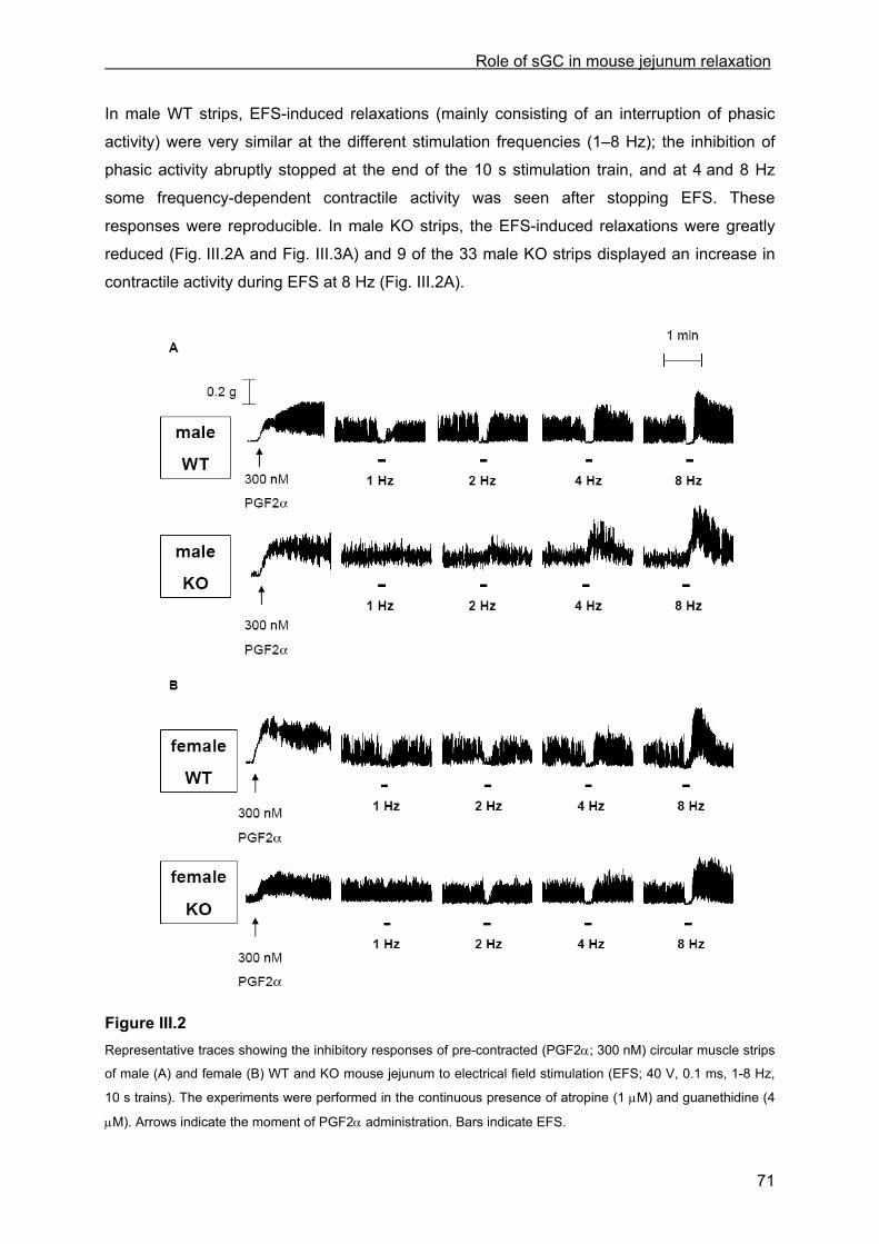
Role of sGC in mouse jejunum relaxation
71
In male WT strips, EFS-induced relaxations (mainly consisting of an interruption of phasic
activity) were very similar at the different stimulation frequencies (1–8 Hz); the inhibition of
phasic activity abruptly stopped at the end of the 10 s stimulation train, and at 4 and 8 Hz
some frequency-dependent contractile activity was seen after stopping EFS. These
responses were reproducible. In male KO strips, the EFS-induced relaxations were greatly
reduced (Fig. III.2A and Fig. III.3A) and 9 of the 33 male KO strips displayed an increase in
contractile activity during EFS at 8 Hz (Fig. III.2A).
Figure III.2 Representative traces showing the inhibitory responses of pre-contracted (PGF2α; 300 nM) circular muscle strips
of male (A) and female (B) WT and KO mouse jejunum to electrical field stimulation (EFS; 40 V, 0.1 ms, 1-8 Hz,
10 s trains). The experiments were performed in the continuous presence of atropine (1 µM) and guanethidine (4
µM). Arrows indicate the moment of PGF2α administration. Bars indicate EFS.
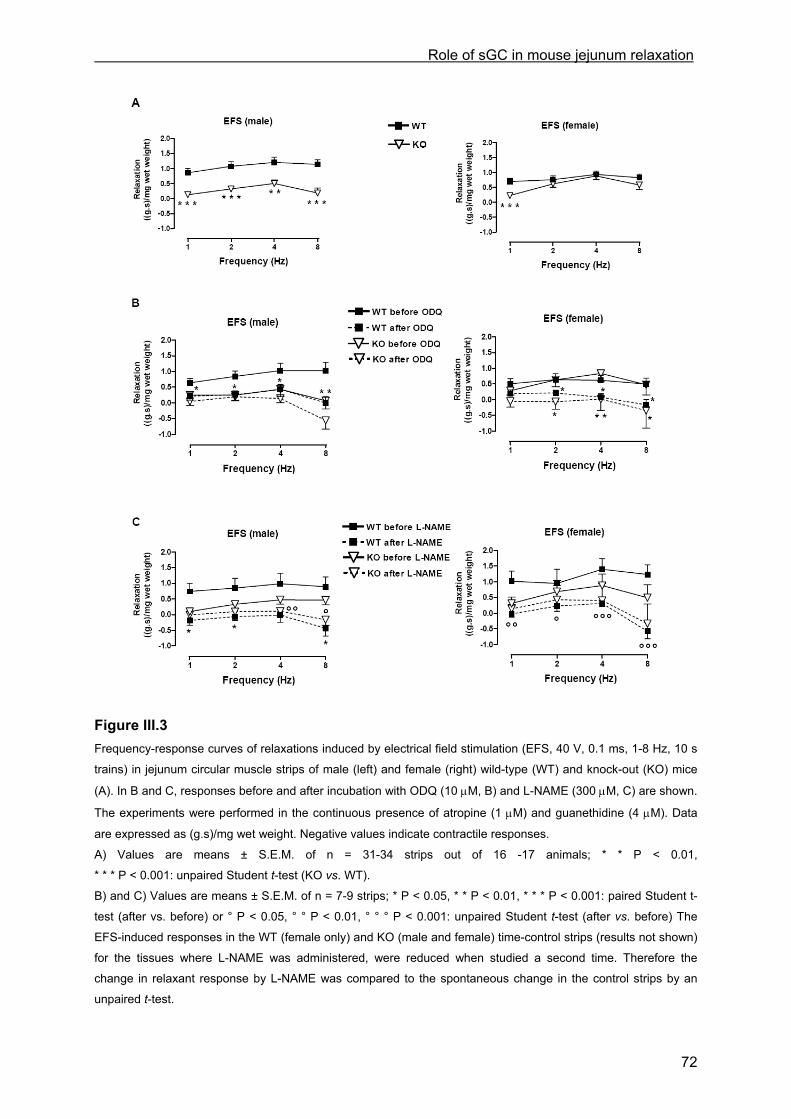
Role of sGC in mouse jejunum relaxation
72
Figure III.3 Frequency-response curves of relaxations induced by electrical field stimulation (EFS, 40 V, 0.1 ms, 1-8 Hz, 10 s
trains) in jejunum circular muscle strips of male (left) and female (right) wild-type (WT) and knock-out (KO) mice
(A). In B and C, responses before and after incubation with ODQ (10 µM, B) and L-NAME (300 µM, C) are shown.
The experiments were performed in the continuous presence of atropine (1 µM) and guanethidine (4 µM). Data
are expressed as (g.s)/mg wet weight. Negative values indicate contractile responses.
A) Values are means ± S.E.M. of n = 31-34 strips out of 16 -17 animals; * * P < 0.01,
* * * P < 0.001: unpaired Student t-test (KO vs. WT).
B) and C) Values are means ± S.E.M. of n = 7-9 strips; * P < 0.05, * * P < 0.01, * * * P < 0.001: paired Student t-
test (after vs. before) or ° P < 0.05, ° ° P < 0.01, ° ° ° P < 0.001: unpaired Student t-test (after vs. before) The
EFS-induced responses in the WT (female only) and KO (male and female) time-control strips (results not shown)
for the tissues where L-NAME was administered, were reduced when studied a second time. Therefore the
change in relaxant response by L-NAME was compared to the spontaneous change in the control strips by an
unpaired t-test.
Role of sGC in mouse jejunum relaxation
73
In male WT strips, the EFS-evoked responses were reduced at 1, 2 and 4 Hz and abolished
at 8 Hz by ODQ (Fig. III.3B), while L-NAME abolished the EFS-evoked responses at all
stimulation frequencies; small contractions occurred upon EFS after application of L-NAME
(Fig. III.3C). In male KO strips, the small relaxations to EFS were significantly reduced at
4 Hz and abolished at 8 Hz by L-NAME (Fig. III.3C); the reduction of the EFS-induced
relaxation by ODQ at 4 Hz and the abolishment at 8 Hz did not reach significance (Fig.
III.3B).
In female WT strips, EFS evoked similar reproducible relaxations as in male WT tissues. The
response by EFS at 1 Hz was significantly reduced in female KO strips as compared to
female WT strips but the responses by EFS at 2-8 Hz were not changed (Fig. III.2B and Fig.
III.3A). In female WT strips, application of ODQ reduced the EFS-induced relaxations at 2
and 4 Hz and abolished them at 8 Hz; in female KO strips, ODQ abolished the EFS-induced
relaxations at 2, 4 and 8 Hz (Fig. III.3B). Application of L-NAME decreased the EFS-induced
relaxations at 2 and 4 Hz and abolished them at 1 and 8 Hz in female WT strips; in female
KO strips, the reduction of the EFS-induced relaxation by L-NAME at 1, 2 and 4 Hz and the
abolishment at 8 Hz did not reach significance (Fig. III.3C).
III.4.2.5 Inhibitory responses to exogenously applied NO in WT and KO mice
In male WT strips, application of exogenous NO interrupted phasic activity; the duration of
the response was concentration-dependent. In male KO strips, the relaxant responses to NO
were almost abolished in comparison to the relaxation obtained in WT strips (Fig. III.4A
and Fig. III.5A). In male WT strips, ODQ reduced the NO-induced relaxations (Fig. III.5B) but
application of L-NAME increased the responses to 1 and 100 µM NO (Fig. III.5C). The small
relaxations to exogenous NO in male KO strips were not influenced by L-NAME (Fig. III.5C)
but abolished by ODQ (Fig. III.5B).
Female WT strips showed similar NO-induced responses as in male WT strips, but in female
KO strips, NO-induced relaxations were only moderately reduced compared to female WT
strips (Fig. III.4B and Fig. III.5A). In female WT strips, the NO-induced relaxations were not
influenced by L-NAME (Fig. III.5C) but were reduced by ODQ, reaching significance at 1 and
10 µM NO (Fig. III.5B). In female KO strips, relaxations induced by NO were reduced by
ODQ again reaching significance at 1 and 10 µM NO (Fig. III.5B); the responses were nearly
not influenced by L-NAME (Fig. III.5C).
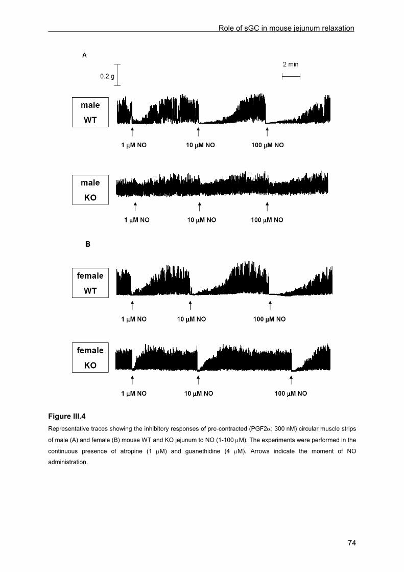
Role of sGC in mouse jejunum relaxation
74
Figure III.4 Representative traces showing the inhibitory responses of pre-contracted (PGF2α; 300 nM) circular muscle strips
of male (A) and female (B) mouse WT and KO jejunum to NO (1-100 µM). The experiments were performed in the
continuous presence of atropine (1 µM) and guanethidine (4 µM). Arrows indicate the moment of NO
administration.
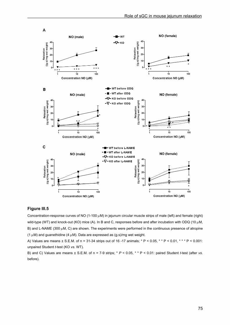
Role of sGC in mouse jejunum relaxation
75
Figure III.5 Concentration-response curves of NO (1-100 µM) in jejunum circular muscle strips of male (left) and female (right)
wild-type (WT) and knock-out (KO) mice (A). In B and C, responses before and after incubation with ODQ (10 µM,
B) and L-NAME (300 µM, C) are shown. The experiments were performed in the continuous presence of atropine
(1 µM) and guanethidine (4 µM). Data are expressed as (g.s)/mg wet weight.
A) Values are means ± S.E.M. of n = 31-34 strips out of 16 -17 animals; * P < 0.05, * * P < 0.01, * * * P < 0.001:
unpaired Student t-test (KO vs. WT).
B) and C) Values are means ± S.E.M. of n = 7-9 strips; * P < 0.05, * * P < 0.01: paired Student t-test (after vs.
before).
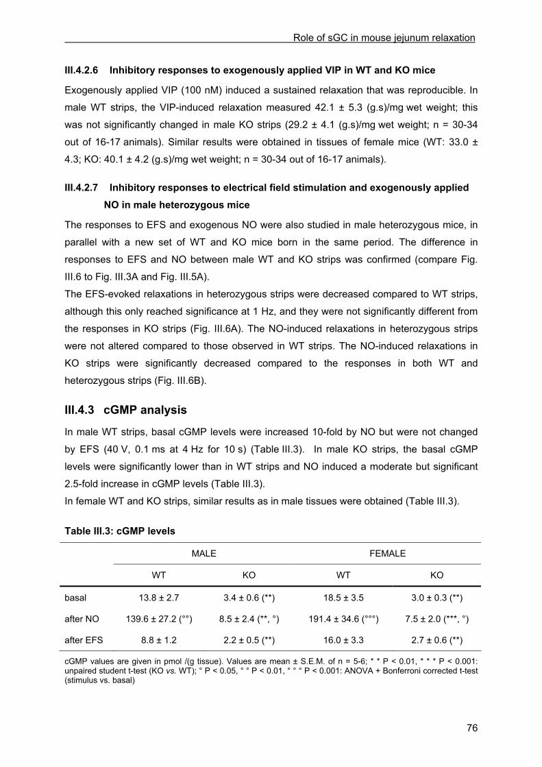
Role of sGC in mouse jejunum relaxation
76
III.4.2.6 Inhibitory responses to exogenously applied VIP in WT and KO mice
Exogenously applied VIP (100 nM) induced a sustained relaxation that was reproducible. In
male WT strips, the VIP-induced relaxation measured 42.1 ± 5.3 (g.s)/mg wet weight; this
was not significantly changed in male KO strips (29.2 ± 4.1 (g.s)/mg wet weight; n = 30-34
out of 16-17 animals). Similar results were obtained in tissues of female mice (WT: 33.0 ±
4.3; KO: 40.1 ± 4.2 (g.s)/mg wet weight; n = 30-34 out of 16-17 animals).
III.4.2.7 Inhibitory responses to electrical field stimulation and exogenously applied NO in male heterozygous mice
The responses to EFS and exogenous NO were also studied in male heterozygous mice, in
parallel with a new set of WT and KO mice born in the same period. The difference in
responses to EFS and NO between male WT and KO strips was confirmed (compare Fig.
III.6 to Fig. III.3A and Fig. III.5A).
The EFS-evoked relaxations in heterozygous strips were decreased compared to WT strips,
although this only reached significance at 1 Hz, and they were not significantly different from
the responses in KO strips (Fig. III.6A). The NO-induced relaxations in heterozygous strips
were not altered compared to those observed in WT strips. The NO-induced relaxations in
KO strips were significantly decreased compared to the responses in both WT and
heterozygous strips (Fig. III.6B).
III.4.3 cGMP analysis
In male WT strips, basal cGMP levels were increased 10-fold by NO but were not changed
by EFS (40 V, 0.1 ms at 4 Hz for 10 s) (Table III.3). In male KO strips, the basal cGMP
levels were significantly lower than in WT strips and NO induced a moderate but significant
2.5-fold increase in cGMP levels (Table III.3).
In female WT and KO strips, similar results as in male tissues were obtained (Table III.3).
Table III.3: cGMP levels
MALE FEMALE
WT KO WT KO
basal
after NO
after EFS
13.8 ± 2.7
139.6 ± 27.2 (°°)
8.8 ± 1.2
3.4 ± 0.6 (**)
8.5 ± 2.4 (**, °)
2.2 ± 0.5 (**)
18.5 ± 3.5
191.4 ± 34.6 (°°°)
16.0 ± 3.3
3.0 ± 0.3 (**)
7.5 ± 2.0 (***, °)
2.7 ± 0.6 (**)
cGMP values are given in pmol /(g tissue). Values are mean ± S.E.M. of n = 5-6; * * P < 0.01, * * * P < 0.001: unpaired student t-test (KO vs. WT); ° P < 0.05, ° ° P < 0.01, ° ° ° P < 0.001: ANOVA + Bonferroni corrected t-test (stimulus vs. basal)
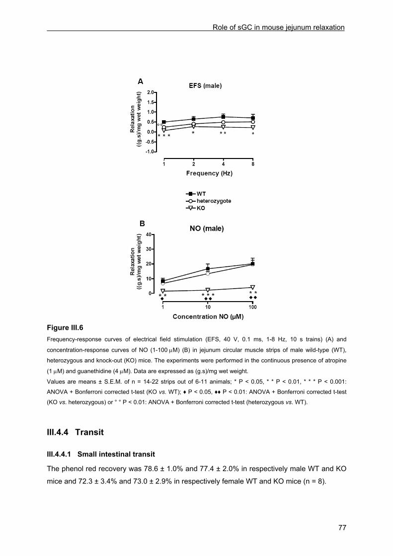
Role of sGC in mouse jejunum relaxation
77
Figure III.6 Frequency-response curves of electrical field stimulation (EFS, 40 V, 0.1 ms, 1-8 Hz, 10 s trains) (A) and
concentration-response curves of NO (1-100 µM) (B) in jejunum circular muscle strips of male wild-type (WT),
heterozygous and knock-out (KO) mice. The experiments were performed in the continuous presence of atropine
(1 µM) and guanethidine (4 µM). Data are expressed as (g.s)/mg wet weight.
Values are means ± S.E.M. of n = 14-22 strips out of 6-11 animals; * P < 0.05, * * P < 0.01, * * * P < 0.001:
ANOVA + Bonferroni corrected t-test (KO vs. WT); ♦ P < 0.05, ♦♦ P < 0.01: ANOVA + Bonferroni corrected t-test
(KO vs. heterozygous) or ° ° P < 0.01: ANOVA + Bonferroni corrected t-test (heterozygous vs. WT).
III.4.4 Transit
III.4.4.1 Small intestinal transit
The phenol red recovery was 78.6 ± 1.0% and 77.4 ± 2.0% in respectively male WT and KO
mice and 72.3 ± 3.4% and 73.0 ± 2.9% in respectively female WT and KO mice (n = 8).
Role of sGC in mouse jejunum relaxation
78
The small bowel length was significantly increased in male KO mice compared to male WT
(from 30.9 ± 0.9 cm to 36.0 ± 0.9 cm, n = 8, P < 0.01). Sixty min after gavage, small intestinal
transit tended to be delayed, but the shift in geometric centre did not reach significance
(geometric centre: male WT: 6.0 ± 0.5; male KO: 4.6 ± 0.6, n = 8) (Fig. III.7A).
The small bowel length was not different between female WT and KO mice (WT: 29.9 ± 1.5;
KO: 33.6 ± 1.2 cm; n = 8). Small intestinal transit was delayed in female KO mice (Fig. III.7A)
as also manifest from the significant decrease in geometric centre (WT: 5.8 ± 0.3; KO: 4.8 ±
0.3, n = 8, P < 0.05) (Fig. III.7A).
III.4.4.2 Intestinal transit
The distribution of fluorescein-labelled dextran in the intestinal tract, sixty min after gavage,
was not different between male WT and KO mice (geometric centre: male WT: 6.1 ± 1.0;
male KO: 6.7 ± 0.4, n = 7-8) and female WT and KO mice (geometric centre: female WT:
6.6 ± 0.3; female KO: 6.5 ± 0.7, n = 7-8) (Fig. III.7B).
III.4.4.3 Whole gut transit time
The whole gut transit time, determined as the time taken for excretion of the first red coloured
faeces after gavage with carmine, was not significantly different between male WT and KO
mice (mean value: male WT: 159 ± 16 min; male KO: 188 ± 21, n = 7-8) nor between female
WT and KO mice (mean value: female WT: 153 ± 16 min; female KO: 154 ± 19, n = 8-9)
(Fig. III.7C).
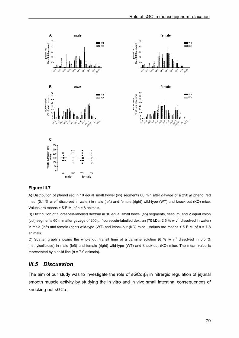
Role of sGC in mouse jejunum relaxation
79
Figure III.7 A) Distribution of phenol red in 10 equal small bowel (sb) segments 60 min after gavage of a 250 µl phenol red
meal (0.1 % w v-1 dissolved in water) in male (left) and female (right) wild-type (WT) and knock-out (KO) mice.
Values are means ± S.E.M. of n = 8 animals.
B) Distribution of fluorescein-labelled dextran in 10 equal small bowel (sb) segments, caecum, and 2 equal colon
(col) segments 60 min after gavage of 200 µl fluorescein-labelled dextran (70 kDa; 2.5 % w v-1 dissolved in water)
in male (left) and female (right) wild-type (WT) and knock-out (KO) mice. Values are means ± S.E.M. of n = 7-8
animals.
C) Scatter graph showing the whole gut transit time of a carmine solution (6 % w v-1 dissolved in 0.5 %
methylcellulose) in male (left) and female (right) wild-type (WT) and knock-out (KO) mice. The mean value is
represented by a solid line (n = 7-9 animals).
III.5 Discussion
The aim of our study was to investigate the role of sGCα1β1 in nitrergic regulation of jejunal
smooth muscle activity by studying the in vitro and in vivo small intestinal consequences of
knocking-out sGCα1.
Role of sGC in mouse jejunum relaxation
80
III.5.1 Role of sGC in male mice
The observation that L-NAME abolished the EFS-induced responses indicates that NO is the
most important inhibitory neurotransmitter in WT jejunal circular muscle strips of male Swiss
129 mice. Additionally, both endogenous NO released upon EFS and exogenous NO
induces relaxation through activation of sGC as the sGC inhibitor ODQ clearly reduced these
nitrergic relaxations. Furthermore, exogenous NO induced a 10-fold increase in cGMP levels
in jejunum strips of male WT mice further supporting the importance of sGC activation in
nitrergic relaxation. In contrast, EFS at 4 Hz for 10 s, although inducing an ODQ-sensitive
relaxation, did not increase cGMP levels. This corresponds to what we reported previously in
the gastric fundus of male Swiss 129 WT mice (Vanneste et al., 2007). Together these data
suggest that the subcellular localization, and recuperability by the assay used, of cGMP
induced by endogenous NO released from nerve varicosities must differ from that raised by
exogenous NO diffusing into the smooth muscle strip (Vanneste et al., 2007). Surprisingly,
application of L-NAME induced an increase of the NO-evoked responses; we have no
clearcut explanation for this observation. Indeed, increased responses to NO-donors upon
removal of endogenous NO have been reported in vascular tissues and were attributed to
desensitization of sGC by tonically released NO (Busse et al., 1989; Luscher et al., 1989;
Shirasaki and Su, 1985). However, if NO were tonically released, L-NAME can be expected
to increase basal tone by abolishing this tonic relaxant drive; this was not the case.
In male sGCα1 KO mice responses evoked by endogenous and exogenous NO were almost
absent, and the increase in cGMP levels by exogenous NO was clearly reduced indicating
that the major sGC isoform involved in nitrergic relaxation of male jejunum is sGCα1β1. Still,
the small NO and EFS-induced responses were sensitive to ODQ, and exogenous NO
induced a 2.5-fold increase in cGMP levels in sGCα1 KO mice illustrating that some response
is present due to activation of sGCα2β1; ODQ indeed equally inhibits sGCα2β1 as sGCα1β1
(Russwurm et al., 1998).
No compensatory mechanisms in cAMP-induced relaxation nor in contractile mechanisms
were observed in sGCα1 KO strips in view of the maintained responses to VIP, carbachol
and PGF2α. This corresponds to observations in cGKI KO mice (Ny et al., 2000; Bonnevier
et al., 2004). As the responses to carbachol are maintained in sGCα1 KO strips, this
suggests that the decreased EFS-induced cholinergic contractions observed in male sGCα1
KO strips are not related to an alternation at the level of the muscarinic receptors located on
the smooth muscle cells or at their transduction mechanism. The Lo of the KO strips was
increased. This might correlate with the partial disappearance of the nitrergic inhibitory
pathway so that the strips require more load to relax and to reach their optimal length-tension
relationship. The weight of WT and KO strips was the same showing that an adaptive

Role of sGC in mouse jejunum relaxation
81
increase in muscle mass in response to the decrease in nitrergic inhibitory influence is
unlikely. Additionally, in gastric fundus strips of KO mice, we did not observe an increase in
Lo (Vanneste et al., 2007).
III.5.2 Role of sGC in female mice
The results in female WT mice were very similar as in male WT mice except that in female
mice L-NAME did not significantly increase the NO-induced response. We have no
explanation for these differences. Most strikingly however, jejunum strips of female sGCα1
KO mice still produced a clear-cut relaxation upon EFS and application of exogenous NO.
The clearly remaining responses to EFS (2-8 Hz) and to exogenous NO were sensitive to
ODQ, suggesting that sGCα2β1 plays a more important role in nitrergic relaxation of jejunum
strips of female sGCα1 KO mice than in male sGCα1 KO mice. Exogenous NO indeed still
induced a significant 2.5-fold increase in cGMP levels in female KO tissue, which was
unexpectedly not more pronounced than the 2.5-fold increase in cGMP levels in male KO
tissues by exogenous NO in view of the better maintained relaxation to exogenous NO in
female KO tissues. As mentioned above, the maintained NO-induced relaxation in female
sGCα1 KO tissues results from the activation of sGCα2β1. It is possible that the subcellular
localization of cGMP raised by the membrane-associated sGCα2β1 (Russwurm et al., 2001)
differs from that raised by the cytosolic sGCα1β1 and that this has consequences on the
recuperability of cGMP by the assay used. Buys et al. (2008) also observed a gender-specific alteration in sGCα1 KO mice when
studying the cardiovascular consequences of the sGCα1 KO mice as male KO mice
developed testosteron-dependent hypertension while female mice did not. As we found no
up-regulation of sGCα2 and sGCβ1 mRNA in jejunum tissue of male as well as female sGCα1
KO mice, a gender-differential compensatory mechanism via an increase in the sGCα2β1
isoform in the female mice seems not present. Whatever mechanism contributes to the less
pronounced decrease in nitrergic relaxation in female sGCα1 KO mice compared to male
sGCα1 KO mice, it does not occur in the whole GI tract as we previously reported that the
influence of knocking-out sGCα1 at the level of the gastric fundus and the colon is the same
in male and female KO mice (Vanneste et al., 2007; Dhaese et al., 2008).
III.5.3 The sGCα1 KO model
Buys et al. (2008) describe the generation of the sGCα1 KO mice by targeted deletion of the
sixth exon which is essential for the enzymatic activity. As a result, sGCα1 KO mice express
an enzymatically inactive mutant sGCα1β1 protein. Mice heterozygous for this deletion
express the active as well as the mutant inactive sGCα1β1 protein. NO-induced increases in
sGC enzyme activity in aortic, lung and left ventricular tissue were intermediate in
Role of sGC in mouse jejunum relaxation
82
heterozygous mice to those measured in WT and sGCα1 KO mice, suggesting a gene dose
effect and indicating that the mutant sGCα1β1 protein does not function as a dominant
negative (Buys et al., 2008). Still, no differences were observed in systolic blood pressure
between male WT and heterozygous mice.
In the jejunum, EFS-induced relaxations were reduced in male heterozygous mice compared
to male WT mice, whereas NO-induced relaxations were not altered in male heterozygous
mice. In the light of the observations made by Buys et al. (2008), we do not believe that the
reduced EFS-evoked relaxations in the heterozygous mice are due to the non-specific effects
of the mutant sGCα1β1 protein. This observation could possibly be related to the fact that
endogenous NO is released from nerve varicosities and thus enters the smooth muscle cell
more locally whereas exogenous NO is diffusing from the surrounding extracellular solution
and therefore enters the smooth muscle over the whole surface. As sGCα1β1 is a cytosolic
enzyme, it is possible that endogenous NO is thus not as effective as exogenous NO in
reaching and thus activating the more limited amount of active sGCα1β1 in heterozygous
mice, resulting in a decreased response to EFS but a maintained response to exogenous
NO.
III.5.4 Implication on intestinal transit
In agreement with observations in cGKI KO mice (Ny et al., 2000), no morphological
abnormalities at the level of the small bowel were observed in sGCα1 KO mice although the
small bowel length was slightly increased in male KO compared to male WT mice; this
increase was not observed in female mice.
Inhibition of nitrergic relaxation at the level of the small intestine is expected to delay
intestinal transit as manifest from studies with NOS-inhibitors in different species illustrating
the essential role of NO in intestinal peristalsis (Chiba et al., 2002; Fraser et al., 2005;
Karmeli et al., 1997). Also cGKI KO mice showed a delay in intestinal transit (Pfeifer et al.,
1998). In male and female sGCα1 KO mice, we found that the small intestinal transit of a
phenol red solution tended to be delayed. However, the intestinal transit of a fluorescein-
labelled dextran solution and the whole gut transit time of a carmine solution were not
delayed in female nor in male sGCα1 KO mice. Taken together, these results suggest that
knocking out sGCα1 has no important in vivo effects on intestinal motility. This seems
plausible for the female sGCα1 KO mice, as also in vitro NO-induced relaxations were only
moderately influenced, indicating that in female mice sGCα2β1 is able to sustain the NO-
induced motility effects. However, in male sGCα1 KO mice, an important decrease in in vitro
NO-induced relaxations was observed. Still, this is not accompanied by a decrease in in vivo
intestinal motility. This might be explained by the concomitant reduction of cholinergic nerve
activity in male sGCα1 KO mice as demonstrated by the decrease in EFS-induced cholinergic

Role of sGC in mouse jejunum relaxation
83
contractions. Indeed, the propagation of peristaltic activity is the result of an interplay
between ascending excitatory and descending inhibitory enteric pathways (Waterman et al.,
1994). The effects of decreased inhibitory (nitrergic) as well as decreased excitatory
(cholinergic) responses in sGCα1 KO mice might neutralize each other, leaving the balance
between these two players of peristaltic activity unaltered.
In conclusion, in vitro, sGCα1β1 is the most important mediator in nitrergic relaxation of
jejunum but nitrergic relaxation can also occur via activation of sGCα2β1. This latter
mechanism is more pronounced in female than in male sGCα1 KO mice. In vivo, no important
implications of knocking out sGCα1 on intestinal motility were observed in male and female
KO mice.
III.6 References Behrends, S., 2003. Drugs that activate specific nitric oxide sensitive guanylyl cyclase isoforms
independent of nitric oxide release. Curr. Med. Chem. 10, 291-301.
Bonnevier, J., Fassler, R., Somlyo, A.P., Somlyo, A.V., Arner, A., 2004. Modulation of Ca2+ sensitivity
by cyclic nucleotides in smooth muscle from protein kinase G-deficient mice. J. Biol. Chem. 279, 5146-
5151.
Busse, R., Pohl, U., Mulsch, A., Bassenge, E., 1989. Modulation of the vasodilator action of SIN-1 by
the endothelium. J. Cardiovasc. Pharmacol. 14, S81-S85.
Buys, E., Sips, P., Vermeersch, P., Raher, M., Rogge, E., Ichinose, F., Dewerchin, M., Bloch, K.D.,
Janssens, S., Brouckaert, P., 2008. Gender-specific hypertension and responsiveness to nitric oxide
in sGCa1 knockout mice. Cardiovasc. Res. 79, 179-186.
Chiba, T., Bharucha, A.E., Thomforde, G.M., Kost, L.J., Phillips, S.F., 2002. Model of rapid
gastrointestinal transit in dogs: effects of muscarinic antagonists and a nitric oxide synthase inhibitor.
Neurogastroenterol. Motil. 14, 535-541.
De Backer, O., Blanckaert, B., Leybaert, L., Lefebvre, R.A., 2008. A novel method for the evaluation of
intestinal transit and contractility in mice using fluorescence imaging and spatiotemporal mapping.
Neurogastroenterol. Motil. 20, 700-707.
de Rosalmeida, M.C., Saraiva, L.D., da Graça, J.R., Ivo, B.B., da Nobrega, M.V., Gondim, F.A., Rola,
F.H., dos Santos, A.A., 2003. Sildenafil, a phosphodiesterase-5 inhibitor, delays gastric emptying and
gastrointestinal transit of liquid in awake rats. Dig. Dis. Sci. 48, 2064-2068.

Role of sGC in mouse jejunum relaxation
84
Dhaese, I., Vanneste, G., Sips, P., Buys, E., Brouckaert, P., Lefebvre, R.A., 2008. Involvement of
soluble guanylate cyclase alpha (1) and alpha(2), and SK(Ca) channels in NANC relaxation of mouse
distal colon. Eur. J. Pharmacol. 589, 251-259.
Fraser, R., Vozzo, R., Di Matteo, A.C., Boeckxstaens, G., Adachi, K., Dent, J., Tournadre, J.P., 2005.
Endogenous nitric oxide modulates small intestinal nutrient transit and activity in healthy adult
humans. Scand. J. Gastroenterol. 40, 1290-1295.
Friebe, A., Mergia, E., Dangel, O., Lange, A., Koesling, D., 2007. Fatal gastrointestinal obstruction and
hypertension in mice lacking nitric oxide-sensitive guanylyl cyclase. Proc. Natl. Acad. Sci. USA 104,
7699-7704.
Huber, A., Trudrung, P., Storr, M., Franck, H., Schusdziarra, V., Ruth, P., Allescher, H.D., 1998.
Protein kinase G expression in the small intestine and functional importance for smooth muscle
relaxation. Am. J. Physiol. 275, G629-G637.
Jaffrey, S.R., Erdjument-Bromage, H., Ferris, C.D., Tempst, P., Snyder, S.H., 2001. Protein S-
nitrosylation: a physiological signal for neuronal nitric oxide. Nat. Cell. Biol. 3, 193-197.
Karmeli, F., Stalnikowicz, R., Rachmilewitz, D., 1997. Effect of colchicine and bisacodyl on rat
intestinal transit and nitric oxide synthase activity. Scand. J. Gastroenterol. 32, 791-796.
Kelm, M., Schrader, J., 1990. Control of coronary vascular tone by nitric oxide. Circ. Res. 66, 1561-
1575.
Luscher, T.F., Richard, V., Yang, Z.H., 1989. Interaction between endothelium-derived nitric oxide and
SIN-1 in human and porcine blood vessels. J. Cardiovasc. Pharmacol. 14, S76-S80.
Martins, S.L., De Oliveira, R.B., Ballejo, G., 1995. Rat duodenum nitrergic-induced relaxations are
cGMP-independent and apamin-sensitive. Eur. J. Pharmacol. 284, 265-270.
Mergia, E., Russwurm, M., Zoidl, G., Koesling, D., 2003. Major occurrence of the new alpha2beta1
isoform of NO-sensitive guanylyl cyclase in brain. Cell. Signal. 15, 189-195.
Murr, M.M., Balsiger, B.M., Farrugia, G., Sarr, M.G., 1999. Role of nitric oxide, vasoactive intestinal
polypeptide, and ATP in inhibitory neurotransmission in human jejunum. J. Surg. Res. 84, 8-12.
Niioka, S., Takeuchi, T., Kishi, M., Ishii, T., Nishio, H., Takewaki, T., Hata, F., 1997. Nonadrenergic,
noncholinergic relaxation in longitudinal muscle of rat jejunum. Jpn. J. Pharmacol. 73, 155-161.
Ny, L., Pfeifer, A., Aszodi, A., Ahmad, M., Alm, P., Hedlund, P., Fassler, R., Andersson, K.E., 2000.
Impaired relaxation of stomach smooth muscle in mice lacking cyclic GMP-dependent protein kinase I.
Br. J. Pharmacol. 129, 395-401.
Role of sGC in mouse jejunum relaxation
85
Patil, C.S., Singh, V.P., Jain, N.K., Kulkarni, S.K., 2005. Inhibitory effect of sildenafil on gastrointestinal
smooth muscle: role of NO-cGMP transduction pathway. Indian J. Exp. Biol. 43, 167-171.
Pfeifer, A., Klatt, P., Massberg, S., Ny, L., Sausbier, M., Hirneiss, C., Wang, G.X., Korth, M., Aszodi,
A., Andersson, K.E., Krombach, F., Mayerhofer, A., Ruth, P., Fassler, R., Hofmann, F., 1998.
Defective smooth muscle regulation in cGMP kinase I-deficient mice. EMBO J. 17, 3045-3051.
Rakestraw, P.C., Snyder, J.R., Woliner, M.J., Sanders, K.M., Shuttleworth, C.W., 1996. Involvement of
nitric oxide in inhibitory neuromuscular transmission in equine jejunum. Am. J. Vet. Res. 57, 1206-
1213.
Russwurm, M., Behrends, S., Harteneck, C., Koesling, D., 1998. Functional properties of a naturally
occurring isoform of soluble guanylyl cyclase. Biochem. J. 335, 125-130.
Russwurm, M., Wittau, N., Koesling, D., 2001. Guanylyl cyclase/PSD-95 interaction: Targeting of the
nitric-sensitive α2β1 guanylyl cyclase to synaptic membranes. J. Biol. Chem. 276, 44647-44652.
Satoh, Y., Takeuchi, T., Yamazaki, Y., Okishio, Y., Nishio, H., Takatsuji, K., Hata, F., 1999. Mediators
of nonadrenergic, noncholinergic relaxation in longitudinal muscle of the intestine of ICR mice. J.
Smooth Muscle Res. 35, 65-75.
Serio, R., Zizzo, M.G., Mule, F., 2003. Nitric oxide induces muscular relaxation via cyclic GMP-
dependent and -independent mechanisms in the longitudinal muscle of the mouse duodenum. Nitric
Oxide 8, 48-52.
Shirasaki, Y., Su, C., 1985. Endothelium removal augments vasodilation by sodium nitroprusside and
sodium nitrite. Eur. J. Pharmacol. 114, 93-96.
Stark, M.E., Bauer ,A.J., Sarr, M.G., Szurszewski, J.H., 1993. Nitric oxide mediates inhibitory nerve
input in human and canine jejunum. Gastroenterology 104, 398-409.
Ueno, T., Duenes, J.A., Zarroug, A.E., Sarr, M.G., 2004. Nitrergic mechanisms mediating inhibitory
control of longitudinal smooth muscle contraction in mouse small intestine. J. Gastrointest. Surg. 8,
831-841.
Vanneste, G., Dhaese, I., Sips, P., Buys, E., Brouckaert, P., Lefebvre, R.A., 2007. Gastric motility in
soluble guanylate cyclase alpha 1 knock-out mice. J. Physiol. 584, 907-920.
Vanneste, G., Robberecht, P., Lefebvre, R.A., 2004. Inhibitory pathways in the circular muscle of rat
jejunum. Br. J. Pharmacol. 143, 107-118.
Waterman, S.A., Tonini, M., Costa, M., 1994. The role of ascending excitatory and descending
inhibitory pathways in peristalsis in the isolated guinea-pig small intestine. J. Physiol. 481, 223-232.
Role of sGC in mouse jejunum relaxation
86
Xue, L., Farrugia, G., Miller, S.M., Ferris, C.D., Snyder, S.H., Szurszewski, J.H., 2000. Carbon
monoxide and nitric oxide as coneurotransmitters in the enteric nervous system: evidence from
genomic deletion of biosynthetic enzymes. Proc. Natl. Acad. Sci .USA 97, 1851-1855.
Zyromski, N.J., Duenes, J.A., Kendrick, M.L., Balsiger, B.M., Farrugia, G., Sarr, M.G., 2001.
Mechanism mediating nitric oxide-induced inhibition in human jejunal longitudinal smooth muscle.
Surgery 130, 489-496.
Chapter IV
INVOLVEMENT OF SOLUBLE GUANYLATE CYCLASE α1 AND α2, AND SKCa CHANNELS IN NANC RELAXATION OF MOUSE DISTAL COLON
Ingeborg Dhaese 1, Gwen Vanneste 1, Patrick Sips 2,3, Emmanuel Buys 2,3,4, Peter Brouckaert 2,3, Romain A. Lefebvre 1
1 Heymans Institute of Pharmacology, Ghent University, Ghent, Belgium 2 Department of Molecular Biomedical Research, VIB, Ghent, Belgium 3 Department of Molecular Biology, Ghent University, Ghent, Belgium 4 Anesthesia Center for Critical Care Research, Department of Anesthesia & Critical Care, Massachusetts General Hospital, Harvard Medical School, Boston, MA 02114, USA Based on Eur. J. Pharmacol. 2008; 589: 251-259

Role of sGC in mouse distal colon relaxation
89
Chapter IV Involvement of soluble guanylate cyclase α1 and α2, and SKCa channels in NANC relaxation of mouse distal colon
IV.1 Abstract
In distal colon, both nitric oxide (NO) and ATP are involved in non-adrenergic non-cholinergic
(NANC) inhibitory neurotransmission. The role of the soluble guanylate cyclase (sGC)
isoforms α1β1 and α2β1, and of the small conductance Ca2+-dependent K+ channels (SKCa
channels) in the relaxation of distal colon by exogenous NO and by NANC nerve stimulation
was investigated, comparing wild type (WT) and sGCα1 knockout (KO) mice. In WT strips,
the relaxation induced by electrical field stimulation (EFS) at 1 Hz but not at 2-8 Hz was
significantly reduced by the NO-synthase inhibitor L-NAME or the sGC inhibitor ODQ. In
sGCα1 KO strips, the EFS-induced relaxation at 1 Hz was significantly reduced and no longer
influenced by L-NAME or ODQ. The SKCa channel blocker apamin alone had no inhibitory
effect on EFS-induced relaxation, but combined with ODQ or L-NAME, apamin inhibited the
relaxation induced by EFS at 2-8 Hz in WT strips and at 8 Hz in sGCα1 KO strips. Relaxation
by exogenous NO was significantly attenuated in sGCα1 KO strips, but could still be reduced
further by ODQ. Basal cGMP levels were lower in sGCα1 KO strips but NO still significantly
increased cGMP levels versus basal.
In conclusion, in the absence of sGCα1β1, exogenous NO is able to partially act through
sGCα2β1. NO, acting via sGCα1β1, is the principal neurotransmitter in EFS-evoked responses
at 1 Hz. At higher stimulation frequencies, NO, acting at sGCα1β1 and/or sGCα2β1, functions
together with another transmitter, probably ATP acting via SKCa channels, with some degree
of redundancy.

Role of sGC in mouse distal colon relaxation
90
IV.2 Introduction
Nitric oxide (NO), synthesized from L-arginine by neuronal NO synthase (nNOS, NOS-1), is
considered to play a major role as non-adrenergic, non-cholinergic (NANC) neurotransmitter
in the gastrointestinal (GI) tract. In colon however, NO is not the sole neurotransmitter as
also ATP was reported to play a role in inhibitory neurotransmission of mouse (Serio et al.,
2003a), rat (Pluja et al., 1999), hamster (El Mahmoudy et al., 2006) and human colon (Benko
et al., 2007; Boeckxstaens et al., 1993; Keef et al., 1993). ATP is suggested to induce
localized Ca2+ release via a mechanism involving P2Y receptors, phospholipase C (PLC) and
inositol 1,4,5-trisphosphate (IP3) receptors. The resulting increase in Ca2+ near the plasma
membrane (Ca2+ puffs) leads to activation of small conductance Ca2+-dependent K+ channels
(SKCa channels) and consequently hyperpolarization (Bayguinov et al., 2000).
The predominant target of the GI NANC neurotransmitter NO in smooth muscle cells is
soluble guanylate cyclase (sGC). Activation of sGC results in increased intracellular cGMP
levels, which consequently activates cGMP-dependent protein kinase (PKG, cGK), leading to
relaxation by lowering the intracellular Ca2+ concentration and by desensitization of the
contractile apparatus to Ca2+ (Lucas et al., 2000; Toda and Herman, 2005). sGC is a
heterodimeric hemoprotein that is composed of a larger α and a smaller β subunit (Harteneck
et al., 1991), both necessary for catalytic activity (Harteneck et al., 1990). For each subunit,
two isoforms (α1/α2 and β1/ β2) have been identified (Harteneck and al., 1991). The sGCα1β1
and the sGCα2β1 heterodimers seem to be the only physiologically active isoforms (Mergia et
al., 2003; Russwurm et al., 1998). Both isoforms are functionally indistinguishable: no
differences in kinetic properties and sensitivity towards NO between the 2 isoforms were
found (Russwurm et al., 1998). In all other tissues than brain, including ileum and colon, the
sGCα1β1 heterodimer is the predominating isoform (Mergia and al., 2003). In brain, the
sGCα2β1 isoform has been reported to be associated with PSD-95. As a consequence of this
interaction, the sGCα2β1 isoform is recruited to the membrane fraction. This suggests that, at
least in brain, the sGCα2β1 isoform works as a sensor for the NO formed by the PSD-95-
associated neuronal NO synthase (Russwurm et al., 2001).
Endogenous NO also has been shown to induce sGC-independent relaxation via the
activation of SKCa channels in mouse (Serio et al., 2003b) and rat duodenum (Martins et al.,
1995) and in rat colon (Van Crombruggen and Lefebvre, 2004).
Recently, sGCα1 knockout (KO) mice were developed, lacking exon 6 of the sGCα1 gene,
coding for an essential part in the catalytical domain (Buys et al., 2008). We previously
reported that sGCα1β1 plays an important role in nitrergic relaxation of gastric smooth muscle
cells, but some nitrergic relaxation can occur in sGCα1 KO mice via sGCα2β1 activation. As a
result, knocking out sGCα1 only has a moderate in-vivo consequence on gastric emptying
Role of sGC in mouse distal colon relaxation
91
(Vanneste et al., 2007). In the present study, the role of the two physiologically active sGC
isoforms and the SKCa channels in relaxation of distal colon by exogenous NO and by
endogenous NANC neurotransmitters was investigated using circular muscle strips of distal
colon isolated from sGCα1 KO mice.
IV.3 Materials and Methods
IV.3.1 Animals
sGCα1 KO mice were generated as described by Buys et al. (2008). Briefly, sGCα1 KO mice
were generated by targeting exon 6 of the sGCα1 gene which codes for an essential part of
the catalytical domain. Chimeras were generated by aggregating R1 embryonic stem cells
carrying the mutant sGCα1 allele with Swiss morula-stage embryos.
sGCα1 KO Swiss/129 mice and wildtype (WT) littermates of both sexes (male: n = 48-50, 8-
37 weeks, 20-50 g; female: n = 28-29, 8-34 weeks, 22-43 g) had free access to water and
commercially available chow (ssniff Spezialdiäten GmbH). All experimental procedures were
approved by the Ethical Committee for Animal Experiments from the Faculty of Medicine and
Health Sciences at Ghent University.
IV.3.2 Muscle tension experiments
IV.3.2.1 Tissue preparation and tension recording
Animals were sacrificed by cervical dislocation. The distal colon was isolated as an
approximately four centimetre segment, taken above the pelvic brim.
This segment was then opened along the mesenteric border and pinned mucosa side up in
Krebs solution (composition in mM: NaCl 118.5, KCl 4.8, KH2PO4 1.2, MgSO4 1.2, CaCl2 1.9,
NaHCO3 25.0 and glucose 10.1). The mucosa was removed by sharp dissection under a
microscope and 2 full-thickness muscle strips (3 × 5 mm) were cut along the circular axis.
After a silk thread (USP 4/0) was attached to both ends of the muscle strips, strips were
mounted in 5 ml organ baths between 2 platinum electrodes (7 mm apart). The organ baths
contained aerated (5% CO2 in O2) Krebs solution maintained at 37°C.
Changes in isometric tension were measured using MLT 050/D force transducers
(ADInstruments) and recorded on a PowerLab/8sp data recording system (ADInstruments)
with Chart software.
Role of sGC in mouse distal colon relaxation
92
IV.3.2.2 Protocol
After an equilibration period of 30 min with refreshing of the Krebs solution every 10 min at a
load of 0.25 g, the length-tension relationship was determined. To determine the optimal
load (Lo; the load at which maximal response to the contractile agent occurred), muscle strips
were stretched by load increments of 0.25 g and at each load level exposed to 1 µM
carbachol. Strips were then allowed to equilibrate for 1 h at Lo with flushing every 10 min in
Krebs solution.
Cumulative contractile responses to carbachol (1 nM – 30 µM) and prostaglandin F2α
(PGF2α) (1 nM – 3 µM) were obtained in separate strips in Krebs solution without atropine
and guanethidine; concentrations were increased at 2 min intervals. All other experiments
were performed after switching to Krebs solution containing 1 µM atropine and 4 µM
guanethidine to block cholinergic and noradrenergic responses respectively (NANC
conditions).
Strips were then pre-contracted with 3 µM PGF2α. 15 Min after its administration, when the
contractile response was surely stabilized, relaxations were induced at 5 min interval by
application of electrical field stimulation (EFS; 40V, 0.1 ms, 1-8 Hz for 30 s) via the platinum
plate electrodes by means of a Hugo Sachs Stimulator I type 215/I, followed by the
application of exogenous NO (1 µM-100 µM) and vasoactive intestinal polypeptide (VIP, 100
nM); these relaxant stimuli were always applied in this order. Strips were then washed and
equilibrated at Lo for 30 min with refreshing of the Krebs solution every 10 min; the NOS
inhibitor Nω-nitro-L-arginine methyl ester (L-NAME; 300 µM) or the sGC inhibitor
1H[1,2,4,]oxadiazolo[4,3-a]quinoxalin-1-one (ODQ; 10 µM) was then incubated for 30 min.
Strips were re-contracted with 3 µM PGF2α and the responses to EFS, NO and VIP were
studied again in the presence of L-NAME or ODQ. The concentrations of ODQ (10 µM) and
L-NAME (300 µM) have been shown to fully inhibit purified sGC (Garthwaite et al., 1995) and
NO-synthases (Heinzel et al., 1992). The responses to EFS and NO were also studied, in
male mice only, in the presence of ODQ and of ODQ plus the SKCa channel blocker apamin
(500 nM) (administered 30 min before a 3rd cycle of PGF2α-EFS-NO). The influence of
apamin and apamin plus ODQ, apamin and apamin plus L-NAME, and L-NAME and L-
NAME plus ODQ was tested in the same way on EFS- and NO-induced relaxation. The
reproducibility of the responses to EFS, NO and VIP were evaluated by running time-control
vehicle treated strips in parallel. The concentration of apamin used has been shown to fully
inhibit SK1, SK2 and SK3 channels expressed in Xenopus oocytes (Grunnet et al., 2001).
These 3 SK channel types are indeed expressed in murine colon (Ro et al., 2001).
In a separate series, the influence of ODQ was also studied versus the relaxing effect of 8-
bromoguanosine 3',5' cyclic monophosphate (8-Br-cGMP; 100 µM).
Role of sGC in mouse distal colon relaxation
93
At the end of each experiment, the wet weight of the muscle tissue between the two silk
thread knots was measured (mg wet weight; see data analysis).
IV.3.2.3 Data analysis
To measure the contractile responses to carbachol and PGF2α, the area under the curve
(AUC) above baseline was determined. The mean phasic activity induced by PGF2α was
determined as the difference between the mean peak level and the mean trough level of the
phasic component of the response to PGF2α. To measure inhibitory responses, the AUC for a
given response was determined and subtracted from the AUC of a corresponding period just
before applying the relaxing drug or stimulus, yielding the area above the curve for the
inhibitory response. Rebound contractions occurring after EFS-induced inhibitory responses
were measured as amplitude above the mean peak level of phasic activity during the 2 min
before EFS.
The duration of the inhibitory responses was determined as 30 s for EFS (i.e. the length of
the stimulus train applied), as the response immediately disappeared upon ending the 30 s
stimulation train. VIP and 8-Br-cGMP induced a sustained response and the duration was
fixed at 5 min for VIP and 10 min for 8-Br-cGMP. NO abolished phasic activity with a
concentration-dependent duration, after which phasic activity progressively reoccurred. The
duration of the inhibitory responses to NO was therefore determined as the time necessary
for phasic activity to regain 50 % of the interval between the mean peak level of phasic
activity during the 2 min before administration of NO and the minimum tone level during the
NO response. This calculation was performed during the 1st cycle of PGF2α-EFS-NO and the
determined duration was further used for the NO-induced responses in the 2nd and 3rd cycle
of PGF2α-EFS-NO. The responses are expressed as (g.s)/mg wet weight, except for the
mean phasic activity induced by PGF2α and the rebound contractions (g/mg wet weight).
IV.3.3 cGMP analysis
From each mouse, 3 circular muscle strips of distal colon were prepared analogous to the
strips prepared for the muscle tension experiments. The muscle strips were weighed to
obtain the tissue wet weight and were mounted as described above, under the mean optimal
load determined in the tension experiments (0.5 g). PGF2α pre-contracted strips were snap
frozen in liquid nitrogen without inducing relaxation (basal), or at maximal relaxation by NO
(10 µM) or upon EFS (40 V, 0.1 ms, 1 Hz for 30 s) via platinum plate electrodes by means of
a Grass S88 Stimulator (Grass, W. Warwick, RI, USA).
Snap frozen tissues were stored at – 80 °C until further processing. They were then
pulverized by a Mikro-dismembrator U (B-Braun Biotech International, Germany) and
dissolved in cold 6 % trichloroacetic acid to give a 10 % (w/v) homogenate. The homogenate
Role of sGC in mouse distal colon relaxation
94
was centrifuged at 2000 x g for 15 min at 4 °C; the supernatant was recovered and washed
four times with five volumes of water saturated diethyl ether. The aqueous extract was then
dried under a stream of nitrogen at 60 °C and dissolved in a 10 or 50 times volume of assay
buffer depending on the cGMP content detected in preliminary experiments. cGMP
concentrations were determined using an enzyme immunoassay kit (EIA Biotrak System;
Amersham Biosciences; UK) after acetylation of the samples and according to the
manufacturer’s instructions. The optimal density (OD) was measured with a 96-well plate
reader (Biotrak II, Amersham Biosciences) at 450 nm. Tissue cGMP concentration was
expressed as pmol per g tissue wet weight.
IV.3.4 Western blot
After removal of the mucosa, colonic muscle tissues were homogenized in denaturing cell
extraction buffer (Invitrogen), containing protease inhibiting cocktail (Roche), subjected to
NuPAGE electrophoresis on Novex 4-12% Bis-Tris gels (Invitrogen) and subsequently
transferred to nitrocellulose membranes (Amersham Bioscience). Blots were incubated with
the following antibodies: rabbit polyclonal antibody specific for sGCα1 (dilution 1:2500),
sGCα2 (dilution 1:200), sGCβ1 (dilution 1:500) and β-tubulin (dilution 1:500) (Abcam). After
incubation with anti-rabbit IgG HRP-linked antibody (Cell signalling Technology), blots were
developed by SuperSignal West Femto Maximum Sensitivity Substrate (Pierce) and
quantification of the bands was done by densitometry (ImageJ).
IV.3.5 Statistics
All results are expressed as means ± S.E.M. n refers to tissues obtained from different
animals unless indicated otherwise. Comparison between sGCα1 KO and WT tissues was
done with an unpaired Student’s t-test. Comparison within tissues of either WT or sGCα1 KO
was done by a paired Student’s t-test; when more than 2 sets of results within the same
tissues had to be compared, repeated measures ANOVA followed by a Bonferroni corrected
t-test was applied. P < 0.05 was considered to be statistically significant (GRAPHPAD, San
Diego, CA, USA).
IV.3.6 Drugs used
The following drugs were used: apamin (obtained from Alomone Labs), atropine sulphate, 8-
bromoguanosine 3',5' cyclic monophosphate sodium salt, guanethidine sulphate, L-NAME,
prostaglandin F2α tris salt, VIP (all obtained from Sigma-Aldrich), carbachol (from Fluka
AG), ODQ (from Tocris Cookson). All drugs were dissolved in de-ionized water except
ODQ, which was dissolved in 100 % ethanol. Saturated NO solution was prepared from gas
(Air Liquide, Belgium) as described by Kelm and Schrader (1990).
Role of sGC in mouse distal colon relaxation
95
IV.4 Results
The series of experiments where the influence of L-NAME or ODQ was tested on relaxation
by EFS, NO and VIP, were performed in mice of both sexes. However, no systematic
differences between sexes were found in distal colon strips. Therefore, results are presented
for male mice and further experiments investigating the effect of apamin alone or in
combination with ODQ or L-NAME were carried out in male mice only. No gross
morphological GI differences between WT and sGCα1 KO animals were observed.
IV.4.1 Western blot
The sGCα1 subunit was not detected in sGCα1 KO distal colon tissues, whereas this subunit
was clearly present in the WT tissues (Fig. IV.1A). The relative expression versus β-tubulin of
the sGCα2 subunit was not significantly up-regulated in the sGCα1 KO compared to the WT
as shown in Fig. IV.1B (0.35 ± 0.02 in the sGCα1 KO tissues vs. 0.32 ± 0.04 in the WT
tissues, n = 5). The sGCβ1 subunit was however significantly lower in the sGCα1 KO versus
the WT (0.44 ± 0.02 in the sGCα1 KO tissues vs. 0.64 ± 0.06 in the WT tissues, n = 5; Fig.
IV.1B).
IV.4.2 General observations of the distal colon muscle strips
In the muscle tension experiments, the length of the strips, measured after equilibration and
determination of optimal load, and the tissue wet weight, measured immediately after the
experiment, were similar in WT and sGCα1 KO mice. The length and weight of the strips was
respectively 5.4 ± 0.2 mm and 0.69 ± 0.06 mg in WT (n = 30 strips from 20 animals) and 5.0
± 0.2 mm and 0.89 ± 0.11 mg in sGCα1 KO tissues (n = 28 strips from 16 animals).
WT strips as well as sGCα1 KO strips showed no or only moderate phasic activity during their
equilibration at optimal load. The total basal contractile activity, measured during 5 min
before the first administration of PGF2α, was similar in sGCα1 KO strips and WT strips
(204.34 ± 25.54 g.s/mg wet weight in sGCα1 KO vs. 254.52 ± 55.20 g.s/mg wet weight in
WT, n = 28-30 strips from 16-20 animals).
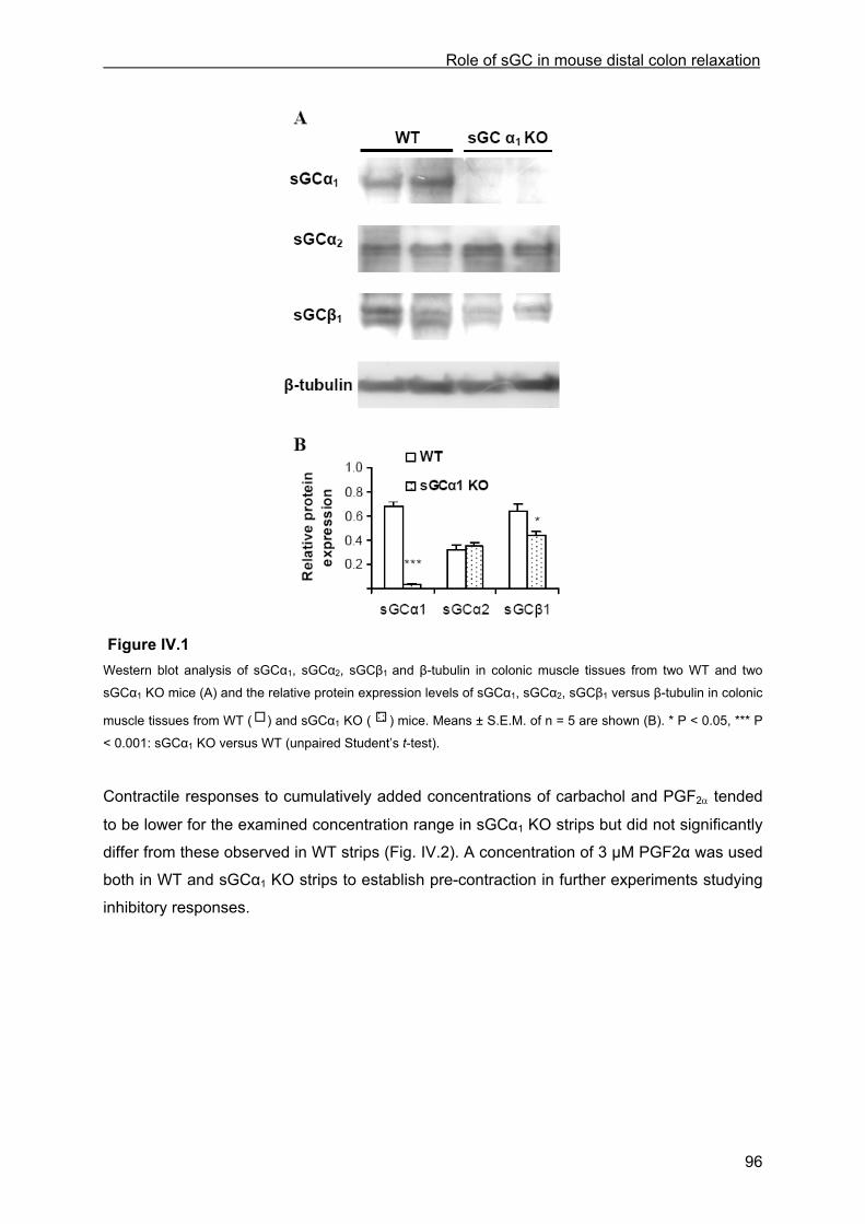
Role of sGC in mouse distal colon relaxation
96
Figure IV.1 Western blot analysis of sGCα1, sGCα2, sGCβ1 and β-tubulin in colonic muscle tissues from two WT and two
sGCα1 KO mice (A) and the relative protein expression levels of sGCα1, sGCα2, sGCβ1 versus β-tubulin in colonic
muscle tissues from WT ( ) and sGCα1 KO ( ) mice. Means ± S.E.M. of n = 5 are shown (B). * P < 0.05, *** P
< 0.001: sGCα1 KO versus WT (unpaired Student’s t-test).
Contractile responses to cumulatively added concentrations of carbachol and PGF2α tended
to be lower for the examined concentration range in sGCα1 KO strips but did not significantly
differ from these observed in WT strips (Fig. IV.2). A concentration of 3 µM PGF2α was used
both in WT and sGCα1 KO strips to establish pre-contraction in further experiments studying
inhibitory responses.

Role of sGC in mouse distal colon relaxation
97
Figure IV.2 Concentration-response curves of A) carbachol (1 nM – 30 µM) and B) PGF2α (1 nM – 3 µM) in WT (■) and
sGCα1 KO (∆) strips. Means ± S.E.M. of n = 8-9 are shown.
IV.4.3 Inhibitory responses
IV.4.3.1 Inhibitory responses to EFS and NO
In WT and sGCα1 KO strips, the addition of 3 µM PGF2α induced an initial fast increase in
tone, which then declined to a lower level with superimposed phasic activity. The mean
phasic activity induced by 3 µM PGF2α, as measured during 5 min before the first EFS at 1
Hz, was not significantly different in sGCα1 KO strips compared to WT strips in these series
of experiments (0.42 ± 0.07 g/mg wet weight in sGCα1 KO strips vs. 0.37 ± 0.06 g/mg wet
weight in WT strips, n = 28-30 strips from 16-20 animals). However, the total contractile
activity induced by 3 µM PGF2α, measured during the same period, was clearly lower in
sGCα1 KO strips (44.21 ± 8.25 g.s/mg wet weight in sGCα1 KO strips vs. 61.95 ± 6.25 g.s/mg
wet weight in WT strips, n = 28-30 strips from 16-20 animals). This means that there is less
PGF2α-induced motor activity to suppress in the sGCα1 KO mice, which might per se lead to
smaller absolute responses to EFS and NO. To compare the AUC of the relaxing responses
in the sGCα1 KO strips to that in WT strips, the AUC in sGCα1 KO strips was multiplied with a
factor “PGF2α response in WT / PGF2α response in sGCα1 KO”.
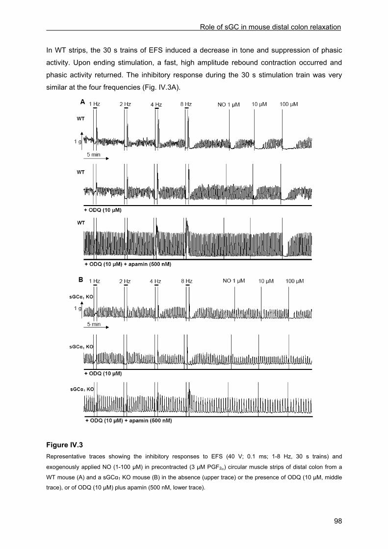
Role of sGC in mouse distal colon relaxation
98
In WT strips, the 30 s trains of EFS induced a decrease in tone and suppression of phasic
activity. Upon ending stimulation, a fast, high amplitude rebound contraction occurred and
phasic activity returned. The inhibitory response during the 30 s stimulation train was very
similar at the four frequencies (Fig. IV.3A).
Figure IV.3 Representative traces showing the inhibitory responses to EFS (40 V; 0.1 ms; 1-8 Hz, 30 s trains) and
exogenously applied NO (1-100 µM) in precontracted (3 µM PGF2α) circular muscle strips of distal colon from a
WT mouse (A) and a sGCα1 KO mouse (B) in the absence (upper trace) or the presence of ODQ (10 µM, middle
trace), or of ODQ (10 µM) plus apamin (500 nM, lower trace).
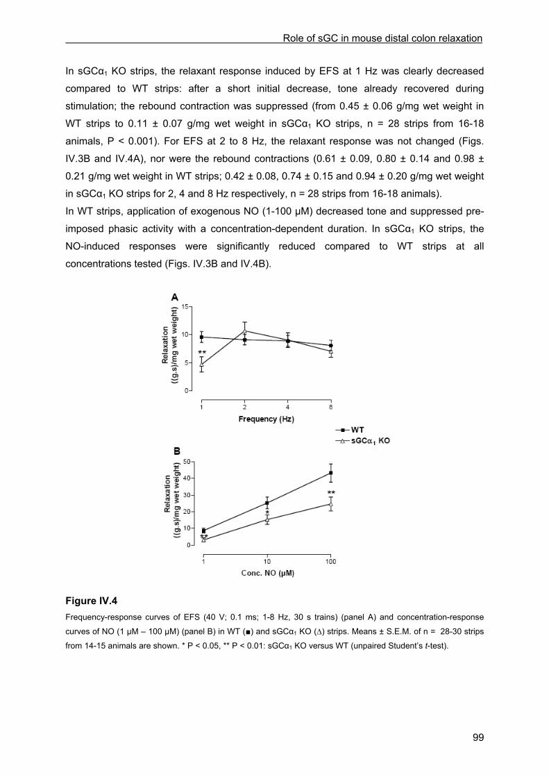
Role of sGC in mouse distal colon relaxation
99
In sGCα1 KO strips, the relaxant response induced by EFS at 1 Hz was clearly decreased
compared to WT strips: after a short initial decrease, tone already recovered during
stimulation; the rebound contraction was suppressed (from 0.45 ± 0.06 g/mg wet weight in
WT strips to 0.11 ± 0.07 g/mg wet weight in sGCα1 KO strips, n = 28 strips from 16-18
animals, P < 0.001). For EFS at 2 to 8 Hz, the relaxant response was not changed (Figs.
IV.3B and IV.4A), nor were the rebound contractions (0.61 ± 0.09, 0.80 ± 0.14 and 0.98 ±
0.21 g/mg wet weight in WT strips; 0.42 ± 0.08, 0.74 ± 0.15 and 0.94 ± 0.20 g/mg wet weight
in sGCα1 KO strips for 2, 4 and 8 Hz respectively, n = 28 strips from 16-18 animals).
In WT strips, application of exogenous NO (1-100 µM) decreased tone and suppressed pre-
imposed phasic activity with a concentration-dependent duration. In sGCα1 KO strips, the
NO-induced responses were significantly reduced compared to WT strips at all
concentrations tested (Figs. IV.3B and IV.4B).
Figure IV.4 Frequency-response curves of EFS (40 V; 0.1 ms; 1-8 Hz, 30 s trains) (panel A) and concentration-response
curves of NO (1 µM – 100 µM) (panel B) in WT (■) and sGCα1 KO (∆) strips. Means ± S.E.M. of n = 28-30 strips
from 14-15 animals are shown. * P < 0.05, ** P < 0.01: sGCα1 KO versus WT (unpaired Student’s t-test).

Role of sGC in mouse distal colon relaxation
100
IV.4.3.2 Influence of ODQ and L-NAME
The inhibitory responses to EFS and exogenous NO were examined following pre-incubation
with ODQ (10 µM) and L-NAME (300 µM) (Fig. IV.5).
Figure IV.5 Panel A, B, E and F: frequency-response curves of EFS (40 V; 0.1 ms; 1-8 Hz, 30 s trains) before and after
incubation with ODQ (10 µM; A and B) and before and after incubation with L-NAME (300 µM; E and F) in WT (A
and E) and sGCα1 KO strips (B and F). Panel C and D: concentration-response curves of NO (1 µM – 100 µM)
before and after incubation with ODQ (10 µM) in WT (C) and sGCα1 KO strips (D). Means ± S.E.M. of n = 11-15
are shown. * P < 0.05, ** P < 0.01, *** P < 0.001: after incubation versus before (paired Student’s t-test).
ODQ and L-NAME had no systematic influence on basal contractility, nor on the PGF2α-
induced response, either when total contractile activity (AUC) or mean phasic activity was
assessed. The response to EFS at 1 Hz was significantly reduced by ODQ in WT strips, but
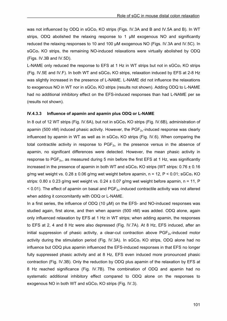
Role of sGC in mouse distal colon relaxation
101
was not influenced by ODQ in sGCα1 KO strips (Figs. IV.3A and B and IV.5A and B). In WT
strips, ODQ abolished the relaxing response to 1 µM exogenous NO and significantly
reduced the relaxing responses to 10 and 100 µM exogenous NO (Figs. IV.3A and IV.5C). In
sGCα1 KO strips, the remaining NO-induced relaxations were virtually abolished by ODQ
(Figs. IV.3B and IV.5D).
L-NAME only reduced the response to EFS at 1 Hz in WT strips but not in sGCα1 KO strips
(Fig. IV.5E and IV.F). In both WT and sGCα1 KO strips, relaxation induced by EFS at 2-8 Hz
was slightly increased in the presence of L-NAME. L-NAME did not influence the relaxations
to exogenous NO in WT nor in sGCα1 KO strips (results not shown). Adding ODQ to L-NAME
had no additional inhibitory effect on the EFS-induced responses than had L-NAME per se
(results not shown).
IV.4.3.3 Influence of apamin and apamin plus ODQ or L-NAME
In 8 out of 12 WT strips (Fig. IV.6A), but not in sGCα1 KO strips (Fig. IV.6B), administration of
apamin (500 nM) induced phasic activity. However, the PGF2α-induced response was clearly
influenced by apamin in WT as well as in sGCα1 KO strips (Fig. IV.6). When comparing the
total contractile activity in response to PGF2α in the presence versus in the absence of
apamin, no significant differences were detected. However, the mean phasic activity in
response to PGF2α, as measured during 5 min before the first EFS at 1 Hz, was significantly
increased in the presence of apamin in both WT and sGCα1 KO strips (WT strips: 0.76 ± 0.16
g/mg wet weight vs. 0.28 ± 0.06 g/mg wet weight before apamin, n = 12, P < 0.01; sGCα1 KO
strips: 0.80 ± 0.23 g/mg wet weight vs. 0.24 ± 0.07 g/mg wet weight before apamin, n = 11, P
< 0.01). The effect of apamin on basal and PGF2α-induced contractile activity was not altered
when adding it concomitantly with ODQ or L-NAME.
In a first series, the influence of ODQ (10 µM) on the EFS- and NO-induced responses was
studied again, first alone, and then when apamin (500 nM) was added. ODQ alone, again
only influenced relaxation by EFS at 1 Hz in WT strips; when adding apamin, the responses
to EFS at 2, 4 and 8 Hz were also depressed (Fig. IV.7A). At 8 Hz, EFS induced, after an
initial suppression of phasic activity, a clear-cut contraction above PGF2α-induced motor
activity during the stimulation period (Fig. IV.3A). In sGCα1 KO strips, ODQ alone had no
influence but ODQ plus apamin influenced the EFS-induced responses in that EFS no longer
fully suppressed phasic activity and at 8 Hz, EFS even induced more pronounced phasic
contraction (Fig. IV.3B). Only the reduction by ODQ plus apamin of the relaxation by EFS at
8 Hz reached significance (Fig. IV.7B). The combination of ODQ and apamin had no
systematic additional inhibitory effect compared to ODQ alone on the responses to
exogenous NO in both WT and sGCα1 KO strips (Fig. IV.3).
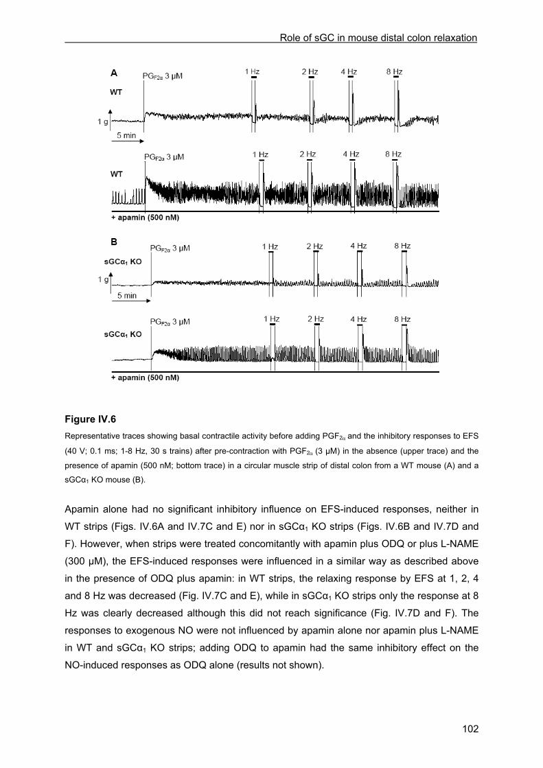
Role of sGC in mouse distal colon relaxation
102
Figure IV.6 Representative traces showing basal contractile activity before adding PGF2α and the inhibitory responses to EFS
(40 V; 0.1 ms; 1-8 Hz, 30 s trains) after pre-contraction with PGF2α (3 µM) in the absence (upper trace) and the
presence of apamin (500 nM; bottom trace) in a circular muscle strip of distal colon from a WT mouse (A) and a
sGCα1 KO mouse (B).
Apamin alone had no significant inhibitory influence on EFS-induced responses, neither in
WT strips (Figs. IV.6A and IV.7C and E) nor in sGCα1 KO strips (Figs. IV.6B and IV.7D and
F). However, when strips were treated concomitantly with apamin plus ODQ or plus L-NAME
(300 µM), the EFS-induced responses were influenced in a similar way as described above
in the presence of ODQ plus apamin: in WT strips, the relaxing response by EFS at 1, 2, 4
and 8 Hz was decreased (Fig. IV.7C and E), while in sGCα1 KO strips only the response at 8
Hz was clearly decreased although this did not reach significance (Fig. IV.7D and F). The
responses to exogenous NO were not influenced by apamin alone nor apamin plus L-NAME
in WT and sGCα1 KO strips; adding ODQ to apamin had the same inhibitory effect on the
NO-induced responses as ODQ alone (results not shown).
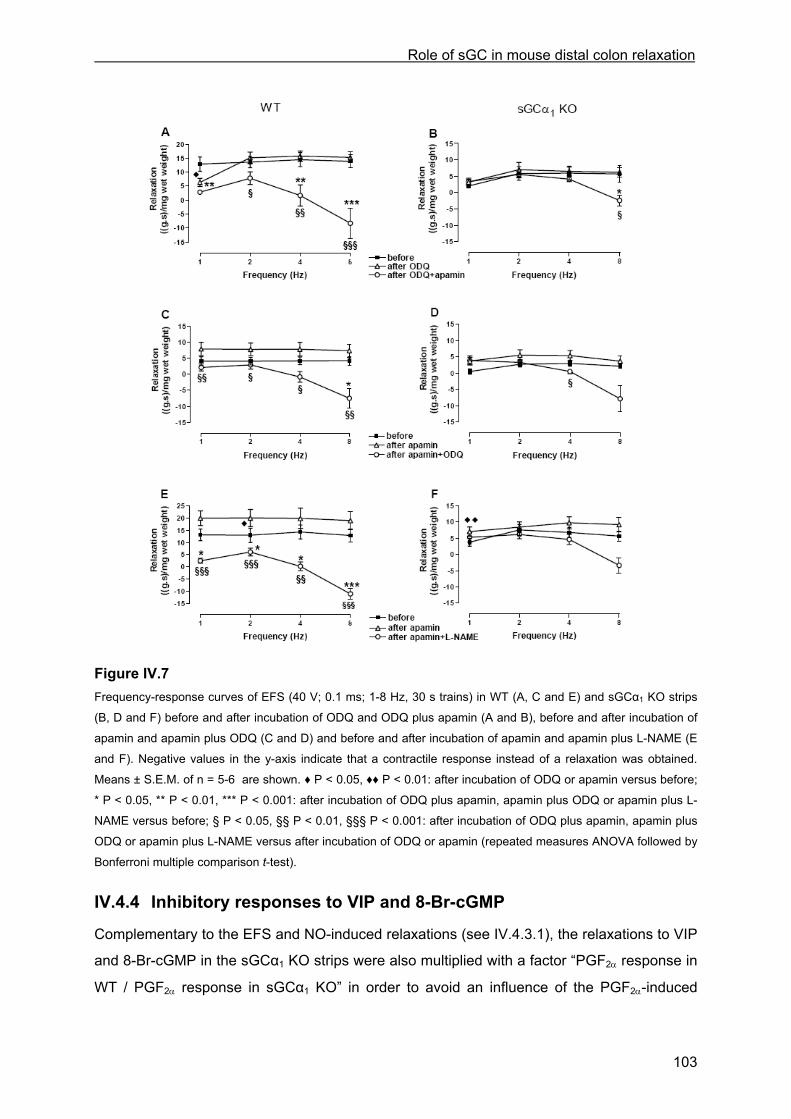
Role of sGC in mouse distal colon relaxation
103
Figure IV.7 Frequency-response curves of EFS (40 V; 0.1 ms; 1-8 Hz, 30 s trains) in WT (A, C and E) and sGCα1 KO strips
(B, D and F) before and after incubation of ODQ and ODQ plus apamin (A and B), before and after incubation of
apamin and apamin plus ODQ (C and D) and before and after incubation of apamin and apamin plus L-NAME (E
and F). Negative values in the y-axis indicate that a contractile response instead of a relaxation was obtained.
Means ± S.E.M. of n = 5-6 are shown. ♦ P < 0.05, ♦♦ P < 0.01: after incubation of ODQ or apamin versus before;
* P < 0.05, ** P < 0.01, *** P < 0.001: after incubation of ODQ plus apamin, apamin plus ODQ or apamin plus L-
NAME versus before; § P < 0.05, §§ P < 0.01, §§§ P < 0.001: after incubation of ODQ plus apamin, apamin plus
ODQ or apamin plus L-NAME versus after incubation of ODQ or apamin (repeated measures ANOVA followed by
Bonferroni multiple comparison t-test).
IV.4.4 Inhibitory responses to VIP and 8-Br-cGMP
Complementary to the EFS and NO-induced relaxations (see IV.4.3.1), the relaxations to VIP
and 8-Br-cGMP in the sGCα1 KO strips were also multiplied with a factor “PGF2α response in
WT / PGF2α response in sGCα1 KO” in order to avoid an influence of the PGF2α-induced
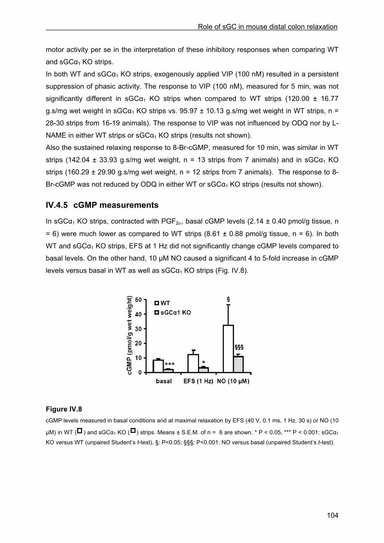
Role of sGC in mouse distal colon relaxation
104
motor activity per se in the interpretation of these inhibitory responses when comparing WT
and sGCα1 KO strips.
In both WT and sGCα1 KO strips, exogenously applied VIP (100 nM) resulted in a persistent
suppression of phasic activity. The response to VIP (100 nM), measured for 5 min, was not
significantly different in sGCα1 KO strips when compared to WT strips (120.00 ± 16.77
g.s/mg wet weight in sGCα1 KO strips vs. 95.97 ± 10.13 g.s/mg wet weight in WT strips, n =
28-30 strips from 16-19 animals). The response to VIP was not influenced by ODQ nor by L-
NAME in either WT strips or sGCα1 KO strips (results not shown).
Also the sustained relaxing response to 8-Br-cGMP, measured for 10 min, was similar in WT
strips (142.04 ± 33.93 g.s/mg wet weight, n = 13 strips from 7 animals) and in sGCα1 KO
strips (160.29 ± 29.90 g.s/mg wet weight, n = 12 strips from 7 animals). The response to 8-
Br-cGMP was not reduced by ODQ in either WT or sGCα1 KO strips (results not shown).
IV.4.5 cGMP measurements
In sGCα1 KO strips, contracted with PGF2α, basal cGMP levels (2.14 ± 0.40 pmol/g tissue, n
= 6) were much lower as compared to WT strips (8.61 ± 0.88 pmol/g tissue, n = 6). In both
WT and sGCα1 KO strips, EFS at 1 Hz did not significantly change cGMP levels compared to
basal levels. On the other hand, 10 µM NO caused a significant 4 to 5-fold increase in cGMP
levels versus basal in WT as well as sGCα1 KO strips (Fig. IV.8).
Figure IV.8 cGMP levels measured in basal conditions and at maximal relaxation by EFS (40 V, 0.1 ms, 1 Hz, 30 s) or NO (10
µM) in WT ( ) and sGCα1 KO ( ) strips. Means ± S.E.M. of n = 6 are shown. * P < 0.05, *** P < 0.001: sGCα1
KO versus WT (unpaired Student’s t-test). §: P<0.05; §§§: P<0.001: NO versus basal (unpaired Student’s t-test).
Role of sGC in mouse distal colon relaxation
105
IV.5 Discussion
In the present study, we investigated the impact of sGCα1-deficiency on distal colon motility.
We used recently developed sGCα1 KO mice lacking exon 6 of the sGCα1 gene, coding for
an essential part in the catalytical domain (Buys et al., 2008). This sGCα1 knockout model
provides a valuable tool to get a better insight in the relative importance of the two sGC
isoforms in inhibitory NANC neurotransmission and in the mechanism of action of the
neurotransmitters involved. There are indeed many reports that NO is not the sole
neurotransmitter responsible for NANC relaxation in colonic tissue, but that also ATP is
involved (Benko et al., 2007; Boeckxstaens et al., 1993; El Mahmoudy et al., 2006; Keef et
al., 1993; Pluja et al., 1999; Serio et al., 2003a; Van Crombruggen and Lefebvre, 2004).
We started the experiments in both male and female mice, as it was found that male but not
female sGCα1 KO mice develop hypertension (Buys et al., 2008). However, no systematic
differences between sexes were found in distal colon strips. The experiments were therefore
continued using male mice only.
IV.5.1 Inhibitory responses to 8-Br-cGMP and VIP
The finding that both WT and sGCα1 KO distal colon strips responded in a similar way to the
cell-permeable cGMP analog 8-Br-cGMP excludes that relaxant mechanisms downstream of
sGC are affected by knocking out sGCα1. This corresponds to observations in the gastric
fundus (Vanneste et al., 2007) and the aorta and femoral artery (Nimmegeers et al., 2007) of
the sGCα1 KO mice. In contrast, in the sGCα1 KO mice developed by Mergia et al. (2006),
increased sensitivity to a cGMP analog in vascular tissue was reported; in these mice exon 5
of the sGCα1 gene is targeted. Because Mergia et al. started their numbering from the first
coding exon while Buys et al. (2008) included the first 5’ non-coding exon in their numbering,
exon 4 of Mergia et al. indeed corresponds to exon 5 in the exon count of Buys et al.. The
difference in response to 8-Br-cGMP suggests that different compensatory regulations might
exist between the 2 knockout models or between the strains of mice used, although other
results were similar (see below).
Knocking out sGCα1 did not induce changes in cAMP-mediated relaxation. This was
demonstrated by the fact that relaxation by the vasoactive intestinal polypeptide (VIP), acting
via activation of adenylate cyclase (Simon and Kather, 1978; Laburthe et al., 2002), was well
maintained in the sGCα1 KO mice. A contribution of muscular NO to the relaxing effect of VIP
as proposed by Grider (1993) in circular muscle strips of rat colon, was not found as L-NAME
did not influence the response to VIP.

Role of sGC in mouse distal colon relaxation
106
IV.5.2 Inhibitory responses to exogenous NO
To assess the role of sGCα2β1 in nitrergic relaxation, we studied the influence of ODQ on the
relaxation by exogenous NO. In sGCα1 KO strips, relaxations induced by exogenous NO
were significantly decreased, but not abolished, when compared to WT strips, and were still
sensitive to ODQ. This suggests that the remaining relaxation by exogenous NO in the
sGCα1 KO strips is due to the activation of sGC, which can only be sGCα2β1. This is
corroborated by the fact that exogenous NO still induced an increase in cGMP levels in
sGCα1 KO strips. Similarly, a contribution of sGCα2β1 in NO-induced relaxation was
previously observed in the aorta, the femoral artery (Nimmegeers et al., 2007) and the
gastric fundus (Vanneste et al., 2007) of the sGCα1 KO mice, and in the aorta of the sGCα1
KO mice developed by Mergia et al. (2006). Similar to what was reported in gastric fundus
(Vanneste et al., 2007) and aorta (Mergia et al., 2006) at mRNA level, no increase in the
sGCα2 gene function was detected in distal colon as the sGCα2 protein level was not
changed. Similar to what has been described in lung tissue of our sGCα1 KO mice
(Vermeersch et al., 2007), we found decreased sGCβ1 protein levels, which is probably due
to the absence of the sGCα1 subunit. The contribution of sGCα2 to GI nitrergic relaxation is
furthermore demonstrated by the fact that no gross GI abnormalities in the sGCα1 KO mice
were observed, whereas recently developed mice deficient for the β1 subunit of sGC,
resulting in a complete loss of enzyme activity, suffered from fatal GI obstruction (Friebe et
al., 2007).
Interestingly, in WT strips, the relaxation to 10 µM and 100 µM NO is not abolished by ODQ.
First, this finding could point in the direction of a sGC-independent mechanism of NO,
responsible for the remaining, ODQ-insensitive, relaxation. NO has for example been
reported to induce relaxation via cGMP-independent SKCa channel activation in GI smooth
muscle (Martins and al., 1995; Serio and al., 2003b). However, the SKCa channel blocker
apamin did not influence the responses to NO, excluding the possibility that NO-induced
relaxation in the murine distal colon is mediated via a direct –cGMP-independent- activation
of SKCa channels. The addition of apamin to ODQ did not add to the inhibitory effect of ODQ
versus the relaxation induced by exogenous NO, which is in contrast to results obtained in
rat distal colon circular muscle (Van Crombruggen and Lefebvre, 2004); in this species, the
responses to higher concentrations of exogenous NO were only blocked by a combination of
apamin and ODQ, indicating that the response to exogenous NO is mediated by both sGC
and SKCa channels. Alternatively, the most plausible explanation for the remaining NO-
induced relaxation in the presence of ODQ in WT strips in our study is the assumption that,
due to the competitive nature of the sGC inhibitor ODQ (Fedele et al., 1996; Schrammel et
al., 1996), 10 µM ODQ might not be sufficient to fully antagonize the sGC activation by
Role of sGC in mouse distal colon relaxation
107
higher concentrations of NO. We indeed reported before in rat gastric fundus that 10 µM
ODQ inhibited the relaxation by NO more extensively than 1 µM ODQ did (Lefebvre,1998). In
sGCα1 KO strips, where the degree of sGC activation to overcome by ODQ might be smaller
in view of the inactivity of sGCα1β1 and the lower cGMP levels when exposed to NO, 10 µM
ODQ is sufficient to abolish the responses to exogenous NO.
IV.5.3 Inhibitory responses to EFS
To study the role of sGC as a messenger and of NO as a neurotransmitter in the NANC
relaxation in mouse distal colon, we investigated the EFS-evoked responses in WT and
sGCα1 KO mice, in the absence and presence of L-NAME and ODQ. In WT strips, the
response to EFS at 1 Hz, but not at 2, 4 and 8 Hz, was significantly reduced by L-NAME and
ODQ, indicating that only at this frequency NO is the main neurotransmitter liberated in the
distal colon, and that the relaxing effect of endogenous NO is mediated via sGC.
Accordingly, only the response to EFS at 1 Hz was reduced in sGCα1 KO strips when
compared to WT strips. The small remaining response to 1 Hz in sGCα1 KO strips was no
longer influenced by L-NAME or ODQ, indicating that the nitrergic response to EFS at 1 Hz in
WT strips must be mediated via sGCα1β1. The cGMP measurements of WT strips snap
frozen during the 30 s EFS-train at 1 Hz did not show a significant increase in cGMP levels
versus basal levels. This finding might be related to very localised cGMP rises induced by
the release of endogenous NO along the varicosities during EFS, which could not be
detected by the cGMP analysis. It should be noted that the cGMP measurements were
performed in the absence of the phosphodiesterase-5 (PDE-5) inhibitor zaprinast, which was
required to measure a cGMP increase for the EFS-evoked response in gastric fundus strips
of WT mice (Vanneste et al., 2007).
The EFS-evoked responses at 2 to 8 Hz were not reduced in sGCα1 KO strips compared to
WT strips, nor were they sensitive to inhibition by L-NAME or ODQ. They were also not
influenced by apamin, excluding a dominant role of an apamin-sensitive factor, such as ATP,
as single neurotransmitter in the EFS-evoked responses in mouse distal colon. However, the
addition of ODQ to apamin, or apamin to ODQ clearly reduced the EFS-induced responses
at 2, 4 and 8 Hz in WT strips, and at 8 Hz in sGCα1 KO strips; similar results were obtained
with the combination of apamin plus L-NAME. These results are indicative of a redundant
action of NO, acting at sGC, and another NANC neurotransmitter, acting at SKCa channels
when NANC neurons in mouse distal colon are stimulated by EFS at 2 to 8 Hz. This implies
that both neurotransmitters can act together such that the loss of any one is compensated by
the other. The fact that this interactive system seems to be maintained in sGCα1 KO strips
points to the ability of endogenous NO, released by EFS at 2 to 8 Hz, to act via sGCα2 in the
absence of sGCα1.

Role of sGC in mouse distal colon relaxation
108
A similar redundant inhibitory mechanism was suggested for the guinea-pig isolated taenia
coli by Selemidis et al. (1997). They proposed that NO acts as a “backup” or redundant
NANC inhibitory neurotransmitter to the dominant apamin-sensitive, nerve-derived
hyperpolarizing factor (NDHF). This hypothesis was based on the observation that L-NOARG
and ODQ had no effect on the EFS-evoked relaxation, but that the apamin-induced reduction
in the EFS-evoked relaxations was increased when L-NOARG or ODQ was added to
apamin. In our experiments only the combinations of apamin with ODQ or L-NAME – and not
apamin alone- inhibited the EFS-evoked responses at 2 to 8 Hz. These results suggest that,
in mouse distal colon, both NO as well as the apamin-sensitive neurotransmitter are
redundant to each other in the EFS-evoked responses at higher frequencies. Also for the
circular muscle of human colon (Boeckxstaens et al., 1993) it was suggested that NO and
another apamin-sensitive neurotransmitter, probably ATP, are involved in the inhibitory
neurotransmission, albeit not in a redundant manner. In rat distal colon (Van Crombruggen
and Lefebvre, 2004), both the nitrergic and the purinergic pathway needed to be blocked to
inhibit maintained EFS-induced relaxations. Indeed, only the combination of L-NAME plus
Reactive Blue 2 (RB2) or L-NAME plus apamin was able to almost abolish the EFS-induced
responses, whereas RB2 or L-NAME alone did not show any effect.
IV.5.4 Basal and PGF2α-induced contractile activity
In colonic tissue of other species such as rat, rabbit and human, NOS inhibition increased the
amplitude or frequency of the spontaneous contractions (Ciccocioppo et al., 1994; Keef et
al., 1993; Mulè et al., 1999). Spontaneous contractions in GI smooth muscle preparations are
related to spontaneous depolarizations, produced by interstitial cells of Cajal (ICC). In
cultured ICCs, NO donors inhibited the frequency of pacemaker currents via cGMP
generation (Koh et al., 2000). However, neither L-NAME nor ODQ had a systematic effect on
basal contractile activity in distal colon strips of WT and sGCα1 KO mice in this study, arguing
against a tonic inhibitory effect of NO via sGC activation. Administration of apamin induced
phasic activity in 67 % of the WT strips, suggesting that SKCa channels might be involved in a
tonic suppression of contractile activity in mouse colon. SKCa channels are expressed in ICCs
(Fujita et al., 2001). The effect of apamin on basal contractile activity corresponds to
observations by Powel and Bywater (2001) in an intact mouse colon preparation, where
apamin induced non-migrating contractions in distal colon. The tonic suppression of
contractile activity via SKCa channels might also counteract PGF2α-induced phasic activity, as
apamin also increased PGF2α-induced mean phasic activity in WT strips. It is not clear why
the effect of apamin on basal contractile activity was not present in sGCα1 KO strips, while
the effect on PGF2α-induced phasic activity was maintained.

Role of sGC in mouse distal colon relaxation
109
IV.5.5 Conclusion
In conclusion, our data indicate that in mouse distal colon exogenous NO is able to act at
least partially through sGCα2β1 in the absence of sGCα1β1 activity. Endogenous NO, acting
through sGCα1β1, is the principal neurotransmitter released by EFS at 1 Hz. At higher
stimulation frequencies, a redundant action of NO, acting at sGC, and another NANC
neurotransmitter, acting at the SKCa channels, is proposed. This interactive system is
maintained in sGCα1 KO strips highlighting the ability of endogenous NO, released in
response to EFS at 2 to 8 Hz, to act via sGCα2β1 in the absence of sGCα1β1 activity.
IV.6 References Bayguinov, O., Hagen, B., Bonev, A.D., Nelson, M.T., Sanders, K.M., 2000. Intracellular calcium events activated by ATP in murine colonic myocytes. Am. J. Physiol Cell Physiol 279, C126-C135.
Benko, R., Undi, S., Wolf, M., Vereczkei, A., Illenyi, L., Kassai, M., Cseke, L., Kelemen, D., Horvath, O.P., Antal, A., Magyar, K., Bartho, L., 2007. P2 purinoceptor antagonists inhibit the non-adrenergic, non-cholinergic relaxation of the human colon in vitro. Neuroscience 147, 146-152.
Boeckxstaens, G.E., Pelckmans, P.A., Herman, A.G., Vanmaercke, Y.M., 1993. Involvement of Nitric-Oxide in the Inhibitory Innervation of the Human Isolated Colon. Gastroenterology 104, 690-697.
Buys, E., Sips, P., Vermeersch, P., Raher, M., Rogge, E., Ichinose, F., Dewerchin, M., Bloch, K.D., Janssens, S., Brouckaert, P., 2008. Gender-specific hypertension and responsiveness to nitric oxide in sGCα1 knockout mice. Cardiovasc. Res. 79, 179-186.
Ciccocioppo, R., Onori, L., Messori, E., Candura, S.M., Coccini, T., Tonini, M., 1994. Role of nitric oxide-dependent and -independent mechanisms on peristalsis and accomodation in the rabbit distal colon. J. Pharmacol. Exp. Ther. 270, 929-937.
El Mahmoudy, A., Khalifa, M., Draid, M., Shiina, T., Shimizu, Y., El Sayed, M., Takewaki, T., 2006. NANC inhibitory neuromuscular transmission in the hamster distal colon. Pharmacol. Res. 54, 452-460.
Fedele, E., Jin, Y., Varnier, G., Raiteri, M., 1996. In vivo microdialysis study of a specific inhibitor of soluble guanylyl cyclase on the glutamate receptor/nitric oxide/cyclic GMP pathway. Br. J. Pharmacol. 119, 590-594.
Fujita, A., Takeuchi, T., Saitoh, N., Hanai, J., Hata, F., 2001. Expression of Ca2+-activated K+ channels, SK3, in the interstitial cells of Cajal in the gastrointestinal tract. Am. J. Physiol. Cell. Physiol. 281, C1727-C1733.
Friebe, A., Mergia, E., Dangel, O., Lange, A., Koesling, D., 2007. Fatal gastrointestinal obstruction and hypertension in mice lacking nitric oxide-sensitive guanylyl cyclase. Proc. Natl. Acad. Sci. U.S.A 104, 7699-7704.
Garthwaite, J., Southam, E., Boulton, C.L., Nielsen, E.B., Schmidt, K., Mayer, B., 1995. Potent and selective inhibition of nitric oxide-sensitive guanylyl cyclase by 1H[1,2,4]oxadiazolo[4,3-a]quinoxalin-1-one. Mol. Pharmacol. 48, 184-188.
Grider, J.R., 1993. Interplay of VIP and nitric oxide in regulation of the descending relaxation phase of peristalsis. Am. J. Physiol. 264, G334-340.
Grunnet, M., Jensen, B.S., Olesen, S., Klaerke, D.A., 2001. Apamin interacts with all subtypes of cloned small-conductance Ca2+-activated K+ channels. Pflugers Arch. 441, 544-550.
Role of sGC in mouse distal colon relaxation
110
Harteneck, C., Koesling, D., Soling, A., Schultz, G., Bohme, E., 1990. Expression of soluble guanylyl cyclase. Catalytic activity requires two enzyme subunits. FEBS Lett. 272, 221-223.
Harteneck, C., Wedel, B., Koesling, D., Malkewitz, J., Bohme, E., Schultz,G., 1991. Molecular cloning and expression of a new alpha-subunit of soluble guanylyl cyclase. Interchangeability of the alpha-subunits of the enzyme. FEBS Lett. 292, 217-222.
Heinzel, B., John, M., Klatt, P., Böhme, E., Mayr, B., 1992. Ca2+/calmodulin-dependent formation of hydrogen peroxide by brain nitric oxide synthase. Biochem. J. 281, 627-630.
Keef, K.D., Du, C., Ward, S.M., McGregor, B., Sanders, K.M., 1993. Enteric inhibitory neural regulation of human colonic circular muscle: role of nitric oxide. Gastroenterology 105, 1009-1016.
Kelm, M., Schrader, J., 1990. Control of coronary vascular tone by nitric oxide. Circ. Res. 66, 1561-1575.
Koh, S.D., Kim, T.W., Jun, J.Y., Glasgow, N.J., Ward, S.M., Sanders, K.M., 2000. Regulation of pacemaker currents in interstitial cells of Cajal from murine small intestine by cyclic nucleotides. J. Physiol., 527, 149-162.
Laburthe, M., Couvineau, A., Marie, J.C., 2002. VPAC receptors for VIP and PACAP. Receptors. Channels 8, 137-153.
Lefebvre, R.A., 1998. Influence of a selective guanylate cyclase inhibitor, and of the contraction level, on nitrergic relaxations in the gastric fundus. Br. J. Pharmacol. 124, 1439-1448.
Lucas, K.A., Pitari, G.M., Kazerounian, S., Ruiz-Stewart, I., Park, J., Schulz, S., Chepenik, K.P., Waldman, S.A., 2000. Guanylyl cyclases and signaling by cyclic GMP. Pharmacol. Rev. 52, 375-414.
Martins, S.L., De Oliveira, R.B., Ballejo, G., 1995. Rat duodenum nitrergic-induced relaxations are cGMP-independent and apamin-sensitive. Eur. J. Pharmacol. 284, 265-270.
Mergia, E., Friebe, A., Dangel, O., Russwurm, M., Koesling, D., 2006. Spare guanylyl cyclase NO receptors ensure high NO sensitivity in the vascular system. J. Clin. Invest 116, 1731-1737.
Mergia, E., Russwurm, M., Zoidl, G., Koesling, D., 2003. Major occurrence of the new alpha2beta1 isoform of NO-sensitive guanylyl cyclase in brain. Cell Signal. 15, 189-195.
Mulè, F., D' Angelo, S., Serio, R., 1999. Tonic inhibitory action by nitric oxide on spontaneous mechanical activity in rat proximal colon: involvement of cyclic GMP and apamin-sensitive K+ channels. Br. J. Pharmacol. 127, 514-520.
Nimmegeers, S., Sips, P., Buys, E., Brouckaert, P., Van de Voorde P., 2007. Functional role of the soluble guanylyl cyclase alpha(1) subunit in vascular smooth muscle relaxation. Cardiovasc. Res. 76, 149-159.
Pluja, L., Fernandez, E., Jimenez, M., 1999. Neural modulation of the cyclic electrical and mechanical activity in the rat colonic circular muscle: putative role of ATP and NO. Br. J. Pharmacol. 126, 883-892.
Powell, A.K., Bywater, A.R., 2001. Endogenous nitric oxide release modulates the direction and frequency of colonic migrating motor complexes in the isolated mouse colon. Neurogastroenterol. Mot. 13, 221-228.
Ro, S., Hatton, W.J., Koh, S.D., Horowitz, B., 2001. Molecular properties of small-conductance Ca2+-activated K+ channels expressed in murine colonic smooth muscle. Am. J. Physiol. gastrointest. Liver Physiol. 281, G964-G973.
Russwurm, M., Behrends, S., Harteneck, C., Koesling, D., 1998. Functional properties of a naturally occurring isoform of soluble guanylyl cyclase. Biochem. J. 335, 125-130.
Role of sGC in mouse distal colon relaxation
111
Russwurm, M., Wittau, N., Koesling, D., 2001. Guanylyl cyclase/PSD-95 interaction. Targeting of the nitric oxide-sensitive α2β1 guanylyl cyclase to synaptic membranes. J. Biol. Chem. 276, 44647-44652.
Schrammel, A., Behrends, S., Schmidt, K., Koesling, D., Mayer, B., 1996. Characterization of 1H-[1,2,4]oxadiazolo[4,3-a]quinoxalin-1-one as a heme-site inhibitor of nitric oxide-sensitive guanylyl cyclase. Mol. Pharmacol. 50, 1-5.
Selemidis, S., Satchell, D.G., Cocks, T.M., 1997. Evidence that NO acts as a redundant NANC inhibitory neurotransmitter in the guinea-pig isolated taenia coli. Br. J. Pharmacol. 121, 604-611.
Serio, R., Alessandro, M., Zizzo, M.G., Tamburello, M.P., Mule, F., 2003a. Neurotransmitters involved in the fast inhibitory junction potentials in mouse distal colon. Eur. J. Pharmacol. 460, 183-190.
Serio, R., Zizzo, M.G., Mule, F., 2003b. Nitric oxide induces muscular relaxation via cyclic GMP-dependent and -independent mechanisms in the longitudinal muscle of the mouse duodenum. Nitric Oxide. 8, 48-52.
Simon, B., Kather, H., 1978. Activation of human adenylate cyclase in the upper gastrointestinal tract by vasoactive intestinal polypeptide. Gastroenterology 74, 722-725.
Toda, N., Herman, A.G., 2005. Gastrointestinal function regulation by nitrergic efferent nerves. Pharmacol. Rev. 57, 315-338.
Van Crombruggen, K., Lefebvre, R.A., 2004. Nitrergic-purinergic interactions in rat distal colon motility. Neurogastroenterol. Motil. 16, 81-98.
Vanneste, G., Dhaese, I., Sips, P., Buys, E., Brouckaert, P., Lefebvre, R.A., 2007. Gastric motility in soluble guanylate cyclase α1 knock-out mice. J. Physiol. 584, 907-920.
Vermeersch, P., Buys, E., Pokreisz, P., Marsboom, G., Ichinose, F., Sips, P., Pellens, M., Gillijns, H., Swinnen, M., Graveline, A., Collen, D., Dewerchin, M., Brouckaert, P., Bloch, K.D., Janssens, S., 2007. Soluble guanylate cyclase-α1 deficiency selectively inhibits the pulmonary vasodilator response to nitric oxide and increases the pulmonary remodeling response to chronic hypoxia. Circulation 116, 936-943.


Chapter V
ROLE OF SOLUBLE GUANYLATE CYCLASE IN GASTROINTESTINAL MOTILITY Gastrointestinal phenotyping of NO resistant sGCbeta1his105phe knock in mice

Role of sGC in gastrointestinal motility
115
Chapter V Role of soluble guanylate cyclase in gastrointestinal motility: Gastrointestinal
phenotyping of NO resistant sGCbeta1his105phe knock in mice
V.1 Abstract
In the gastrointestinal (GI) tract, the importance of nitric oxide (NO) in non-adrenergic non-
cholinergic (NANC) relaxation is well established. The aim of this study was to investigate the
role of nitrergic activation of sGC in regulation of gastrointestinal motility, comparing wild-type
(WT) and sGCβ1his105phe knock in (KI) mice, which lack heme-bound, NO-sensitive sGC.
As a consequence, diethylenetriamine NONOate (DETA-NO) failed to increase sGC activity
in GI tissue of KI mice and exogenous NO did not induce relaxation in KI fundic, jejunal and
colonic strips. The KI mice showed an enlarged stomach with hypertrophy of the circular
muscle of the fundus and delayed gastric emptying. In addition, delayed intestinal transit and
increased whole gut transit time were observed in the KI mice. The nitrergic relaxant
responses to electrical field stimulation (EFS) at 1-8 Hz were abolished in the KI fundic and
jejunal strips; in the KI colonic strips, only the response at 1 Hz was abolished.
In conclusion, the GI consequences of lacking NO-sensitive sGC were most pronounced at
the level of the stomach establishing the pivotal role of the activation of sGC by NO in normal
gastric functioning. In addition, delayed transit was observed in the absence of NO-sensitive
sGC, indicating that nitrergic activation of sGC also plays a role in the lower GI tract. In the
distal colon, other neurotransmitters besides NO take part in the relaxations to higher
stimulation frequencies.
Role of sGC in gastrointestinal motility
116
V.2 Introduction
In the GI tract, nitric oxide (NO) is synthesized from L-arginine by neuronal NO synthase
(nNOS, NOS-1) in non-adrenergic non-cholinergic (NANC) neurons, from where it diffuses to
the smooth muscle cells. The importance of NO in NANC relaxation and hence GI motility is
well established. Experiments with NOS inhibitors resulted in delayed small intestinal transit
in rats (Karmeli et al., 1997), dogs (Chiba et al., 2002) and humans (Fraser et al., 2005) and
delayed colonic transit in rats (Mizuta et al., 1999) and dogs (Chiba et al., 2002). nNOS
knockout (KO) mice, which are chronically deprived of nNOS generated NO, showed an
enlarged stomach and delayed gastric emptying of solids and liquids (Huang et al., 1993;
Mashimo et al., 2000). The principal intracellular target of NO is the heme-protein soluble
guanylate cyclase (sGC). Once activated by NO, sGC leads to the production of the second
messenger cyclic guanosine 3’-5’-monophosphate (cGMP) and subsequently relaxation
(Toda and Herman, 2005). However, relaxant effects of NO not involving the sGC-cGMP
pathway but related to activation of small conductance Ca2+-dependent K+ channels have
been described in mouse (Serio et al., 2003) and rat duodenum (Martins et al., 1995) and in
rat colon (Van Crombruggen and Lefebvre, 2004), corresponding with direct activation of K+
channels by NO observed in colonic smooth muscle cells by Koh et al. (1995) and Lang and
Watson (1998). Additionally, sGC can be activated by other stimuli than NO such as carbon
monoxide (CO; Schmidt et al., 1992). CO represents another putative NANC
neurotransmitter, albeit the evidence for this is much weaker compared to NO.
sGC is composed of an α subunit and a β subunit (Harteneck et al., 1991). The
physiologically active isoforms are sGCα1β1 and sGCα2β1 with sGCα1β1 being the
predominant isoform in the GI tract (Mergia et al., 2003). However, we showed before that
sGCα2β1 can compensate at least partially for the absence of sGCα1β1 such that knocking
out sGCα1 does not induce important implications on in vivo GI motility (Vanneste et al.,
2007; Dhaese et al., 2009). Knocking out exon 10 of the β1 subunit, eliminating activation of
both sGC isoforms by NO but also basal activity, led to a phenotype with severely delayed
gut transit, grossly enlarged and dilated caecum, growth retardation and premature death
(Friebe et al., 2007). Essential for the activation of both sGC isoforms by NO is the prosthetic
heme group. The histidine 105 residu of the β1 subunit is a crucial amino acid for the binding
of the heme group to the sGC protein (Schmidt et al., 2004). Recently, sGCβ1his105phe
knock in (KI) mice in which the histidine 105 amino acid was replaced by phenylalanine were
developed by Thoonen et al. (2009); the resulting heme-deficient sGC isoforms retain their
basal activity but can no longer be activated by NO (Wedel et al., 1994). The
sGCβ1his105phe KI mice show a reduced live span, growth retardation and cardiovascular
implications such as elevated blood pressure and reduced heart rate (Thoonen et al., 2009).

Role of sGC in gastrointestinal motility
117
In the present study, the importance of nitrergic activation of sGC in regulation of GI motility
was investigated using sGCβ1his105phe KI mice.
V.3 Methods
V.3.1 Ethical approval
All experimental procedures were approved by the Ethical Committee for Animal
Experiments from the Faculty of Medicine and Health Sciences at Ghent University.
V.3.2 Animals
Homozygous sGCβ1his105phe knock in (KI) mice and wild type (WT) controls were derived
from a heterozygous breeding (genetic background: mixed 129/SvJ-C57BL/6J; Thoonen et
al., 2009). WT and KI mice of both sexes (male: n = 47 (WT) and 48 (KI), 7-13 weeks;
female: n = 36 (WT) and 36 (KI), 7-16 weeks) had free access to water and commercially
available chow. However, when investigating transit using the phenol red or fluorescein-
labelled dextran method (see below), food was withheld for 16 hours overnight with free
access to water.
V.3.3 Muscle tension experiments
V.3.3.1 Tissue preparation
Animals were killed by cervical dislocation and the GI tract was removed and put in aerated
Krebs solution (composition in mM: NaCl 118.5, KCl 4.8, KH2PO4 1.2, MgSO4 1.2, CaCl2 1.9,
NaHCO3 25.0 and glucose 10.1). The stomach was emptied from its contents and weighed.
Then the gastric fundus was separated from the rest of the stomach and two full wall
thickness fundus strips (2 x 11 mm) were prepared by cutting in the direction of the circular
muscle layer. A ± 5 cm long fragment of small bowel, starting approximately 10 cm distal to
the pylorus, and a ± 4 cm long segment of distal colon, taken above the pelvic brim, were
isolated and opened along the mesenteric border. The fragments were pinned mucosa side
up in Krebs solution. The mucosa was removed by sharp dissection under a microscope and
two full-thickness muscle strips (4 × 5 mm) were cut along the circular axis.
V.3.3.2 Isometric tension recording
After a cotton thread (fundus) or a silk thread (USP 4/0; jejunum and colon) was attached to
both ends of the strips, strips were mounted in 5 (jejunum and colon) or 15 ml (fundus) organ
baths between 2 platinum plate electrodes (6 or 7 mm apart). The organ baths contained
aerated (5% CO2 in O2) Krebs solution, maintained at 37°C. Changes in isometric tension
Role of sGC in gastrointestinal motility
118
were measured using MLT 050/D force transducers (ADInstruments) and recorded on a
Graphtec linearcorder F WR3701 (Graphtec, Yokohama, Japan; fundus)) or on a
PowerLab/8sp data recording system (ADInstruments) with Chart software (jejunum and
colon).
After an equilibration period of 30 min with flushing every 10 min at a load of 0.75 g (fundus),
0.25 g (colon) or 0.125 g (jejunum), the length-tension relationship was determined. Muscle
tissues were stretched by load increments of 0.25 g (fundus and colon) or 0.125 g (jejunum)
and at each load level exposed to 0.1 (fundus and jejunum) or 1 (colon) µM carbachol to
determine the optimal load (Lo; the load at which maximal response to the contractile agent
occurred). Tissues were allowed to equilibrate for 60 min at Lo with flushing every 15 min in
Krebs solution.
V.3.3.3 Protocols
All experiments were performed after switching to Krebs solution containing 1 µM atropine
and 4 µM guanethidine to block cholinergic and noradrenergic responses respectively
(NANC conditions), except for the cumulative contractile responses to carbachol and
prostaglandin F2α (PGF2α), which were obtained in Krebs solution without atropine and
guanethidine. All relaxant stimuli were examined after pre-contraction of the strips with
300 nM (fundus and jejunum) or 3 µM (colon) PGF2α. In a first series, 10 to 15 min after
adding PGF2α and when the contractile response was surely stabilized, relaxations were
induced by application of EFS (40V, 0.1 ms, 1-2-4-8 Hz for 10 s [jejunum], 30 s [colon] or 60
s [fundus] at 5 min interval) via the platinum plate electrodes by means of a Grass S88
Stimulator (Grass, W. Warwick, RI, USA; fundus) or a Hugo Sachs Stimulator I type 215/I
(jejunum and colon). This was followed by the application of exogenous NO (1-10-100 µM
with an interval of at least 5 min during which the effect of a given concentration of NO had
disappeared) and finally vasoactive intestinal polypeptide (VIP, 100 nM), which was left in
contact with the tissue for 5 min. Strips were washed for 30 min, and were subsequently
incubated with the sGC inhibitor 1H[1,2,4,]oxadiazolo [4,3-a]quinoxalin-1-one (ODQ; 10 µM)
for 30 min. PGF2α was then applied again and the responses to EFS, NO and VIP were
studied again in the presence of ODQ. In a second series, cumulative contractile responses
to carbachol (1 nM – 30 µM) or PGF2α (1 nM – 30 µM) were obtained. In jejunal and colonic
strips, the contact time for each concentration of carbachol and PGF2α was fixed at 2 min; in
fundic strips, a higher concentration of carbachol and PGF2α was given when the former
concentration reached its maximal contractile effect. Then, the influence of ODQ (10 µM)
versus 10 µM (fundus) or 100 µM (jejunum and colon) 8-bromoguanosine 3’,5’ cyclic
monophosphate (8-Br-cGMP) was studied. 8-Br-cGMP was left in contact with the tissue for
Role of sGC in gastrointestinal motility
119
10 min. In a third series, the influence of the NOS inhibitor Nω-nitro-L-arginine methyl ester
(L-NAME; 300 µM) was tested against the relaxation evoked by EFS. In colonic tissues, the
PGF2α-EFS cycle was repeated a third time in order to test subsequently the combination of
L-NAME (300 µM) plus the small conductance Ca2+-dependent K+ channel blocker apamin
(500 nM). In all series, the reproducibility of the relaxant responses was evaluated by running
time-control vehicle treated strips in parallel. At the end of each experiment, the tissue wet
weight was determined (mg wet weight, see data analysis).
V.3.3.4 Data analysis
As jejunal and colonic strips showed phasic activity, the area under the curve (AUC) above
baseline was determined to measure the contractile responses to carbachol and PGF2α. EC50
values of the concentration-response curves were calculated by linear interpolation. To
measure relaxant responses in jejunal and colonic strips, the AUC for a given response was
determined and subtracted from the AUC of a corresponding period just before applying the
relaxing drug or stimulus, yielding the area above the curve for the relaxant response. The
duration of the relaxant responses was determined as 10 s (jejunum) or 30 s (colon) for EFS
(i.e. the length of the stimulus train applied). VIP and 8-Br-cGMP induced a sustained
response and the duration was fixed at 5 min for VIP and 10 min for 8-Br-cGMP. NO
abolished phasic activity for a concentration-dependent period, after which phasic activity
progressively reoccurred. The duration of the relaxant responses to NO was therefore
determined as the time necessary for phasic activity to regain 50 % of the interval between
the mean peak level of phasic activity during the 2 min before administration of NO and the
minimum tone level during the NO response. This calculation was performed during the 1st
cycle of PGF2α-EFS-NO-VIP and the determined duration was further used for the NO-
induced responses in the 2nd cycle of PGF2α-EFS-NO-VIP. The responses are expressed as
(g.s)/mg wet weight.
Fundic strips showed tonic responses and the amplitude of the response to EFS was
measured at the end of the 60 s stimulation train; the responses to carbachol, PGF2α, NO
and 8-br-cGMP were measured at their maximal effect. The amplitude of contractile and
relaxant responses was expressed in g/g wet weight. The relaxant responses were then
expressed as % of the contraction evoked by PGF2α. EC50 values of the concentration-
response curves were calculated by linear interpolation.
V.3.4 Gastric emptying
As modified from de Rosalmeida et al. (2003), mice were, after food was withheld overnight,
administered 250 µl of a phenol red meal (0.1 % w v-1 dissolved in water) by gavage with a
Role of sGC in gastrointestinal motility
120
feeding needle. 15 min later, mice were killed by cervical dislocation and the stomach and
small bowel were clamped at both sides. Both organs were cut into small fragments and
placed into 20 ml of 0.1 N NaOH in a 50 ml Falcon tube. This mixture was homogenised for
approximately 30 s and allowed to stand for 20 min at room temperature. 10 ml of
supernatant was placed into a 15 ml Falcon tube and centrifuged for 10 min at 2800 rpm.
Proteins in 5 ml supernatant were precipitated with 0.5 ml of 20 % (w/v) trichloroacetic acid
and the solution was centrifuged for 20 min at 2800 rpm. 0.5 ml of supernatant was added to
0.667 ml of 0.5 N NaOH and the absorbance of 300 µl of this mixture was
spectrophotometrically determined at 540 nm in a Biotrak II plate reader (Amersham
Biosciences). Gastric emptying was calculated as the amount of phenol red that left the
stomach as % of the total amount of phenol red recovered and the phenol red recovery was
determined as the amount of phenol red recovered, expressed as % of the amount of phenol
red administered.
V.3.5 Transit
V.3.5.1 Intestinal transit (fluorescein-labelled dextran method)
Mice were, after food was withheld overnight, administered 200 µl of non-absorbable
fluorescein-labelled dextran (FD70; 70 kDa, 2.5 % w v-1 dissolved in water) by gavage with a
feeding needle. Ninety minutes later, mice were killed by cervical dislocation. For a full
description of the technical details of this method, we refer to De Backer et al. (2008). Briefly,
the entire GI tract was excised and the mesenterium was removed. The GI tract was then
pinned down in a custom-made Petri dish filled with Krebs solution. Immediately after, FD70
was visualized using the Syngene Geneflash system (Syngene, Cambridge, UK). Two full-
field images- one in normal illumination mode and another in fluorescent mode- were taken
and matched for analysis. The fluorescent intensity throughout the intestinal tract was
calculated and data were expressed as the percentage of fluorescence intensity per segment
(sb, small bowel segments 1-10; caecum; col, colon segments 1-2). The geometric centre
was calculated as (Σ (% FD70 per segment x segment number))/100.
V.3.5.2 Whole gut transit time (carmine method)
As adapted from Friebe et al. (2007), mice were administered 200 µl carmine (6 % w v-1
dissolved in 0.5 % methylcellulose) by gavage with a feeding needle. Mice were then
returned to individual cages, without food deprivation. The time taken for excretion of the first
red coloured faeces was determined at 30 min intervals.
Role of sGC in gastrointestinal motility
121
V.3.6 Small intestinal contractility
Immediately after the evaluation of intestinal transit by fluorescence imaging (see above), the
spontaneous contractile activity in the jejunum was recorded. For a full description of the
technical details of this method, we refer to De Backer et al. (2008). Briefly, a 6 cm long
segment of jejunum was recorded for 30 s and the video files were imported in ImageJ. After
the contrast threshold value was set, the images were converted to black-and-white and the
mean diameter of the jejunal segment was measured in the first frame. Next, all 750 frames
were sequentially analysed using Amplitude Profiler software- written as ImageJ plugin. The
change in intestinal diameter within this 30 s-period for every pixel (768 pixels) along this 6
cm long jejunal segment was calculated as % contraction amplitude by the following
equation: [(maximal diameter – minimal diameter)/maximal diameter] * 100. Finally, the mean
value of these 768 amplitude values was calculated. The oscillatory changes were also
represented in a three-dimensional (3-D) plot using Spatiotemporal Motility Mapping software
- written as ImageJ plugin with a GnuPlot backend – allowing to see contractility in function
of time.
V.3.7 Histology
Specimens of fundic, jejunal and colonic tissues were taken from 1 WT and 1 KI mouse of
both sexes. The tissues were fixed in 4% neutral buffered formalin (NBF), dehydrated
through a graded series of ethanol and embedded in paraffin wax. Serial transverse sections
of 5 µm thickness were cut at 500 µm intervals, using a rotary microtome (SLEE CUT 4060)
and stained with hematoxylin and eosin for morphological observation.
V.3.8 sGC enzyme activity
sGC enzyme activity was measured as described (Buys et al., 2008). Fundus, jejunum and
distal colon were harvested. The fundus was immediately snap-frozen in liquid nitrogen; the
jejunum and distal colon were snap-frozen after removal of the mucosa. The tissues were
stored at -80°C until further processing. They were then thawn and homogenized in buffer
containing 50 mM tris(hydroxymethyl)aminomethane (Tris).HCl (pH 7.6), 1 mM EDTA, 1 mM
dithiothreitol (DTT), and 2 mM phenylmethylsulfonyl fluoride. Extracts were centrifuged at
20,000 g for 20 min at 4°C. Supernatants (containing 40 µg protein) were incubated for 10
min at 37°C in a reaction mixture containing 50 mM Tris.HCl (pH 7.5), 4 mM MgCl2, 0.5 mM
1-methyl-3-isobutylxanthine, 7.5 mM creatine phosphate, 0.2 mg ml-1 creatine
phosphokinase, 1 mM L-NAME, and 1 mM GTP with or without 1 mM diethylenetriamine
NONOate (DETA-NO). The reaction was terminated by the addition of 0.9 ml of 0.05 M HCl.
cGMP in the reaction mixture was measured using a commercial radioimmunoassay
Role of sGC in gastrointestinal motility
122
(Biomedical Technologies, Stoughton, MA). sGC enzyme activity is expressed as pmol of
cGMP produced per min per milligram of protein in GI extract supernatant.
V.3.9 Drugs used
The following drugs were used: apamin (obtained from Alomone Labs), atropine sulphate, 8-
Br-cGMP sodium salt, carmine, guanethidine sulphate, L-NAME, phenol red, PGF2α tris salt,
VIP (all obtained from Sigma-Aldrich), DETA-NO (from Alexis Biochemicals), carbachol (from
Fluka AG), fluorescein-labelled dextran (70 kDa, FD70; Invitrogen), ODQ (from Tocris
Cookson). All drugs were dissolved in de-ionized water except for the following: ODQ, which
was dissolved in 100 % ethanol and DETA-NO, which was dissolved in sGC enzyme activity
buffer (see above). Saturated NO solution was prepared from gas (Air Liquide, Belgium) as
described by Kelm and Schrader (1990).
V.3.10 Statistics
All results are expressed as means ± S.E.M. n refers to tissues obtained from different
animals unless otherwise indicated. Comparison between KI and WT tissues was done with
an unpaired Student’s t-test. Comparison within tissues of either WT or KI was done by a
paired Student’s t-test. When more than 2 sets of results within the same tissue had to be
compared, repeated measures ANOVA followed by a Bonferroni corrected t-test was applied.
A P-value less than 0.05 was considered to be statistically significant (GRAPHPAD, San
Diego, CA, USA).
V.4 Results
All in vivo experiments and all in vitro experiments were performed in mice of both sexes,
except for the measurement of sGC activity which was investigated in male mice only. As no
systematic differences between the sexes were observed, results are presented for male
mice.
V.4.1 General observations
The body weight of the KI mice was significantly smaller than that of the WT mice (Table
V.1). However, in contrast, the stomach of the KI mice was significantly enlarged; in some KI
mice the stomach occupied most of the abdominal cavity. The mean empty stomach weight
of the KI mice was significantly larger than this of the WT mice (Table V.1).

Role of sGC in gastrointestinal motility
123
Table V.1: Body weight, stomach weight and weight of the gastro-intestinal preparations.
WT KI
Body weight (g) 30 ± 1 (n = 23) 22 ± 1 (n = 25) ***
Stomach weight (mg) 209 ± 10 (n = 23) 379 ± 28 (n = 25) ***
Fundus strips (mg) 5.83 ± 0.27 (n = 46) 15.73 ± 1.55 (n = 50) ***
Jejunum strips (mg) 0.54 ± 0.05 (n = 47) 0.45 ± 0.05 (n = 45)
Colon strips (mg) 0.53 ± 0.03 (n = 41) 0.44 ± 0.03 (n = 41) *
For the weight of the strips (2 strips per animal), values are means ± SEM of n = 41-50 out of 23-25 animals, i.e. all male animals used in the muscle tension experiments. * P < 0.05, *** P < 0.001: unpaired Student’s t test (KI vs. WT).
V.4.2 Gastric emptying, small intestinal transit and whole gut transit time
The phenol red recovery was 74 ± 5 % in WT mice (n = 10) and 77 ± 4 % in KI mice (n = 8).
Fifteen min after gavage, gastric emptying of a phenol red solution was significantly delayed
in KI mice compared to WT mice (Fig. V.1A). At ninety min after gavage, we observed a
delayed intestinal transit of a fluorescein-labelled dextran solution in KI versus WT mice as
manifested from the significant decrease in geometric center (Fig. V.1C). Small intestinal
contractility at that time point was however not different between WT and KI mice (%
contraction amplitude in KI: 23 ± 3 % versus WT: 22 ± 3 %, n = 5-6), which is illustrated in
Fig. V.1D. The whole gut transit time, determined as the time taken for excretion of the first
red-coloured faeces after gavage with a carmine solution, was between 120 and 180 min in
WT mice. In KI mice, the whole gut transit time was more variable and the mean value was
significantly increased (KI: 320 ± 25 min versus WT: 146 ± 10 min, n = 6-7; P < 0.001; Fig.
V.1B).
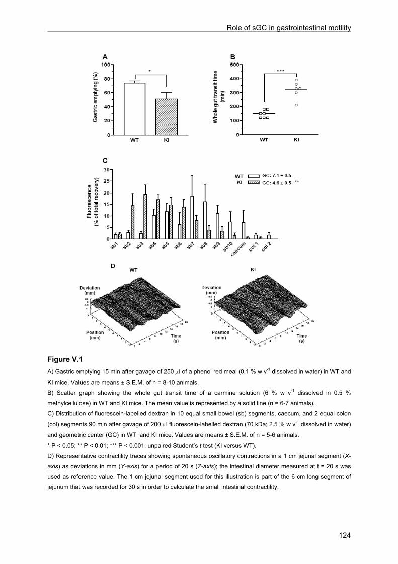
Role of sGC in gastrointestinal motility
124
Figure V.1 A) Gastric emptying 15 min after gavage of 250 µl of a phenol red meal (0.1 % w v-1 dissolved in water) in WT and
KI mice. Values are means ± S.E.M. of n = 8-10 animals. B) Scatter graph showing the whole gut transit time of a carmine solution (6 % w v-1 dissolved in 0.5 %
methylcellulose) in WT and KI mice. The mean value is represented by a solid line (n = 6-7 animals).
C) Distribution of fluorescein-labelled dextran in 10 equal small bowel (sb) segments, caecum, and 2 equal colon
(col) segments 90 min after gavage of 200 µl fluorescein-labelled dextran (70 kDa; 2.5 % w v-1 dissolved in water)
and geometric center (GC) in WT and KI mice. Values are means ± S.E.M. of n = 5-6 animals.
* P < 0.05; ** P < 0.01; *** P < 0.001: unpaired Student’s t test (KI versus WT).
D) Representative contractility traces showing spontaneous oscillatory contractions in a 1 cm jejunal segment (X-
axis) as deviations in mm (Y-axis) for a period of 20 s (Z-axis); the intestinal diameter measured at t = 20 s was
used as reference value. The 1 cm jejunal segment used for this illustration is part of the 6 cm long segment of
jejunum that was recorded for 30 s in order to calculate the small intestinal contractility.
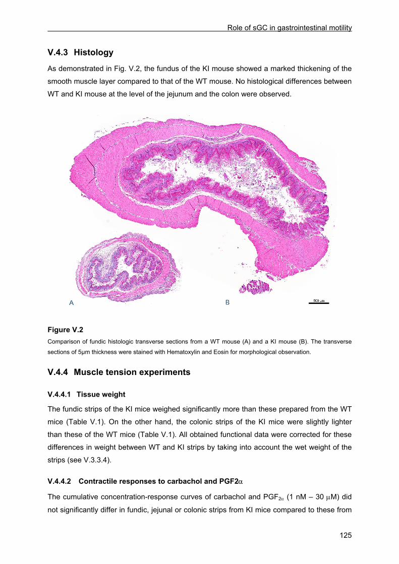
Role of sGC in gastrointestinal motility
125
V.4.3 Histology
As demonstrated in Fig. V.2, the fundus of the KI mouse showed a marked thickening of the
smooth muscle layer compared to that of the WT mouse. No histological differences between
WT and KI mouse at the level of the jejunum and the colon were observed.
Figure V.2 Comparison of fundic histologic transverse sections from a WT mouse (A) and a KI mouse (B). The transverse
sections of 5µm thickness were stained with Hematoxylin and Eosin for morphological observation.
V.4.4 Muscle tension experiments
V.4.4.1 Tissue weight
The fundic strips of the KI mice weighed significantly more than these prepared from the WT
mice (Table V.1). On the other hand, the colonic strips of the KI mice were slightly lighter
than these of the WT mice (Table V.1). All obtained functional data were corrected for these
differences in weight between WT and KI strips by taking into account the wet weight of the
strips (see V.3.3.4).
V.4.4.2 Contractile responses to carbachol and PGF2α
The cumulative concentration-response curves of carbachol and PGF2α (1 nM – 30 µM) did
not significantly differ in fundic, jejunal or colonic strips from KI mice compared to these from
Role of sGC in gastrointestinal motility
126
WT mice. For example, in colonic strips, the EC50 (nM) and Emax ([g.s]/mg wet weight) for
carbachol were 433 ± 72 and 170 ± 35 in WT (n = 6) and 709 ± 144 and 211 ± 77 in KI (n =
6). For PGF2α, the EC50 (µM) and Emax ([g.s]/mg wet weight) in this tissue were 7.68 ± 1.04
and 53 ± 13 in WT (n = 6) and 9.14 ± 1.86 and 44 ± 9 in KI (n = 6). PGF2α at a concentration
of 300 nM was chosen to pre-contract fundic and jejunal strips in order to investigate the
relaxant responses to EFS, NO, VIP and 8-br-cGMP (see below); in colonic strips 3 µM
PGF2α was used. In fundic strips, the PGF2α-induced response consisted solely of an
increase in tone. Instead, in jejunal and colonic strips, albeit PGF2α induced an initial fast
increase in tone, this then declined to a lower level, or in the case of the jejunal strips even to
baseline, with superimposed phasic activity (Fig. V.3). Despite the similar concentration-
response curve of PGF2α in the WT and KI colonic strips, the response to 3 µM PGF2α was
decreased in the KI versus WT colonic strips in the first series where ODQ was tested
against the responses to EFS, NO and VIP (see below).
V.4.4.3 Relaxant responses to EFS
In fundic strips of WT mice, EFS (1-8 Hz) induced frequency-dependent relaxations,
consisting of a progressive decline in tone which recovered upon ending stimulation (see Fig.
V.3A for the response at 8 Hz and Fig. V.4A for mean responses). In WT jejunal and colonic
strips, the magnitude of the EFS-induced relaxations was rather similar at the different
stimulation frequencies (Fig. V.3B and C and Fig. V.4B and C). In WT jejunal strips, EFS only
induced suppression of phasic activity; in WT colonic strips, EFS induced suppression of
phasic activity as well as a decrease in tone. Upon ending stimulation, a rebound contraction
was observed at 8 Hz in WT jejunal strips and at all tested frequencies in WT colonic strips
(Fig. V.3B and C). ODQ (10 µM; Fig. V.4A and B) and L-NAME (300 µM, data not shown)
(nearly) abolished the EFS-induced responses at all frequencies in WT fundic and jejunal
strips. In WT colonic strips, only the response to 1 Hz was reduced by ODQ (Fig. V.4C) or L-
NAME, albeit the latter did not reach significance (Fig. V.5); the combination of L-NAME plus
apamin was able to decrease the response at all frequencies in these strips but did not
abolish them (Fig. V.5).
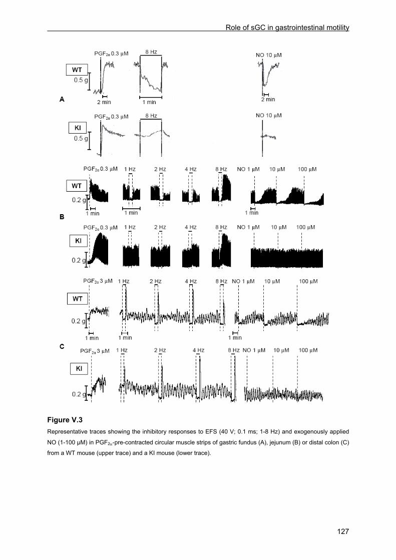
Role of sGC in gastrointestinal motility
127
Figure V.3 Representative traces showing the inhibitory responses to EFS (40 V; 0.1 ms; 1-8 Hz) and exogenously applied
NO (1-100 µM) in PGF2α-pre-contracted circular muscle strips of gastric fundus (A), jejunum (B) or distal colon (C)
from a WT mouse (upper trace) and a KI mouse (lower trace).
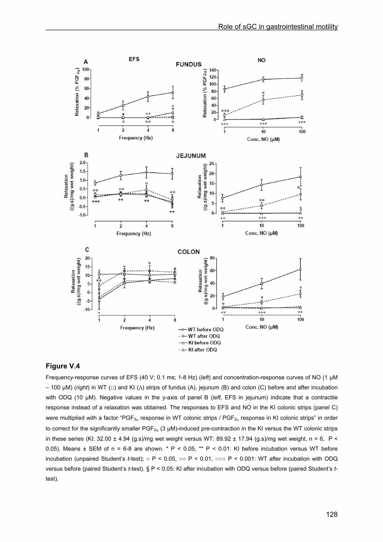
Role of sGC in gastrointestinal motility
128
Figure V.4 Frequency-response curves of EFS (40 V; 0.1 ms; 1-8 Hz) (left) and concentration-response curves of NO (1 µM
– 100 µM) (right) in WT (□) and KI (∆) strips of fundus (A), jejunum (B) and colon (C) before and after incubation
with ODQ (10 µM). Negative values in the y-axis of panel B (left, EFS in jejunum) indicate that a contractile
response instead of a relaxation was obtained. The responses to EFS and NO in the KI colonic strips (panel C)
were multiplied with a factor “PGF2α response in WT colonic strips / PGF2α response in KI colonic strips” in order
to correct for the significantly smaller PGF2α (3 µM)-induced pre-contraction in the KI versus the WT colonic strips
in these series (KI: 32.00 ± 4.94 (g.s)/mg wet weight versus WT: 89.92 ± 17.94 (g.s)/mg wet weight, n = 6, P <
0.05). Means ± SEM of n = 6-8 are shown. * P < 0.05, ** P < 0.01: KI before incubation versus WT before
incubation (unpaired Student’s t-test); ○ P < 0.05, ○○ P < 0.01, ○○○ P < 0.001: WT after incubation with ODQ
versus before (paired Student’s t-test). § P < 0.05: KI after incubation with ODQ versus before (paired Student’s t-
test).
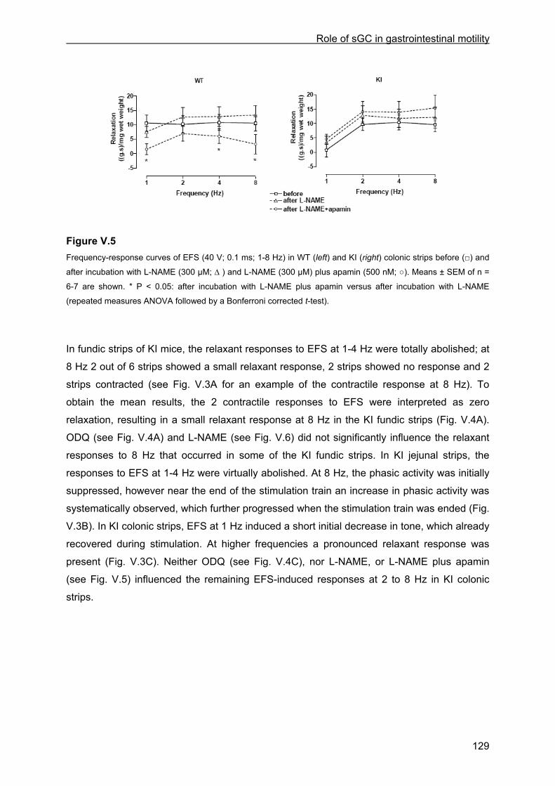
Role of sGC in gastrointestinal motility
129
Figure V.5 Frequency-response curves of EFS (40 V; 0.1 ms; 1-8 Hz) in WT (left) and KI (right) colonic strips before (□) and
after incubation with L-NAME (300 µM; ∆ ) and L-NAME (300 µM) plus apamin (500 nM; ○). Means ± SEM of n =
6-7 are shown. * P < 0.05: after incubation with L-NAME plus apamin versus after incubation with L-NAME
(repeated measures ANOVA followed by a Bonferroni corrected t-test).
In fundic strips of KI mice, the relaxant responses to EFS at 1-4 Hz were totally abolished; at
8 Hz 2 out of 6 strips showed a small relaxant response, 2 strips showed no response and 2
strips contracted (see Fig. V.3A for an example of the contractile response at 8 Hz). To
obtain the mean results, the 2 contractile responses to EFS were interpreted as zero
relaxation, resulting in a small relaxant response at 8 Hz in the KI fundic strips (Fig. V.4A).
ODQ (see Fig. V.4A) and L-NAME (see Fig. V.6) did not significantly influence the relaxant
responses to 8 Hz that occurred in some of the KI fundic strips. In KI jejunal strips, the
responses to EFS at 1-4 Hz were virtually abolished. At 8 Hz, the phasic activity was initially
suppressed, however near the end of the stimulation train an increase in phasic activity was
systematically observed, which further progressed when the stimulation train was ended (Fig.
V.3B). In KI colonic strips, EFS at 1 Hz induced a short initial decrease in tone, which already
recovered during stimulation. At higher frequencies a pronounced relaxant response was
present (Fig. V.3C). Neither ODQ (see Fig. V.4C), nor L-NAME, or L-NAME plus apamin
(see Fig. V.5) influenced the remaining EFS-induced responses at 2 to 8 Hz in KI colonic
strips.
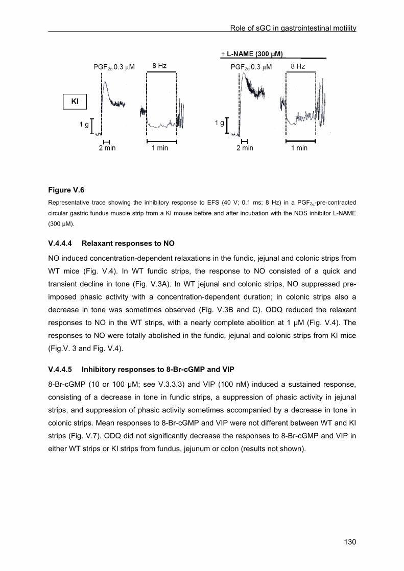
Role of sGC in gastrointestinal motility
130
Figure V.6 Representative trace showing the inhibitory response to EFS (40 V; 0.1 ms; 8 Hz) in a PGF2α-pre-contracted
circular gastric fundus muscle strip from a KI mouse before and after incubation with the NOS inhibitor L-NAME
(300 µM).
V.4.4.4 Relaxant responses to NO
NO induced concentration-dependent relaxations in the fundic, jejunal and colonic strips from
WT mice (Fig. V.4). In WT fundic strips, the response to NO consisted of a quick and
transient decline in tone (Fig. V.3A). In WT jejunal and colonic strips, NO suppressed pre-
imposed phasic activity with a concentration-dependent duration; in colonic strips also a
decrease in tone was sometimes observed (Fig. V.3B and C). ODQ reduced the relaxant
responses to NO in the WT strips, with a nearly complete abolition at 1 µM (Fig. V.4). The
responses to NO were totally abolished in the fundic, jejunal and colonic strips from KI mice
(Fig.V. 3 and Fig. V.4).
V.4.4.5 Inhibitory responses to 8-Br-cGMP and VIP
8-Br-cGMP (10 or 100 µM; see V.3.3.3) and VIP (100 nM) induced a sustained response,
consisting of a decrease in tone in fundic strips, a suppression of phasic activity in jejunal
strips, and suppression of phasic activity sometimes accompanied by a decrease in tone in
colonic strips. Mean responses to 8-Br-cGMP and VIP were not different between WT and KI
strips (Fig. V.7). ODQ did not significantly decrease the responses to 8-Br-cGMP and VIP in
either WT strips or KI strips from fundus, jejunum or colon (results not shown).

Role of sGC in gastrointestinal motility
131
Figure V.7 Relaxant responses to 10 µM (fundus, A) or 100 µM (jejunum, B and colon, C) 8-Br-cGMP (left) and to 100 nM
VIP (right) in WT and KI strips. Complementary to the EFS- and NO-induced relaxations (see legend Fig. V.4C),
the responses to VIP in the KI colonic strips were multiplied with the correction factor “PGF2α response in WT
colonic strips / PGF2α response in KI colonic strips”. Means ± SEM of n = 9-12 out of 5-6 animals are shown.
V.4.5 sGC enzyme activity
Baseline sGC activity was slightly increased in KI tissues compared to WT tissues; this
reached only significance in the colonic tissues (Fig. V.8). DETA-NO significantly increased
sGC activity in WT tissues, but not in KI tissues. The level of sGC activity in the presence of
DETA-NO was as a result significantly lower in the KI versus the WT tissues.

Role of sGC in gastrointestinal motility
132
Figure V.8 Un-stimulated (baseline) and DETA-NO-stimulated sGC enzyme activity (expressed as picomoles cGMP
produced per mg protein per minute) in fundic (A), jejunal (B) and colonic (C) extracts of WT and KI mice. Means
± SEM of n = 5-6. ** P < 0.01, *** P < 0.001: KI versus WT (unpaired Student’s t-test); §§ P < 0.01, §§§ P < 0.001:
DETA-NO versus baseline (paired Student’s t-test).
Role of sGC in gastrointestinal motility
133
V.5 Discussion
The aim of this study was to investigate the importance of the nitrergic activation of sGC in
regulation of GI motility by use of sGCβ1his105phe knock-in (KI) mice, possessing heme-
deficient sGC. Heme-deficient sGC isoforms retain their basal activity but can not be
activated by NO (Wedel et al., 1994). Indeed exogenous NO was not able to induce in vitro
relaxation in the fundic, jejunal and colonic smooth muscle strips of KI mice.
Correspondingly, the NO-donor DETA-NO failed to increase the sGC activity in the GI
tissues of KI mice.
In vivo, the observed GI phenotype of the KI mice included delayed gastric emptying,
delayed intestinal transit and increased whole gut transit time. The maintained responses to
respectively the cell-permeable cGMP analogue 8-Br-cGMP and VIP, that acts through
adenylate cyclase coupled VIP receptors, in the KI fundic, jejunal and colonic strips, indicate
that there are no compensatory mechanisms in the relaxant pathway downstream of sGC nor
in the cAMP-induced relaxation in the KI mice. Also contractile mechanisms are not
influenced as contractions to carbachol and PGF2α did not consistently differ between KI and
WT mice. The severely disturbed gastric emptying and intestinal transit in KI mice illustrate
the importance of NO-sensitive sGC in GI motility and probably contribute to the reduced
lifespan of KI mice (median survival of 29-36 weeks; Thoonen et al., 2009). The latter values
contrast with the much shorter lifespan of sGC β1 KO mice, where less than 10 % survived
for 1 month due to intestinal dysmotility (Friebe et al., 2007), suggesting that basal sGC
activity, maintained in the KI mice, is important for longer survival.
NO is the principal relaxant neurotransmitter in the mouse gastric fundus, as EFS-induced
relaxations are totally abolished by L-NAME in WT fundic strips. The fact that ODQ has a
similar effect as L-NAME in the WT fundic strips indicates that these NO-induced relaxations
are mediated through activation of sGC. However, a relaxant response to EFS occurred in
response to EFS at 8 Hz in some KI fundic strips, being insensitive to L-NAME. The latter
suggests the release of another neurotransmitter than NO, at higher stimulation frequencies
in the gastric fundus of some KI mice. We could speculate this neurotransmitter to be VIP, as
VIP was shown to be released at higher stimulation frequencies in rat gastric fundus strips
(D’Amato et al., 1992). In compensation for the loss of nitrergic relaxation, VIP release might
start at lower frequencies in some KI mice. Fundic nitrergic relaxation is essential for gastric
accommodation (Desai et al., 1991) and its deficiency in KI mice might be expected to speed
up liquid gastric emptying as fundic storage of the liquids is impaired. Instead, we observed
delayed liquid gastric emptying in the KI mice, which is in accordance with reports of delayed
gastric emptying in nNOS KO mice (Mashimo et al., 2000) and cGMP-dependent protein
kinase (cGKI) KO mice (Pfeifer et al., 1998). Whereas nNOS is involved in the synthesis of
Role of sGC in gastrointestinal motility
134
NO and thus is situated upstream of sGC, cGKI is an important downstream effector of sGC.
Moreover, the KI mice, similar to the nNOS (Huang et al., 1993; Mashimo et al., 2000) and
cGKI (Pfeifer et al., 1998) KO mice, showed a marked enlargement of the stomach with
hypertrophy of the circular muscle of the fundus. Mashimo et al. (2000) suggested this
gastric smooth muscle thickening to be secondary to functional pyloric obstruction and to
represent work hypertrophy. NOS inhibition is indeed reported to increase the pyloric tone
(Anvari et al., 1998) and impairment of pyloric relaxation will counteract the accelerating
effect of the deficient fundic relaxation on gastric emptying (Anvari et al., 1998; Mashimo et
al., 2000), as such explaining the observed delay in gastric emptying in the KI mice in this
study.
The delay in small intestinal transit might at least in part result from the disturbances in
gastric emptying, retarding the liquid solution to enter the small intestine in KI mice and
creating a lag compared to the WT mice that cannot be compensated anymore. The abolition
of the EFS-induced relaxations in the WT jejunal strips by L-NAME points to NO as the
principal neurotransmitter in mouse jejunum. In the KI jejunal strips, due to the lack of NO-
sensitive sGC, nitrergic relaxation is abolished. It seems therefore inevitable that in the KI
mice an imbalance between the inhibitory (nitrergic) and excitatory (cholinergic) input during
peristalsis develops, which is expected to contribute to the delay in intestinal transit observed
in these mice. Similarly, interrupting the pathway downstream of NO-sGC in the cGKI KO
mice indeed resulted in disturbed intestinal motility as spastic contractions of long intestinal
segments followed by scarce and slow relaxations occurred (Pfeifer et al., 1998). Still, in the
KI mice, the in vitro small intestinal contractility was not different from that in the WT mice,
which seems in contrast to the observed delay in intestinal transit in the KI mice. The in vitro
small intestinal contractility was assessed by measuring the change in intestinal diameter
along jejunal segments within a 30 s-period. Although the results thus indicate that the in
vitro spontaneous oscillatory contractions are preserved in the jejunum of the KI mice, the
coordinated interplay between the ascending contractions and descending relaxations,
essential for the propagation of the peristaltic contraction (Waterman et al., 1994), is most
probably disturbed in KI mice and contributes to the delay in small intestinal transit.
The delays in gastric emptying and in intestinal transit certainly contribute to the increase in
whole gut transit time in KI mice. In addition to the delays in the upper GI transit, also
retardation of the colonic transit might take part in the increase in whole gut transit time in the
KI mice. NOS inhibition was previously indeed found to inhibit the colonic propulsion of
artificial pellets in guinea pig colonic segments (Foxx-Orenstein & Grider, 1996) and to delay
colonic transit in rats (Mizuta et al., 1999). Still, in the KI mice, the extent of delay in colonic
transit per se will probably be limited, considering the fact that in mouse distal colon, NO -
acting via sGC- is only the principal neurotransmitter at a stimulation frequency of 1 Hz.

Role of sGC in gastrointestinal motility
135
Indeed, in WT colonic strips, only the response to EFS at 1 Hz was reduced by L-NAME or
ODQ. Accordingly, in KI colonic strips, only the relaxant response to EFS at 1 Hz was
abolished, whereas the responses to EFS at 2 to 8 Hz were not influenced. We previously
suggested that a redundant action of NO, acting at sGC, and another neurotransmitter,
acting at the small conductance Ca2+-dependent K+ channels, is responsible for the relaxant
responses to EFS at 2 to 8 Hz in mouse distal colon. This proposal was based on the
observation that in mouse colonic strips, the relaxant responses at these higher stimulation
frequencies were nearly abolished by the combined addition of L-NAME plus apamin, or
ODQ plus apamin (Dhaese et al., 2008). However, in the KI mice, L-NAME plus apamin
failed to influence the relaxant responses by EFS at 2 to 8 Hz. This indicates the presence of
another, possibly third, neurotransmitter, responsible for the relaxations at 2 to 8 Hz, and
could represent a compensatory mechanism in the KI mice. In the WT colonic strips in the
actual study, L-NAME plus apamin could only reduce -and not abolish- the relaxations to
EFS at 2 to 8 Hz; thus also in the WT mice used in this study, a third neurotransmitter is
likely to be present. The presence of this unidentified neurotransmitter could be related to the
genetic background of the mice, as the latter is different between this study (mixed 129/SvJ-
C57BL/6J) and the referred study (mixed Swiss-129; Dhaese et al., 2008), where L-NAME
plus apamin was able to nearly abolish the responses to EFS at 2 to 8 Hz in the WT colonic
strips. The presence of a third neurotransmitter, besides NO and ATP, was also reported in
rat distal colon by Van Crombruggen & Lefebvre (2004).
In conclusion, the GI consequences of lacking NO-sensitive sGC were most pronounced at
the level of the stomach; the observed enlargement of the stomach with hypertrophy of the
circular muscle of the fundus and the delayed gastric emptying establish the pivotal role of
the activation of sGC by NO in normal gastric functioning. In addition, the absence of NO-
sensitive sGC induced delayed intestinal transit and increased whole gut transit time. In
mouse distal colon, other neurotransmitters besides NO play a role in the in vitro relaxant
responses to higher stimulation frequencies.
V.6 References Anvari, M., Paterson, C.A., Daniel, E.E., 1998. Role of nitric oxide mechanisms in control of pyloric motility and transpyloric flow of liquids in conscious dogs. Dig. Dis. Sci. 43, 506-512.
Buys, E., Sips, P., Vermeersch, P., Raher, M., Rogge, E., Ichinose, F., Dewerchin, M., Bloch, K.D., Janssens, S., Brouckaert, P., 2008. Gender-specific hypertension and responsiveness to nitric oxide in sGCα1 knockout mice. Cardiovasc. Res. 79, 179-186.
Chiba, T., Bharucha, A.E., Thomforde, G.M., Kost, L.J., Phillips, S.F., 2002. Model of rapid gastrointestinal transit in dogs: effects of muscarinic antagonists and a nitric oxide synthase inhibitor. Neurogastroenterol. Motil. 14, 535-541.
Role of sGC in gastrointestinal motility
136
D’Amato, M., Curro, D., Montuschi, P., Ciabattoni, G., Ragazzoni, E., Lefebvre, R.A., 1992. Release of vasoactive intestinal polypeptide from the rat gastric fundus. Br. J. Pharmacol. 105, 691-695.
De Backer, O., Blanckaert, B., Leybaert, L., Lefebvre, R.A., 2008. A novel method for the evaluation of intestinal transit and contractility in mice using fluorescence imaging and spatiotemporal mapping. Neurogastroenterol. Motil. 20, 700-707.
de Rosalmeida, M.C., Saraiva, L.D., da Graça, J.R., Ivo, B.B., da Nobrega, M.V., Gondim, F.A., Rola, F.H., dos Santos, A.A., 2003. Sildenafil, a phosphodiesterase-5 inhibitor, delays gastric emptying and gastrointestinal transit of liquid in awake rats. Dig. Dis. Sci. 48, 2064-2068.
Desai, K.M., Sessa, W.C., Vane, J.R., 1991. Involvement of nitric oxide in the reflex relaxation of the stomach to accommodate food or fluid. Nature 351, 477-479.
Dhaese, I., Vanneste, G., Sips, P., Buys, E., Brouckaert, P., Lefebvre, R.A., 2008. Involvement of soluble guanylate cyclase α1 and α2, and SKCa channels in NANC relaxation of mouse distal colon. Eur. J. Pharmacol. 589, 251-259.
Dhaese, I., Vanneste, G., Sips, P., Buys, E.S., Brouckaert, P., Lefebvre, R.A., 2009. Small intestinal motility in soluble guanylate cyclase alpha(1) knockout mice: (Jejunal phenotyping of sGCalpha(1) knockout mice). Naunyn Schmiedebergs Arch. Pharmacol. 379, 473-487.
Foxx-Orenstein, A.A., Grider, J.R., 1996. Regulation of colonic propulsion by enteric excitatory and inhibitory neurotransmitters. Am. J. Physiol. Gastrointest. Liver Physiol. 271, G433-G437.
Fraser, R., Vozzo, R., Di Matteo, A.C., Boeckxstaens, G., Adachi, K., Dent, J., Tournadre, J.P., 2005. Endogenous nitric oxide modulates small intestinal nutrient transit and activity in healthy adult humans. Scan. J. Gastroenterol. 40, 1290-1295.
Friebe, A., Mergia, E., Dangel, O., Lange, A., Koesling, D., 2007. Fatal gastrointestinal obstruction and hypertension in mice lacking nitric oxide-sensitive guanylyl cyclase. Proc. Natl. Acad. Sci. USA 104, 7699-7704.
Harteneck, C., Wedel, B., Koesling, D., Malkewitz, J., Bohme, E., Schultz, G., 1991. Molecular cloning and expression of a new alpha-subunit of soluble guanylyl cyclase. Interchangeability of the alpha-subunits of the enzyme. FEBS Lett. 292, 217-222.
Huang, P.L., Dawson, T.M., Bredt, D.S., Snyder, S.H., Fishman, M.C., 1993. Targeted disruption of the neuronal nitric oxide synthase gene. Cell 75, 1273-1286.
Karmeli, F., Stalnikowicz, R., Rachmilewitz, D., 1997. Effect of colchicine and bisacodyl on rat intestinal transit and nitric oxide synthase activity. Scand. J. Gastroenterol. 32, 791-796.
Kelm, M., Schrader, J., 1990. Control of coronary vascular tone by nitric oxide. Circ. Res. 66, 1561-1575.
Koh, S.D., Campbell, J.D., Carl, A., Sanders, K.M., 1995. Nitric oxide activates multiple potassium channels in canine colonic smooth muscle. J. Physiol. 489, 735-743.
Lang, R.J., Watson, M.J., 1998. Effects of nitric oxide donors, S-nitroso-L-cysteine and sodium nitroprusside, on the whole-cell and single channel currents in single myocytes of the guinea-pig proximal colon. Br. J. Pharmacol. 123, 505-517.
Martins, S.L., De Oliveira, R.B., Ballejo, G., 1995. Rat duodenum nitrergic-induced relaxations are cGMP-independent and apamin-sensitive. Eur. J. Pharmacol. 284, 265-270.
Mashimo, H., Kjellin, A., Goyal, R.K., 2000. Gastric stasis in neuronal nitric oxide synthase-deficient knockout mice. Gastroenterology 119, 766-773.
Role of sGC in gastrointestinal motility
137
Mergia, E., Russwurm, M., Zoidl, G., Koesling, D., 2003. Major occurrence of the new alpha2beta1 isoform of NO-sensitive guanylyl cyclase in brain. Cell. Signal. 15, 189-195.
Mizuta, Y., Takahashi, T., Owyang, C., 1999. Nitrergic regulation of colonic transit in rats. Am. J. Physiol. Gastrointest. Liver Physiol. 277, G275-G279.
Pfeifer, A., Klatt, P., Massberg, S., Ny, L., Sausbier, M., Hirneiβ, C., Wang, G.-X., Korth, M., Aszodi, A., Andersson, K.-E., Krombach, F., Mayerhofer, A., Ruth, P., Fassler, R., Hofmann, F., 1998. Defective smooth muscle regulation in cGMP kinase I-deficient mice. EMBO J. 17, 3045-3051.
Schmidt, H.H.H.W., 1992. NO., CO and .OH: endogenous soluble guanylyl cyclase-activating factors. FEBS 307, 102-107.
Schmidt, P.M., Schramm, M., Schröder, H., Wunder, F., Stasch, J.-P., 2004. Identification of residues crucially involved in the binding of the heme moiety of soluble guanylate cyclase. J. Biol. Chem. 279, 3025-3032.
Serio, R., Zizzo, M.G., Mule, F., 2003. Nitric oxide induces muscular relaxation via cyclic GMP-dependent and -independent mechanisms in the longitudinal muscle of the mouse duodenum. Nitric Oxide, 8, 48-52.
Thoonen, R., Buys, E., Cauwels, A., Rogge, E., Nimmegeers, S., Van den Hemel, M., Hochepied, T., Van de Voorde, J., Stasch, J.-P., Brouckaert, P., 2009. NO-insensitive sGCbeta1 H105F knockin mice: if NO has no place to go. BMC Pharmacology, 9, S41 (Abstract).
Toda, N., Herman, A.G., 2005. Gastrointestinal function regulation by nitrergic efferent nerves. Pharmacol. Rev. 57, 315-338.
Van Crombruggen, K., Lefebvre, R.A., 2004. Nitrergic-purinergic interactions in rat distal colon motility. Neurogastroenterol. Motil. 16, 81-98.
Vanneste, G., Dhaese, I., Sips, P., Buys, E., Brouckaert, P., Lefebvre, R.A., 2007. Gastric motility in soluble guanylate cyclase α1 knock-out mice. J. Physiol. 584, 907-920.
Waterman, S.A., Tonini, M., Costa, M., 1994. The role of ascending excitatory and descending inhibitory pathways in peristalsis in the isolated guinea-pig small intestine. J. Physiol. 481, 223-232
Wedel, B., Humbert, P., Harteneck, C., Foerster, J., Malkewitz, J., Böhme, E., Schultz, G., Koesling, D., 1994. Mutation of His-105 in the beta1 subunit yields a nitric oxide-insensitive form of soluble guanylate cyclase. Proc. Natl. Acad. Sci. USA 91, 2592-2596.


Chapter VI
MYOSIN LIGHT CHAIN PHOSPHATASE ACTIVATION IS INVOLVED IN THE HYDROGEN SULFIDE-INDUCED RELAXATION IN MOUSE GASTRIC FUNDUS
Ingeborg Dhaese, Romain A. Lefebvre
Based on Eur. J. Pharmacol. 2009; 606: 180-186


H2S-induced relaxation in mouse gastric fundus
141
Chapter VI Myosin light chain phosphatase activation is involved in the hydrogen sulfide-induced relaxation in mouse gastric fundus
VI.1 Abstract
The relaxant effect of hydrogen sulfide (H2S) in the vascular tree is well established but its
influence and mechanism of action in gastrointestinal (GI) smooth muscle was hardly
investigated. The influence of H2S on contractility in mouse gastric fundus was therefore
examined. Sodium hydrogen sulfide (NaHS; H2S donor) was administered to prostaglandin
F2α (PGF2α)-contracted circular muscle strips of mouse gastric fundus, before and after
incubation with interfering drugs. NaHS caused a concentration-dependent relaxation of the
pre-contracted mouse gastric fundus strips. The K+ channels blockers glibenclamide,
apamin, charybdotoxin, 4-aminopyridin and barium chloride had no influence on the NaHS-
induced relaxation. The relaxation by NaHS was also not influenced by L-NAME, ODQ and
SQ 22536, inhibitors of the cGMP and cAMP pathway, by nerve blockers capsazepine, ω-
conotoxin and tetrodotoxin or by several channel and receptor blockers (ouabain, nifedipine,
2-aminoethyl diphenylborinate, ryanodine and thapsigargin). The myosin light chain
phosphatase (MLCP) inhibitor calyculin-A reduced the NaHS-induced relaxation, but the
Rho-kinase inhibitor Y-27632 had no influence.
We show that NaHS is able to relax PGF2α-contracted mouse gastric fundus strips. The
results suggest that in the mouse gastric fundus, H2S causes relaxation at least partially via
activation of MLCP.

H2S-induced relaxation in mouse gastric fundus
142
VI.2 Introduction
Hydrogen sulfide (H2S), a colourless gas with the smell of rotten eggs, has traditionally been
considered to be toxic leading to brain intoxication and the inhibition of the respiratory system
(Beauchamp et al., 1984). However, it was found that H2S can also be produced
endogenously in mammalian tissues from L-cysteine by two enzymes, cystathionine β-
synthase (CBS) and cystathionine γ-lyase (CSE) (Stipanuk and Beck, 1982). It was
suggested that H2S may operate as an endogenous neurotransmitter based on its
endogenous production and its biological effects at physiological concentrations. H2S is as
such a possible third gaseous transmitter, besides nitric oxide (NO) and carbon monoxide
(CO), and has been reported to play a role in hippocampal long-term potentiation (Abe and
Kimura, 1996) and to exert antinociceptive (Distrutti et al., 2006), exocrine prosecretory
(Schicho et al., 2006) and smooth muscle relaxing (Cheng et al., 2004, Hosoki et al., 1997,
Kubo et al., 2007a, Teague et al., 2002, Wang et al., 2008, Zhao et al., 2001) effects.
Smooth muscle relaxation by H2S has been shown in different parts of the vascular tree. In
this system, the ATP-dependent K+ channel (KATP channel) blocker glibenclamide generally
inhibits at least partially the vasorelaxation induced by H2S, indicating that H2S is an activator
of KATP channels, leading to hyperpolarization and subsequently relaxation (Cheng et al.,
2004, Tang et al., 2005, Webb et al., 2008; Zhao et al., 2001). Both H2S-synthetizing
enzymes have been shown in human and guinea-pig myenteric neurons (Schicho et al.,
2006). Recently, CSE but not CBS was shown to be present in mouse colon myenteric
neurons, and the intact colonic muscle layer containing the myenteric plexus generated
detectable levels of H2S (Linden et al., 2008). Still, there are only a few reports concerning
the influence of H2S on GI smooth muscle. H2S relaxed concentration-dependently
acetylcholine-induced contraction in guinea-pig ileum (Hosoki et al., 1997). This was
confirmed by Teague et al. (2002); these authors also showed that H2S inhibited
spontaneous contractions of rabbit ileum and electrically induced contractions of the guinea-
pig ileum. The latter response was unaffected by glibenclamide, indicating that the
mechanism by which H2S induces inhibition of GI smooth muscle differs from that in the
cardiovascular system.
The gastric fundus contains a smooth muscle layer with tonic activity, essential in gastric
receptive relaxation. Although a principal role for NO in nonadrenergic noncholinergic
relaxation at this level was established, evidence for neuronal release of a relaxant agent
that is not NO, vasoactive intestinal peptide (VIP), ATP or CO has been reported (Curro et
al., 2004). The aim of this study was to investigate the influence of H2S on contractility in
mouse gastric fundus, and to explore its mechanism of action.

H2S-induced relaxation in mouse gastric fundus
143
The data obtained in this study indicate that in mouse gastric fundus, H2S induces relaxation
at least partially via activation of myosin light chain phosphatase; this seems not related to an
inhibition of the RhoA/Rho-kinase pathway.
VI.3 Methods
VI.3.1 Animals
Male Swiss (SPF Orl) mice (7-12 weeks, 31-43 g, n = 102) were purchased from Janvier, Le
Genest St-Isle, France and had free access to water and commercially available chow. All
experimental procedures were approved by the Ethical Committee for Animal Experiments
from the Faculty of Medicine and Health sciences at Ghent university.
VI.3.2 Tissue preparation
Mice were sacrificed by cervical dislocation. The gastric fundus was isolated and put in Krebs
solution (composition in mM: NaCl 118.5, KCl 4.8, KH2PO4 1.2, MgSO4 1.2, CaCl2 1.9,
NaHCO3 25.0 and glucose 10.1).
Two full-thickness fundus strips (2 x 15 mm) were prepared from the gastric fundus by
cutting in the direction of the circular muscle layer. After a cotton thread was attached to both
ends of the fundus strips, strips were mounted in 5 or 10 ml organ baths, containing aerated
(5% CO2 in O2) Krebs solution maintained at 37°C.
Changes in isometric tension were measured using Grass force transducers or MLT 050/D
force transducers (ADInstruments) and recorded on a PowerLab/8sp data recording system
(ADInstruments) with Chart software.
All experiments were performed at optimal load. Therefore, after an equilibration period of 30
min at a load of 0.75 g with refreshing of the Krebs solution every 10 min, tissues were
exposed to 100 nM carbachol. Muscle strips were then stretched by load increments of 0.25
g and at each load level exposed to 100 nM carbachol, to determine the optimal load (the
load at which maximal response to the contractile agent occurred). Once this was
determined, strips were allowed to further equilibrate for 45 min at their optimal load with
flushing every 10 min in Krebs solution.
VI.3.3 Protocols
Preliminary experiments had shown that, at basal tone, NaHS, the experimental source of
H2S, had no contractile effect but induced small relaxations. After equilibration and
determination of optimal load, strips were therefore contracted with 300 nM PGF2α. When the
contractile response to PGF2α was stabilized, NaHS was cumulatively added. Strips were
then washed with Krebs solution every 10 min for 1 hour. Inhibitors (4-aminopyridine (4-AP),
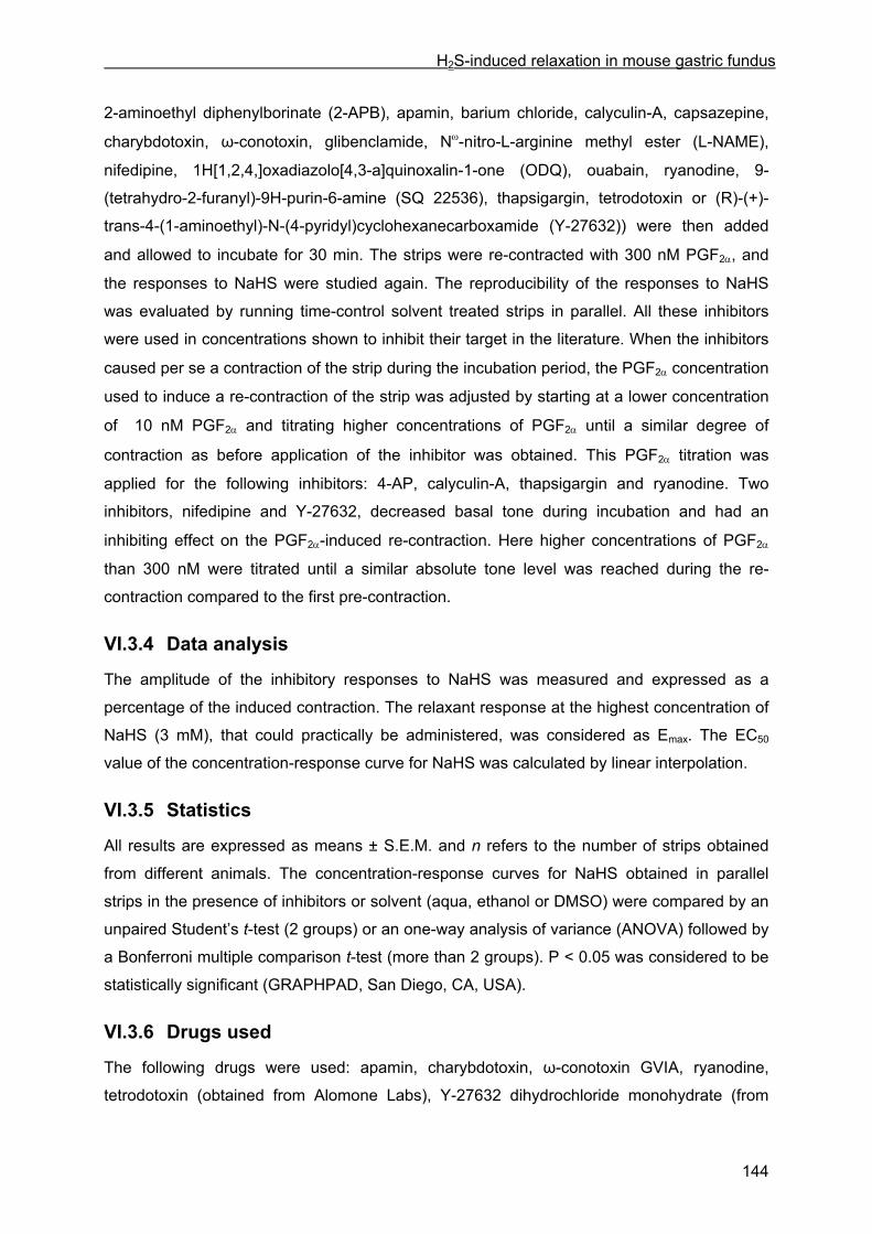
H2S-induced relaxation in mouse gastric fundus
144
2-aminoethyl diphenylborinate (2-APB), apamin, barium chloride, calyculin-A, capsazepine,
charybdotoxin, ω-conotoxin, glibenclamide, Nω-nitro-L-arginine methyl ester (L-NAME),
nifedipine, 1H[1,2,4,]oxadiazolo[4,3-a]quinoxalin-1-one (ODQ), ouabain, ryanodine, 9-
(tetrahydro-2-furanyl)-9H-purin-6-amine (SQ 22536), thapsigargin, tetrodotoxin or (R)-(+)-
trans-4-(1-aminoethyl)-N-(4-pyridyl)cyclohexanecarboxamide (Y-27632)) were then added
and allowed to incubate for 30 min. The strips were re-contracted with 300 nM PGF2α, and
the responses to NaHS were studied again. The reproducibility of the responses to NaHS
was evaluated by running time-control solvent treated strips in parallel. All these inhibitors
were used in concentrations shown to inhibit their target in the literature. When the inhibitors
caused per se a contraction of the strip during the incubation period, the PGF2α concentration
used to induce a re-contraction of the strip was adjusted by starting at a lower concentration
of 10 nM PGF2α and titrating higher concentrations of PGF2α until a similar degree of
contraction as before application of the inhibitor was obtained. This PGF2α titration was
applied for the following inhibitors: 4-AP, calyculin-A, thapsigargin and ryanodine. Two
inhibitors, nifedipine and Y-27632, decreased basal tone during incubation and had an
inhibiting effect on the PGF2α-induced re-contraction. Here higher concentrations of PGF2α
than 300 nM were titrated until a similar absolute tone level was reached during the re-
contraction compared to the first pre-contraction.
VI.3.4 Data analysis
The amplitude of the inhibitory responses to NaHS was measured and expressed as a
percentage of the induced contraction. The relaxant response at the highest concentration of
NaHS (3 mM), that could practically be administered, was considered as Emax. The EC50
value of the concentration-response curve for NaHS was calculated by linear interpolation.
VI.3.5 Statistics
All results are expressed as means ± S.E.M. and n refers to the number of strips obtained
from different animals. The concentration-response curves for NaHS obtained in parallel
strips in the presence of inhibitors or solvent (aqua, ethanol or DMSO) were compared by an
unpaired Student’s t-test (2 groups) or an one-way analysis of variance (ANOVA) followed by
a Bonferroni multiple comparison t-test (more than 2 groups). P < 0.05 was considered to be
statistically significant (GRAPHPAD, San Diego, CA, USA).
VI.3.6 Drugs used
The following drugs were used: apamin, charybdotoxin, ω-conotoxin GVIA, ryanodine,
tetrodotoxin (obtained from Alomone Labs), Y-27632 dihydrochloride monohydrate (from

H2S-induced relaxation in mouse gastric fundus
145
Calbiochem), 4-AP, 2-APB, capsazepine, glibenclamide, L-NAME, nifedipine, ouabain
octahydrate, prostaglandin F2α tris salt, NaHS hydrate, SQ 22536, thapsigargin (from
Sigma-Aldrich), barium chloride (from Merck), carbachol (from Fluka AG), calyculin-A, ODQ
(from Tocris Cookson). Drugs were dissolved in de-ionized water except 2-APB,
capsazepine, glibenclamide, nifedipine, ODQ, ryanodine, thapsigargin which were dissolved
in ethanol and calyculin-A which was dissolved in dimethyl sulfoxide (DMSO). Solutions of
NaHS were prepared fresh on the morning of each experiment and kept on ice.
VI.4 Results
VI.4.1 Basic observations
In PGF2α-contracted gastric fundus strips, cumulatively added NaHS induced concentration-
dependent relaxations. Concentrations higher than 30 µM NaHS were needed to induce
relaxation (Fig. VI.1). In the majority of the strips, the NaHS-induced relaxations progressed
to a stabile sustained degree. However, in 31/145 (21 %) of the strips tested, at one or more
concentrations of NaHS, the tone partially recovered after reaching a maximal degree of
relaxation; the latter was used for calculations. In all strips treated with aqua, ethanol or
DMSO (i.e. control strips, n = 54), this yielded an overall mean Emax and EC50 of respectively
132 ± 3 % and 514 ± 36 µM for the NaHS-induced relaxation.
The cumulative concentration-response curves were reproducible within the same strip,
except for 300 µM NaHS. In the majority of the control strips, independently whether they
were treated with aqua, ethanol (Fig. VI.1) or DSMO before constructing the second
concentration-response curve to NaHS, NaHS at a concentration of 300 µM induced a larger
relaxation during the second compared to the first concentration-response curve. As the first
concentration-response curves of NaHS in parallel strips were always comparable, the
influence of inhibitors on the relaxations by NaHS was thus assessed by comparing the
concentration-response curves in the presence of the inhibitors with those in the presence of
their solvent in the parallel tissues (see VI.3.5).
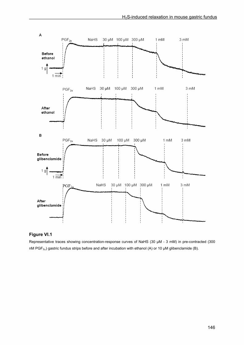
H2S-induced relaxation in mouse gastric fundus
146
Figure VI.1 Representative traces showing concentration-response curves of NaHS (30 µM - 3 mM) in pre-contracted (300
nM PGF2α) gastric fundus strips before and after incubation with ethanol (A) or 10 µM glibenclamide (B).
H2S-induced relaxation in mouse gastric fundus
147
VI.4.2 Influence of glibenclamide, ODQ plus SQ 22536, L-NAME, tetrodotoxin and ω-conotoxin
The influence of the KATP channel blocker glibenclamide on the NaHS-induced relaxation was
first examined; the NaHS-induced relaxation was unaltered by incubation of the strips with
glibenclamide (10 µM) (Figs. VI.1B and VI.2A). The combination of ODQ (10 µM) and SQ
22536 (100 µM), inhibitors of respectively soluble guanylate cyclase (sGC) and adenylate
cyclase (AC), did also not influence the NaHS-induced relaxation (Fig. VI.2B). The NO
synthase inhibitor L-NAME (300 µM), the Na+ channel blocker tetrodotoxin (3 µM) and the N-
type voltage dependent Ca2+ channel blocker ω-conotoxin (3 µM) did not have any effect on
the NaHS-induced relaxation (Fig. VI.2C and D).
Figure VI.2
Concentration-response curves of NaHS (30 µM - 3 mM) in the presence of A) ethanol ( ) or glibenclamide (10
µM ; ∆), B) ethanol ( ) or ODQ (10 µM) plus SQ 22536 (100 µM ; ∆), C) aqua ( ) or L-NAME (300 µM, ∆) or
tetrodotoxin (3 µM , ○), D) aqua ( ) or ω-conotoxin (3 µM, ∆). Means ± S.E.M. of n = 8 (except for 3 mM: n = 4)
are shown in A, means ± S.E.M. of n = 4-5 are shown in B, C and D.

H2S-induced relaxation in mouse gastric fundus
148
VI.4.3 Influence of channel and receptor blockers
A second set of inhibitors was tested, targeting ion channels/receptors located in the plasma
membrane of smooth muscle cells or neurons or the membrane of the sarcoplasmatic
reticulum of smooth muscle cells, possibly involved in relaxation. The full list of the tested
inhibitors is shown in Table VI.1. 4-AP, ryanodine and thapsigargin caused per se a
contraction and elevated the basal tone by respectively 93 ± 5 % (n = 4), 45 ± 3 % (n = 6)
and 68 ± 12 % (n = 6) expressed as a percentage of the first PGF2α-induced contraction.
Nifedipine (30 nM) on the other hand decreased the basal tone by 14 ± 4 % (n = 6)
compared to the first PGF2α-induced contraction. None of the inhibitors listed in Table VI.1
had a significant influence on the Emax and EC50 of the NaHS-induced relaxation.
Table VI.1: Emax (%) and EC50 (µM) for the relaxant effect of NaHS in the presence of inhibitors of the listed targets, or their solvent (control)
inhibitor target n Emax (%) EC50 (µM)
* Control (ethanol) 4 120 ± 1 429 ± 147
ODQ (10 µM) +
Apamin (500 nM)
sGC +
small conductance Ca2+ dependent K+
channel
4 117 ± 7 301 ± 57
* Control (aqua) 5 117 ± 5 364 ± 73
Apamin (500 nM) +
Charybdotoxin (100 nM)
small conductance +
intermediate and large conductance
Ca2+ dependent K+ channel
4 131 ± 8 354 ± 77
* Control (aqua) 3 117 ± 1 369 ± 64
Ouabain (100 µM) Na+/K+ ATPase 4 123 ± 9 476 ± 150
4-AP (5 mM) voltage dependent K+ channel 4 122 ± 6 360 ± 84
Barium chloride (30 µM) inward rectifier K+ channel 4 131 ± 3 445 ± 63
* Control (ethanol) 4 120 ± 1 334 ± 56
2-APB (50 µM) IP3 receptor 4 110 ± 3 442 ± 179
Capsazepine (3 µM) transient receptor potential vanilloid
type 1 (TRPV1) 4 118 ± 3 407 ± 116
* Control (ethanol) 6 123 ± 6 411 ± 85
Nifedipine (30 nM) L-type voltage dependent Ca2+ channel 6 113 ± 4 563 ± 67
* Control (ethanol) 6 141 ± 5 590 ± 119
Ryanodine (10 µM) ryanodine receptor 6 133 ± 7 428 ± 61
* Control (ethanol) 5 129 ± 6 820 ± 96
Thapsigargin (1 µM) sarcoplasmatic reticulum Ca2+ ATPase 6 161 ± 20 633 ± 37

H2S-induced relaxation in mouse gastric fundus
149
VI.4.4 Influence of calyculin-A and Y-27632
Calyculin-A is an inhibitor of myosin light chain phosphatase (MLCP) and Y-27632 an
inhibitor of Rho-kinase. Both targets are involved in the desensitization of the smooth muscle
contractile apparatus to the intracellular Ca2+-concentration.
Calyculin-A (1 µM) elevated the basal tone by 79 ± 10 % (n = 8) compared to the first PGF2α-
induced contraction (Fig. VI.3A). PGF2α administered on top of the calyculin-A-induced
contraction only induced a minor additional contraction. Still, the active contraction level
obtained in the presence of calyculin-A plus PGF2α attained 86 ± 9 % (n = 8) of that by the
first administration of PGF2α.
Figure VI.3 Representative traces showing concentration-response curves of NaHS (30 µM, 100 µM, 300 µM, 1 mM, 3mM
administered at the arrows) in PGF2α-pre-contracted gastric fundus strips before and after incubation with 1 µM
calyculin-A (A) or 10 µM Y-27632 (B). For the first pre-contraction 300 nM PGF2α was administered. For the
second pre-contraction, PGF2α was administered in concentrations of 10, 30, 100 and 300 nM after incubation
with calyculin-A (A) or of 0.3, 0.5, 0.7, 1, 3, 5 and 7 µM after incubation with Y-27632 (B), aiming for a similar
absolute tone level (passive plus active) after versus before incubation with calyculin-A or Y-27632. The reference
contraction level used to express the NaHS-induced relaxations is indicated as 100 %.
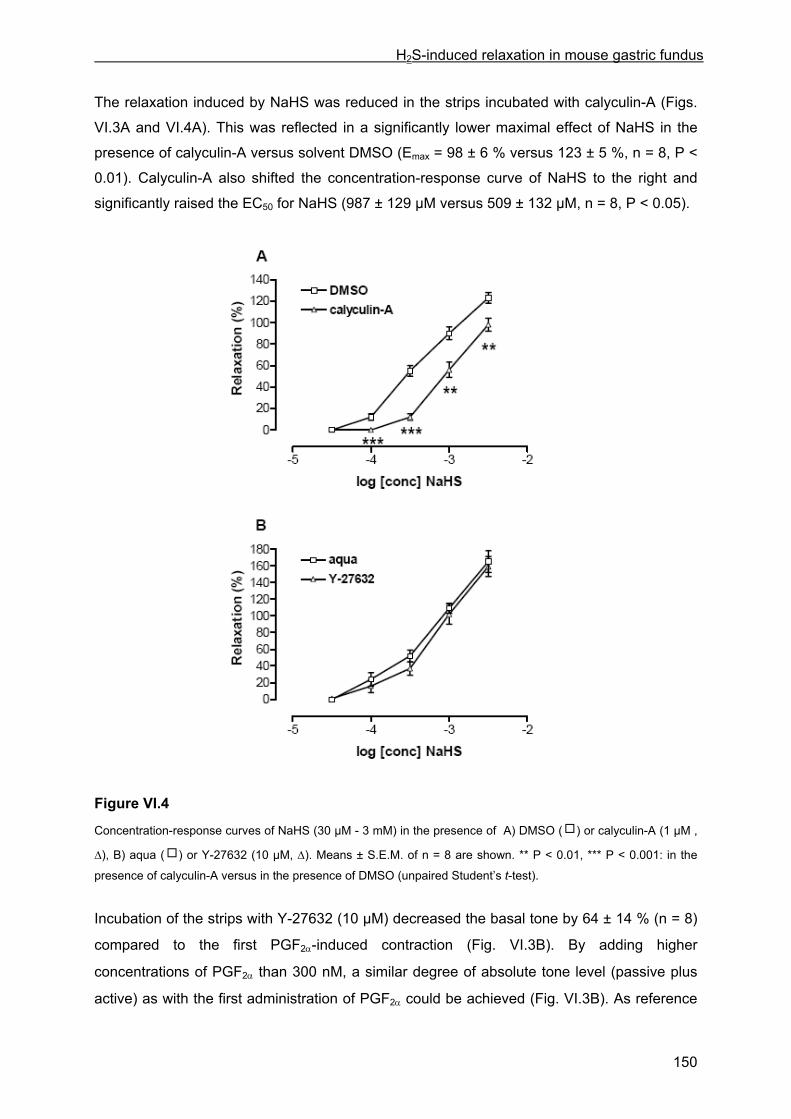
H2S-induced relaxation in mouse gastric fundus
150
The relaxation induced by NaHS was reduced in the strips incubated with calyculin-A (Figs.
VI.3A and VI.4A). This was reflected in a significantly lower maximal effect of NaHS in the
presence of calyculin-A versus solvent DMSO (Emax = 98 ± 6 % versus 123 ± 5 %, n = 8, P <
0.01). Calyculin-A also shifted the concentration-response curve of NaHS to the right and
significantly raised the EC50 for NaHS (987 ± 129 µM versus 509 ± 132 µM, n = 8, P < 0.05).
Figure VI.4
Concentration-response curves of NaHS (30 µM - 3 mM) in the presence of A) DMSO ( ) or calyculin-A (1 µM ,
∆), B) aqua ( ) or Y-27632 (10 µM, ∆). Means ± S.E.M. of n = 8 are shown. ** P < 0.01, *** P < 0.001: in the
presence of calyculin-A versus in the presence of DMSO (unpaired Student’s t-test).
Incubation of the strips with Y-27632 (10 µM) decreased the basal tone by 64 ± 14 % (n = 8)
compared to the first PGF2α-induced contraction (Fig. VI.3B). By adding higher
concentrations of PGF2α than 300 nM, a similar degree of absolute tone level (passive plus
active) as with the first administration of PGF2α could be achieved (Fig. VI.3B). As reference

H2S-induced relaxation in mouse gastric fundus
151
value to express the NaHS-induced relaxations, the second pre-contraction was measured
as the difference between the PGF2α-induced absolute tone level and the tone level present
before the administration of Y-27632 (Fig. VI.3B). Using active PGF2α-induced contraction as
reference would indeed mean that NaHS-induced relaxations are expressed as percentage
of a much higher value than for the first administration cycle. In the presence of Y-27632, the
maximal relaxing effect of NaHS as well as the EC50 for NaHS were not significantly
influenced versus in the presence of the solvent aqua (Emax = 159 ± 12 % versus 165 ± 13 %;
EC50 = 805 ± 127 µM versus 630 ± 85 µM, n = 8) (Fig. VI.4B).
VI.5 Discussion
In this study, we investigate the relaxing effect of H2S on the tonic smooth muscle of the
mouse gastric fundus. Using NaHS makes it possible to define the concentrations of H2S in
solution more accurately and reproducibly than bubbling H2S gas (Hosoki et al., 1997).
In pre-contracted mouse gastric fundus strips, NaHS induced concentration-dependent
relaxations with a threshold of 100 µM and an EC50 of 514 ± 36 µM. These values are higher
than previously reported for NaHS in GI tissues. For inhibition of spontaneous contractions in
rabbit ileum: EC50, 76 µM, threshold, 10 µM; inhibition of electrically evoked contractions in
rat and guinea-pig ileum: EC50, 65 µM and 63 µM, threshold, 10 µM (Teague et al., 2002).
For relaxation of acetylcholine pre-contracted guinea-pig ileum: EC50, 180 µM, lowest tested
concentration 100 µM (Hosoki et al.,1997). Also vascular tissues seem more sensitive to
NaHS: rat aorta, EC50, 136 µM, threshold, 60 µM (Zhao and Wang, 2002); rat mesenteric
artery, EC50, 104 µM, threshold, 0.1 µM (Cheng et al., 2004). But the concentration-response
curve is similar to that reported in mouse carbachol-contracted bronchial rings (Kubo et al.,
2007a, EC50 not reported) so that species differences might be important.
In our study, the KATP channel blocker glibenclamide had no influence on the relaxing effect
of NaHS, arguing against a role of KATP channels in the NaHS-induced relaxation in mouse
gastric fundus. This is in contrast to cardiovascular tissues, where KATP channels are
reported to be involved in the NaHS-induced relaxation, such as in rat aorta (Zhao et al.,
2001), rat mesenteric artery (Cheng et al., 2004) and human internal mammary artery (Webb
et al., 2008). An exception however is the mouse aorta, where glibenclamide failed to affect
the relaxing effect of NaHS (Kubo et al., 2007b). Our result is also in correspondence with
the non-effect of glibenclamide on the NaHS-induced bronchodilation in mice (Kubo et al.,
2007a) and on the NaHS-induced inhibition of the electrically induced contractions in the
guinea-pig ileum (Teague et al., 2002).
Zhao and Wang (2002) reported the release of endothelium-derived NO by H2S in
endothelium-intact rat aortic tissues. The possible contribution of NO, released from nitrergic
nerves, in the relaxation by NaHS of mouse gastric fundus was excluded as the NO-

H2S-induced relaxation in mouse gastric fundus
152
synthase inhibitor L-NAME had no influence. This is in correspondence with Teague et al.
(2002), who reported that the inhibitory effect of NaHS on electrically induced contractions of
the guinea-pig ileum was unaffected by L-NAME, and is corroborated by our observation that
inhibiting the principal target of NO in mouse gastric fundus (Vanneste et al., 2007), i.e.
soluble guanylate cyclase, with ODQ did not influence the NaHS-induced relaxation.
Activation of another non-nitrergic relaxant neuron type is also excluded as the Na+ channel
blocker tetrodotoxin, inhibiting action potential propagation, and the N-type voltage
dependent Ca2+ channel blocker ω-conotoxin, interfering with exocytosis at the nerve ending,
had no influence. In human and guinea-pig colon, the prosecretory effect of NaHS involves
the activation of the transient receptor potential vanilloid type 1 (TRPV1) on the extrinsic
primary afferent nerve terminals, which in turn activates the enteric neurons (Schicho et al.,
2006). In the rat urinary bladder, NaHS stimulates the primary afferent nerve terminals to
induce contraction (Patacchini et al., 2004). But the TRPV1 blocker capsazepine had no
influence on NaHS-induced relaxation in mouse gastric fundus.
ODQ was tested together with the adenylate cyclase inhibitor SQ 22536, as cAMP
generation is also a classic relaxant transduction pathway in smooth muscle e.g. for VIP
(Hagen et al., 2006), a peptide involved in sustained non-adrenergic non-cholinergic
relaxation of gastric fundus (De Beurme and Lefebvre, 1987). As ODQ plus SQ 22536 was
ineffective also the cAMP transduction pathway can be excluded for the relaxant effect of
NaHS. ODQ was also tested in combination with the small conductance Ca2+ dependent K+
channel (SKCa channel) blocker apamin, as this combination was required to fully inhibit the
relaxation by exogenous NO in rat colon (Van Crombruggen and Lefebvre, 2004), but this
had no influence versus NaHS. In mouse gastric fundus, the relaxation by exogenous CO is
significantly reduced by apamin plus the intermediate (IKCa) and large (BKCa) conductance
Ca2+ dependent K+ channel blocker charybdotoxin, while either agent alone had no influence
(De Backer et al., 2007). However, the relaxant effect of NaHS in mouse gastric fundus was
not influenced by apamin plus charybdotoxin, excluding an interactive contribution of a SKCa
channel and a IKCa or BKCa channel. The influence of barium chloride and 4-AP, blockers of
respectively the voltage dependent and the inward rectifier K+ channel, was therefore tested.
4-AP caused per se a contraction of the gastric fundus strips, which was also observed in
mouse trachea rings by Li et al. (1998). An inhibiting effect of 4-AP and barium on the
acetylcholine-induced relaxation in canine basilar artery was previously shown by Elliott et al.
(1991), indicating that the voltage dependent and the inward rectifier K+ channel can play a
role in smooth muscle relaxation. However, 4-AP as well as barium chloride did not affect the
NaHS-induced relaxation. Taken together, this indicates that the above mentioned K+
channels, which were reported to play a role in neurotransmitter-induced relaxation, are not
involved in the NaHS-induced relaxation in mouse gastric fundus.

H2S-induced relaxation in mouse gastric fundus
153
In rat aortic tissues, the H2S-induced vasorelaxation was reduced by the L-type voltage
dependent Ca2+ channel blocker nifedipine and was as such shown to be dependent on
interference with extracellular calcium entry (Zhao and Wang, 2002). In mouse gastric fundus
strips, nifedipine per se decreased the basal tone, as was previously observed in dog
cerebral arteries by Asano et al. (1996), and reduced the PGF2α-induced contraction, as was
previously reported in rat gastric fundus by Van Geldre and Lefebvre (2004). Consequently,
higher concentrations of PGF2α were needed to induce contraction in the presence of
nifedipine but in these conditions, nifedipine had no effect on the NaHS-induced relaxation in
mouse gastric fundus. As stimulation of Na+/K+ ATPase was reported to be involved in the
NO-induced relaxation in mouse gastric fundus strips and rabbit aorta by respectively
Yaktubay et al. (1998) and Gupta et al. (1994), we investigated whether Na+/K+ ATPase
could also be involved in the NaHS-induced relaxation. However, the Na+/K+ ATPase
inhibitor ouabain had no influence on the relaxation induced by NaHS. The sarcoplasmatic
reticulum Ca2+ ATPase (SERCA) inhibitor thapsigargin was reported to decrease the sodium
nitroprusside (SNP)-induced inhibitory effects in mouse ileum (Zizzo et al., 2005), whereas
ryanodine and 2-APB, inhibitors of respectively the ryanodine receptor and the IP3 receptor,
decreased the SNP-induced relaxation of mouse gastric fundus (Kim et al., 2006).
Thapsigargin and ryanodine caused per se a contraction of the mouse gastric fundus strips,
as previously observed in respectively mouse anococcygeus (Wallace et al., 1999) and dog
cerebral arteries (Asano et al., 1996). Thapsigargin nor ryanodine or 2-APB influenced the
NaHS-induced relaxation, arguing against the involvement of calcium
mobilization/sequestration at the level of the sarcoplasmatic reticulum in the NaHS-induced
relaxation.
Relaxation in smooth muscle cells can only be induced through a decrease in the
intracellular calcium concentration, and/or through a desensitization of the contractile
proteins to the intracellular calcium concentration. Calcium desensitization is accomplished
by the activation of myosin light chain phosphatase (MLCP), leading to a higher degree of
dephosphorylation of myosin light chain (MLC) and subsequently relaxation. The
phosphatase inhibitor calyculin-A (1 µM) was reported to induce contraction of canine antral
smooth muscle together with MLC phosphorylation but without increasing the intracellular
calcium concentration (Ozaki et al., 1991). Similarly, calyculin-A (1 µM) per se caused
contraction of mouse gastric fundus strips, illustrating that MLCP is tonically active in this
tissue. Although Ozaki et al. (1991) mention in their discussion that calyculin-A is much more
effective versus phosphatase type 1 than versus phosphatase type 2, Ishihara et al. (1989a)
reported similar IC50 values of calyculin-A versus type 1 and type 2A phosphatase (0.5-2
nM). Phosphatase type 1c is part of the physiologically active MLCP in smooth muscle cells
(Somlyo and Somlyo, 2003; Ito et al., 2004) so that the effects of calyculin-A in smooth
H2S-induced relaxation in mouse gastric fundus
154
muscle can probably be ascribed to interference with phosphatase type 1. In intact smooth
muscle preparations, higher concentrations than the low nM range are required to induce
effects. In guinea pig taenia caeci e.g., calyculin-A induced contraction with an EC50 of 210
nM and an Emax at 3 µM (Ishihara et al., 1989b). In the mouse anococcygeus, calyculin-A (1
µM) nearly abolished relaxations induced by endogenous NO, liberated by electrical field
stimulation; this implies that calcium desensitization by activation of MLCP is the major
cellular mechanism underlying nitrergic relaxation in this tissue (Gibson et al., 2003).
Similarly, calyculin-A (1 µM) reduced the NaHS-induced relaxations in mouse gastric fundus,
suggesting that activation of MLCP is also involved in the relaxant effect of NaHS.
An important pathway controlling smooth muscle contraction is the RhoA/Rho-kinase
pathway, acting by inhibition of MLCP through phosphorylation of the MYPT1 unit and thus
promoting calcium sensitization and contraction (Somlyo and Somlyo, 2003). Inhibition of the
RhoA/Rho-kinase pathway will thus lead to relaxation. The Rho-kinase inhibitor Y-27632
lowered the basal tone of the mouse gastric strips, as was also observed in the rat internal
anal sphincter by Patel and Rattan (2006), illustrating that the RhoA/Rho-kinase pathway is
tonically active in the mouse gastric fundus under basal conditions. Büyükafşar and Levent
(2003) have shown before that Y-27632 concentration-dependently relaxed carbachol-
contracted mouse gastric fundus illustrating that also muscarinic receptor activation in this
tissue activates RhoA/Rho-kinase. In blood vessels, it was shown that the vasorelaxant
effect of NO can be mediated through inhibition of the RhoA/Rho-kinase pathway, and thus
subsequent increased activity of MLCP (Sauzeau et al., 2000). To investigate whether the
activation of MLCP by NaHS in the mouse gastric fundus, as revealed in the experiments
with calyculin-A, might also be related to suppression of the RhoA/Rho-kinase pathway, we
inhibited the latter with Y-27632; but in this condition, the relaxant effect of NaHS was
maintained excluding this possibility. Our results are similar to those reported for the relaxant
effect of the β-receptor agonist isoprenaline in guinea-pig tracheal smooth muscle, that was
abolished by calyculin-A but not influenced by Y-27632; also in this study it was concluded
that isoprenaline acts through activation of MLCP, without affecting the RhoA/Rho-kinase
pathway (Oguma et al., 2006).
In conclusion, we show that NaHS (donor of H2S) is able to relax PGF2α-contracted mouse
gastric fundus strips. The mechanism of action of the NaHS-induced relaxation in mouse
gastric fundus is different from that in the cardiovascular system as the relaxation induced by
NaHS does not involve activation of KATP channels. The results obtained in this study
suggest that in the mouse gastric fundus, H2S causes relaxation at least partially via
activation of myosin light chain phosphatase; this seems not related to an inhibition of the
RhoA/Rho-kinase pathway.
H2S-induced relaxation in mouse gastric fundus
155
VI.6 References Abe, K., Kimura, H., 1996. The possible role of hydrogen sulfide as an endogenous neuromodulator.
J. Neurosci. 16, 1066-1071.
Asano, M., Kuwako, M., Nomura, Y., Suzuki, Y., Shibuya, M., Sugita, K., Ito, K., 1996. Possible
mechanism of the potent vasoconstrictor responses to ryanodine in dog cerebral arteries. Eur. J.
Pharmacol. 311, 53-60.
Beauchamp, O. Jr, Bus, J.S., Popp, J.A., Boreiko, C.J., Andjelkovich, D.A., 1984. A critical review of
the literature on hydrogen sulfide toxicity. Crit Rev. Toxicol. 13, 25-97.
Büyükafşar, K., Levent, A., 2003. Involvement of Rho/Rho-kinase signalling in the contractile activity
and acetylcholine release in the mouse gastric fundus. Biochem. Biophys. Commun. 303, 777-781.
Cheng, Y., Ndisang, J.F., Tang, G., Cao, K., Wang, R., 2004. Hydrogen sulfide-induced relaxation of
resistance mesenteric artery beds of rats. Am. J. Physiol Heart Circ. Physiol 287, H2316-H2323.
Curro, D., De Marco, T., Preziosi, P., 2004. Evidence for an apamin-sensitive, but not purinergic,
component in the nonadrenergic noncholinergic relaxation of the rat gastric fundus. Br. J. Pharmacol.
143, 785-793.
De Backer, O., Lefebvre, R.A., 2007. Mechanisms of relaxation by carbon monoxide-releasing
molecule-2 in murine gastric fundus and jejunum. Eur. J. Pharmacol. 572, 197-206.
De Beurme, F.A., Lefebvre, R.A., 1987. Influence of α-chymotrypsin and trypsin on the non-adrenergic
non-cholinergic relaxation in the rat gastric fundus. Br. J. Pharmacol. 91, 171-177.
Distrutti, E., Sediari, L., Mencarelli, A., Renga, B., Orlandi, S., Antonelli, E., Roviezzo, F., Morelli, A.,
Cirino, G., Wallace, J.L., Fiorucci, S., 2006. Evidence that hydrogen sulfide exerts antinociceptive
effects in the gastrointestinal tract by activating KATP channels. J. Pharmacol. Exp. Ther. 316, 325-
335.
Elliott, D.A., Gu, M., Ong, B.Y., Bose, D., 1991. Inhibition of the acetylcholine-induced relaxation of
canine isolated basilar artery by potassium-conductance blockers. Can. J. Physiol. Pharmacol. 69,
786-791.
Gibson, A., Wallace, P., McFadzean, I., 2003. Calyculin-A inhibits nitrergic relaxations of the mouse
anococcygeus. Eur. J. Pharmacol. 471, 213-215.
Gupta, S., McArthur, C., Grady, C., Ruderman, N.B., 1994. Stimulation of vascular Na+K+-ATPase
activity by nitric oxide: a cGMP-independent effect. Am. J. Physiol. 266, H2146-2151.
H2S-induced relaxation in mouse gastric fundus
156
Hagen, B.M., Bayguinov, O., Sanders, K.M., 2006. VIP and PACAP regulate localized Ca2+ transients
via cAMP-dependent mechanism. Am. J. Physiol. Cell Physiol. 291, C375-C385.
Hosoki, R., Matsuki, N., Kimura, H., 1997. The possible role of hydrogen sulfide as an endogenous
smooth muscle relaxant in synergy with nitric oxide. Biochem. Biophys. Res. Commun. 237, 527-531.
Ishihara, H., Martin, B.L., Brautigan, D.L., Karaki, H., Ozaki, H., Kato, Y., Fusetani, N., Watabe, S.,
Hashimoto, K., Uemura, D., Hartshorne, D.J., 1989a. Calyculin A and okadaic acid. Inhibitors of
protein phosphatase activity. Biochem. Biophys. Commun. 159, 871-877.
Ishihara, H., Ozaki, H., Sato, K., Hori, M., Karaki, H., Watabe, S., Kato, Y., Fusetani, N., Hashimoto,
K., Uemura, D., Hartshorne, D.J., 1989b. Calcium-independent activation of contractile apparatus in
smooth muscle by calyculin-A. J. Pharmacol. Exp. Ther. 250, 388-396.
Ito, M., Nakano, T., Erdődi, F., Hartshorne, D.J., 2004. Myosin phosphatase: Structure, regulation and
function. Mol. Cell. Biochem. 259, 197-209.
Kim, M., Han, I.S., Koh, S.D., Perrino, B.A., 2006. Roles of CaM kinase II and phospholamban in
SNP-induced relaxation of murine gastric fundus smooth muscles. Am. J. Physiol. Cell Physiol. 291,
C337-C347.
Kubo, S., Doe, I., Kurokawa, Y., Kawabata, A., 2007a. Hydrogen sulfide causes relaxation in mouse
bronchial smooth muscle. J. Pharmacol. Sci. 104, 392-396.
Kubo, S., Doe, I., Kurokawa, Y., Nishikawa, H., Kawabata, A., 2007b. Direct inhibition of endothelial
nitric oxide synthase by hydrogen sulfide: contribution to dual modulation of vascular tension.
Toxicology 232, 138-146.
Li, L., Paakkari, I., Vapaatalo, H., 1998. Effects of K+ channel inhibitors on the basal tone and KCl- or
methacholine-induced contraction of mouse trachea. Eur. J. Pharmacol. 346, 255-260
Linden, D.R., Sha, L., Mazzone, A., Stoltz, G.J., Bernard, C.E., Furne, J.K., Levitt, M.D., Farrugia, G.,
Szurszewski, J.H., 2008. Production of the gaseous signal molecule hydrogen sulfide in mouse
tissues. J. Neurochem. 106, 1577-1585.
Oguma, T., Kume, H., Ito, S., Takeda, N., Honjo, H., Kodama, I., Shimokata, K., Kamiya, K., 2006.
Involvement of reduced sensitivity to Ca2+ in β-adrenergic action on airway smooth muscle. Clin. Exp.
Allergy 36, 183-191.
Ozaki, H., Gerthoffer, W.T., Publicover, N.G., Fusetani, N., Sanders, K.M., 1991. Time-dependent
changes in Ca2+ sensitivity during phasic contraction of canine antral smooth muscle. J. Physiol. 440,
207-224.
H2S-induced relaxation in mouse gastric fundus
157
Patacchini, R., Santicioli, P., Giuliani, S., Maggi, C.A., 2004. Hydrogen sulfide (H2S) stimulates
capsaicin-sensitive primary afferent neurons in the rat urinary bladder. Br. J. Pharmacol. 142, 31-34.
Patel, C.A., Rattan, S., 2006. Spontaneously tonic smooth muscle has characteristically higher levels
of RhoA/ROK compared with the phasic smooth muscle. Am. J. Gastrointest. Liver Physiol. 291,
G830-G837.
Sauzeau, V., Le Jeune, H., Cario-Toumaniantz, C., Smolenski, A., Lohmann, M., Bertoglio, J.,
Chardin, P., Pacaud, P., Loirand, G., 2000. Cyclic GMP-dependent protein kinase signaling pathway
inhibits RhoA-induced Ca2+ sensitization of contraction in vascular smooth muscle. J. Biol. Chem. 275,
21722-21729.
Schicho, R., Krueger, D., Zeller, F., Von Weyhern, C.W.H., Frieling, T., Kimura, H., Ishii, I., De Giorgio,
R., Campi, B., Schemann, M., 2006. Hydrogen sulfide is a novel prosecretory neuromodulator in the
guinea-pig and human colon. Gastroenterology 131, 1542-1552.
Somlyo, A.P., Somlyo, A., 2003. Ca2+ sensitivity of smooth muscle and nonmuscle myosin II:
modulated by G proteins, kinases, and myosine phosphatase. Physiol.Rev. 83, 1325-1358.
Stipanuk, M.H., Beck, P.W., 1982. Characterization of the enzymic capacity for cysteine
desulphhydration in liver and kidney of the rat. Biochem. J. 206, 267-277.
Tang, G., Wu, L., Liang, W., Wang, R., 2005. Direct stimulation of KATP channels by exogenous and
endogenous hydrogen sulfide in vascular smooth muscle cells. Mol. Pharmacol. 68, 1757-1764.
Teague, B., Asiedu, S., Moore, P.K., 2002. The smooth muscle relaxant effect of hydrogen sulphide in
vitro: evidence for a physiological role to control intestinal contractility. Br. J. Pharmacol. 137, 139-145.
Van Crombruggen, K., Lefebvre, R.A., 2004. Nitrergic-purinergic interactions in rat distal colon motility.
Neurogastroenterol. Motil. 16, 81-98.
Van Geldre, L.A., Lefebvre, R.A., 2004. Nitrergic relaxation in rat gastric fundus: influence of
mechanism of induced tone and possible role of sarcoplasmatic/endoplasmatic reticulum Ca2+
ATPase. Life Sci. 74, 3259-3274.
Vanneste ,G., Dhaese, I., Sips, P., Buys, E., Brouckaert, P., Lefebvre, R.A., 2007. Gastric motility in
soluble guanylate cyclase alpha1 knockout mice. J. Physiol 584, 907-920.
Wallace, P., Ayman, S., McFadzean, I., Gibson, A., 1999. Thapsigargin-induced tone and capacitative
calcium influx in mouse anococcygeus smooth muscle cells. Naunyn- Schmiedeberg's Arch.
Pharmacol. 360, 368-375.
H2S-induced relaxation in mouse gastric fundus
158
Wang, Y.F., Mainali, P., Tang, C.S., Shi, L., Zhang, C.Y., Yan, H., Liu, X-q., Du, J-b., 2008. Effects of
nitric oxide and hydrogen sulfide on the relaxation of pulmonary arteries in rats. Chin Med. J. 121,
420-423.
Webb, G.D., Lim, L.H., Oh, V.M., Yeo, S.B., Cheong, Y.P., Ali, M.Y., Oakley, R.E., Lee, C.N., Wong,
P.S., Caleb, M.G., Salto-Tellez, M., Bhatia, M., Chan, .S.Y., Taylor, E.A., Moore, P.K., 2008.
Contractile and vasorelaxant effects of hydrogen sulfide and its biosynthesis in the human internal
mammary artery. J. Pharmacol. Exp. Ther. 324, 876-882.
Yaktubay, N., Ögülener, N., Önder, S., Baysal, F., 1999. Possible stimulation of Na+-K+-ATPase by
NO produced from sodium nitrite by ultraviolet light in mouse gastric fundal strip. Gen. Pharmac. 32,
159-162.
Zhao, W., Zhang, H.J., Lu, Y., Wang, R., 2001. The vasorelaxant effect of H2S as a novel endogenous
gaseous KATP channel opener. EMBO J. 20, 6008-6016.
Zhao, W., Wang, R., 2002. H2S-induced vasorelaxation and underlying cellular and molecular
mechanisms. Am. J. Physiol. Heart Circ. Physiol. 283, H474-H480.
Zizzo, M.G., Mulé, F., Serio, R., 2005. Mechanisms underlying the nitric oxide inhibitory effects in
mouse ileal longitudinal muscle. Can. J. Physiol. Pharmacol. 83, 805-810.
Chapter VII
MECHANISMS OF ACTION OF HYDROGEN SULFIDE IN RELAXATION OF MOUSE DISTAL COLONIC SMOOTH MUSCLE Based on Eur. J. Pharmacol. 2010; 628: 179-186


H2S-induced relaxation in mouse distal colon
161
Chapter VII Mechanisms of action of hydrogen sulfide in relaxation of mouse distal colonic
smooth muscle
VII.1 Abstract
Hydrogen sulfide (H2S) has been suggested as a gaseous neuromodulator in mammals. The
aim of this study was to examine the influence of H2S on contractility in mouse distal colon.
The effect of sodium hydrogen sulfide (NaHS; H2S donor) on prostaglandin F2α (PGF2α)-
contracted circular muscle strips of mouse distal colon was investigated. In addition, tension
and cytosolic calcium concentration ([Ca2+]cyt) in the mouse distal colon strips were measured
simultaneously in the presence of NaHS. NaHS caused concentration-dependent relaxation
of the pre-contracted mouse distal colon strips. The NaHS-induced relaxation was not
influenced by the K+ channels blockers glibenclamide, apamin, charybdotoxin, barium
chloride and 4-aminopyridine. The relaxation by NaHS was also not influenced by the nitric
oxide inhibitor L-NAME, by the soluble guanylate cyclase respectively adenylate cyclase
inhibitors ODQ and SQ 22536, by the nerve blockers capsazepine, ω-conotoxin and
tetrodotoxin or by several channel and receptor blockers (ouabain, nifedipine, 2-aminoethyl
diphenylborinate, ryanodine and thapsigargin). The initiation of the NaHS-induced relaxation
was accompanied by an increase in [Ca2+]cyt, but once the relaxation was maximal and
sustained, no change in [Ca2+]cyt was measured. This calcium desensitization is not related to
the best known calcium desensitizing mechanism as the myosin light chain phosphatase
(MLCP) inhibitor calyculin-A and the Rho-kinase inhibitor Y-27632 had no influence.
We conclude that NaHS caused concentration-dependent relaxations in mouse distal colon
not involving the major known K+ channels and without a change in [Ca2+]cyt. This calcium
desensitization is not related to inhibition of Rho-kinase or activation of MLCP.
H2S-induced relaxation in mouse distal colon
162
VII.2 Introduction
Hydrogen sulfide (H2S) has been suggested as a third gaseous signal molecule and
neuromodulator in mammals, besides nitric oxide (NO) and carbon monoxide (CO). H2S can
be produced endogenously in mammalian tissues from L-cysteine by two enzymes,
cystathionine β-synthase (CBS) and cystathionine γ-lyase (CSE) (Stipanuk and Beck, 1982). Besides the detection of CBS mRNA and/or CSE mRNA and H2S generation in the brain
(Abe and Kimura, 1996; Linden et al., 2008) and in cardiovascular tissues (Zhao et al., 2001),
CBS and CSE mRNA as well as H2S generation were reported in guinea pig ileum (Hosoki et
al., 1997). Both CBS and CSE have also been shown in human and guinea-pig colon
myenteric neurons (Schicho et al., 2006).
In the cardiovascular system, H2S induces vasorelaxation at least partially due to activation
of ATP-dependent K+ channels (KATP channels) (Cheng et al., 2004; Tang et al., 2005; Webb
et al., 2008; Zhao et al., 2001). In the gastrointestinal (GI) tract, H2S inhibits acetylcholine-
induced (Hosoki et al., 1997) and electrically induced contractions of the guinea-pig ileum;
the latter effect was unaffected by the KATP channel inhibitor glibenclamide (Teague et al.,
2002). Similarly, we recently reported that the H2S-induced relaxation of mouse gastric
fundus is not sensitive to glibenclamide but appears at least partially related to the activation
of myosin light chain phosphatase and calcium desensitization (Dhaese and Lefebvre, 2009).
In mouse colon, CSE but not CBS was shown to be present in myenteric neurons, and the
intact colonic muscle layer containing the myenteric plexus generated detectable levels of
H2S (Linden et al., 2008). It is thus important to know the effect and mechanism of action of
H2S on the distal colon circular muscle layer, as H2S might play a physiological role in
smooth muscle regulation; a role for H2S as oxygen sensor, as suggested in vascular smooth
muscle by Olson et al. (2006), can not be excluded in the distal colon. Additionally, the
colonic muscle layer might be reached by luminally produced H2S in pathological conditions.
Indeed, ulcerative colitis is associated with an increased fecal H2S production (Levine et al.,
1998).
In the present experiments, we therefore investigated the influence and mechanism of action
of H2S on pre-contracted mouse distal colon circular muscle strips. Recently, while finishing
our study, Gallego et al. (2008) reported that H2S inhibited spontaneous motor complexes in
perfused 3 cm segments of mouse colon, an effect reduced by the small conductance Ca2+-
dependent K+ channel (SKCa channel) blocker apamin. In the present study however, we
show that in mouse distal colon strips apamin was not effective versus H2S, nor were
inhibitors of other major known K+ channels and ion channels; the sustained H2S-induced
relaxation in mouse distal colon strips was not accompanied by a change in [Ca2+]cyt.

H2S-induced relaxation in mouse distal colon
163
VII.3 Material and Methods
VII.3.1 Animals
Male Swiss (SPF Orl) mice (7-12 weeks, 26-46 g, n = 91) were purchased from Janvier, Le
Genest St-Isle, France and had free access to water and commercially available chow. All
experimental procedures were approved by the Ethical Committee for Animal Experiments
from the Faculty of Medicine and Health sciences at Ghent university.
VII.3.2 Tissue preparation
Animals were sacrificed by cervical dislocation. The distal colon was taken above the pelvic
brim as an approximately four cm segment and was put in Krebs solution (composition in
mM: NaCl 118.5, KCl 4.8, KH2PO4 1.2, MgSO4 1.2, CaCl2 1.9, NaHCO3 25.0 and glucose
10.1). The segment of distal colon was opened along the mesenteric border and pinned
mucosa side up. The mucosa was removed by sharp dissection under a microscope and four
full-thickness muscle strips (3 × 5 mm) were cut along the circular axis. After a silk thread
(USP 4/0) was attached to both ends of the muscle strips, strips were mounted along the
circular axis in 5 ml organ baths. The organ baths contained aerated (5% CO2 in O2) Krebs
solution maintained at 37°C. Changes in isometric tension were measured using MLT 050/D
force transducers (ADInstruments) and recorded on a PowerLab/8sp data recording system
(ADInstruments) with Chart software.
VII.3.3 Measurement of contractile tension
NaHS, the experimental source of H2S was used. Preliminary experiments showed that
cumulative administration of NaHS yielded less pronounced relaxant responses than isolated
administration with a threshold concentration varying between strips; this cumulative
concentration-response curve was not reproducible nor was the response to a single dose of
NaHS within the same strip. After an equilibration period of 1 hour at a preliminary
determined optimal pre-load of 0.4 g with refreshing of the Krebs solution every 10 min, strips
were pre-contracted with 3 µM PGF2α. 15 Min after its administration, each of the four strips
was given a single concentration of NaHS (0.1, 0.3, 1 or 3 mM) and the effect was followed
for 10 min. From the isolated concentration-response curve of NaHS, a concentration of 1
mM NaHS was chosen to investigate the mechanism of action of NaHS in mouse distal
colon.
In these experiments, inhibitors were added after 1 hour of equilibration at 0.4 g. The drugs
were allowed to incubate for 30 min before inducing pre-contraction with 3 µM PGF2α
followed by the administration of 1 mM NaHS, again examining the effect for 10 min. Control

H2S-induced relaxation in mouse distal colon
164
strips that received the solvent of these drugs (aqua, ethanol or DMSO), were evaluated in
parallel. When studying the influence of calyculin-A versus NaHS, an additional series was
performed using 80 mM KCl as the contractile agent instead of PGF2α. When we examined
the influence of glibenclamide, we used carbachol instead of PGF2α to induce pre-contraction
as preliminary experiments showed that in 3 out of 4 strips the PGF2α-induced contraction
was abolished after incubation with glibenclamide; this was previously observed in rat
arteries (Delaey and Van de Voorde, 1997). In parallel, carbachol-precontracted control
strips receiving ethanol (solvent of glibenclamide) were evaluated.
VII.3.4 Simultaneous measurement of contractile tension and cytosolic calcium concentration
After removal of the mucosa, thin sheets of full-thickness muscle of distal colon (5 x 5 mm)
were cut along the circular axis. The tissue was clipped at both ends with titanium clips and
incubated for 4 h at 37°C in physiological salt solution (PSS; Salomone et al., 1995;
composition in mM: NaCl 122, KCl 5.9, MgCl2 1.25, CaCl2 1.25, NaHCO3 15 and glucose
10.1), containing 5 µM of the fluorescent calcium indicator fura-2 acetoxymethyl ester (fura-2
AM) and 0.05% cremophor EL. The fura-2 loaded tissues were then transferred to fresh
PSS, washed for 5 min to remove uncleaved fura-2 AM, and mounted in a 3 ml cuvette filled
with PSS kept at 37°C and aerated with 5% CO2 in O2. The cuvette was part of a
spectrofluorimeter (Quantamaster QM-2000-4, Photon Technology International (PTI)).
Changes in isometric tension were measured using an isometric force transducer (Grass
FT03). The flat rectangular area of the tissue between the clips was positioned in the cuvette
with the luminal side (i.e. circular muscle layer) towards the excitation light beam. Changes in
cytosolic calcium concentration were measured simultaneously with the contractile tension
by alternatively illuminating the circular muscle side with two excitation wavelengths (340 and
380 nm). The emitted fluorescence light from the circular muscle surface was collected by a
photomultiplier through an emission monochromator set at 510 nm. The fluorescence signals
and the contractile tension were recorded using the data acquisition hardware (PTI) and the
data recording software FeliXTM (PTI).
After being mounted in the cuvette, the muscle sheet was equilibrated for 30 min at a pre-
load of 0.4 g, meanwhile being perfused with PSS at a flow rate of 8 ml/min. Subsequently,
the perfusion was stopped and 3 µM PGF2α was added and left in contact with the tissues for
16 min. 6 Min after the addition of PGF2α, nitric oxide (NO; 10 µM) or NaHS (1 mM) was
administered in 1/3 of the tissues respectively.
At the end of each experiment, the fura-2 Ca2+ signal was calibrated. The maximal ratio
(Rmax) was measured in calcium saturating medium by adding ionomycin (20 µM) in high KCl
H2S-induced relaxation in mouse distal colon
165
solution (100 mM), while the minimal ratio (Rmin) was obtained in calcium-free medium in the
presence of EGTA (8 mM). The autofluorescence was measured at 340 and at 380 nm by
quenching the fura-2 signal with MnCl2 (5 mM) and was subtracted from all values.
VII.3.5 Data analysis and statistics
To evaluate contractile activity , the area under the curve (AUC; g.s; measured from the zero
baseline) was calculated. Measurements were done for 10 min just before adding the
inhibitor or solvent, from 21 to 30 min after adding the inhibitor or solvent and from 6 to 15
min after adding the contractile agent. To measure the inhibitory responses to NaHS, the
AUC (again from the zero baseline) from 1 to 10 min after adding NaHS was calculated and
subtracted from the AUC of the pre-contraction, yielding the area above the curve or the
relaxing effect. This relaxing effect was then expressed as % of the AUC of the pre-
contraction. The AUCs were measured from the zero baseline and thus integrated passive
tone (= 0.4 g) and PGF2α-induced contractile activity. This was done because the troughs of
PGF2α-induced phasic activity sometimes reached levels below the imposed passive tone of
0.4 g (see for example bottom trace of Fig. VII.1A), making it impossible to use active PGF2α-
induced contractility as a reference for the inhibitory responses to NaHS.
The cytosolic calcium concentration ([Ca2+]cyt) was calculated according to the equation of
Grynkiewicz et al. (1985). PGF2α induced an increase in [Ca2+]cyt versus basal. The mean
[Ca2+]cyt was determined for the time interval 1-6 min after adding PGF2α and used as a
reference. The mean [Ca2+]cyt in the time intervals 0-1 min, 4-5 min and 9-10 min after adding
NaHS or NO, or in corresponding time intervals in the control tissues was calculated and the
difference versus the PGF2α-induced reference was determined; these values were
expressed as % of the PGF2α-induced increase in [Ca2+]cyt.
All results are expressed as means ± S.E.M. and n refers to the number of strips obtained
from different animals unless indicated otherwise. In the four groups of parallel strips
receiving 0.1, 0.3, 1 or 3 mM NaHS, the AUC for the level of pre-contraction after adding
PGF2α was compared by a one-way ANOVA followed by Bonferroni multiple comparison t-
test. In the solvent- or inhibitor-treated strips, the AUC for the spontaneous activity before
and after adding the solvent or inhibitor and for the level of pre-contraction after adding the
contractile agent were compared by a repeated measures ANOVA followed by Bonferroni
multiple comparison t-test. The level of pre-contraction and the inhibitory responses to NaHS
obtained in parallel strips in the presence of inhibitors versus in the presence of solvent
(aqua, ethanol or DMSO) were compared by an unpaired Student’s t-test (2 groups) or an
one-way analysis of variance (ANOVA) followed by a Bonferroni multiple comparison t-test
(more than 2 groups). The changes in mean [Ca2+]cyt at the different time intervals after
H2S-induced relaxation in mouse distal colon
166
adding NaHS or NO or at the corresponding time intervals in the control strips were
compared by an one-way ANOVA followed by a Bonferroni multiple comparison t-test. P <
0.05 was considered to be statistically significant (GRAPHPAD, San Diego, CA, USA).
VII.3.6 Drugs used
Apamin, charybdotoxin, ω-conotoxin GVIA, ryanodine, tetrodotoxin (obtained from Alomone
Labs), fura-2 AM, (R)-(+)-trans-4-(1-aminoethyl)-N-(4-pyridyl)cyclohexanecarboxamide (Y-
27632) dihydrochloride monohydrate (from Calbiochem), 4-aminopyridine (4-AP), 2-
aminoethyl diphenylborinate (2-APB), capsazepine, cremophor EL, EGTA, glibenclamide,
ionomycin calcium salt, Nω-nitro-L-arginine methyl ester (L-NAME), manganese chloride
tetrahydrate, nifedipine, ouabain octahydrate, prostaglandin F2α tris salt, sodium
hydrosulfide hydrate, 9-(tetrahydro-2-furanyl)-9H-purin-6-amine (SQ 22536), thapsigargin
(from Sigma-Aldrich), barium chloride, potassium chloride (from Merck), carbachol (from
Fluka AG), calyculin-A, 1H[1,2,4,]oxadiazolo[4,3-a]quinoxalin-1-one (ODQ) (from Tocris
Cookson). All drugs were dissolved in de-ionized water except 2-APB, capsazepine,
glibenclamide, ionomycin calcium salt, nifedipine, ODQ, ryanodine and thapsigargin which
were dissolved in ethanol and calyculin-A which was dissolved in DMSO. Solutions of NaHS
were prepared fresh on the morning of each experiment and kept on ice. Saturated NO
solution was prepared from gas (Air Liquide, Belgium) as described by Kelm and Schrader
(1990).
H2S-induced relaxation in mouse distal colon
167
VII.4 Results
VII.4.1 Effect of NaHS on contractility
At the imposed passive tone, only in 2 out of 4 mouse distal colon strips tested a response,
which consisted of a decrease in tone, was observed after NaHS (1 mM) administration.
Therefore, the responses to NaHS were investigated in the presence of PGF2α. PGF2α
induced (an increase in) phasic activity or an increase of tone plus superimposed phasic
activity (Fig. VII.1A). In the four groups of parallel strips receiving 0.1, 0.3, 1 or 3 mM NaHS,
the AUC for contractile activity increased from 234 ± 6, 230 ± 6, 239 ± 15 and 243 ± 3 g.s
before administration of PGF2α to 300 ± 24, 310 ± 39, 383 ± 72 and 332 ± 13 g.s (n = 6 or 7)
after its administration; the AUC after PGF2α did not significantly differ between the four
groups.
In the PGF2α-pre-contracted mouse distal colon circular muscle strips, NaHS induced
concentration-dependent relaxations (Fig. VII.1). At higher concentrations, phasic activity
was often completely suppressed in the course of the 10 min incubation time, so that
responses were measured as the decrease in overall mechanical activity (AUC before NaHS
minus that after NaHS). The concentration-response curve of NaHS yielded a mean Emax and
EC50 of respectively 51 ± 6 % and 444 ± 71 µM (n = 7) for the NaHS-induced relaxation.
A concentration of 1 mM NaHS was chosen to investigate the mechanism of action of NaHS
in the mouse distal colon strips, as this concentration induced the largest relaxation; at a
higher concentration, the NaHS-induced relaxation started to decline (Fig. VII.1B).
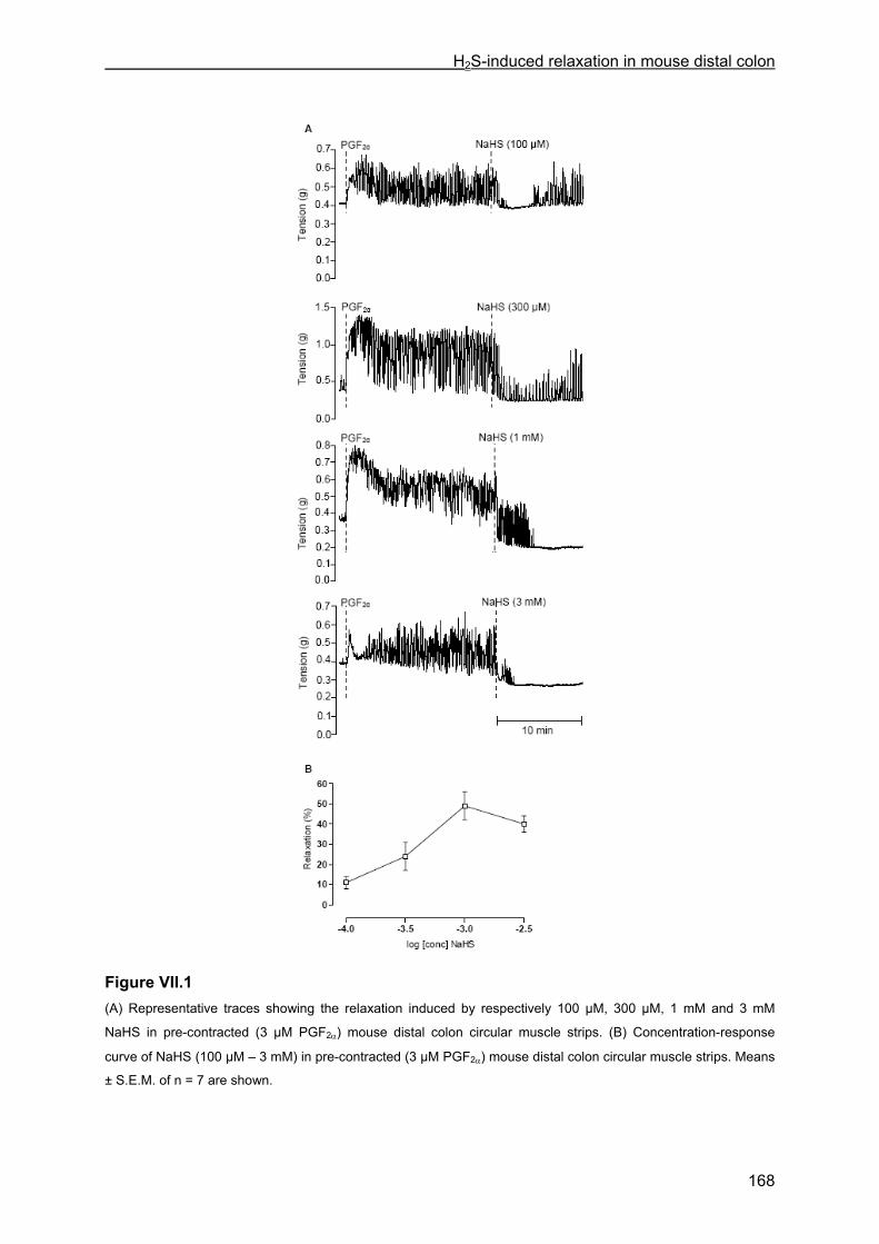
H2S-induced relaxation in mouse distal colon
168
Figure VII.1 (A) Representative traces showing the relaxation induced by respectively 100 µM, 300 µM, 1 mM and 3 mM
NaHS in pre-contracted (3 µM PGF2α) mouse distal colon circular muscle strips. (B) Concentration-response
curve of NaHS (100 µM – 3 mM) in pre-contracted (3 µM PGF2α) mouse distal colon circular muscle strips. Means
± S.E.M. of n = 7 are shown.
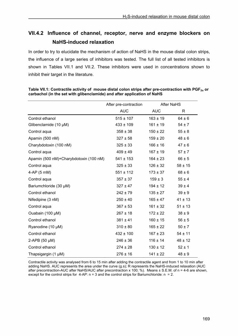
H2S-induced relaxation in mouse distal colon
169
VII.4.2 Influence of channel, receptor, nerve and enzyme blockers on NaHS-induced relaxation
In order to try to elucidate the mechanism of action of NaHS in the mouse distal colon strips,
the influence of a large series of inhibitors was tested. The full list of all tested inhibitors is
shown in Tables VII.1 and VII.2. These inhibitors were used in concentrations shown to
inhibit their target in the literature.
Table VII.1: Contractile activity of mouse distal colon strips after pre-contraction with PGF2α or carbachol (in the set with glibenclamide) and after application of NaHS After pre-contraction After NaHS AUC AUC R
Control ethanol 515 ± 107 163 ± 19 64 ± 6 Glibenclamide (10 µM) 433 ± 109 161 ± 19 54 ± 7
Control aqua 358 ± 38 150 ± 22 55 ± 8
Apamin (500 nM) 327 ± 58 159 ± 20 48 ± 6
Charybdotoxin (100 nM) 325 ± 33 166 ± 16 47 ± 6
Control aqua 409 ± 49 167 ± 19 57 ± 7
Apamin (500 nM)+Charybdotoxin (100 nM) 541 ± 153 164 ± 23 66 ± 5
Control aqua 325 ± 33 126 ± 32 58 ± 15
4-AP (5 mM) 551 ± 112 173 ± 37 68 ± 6
Control aqua 357 ± 37 159 ± 3 55 ± 4
Bariumchloride (30 µM) 327 ± 47 194 ± 12 39 ± 4
Control ethanol 242 ± 79 135 ± 27 39 ± 9
Nifedipine (3 nM) 250 ± 40 165 ± 47 41 ± 13
Control aqua 367 ± 53 161 ± 32 51 ± 13
Ouabain (100 µM) 267 ± 18 172 ± 22 38 ± 9
Control ethanol 381 ± 41 160 ± 15 56 ± 5
Ryanodine (10 µM) 310 ± 80 165 ± 22 50 ± 7
Control ethanol 432 ± 100 167 ± 23 54 ± 11
2-APB (50 µM) 246 ± 36 116 ± 14 48 ± 12
Control ethanol 274 ± 28 130 ± 12 52 ± 1
Thapsigargin (1 µM) 276 ± 16 141 ± 22 48 ± 9
Contractile activity was analysed from 6 to 15 min after adding the contractile agent and from 1 to 10 min after adding NaHS. AUC represents the area under the curve (g.s); R represents the NaHS-induced relaxation (AUC after precontraction-AUC after NaHS/AUC after precontraction x 100; %). Means ± S.E.M. of n = 4-6 are shown, except for the control strips for 4-AP: n = 3 and the control strips for Bariumchloride: n = 2.

H2S-induced relaxation in mouse distal colon
170
Table VII.2: Contractile activity of mouse distal colon strips after pre-contraction with PGF2α or KCl (in one of the 2 sets with calyculin-A) and after application of NaHS After pre-contraction After NaHS AUC AUC R
Control aqua 319 ± 35 169 ± 18 47 ± 3 Tetrodotoxin (3 µM) 412 ± 82 201 ± 16 48 ± 6
Control aqua 272 ± 32 171 ± 14 38 ± 8
ω-Conotoxin (3 µM) 320 ± 11 124 ± 22 58 ± 7
Control ethanol 287 ± 18 166 ± 23 42 ± 7
Capsazepine (3 µM) 233 ± 21 158 ± 18 30 ± 10
Control ethanol 356 ± 43 160 ± 26 51 ± 11
L-NAME (300 µM) 420 ± 82 169 ± 30 47 ± 13
ODQ (10 µM) 364 ± 49 148 ± 13 56 ± 9
Control ethanol 383 ± 64 184 ± 48 53 ± 5
ODQ (10 µM) + Apamin (500 nM) 349 ± 30 126 ± 25 62 ±10
Control aqua 333 ± 39 157 ± 16 51 ± 10
SQ 22536 (100 µM) 363 ± 30 149 ± 11 58 ± 5
Control ethanol 330 ± 28 156 ± 12 51 ± 7
SQ 22536 (100 µM) + ODQ (10 µM) 355 ± 69 138 ± 27 53 ± 15
Control DMSO (†) 442 ± 137 160 ± 32 55 ± 11
Calyculin-A (1 µM; †) 1013 ± 223 566 ± 125 44 ± 1
Control DMSO (††) 524 ± 34 275 ± 9 46 ± 5
Calyculin-A (1 µM; ††) 989 ± 129 ** 591 ± 102 42 ± 4
Control aqua 513 ± 126 215 ± 16 48 ± 11
Y-27632 (10 µM) 311 ± 31 212 ± 23 32 ± 6
Contractile activity was analysed from 6 to 15 min after adding the contractile agent and from 1 to 10 min after adding NaHS. AUC represents the area under the curve (g.s); R represents the NaHS-induced relaxation (AUC after precontraction-AUC after NaHS/AUC after precontraction x 100; %). Means ± S.E.M. of n = 4-6 are shown, except for the control strips for SQ 22536: n = 3 and the control strips for ODQ + Apamin: n = 2. (†): series of calyculin-A in PGF2α-precontracted strips; (††): series of calyculin-A in KCl-precontracted strips. ** P < 0.01: in the presence of inhibitor versus in the presence of solvent (unpaired Student’s t-test).
Only two inhibitors influenced the spontaneous activity of the mouse distal colon strips.
Calyculin-A induced a significant increase of the AUC from 210 ± 18 g.s during the 10 min
before its incubation to 1143 ± 198 g.s (n = 4, P < 0.001) during the 21st to 30th min after its
administration, due to a fully tonic response (Fig. VII.2). Y-27632 induced a moderate
reduction in AUC (208 ± 15 g.s after incubation with Y-27632 versus 261 ± 15 g.s before
incubation with Y-27632, n = 4, P < 0.001).
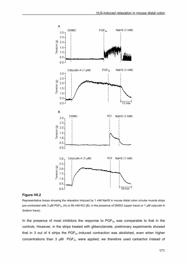
H2S-induced relaxation in mouse distal colon
171
Figure VII.2 Representative traces showing the relaxation induced by 1 mM NaHS in mouse distal colon circular muscle strips
pre-contracted with 3 µM PGF2α (A) or 80 mM KCl (B), in the presence of DMSO (upper trace) or 1 µM calyculin-A
(bottom trace).
In the presence of most inhibitors the response to PGF2α was comparable to that in the
controls. However, in the strips treated with glibenclamide, preliminary experiments showed
that in 3 out of 4 strips the PGF2α-induced contraction was abolished, even when higher
concentrations than 3 µM PGF2α were applied; we therefore used carbachol instead of
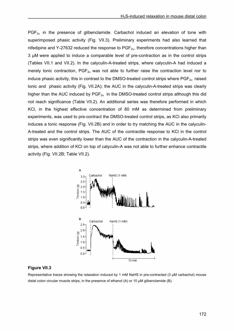
H2S-induced relaxation in mouse distal colon
172
PGF2α in the presence of glibenclamide. Carbachol induced an elevation of tone with
superimposed phasic activity (Fig. VII.3). Preliminary experiments had also learned that
nifedipine and Y-27632 reduced the response to PGF2α, therefore concentrations higher than
3 µM were applied to induce a comparable level of pre-contraction as in the control strips
(Tables VII.1 and VII.2). In the calyculin-A-treated strips, where calyculin-A had induced a
merely tonic contraction, PGF2α was not able to further raise the contraction level nor to
induce phasic activity, this in contrast to the DMSO-treated control strips where PGF2α raised
tonic and phasic activity (Fig. VII.2A); the AUC in the calyculin-A-treated strips was clearly
higher than the AUC induced by PGF2α in the DMSO-treated control strips although this did
not reach significance (Table VII.2). An additional series was therefore performed in which
KCl, in the highest effective concentration of 80 mM as determined from preliminary
experiments, was used to pre-contract the DMSO-treated control strips, as KCl also primarily
induces a tonic response (Fig. VII.2B) and in order to try matching the AUC in the calyculin-
A-treated and the control strips. The AUC of the contractile response to KCl in the control
strips was even significantly lower than the AUC of the contraction in the calyculin-A-treated
strips, where addition of KCl on top of calyculin-A was not able to further enhance contractile
activity (Fig. VII.2B; Table VII.2).
Figure VII.3 Representative traces showing the relaxation induced by 1 mM NaHS in pre-contracted (3 µM carbachol) mouse
distal colon circular muscle strips, in the presence of ethanol (A) or 10 µM glibenclamide (B).
H2S-induced relaxation in mouse distal colon
173
None of the inhibitors listed in Tables VII.1 and VII.2 had a significant influence on the NaHS-
induced relaxation. In calyculin-A-treated strips, the response to NaHS was not different from
that in control strips, either when these were contracted by PGF2α or by KCl.
VII.4.3 Effect of NaHS on cytosolic calcium concentration
We simultaneously measured tension and cytosolic calcium concentration ([Ca2+]cyt) in the
mouse distal colon strips. The mean resting [Ca2+]cyt was 181 ± 10 nM (n = 22).
Administration of PGF2α induced an increase in tension/phasic activity, that stabilized within 5
min after its administration; PGF2α induced an elevation of [Ca2+]cyt to 331 ± 19 µM (n = 22)
as measured for the 2nd to 6th min after its administration.
In control strips, the contractile response to PGF2α remained stable from 6 to 16 min after its
administration; in this period the [Ca2+]cyt at 0-1 min, and to a lesser extent at 4-5 and 9-10
min, was slightly increased (Figs. VII.4A and VII.5A).
When NaHS was administered, relaxation was initiated which progressively evolved to a
sustained level (Fig. VII.4B). The [Ca2+]cyt however significantly increased immediately after
adding NaHS (increase of 38 ± 9 % versus 11 ± 3 % in control strips, n = 7, P < 0.01) (Figs.
VII.4B and VII.5B). At 4-5 and 9-10 min after administration of NaHS, no differences in
[Ca2+]cyt between NaHS-treated and control strips were observed.
The quick relaxation induced by NO was clearly accompanied by an immediate significant (P
< 0.001) reduction in [Ca2+]cyt of 39 ± 3 % (n = 7-8). The NO-induced relaxation and decrease
in [Ca2+]cyt were transient (Figs. VII.4C and VII.5C).
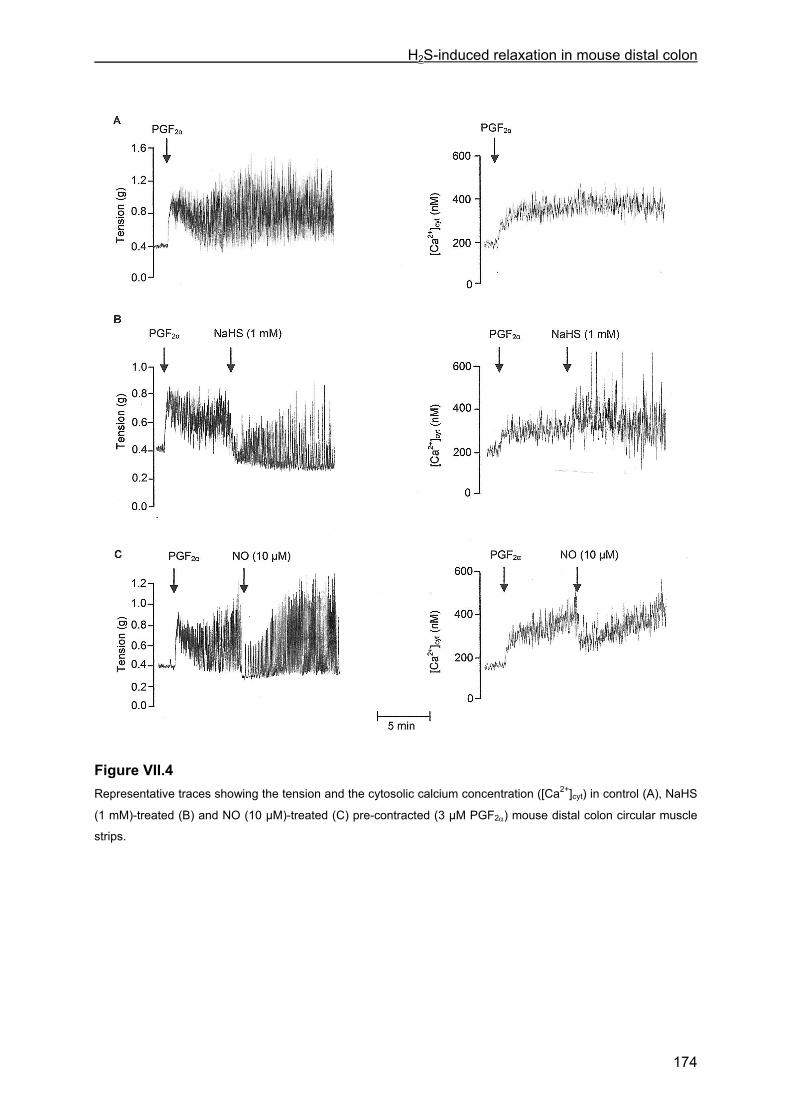
H2S-induced relaxation in mouse distal colon
174
Figure VII.4 Representative traces showing the tension and the cytosolic calcium concentration ([Ca2+]cyt) in control (A), NaHS
(1 mM)-treated (B) and NO (10 µM)-treated (C) pre-contracted (3 µM PGF2α) mouse distal colon circular muscle
strips.

H2S-induced relaxation in mouse distal colon
175
Figure VII.5 The change in cytosolic calcium concentration ([Ca2+]cyt) in the time intervals 0-1 min, 4-5 min and 9-10 min after
administration of NaHS (1 mM) (B) or NO (10 µM) (C) or in corresponding time intervals (control; A) in pre-
contracted (3 µM PGF2α) mouse distal colon circular muscle strips. Values are represented as % of PGF2α-
induced rise in [Ca2+]cyt. The positive and negative values in the y-axis represent respectively increases and
decreases in [Ca2+]cyt. Means ± S.E.M. of n = 7-8 are shown. * P < 0.05, ** P < 0.01, *** P < 0.001: NaHS- or NO-
treated versus control (one-way ANOVA followed by Bonferroni multiple comparison t-test).
H2S-induced relaxation in mouse distal colon
176
VII.5 Discussion
The aim of this study was to examine the influence and mechanism of action of H2S on
contractility in mouse distal colon. In pre-contracted mouse distal colon circular muscle strips,
H2S-donor NaHS induced concentration-dependent relaxations with an EC50 of 444 ± 71 µM.
This value is somewhat higher than the previously reported EC50 values for NaHS, ranging
from 31 to 261 µM, in GI tissues of other species (Gallego et al., 2008; Hosoki et al., 1997;
Teague et al., 2002). Also rat vascular tissues seem more sensitive to NaHS with EC50
values between 104 and 136 µM (Cheng et al., 2004; Zhao and Wang, 2002). However, in
pre-contracted mouse gastric fundus strips, we found that NaHS induced relaxations with an
EC50 of 514 ± 36 µM (Dhaese and Lefebvre, 2009). In mouse bronchial rings, an EC50 above
300 µM was observed for NaHS (Kubo et al., 2007a). These observations suggest that a
species difference could exist with mouse smooth muscle being less sensitive to NaHS. Still,
an EC50 of 121 and 150 µM was reported for the NaHS-induced effects in isolated segments
of mouse colon and jejunum (Gallego et al., 2008).
The non-effect of tetrodotoxin and L-NAME on spontaneous contractile activity corresponds
with previous data reported in mouse colon strips (Porcher et al., 2004; Dhaese et al., 2008)
and illustrates that there is no prevailing tonic neurogenic nitrergic inhibition of mouse distal
colon under the conditions used. The inhibitor of myosin light chain phosphatase (MLCP)
calyculin-A induced tonic contraction illustrating that MLCP is tonically active in this tissue,
possibly under the inhibitory control of Rho-kinase as the Rho-kinase inhibitor Y-27632
reduced spontaneous contractile activity.
NaHS seems to act at the muscular level in mouse distal colon strips since the Na+ channel
blocker tetrodotoxin and the N-type voltage dependent Ca2+ channel blocker ω-conotoxin did
not influence NaHS-induced relaxation. Additionally, the transient receptor potential vanilloid
type 1 (TRPV1) blocker capsazepine, tested since TRPV1 receptors were reported to be
involved in the prosecretory effect of NaHS in human and guinea-pig colon (Schicho et al.,
2006) and in the contractile effect of NaHS in the rat urinary bladder (Patacchini et al., 2004),
did not influence NaHS-induced relaxation in the mouse distal colon strips.
KATP channels are reported to play a role in the NaHS-induced relaxation in cardiovascular
tissues (Cheng et al., 2004; Webb et al., 2008; Zhao et al., 2001), and recently, also in
human and rat colonic strips (Gallego et al., 2008). Glibenclamide, a blocker of the KATP
channels, however did not inhibit the NaHS-induced relaxation in mouse distal colon strips,
corresponding with the non-effect of glibenclamide on the NaHS-induced inhibition in mouse
aorta (Kubo et al., 2007b), mouse bronchial rings (Kubo et al., 2007a), mouse gastric fundus
(Dhaese and Lefebvre, 2009) and guinea pig ileum (Teague et al., 2002). The SKCa channel
blocker apamin (3 µM) significantly reduced the NaHS-induced effects in rat and human

H2S-induced relaxation in mouse distal colon
177
colonic strips and in mouse distal colon full segments (Gallego et al., 2008). In our study, 500
nM apamin did not influence the NaHS-induced relaxation. IC50 values as low as 83 pM to 28
nM were reported for apamin to block the three SKCa channel subtypes (SK1, SK2 and SK3;
Ro et al., 2001; Strøbæk et al., 2000) and Van Crombruggen and Lefebvre (2004) showed
that 500 nM apamin reduced the relaxation by ATP in rat distal colon to the same extent as
30 nM apamin. 500 nM apamin is thus sufficient to investigate the possible role of SKCa
channels. The difference in effect of apamin between our study and that of Gallego et al.
(2008) might be related to the difference in tissues used: circular muscle strips in the actual
study versus full segments. In the latter preparation, NaHS inhibited spontaneous motor
complexes that were abolished by tetrodotoxin; precisely this effect of NaHS on the
neuronally driven motor complexes was inhibited by apamin; in the presence of tetrodotoxin,
NaHS reduced basal tone but the influence of apamin on this effect is not reported (Gallego
et al., 2008). Thus probably in mouse distal colon, NaHS has a direct muscular effect not
involving SKCa channels, as seen in smooth muscle strips in our study, and a neurogenic
effect involving SKCa channels, as seen in full segments in the study of Gallego et al. (2008).
Recently, SK2 channels were indeed detected in enteric neurons and were found to
negatively regulate the acetylcholine release (Nakajima et al., 2007). Still, our results differ
from these in human and rat colon circular muscle strips where Gallego et al. (2008) showed
that the NaHS-induced inhibition of spontaneous activity is sensitive to apamin in the
presence of tetrodotoxin. Also the intermediate (IKCa) and large (BKCa) conductance Ca2+
dependent K+ channel blocker charybdotoxin did not influence the NaHS-induced relaxation
in mouse distal colon strips, neither alone, nor in combination with apamin; the latter
combination was required to reduce significantly the relaxation by exogenous CO in mouse
gastric fundus (De Backer and Lefebvre, 2007). Finally, the influence of barium chloride and
4-AP, blockers of respectively the inward rectifier and the voltage dependent K+ channel, was
tested as both K+ channels can play a role in smooth muscle relaxation (Horinouchi et al.,
2003; Orie et al., 2006). However, 4-AP and barium chloride did not affect the NaHS-induced
relaxation.
Zhao and Wang (2002) reported the release of endothelium-derived NO by H2S in rat aortic
tissues. Contribution of endogenous NO and its downstream target soluble guanylate cyclase
(sGC) in the relaxation by NaHS of mouse distal colon strips was excluded as the NO
synthase inhibitor L-NAME nor the sGC inhibitor ODQ had an influence. As in rat colon the
combination of ODQ plus apamin was required to fully inhibit the relaxation by exogenous
NO (Van Crombruggen and Lefebvre, 2004), ODQ plus apamin was tested, but this had no
influence versus NaHS. The non-effect of the adenylate cyclase (AC) inhibitor SQ 22536
indicates that also the cAMP pathway is not involved in the relaxant effect of NaHS.
H2S-induced relaxation in mouse distal colon
178
In rat aortic tissues, the H2S-induced vasorelaxation was reduced by the L-type voltage
dependent Ca2+ channel blocker nifedipine (Zhao and Wang, 2002). In our study, nifedipine
was used at a concentration (3 nM) able to reduce the PGF2α (3 µM)-induced contractile
response by 50 %. Higher concentrations of PGF2α brought about a comparable degree of
pre-contraction as in the control strip. Nifedipine showed no effect on the NaHS-induced
relaxation in mouse distal colon. As the Na+/K+ ATPase, the ryanodine receptor, the IP3
receptor and the sarcoplasmatic reticulum Ca2+ ATPase (SERCA) can be involved in
relaxation of mouse GI tissues (Kim et al., 2006; Yaktubay et al., 1999; Zizzo et al., 2005),
we tested respectively ouabain, ryanodine, 2-APB and thapsigargin against the NaHS-
induced relaxation. However, none of these inhibitors had an influence on the relaxant effect
of NaHS.
Relaxation in smooth muscle cells results from a decrease in the intracellular calcium
concentration, and/or a desensitization of the contractile proteins to the intracellular calcium
concentration. We therefore performed experiments simultaneously measuring tension and
[Ca2+]cyt in the mouse distal colon strips. Exogenous NO induced a relaxation accompanied
by a decrease in [Ca2+]cyt. This was previously reported for the sodium nitroprusside-induced
relaxation in guinea pig taenia coli (Kwon et al., 2000) and the NO-induced relaxation in rat
distal colon (Colpaert et al., 2005). The relaxation starting immediately after administration of
NaHS was however accompanied by a significant increase in [Ca2+]cyt. We have no clear-cut
explanation for this rise in [Ca2+]cyt. In the literature, only localized increases in [Ca2+]cyt
caused by calcium release from the sarcoplasmatic reticulum via ryanodine receptors (Ca2+
sparks) or IP3 receptors (Ca2+ puffs), leading to the activation of BKCa or SKCa channels
(Bayguinov et al., 2000; Jaggar et al., 2000), have been associated with relaxation. These
localized increases in [Ca2+]cyt can however not account for the rise in global [Ca2+]cyt that we
measured. Moreover, as already discussed, inhibiting the ryanodine receptor or the IP3
receptor did not affect the NaHS-induced relaxation, nor did inhibition of the BKCa and SKCa
channels. At later time intervals, [Ca2+]cyt was not different from that in control strips, despite
the fact that the maximal relaxing effect of NaHS was reached and sustained. Smooth
muscle relaxation without a change in [Ca2+]cyt is mediated through calcium desensitization.
One of the most important calcium desensitizing mechanisms is inhibition of the RhoA/Rho-
kinase pathway leading to the activation of myosin light chain phosphatase (MLCP; Somlyo
and Somlyo, 2003) but this pathway is excluded as the Rho-kinase inhibitor Y-27632 did not
influence NaHS-induced relaxation. Also other pathways leading to MLCP activation can be
excluded as the inhibitor of MLCP calyculin-A did not affect NaHS-induced relaxation, which
is in contrast to our previous data obtained in the mouse gastric fundus (Dhaese and
Lefebvre, 2009). A calcium desensitization mechanism, independent of MLCP, is the
H2S-induced relaxation in mouse distal colon
179
inhibitory phosphorylation of the myosin light chain kinase (MLCK) by activation of
Ca2+/calmodulin-dependent protein kinase II (CaMKII; Tansey et al., 1994). However, this
requires an increase in [Ca2+]cyt. There was an initial short lasting increase in [Ca2+]cyt by
NaHS in our experiments, but in the desensitization model through CaMKII , this increase is
expected to first activate MLCK and thus to induce contraction which was not the case
(Tansey et al., 1994). Some kinases, such as integrin-linked kinase, p21-activated protein
kinase and zip kinase that have been shown to be present in smooth muscle cells and were
obtained by recombinant technology, have been shown to directly phosphorylate the myosin
light chain (MLC) in an Ca2+-independent manner in an in vitro assay and to induce smooth
muscle contraction when their catalytic domains were introduced as recombinant proteins
into permeabilized smooth muscle tissues (Hirano et al., 2004). Inhibition of these kinases
would, independently of MLCP, lead to a lower amount of phosphorylated MLC and
relaxation. As it still remains to be clarified whether this(these) kinase(s) play(s) a
physiological role in the regulation of smooth muscle contractility (Hirano et al., 2004), we
can only speculate that NaHS might induce relaxation by inhibition of one of these kinases.
In conclusion, our study demonstrates that in mouse distal colon strips H2S induced
concentration-dependent relaxations, not involving the major known K+ channels and not
accompanied by a decrease in [Ca2+]cyt. The latter indicates that the H2S -induced relaxations
are mediated via calcium desensitization. This calcium desensitizing mechanism seems not
related to inhibition of Rho-kinase or activation of MLCP.
VII.6 References Abe, K., Kimura, H., 1996. The possible role of hydrogen sulfide as an endogenous neuromodulator.
J. Neurosci. 16, 1066-1071.
Bayguinov, O., Hagen, B., Bonev, A.D., Nelson, M.T., Sanders, K.M., 2000. Intracellular calcium
events activated by ATP in murine colonic myocytes. Am. J. Physiol. Cell Physiol. 279, C126-C135.
Cheng, Y., Ndisang, J.F., Tang, G., Cao, K., Wang, R., 2004. Hydrogen sulfide-induced relaxation of
resistance mesenteric artery beds of rats. Am. J. Physiol. Heart Circ. Physiol. 287, H2316-H2323.
Colpaert, E.E., Levent, A., Lefebvre, R.A., 2005. Nitric oxide relaxes circular smooth muscle of rat
distal colon through RhoA/Rho-kinase independent Ca2+ desensitization. Br. J. Pharmacol. 144, 588-
594.
De Backer, O., Lefebvre, R.A., 2007. Mechanisms of relaxation by carbon monoxide-releasing
molecule-2 in murine gastric fundus and jejunum. Eur. J. Pharmacol. 572, 197-206.
H2S-induced relaxation in mouse distal colon
180
Delaey, C., Van de Voorde, J., 1997. Heterogeneity of the inhibitory influence of sulfonylureas on
prostanoid-induced smooth muscle contraction. Eur. J. Pharmacol. 325, 41-46.
Dhaese, I., Lefebvre, R.A., 2009. Myosin light chain phosphatase activation is involved in the
hydrogen sulfide-induced relaxation in mouse gastric fundus. Eur. J. Pharmacol. 606, 180-186.
Dhaese, I., Vanneste, G., Sips, P., Buys, E., Brouckaert, P., Lefebvre, R.A., 2008. Involvement of
soluble guanylate cyclase α1 en α2, and SKCa channels in NANC relaxation of mouse distal colon. Eur.
J. Pharmacol. 589, 251-259.
Gallego, D., Clavé, P., Donovan, J., Rahmati, R., Grundy, D., Jiménez, M., Beyak, M.J., 2008. The
gaseous mediator, hydrogen sulphide, inhibits in vitro motor patterns in the human, rat and mouse
colon and jejunum. Neurogastroenterol. Motil. 20, 1306-1316.
Grynkiewicz, G., Poenie, M., Tsien, R.Y., 1985. A new generation of Ca2+ indicators with greatly
improved fluorescence properties. J. Biol. Chem. 260, 3440-3450.
Hirano, K., Hirano, M., Kanaide, H., 2004. Regulation of myosin phosphorylation and myofilament Ca2+
sensitivity in vascular smooth muscle. J. Smooth Muscle Res. 40, 219-236.
Horinouchi, T., Tanaka, Y., Koike, K., 2003. Evidence for the primary role of 4-aminopyridine-sensitive
Kv channels in β3-adrenoceptor-mediated, cyclic AMP-independent relaxations of guinea pig
gastrointestinal smooth muscles. Naunyn Schmiedebergs Arch. Pharmacol. 367, 193-203.
Hosoki, R., Matsuki, N., Kimura, H., 1997. The possible role of hydrogen sulfide as an endogenous
smooth muscle relaxant in synergy with nitric oxide. Biochem. Biophys. Res. Commun. 237, 527-531.
Jaggar, J.H., Porter, V.A., Lederer, W.J., Nelson, M.T., 2000. Calcium sparks in smooth muscle. Am.
J. Physiol. Cell physiol. 278, C235-C256.
Kelm, M., Schrader, J., 1990. Control of coronary vascular tone by nitric oxide. Circ. Res. 66, 1561-
1575.
Kim, M., Han, I.S., Koh, S.D., Perrino, B.A, 2006. Roles of CaM kinase II and phospholamban in SNP-
induced relaxation of murine gastric fundus smooth muscles. Am. J. Physiol. Cell. Physiol. 291, C337-
C347.
Kubo, S., Doe, I., Kurokawa, Y., Kawabata, A., 2007a. Hydrogen sulfide causes relaxation in mouse
bronchial smooth muscle. J. Pharmacol. Sci. 104, 392-396.
Kubo, S., Doe, I., Kurokawa, Y., Nishikawa, H., Kawabata, A., 2007b. Direct inhibition of endothelial
nitric oxide synthase by hydrogen sulfide: contribution to dual modulation of vascular tension.
Toxicology 232, 138-146.
H2S-induced relaxation in mouse distal colon
181
Kwon, S.-C., Ozaki, H., Karaki, H., 2000. NO donor sodium nitroprusside inhibits excitation-contraction
coupling in guinea pig taenia coli. Am. J. Physiol. Gastrointest. Liver Physiol. 279, G1235-G1241.
Levine, J., Ellis, C.J., Furne, J.K., Springfield, J., Levitt, M.D., 1998. Fecal hydrogen sulphide
production in ulcerative colitis. Am. J. Gastroenterol. 93, 83-87.
Linden, D.R., Sha, L., Mazzone, A., Stoltz, G.J., Bernard, C.E., Furne, J.K., Levitt, M.D., Farrugia, G.,
Szurszewski, J.H., 2008. Production of the gaseous signal molecule hydrogen sulfide in mouse
tissues. J. Neurochem. 106, 1577-1585.
Nakajima, H., Goto, H., Azuma, Y.-T., Fujita, A., Takeuchi, T., 2007. Functional interactions between
the SK2 channel and the nicotinic acetylcholine receptor in enteric neurons of the guinea pig ileum. J.
Neurochem. 103, 2428-2438.
Olson, K.R., Dombkowski, R.A., Russell, M.J., Doellman, M.M., Head, S.K., Whitfield, N.L., Madden,
J.A., 2006. Hydrogen sulfide as an oxygen sensor/transducer in vertebrate hypoxic vasocontraction
and hypoxic vasodilation. J. Exp. Biol. 209, 4011-4023.
Orie, N.N., Fry, C.H., Clapp, L.H., 2006. Evidence that inward rectifier K+ channels mediate relaxation
by the PGI2 receptor agonist cicaprost via a cyclic AMP-independent mechanism. Cardiovasc. Res.
69, 107-115.
Patacchini, R., Santicioli, P., Giuliani, S., Maggi, C.A., 2004. Hydrogen sulfide (H2S) stimulates
capsaicin-sensitive primary afferent neurons in the rat urinary bladder. Br. J. Pharmacol. 142, 31-34.
Porcher, C., Horowitz, B., Ward, S.M., Sanders, K.M., 2004. Constitutive and functional expression of
cyclooxygenase 2 in the murine proximal colon. Neurogastroenterol. Motil. 16, 785-799.
Ro, S., Hatton, W.J., Koh, S.D., Horowitz, B., 2001. Molecular properties of small-conductance Ca2+-
activated K+ channels expressed in murine colonic smooth muscle. Am. J. Physiol. Gastrointest. Liver
Physiol. 281, G964-G973.
Salomone, S., Morel, N., Godfraind, T., 1995. Effects of 8-bromo-cyclic GMP and verapamil on
depolarization-evoked Ca2+ signal and contraction in rat aorta. Br. J. Pharmacol. 114, 1731-1737.
Schicho, R., Krueger, D., Zeller, F., Von Weyhern,C.W.H., Frieling, T., Kimura, H., Ishii, I., De Giorgio,
R., Campi, B., Schemann, M., 2006. Hydrogen sulfide is a novel prosecretory neuromodulator in the
guinea-pig and human colon. Gastroenterology 131, 1542-1552.
Somlyo, A.P., Somlyo, A., 2003. Ca2+ sensitivity of smooth muscle and nonmuscle myosin II:
modulated by G proteins, kinases, and myosine phosphatase. Physiol. Rev. 83, 1325-1358.
Stipanuk, M.H., Beck, P.W., 1982. Characterization of the enzymic capacity for cysteine
desulphhydration in liver and kidney of the rat. Biochem. J. 206, 267-277.
H2S-induced relaxation in mouse distal colon
182
Strøbæk, D., Jørgensen, T.D., Christophersen, P., Ahring, P.K., Olesen, S.-P., 2000. Pharmacological
characterization of small-conductance Ca2+-activated K+ channels stably expressed in HEK 293 cells.
Br. J. Pharmacol. 129, 991-999.
Tang, G., Wu, L., Liang, W., Wang, R., 2005. Direct stimulation of KATP channels by exogenous and
endogenous hydrogen sulfide in vascular smooth muscle cells. Mol. Pharmacol. 68, 1757-1764.
Tansey, M.G., Luby-Phelps, K., Kamm, K.E., Stull J.T., 1994. Ca2+ dependent phosphorylation of
myosin light chain kinase decreases the Ca2+ sensitivity of light chain phosphorylation within smooth
muscle cells. J. Biol. Chem. 269, 9912-9920.
Teague, B., Asiedu, S., Moore, P.K., 2002. The smooth muscle relaxant effect of hydrogen sulphide in
vitro: evidence for a physiological role to control intestinal contractility. Br. J. Pharmacol. 137, 139-145.
Van Crombruggen, K., Lefebvre, R.A., 2004. Nitrergic-purinergic interactions in rat distal colon motility.
Neurogastroenterol. Motil. 16, 81-98.
Webb, G.D., Lim, L.H., Oh, V.M., Yeo, S.B., Cheong, Y.P., Ali, M.Y., Oakley, R.E., Lee, C.N., Wong,
P.S., Caleb, M.G., Salto-Tellez, M., Bhatia, M., Chan, E.S.Y., Taylor, E.A., Moore, P.K., 2008.
Contractile and vasorelaxant effects of hydrogen sulfide and its biosynthesis in the human internal
mammary artery. J. Pharmacol. Exp. Ther. 324, 876-882.
Yaktubay, N., Ögülener, N., Önder, S., Baysal, F., 1999. Possible stimulation of Na+-K+-ATPase by
NO produced from sodium nitrite by ultraviolet light in mouse gastric fundal strip. Gen. Pharmac. 32,
159-162.
Zhao, W., Wang, R., 2002. H2S-induced vasorelaxation and underlying cellular and molecular
mechanisms. Am. J. Physiol. Heart Circ. Physiol. 283, H474-H480.
Zhao, W., Zhang, H.J., Lu, Y., Wang, R., 2001. The vasorelaxant effect of H2S as a novel endogenous
gaseous KATP channel opener. EMBO J. 20, 6008-6016.
Zizzo, M.G., Mulé, F., Serio, R., 2005. Mechanisms underlying the nitric oxide inhibitory effects in
mouse ileal longitudinal muscle. Can. J. Physiol. Pharmacol. 83, 805-810.

Chapter VIII
GENERAL DISCUSSION AND CONCLUSION

General discussion and conclusion
185
Chapter VIII General Discussion and Conclusion
VIII.1 The relative importance of the sGC isoforms α1β1 and α2β1 in
the nitrergic relaxation of gastrointestinal smooth muscle
During the past two decades, several studies established a role for the neurotransmitter NO
in relaxation of gastrointestinal smooth muscle and hence motility of the gastrointestinal (GI)
tract. The principal target of NO is the heme-containing enzyme sGC (Lucas et al., 2000).
Two physiological active isoforms of sGC have been described: the predominantly expressed
heterodimer sGCα1β1 and the less abundantly expressed sGCα2β1. In mouse ileum and
colon, the presence of α1, α2 and β1 subunit mRNA was reported (Mergia et al., 2003), albeit
the amount of α2 mRNA was clearly lower than this of α1 mRNA, as is the case in most
tissues. Still, the presence of sGCα1β1 as well as sGCα2β1 in GI tissues triggers the question
on the relative importance of each isoform in the NO-induced relaxation in these tissues.
Research on this subject has however been hampered by the fact that there are no isoform-
specific inhibitors. We tried to address this topic using genetically engineered mice, focussing
our research on the NO-mediated relaxation in the jejunum and the distal colon of the sGCα1
knockout (KO) mice in which a mutant sGCα1β1 isoform is no longer functionally active.
Exogenous NO. We found that the relaxant response to exogenous NO was reduced in the
jejunal and distal colonic strips isolated from sGCα1 KO mice, pointing to a role of sGCα1β1 in
exogenous NO-induced relaxation in these tissues. The (small) remaining responses in the
sGCα1 KO mice were still sensitive to the sGC inhibitor ODQ, indicating that also sGCα2β1
contributes to the relaxing effect of NO. The sGCα1 KO mice did not show an increase in α2
mRNA in the jejunum nor an elevated expression of the α2 subunit protein in the distal colon,
indicating that the participation of sGCα2β1 in the exogenous NO-induced relaxation is not a
compensation mechanism by upregulation of the sGCα2β1 isoform due to sGCα1 deficiency.
At the level of the distal colon, the contribution of sGCα2β1 in the relaxant response to
exogenous NO did not differ between male and female mice. In the jejunum however, the
participation of sGCα2β1 in the exogenous NO-induced relaxation differed depending on the
gender. The contribution of sGCα2β1 in the relaxation by exogenous NO was most
pronounced in the jejunum of female mice, as the relaxant responses to exogenous NO were
only slightly reduced in the jejunal strips from female sGCα1 KO mice. In contrast, the role of
sGCα2β1 in the exogenous NO-induced responses was least pronounced in the jejunum of

General discussion and conclusion
186
male mice, as a near abolition of these responses was observed in the jejunal strips from
male sGCα1 KO mice. These observations contrast with previous results of our group in
pyloric tissue, where the opposite was found i.e. a more pronounced reduction of relaxation
by exogenous NO in female than in male sGCα1 KO mice (Vanneste et al., 2007). The more
pronounced consequences of knocking out sGCα1 on the exogenous NO-induced relaxation
at the level of the jejunum in the male versus the female mice are however in line with the
finding that male sGCα1 KO mice developed hypertension whereas female sGCα1 KO mice
maintained normal blood pressure (Buys et al., 2008). But these in vivo data were not
accompanied by gender differences in exogenous NO-induced relaxation of aortic rings in
vitro, as the latter was similarly attenuated in both male and female sGCα1 KO mice
(Nimmegeers et al., 2007), leading to the conclusion that the observed gender dimorphism in
blood pressure in the sGCα1 KO mice is not per se related to gender-specific impairments in
NO-induced relaxation. Further findings in the study by Buys et al. (2008) suggested a role of
androgens in the hypertension associated with sGCα1 deficiency. Taken our results and the
results previously reported by Vanneste et al. (2007) together, we can state that exogenous
NO is able to induce relaxation via the activation of sGCα1β1 as well as sGCα2β1, but that the
relative contribution of sGCα2β1 to the response to exogenous NO can somewhat differ
depending on GI tissue and even gender.
Endogenous NO (in comparison to exogenous NO). Whereas exogenous NO diffuses
from the surrounding extracellular solution and enters the smooth muscle cells over the
whole surface, endogenous i.e. nNOS-derived NO is released from the nerve varicosities
and thus enters the smooth muscle cell more locally. The different mechanism of entering
could have implications towards the relative contribution of each sGC isoform in mediating
relaxation by exogenous respectively endogenous NO. We assessed the responses to
endogenous NO by electrical field stimulation (EFS) of the GI tissues in NANC conditions.
Within the experimental conditions used, this yielded purely nitrergic responses at all
frequencies tested in the jejunum, corresponding to what our group reported before for the
gastric fundus (Vanneste et al., 2007) but in the distal colon only the response at 1 Hz was a
purely nitrergic one. In the gastric fundus of the sGCα1 KO mice, the responses to
endogenous NO, released during 10 s trains of EFS, were reduced to a much greater extent
than the relaxations by exogenous NO (Vanneste et al., 2007). These results point to a more
dominant role of sGCα1β1 in the relaxant response to endogenous versus exogenous NO in
the gastric fundus. Similar findings were observed in the distal colon, where sGCα1β1 as well
as sGCα2β1 are involved in the responses to exogenous NO, but the response to
endogenous NO released by EFS at 1 Hz was mediated solely through activation of
sGCα1β1, as it was abolished in the sGCα1 KO mice. In the jejunum however, the relative
General discussion and conclusion
187
contribution of sGCα2β1 versus sGCα1β1 in response to endogenous NO was similar as this
in response to exogenous NO, with sGCα2β1 being only minimally involved in the NO-
induced relaxation in the jejunum of male mice but contributing in a much more pronounced
way in female mice.
At the level of the gastric fundus and the distal colon, endogenous NO seems to act mainly
via sGCα1β1. Although both sGCα1β1 and sGCα2β1 are regarded to be soluble, it was shown
in rat brain that, whereas the sGCα1β1 isoform was found in the cytosol, the α2β1 isoform is
actually membrane-associated via interaction with the PDZ-containing post-synaptic density
protein-95 (PSD-95; Russwurm et al., 2001). Also in epithelial cells in human colon, sGCα2β1
was found to be localized to the apical plasma membrane (Bellingham & Evans, 2007). It is
possible that the spatially more confined and lower concentrations of endogenous NO are
not as effective as the more diffused and higher concentrations of exogenous NO in reaching
the membrane-located sGCα2β1, which is present in lower amounts in the GI smooth muscle
cells than the cytosolic sGCα1β1.
VIII.2 sGC-dependency of the nitrergic component of GI smooth
muscle relaxation
Exogenous NO. The sGCα1 KO model showed us that in the absence of sGCα1β1, sGCα2β1
seems able to maintain some degree of nitrergic relaxation in the investigated GI tissues.
Conclusive evidence for this supporting role of sGCα2β1 came from our observations in the
heme-deficient sGCβ1 knock in (KI) mice. Due to the impairment of the heme-dependent and
thus nitrergic activation of sGCα1β1 as well as sGCα2β1, the relaxant responses to
exogenous NO were abolished in the gastric fundic, jejunal and distal colonic strips isolated
from the sGCβ1 KI mice. This illustrates that it is indeed sGCα2β1 that is responsible for the
remaining nitrergic responses in the sGCα1 KO mice. Moreover, this indicates that
exogenous NO, within the experimental conditions used, completely relies on sGC to induce
relaxation; our results do consequently not comply with possible sGC-independent actions,
such as the direct activation of K+ channels (Lang & Watson, 1998; Serio et al., 2003b) by
NO in the investigated GI tissues. In accordance with this conclusion, one would expect that
in the GI tissues isolated from the WT mice, responses to exogenous NO should be
abolished by ODQ, an inhibitor of both sGC isoforms by oxidation of their prosthetic heme
group. Our observations that ODQ only reduced –not abolished- the relaxation induced by
higher concentrations of exogenous NO in the gastric fundic, jejunal and distal colonic strips
from the WT mice can however be explained by the NO-competitive nature of ODQ
(Schrammel et al., 1996). The latter should indeed be taken into account when interpreting
General discussion and conclusion
188
results obtained with ODQ, illustrating the additional value of our mouse transgenic approach
for differentiating and evaluating sGC-dependent versus sGC-independent effects of NO.
Endogenous NO in gastric fundus. Besides exogenous NO, also endogenous NO seems
to act solely via sGC in the investigated GI tissues. Indeed, in the gastric fundic strips, the
EFS-induced relaxations, which were found to be L-NAME-sensitive in the WT mice, were
virtually abolished in the sGCβ1 KI mice, indicating that NO mediates its relaxation via sGC,
and corroborating that NO is the main neurotransmitter in mouse gastric fundus. An
exception was the remaining relaxant response to EFS at 8 Hz in some gastric fundic strips
of the sGCβ1 KI mice, notwithstanding this response was found to be abolished by L-NAME
in the WT mice. This pointed to a possible sGC-independent action of endogenous NO in the
gastric fundus; however further experiments showed that this remaining response in the
sGCβ1 KI mice was unsensitive to L-NAME. The latter thus suggests the emerge of a
compensatory mechanism, involving another neurotransmitter than NO, at higher stimulation
frequencies in the sGCβ1 KI mice. We speculate this neurotransmitter to be VIP, as VIP is
reported to be released at higher stimulation frequencies in rat gastric fundus strips (D’Amato
et al., 1992). In compensation for the loss of nitrergic relaxation in the gastric fundus, VIP
release might start at lower frequencies in some sGCβ1 KI mice.
Endogenous NO in jejunum. NO –acting via sGC- seems also the main neurotransmitter in
the mouse jejunum, as the L-NAME-sensitive relaxations observed at the four applied
stimulation frequencies in the jejunal strips from the WT mice were abolished in the sGCβ1 KI
mice. This is in contrast to the report of De Man et al. (2003) in which a role for ATP (De Man
et al., 2003) in mouse jejunum was suggested. In this study, EFS was applied at the same
four frequencies and for 10 s as in our study, but the applied pulse duration and voltage of
the stimulations (respectively 0.1 ms and 40 V in our study) are not mentioned and could
thus differ from our study. Our results obtained in mouse jejunum however correspond to
what our group reported in rat jejunum under similar - but not identical - stimulation
parameters (40 V; 0.1 ms; 1-8 Hz; but 20 s trains; Vanneste et al., 2004).
Endogenous NO in distal colon. In mouse distal colon, the response to EFS at 1 Hz, being
purely nitrergic, was abolished in distal colonic strips of the sGCα1 KO and sGCβ1 KI mice,
whereas the responses to EFS at 2 to 8 Hz were not influenced. For the relaxant responses
to EFS at 2 to 8 Hz, a redundant action of NO, acting at sGC, and another neurotransmitter,
acting at small conductance Ca2+-dependent K+ channels, is suggested. This proposal is
based on the observation that the combined addition of L-NAME plus apamin or ODQ plus
apamin was able to nearly abolish the relaxant responses at these higher stimulation
frequencies in distal colonic strips of the WT mice, studied in comparison with the sGCα1 KO
mice. This second neurotransmitter, besides NO, is probably ATP, as ATP indeed leads to

General discussion and conclusion
189
activation of small conductance Ca2+-dependent K+ channels (Bayguinov et al., 2000) and
has previously been suggested to be involved in the NANC inhibitory neurotransmission in
the mouse (Serio et al., 2003a), rat (Pluja et al., 1999; Van Crombruggen & Lefebvre, 2004),
hamster (El Mahmoudy et al., 2006) and human (Benko et al., 2007) colon. In the WT mice
from the sGCβ1 KI study, L-NAME plus apamin could only reduce -and not abolish- the
relaxations to EFS at 2 to 8 Hz, suggesting the presence of another, possibly third,
neurotransmitter in these mice. The latter could be related to the genetic background of the
mice, as this was different between the study of the sGCα1 KO mice (mixed Swiss/129) and
the study of the sGCβ1 KI mice (mixed 129/SvJ-C57BL/6J). Consistent with this, the
involvement of a third neurotransmitter was also suggested in the sGCβ1 KI mice, as L-
NAME plus apamin failed to influence the relaxant responses by EFS at 2 to 8 Hz in the
distal colonic strips of these mice. Previously, the presence of a third neurotransmitter,
besides NO and ATP, was suggested in rat distal colon by our group (Van Crombruggen &
Lefebvre, 2004) and in hamster distal colon by El-Mahmoudy et al. (2006).
VIII.3 Role of the nitrergic activation of sGC in the regulation of GI
motility
In the sGCα1 KO mice, no morphological GI abnormalities or important GI malfunctions were
observed. However, the total impairment of the heme-dependent and thus nitrergic activation
of sGCα1β1 as well as sGCα2β1 induced delayed gastric emptying, delayed intestinal transit
and increased whole gut transit time in the sGCβ1 KI mice.
Gastric emptying. Disturbances in liquid gastric emptying were indeed expected in the
sGCβ1 KI mice as fundic nitrergic relaxation, essential for gastric accommodation (Desai et
al., 1991), was abolished. The impaired fundic storage of the liquids should be expected to
enhance liquid gastric emptying, but we observed delayed liquid gastric emptying in the
sGCβ1 mice. This is in accordance with reports of delayed gastric emptying in nNOS KO
mice (Mashimo et al., 2000) and cGMP-dependent protein kinase I (cGKI) KO mice (Pfeifer
et al., 1998). The sGCβ1 KI mice also showed, similar to the nNOS (Huang et al., 1993;
Mashimo et al., 2000) and cGKI (Pfeifer et al., 1998) KO mice, a marked enlargement of the
stomach with hypertrophy of the circular muscle of the fundus. Mashimo et al. (2000)
suggested that this gastric smooth muscle thickening is secondary to functional pyloric
obstruction and represents work hypertrophy. NOS inhibition is indeed reported to increase
the pyloric tone (Anvari et al., 1998) and impairment of pyloric relaxation will counteract the
accelerating effect of the deficient fundic relaxation on gastric emptying (Mashimo et al.,
2000; Anvari et al., 1998), as such explaining the observed delay in gastric emptying in the
sGCβ1 KI mice. With the study of the sGCβ1 KI mice, we provide the link between the nNOS
General discussion and conclusion
190
KO mice and the cGKI KO mice. The severe morphological changes at the level of the
stomach and the delayed gastric liquid emptying observed in these three transgenic mouse
models illustrate the pivotal role of the NO-sGC-cGMP-cGKI pathway in normal gastric
functioning.
Small intestinal transit. In addition to and most probably in part resulting from the
disturbances in gastric emptying, sGCβ1 KI mice showed delayed small intestinal transit.
Previously, studies in different species with NOS-inhibitors showed delayed small intestinal
transit (Chiba et al., 2002; Fraser et al., 2005; Karmeli et al., 1997). In the cGKI KO mice,
spastic contractions of long intestinal segments followed by scarce and slow relaxations were
observed as a result of the impairment of the pathway downstream of NO (Pfeifer et al.,
1998). Considering the impaired response to neurally released NO in the jejunal strips from
the sGCβ1 KI mice, it seems inevitable that also in the sGCβ1 KI mice an imbalance between
the excitatory (cholinergic) and inhibitory (nitrergic) input during peristalsis develops,
interrupting the coordinated interplay between the ascending contractions and descending
relaxations, which is essential for the propagation of the peristaltic contraction (Waterman et
al., 1994).
Colonic transit. Concomitant to the delays in the upper GI transit, also retardation of the
colonic transit might take place in the sGCβ1 KI mice, as an increase in whole gut transit time
was observed. NOS inhibition was previously indeed found to inhibit the colonic propulsion of
artificial pellets in guinea pig colonic segments (Foxx-Orenstein & Grider, 1996) and to delay
colonic transit in rats (Mizuta et al., 1999). Still, in sGCβ1 KI mice, the extent of delay in
colonic transit per se will probably be limited, considering the fact that in mouse distal colon,
NO - acting via sGC- is only the principal neurotransmitter at a stimulation frequency of 1 Hz.
The severely disturbed gastric emptying and intestinal transit in sGCβ1 KI mice probably
contribute to the reduced lifespan of sGCβ1 KI mice (median survival of 29-36 weeks;
Thoonen et al., 2009). However, the latter values contrast with the much shorter lifespan of
sGC β1 KO mice, generated by knocking out exon 10 of the β1 subunit, eliminating activation
of both sGC isoforms by NO but also basal activity (Friebe et al., 2007). Less than 10 % of
the sGC β1 KO mice survived for 1 month due to intestinal dysmotility (Friebe et al., 2007),
suggesting that basal sGC activity, maintained in the sGC β1 KI mice, is important for longer
survival.
In conclusion, our findings indicate that NO is the principal relaxant neurotransmitter in the
mouse gastric fundus and the mouse jejunum. In the mouse distal colon, NO is the sole
relaxant neurotransmitter released by EFS at 1 Hz, however at higher stimulation
frequencies, other neurotransmitters contribute to the smooth muscle relaxation. Our results
General discussion and conclusion
191
further show that the relaxant effect of endogenous and exogenous NO in the investigated GI
tissues completely relies on the activation of sGC isoforms sGCα1β1 and α2β1. Interestingly,
at the level of the gastric fundus and the distal colon, endogenous NO seems less efficient
than exogenous NO in activating sGCα2β1. Still, in the absence of sGCα1β1, sGCα2β1 is able
to maintain normal GI functioning. Impairment of the NO-induced activation of sGCα1β1 and
sGCα2β1 induces GI malfunctions, which are most pronounced at the level of the stomach,
indicating the important role of the NO-sGC pathway in GI motility.
VIII.4 Influence of H2S on GI contractility
After NO and CO, a third gaseous molecule, H2S, was recently shown to be generated
naturally in mammalian tissues and to exert a number of physiological effects such as
smooth muscle relaxation. As H2S-induced smooth muscle relaxation was initially observed
in the vascular system, the mechanism of action of H2S is already most extensively studied in
this system. Interestingly, the sGC-cGMP pathway seems not to be involved in the H2S-
induced vasorelaxation (Zhao & Wang, 2002), indicating that the mechanism of action of H2S
thus differs from that of NO and CO, as the latter both induce relaxation via the activation of
sGC. Instead, most of the studies report the involvement of the ATP-dependent K+ channel
(KATP channel) in the H2S-induced vasorelaxation (Cheng et al., 2004; Webb et al., 2008;
Zhao et al., 2001). In a number of GI preparations, H2S was shown to inhibit spontaneous or
induced contractions (Hosoki et al., 1997; Teague et al., 2002; Gallego et al., 2008). In the GI
tract however, the possible role of the KATP channel in the relaxant effect of NaHS remains
more controversial with results reported in favour (Distrutti et al., 2006) as well as against
(Teague et al.; 2002) its involvement. As the mechanism of action by which H2S brings about
relaxation is thus far from being understood in the GI tract, our aim was to investigate the
effect of H2S on the contractility of the mouse gastric fundus and distal colon and to explore
its mechanism of action in these two tissues. We used sodium hydrogen sulfide (NaHS) as
experimental source of H2S.
In pre-contracted mouse gastric fundic and distal colonic strips, NaHS was able to induce
concentration-dependent relaxations with an EC50 of respectively 514 µM and 444 µM. These
values are higher than those previously reported for the inhibiting effect of NaHS in GI
tissues of other species, ranging from 31 to 261 µM (Gallego et al., 2008; Hosoki et al.,1997;
Teague et al., 2002), indicating that a species difference could exist with mouse smooth
muscle being less sensitive to NaHS. However, Gallego et al. (2008) recently reported an
EC50 of 121 µM for the H2S-induced inhibition of spontaneous motor complexes in isolated
segments of mouse colon. The difference in tissues used, circular muscle strips in our study
versus full segments in the study by Gallego et al. (2008), cannot serve as an explanation for
the observed difference in EC50 between both studies, as on the contrary, the use of muscle
General discussion and conclusion
192
strips is expected to optimize the penetration of the drugs under study and thus potentially
allow a higher effect of lower drug concentrations. Still, we have to take notice of the fact that
Gallego et al. (2008) investigate the NaHS-induced inhibition of spontaneous motor
complexes whereas we measure the NaHS-induced inhibition of PGF2α-induced pre-
contraction; these different motility patterns might explain the difference in EC50 between both
studies.
In agreement with the observations in rat aortic tissues (Zhao & Wang, 2002), the sGC
inhibitor ODQ failed to reduce the NaHS-mediated relaxation in the mouse gastric fundic and
distal colonic strips, indicating that also in these two GI tissues H2S does not induce
relaxation via the sGC-cGMP pathway. Furthermore, this indicates that NO, released from
nitrergic nerves, does probably not contribute in the relaxation by NaHS, which was
confirmed by the non-effect of the NOS-inhibitor L-NAME versus the NaHS-mediated
relaxation in the mouse gastric fundic and distal colonic strips. Previously, the contribution of
NO in the relaxation by NaHS was also excluded in the guinea pig ileum and the mouse
colon and jejunum by respectively Teague et al. (2002) and Gallego et al. (2008). In addition,
we excluded the involvement of non-nitrergic relaxant neuron types as the Na+ channel
blocker tetrodotoxin, inhibiting action potential propagation, and the N-type voltage
dependent Ca2+ channel blocker ω-conotoxin, interfering with exocytosis at the nerve ending,
did not affect the NaHS-mediated relaxation in the mouse gastric fundic and distal colonic
strips. This indicates that NaHS seems to act directly at the muscular level in the mouse
gastric fundic and distal colonic strips. In contrast to the observations in cardiovascular
tissues, which indicate a role of the KATP channel in the NaHS-induced relaxation, we could
not find any evidence supporting a role of the KATP channel - nor another major K+ channel -
in the NaHS-induced relaxation in the mouse gastric fundic and distal colonic strips. This is
also in contrast to the results recently reported by Gallego et al. (2008) in mouse distal
colonic segments, where the inhibition of neuronally driven spontaneous motor complexes by
NaHS was inhibited by the small conductance Ca2+-dependent K+ channel (SKCa channel)
blocker apamin. Probably, in mouse distal colon, NaHS has a direct muscular effect not
involving SKCa channels, as seen in smooth muscle strips in our study, and a neuronal effect
involving SKCa channels, as seen in full segments in the study of Gallego et al. (2008).
Besides the major K+ channels, also the L-type voltage dependent Ca2+ channel does not
seem to play a role in the NaHS-induced relaxation in the mouse gastric fundic and distal
colonic strips, arguing against an interference of NaHS with the extracellular calcium entry to
induce relaxation. Relaxation can also be achieved by increasing the extrusion of intracellular
calcium through the activation of Na+/K+ ATPase, which leads to hyperpolarization and
increased calcium extrusion via the Na+/Ca2+ exchanger. However, inhibiting the Na+/K+
ATPase did not affect the NaHS-induced relaxation, excluding that NaHS induces relaxation

General discussion and conclusion
193
via this mechanism. As inhibitors of the sarcoplasmatic reticulum Ca2+ ATPase (SERCA), the
ryanodine receptor and the IP3 receptor did not influence the NaHS-induced relaxation, also
the involvement of calcium mobilization/sequestration at the level of the sarcoplasmatic
reticulum in the NaHS-induced relaxation in the mouse gastric fundic and distal colonic strips
is unlikely. Taken together, we could thus not find any evidence that would point to NaHS
being able to induce relaxation via a decrease in the intracellular calcium concentration
([Ca2+]cyt).
Relaxation in smooth muscle cells can also take place without an underlying decrease in
[Ca2+]cyt; this mechanism of action is called calcium desensitization as relaxation of the
contractile apparatus then occurs in the presence of unaltered [Ca2+]cyt (Somlyo & Somlyo,
2003). Calcium desensitization is most frequently achieved by the activation of myosin light
chain phosphatase (MLCP), leading to a higher degree of dephosphorylation of myosin light
chain (MLC) and subsequently relaxation. The MLCP inhibitor calyculin-A reduced the
NaHS-induced relaxations in the mouse gastric fundic strips, suggesting that the activation of
MLCP is indeed involved in the relaxant effect of NaHS in mouse gastric fundus. The most
important mechanism leading to an increased activity of MLCP is relieving MLCP from the
inhibitory actions of the Rho-A/Rho-kinase pathway (Somlyo & Somlyo, 2003). But as the
Rho-kinase inhibitor Y-27632 did not influence NaHS-induced relaxation, the activation of
MLCP by NaHS in the mouse gastric fundus is not related to suppression of the RhoA/Rho-
kinase pathway. Similar to the mouse gastric fundus, also in the mouse distal colon, the
NaHS-induced relaxation is mediated via a calcium desensitizing mechanism, as
simultaneous measurement of tension and [Ca2+]cyt in the mouse distal colonic strips showed
that the maximal relaxing effect of NaHS was reached without a concomitant decrease in
[Ca2+]cyt. However, MLCP activation seems not involved as calyculin-A did not affect the
NaHS-induced relaxation.
Altogether, our results suggest that the effectors involved in the relaxation by NaHS differ
between the mouse gastric fundus and distal colon. This implies that the mechanism of
action of NaHS to induce smooth muscle relaxation not only varies between the
cardiovascular system and the GI tract (involvement or not of the KATP channel), but also
within the GI tract (involvement or not of MLCP activation). The fact that the mechanism of
action of a substance can vary between organs – even parts of the same body system - was
previously illustrated by Cogolludo et al. (2001). They showed that NO-donor sodium
nitroprusside (SNP) relaxes the piglet pulmonary and mesenteric arteries through different
effectors, as only in the mesenteric arteries the activation of Na+/K+ ATPase plays a role in
the SNP-induced decrease in [Ca2+]cyt.
General discussion and conclusion
194
In conclusion, our study demonstrates that in pre-contracted mouse gastric fundic and distal
colonic strips, NaHS (donor of H2S) induces concentration-dependent relaxations. The
relaxing effect of NaHS in the mouse gastric fundus and distal colon is not mediated by the
activation of the KATP channel, the latter being an important mechanism of action of the
NaHS-induced relaxation in the cardiovascular system. We propose that a calcium
desensitizing mechanism is involved in the NaHS-induced relaxation in the mouse gastric
fundus and distal colon. However, the underlying mechanism can differ between organs as
this calcium desensitization is mediated via the activation of MLCP in the mouse gastric
fundus but not in the mouse distal colon.
VIII.5 Future perspectives
While a role for the NO-sGC pathway in GI motility under physiological conditions is
established, it is still unclear as to which role this pathway plays in inflammatory bowel
diseases and in postoperative ileus. Conflicting results have been reported, demonstrating
that NO may promote, attenuate or have little effect on gut inflammation and injury (Grisham
et al., 2002). Moreover, the possible contribution of sGC in these pro- or anti-inflammatory
effects of NO still needs to be elucidated. Our group previously tried to address this issue by
studying the effect of the sGC inhibitor ODQ in an experimental rat model for inflammatory
bowel diseases (Van Crombruggen et al., 2008) and postoperative ileus (Vanneste et al.,
2008). In rats with colitis induced by exposure to dextran sulphate sodium (DSS) in the
drinking water for 6 days, concomitant intraperitoneal treatment with ODQ moderately
reduced the colonic and plasmatic levels of cGMP, but this did not prevent the colonic
inflammation and motility changes induced by DSS (Van Crombruggen et al., 2008).
Subcutaneous injection of ODQ 30 min before and 12 h after laparotomy and small intestinal
manipulation, was not able to reduce the plasmatic and small intestinal levels of cGMP
measured at 24 h after intestinal manipulation so that the pro-/anti-inflammatory role of sGC
in this model could not be assessed (Vanneste et al., 2008). Ideal sGC inhibitors for in vivo
use are currently not available. This problem can be circumvented by using the sGC β1 KI
mice to investigate the pro-/anti-inflammatory effect of sGC in inflammatory models of
postoperative ileus and inflammatory bowel disease.
In the second part of this thesis, we showed that the H2S-donor NaHS is able to induce
relaxation in the mouse gastric fundus and distal colon and that calcium desensitization is
involved. In the mouse gastric fundus, NaHS induces calcium desensitization via activation of
MLCP, but the most important mechanism leading to an increased activity of MLCP, i.e.
relieving MLCP from the inhibitory actions of the Rho-A/Rho-kinase pathway, is not involved.
Besides the Rho-A/Rho-kinase pathway, another frequently-cited mechanism for inhibition of
MLCP is via the peptide CPI-17, whose phosphorylation enhances its potency for inhibiting
General discussion and conclusion
195
MLCP. The kinase responsible for the phosphorylation of CPI-17 is primarily protein kinase C
(PKC) (Ito et al., 2004; Somlyo & Somlyo, 2003). It might thus be useful to investigate
whether inhibition of PKC, thereby relieving MLCP from the inhibitory action of CPI-17, is
involved in the NaHS-induced relaxation in mouse gastric fundus.
Also the role – if any - of H2S in the inhibitory neurotransmission in the mouse GI tissues
should be clarified. Recently, cystathionine γ-lyase (CSE), one of the two H2S-synthetizing
enzymes, was shown to be present in mouse colon myenteric neurons, and the intact colonic
muscle layer containing the myenteric plexus generated detectable levels of H2S (Linden et
al., 2008). The observation that mouse colon thus generates and releases H2S together with
our finding that mouse colon responds to exogenously applied NaHS, suggest that H2S could
function as an endogenous gaseous neurotransmitter (“gasotransmitter”) in mouse colon,
however further research is warranted. It might thus be useful to investigate the influence of
CSE inhibition on the EFS-induced responses in the mouse distal colon. Although the
membrane permeability of CSE-inhibitors D,L-propargylglycine (PAG) and β -cyano-L-
alanine (β-CA) has been unclear, it would appear that at least PAG is well absorbed and
readily crosses biological membranes (Teague et al., 2002; Zhao et al., 2003), making PAG
suitable for use in in vitro intact tissue studies.
VIII.6 References ANVARI, M., PATERSON, C.A. & DANIEL, E.E. (1998). Role of nitric oxide mechanisms in control of pyloric motility and transpyloric flow of liquids in conscious dogs. Dig. Dis. Sci., 43, 506-512.
BAYGUINOV, O., HAGEN, B., BONEV, A.D., NELSON, M.T. & SANDERS, K.M. (2000). Intracellular calcium events activated by ATP in murine colonic myocytes. Am. J. Physiol Cell Physiol., 279, C126-C135.
BELLINGHAM, M. & EVANS, T.J. (2007). The alpha2beta1 isoform of guanylyl cyclase mediates plasmamembrane localized nitric oxide signalling. Cell Signal., 19, 2183-2193.
BENKO, R., UNDI, S., WOLF, M., VERECZKEI, A., ILLENYI, L., KASSAI, M., CSEKE, L., KELEMEN, D., HORVATH, O.P., ANTAL, A., MAGYAR, K. & BARTHO, L. (2007). P2 purinoceptor antagonists inhibit the non-adrenergic, non-cholinergic relaxation of the human colon in vitro. Neuroscience, 147, 146-152.
BUYS, E., SIPS, P., VERMEERSCH, P., RAHER, M., ROGGE, E., ICHINOSE, F., DEWERCHIN, M., BLOCH, K.D., JANSSENS, S. & BROUCKAERT, P. (2008). Gender-specific hypertension and responsiveness to nitric oxide in sGCα1 knockout mice. Cardiovasc. Res., 79, 179-186.
CHENG, Y., NDISANG, J.F., TANG, G., CAO, K. & WANG, R. (2004). Hydrogen sulfide-induced relaxation of resistance mesenteric artery beds of rats. Am. J. Physiol. Heart Circ. Physiol., 287, H2316-H2323.
CHIBA, T., BHARUCHA, A.E., THOMFORDE, G.M., KOST, L.J. & PHILLIPS, S.F. (2002). Model of rapid gastrointestinal transit in dogs: effects of muscarinic antagonists and a nitric oxide synthase inhibitor. Neurogastroenterol Motil., 14, 535-541
General discussion and conclusion
196
COGOLLUDO, A.L., PEREZ-VIZCAINO, F, ZARAGOZA-ARNAEZ, F., IBARRA, M., LOPEZ-LOPEZ, G., LOPEZ-MIRANDA, V. & TAMARGO, J. (2001). Mechanisms involved in SNP-induced relaxation and [Ca2+]I reduction in piglet pulmonary and systemic arteries. Br. J. Pharmacol., 132, 959-967.
D’AMATO, M., CURRO, D. & MONTUSCHI, P. (1992). Evidence for dual components in the non-adrenergic non-cholinergic relaxation in the rat gastric fundus: role of endogenous nitric oxide and vasoactive intestinal polypeptide. J. Auton. Nerv. Syst., 37, 175-186.
DE MAN, J.G., DE WINTER, B.Y., SEERDEN, T.C., DE SCHEPPER, H.U., HERMAN, A.G. & PELCKMANS, P.A. (2003). Functional evidence that ATP or a related purine is an inhibitory NANC neurotransmitter in the mouse jejunum: study on the identity of P2X and P2Y purinoceptors involved. Br. J. Pharmacol., 140, 1108-1116.
DESAI, K.M., SESSA, W.C. & VANE, J.R. (1991). Involvement of nitric oxide in the reflex relaxation of the stomach to accommodate food or fluid. Nature, 351, 477-479.
DISTRUTTI, E., SEDIARI, L., MENCARELLI, A., RENGA, B., ORLANDI, S., ANTONELLI, E., ROVIEZZO, F., MORELLI, A ., CIRINO, G., WALLACE, J.L. & FIORUCCI, S. (2006). Evidence that hydrogen sulfide exerts antinociceptive effects in the gastrointestinal tract by activating KATP channels. J. Pharmacol. Exp. Ther., 316, 325-335.
EL MAHMOUDY, A., KHALIFA, M., DRAID, M., SHIINA, T., SHIMIZU, Y., EL SAYED, M. & TAKEWAKI, T. (2006). NANC inhibitory neuromuscular transmission in the hamster distal colon. Pharmacol. Res., 54, 452-460.
FOXX-ORENSTEIN, A.E. & GRIDER, J.R. (1996). Regulation of colonic propulsion by enteric excitatory and inhibitory neurotransmitters. Am. J. Physiol. Gastrointest. Liver Physiol., 271, G433-G437.
FRASER, R., VOZZO, R., DI MATTEO, A.C., BOECKXSTAENS, G., ADACHI, K., DENT, J. & TOURNADRE, J.P. (2005). Endogenous nitric oxide modulates small intestinal nutrient transit and activity in healthy adult humans. Scand. J. Gastroenterol., 40, 1290-1295.
FRIEBE, A., MERGIA, E., DANGEL, O., LANGE, A. & KOESLING, D. (2007). Fatal gastrointestinal obstruction and hypertension in mice lacking nitric oxide-sensitive guanylyl cyclase. Proc. Natl. Acad. Sci. USA, 104, 7699-7704.
GALLEGO, D., CLAVÉ, P., DONOVAN, J., RAHMATI, R., GRUNDY, D., JIMÉNEZ, M. & BEYAK, M.J. (2008). The gaseous mediator, hydrogen sulphide, inhibits in vitro motor patterns in the human, rat and mouse colon and jejunum. Neurogastroenterol. Motil., 20, 1306-1316.
GRISHAM, M.B., PAVLICK, K.P., LAROUX, F.S., HOFFMAN, J., BHARWANI, S. & WOLF, R.E. (2002). Nitric oxide and chronic gut inflammation: controversies in inflammatory bowel disease. J. Investig. Med., 50, 272-283.
HOSOKI, R., MATSUKI, N. & KIMURA, H. (1997). The possible role of hydrogen sulfide as an endogenous smooth muscle relaxant in synergy with nitric oxide. Biochem. Biophys. Res. Commun., 237, 527-531.
HUANG, P.L., DAWSON, T.M., BREDT, D.S., SNYDER, S.H. & FISHMAN, M.C. (1993). Targeted disruption of the neuronal nitric oxide synthase gene. Cell, 75, 1273-1286.
ITO, M., NAKANO, T., ERDODI, F. & HARTSHORNE, D.J. (2004). Myosin phosphatase: Structure, regulation and function. Mol. Cell. Biochem., 259, 197-209.
KARMELI, F., STALNIKOWICZ, R. & RACHMILEWITZ, D. (1997). Effect of colchicine and bisacodyl on rat intestinal transit and nitric oxide synthase activity. Scand. J. Gastroenterol., 32, 791-796.

General discussion and conclusion
197
LANG, R.J. & WATSON, M.J. (1998). Effects of nitric oxide donors, S-nitroso-L-cysteine and sodium nitroprusside, on the whole-cell and single channel currents in single myocytes of the guinea-pig proximal colon. Br. J. Pharmacol., 123, 505-517.
LINDEN, D.R., SHA, L., MAZZONE, A., STOLTZ, G.J., BERNARD, C.E., FURNE, J.K., LEVITT, M.D., FARRUGIA, G. & SZURSZEWSKI, J.H. (2008). Production of the gaseous signal molecule hydrogen sulfide in mouse tissues. J. Neurochem., 106, 1577-1585.
LUCAS, K.A., PITARI, G.M., KAZEROUNIAN, S., RUIZ-STEWART, I., PARK, J., SCHULZ, S., CHEPENIK, K.P. & WALDMAN, S.A. (2000). Guanylyl cyclases and signaling by cyclic GMP. Pharmacol. Rev., 52, 375-414.
MASHIMO, H., KJELLIN, A. & GOYAL, R.K. (2000). Gastric stasis in neuronal nitric oxide synthase-deficient knockout mice. Gastroenterology, 119, 766-773.
MERGIA, E., RUSSWURM, M., ZOIDL, G. & KOESLING, D. (2003). Major occurrence of the new alpha2beta1 isoform of NO-sensitive guanylyl cyclase in brain. Cell Signal., 15, 189-195.
MIZUTA, Y., TAKAHASHI, T. & OWYANG, C. (1999). Nitrergic regulation of colonic transit in rats. Am. J. Physiol. Gastrointest. Liver Physiol., 277, G275-G279.
NIMMEGEERS, S., SIPS, P., BUYS, E., BROUCKAERT, P. & VAN DE VOORDE, J. (2007). Functional role of the soluble guanylyl cyclase alpha(1) subunit in vascular smooth muscle relaxation. Cardiovasc. Res., 76, 149-159.
PFEIFER, A., KLATT, P., MASSBERG, S., NY, L., SAUSBIER, M., HIRNEIβ, C., WANG, G.-X., KORTH, M., ASZODI, A., ANDERSSON, K.-E., KROMBACH, F., MAYERHOFER, A., RUTH, P., FASSLER, R. & HOFMANN, F. (1998). Defective smooth muscle regulation in cGMP kinase I-deficient mice. EMBO J., 17, 3045-3051.
PLUJA, L., FERNANDEZ, E. & JIMENEZ, M. (1999). Neural modulation of the cyclic electrical and mechanical activity in the rat colonic circular muscle: putative role of ATP and NO. Br. J. Pharmacol., 126, 883-892.
RUSSWURM, M., WITTAU, N. & KOESLING, D. (2001). Guanylyl cyclase/PSD-95 interaction. Targeting of the nitric oxide-sensitive α2β1 guanylyl cyclase to synaptic membranes. J. Biol. Chem., 276, 44647-44652.
SATOH, Y., TAKEUCHI, T., YAMAZAKI, Y., OKISHIO, Y, NISHIO, H, TAKASUJI, K. & HATA, F. (1999). Mediators of nonadrenergic, noncholinergic relaxation in longitudinal muscle of the intestine of ICR mice. J. Smooth Muscle Res., 35, 65-75.
SCHRAMMEL, A. BEHRENDS, S., SCHMIDT, K., KOESLING, D. & MAYER, B. (1996). Characterization of 1H[1,2,4,]oxadiazolo [4,3-a]quinoxalin-1-one as a heme-site inhibitor of nitric oxide-sensitive guanylyl cyclase. Mol. Pharmacol., 50, 1-5.
SERIO, R., ALESSANDRO, M., ZIZZO, M.G., TAMBURELLO, M.P. & MULE, F. (2003a). Neurotransmitters involved in the fast inhibitory junction potentials in mouse distal colon. Eur. J. Pharmacol., 460, 183-190.
SERIO, R., ZIZZO, M.G. & MULE, F. (2003b). Nitric oxide induces muscular relaxation via cyclic GMP-dependent and -independent mechanisms in the longitudinal muscle of the mouse duodenum. Nitric Oxide, 8, 48-52.
SOMLYO, A.P. & SOMLYO, A. (2003). Ca2+ sensitivity of smooth muscle and nonmuscle myosin II: modulated by G proteins, kinases, and myosine phosphatase. Physiol. Rev., 83, 1325-1358.
STASCH, J.-P., SCHMIDT, P., ALONSO-ALIJA, C., APELER, H., DEMBOWSKY, K., HAERTER, M., HEIL, M., MINUTH, T., PERZBORN, E., PLEISS, U., SCHRAMM, M., SCHROEDER, W., SCHRODER, H., STAHL, E., STEINKE, W. & WUNDER, F. (2002). NO- and haem-independent
General discussion and conclusion
198
activation of soluble guanylate cyclase: molecular basis and cardiovascular implications of a new pharmacological principle. Br. J. Pharmacol., 136, 773-783.
TEAGUE, B., ASIEDU, S., & MOORE, P.K. (2002). The smooth muscle relaxant effect of hydrogen sulphide in vitro: evidence for a physiological role to control intestinal contractility. Br. J. Pharmacol., 137, 139-145.
THOONEN, R., BUYS, E., CAUWELS, A., ROGGE, E., NIMMEGEERS, S., VAN DEN HEMEL, M., HOCHEPIED, T., VAN DE VOORDE, J., STASCH, J.-P. & BROUCKAERT, P. (2009). NO-insensitive sGCbeta1 H105F knockin mice: if NO has no place to go. BMC Pharmacology, 9, S41 (Abstract).
VAN CROMBRUGGEN, K. & LEFEBVRE, R.A. (2004). Nitrergic-purinergic interactions in rat distal colon motility. Neurogastroenterol. Motil., 16, 81-98.
VAN CROMBRUGGEN, K., VAN NASSAUW, L., DEMETTER, P., CUVELIER, C., TIMMERMANS, J.-P. & LEFEBVRE, R.A. (2008). Influence of soluble guanylate cyclase inhibition on inflammation and motility disturbances in DSS-induced colitis. Eur. J. Pharmacol., 579, 337-349.
VANNESTE, G., DHAESE, I., SIPS, P., BUYS, E., BROUCKAERT, P. & LEFEBVRE, R.A. (2007). Gastric motility in soluble guanylate cyclase α1 knock-out mice. J. Physiol., 584, 907-920.
VANNESTE, G., ROBBERECHT, P. & LEFEBVRE, R.A. (2004). Inhibitory pathways in the circular muscle of rat jejunum. Br. J. Pharmacol., 143, 107-118.
VANNESTE, G., VAN NASSAUW, L., KALFIN, R., VAN COLEN, I., ELINCK, E., VAN CROMBRUGGEN, K., TIMMERMANS, J.-P. & LEFEBVRE, R.A. (2008). Jejunal cholinergic, nitrergic and soluble guanylate cyclase activity in postoperative ileus. Surgery, 144, 410-426.
WATERMAN, S.A., TONINI, M. & COSTA, M. (1994). The role of ascending excitatory and descending inhibitory pathways in peristalsis in the isolated guinea-pig small intestine. J. Physiol., 481, 223-232.
WEBB, G.D., LIM, L.H., OH, V.M., YEO, S.B., CHEONG, Y.P., ALI, M.Y., OAKLEY, R.E., LEE, C.N., WONG, P.S., CALEB, M.G., SALTO-TELLEZ, M., BHATIA, M., CHAN, E.S.Y., TAYLOR, E.A & MOORE, P.K. (2008). Contractile and vasorelaxant effects of hydrogen sulfide and its biosynthesis in the human internal mammary artery. J. Pharmacol. Exp. Ther., 324, 876-882.
ZHAO, W., NDISANG, J.F. & WANG, R. (2003). Modulation of endogenous production of H2S in rat tissues. Can. J. Physiol. Pharmacol., 81, 848-853.
ZHAO, W. & WANG, R. (2002). H2S-induced vasorelaxation and underlying cellular and molecular mechanism. Am. J. Physiol. Heart Circ. Physiol., 283, H474-H480.
ZHAO, W., ZHANG, H.J., LU, Y. & WANG, R. (2001). The vasorelaxant effect of H2S as a novel endogenous gaseous KATP channel opener. EMBO J., 20, 6008-6016.

Chapter IX
SUMMARY


Summary
201
Chapter IX Summary Neuronal control mechanisms, mediated via the extrinsic nervous system and the enteric
nervous system (ENS), play an important role in the regulation of gastrointestinal (GI)
motility. As part of the ENS, the myenteric plexus, which lies between the longitudinal and
circular smooth muscle layers, controls the contraction and relaxation of GI smooth muscle
through the release of respectively contractile and inhibitory neurotransmitters. Besides
contractile cholinergic and inhibitory adrenergic neurotransmitters, also other, so-called non-
cholinergic non-adrenergic (NANC) neurotransmitters are released. An important inhibitory
NANC neurotransmitter is nitric oxide (NO), generated by neuronal NO synthase (nNOS).
NO induces relaxation of GI smooth muscle through the activation of its principal intracellular
target, soluble guanylate cyclase (sGC). sGC has 2 physiologically active isoforms, i.e. the
predominantly expressed sGCα1β1 and the less abundantly expressed sGCα2β1. However,
also sGC-independent actions of NO have been shown, such as nitration and S-nitrosylation
of proteins. Reversely, sGC can also be activated by other stimuli than NO, such as carbon
monoxide (CO). In addition, depending on the region of the GI tract, besides NO, also other
inhibitory NANC neurotransmitters such as adenosine triphosphate (ATP), vasoactive
intestinal peptide (VIP), pituitary adenylate cyclase activating peptide (PACAP) and CO are
reported to be involved in the relaxation of GI smooth muscle. In the first part of this work, the
relative importance of the sGC isoforms α1β1 and α2β1 in GI nitrergic relaxation, the
dependency of GI nitrergic relaxation upon sGC and the importance of sGC for GI motility
were investigated using genetically modified mice.
Previously, our group studied the relative importance of the two isoforms of sGC in the NO-
mediated relaxation at the level of the stomach. As no isoform-specific inhibitors are
available, this topic was addressed using sGCα1 knockout (KO) mice in which a mutant
sGCα1β1 isoform is no longer functionally active. We have now investigated the
consequences of knocking out the α1-subunit of sGC in the jejunum (Chapter III) and the
distal colon (Chapter IV). In the jejunum exogenous and endogenous NO are able to induce
relaxation via the activation of sGCα1β1 as well as sGCα2β1. However, the relative
contribution of sGCα2β1 versus sGCα1β1 to the response to exogenous and endogenous NO
depends on gender, with sGCα2β1 being only minimally involved in the NO-induced
relaxation in the jejunum of male mice but contributing in a much more pronounced way in
female mice. Still, in the jejunum of both sexes, the less abundantly expressed sGCα2β1
seems able to maintain some degree of nitrergic relaxation in the absence of sGCα1β1, so

Summary
202
that no important in vivo implications on intestinal motility were observed in the sGCα1 KO
mice. Also in the distal colon, we found that exogenous NO is able to induce relaxation via
the activation of sGCα1β1 as well as sGCα2β1. The relative contribution of sGCα2β1 versus
sGCα1β1 in the NO-induced response did not differ between sexes. Opposed to exogenous
NO, endogenous NO released by electrical field stimulation (EFS) of the intrinsic neurones at
the lowest frequency applied - 1Hz – acts solely via sGCα1β1. At higher stimulation
frequencies (2 to 8 Hz), endogenous NO seems able to also act via sGCα2β1. An important
finding is that at these higher stimulation frequencies, NO functions together with another
neurotransmitter, probably ATP acting via small conductance Ca2+-dependent K+ channels
(SKCa channels), with some degree of redundancy. This implies that both neurotransmitters
can interact such that the loss of one is compensated by the other.
In a further step, we investigated to what extent 1) nitrergic relaxation of stomach, jejunum
and colon is sGC-dependent and 2) in vivo GI motility is influenced when heme-dependent
sGC activation is lacking (Chapter V). As reliable specific sGC inhibitors with in vivo
usefulness are lacking, we approached these questions using sGCβ1his105phe knock in (KI)
mice, where neither sGCα1β1 nor sGCα2β1 can be activated by heme-dependent sGC
activators such as NO. Within our experimental conditions, both exogenous and endogenous
NO seem to act solely via sGC in the investigated gastrointestinal tissues i.e. gastric fundus,
jejunum and distal colon. Our results do consequently not comply with possible sGC-
independent actions of NO. The total impairment of the heme-dependent and thus nitrergic
activation of sGCα1β1 as well as sGCα2β1 induced delayed gastric emptying, delayed
intestinal transit and increased whole gut transit time in the sGCβ1 KI mice. As our results
show that NO - acting via sGC - is the main neurotransmitter in the gastric fundus and the
jejunum, disturbances in gastric emptying and intestinal transit were expected in the sGCβ1
KI mice, in which nitrergic relaxation is abolished due to the lack of NO-sensitive sGC,
corresponding to observations obtained in animals treated with NO-synthase inhibitors. The
sGCβ1 KI mice showed also a marked enlargement of the stomach with hypertrophy of the
circular muscle of the fundus, which is suggested to be secondary to deficient pyloric
relaxation. As our results confirm that in the distal colon, NO - acting via sGC - is only the
principal neurotransmitter at a stimulation frequency of 1 Hz, the extent of delay in colonic
transit will probably be limited. In addition to NO and probably ATP, released and interacting
in a redundant way in response to EFS at 2 to 8 Hz, as observed in the sGCα1 KO mice and
their wild type (WT) controls, evidence for the release of yet another neurotransmitter in
response to EFS at these higher stimulation frequencies was obtained in the distal colon of
the sGCβ1 KI mice and their WT controls. The presence of an additional neurotransmitter in
the latter mice could be related to their genetic background, i.e. a mixed 129/SvJ-C57BL/6J
Summary
203
background; this is different from the mixed Swiss/129 background of the sGCα1 KO mice
and their WT controls.
Recently, after NO and CO, a third gaseous molecule, H2S, came to the notice as it was also
shown to be generated endogenously and to induce smooth muscle relaxation. As H2S-
induced smooth muscle relaxation was initially observed in the vascular system, the
mechanism of action of H2S is already most extensively studied in this system. Interestingly,
in contrast to NO and CO, H2S does not relax vascular smooth muscle through activation of
sGC but mainly by activation of ATP-dependent K+ channels (KATP channels). As up to date
only a few reports are available concerning the influence of H2S on GI smooth muscle, the
second part of this work focused on the effect of H2S on the contractility of the mouse gastric
fundus (Chapter VI) and distal colon (Chapter VII) and its mechanism of action in these two
tissues.
In the mouse gastric fundus and distal colon, NaHS, an H2S-donor, is able to induce
concentration-dependent relaxations that are not linked to activation of the sGC-cGMP
pathway. In contrast to the cardiovascular tissues however, the KATP channel - nor any other
major K+ channel – plays a role in the NaHS-induced relaxation in the mouse gastric fundus
and the distal colon. By extension, we could not find any evidence that would point to NaHS
being able to induce relaxation via a decrease in the intracellular calcium concentration
([Ca2+]cyt). However, relaxation in smooth muscle cells can also take place without an
underlying decrease in [Ca2+]cyt; this mechanism of action is called calcium desensitization as
relaxation of the contractile apparatus then occurs in the presence of unaltered [Ca2+]cyt.
Calcium desensitization is most frequently achieved by the activation of myosin light chain
phosphatase (MLCP), leading to a higher degree of dephosphorylation of myosin light chain
(MLC) and subsequently relaxation. The MLCP inhibitor calyculin-A reduced the NaHS-
induced relaxations in the mouse gastric fundic strips, suggesting that the activation of MLCP
is indeed involved in the relaxant effect of NaHS in mouse gastric fundus. The most
important mechanism leading to an increased activity of MLCP is relieving MLCP from the
inhibitory actions of the Rho-A/Rho-kinase pathway. However, the Rho-kinase inhibitor Y-
27632 did not influence NaHS-induced relaxation; the activation of MLCP by NaHS in the
mouse gastric fundus is thus not related to suppression of the Rho-A/Rho-kinase pathway.
Similar to the mouse gastric fundus, also in the mouse distal colon, the NaHS-induced
relaxation is mediated via a calcium desensitizing mechanism, as simultaneous
measurement of tension and [Ca2+]cyt in the mouse distal colonic strips showed that the
maximal relaxing effect of NaHS was reached without a concomitant decrease in [Ca2+]cyt.
However, MLCP activation seems not involved as calyculin-A did not affect the NaHS-
induced relaxation in mouse distal colon.
Summary
204
In conclusion, our findings indicate that NO is the principal relaxant neurotransmitter in the
mouse gastric fundus and the mouse jejunum. In the mouse distal colon, NO is the sole
relaxant neurotransmitter released by EFS at 1 Hz, however at higher stimulation
frequencies, other neurotransmitters contribute to the smooth muscle relaxation. Our results
further show that the relaxant effect of endogenous and exogenous NO in the investigated GI
tissues completely relies on the activation of sGC isoforms sGCα1β1 and sGCα2β1. In the
absence of sGCα1β1, sGCα2β1 is able to maintain normal GI functioning. Impairment of the
NO-induced activation of sGCα1β1 and sGCα2β1 induces GI malfunctions, indicating the
important role of the NO-sGC pathway in GI motility.
In the mouse gastric fundus and distal colon, the H2S-donor NaHS is able to induce
concentration-dependent relaxations via a calcium desensitizing mechanism. In the mouse
gastric fundus, this NaHS-induced calcium desensitization involves activation of MLCP; the
most important mechanism leading to an increased activity of MLCP, i.e. suppression of the
Rho-A/Rho-kinase pathway, is however not involved. In the mouse distal colon, the NaHS-
induced calcium desensitization is not related to activation of MLCP.

205
Chapter X
SAMENVATTING

206
Samenvatting
207
Chapter X Samenvatting
Neuronale controle-mechanismen, tot stand gebracht via het extrinsieke zenuwstelsel en het
intrinsieke of enterische zenuwstelsel, spelen een belangrijke rol in de regulatie van de
gastro-intestinale motiliteit. De myenterische plexus, die een onderdeel is van het enterische
zenuwstelsel en gelegen is tussen de longitudinale en circulaire gladde spierlaag, controleert
de contractie en relaxatie van gastro-intestinale gladde spieren via de vrijstellling van
respectievelijk contractiele en relaxerende neurotransmitters. Naast contractiele cholinerge
en relaxerende adrenerge neurotransmitters worden ook andere, zogeheten niet-adrenerge
niet-cholinerge (NANC) neurotransmitters vrijgesteld. Een belangrijke relaxerende NANC
neurotransmitter is stikstofmonoxide (NO), geproduceerd door het neuronale NO synthase
(nNOS). NO veroorzaakt relaxatie van gastro-intestinale gladde spiercellen door middel van
activering van oplosbaar guanylaat cyclase (sGC), zijn belangrijkste intracellulaire doelwit.
sGC bestaat in twee fysiologisch actieve isovormen namelijk sGCα1β1 en sGCα2β1, waarvan
sGCα1β1 de meest voorkomende isovorm is in de gastro-intestinale tractus. Niettemin
werden ook sGC-onafhankelijke effecten van NO aangetoond, zoals nitratie en S-nitrosylatie
van eiwitten. Daarentegen kan sGC ook geactiveerd worden door andere neurotransmitters
dan NO, zoals koolstofmonoxide (CO). Bovendien blijken, afhankelijk van de onderzochte
diersoort en gastro-intestinale regio, ook andere NANC neurotransmitters, naast NO,
betrokken te zijn in de relaxatie van gastro-intestinale gladde spiercellen, zoals adenosine-
trifosfaat (ATP), vaso-actief intestinaal peptide (VIP), hypofysair adenylaat cyclase
activerend peptide (PACAP) en CO. In het eerste deel van dit proefschrift onderzochten we,
gebruikmakend van genetisch gemanipuleerde muizen, het relatieve belang van de sGC
isovormen α1β1 and α2β1 in de gastro-intestinale nitrerge relaxatie, de afhankelijkheid van de
gastro-intestinale nitrerge relaxatie ten opzichte van sGC en het belang van sGC in de
gastro-intestinale motiliteit.
In een vorige studie onderzocht onze groep het relatief belang van de twee sGC isovormen
in de nitrerge relaxatie ter hoogte van de maag. Aangezien geen isovorm-specifieke
remmers beschikbaar zijn, werd dit onderwerp onderzocht door middel van sGCα1 knock-out
(KO) muizen, waarbij de mutante sGCα1β1 isoform niet langer functioneel actief is. In deze
studie werden de gevolgen van het elimineren van de α1-subeenheid van het sGC enzym
bestudeerd ter hoogte van het jejunum (Hoofdstuk III) en het distaal colon (Hoofdstuk IV).
In het jejunum kunnen zowel exogeen NO als endogeen NO relaxatie veroorzaken door

Samenvatting
208
activering van sGCα1β1 en sGCα2β1. De relatieve bijdrage van sGCα1β1 versus sGCα2β1 in
het antwoord op exogeen NO en endogeen NO is echter geslachtsafhankelijk, waarbij
sGCα2β1 slechts in beperkte mate betrokken is in de nitrerge relaxatie in het jejunum van
mannelijke muizen maar een meer uitgesproken bijdrage levert in de vrouwelijke muizen.
Niettemin is, in de afwezigheid van sGCα1β1, het minder voorkomende sGCα2β1 in staat om
een zekere mate van nitrerge relaxatie te behouden in het jejunum van zowel mannelijke als
vrouwelijke muizen. Er werden dan ook geen belangrijke in vivo implicaties betreffende de
intestinale relaxatie geobserveerd in de sGCα1 KO muizen. Ook in het distaal colon vonden
we dat exogeen NO relaxaties kan veroorzaken via activering van sGCα1β1 en sGCα2β1. De
relatieve bijdrage van sGCα2β1 versus sGCα1β1 in de NO-geïnduceerde relaxatie was niet
verschillend in beide geslachten. In tegenstelling tot exogeen NO, werkt endogeen NO, dat
vrijgesteld wordt na elektrische veldstimulatie (EVS) van de intrinsieke neuronen aan de
laagst toegepaste frequentie, namelijk 1 Hz, enkel via sGCα1β1. Endogeen NO dat vrijgesteld
wordt bij hogere stimulatie-frequenties (2 tot 8 Hz) blijkt wel in staat te zijn om ook via
sGCα2β1 te functioneren. Een belangrijke bevinding is dat bij deze hogere stimulatie-
frequenties, NO op een redundante wijze samenwerkt met een andere neurotransmitter.
Deze tweede neurotransmitter is waarschijnlijk ATP, dat relaxatie veroorzaakt via de
activering van de Ca2+-afhankelijke K+ kanalen met lage begeleiding (SKCa kanalen). Beide
neurotransmitters werken op zodanige wijze samen dat de afwezigheid van de ene
neurotransmitter gecompenseerd wordt door de aanwezigheid van de andere
neurotransmitter.
In een volgende stap gingen we na in hoeverre 1) de nitrerge relaxatie in de maag, het
jejunum en het distaal colon sGC-afhankelijk is, en 2) de in vivo motiliteit beïnvloed wordt
wanneer haem-afhankelijke sGC-activatie afwezig is (Hoofdstuk V). Aangezien betrouwbare
en specifieke remmers van sGC met in vivo bruikbaarheid niet beschikbaar zijn, hebben we
deze vragen benaderd aan de hand van sGCβ1his105phe knock-in (KI) muizen, waarbij
sGCα1β1 noch sGCα2β1 geactiveerd kunnen worden door haem-afhankelijke sGC activatoren
zoals NO. Binnen onze experimentele condities blijken zowel exogeen NO als endogeen NO
enkel via sGC te functioneren in de onderzochte gastro-intestinale weefsels, namelijk de
maagfundus, het jejunum en het distaal colon. Onze resultaten zijn bijgevolg niet verenigbaar
met mogelijke sGC-onafhankelijke acties van NO. De totale afwezigheid van de haem-
afhankelijke en dus ook nitrerge activatie van zowel sGCα1β1 als sGCα2β1 gaf aanleiding tot
vertraagde maaglediging, vertraging in de darmtransit en een verhoging van de totale transit-
tijd in de sGCβ1 KI muizen. Aangezien onze resultaten aantoonden dat NO – werkzaam via
sGC – de belangrijkste neurotransmitter is in de maagfundus en het jejunum, werden deze
stoornissen in maaglediging en darmtransit, vergelijkbaar met deze geobserveerd in dieren
behandeld met remmers van NO-synthase, verwacht in de sGCβ1 KI muizen, waarin de
Samenvatting
209
nitrerge relaxatie uitgeschakeld is wegens het afwezig zijn van NO-activeerbaar sGC. De
sGCβ1 KI muizen vertoonden ook een sterk vergrootte maag en een hypertrofe circulaire
spierlaag ter hoogte van de fundus, die te wijten zou zijn aan de gebrekkige relaxatie van de
pylorus. Onze resultaten bevestigen dat in het distaal colon NO – werkzaam via sGC- enkel
een essentiële rol speelt bij een stimulatie-frequentie van 1 Hz. Hieruit kunnen we afleiden
dat de transit in het colon wellicht slechts in beperkte mate vertraagd is in de sGCβ1 KI
muizen. Bovenop NO en ATP, die vrijgesteld worden in antwoord op EVS aan 2 tot 8 Hz en
op een redundante wijze samenwerken in de sGCα1 KO muizen en de overeenkomstige
wild-type (WT) controle muizen, werden aanwijzigingen voor de vrijstelling van een
bijkomende neurotransmitter in antwoord op EVS aan deze hogere stimulatie-frequenties
bekomen in het distaal colon van de sGCβ1 KI muizen en hun overeenkomstige WT controle
muizen. De aanwezigheid van een bijkomende neurotransmitter in deze laatst vermelde
muizen zou gerelateerd kunnen zijn aan hun genetische achtergrond, namelijk een gemixte
129/SvJ-C57BL/6J achtergrond; dit is verschillend van de gemixte Swiss/129 achtergrond
van de sGCα1 KO muizen en hun overeenkomstige WT controle muizen.
Na NO en CO komt nu ook een derde gasvormige molecule, namelijk H2S, in de
belangstelling nadat aangetoond werd dat ook H2S endogeen aangemaakt wordt en in staat
is relaxatie van gladde spiercellen te induceren. Het werkingsmechanisme van H2S is reeds
het meest bestudeerd in het vasculaire stelsel, waar de H2S-geïnduceerde relaxatie initieel
werd vastgesteld. In tegenstelling tot NO en CO relaxeert H2S de vasculaire gladde
spiercellen niet via de activatie van sGC maar voornamelijk via de activatie van de ATP-
afhankelijke K+ kanalen (KATP kanalen). Aangezien enkel een beperkt aantal studies
betreffende de invloed van H2S op gastro-intestinale gladde spiercellen beschikbaar zijn,
werd in het tweede deel van dit proefschrift de invloed van H2S op de contractiliteit van de
maagfundus (Hoofdstuk VI) en het distaal colon (Hoofdstuk VII) nagegaan en werd het
werkingsmechanisme van het H2S in deze twee weefsels onderzocht.
In de maagfundus en het distaal colon van de muis was NaHS, een H2S-donor, in staat
concentratie-afhankelijke relaxaties te induceren die niet door de activatie van de sGC-cGMP
transductieweg bewerkstelligd werden. In tegenstelling tot de cardiovasculaire weefsels
speelde noch het KATP kanaal noch één van de andere belangrijke K+ kanalen een rol in de
NaHS-geïnduceerde relaxatie in de maagfundus en het distaal colon. Bij uitbreiding vonden
we geen enkel bewijs dat zou kunnen suggereren dat NaHS relaxatie veroorzaakt via een
daling in de intracellulaire calciumconcentratie ([Ca2+]cyt). Relaxatie in gladde spiercellen kan
echter ook bewerkstelligd zonder een daling in [Ca2+]cyt; dit werkingsmechanisme noemt men
calcium desensitisatie aangezien relaxatie van het contractiele apparaat gebeurt zonder
verandering in [Ca2+]cyt. Calcium desensitisatie wordt meestal tot stand gebracht door de

Samenvatting
210
activatie van het fosfatase van de lichte keten van myosine (MLCP), hetgeen leidt tot een
hogere graad van defosforylatie van de lichte keten van myosine (MLC) met relaxatie tot
gevolg. Calyculin-A, een remmer van het MLCP, verminderde de NaHS-geïnduceerde
relaxatie in de maagfundus, hetgeen aangeeft dat activatie van het MLCP inderdaad een rol
speelt in het relaxerend effect van NaHS in de maagfundus van de muis. Het belangrijkste
mechanisme om een verhoogde activiteit van het MLCP te bewerkstelligen is de opheffing
van de remmende effecten van de Rho-A/Rho-kinase transductieweg. De Rho-kinase
inhibitor Y-27632 beïnvloedde echter de NaHS-geïnduceerde relaxatie niet; de activatie van
het MLCP door NaHS in de maagfundus van de muis is dus niet gerelateerd met
onderdrukking van de Rho-A/Rho-kinase transductieweg. Gelijkaardig aan de maagfundus,
vonden we dat ook in het distaal colon de NaHS-geïnduceerde relaxatie tot stand komt via
een calcium desensitizerend mechanisme. Inderdaad, door middel van simultane meting van
tensie en [Ca2+]cyt in het distaal colon toonden we aan dat NaHS een maximale relaxatie
veroorzaakt zonder een daling in [Ca2+]cyt te bewerkstelligen. Echter, activatie van het MLCP
blijkt niet betrokken te zijn in de relaxatie door NaHS in het distaal colon van de muis
aangezien calyculin-A geen invloed had.
Samenvattend tonen de resultaten in dit proefschrift aan dat NO de belangrijkste
relaxerende neurotransmitter is in de maagfundus en het jejunum van de muis. In het distaal
colon van de muis is NO de enige relaxerende neurotransmitter die vrijgesteld wordt door
elektrische stimulatie aan een frequentie van 1 Hz, bij hogere stimulatie-frequenties zijn ook
andere neurotransmitters betrokken in de relaxatie van de gladde spiercellen. Verder
toonden we aan dat het relaxerend effect van endogeen NO en exogeen NO in de drie
onderzochte gastro-intestinale weefsels volledig afhangt van de activatie van de sGC
isovormen sGCα1β1 en sGCα2β1. In de afwezigheid van sGCα1β1 is sGCα2β1 in staat om het
normale gastro-intestinale functioneren te vrijwaren. De totale afwezigheid van de nitrerge
activatie van zowel sGCα1β1 als sGCα2β1 veroorzaakt gastro-intestinale stoornissen; dit wijst
op de belangrijke rol van de NO-sGC transductieweg in de gastro-intestinale motiliteit.
In de maagfundus en het distaal colon van de muis is H2S-donor NaHS in staat om
concentratie-afhankelijke relaxaties te veroorzaken door middel van een calcium
desensitizerend mechanisme; in de maagfundus van de muis induceert NaHS calcium
desensitisatie door middel van activatie van het MLCP; het belangrijkste mechanisme dat tot
een verhoogde activiteit van het MLCP leidt, namelijk de onderdrukking van de Rho-A/Rho-
kinase transductieweg, is echter niet betrokken. In het distaal colon van de muis is de
calcium desensitisatie die door NaHS geïnduceerd wordt niet bewerkstelligd door middel van
activatie van het MLCP.

DANKWOORD


Dankwoord
213
Dankwoord Een dankwoord schrijven. Hier heb ik stiekem naar uitgekeken, want dit betekent dat het
voltooien van mijn proefschrift een feit is en dat ik eindelijk iedereen die mij hierbij geholpen
heeft “officieel” kan bedanken.
In de eerste plaats gaat mijn dank uit naar mijn promotor prof. dr. Romain Lefebvre. Hij gaf
mij de kans om als doctoraatsbursaal aan het werk te gaan in de vakgroep Farmacologie.
Zodoende kon ik kennis maken met het wetenschappelijk onderzoek en alles wat daarmee
gepaard gaat: de voorafgaande literatuurstudie, de eigenlijke experimenten en
daaruitvloeiende publicaties en presentaties. In al deze deelaspecten kreeg ik steeds de
nodige begeleiding en raad. Professor, een welgemeende dank voor alle moeite en tijd die
ook u besteed hebt aan het tot stand brengen van dit proefschrift.
Ten tweede wens ik ook prof. P. Brouckaert te bedanken voor de samenwerking in dit
project. Verder wil ik Patrick en Rob bedanken voor de kweek en levering van de transgene
muisjes en Manu voor het kritisch nalezen van de manuscripten omtrent de transgene
muizen en het uitvoeren van de sGC-activiteitsbepalingen.
Professor L. Leybaert wens ik te bedanken voor het ter beschikking stellen van zijn
technische apparatuur, die goed van pas kwam bij de evaluatie van de GI motiliteit van de
muisjes.
Ook wil ik de leden van de examencommissie – de professoren Cuvelier (UGent), Bult
(U.A.), Depoortere (K.U.Leuven), Colle (UGent), Joos (UGent), Leybaert (UGent), Van de
Voorde (UGent) - bedanken voor het zorgvuldig lezen van mijn proefschrift en voor de
zinvolle opmerkingen.
Ik ben erg blij dat ik mijn doctoraatsjaren heb mogen doorbrengen in een heel aangename
werksfeer. Hiervoor wil ik van harte al mijn (ex-)collega’s van het Heymans Instituut danken.
Mevr. De Smet, bedankt voor alle administratieve hulp, voor het steeds snel en zorgvuldig in
orde brengen van de vele bestellingen en voor jouw luisterend oor. Valère, het was heel
aangenaam om met jou samen te werken in het labo. Je hebt me al die jaren fantastisch
goed ondersteund zowel bij het praktisch voorbereiden en uitvoeren van experimenten als
Dankwoord
214
het oplossen van allerhande orgaanbad-gerelateerde problemen. Bovendien monterde je mij
op wanneer de experimenten niet echt vlot verliepen. Bedankt voor alle medewerking en
medeleven tijdens mijn doctoraat. Inge, jou wil ik bedanken voor alle hulp i.v.m. de calcium
experimenten. Ik weet uit eigen ondervinding dat dit helemaal geen vlotte en gemakkelijke
experimenten zijn. Ik wil je dan ook heel erg te bedanken voor al jouw inzet om deze
experimenten tot een goed einde te brengen. Ellen, bedankt voor de hulp bij de western blot
experimenten en de cGMP experimenten. Roland en Bart, bedankt voor alle technische hulp.
Ook mijn (ex-)collega’s-doctoraatsstudenten Gwen, Sofie, Ole, Koen, Joris, Nele, Kelly en
Evelien wil ik bedanken voor de steun en de vele toffe momenten. Gwen, ik kijk met veel
plezier terug op de tijd dat we tesamen in het labo stonden. Je stond altijd klaar met raad en
daad, ik wil je hiervoor heel erg bedanken. Sofie, ik ben blij dat ik met jou de muisjes kon
delen. Onze samenwerking verliep steeds vlot, gewoon omdat we zo op elkaar ingesteld
waren. We waren een excellent team.
Ik ben ook blij dat dit onderzoeksproject voortloopt. Sarah, ik wens je veel succes met het
verder onderzoek.
Tenslotte wil ik mijn familie en vrienden bedanken voor hun interesse en steun. Mama en
papa, wat jullie allemaal voor mij reeds gedaan hebben is zelfs te veel voor woorden. Weet
dat jullie heel erg veel voor mij tekenen en dank je wel voor alles. Sammy, ook aan jou heb ik
zoveel te danken. Tijdens mijn doctoraatsjaren zijn we gaan samenwonen, getrouwd en
hebben we een zoontje gekregen. Kortom, je hebt voor de perfecte afleidingen gezorgd om
thuis de muisjes en proeven even te kunnen vergeten zodat ik er dan weer met volle moed
en energie tegenaan kon gaan. Ik zie je heel erg graag en wil jou bedanken voor alle steun
en liefde. Jasper, als er ééntje is die erin slaagt mij altijd op te monteren en mij leert de
hoofd- van bijzaken te scheiden, dan ben jij het wel! Bedankt hiervoor lieve Jasper, en een
grote knuffel van mama.