Embed Size (px)
Citation preview

Mechanics of Materials 44 (2012) 174–188
Contents lists available at SciVerse ScienceDirect
Mechanics of Materials
journal homepage: www.elsevier .com/locate /mechmat
Role of structural anisotropy of biological tissues in poroelasticwave propagation
Luis Cardoso ⇑, Stephen C. CowinDepartment of Biomedical Engineering, City University of New York, New York, NY 10031, USA
a r t i c l e i n f o
Article history:Received 25 January 2011Received in revised form 1 August 2011Available online 24 August 2011
Keywords:AnisotropyBiological tissuesFabric tensorPoroelasticityWave propagation
0167-6636/$ - see front matter � 2011 Elsevier Ltddoi:10.1016/j.mechmat.2011.08.007
⇑ Corresponding author.E-mail address: [email protected] (L.
a b s t r a c t
Ultrasound waves have a broad range of clinical applications as a non-destructive testingapproach in imaging and in the diagnoses of medical conditions. Generally, biological tis-sues are modeled as an homogenized equivalent medium with an apparent densitythrough which a single wave propagates. Only the first wave arriving at the ultrasoundprobe is used for the measurement of the speed of sound. However, the existence of a sec-ond wave in tissues such as cancellous bone has been reported and its existence is anunequivocal signature of Biot type poroelastic media. To account for the fact that ultra-sound is sensitive to microarchitecture as well as density, a fabric-dependent anisotropicporoelastic ultrasound (PEU) propagation theory was recently developed. Key to this devel-opment was the inclusion of the fabric tensor – a quantitative stereological measure of thedegree of structural anisotropy of bone – into the linear poroelasticity theory. In the pres-ent study, this framework is extended to the propagation of waves in several soft and hardtissues. It was found that collagen fibers in soft tissues and the mineralized matrix in hardtissues are responsible for the anisotropy of the solid tissue constituent through the fabrictensor in the model.
� 2011 Elsevier Ltd. All rights reserved.
1. Introduction
Ultrasound waves have a broad range of clinical appli-cations as a non-invasive testing approach to imagingand the diagnoses of medical conditions. For instance,ultrasound elastography, intravascular ultrasound andquantitative ultrasound have been considered as attractivealternatives to the use of magnetic resonance imaging(MRI), computed tomography (CT) or dual-energy X-rayabsorptiometry (DXA) to diagnose cancer, atherosclerosisor osteoporosis among many other diseases (Siffert andKaufman, 2006; Hans et al., 1996; Grimm and Williams,1997). Ultrasound waves are elastic vibrations that canprovide direct information on the mechanical propertiesof the medium in which they propagate. These waves havepotential diagnostic information, they are non-ionizing,
. All rights reserved.
Cardoso).
inexpensive to measure and non-invasive. On one hand,most clinical ultrasound systems use wave echography tocompose images of tissues morphology or to estimate themechanical properties of tissues based on their character-istic wave absorption and mass density. On the other hand,ultrasound bone densitometers use a through transmissionapproach to measure the speed of sound (SOS) and broad-band ultrasound attenuation (BUA) in cancellous bone todiagnose bone loss and osteoporosis. Nevertheless, thesetwo approaches in either soft or hard tissues consider tis-sues as a single-phase medium with an apparent densitythrough which a single wave propagates, and in which onlythe first wave arriving at the ultrasound probe is used forthe measurement of the SOS.
If only one wave is measured, the solid matrix structurecannot be distinguished from the fluid within the tissue.However, the existence of a second wave in cancellousbone has been reported (Hosokawa and Otani, 1997,1998; Cardoso et al., 2001, 2003; Mizuno et al., 2008,
L. Cardoso, S.C. Cowin / Mechanics of Materials 44 (2012) 174–188 175
2009; Anderson et al., 2008, 2009). The existence of thiswave is an unequivocal signature of a poroelastic medium.These two waves propagate with different velocities andhave been shown to correspond to the fast and slow wavespredicted by Biot’s poroelastic wave propagation theory(Biot 1941, 1955, 1956a, b, 1962a, b). Therefore, poroelasticwave propagation theory is conceptually more appropriatethan the homogenized equivalent media approach to char-acterize the properties of a porous medium. Isotropic poro-elasticity theory has been used for many years to analyzewave propagation in cancellous bone (Williams 1992; Hos-okawa and Otani, 1997, 1998; Haire and Langton 1999;Kaczmarek et al., 2002; Fellah et al., 2004; Wear et al.,2005; Wear, 2009, 2010; Pakula et al., 2008; Cardosoet al., 2008), but it is only recently that the role of microar-chitecture (Xia et al., 2007; Cardoso et al., 2008; Sassoet al., 2008; Haïat et al., 2008; Pakula et al., 2009; Linet al., 2009; Nguyen et al., 2010) has been included in poro-elasticity theory through the fabric tensor (Cowin and Car-doso 2011). The fabric-dependent anisotropic poroelasticultrasound (PEU) approach developed by Cowin and Car-doso (2011) has the advantage of providing a theoreticalframework to describe the relationship between measur-able wave properties (i.e. wave velocity and attenuation)and the elastic constants of the porous structure. Key tothe development of such a theory was the incorporationof the fabric tensor into the governing equations for wavemotion in the linear theory of anisotropic poroelasticmaterials (Cowin 1985, 2004). Fabric is a quantitative ster-eological measure of the degree of structural anisotropy inthe pore architecture of a porous medium (Hilliard, 1967;Whitehouse 1974; Whitehouse and Dyson, 1974; Cowinand Satake, 1978; Satake, 1982; Kanatani, 1983, 1984a;Kanatani, 1984b; Kanatani, 1985; Harrigan and Mann,1984; Odgaard, 1997a, 2001; Odgaard et al., 1997b; Matsu-ura et al., 2008). This new approach resulted in a poroelas-tic Christoffel equation for anisotropic poroelastic mediarepresented by an eigenvalue problem with a characteris-tic polynomial equation of order six. Four of those six rootsare nonzero, and correspond to the four wave modes ofpropagation in porous media, two of which are longitudi-nal and two are shear wave modes. Analytical expressionswere given in Cowin and Cardoso (2011) and Cardoso andCowin (2011) for the velocity and attenuation of each wavemode along an arbitrary direction in orthotropic porousmedia. Since this poroelastic wave propagation theory de-pends on anisotropy of the structure and tissue composi-tion in addition to tissue’s mass density, it represents analternative to improve over the characterization of biolog-ical tissue as provided by single-phase ultrasound elastog-raphy and quantitative ultrasound.
In the present study, the theoretical framework of Cow-in and Cardoso (2011) is extended to the propagation ofwaves in several soft and hard tissues. The plane waveequation in an anisotropic fluid-saturated poroelastic med-ium developed in Cowin and Cardoso (2011) is reviewed inSection 2. The fabric dependence of tensors appearing inthe poroelastic model of wave propagation is summarizedin Section 3. The propagation of plane waves in an aniso-tropic, fabric dependent, saturated porous medium alongan arbitrary direction is non-dimensionalized in Section
4, and the practical application of these results to the wavepropagation in soft and hard tissues is presented in Section5. Speed of sound of blood vessels, brain, breast, cartilage,eye lens, eye aqueous humor, adipose, heart, kidney, liver,skeletal muscle, skin, spleen, tendon, testis, tongue anduterus are compared with the predictions of the proposedporoelastic model of wave propagation. Among the hardtissues included are cortical bone, cancellous bone, tooth’sdentin and tooth’s enamel. In order to calculate the wavepropagation in biological tissues, the material propertiesof tissues’ constituents, such as the elastic modulus anddensity of the tissue matrix, as well as the bulk modulusand density of the fluid were obtained from the literature.The final section, Section 6, contains our discussion andconcluding remarks.
2. Plane wave equation in an anisotropic fluid-saturatedporoelastic medium
The field equations of motion in an anisotropic porousmedium (Biot 1941) were obtained from the conservationof linear momentum and the conservation of mass bysubstituting constitutive equations for stress and fluid flux(Cowin and Cardoso, 2011). Einstein’s convention of sum-ming over repeated indices is adopted for this presenta-tion. The field equations describe the solid displacementfield u and the displacement field w of the fluid relativeto the solid (Cowin and Cardoso, 2011),
Zijkm@2uk
@xm@xjþMij
@2wk
@xk@xj¼ q€ui þ qf €wi; ð1Þ
Mkm@2uk
@xm@xiþM
@2wk
@xk@xi¼ qf ð€ui þ Jij €wjÞ þ lRij _wj; ð2Þ
where q is the bulk density of the solid matrix, qf the den-sity of the pore fluid, l the viscosity of the pore fluid and Mthe constant of proportionality between the fluid porepressure p and the variation in fluid content, f. The varia-tion in fluid content f, a traditional Biot variable, is definedas the divergence of the displacement vector w of the fluidrelative to the solid, f ¼ �r �w. Also, the Biot effectivestress coefficient tensor Aij represents the proportionalityfactor between the stress tensor Tij and the pore fluid pres-sure p, Tij ¼ Cd
ijkmEkm � Aijp, where Tij are the components ofthe stress tensor and Cd
ijkm represents the components ofthe drained elasticity tensor. The four constitutive tensors,Zijkm, Mij, Jij and Rij, appearing in (1) and (2) are identified asfollows: Zijkm is Biot’s elasticity tensor, Mij is directly re-lated to the Biot effective stress coefficient tensor Aij andthe scalar M by Mij = MAij; The constant M is related tothe effective drained elastic stiffness tensor Cd
ijkm, thedrained compliance tensor Sd
ijkm and the Biot’s effectivestress tensor Aij by
M ¼ ðCdeff � AijS
dijkmAkmÞ�1 ð3Þ
Jij is the micro-macro velocity average tensor – it functionsas a density distribution function that relates the relativemicro-solid-fluid velocity to its bulk volume average _w;and Rij is the flow-resistivity tensor, the inverse of the per-meability tensor Kij. Note Zijkm differs from the drained

176 L. Cardoso, S.C. Cowin / Mechanics of Materials 44 (2012) 174–188
elasticity tensor Cdijkm by the term MAijAkm, which is the
open product of the Biot effective stress coefficient tensorAij with itself.
The propagation of plane waves in an anisotropic fluid-saturated porous medium is represented kinematically bya direction of propagation, denoted by n = (n1,n2,n3)T a unitnormal to the wave front. The directions of displacementfor the wave fronts, a or b, are associated with u and w,respectively. These two plane waves are represented by
uðx; tÞ ¼ aeið~k�x�xtÞ and wðx; tÞ ¼ beið~k�x�xtÞ; ð4Þ
where x is the position vector, x is the angular frequency, tis time, and ~k is the complex wave vector,
~k ¼ ðjþ iaÞ ¼ ðk1; k2; k3Þ ð5Þ
where j is the real valued wave vector, indicating thedirection of wave propagation and a is the attenuation vec-tor, indicating the direction of wave attenuation (Carcione,2001; Cerveny and Psencik, 2006; Sharma, 2010). Forhomogeneous waves, the propagation and attenuationdirections coincide and the complex wave vector can bewritten as
~k ¼ ðjþ iaÞn ¼ ~kn ð6Þ
where
n ¼ ðn1;n2;n3ÞT ð7Þ
defines the propagation direction through the direction co-sines n1, n2 and n3. A transverse wave is characterized bya�n = 0, a longitudinal wave by a�n = 1. Substituting therelations (4) for the plane waves into the field equations(1) and (2) leads to the poroelastic Christoffel equation (8):
Q � q~z1 C� qf~z1
CT � qf~z1 H� qf
~z~S
" #ab
� �¼ 0; ð8Þ
where the complex valued quantity ~z represents the squareof the frequency to complex wave vector ratio
~z ¼ x~k
� �2
ð9Þ
and the notation Qik ¼ Zijkmnmnj, Cik ¼ Mijnjnk, Hik ¼ Mni�nk, and ~Sik ¼ Jik þ i l
qf xRik has been introduced. The detailed
algebraic structure of the tensors Q, C, H and ~S as functionsof the general direction of wave propagation n are recordedin Cardoso and Cowin (2011).
3. Fabric dependence of tensors appearing in theporoelastic Christoffel equation
The second rank fabric tensor F is a non-dimensionalquantitative stereological measure of the degree of struc-tural anisotropy in the pore architecture of a porous med-ium. The experimental procedure for the surface areaorientation measurement of cancellous bone is describedby Whitehouse (1974), Whitehouse and Dyson (1974),Harrigan and Mann (1984) and (Turner and Cowin, 1987;Turner et al., 1990). The work of these authors, and Odg-aard (1997a; 2001), Odgaard et al. (1997b), van Rietbergenet al. (1996, 1998), Matsuura et al. (2008) and others, has
shown that the fabric tensor is a good measure of thestructural anisotropy in cancellous bone tissue (Cowin,1997).
The second rank fabric tensor F is symmetric, thereforeits invariants IF, IIF and IIIF are related to the traces of F, F2
and F3 by the formulas recorded, for example, in Ericksen(1960):
IF ¼ trF; IIF ¼12½ðtrFÞ2 � trF2�;
IIIF ¼16ðtrF� 3trF2 þ 2trF3Þ: ð10Þ
The fact that a matrix satisfies its own characteristicequation, the Cayley–Hamilton theorem, is then writtenin the form:
F3 � IFF2 þ IIF F� IIIF1 ¼ 0: ð11Þ
The significance of this result is that any power of F of theorder three or higher may be eliminated by repetitive useof this result. From the first and second equations of (10)one can see that trF2 ¼ I2
F � 2IIF . Using the Cayley–Hamil-ton theorem it is easy to show that
trF3 ¼ I3F � 3IF � IIF þ 3IIIF and
trF4 ¼ I4F � 3I2
F � IIF þ 2II2F þ 4IF � IIIF ; ð12Þ
these results will be used below. Finally, we normalize thefabric tensor by setting IF ¼ trF ¼ 1. Thus in the applica-tions of the formulas trF2 ¼ I2
F � 2IIF and (11), IF is replacedby 1. The Cayley–Hamilton theorem is here used not onlyon the fabric tensor, but on other second rank tensors suchas R, K, M and J. Formulas relating the Biot’s elasticity ten-sor Z, the flow resistivity tensor R and the tensor M, repre-senting the interaction of the velocity fields u and w, to thefabric tensor F were obtained in Cowin and Cardoso(2011).Briefly, the dependence of the Biot’s elasticity ten-sor Z upon the fabric tensor F is described by the followingrelationships:
Zijkm ¼ acd1 þM
3Km � ado
3Km
� �2" #
dijdkm
þ acd2 �
Mð3Km � adoÞad
I
ð3KmÞ2
" #ðFijdkm þ dijFkmÞ
þ acd3 �M
ð3Km � acdo Þacd
II
ð3KmÞ2
" #ðdijFkqFqm þ dkmFiqFqjÞ
þ bcd1 þM
acdI
3Km
� �2" #
FijFkm
þ bcd2 þM
acdI acd
II
ð3KmÞ2
" #ðFijFkqFqm þ FkmFiqFqjÞ
þ bcd3 þM
acdII
3Km
� �2" #
FisFsjFkqFqm þ ccd1 ðdkidmj þ dmidkjÞ
þ ccd2 Fkidmj þ Fkjdmi þ Fimdkj þ Fmjdki� �
þ ccd3 ðFirFrkdmj þ FkrFrjdmi þ FirFrmdkj þ FmrFrjdikÞ:
ð13Þ
The tensor M represents the elastic coupling between thesolid and the fluid phases,

L. Cardoso, S.C. Cowin / Mechanics of Materials 44 (2012) 174–188 177
Mij ¼ Mdij �M
3Km ðacdo dij þ acd
I Fij þ acdII FiqFqjÞ ð14Þ
the micro–macro velocity average tensor J is related to thefabric by
Jij ¼ j1dij þ j2Fij þ j3FiqFqj ð15Þ
similarly, the flow-resistivity tensor R, is related to the fab-ric by
Rij ¼ r1dij þ r2Fij þ r3FiqFqj; ð16Þ
where the quantities acd1 ; a
cd2 ; a
cd3 ; b
cd1 ; b
cd2 ; b
cd3 ; c
cd1 ; c
cd2 ; c
cd3 ; a
cdo ;
acdI ; a
cdII ; j1; j2; j3; r1; r2; andr3 are scalar-valued functions of
/, IIF and IIIF (Cowin, 1985; Cowin and Cardoso, 2011).R is the inverse of the second-rank intrinsic permeabilitytensor K and it represents dissipation phenomena due toviscous losses at low frequencies of fluid motion. The con-ception of K was extended to take into account thechange in fluid flow regime occurring between low andhigh frequencies of wave propagation (Johnson et al.,1987):
KijðxÞ ¼ j0 1� 2J1ðdvÞ
dvJ0ðdvÞ
� �K1dij þ K2Fij þ K3FiqFqj� �
;
ð17Þ
where the dynamic permeability tensor K is a function ofthe average intrinsic permeability j0, the fabric tensorand Bessel functions that characterize the dynamics ofthe oscillatory fluid flow inside a cylindrical channel. Inthis equation, J1 and J0 are, respectively, the first orderand zeroth order Bessel functions of the first kind, and dcorresponds to the average characteristic pore dimension.The inverse of the viscous skin depth v is defined as a func-tion of the angular frequency x, the fluid mass density qf
and the dynamic viscosity of the fluid l:
v ¼ixqf
l
� �1=2
: ð18Þ
4. Non-dimensional poroelastic Christoffel equation
Eq. (8) is made dimensionless by dividing through by Kf,the bulk modulus of the fluid in the pores,
Q o � q0~zo1 Co � ~zo1
CTo � ~zo1 Ho � ~zo
~So
24
35 a
b
" #¼ 0; ð19Þ
where the fluid wave velocity, vf, and the characteristic fre-quency, xc, were used to define the non dimensional com-plex quantity, ~zo, non dimensional frequency xo, and nondimensional elastic coefficients Qo, Co, Ho, and ~bfSo as
~zo ¼~zv2
f
; v f ¼ffiffiffiffiffiKf
qf
s; xo ¼
xxc
; xc ¼lqf;
Q o ¼QKf; Co ¼
CKf; Ho ¼
HKf; ~So ¼ Jþ i
Rxo
ð20Þ
and the non dimensional density q0 is considered as afunction of the porosity /, the mass density of the solid tis-sue matrix, qm, and the mass density of the fluid, qf,
q0 ¼qqf¼ð1� /Þqm þ /qf
qf¼ qm
qfþ / 1� qm
qf
!: ð21Þ
Eq. (19) represent an eigenvalue problem, the non-dimen-sional complex quantity ~zo representing the eigenvaluesand the vectors a and b representing the eigenvectors.Since the right hand side of this linear system of equationsis a zero 6D vector, it follows from Cramer’s rule that, in or-der to avoid the trivial solution, it is necessary to set thedeterminant of the 6 by 6 matrix equal to zero, thus
Q o Co
CTo Ho
� �� ~zo
qo � 1 1
1 ~So
" # ¼ 0: ð22Þ
The characteristic equation of system (22) is representedby a sixth order polynomial in ~z, given by
h6~z6o þ h5~z5
o þ h4~z4o þ h3~z3
o þ h2~z2o þ h1~zo þ h0 ¼ 0; ð23Þ
from which six complex valued eigenvalues, ~zom, (m = 1,...,6) are obtained, and their square root,ffiffiffiffiffiffiffi
~zom
p¼ xo
~kom
¼ ~vom; ð24Þ
is used to obtain the values of the phase velocity and atten-uation for each wave mode in a fluid saturated porousmedium. The complex wave vector is decomposed asbelow
~ko ¼xo
~vo¼ xoðRe~voÞðRe~voÞ2 þ ðIm~voÞ2
� ixoðIm~voÞ
ðRe~voÞ2 þ ðIm~voÞ2ð25Þ
from which the real valued phase velocity is obtained
vo ¼xo
Re~k¼ ðRe~voÞ2 þ ðIm~voÞ2
Re~vo; ð26Þ
as well as the attenuation
a ¼ � xoðIm~voÞðRe~voÞ2 þ ðIm~voÞ2
: ð27Þ
For each value of ~zom substituted back into (8), two 3Dpolarization vectors, a and b, are determined subject tothe condition that they are both unit vectors. For an isotro-pic medium, the complex wave vector ~k becomes a scalarquantity (i.e. wave number) and the phase velocity v0 isconstant for any direction n of wave propagation. If theporous medium is anisotropic, the direction of phase prop-agation is described by the complex wave vector ~k (Equa-tion (5)) and the associated phase velocity vector v0
varies with the direction of wave propagation n.In a general direction n there will be six roots of which
four are non-trivial wave speeds, two shear waves and twolongitudinal modes, representing Biot’s fast and slowwaves. Only waves propagating along the axes of symme-try are considered as pure wave modes (P1,P2,S1 and S2),while waves propagating off axes of symmetry are com-posed of mixed modes and called quasi-waves (qP1, qP2,qS1 and qS2). The two zero roots obtained from the poro-elastic Christoffel equation indicate that the two possible‘‘slow shear waves’’ have zero velocity. In other words,transverse wave modes in which the polarization of the so-lid and fluid displacements, a and b, are out of phase with

Fig. 1. Spherical coordinate system, where the radial distance from a fixed origin, is the magnitude of v, h is the inclination angle measured from a fixedzenith direction (n3), and u is the azimuth angle of its orthogonal projection on a reference plane that passes through the origin and is orthogonal to thezenith, measured from a fixed reference direction (n1) on that plane (a). Phase (v) and group (Vgr) wave velocities of quasi-waves propagating in a dispersiveanisotropic medium, exhibiting different magnitude and direction W (b).
178 L. Cardoso, S.C. Cowin / Mechanics of Materials 44 (2012) 174–188
each other, and orthogonal to the direction of wave propa-gation, n, have zero velocity of propagation. Using a spher-ical coordinate system, the phase velocity vectorv0ðv0; h;uÞ along the direction n = (sinh cos/, sinh sin/,cosh) is expressed in terms of its magnitude (radial dis-tance from a fixed origin, v0), its inclination angle (h) mea-sured from a fixed zenith direction (n3), and the azimuthangle (/) of its orthogonal projection on a reference planethat passes through the origin and is orthogonal to the ze-nith, measured from a fixed reference direction (n1) on thatplane (Fig. 1a).
In anisotropic media, waves traveling off axes of sym-metry propagate with a group velocity, slightly differentfrom their phase velocity (Cardoso and Cowin, 2011). Thegroup velocity Vgr propagates along a ray at an angle(hgr, /gr) to the phase propagation direction (hm, /m) foreach mode of wave propagation (qP1, qP2, qS1, qS2). Dif-ferent phase and group velocities may exist for each ofthe qP1, qP2, qS1 and qS2 wave modes. In isotropic, non-dispersive media, the phase and group velocities are thesame. However, phase and group velocities may be differ-ent due to dispersion, or anisotropy or both. The groupvelocity is given by
Vgr ¼ @x@j
: ð28Þ
The wave vector magnitude is proportional to the ratio ofthe frequency and the wave phase velocity, and its direc-tion is perpendicular to the wavefront. If the difference be-tween the phase and group velocities depends only on themagnitude of k, the difference Vgr � v is caused by disper-sion. In a dispersive medium, the phase and group veloci-ties have the same direction but different magnitudes. Ifthe difference between phase and group velocities dependsonly on the direction of j, the difference Vgr � v is causedby anisotropy (Fig. 1b), and the group velocity can be com-puted in cylindrical coordinates as
Vgr ¼ rvðv; h;uÞ ¼ @v@v v þ 1
v@v@h
hþ 1v sin h
@v@u
u; ð29Þ
where the two angular components of the group velocity in(29) can be obtained by differentiation of the dispersionequation (8):
Vgrh ðv;h;uÞ ¼
@vðv;h;uÞ@h
;Vgru ðv;h;uÞ ¼
@vðv;h;uÞ@u
; ð30Þ
The magnitude and direction of phase and group velocitiesare both different in dispersive anisotropic media. In thecase of anisotropy, the phase velocity is the projection ofthe velocity of energy transport in the direction of thewave normal (Fig. 1b). The phase and group velocities arethus characterized by the phase direction (h, u) and raydirection (hgr, ugr) respectively. The difference betweenphase and ray directions is shown in Fig. 1b by the angleW. Anisotropy and dispersion may co-exist in biologicaltissues.
Alternate approaches to the solution of the poroelasticwave equation has been undertaken by Carcione (2001)and Sharma (2005, 2008, 2010). These authors solved theporoelastic wave equation for anisotropic media based oneither the balance of elastic, kinetic, and dissipative energy,or based on the field equations of motion as presented inCowin and Cardoso (2011). The approach by Sharma is re-stricted to the four nonzero roots of the poroelastic equa-tion, while the approaches by Carcione as well as Cowinand Cardoso lead to four nonzero and two zero roots. Thislast approach by Cowin and Cardoso is the first to include atensorial descriptor of the pore medium architecture, thefabric, and does not require a priori knowledge of theanisotropic drained elastic constants of the porous med-ium. The drained elastic constants are a function of the fab-ric, tissue matrix material properties and the volumefraction.
5. Results
5.1. Poroelastic wave propagation in biological tissues
The anisotropic poroelastic model of wave propagationis now applied to the case of several soft and hard (calci-fied) biological tissues. The soft tissues considered areblood vessels, brain, breast, cartilage, eye lens, eye aqueoushumor, adipose, heart, kidney, liver, skeletal muscle, skin,spleen, tendon, testis, tongue and uterus. Among the hardtissues included are cortical bone, cancellous bone, tooth’sdentin and enamel. In order to calculate the wave

Table 1Volume fraction of main constituents of biological tissues from Goss et al. (1980a), Cowin and Cardoso (2011) and Turner et al., (1999).
Tissue % Total protein % Collagen % Lipid % Water
Blood vessel 24(23–27) 5.7(5–6.5) 1.8(1.5–1.9) 70Brain 10(8–12) 0.16(0.05–0.28) 11(9–17) 77.4(76–78)Breast 20 3 50–75Cartilage 21(18–24) 15.5(14–17) 1.3 72(55–85)Eye, lens 35.5 0.01–0.05 1.7–2.3 68Eye, aqueous 0.013(0.011–0.016) 99Adipose Fat 5 – 80 15(10.0–21)Heart 16.5(14–19) 1.7(1.4–2.0) 2.6(2.7–17) 72(63–83)Kidney 17(14.7–19.3) 0.865(0.43–1.3) 5(1.8–7.2) 76(71–81)Liver 18(16–22) 0.4(0.1–0.7) 6.9(1.1–11.5) 71(63.6–73.9)Muscle 17.2(13–20) 0.6(0.4–0.8) 2.2(2.2–9.4) 79(68.9–80.3)Skin 33(32–34) 30 0.3–19 62(53.7–72.5)Spleen 19.5(18.8–20.2) 0.6 1.6(0.85–3) 77(72–79)Tendon 37.5(35–40) 32 1 63Testis 12 3 81Tongue 16–18 15–24 60–72Uterus 20 17 1.4(0.9–2.2) 79Bone, cortical 1–3% of tissue matrix 30% Col & 65–70% mineral – (5–15)Bone, trabecular 1–3% of tissue matrix 30% Col & 65–70% mineral – (60–95)Tooth, dentin 2.5% of tissue matrix 25.5% Col & 48% mineral – 24Tooth, enamel 0.1% of tissue matrix 1% Col & 95% mineral – 4
Table 2Physiological values of main constituents of biological tissues summarized from Fratzl (2008), Silver et al., (2000; 2001a,b,c; 2002a,b; 2003), Noda (1972),Cowin (1986; 1999); Cowin and Mehrabadi (2007); Cowin and Cardoso (2011), and Turner et al. (1999).
Parameter Symbol Value Units
Bulk modulus of lipid KL 0.5 GPaBulk modulus of water KW 2.25 GPaMass density of water qf 1000 kg/m3
Viscosity of water m 1 � 10�3 Pa-sYoung’s modulus of collagen EC 5.95 (4.2–7.69) GPaYoung’s modulus of other proteins EP 4.0 GPaShear modulus of collagen/protein matrix GC 2.14 GPaPoisson ratio of collagen/protein matrix mC 0.4Mass density of collagen qC 1430 kg/m3
Young’s modulus of bone mineralized matrix Em 18 GPaShear modulus of bone mineralized matrix Gm 7.2 GPaPoisson ratio of bone mineralized matrix mm 0.25Mass density of bone mineralized matrix qm 2000 kg/m3
L. Cardoso, S.C. Cowin / Mechanics of Materials 44 (2012) 174–188 179
propagation in such tissues, the theoretical model requiresthe material properties of tissues’ constituents, such as theelastic modulus and density of the tissue matrix, as well asthe bulk modulus and density of the fluid. Soft tissues aremainly made of water, collagen, other proteins and fat.Therefore, water and fat will be considered part of the fluidphase, while proteins and collagen will be part of theanisotropic solid tissue matrix. The bulk modulus of thefluid constituent (Berryman, 1997) was calculated as
/Kf¼ /L
KLþ /W
KW; ð31Þ
using measurements of the volume fraction of lipid andwater (/L and /W) for each kind of soft tissue (Table 1)and the bulk modulus of lipid and water (KL and KW)reported in Table 2. The fluid volume fraction, or porosity/, is given by
/ ¼ /L þ /W ð32Þ
The elastic modulus of the solid tissue matrix, Em, was esti-mated using the volume fraction of the solid constituents
(/P and /C) of the soft tissue (Table 1), and the elastic prop-erties of proteins and collagen (EP and EC) reported in Table2 as
/s
Em¼ /C
ECþ /P
EP; ð33Þ
where the solid volume fraction is calculated as
/s ¼ /C þ /P ¼ 1� / ð34Þ
The elastic properties estimated using equations 31 and 33are summarized in Table 3. The bulk modulus of lipid andwater, the Young’s elastic modulus of the collagen, EC, andproteins, EP, were obtained from Fratzl (2008), Silver et al.(2000, 2001a,b,c, 2002a,b, 2003), Noda (1972), Cowin,1986, 1999); Cowin and Mehrabadi (2007); Cowin and Car-doso (2011), Turner et al., 1999 and are summarized inTable 2.

Table 3MASS density and speed of sound of biological tissues were obtained from the literature (Goldman and Richards, 1954; Goldman and Hueter, 1956; Chivers andParry, 1978; Goss et al., 1978, 1980a,b; Goss and Dunn, 1980; Hoffmeister et al., 1995; Mast, 2000; Haïat et al., 2006). The fluid bulk modulus Kf and tissuematrix modulus Em were computed using Eqs. 31 and 33 respectively, and corresponding data from Table 1 and 2.
Tissue Mass density (kg/m3) Speed of sound (m/s) Fluid bulk modulus Kf (GPa) Tissue matrix modulus Em (GPa)
Blood vessel 1500–1532 2.069 6.15Brain 1040 1540–1580 1.541 6.90Breast 1020 1490–1530 1.846 7.00Cartilage 1565–1770 2.100 5.31Eye, lens 1070 1635–1655 2.045 7.00Eye, aqueous 1010 1597–1518 2.250 4.44Fat 0950 1468–1488 0.570 7.00Heart 1060 1526–1623 1.727 6.49Kidney 1050 1530–1590 1.854 6.72Liver 1060 1555–1635 1.644 6.89Muscle 1050 1517–1600 1.980 6.83Skin 1090 1595–1635 2.028 6.59Spleen 1054 1537–1597 2.069 6.85Tendon 1130 1654–1852 2.133 4.20Testis 1044 1556–1535 2.010 7.00Tongue 1500–1600 1.235 7.00Uterus 1589–1669 2.071 4.19Bone, Cortical 1800–2200 3200–4000 0.225 18.00Trabecular 400–1600 1500–2400 1.22–2.20 1.00–15.00Tooth, dentin 2000–2200 3800–4200 0.20 19.00–25.00Tooth, enamel 2840–3000 5000–5900 0.02 16.00–23.00
180 L. Cardoso, S.C. Cowin / Mechanics of Materials 44 (2012) 174–188
5.2. Phase velocity
The phase velocity of the two longitudinal modes ofwave propagation in isotropic soft and hard tissues(Fig. 2a), and along the axes of symmetry anisotropic onsoft tissues (Fig. 2b), as well as anisotropic hard tissues(Fig. 2c) are shown as functions of the porosity in Fig. 2.The fast wave velocity (squares) depicted in Fig. 2 linearlydecrease as the porosity increases at low porosities; con-versely, the slow wave velocity (diamonds) increase withthe porosity within the same range of porosity. However,this monotonic behavior changes drastically for porositieshigher than about 80% in hard tissues, and 60% in soft tis-sues. The fast wave velocity becomes almost constant,reaching a value close to the velocity of sound in the fluid,vf. At the same high porosity level, the slow wave velocityshows a clear inflexion, becoming inversely related to theporosity.
Fig. 2b and c are presented to illustrate the much great-er variability possible with an orthotropic matrix materialcompared to the isotropic material illustrated in Fig. 2a. InFig. 2b the two longitudinal wave modes are shown prop-agating along all three axes of symmetry of an orthotropichard tissue sample. Anisotropy is characterized by threedistinct principal values of fabric, F1, F2 and F3. The direc-tion F1 represents the direction parallel to the collagen fi-bers, matrix fibrils or bone trabeculae. The directions F2
and F3 are perpendicular to the fibers, and in the case oftrabecular bone, F2 – F3. In Fig. 2b note the variability ofthe fast wave for porosities lower than about 60% porosityfor the three different directions, as well as in the variabil-ity of the slow wave at porosities higher than 60%, for thethree directions. In contrast, the slow wave velocity below60% porosity and fast wave velocity above 60%, are practi-cally insensitive to the anisotropy of the collagen/matrixstructure. A similar effect of the anisotropic mineralized
matrix is observed for hard tissues but at a higher porosityvalue, of 75% approximately.
These theoretical results indicate that changes in bothporosity and anisotropy are mainly shown in the fast wavevelocity at low and mild porosities, while these changesare observed in the slow wave velocity mainly at highporosities. In contrast, the slow wave at low and mildporosities is slightly sensitive to changes in porosity andpractically insensitive to bone anisotropy; and the fastwave at high porosities is independent of both porosityand anisotropy. All together, these findings indicate theexistence of a wave mode transition between the longitudi-nal wave mode (fast or slow) that is most sensitive tochanges in porosity and anisotropy. The wave mode transi-tion occurs at the porosity level at which the drained elas-tic constant values of the solid fraction are equal to thebulk modulus of the fluid fraction. This wave mode transi-tion occurs at a different value of porosity for soft and hardtissues, since the drained elastic constants are different insoft and hard tissues.
To further study the role of the structural anisotropy(fabric) of biological tissues, Fig. 3 is analyzed. Fig. 3 showsa typical set of signals obtained in a single direction of ahuman cancellous sample: (i) the signal received afterpropagating through the fluid-saturated cancellous bonesample (Fig. 3a), (ii) the signal received through the samplewhen the water medium was removed (Fig. 3b) and (iii)the signal received when the cancellous bone sample wasremoved, thus representing the propagation through thefluid alone (Fig. 3c). From these figures, it is clear thatpropagation through the cancellous bone structure dra-matically alters the waveform, which after propagation ismade of at least two distinguishable waves. When remov-ing the water from the sample (Fig. 3b), only the very firstpart of the signal remains. On the contrary, when removingthe sample while leaving the transducers in place, this first

Fig. 2. Phase velocity as a function of porosity of the four wave modes in isotropic bone medium (a), along the axes of symmetry in anisotropic soft tissues(b), and in anisotropic hard tissues (c).
L. Cardoso, S.C. Cowin / Mechanics of Materials 44 (2012) 174–188 181
signal disappears and the remaining signal is very similarto the second part of the transmitted signal of Fig. 3a.The genesis of two waves in bone is confirmed in the
spectrograms in Fig. 3d, when the fluid is removed fromthe pores (3e), and when the porous sample is removedand the wave propagates in the fluid only (3f). From these

Fig. 3. Ultrasound wave after propagation through a fluid saturated human cancellous bone sample (a) signal propagated through the same human sampleafter the water was removed from the pores (b), and detected pulse after propagating in water on a distance identical to the sample’s size (c). Correspondingspectrograms of a human signal showing the two waves having different frequency compounds and time localization (d), when the fluid is removed fromthe pores (e) and when the porous sample is removed and the wave propagates in the fluid only (f). The color bar indicates the respective power spectradensity value (Vrms2).
182 L. Cardoso, S.C. Cowin / Mechanics of Materials 44 (2012) 174–188
results, one may conclude that the two waves observedwith fluid saturated cancellous bone correspond in generalto: (i) a first propagation mode related to the presence of asolid phase within the biphasic material and (ii) a secondwave highly related to the effect of the fluid phase.
This observation is also verified by analyzing thedrained elastic constants of the porous medium as a func-tion of the fabric. Fig. 4 shows the Cd
o11;Cdo22; and Cd
o33
drained elastic constants for isotropic soft and hard tissues(Fig. 4a), anisotropic soft tissues (Fig. 4b) and anisotropichard tissues (Fig. 4c). Here, it can be observed that the va-lue of Cd
o11 is equal to that of Kf at 50% porosity in soft tis-sues and 70% porosity in hard tissues. The transition in theratio between Cd
o11 and Kf occurs at the same porosity as thechange from solid wave to fluid wave observed in Fig. 2.
5.3. Comparison of the theory with reported measurements
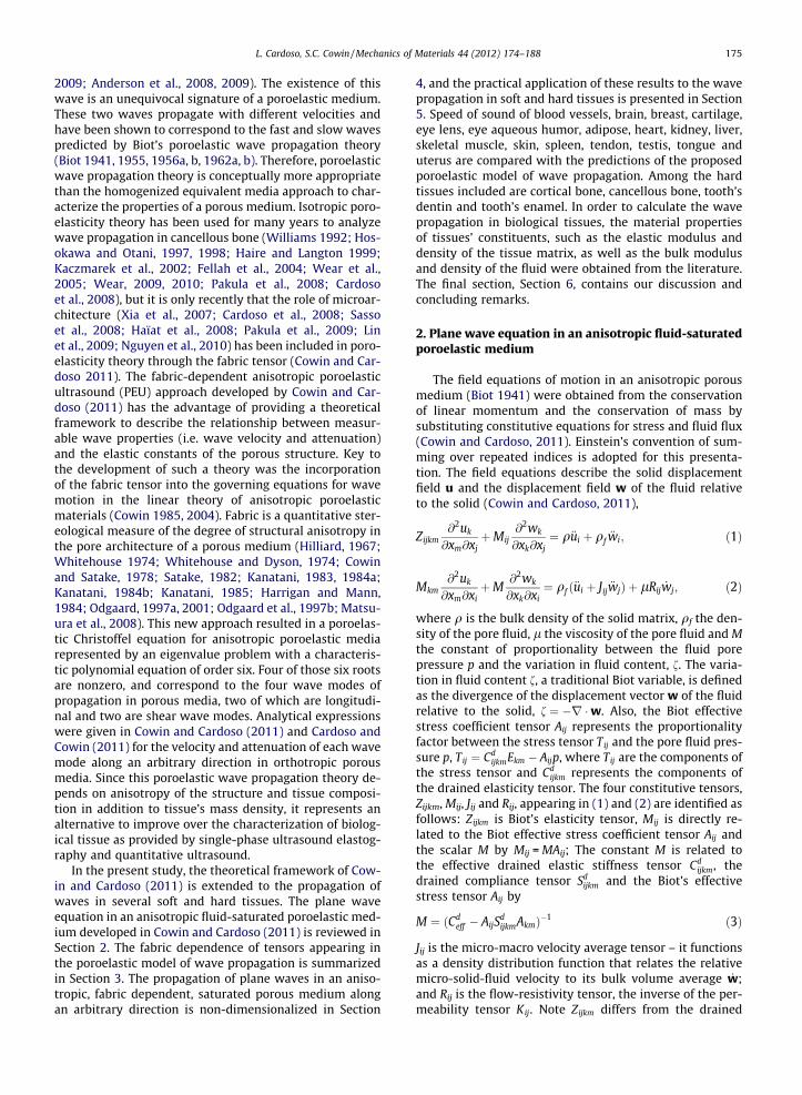
Fig. 5 shows the behavior of the non-dimensional fastand slow waves (P1 & P2) as a function of porosity in iso-tropic hard tissues along the direction n = (1,0,0)T, whichare compared to the curves of non dimensional elastic-density ratios, RQ
o ;RCdo ; and RMAA
o , involving the acoustictensor Q o11, the drained elastic constants Cd
o11 and the dif-ference between them, MA11A11, namely the product ofthe constant M times the open product of the Biot’s effec-tive stress tensor with itself,
RQo ¼
ffiffiffiffiffiffiffiffiffiffiQ o11
qo
s; RMAA
o ¼ffiffiffiffiffiffiffiffiffiffiffiffiffiffiffiffiffiffiMA11A11
qo
s; and RCd
o ¼
ffiffiffiffiffiffiffiffiffiCd
o11
qo
s
ð22Þ
Fig. 5 illustrates how RCdo decreases linearly from a value of
2.3 to zero (circles), while the ratio RMAAo increases linearly
with the porosity from 0 to 1 (triangles). The formerillustrates the behavior of the drained elastic constants asa function of the porosity, which depends on the propertiesof the solid phase, the anisotropic tissue matrix. The laterRMAA
o ratio is related to the fluid fraction in the porous med-ium, and is equal to the fluid properties at 100% porosity.The RQ
o ratio (pentagrams) is in fact the combination ofthe RMAA
o and RCdo ratios since it follows the behavior of
the drained elastic constants ratio RCdo from 0 to �70%
porosity, and then changing to the behavior of the RMAAo ra-
tio from 70% to 100% porosity. Therefore, RQo changes in
behavior from solid-like to fluid-like as a function of theporosity. Importantly, when these non dimensional ratiosare compared to the non dimensional fast and slow wavevelocities, it shows how the fast wave exhibits a behaviorsimilar to the drained elastic constants at low porosity,but it changes in behavior around 70% porosity to becomemuch more like a fluid material from 70 to 100% porosity.
Speed of sound of blood vessels, brain, breast, cartilage,eye lens, eye aqueous humor, adipose, heart, kidney, liver,skeletal muscle, skin, spleen, tendon, testis, tongue anduterus are compared with the predictions of the proposedporoelastic model of wave propagation. Such values ofspeed of sound were obtained from the literature (Gold-man and Richards 1954; Goldman and Hueter 1956; Chi-vers and Parry 1978; Goss et al., 1978, 1980a,b; Goss andDunn, 1980; Hoffmeister et al., 1995; Mast 2000; Haïatet al., 2006) and summarized in Table 3. Ultrasound wavevelocities reported in Table 3 are shown as different re-gions for enamel, cortical bone, dentine, cancellous boneand soft tissues (Fig. 6a). The soft and hard tissues regionsoverlap slightly at high porosities. Importantly, the speedof sound of either soft or hard tissues at high porosity isvery similar to the speed of sound in water, due to the high

Fig. 4. Non-dimensional drained elastic constants as a function of porosity in isotropic tissues (a), along the axes of symmetry in anisotropic soft tissues (b)and in anisotropic hard tissues (c).
L. Cardoso, S.C. Cowin / Mechanics of Materials 44 (2012) 174–188 183
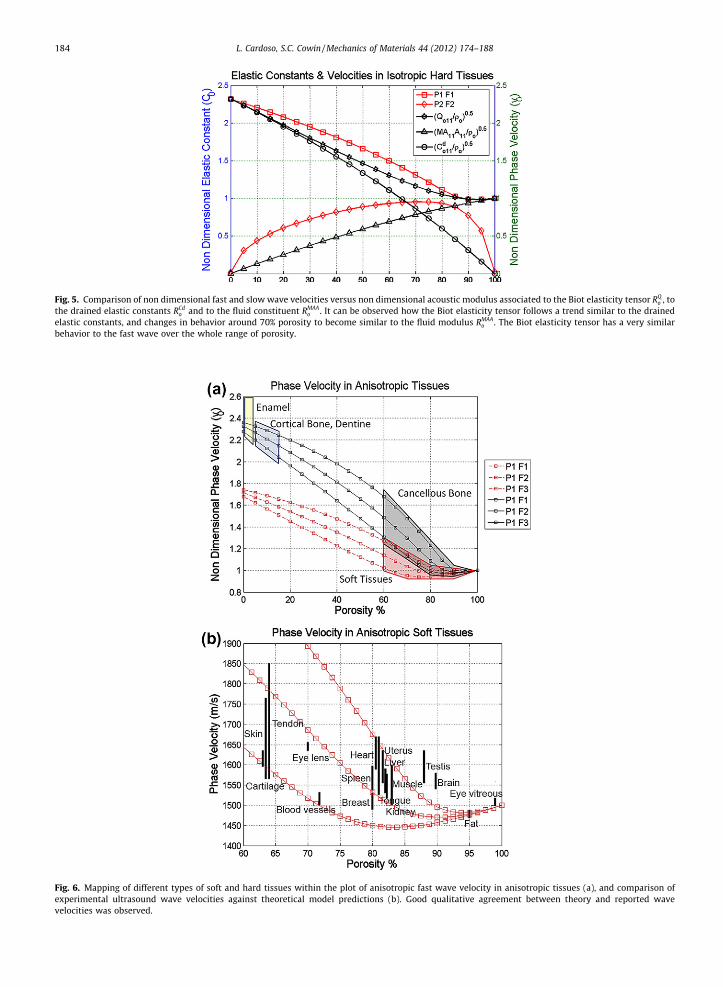
Fig. 5. Comparison of non dimensional fast and slow wave velocities versus non dimensional acoustic modulus associated to the Biot elasticity tensor RQo , to
the drained elastic constants RCdo and to the fluid constituent RMAA
o . It can be observed how the Biot elasticity tensor follows a trend similar to the drainedelastic constants, and changes in behavior around 70% porosity to become similar to the fluid modulus RMAA
o . The Biot elasticity tensor has a very similarbehavior to the fast wave over the whole range of porosity.
Fig. 6. Mapping of different types of soft and hard tissues within the plot of anisotropic fast wave velocity in anisotropic tissues (a), and comparison ofexperimental ultrasound wave velocities against theoretical model predictions (b). Good qualitative agreement between theory and reported wavevelocities was observed.
184 L. Cardoso, S.C. Cowin / Mechanics of Materials 44 (2012) 174–188

Fig. 7. Comparison of experimental ultrasound wave velocities against theoretical model predictions when considering (a) isotropic fabric and (b)anisotropic fabric measurements. The predictability of experimental values by the poroelastic theory is much increased when the fabric anisotropy is takeninto account.
L. Cardoso, S.C. Cowin / Mechanics of Materials 44 (2012) 174–188 185
fluid content in soft tissues and cancellous bone at highporosity. Therefore, at high porosities (i.e. 90% porosity),the fast wave velocity is practically indicative of the fluidphase of biological tissues, as opposed to the tissue matrix.Fig. 6b shows a magnified view of the region correspondingto the soft tissues, in which each of them is located basedon their porosity and speed of sound. Reported values of
speed of sound fall between the theoretical predictionsby the model for most of them, except for brain and testis.
The predictions of the fast and slow wave velocities usingthis theoretical model are now compared with experimentalmeasurements in trabecular bone previously reported (Car-doso et al., 2003). Briefly, fourteen bovine and sixty humantrabecular bone samples were retrieved from bovine femo-

186 L. Cardoso, S.C. Cowin / Mechanics of Materials 44 (2012) 174–188
ral heads, human femoral heads and femoral and tibial con-dyles. Samples of approximately 1 � 1 � 1 cm in size wereprepared, followed by removal of fat and marrow, and satu-rated with water under vacuum for 30 min. Group velocitiesof quasi-wave modes were measured in immersion withdistillated water at room temperature, using two broadbandultrasound transducers (Panametrics V323-SU) at a centralfrequency of 2.25 MHz (0.25 in diameter). The emitter wasexcited by a damped single pulse generated by an ultrasonicsource (Panametrics 5052 UA) operated in a transmissionmode. The signal was amplified in 40 dB, digitized by a100 MHz Digital Oscilloscope (Tektronic model 2430), andanalyzed in MATLAB. Importantly, bone cubes were cutwithout aligning their orthogonal faces with the planes ofsymmetry of the sample. Since bone samples were interro-gated along the normal direction to each face of the sample(e.g. arbitrary directions labeled as A, B and C), which do notcoincide with the normals to the planes of symmetry of thesample, it was considered that propagated signals corre-spond to quasi-wave modes for each of the three interro-gated directions. It was shown in that study that thewavelengths of the fast and slow waves are significantly dif-ferent and that this property can be used to separate the twowaves using digital filtering. Therefore, acquired signalswere filtered out using bandpass filters with a frequencybandwidth of 0.5 ± 0.25 MHz for the fast wave and a fre-quency bandwidth of 1.6 ± 0.6 MHz for the slow wave. Thefabric tensor describing the microarchitecture of each sam-ple was measured using the Mean Intercept Length (MIL)approach as described in Whitehouse (1974), Whitehouseand Dyson (1974), Harrigan and Mann (1984), Turner andCowin (1987) and Turner et al. (1990). The phase angles h,u were determined as the relative orientation of the princi-pal axes of fabric in respect to the measurement directionsA, B and C of the cube samples.
In order to investigate the effect of fabric on the fast andslow wave velocity measurements, two cases were ana-lyzed by comparing experimental values with theoreticalpredictions. First, the theoretical phase velocities for qP1and qP2 were computed using the experimental values ofporosity, tissue density, and without taking into accountthe fabric anisotropy, but considering the fabric as isotro-pic (F1 = F2 = F3 = 1=3). Then, the ray direction and groupvelocity were calculated for each quasi-longitudinal mode.Fig. 7a shows a comparison between the group velocitiespredicted from the model (under the assumption of isot-ropy) and the experimental wave velocity measurementsobtained on three orthogonal directions on each sample.In the second case, the theoretical phase velocities forqP1 and qP2 were computed as in the previous case, butthis time using the images-derived fabric anisotropy val-ues for each sample. Again, the ray direction and groupvelocity were calculated for each quasi-longitudinal mode.Fig. 7b shows the comparison between experimental groupwave velocities, for all three directions A, B and C and pre-dicted wave velocity values when the fabric was taken intoaccount. The correlation coefficient between experimentaland theoretical predictions when the fabric anisotropy isnot taken into account was R2 = 0.53, and R2 = 0.86 whenthe fabric anisotropy was included in the theoretical mod-el. This analysis indicates a much higher quantitative
agreement between experimental and theoretical valuesof group quasi-wave velocity when the fabric anisotropyis included in the model.
6. Discussion
A theoretical framework for analysis of anisotropicporoelastic wave propagation in a porous medium was re-cently developed by introducing the dependence of thewave motion equations upon fabric, a tensorial descriptorof the porous microarchitecture (Cowin and Cardoso,2011). Solution of the constitutive equations for harmonicdisplacements of the solid and fluid constituents leads to amodified Christoffel equation for anisotropic porous mediathat includes the acoustic tensor Q, the solid-fluid interac-tion tensor C, and the permeability tensor K(x). These ten-sors describe the elastic and viscous effects in the waveequation, and they all depend on the measurable fabrictensor, F. The modified Christoffel equation represents aneigenvalue problem with a sixth order characteristic equa-tion and four non-zero roots. When the porosity / is equalto zero, the model is reduced to that of a linear elastic solidand the Christoffel equation is obtained. When the porosity/ is equal to one, the model is reduced to the wave equa-tion in a fluid. Also, this system reduces to the isotropic for-mulation developed by Biot when the fabric tensor isisotropic. Two eigenvalues represent the longitudinal wavemodes P1 and P2 and the other two correspond to theshear wave modes S1 and S2. Such eigenvalues are com-plex valued, and describe the phase velocity and attenua-tion due to absorption of the four wave modes. Then, theporoelasticity theory was also extended to the case of anarbitrary phase direction (h, u) of wave propagation (Car-doso and Cowin, 2011). The advantage of that developmentis the ability to distinguish the role of fabric anisotropy inthe predictions of phase and group velocities of the fourwave modes propagating in porous media at any phaseor ray direction.
In those two previous studies, the poroelasticity theorywas only applied to the case of wave propagation in trabec-ular bone. That analysis was here extended here to manyother anisotropic soft and hard tissues. Elastic constantand density values for the tissue matrix constituents andwater were used in the poroelastic model to study thewave propagation in both soft and hard tissues. Only twoindependent variables (/ and F), one scalar and the othertensorial respectively, were used in the model to studythe influence of material properties on both global anddirectional changes in the velocity and attenuation of thefour wave modes generated in porous media. The collagenfibers in soft tissues and the mineralized matrix in hard tis-sues are responsible for the anisotropy of the solid tissueconstituent through the fabric tensor in the model. The re-ported measurements in the literature were found to fallbetween the predicted values of fast wave velocity by themodel.
Poroelasticity theory predicts the genesis of two longi-tudinal waves, however, the second wave, known as ‘‘slowwave’’, has only been reported in hard tissues such as can-cellous bone. We may speculate on two possible reasonswhy this has not been reported in soft tissues: first,

L. Cardoso, S.C. Cowin / Mechanics of Materials 44 (2012) 174–188 187
because the predicted velocity of both waves (fast andslow) in soft tissues is very similar, which may result insuperposition of both waves in the time domain, and sec-ond, because the attenuation of this slow wave may betoo high to be actually measured. Interestingly, our numer-ical analysis suggests that both fast and slow waves may begenerated in soft tissues, for instance, in tendons, whichexhibit large acoustic anisotropy above the speed of soundin water. Careful experimentation is required to determinewhether both fast and slow wave modes can be generatedor not in soft tissues.
In hard tissues, the theoretical model predicted the highvariability of fast and slow wave velocities observed in bo-vine and human bone in our experimental study. The devel-opment of the fabric-dependent anisotropic theory ofpropagation of quasi-waves in porous media (Cardoso andCowin, 2011) was able to predict the high variability of fastand slow wave velocities observed in bovine and humanbone in our experimental study. Fig. 7 indicates that the fab-ric tensor measurement alone is able to drastically increasethe model predictability on wave velocities from 53% to 86%.In other words, directional variability within a sample waseffectively explained by the theoretical model after inclu-sion of the fabric; this directional variability could not be ex-plained by the porosity only. The agreement betweenexperimental and theoretical values indicates that despitethe complexity added to the poroelastic theory, a tensorialvariable describing the bone microstructure is required toexplain the directional variability of the wave propagationwith bone architecture. Therefore, the fabric tensor –a mea-sure of bone microarchitecture–exhibited a role as impor-tant as the mass density in determining the acousticproperties of anisotropic porous bone samples.
Overall, the results from the present study demonstratethe ability of the proposed model to describe the acousticbehavior of the fast and slow wave velocities in both softand hard tissues. The phase velocity depends on the archi-tecture (porosity and fabric) and the composition of themedium (solid and fluid mass density, solid matrix elasticmodulus, fluid bulk modulus and fluid viscosity). For givenfrequency and material parameter values, the behaviors ofthe fast and slow waves are governed by the extrinsicproperties of the media: the porosity and fabric anisotropy.These theoretical predictions also corroborate our experi-mental observations that indicate that at high porositiesthe fast wave is mostly related to the propagation in thefluid constituent and the slow wave is highly related tothe solid matrix structure.
Acknowledgments
This work was supported by the National Institutes ofHealth (AG34198 & HL069537-07 R25 Grant for MinorityBME Education), the National Science Foundation (NSF0723027, PHY-0848491), and the PSC-CUNY ResearchAward Program of the City University of New York.
References
Anderson, C.C., Marutyan, K.R., Holland, M.R., Wear, K.A., Miller, J.G., 2008.Interference between wave modes may contribute to the apparent
negative dispersion observed in cancellous bone. J. Acoust. Soc. Am124, 1781–1789.
Anderson, C.C., Pakula, M., Holland, M.R., Bretthorst, G.L., Laugier, P.,Miller, J.G., (2009). Extracting fast and slow wave velocities andattenuations from experimental measurements of cancellous boneusing Bayesian probability theory. In: Ultrasonics Symposium (IUS),2009 IEEE International. 20–23 Sept. 2009 pp.546–549. doi:10.1109/ULTSYM.2009.5441732.
Berryman, J., (1997). Analysis of ultrasonic velocities in hydrocarbonmixtures. Stanford exploration project report. 75, 479–486.
Biot, M.A., 1955. Theory of elasticity and consolidation for a porousanisotropic solid. J. Appl. Phys. 26, 182-182.
Biot, M.A., 1956a. Theory of propagation of elastic waves in a fluidsaturated porous solid I low frequency range. J. Acoust. Soc. Am. 28,168–178.
Biot, M.A., 1956b. Theory of propagation of elastic waves in a fluidsaturated porous solid II higher frequency range. J. Acoust. Soc. Am.28, 179–191.
Biot, M.A., 1962a. Mechanics of deformation and acoustic propagation inporous media. J. Appl. Phys. 33, 1482–1498.
Biot, M.A., 1962b. Generalized theory of acoustic propagation in porousdissipative media. J. Acoust. Soc. Am. 28, 254–1264.
Biot, M.A., 1941. General theory of three-dimensional consolidation. J.Appl. Phys. 12, 155–164.
Carcione, J.M., 2001. Energy balance and fundamental relations indynamic anisotropic poro-viscoelasticity. Proc. R. Soc. Lond. A 457,331–348.
Cardoso, L., Cowin, S.C., 2011. Fabric dependence of quasi-waves inanisotropic porous media. J. Acoust. Soc. Am. 129 (5), 3302–3316.
Cardoso, L., Meunier, A., Oddou, C., 2008. In vitro acoustic wavepropagation in human and bovine cancellous bone as predicted bythe Biot’s theory. J. Mech. Med. Biol. 8 (2), 1–19.
Cardoso, L., Teboul, F., Meunier, A., Oddou, C., 2001. Ultrasoundcharacterization of cancellous bone: Theoretical and experimentalanalysis. I EEE Trans. Ultrason. Symp. 2, 1213–1216.
Cardoso, L., Teboul, F., Sedel, L., Meunier, A., Oddou, C., 2003. In vitroacoustic waves propagation in human and bovine cancellous bone. J.Bone Miner. Res. 18 (10), 1803–1812.
Cerveny, V., Psencik, I., 2006. Energy flux in viscoelastic anisotropicmedia. Geophys. J. Int. 166, 1299–1317.
Chivers, R.C., Parry, R.J., 1978. Ultrasonic velocity and attenuation inmammalian tissues. J. Acoust. Soc. Am. 63 (3), 940–953.
Cowin, S.C., Cardoso, L., (2011). Fabric dependence of poroelastic wavepropagation. Biomechan. Model Mechanobiol., 10 (1), 39–65, OnlineFirst May 12th, 2010, Open Access. doi:10.1007/s10237-010-0217-7.
Cowin, S.C., Mehrabadi, M.M., 2007. Compressible and incompressibleconstituents in anisotropic poroelasticity: The problem of unconfinedcompression of a disk. J. Mech. Phys. Solids 55, 161–193.
Cowin, S.C., 2004. Anisotropic poroelasticity: Fabric tensor formulation.Mechanics of Materials 36, 665–677.
Cowin, S.C., Satake, M., (Eds.), 1978. Continuum Mechanical and StatisticalApproaches in the Mechanics of Granular Materials, GakujutsuBunken Fukyu-Kai, Tokyo, p. 350.
Cowin, S.C., 1985. The relationship between the elasticity tensor and thefabric tensor. Mech. Mater. 4, 137–147.
Cowin, S.C., 1986. Wolff’s law of trabecular architecture at remodelingequilibrium. J. Biomech. Eng. 108, 83–88.
Cowin, S.C., 1997. Remarks on the paper entitled Fabric and elasticprincipal directions of cancellous bone are closely related. J. Biomech.30, 1191–1192.
Cowin, S.C., 1999. Bone poroelasticity. J. Biomech. 32, 218–238.Ericksen, J.L., (1960). Tensor fields in Encyclopedia of Physics, Truesdell C.
A., (Ed.), Springer, Berlin, pp. 794–858.Fellah, Z.E.A., Chapelon, J.Y., Berger, S., Lauriks, W., Depollier, C., 2004.
Ultrasonic wave propagation in human cancellous bone: Applicationof Biot theory. J. Acoust. Soc. Am. 116, 61–73, doi:10.1121/1.1755239.
Fratzl, P., 2008. Collagen: Structure and Mechanics. Springer, P 516.Goldman, D.E., Hueter, T.F., 1956. Tabular data of the velocity and
absorption of high-frequency sound in mammalian tissues. J. Acoust.Soc. Am. 28 (1), 35–37.
Goldman, D.E., Richards, J.R., 1954. Measurement of high-frequencysound velocity in mammalian soft tissues. J. Acoust. Soc. Am. 26 (6),981–983.
Goss, S.A., Dunn, F., 1980. Ultrasonics propagation properties of collagen.Phys. Med. Biol. 25 (5), 827–837.
Goss, S.A., Frizzell, L.A., Dunn, F., 1980a. Dependence of the ultrasonicproperties of biological tissue on constituent proteins. J. Acoust. Sec.Am. 67 (3), 1041–1044.

188 L. Cardoso, S.C. Cowin / Mechanics of Materials 44 (2012) 174–188
Goss, S.A., Johnston, R.L., Dunn, F., 1978. Comprehensive compilation ofempirical ultrasonic properties of mammalian tissues. J. Acoust. Soc.Am. 64 (2), 423–457.
Goss, S.A., Johnston, R.L., Dunn, F., 1980b. Compilation of empiricalultrasonic properties of mammalian tissues II. J. Acoust. Soc. Am. 68(1), 93–108.
Grimm, M.J., Williams, J.L., 1997. Assessment of bone quantity and‘quality’ by ultrasound attenuation and velocity in the heel. Clin.Biomech. (Bristol, Avon) 12, 281–285.
Haïat, G., Padilla, F., Cleveland, R.O., Laugier, P., 2006. Effects of frequency-dependent attenuation and velocity dispersion on in vitro ultrasoundvelocity measurements in intact human femur specimens. IEEE Trans.Ultrason. Ferroelectr. Freq. Control 53 (1), 39–51.
Haïat, G., Padilla, F., Peyrin, F., Laugier, P., 2008. Fast wave ultrasonicpropagation in trabecular bone: Numerical study of the influence ofporosity and structural anisotropy. J. Acoust. Soc. Am. 123, 1694–1705.
Haire, T.J., Langton, C.M., 1999. Biot theory: A review of its application toultrasound propagation through cancellous bone. Bone 24, 291–295.
Hans, D., Fuerst, T., Uffmann, M., 1996. Bone density and qualitymeasurement using ultrasound. Curr. Opin. Rheumatol. 8, 370–375.
Harrigan, T., Mann, R.W., 1984. Characterization of microstructuralanisotropy in orthotropic materials using a second rank tensor. J.Mat. Sci. 19, 761–769.
Hilliard, J.E., 1967. Determination of structural anisotropy, stereology. In:Proc. 2nd Int. Congress for Stereology, Chicago, 1967, Springer, Berlin,p. 219.
Hoffmeister, B.K., Handley, S.M., Verdonk, E.D., Wickline, S.A., Miller, J.G.,1995. Estimation of the elastic stiffness coefficient c13 of fixed tendonand fixed myocardium. J. Acoust. Soc. Am. 97 (5 Pt 1), 3171–3176.
Hosokawa, A., Otani, T., 1997. Ultrasonic wave propagation in bovinecancellous bone. J. Acoust. Soc. Am. 101, 558–562.
Hosokawa, A., Otani, T., 1998. Acoustic anisotropy in bovine cancellousbone. J. Acoust. Soc. Am. 103, 2718–2722.
Johnson, D.L., Koplik, J., Dashen, R., 1987. Theory of dynamic permeabilityand tortuosity in fluid-saturated porous media. J. Fluid Mech. 176,379–402.
Kaczmarek, M., Kubik, J., Pakula, M., 2002. Short ultrasonic waves incancellous bone. Ultrasonics 40, 95–100.
Kanatani, K., 1983. Characterization of structural anisotropy by fabrictensors and their statistical test. J. Japanese Soil Mech. Found. Eng. 23,171.
Kanatani, K., 1984a. Distribution of directional data and fabric tensors. Int.J. Eng. Sci. 22, 149–164.
Kanatani, K., 1984b. Stereological determination of structural anisotropy.Int. J. Eng. Sci. 22, 531–546.
Kanatani, K., 1985. Procedures for stereological estimation of structuralanisotropy. Int. J. Eng. Sci. 23, 587–596.
Lin, W., Xia, Y., Qin, Y.X., 2009. Characterization of the trabecular bonestructure using frequency modulated ultrasound pulse. J. Acoust. Soc.Am. 125 (6), 4071–4077.
Mast, T.D., 2000. Empirical relationships between acoustic parameters inhuman soft tissues. Acous. Res. Lett. Online 1 (2), 37–42.
Matsuura, M., Eckstein, F., Lochmüller, E-M., Zysset, P.K., 2008. The role offabric in the quasi-static compressive mechanical properties ofhuman trabecular bone from various anatomical locations. Biomech.Model Mechanobiol. 7, 27–42.
Mizuno, K., Matsukawa, M., Otani, T., Takada, M., Mano, I., Tsujimoto, T.,2008. Effects of structural anisotropy of cancellous bone on speed ofultrasonic fast waves in the bovine femur. IEEE Trans Ultrason.Ferroelectr. Freq. Control 55 (7), 1480–1487.
Mizuno, K., Matsukawa, M., Otani, T., Laugier, P., Padilla, F., 2009.Propagation of two longitudinal waves in human cancellous bone:an in vitro study. J. Acoust. Soc. Am. 125, 3460–3466.
Nguyen, V.H., Naili, S., Sansalone, V., 2010. Simulation of ultrasonic wavepropagation in anisotropic cancellous bone immersed in fluid. WaveMotion 47 (2), 117–129, ISSN 0165-2125, DOI:10.1016/j.wavemoti.2009.09.002.
Noda, H., 1972. Partial specific volume of collagen. J. Biochem. 71, 699–703.Odgaard, A., 1997a. Three- dimensional methods for quantification of
cancellous bone architecture. Bone 20, 315–328.Odgaard, A., (2001). Quantification of cancellous bone architecture. In:
Cowin, S. C., (Ed.), Bone Mechanics Handbook. Springer, Berlin, pp.14.1-19.
Odgaard, A., Kabel, J., van Rietbergen, B., Dalstra, M., Huiskes, R., 1997b.Fabric and elastic principal directions of cancellous bone are closelyrelated. J. Biomech. 30, 487–495.
Pakula, M., Padilla, F., Laugier, P., 2009. Influence of the filling fluid onfrequency-dependent velocity and attenuation in cancellous bonesbetween 0.35 and 2.5 MHz. J. Acoust. Soc. Am. 126 (6), 3301–3310.
Pakula, M., Padilla, F., Laugier, P., Kaczmarek, M., 2008. Application ofBiot’s theory to ultrasonic characterization of human cancellousbones: determination of structural, material, and mechanicalproperties. J. Acoust. Soc. Am. 123 (4), 2415–2423.
Sasso, M., Haïat, G., Yamato, Y., Naili, S., Matsukawa, M., (2008).Dependence of ultrasonic attenuation on bone mass andmicrostructure in bovine cortical bone. J Biomech. 41(2), 347–55.Epub 2007 Oct 29.
Satake, M., 1982. Fabric tensor in granular materials. In: Vermeer,P.A.,Lugar, H.J., (Eds.), Deformation and Failure of Granular Materials,Balkema, Rotterdam, p. 63
Sharma, M.D., 2005. Propagation of inhomogeneous plane waves indissipative anisotropic poroelastic solids. Geophys. J. Int. 163, 981–990.
Sharma, M.D., 2008. Propagation of harmonic plane waves in a generalanisotropic porous solid. Geophys. J. Int. 172 (3), 982–994.
Sharma, M.D., 2010. Energy velocity and quality factor of plane harmonicinhomogeneous waves in anisotropic poro-viscoelastic media.Geophys. J. Int. 180 (3), 1265–1273.
Siffert, R., Kaufman, J., 2006. Ultrasonic bone assessment: ‘‘The time hascome. Bone 40 (1), 5.
Silver, F.H., Christiansen, D.L., Snowhill, P., Chen, Y., 2000. Role of storageon changes in the mechanical properties of tendon and self-assembled collagen fibers. Connective Tissue Res. 41, 155–164.
Silver, F.H., Bradica, G., Tria, A., 2002a. Elastic energy storage in humanarticular cartilage: estimation of the elastic spring constant for type IIcollagen and changes associated with osteoarthritis. Matrix Biol. 21,129–137.
Silver, F.H., Christiansen, D.L., Snowhill, P.B., Chen, Y., 2001a. Transitionfrom viscous to elastic-based dependency of mechanical properties ofself-assembled type I collagen fibers. J. Appl. Polymer Sci. 79, 134–142.
Silver, F.H., Freeman, J., DeVore, D., 2001b. Viscoelastic properties ofhuman skin and processed dermis. Skin Res. Technol. 7, 18–23.
Silver, F.H., Horvath, I., Foran, D., 2001c. Viscoelasticity of the vessel wall:role of collagen and elastic fibers. Critical Rev. Biomed. Eng. 29, 279–302.
Silver, F.H., Horvath, I., Foran, D.J., 2002b. Mechanical implications of thedomain structure of fibril forming collagens: Comparison of themolecular and fibrillar flexibility of alpha1-chains found in types I, IIand III collagens. J. Theor. Biol. 216, 243–254.
Silver, F.H., Snowhill, P.B., Foran, D., 2003. Mechanical behavior of vesselwall: A comparative study of aorta, vena cava, and carotid artery. Ann.Biomed. Eng. 31, 793–803.
Turner, C.H., Cowin, S.C., 1987. On the dependence of the elastic constantsof an anisotropic porous material upon porosity and fabric. J.Materials Sci. 22, 3178–3184.
Turner, C.H., Cowin, S.C., Rho, J.Y., Ashman, R.B., Rice, J.C., 1990. The fabricdependence of the orthotropic elastic properties of cancellous bone. J.Biomech. 23, 549–561.
Turner, C.H., Rho, J.Y., Takano, Y., Tsui, T.Y., Pharr, G.M., 1999. The elasticproperties of trabecular and cortical bone tissues are similar: resultsfrom two microscopic measurement techniques. J. Biomech. 32, 437–441.
Van Rietbergen, B., Odgaard, A., Kabel, J., Huiskes, R., 1996. Directmechanical assessment of elastic symmetries and properties oftrabecular bone architecture. J. Biomech. 29, 1653–1657.
Van Rietbergen, B., Odgaard, A., Kabel, J., Huiskes, R., 1998. Relationshipsbetween bone morphology and bone elastic properties can beaccurately quantified using high-resolution computerreconstructions. J. Orthop. Res. 16, 23–28.
Wear, K.A., 2009. Frequency dependence of average phase shift fromhuman calcaneus in vitro. J. Acoust. Soc. Am. 126 (6), 3291–3300.
Wear, K.A., 2010. Decomposition of two-component ultrasound pulses incancellous bone using modified least squares Prony method –phantom experiment and simulation. Ultrasound Med. Biol. 36 (2),276–287.
Wear, K.A., Laib, A., Stuber, A.P., Reynolds, J.C., 2005. Comparison ofmeasurements of phase velocity in human calcaneus to Biot theory. J.Acoust. Soc. Am. 117 (5), 3319–3324.
Whitehouse, W.J., Dyson, E.D., 1974. Scanning electron microscopestudies of trabecular bone in the proximal end of the human femur.J. Anatomy 118, 417–444.
Whitehouse, W.J., 1974. The quantitative morphology of anisotropictrabecular bone. J. Microscopy 101, 153–168.
Williams, J.L., 1992. Ultrasonic wave propagation in cancellous andcortical bone: Prediction of some experimental results by Biot’stheory. J. Acoust. Soc. Am. 91, 1106–1112.
Xia, Y., Lin, W., Qin, Y.X., 2007. Bone surface topology mapping and its rolein trabecular bone quality assessment using scanning confocalultrasound. Osteoporos. Int. 18 (7), 905–913, Epub 2007 Mar 15.