Embed Size (px)
Citation preview
Role of Tet proteins in enhancer activityand telomere elongation
Falong Lu,1,2,3,6 Yuting Liu,1,2,3,6 Lan Jiang,1,2,3,6 Shinpei Yamaguchi,1,2,3 and Yi Zhang1,2,3,4,5
1Howard Hughes Medical Institute, Boston Children’s Hospital, Boston, Massachusetts 02115, USA; 2Program in Cellular andMolecular Medicine, Boston Children’s Hospital, Boston, Massachusetts 02115, USA; 3Division of Hematology/Oncology,Department of Pediatrics, Boston Children’s Hospital, Boston, Massachusetts 02115, USA; 4Department of Genetics, HarvardMedical School, Boston, Massachusetts 02115, USA; 5Harvard Stem Cell Institute, Boston, Massachusetts 02115, USA
DNA methylation at the C-5 position of cytosine (5mC) is one of the best-studied epigenetic modifications and playsimportant roles in diverse biological processes. Iterative oxidation of 5mC by the ten-eleven translocation (Tet) familyof proteins generates 5-hydroxymethylcytosine (5hmC), 5-formylcytosine (5fC), and 5-carboxylcytosine (5caC). 5fCand 5caC are selectively recognized and excised by thymine DNA glycosylase (TDG), leading to DNA demethylation.Functional characterization of Tet proteins has been complicated by the redundancy between the three familymembers. Using CRISPR/Cas9 technology, we generated mouse embryonic stem cells (ESCs) deficient for all three Tetproteins (Tet triple knockout [TKO]). Whole-genome bisulfite sequencing (WGBS) analysis revealed that Tet-mediatedDNA demethylation mainly occurs at distally located enhancers and fine-tunes the transcription of genes associatedwith these regions. Functional characterization of Tet TKO ESCs revealed a role for Tet proteins in regulating the two-cell embryo (2C)-like state under ESC culture conditions. In addition, Tet TKO ESCs exhibited increased telomere–sister chromatid exchange and elongated telomeres. Collectively, our study reveals a role for Tet proteins in not onlyDNA demethylation at enhancers but also regulating the 2C-like state and telomere homeostasis.
[Keywords: DNA methylation; demethylation; enhancer; embryonic stem cell; telomere]
Supplemental material is available for this article.
Received June 26, 2014; revised version accepted August 25, 2014.
DNA methylation at the C-5 position of cytosine (5mC)plays an important role in diverse biological processes(Goll and Bestor 2005; Ooi and Bestor 2008; Wu andZhang 2010). DNA methylation is established and main-tained by a family of DNA methyltransferases (Goll andBestor 2005). Growing evidence supports a biochemicalpathway of active DNA demethylation involving ten-eleven translocation (Tet) family proteins and thymineDNA glycosylase (TDG) (Tahiliani et al. 2009; He et al.2011; Ito et al. 2011; Pastor et al. 2013;Wu and Zhang 2014).Tet proteins can oxidize 5mC to 5-hydroxymethylcytosine(5hmC) (Tahiliani et al. 2009; Ito et al. 2010), which in turncan be further oxidized to 5-formylcytosine (5fC) and5-carboxylcytosine (5caC) and then excised by TDG fol-lowed by base excision repair to complete the demethyla-tion cycle (He et al. 2011; Ito et al. 2011; Shen et al. 2013).Mouse embryonic stem cells (mESCs) are derived from
preimplantation epiblasts and have the capacity for self-renewal and the ability to differentiate into all embryoniclineages (Rossant 2008; Silva and Smith 2008; Hanna
et al. 2010; Boroviak et al. 2014). ESCs are heterogeneousand consist of subpopulations bearing distinct character-istics. A small population (<0.5%) of ESCs, called two-cellembryo (2C)-like cells, has the ability to contribute toboth embryonic and extraembryonic tissues (Macfarlanet al. 2012). ESCs enter the transient 2C-like state regu-larly (Macfarlan et al. 2012). Physiologically, the activationof Zscan4, one of the 2C-specific genes, is important fortelomerase-independent telomere elongation and restora-tion of developmental capacity, two properties required forlong-term maintenance of ESC pluripotency (Zalzmanet al. 2010; Amano et al. 2013).Tet1 and Tet2 are highly expressed in mESCs, which is
consistent with the relative high level of 5hmC anddetectable levels of 5fC and 5caC in these cells (Itoet al. 2010, 2011; Ficz et al. 2011; Koh et al. 2011).Knockdown of Tet1 in mESCs leads to reduced 5hmClevels and substantial transcriptional and promoter DNAmethylation changes (Ito et al. 2010; Ficz et al. 2011; Koh
� 2014 Lu et al. This article is distributed exclusively by Cold SpringHarbor Laboratory Press for the first six months after the full-issuepublication date (see http://genesdev.cshlp.org/site/misc/terms.xhtml).After six months, it is available under a Creative Commons License(Attribution-NonCommercial 4.0 International), as described at http://creativecommons.org/licenses/by-nc/4.0/.
6These authors contributed equally to this work.Corresponding author: [email protected] published online ahead of print. Article and publication date are onlineat http://www.genesdev.org/cgi/doi/10.1101/gad.248005.114.
GENES & DEVELOPMENT 28:2103–2119 Published by Cold Spring Harbor Laboratory Press; ISSN 0890-9369/14; www.genesdev.org 2103
Cold Spring Harbor Laboratory Press on February 26, 2019 - Published by genesdev.cshlp.orgDownloaded from
et al. 2011; Williams et al. 2011; Wu et al. 2011; Xu et al.2011). While Tet1-deficient mESCs maintain pluripotencyand are largely normal, they showed skewed differentia-tion in vitro and in the teratoma assay (Dawlaty et al.2011; Koh et al. 2011). Similarly, both Tet1/2 double-knockout (DKO) and Tet triple-knockout (TKO) mESCsare largely normal under ESC culture conditions, butsevere abnormalities in lineage differentiation of TetDKO and Tet TKO mESCs have been reported (Dawlatyet al. 2013, 2014; Hu et al. 2014). In contrast to theaccumulating knowledge regarding the loss-of-functionphenotypes of Tet proteins, it remains unclear where andto what extent Tet proteins contribute to DNA demeth-ylation within the genome. Thus, quantitative and high-resolution mapping of DNA methylation changes in thegenome of mESCs deficient for all Tet proteins willprovide insights into the role of these 5mC oxidases inregulating the methylome of ESCs.To identify targets of Tet-mediated active DNA demeth-
ylation in the genome and their biological significance, wegenerated Tet TKO mESCs and performed base-resolutionDNA methylome and transcriptome analyses. We foundthat DNA hypermethylation occurred extensively atdistal enhancers in Tet TKO mESCs. Parallel transcrip-tome analysis demonstrated that Tet TKO cells showedactivation of 2C-specific genes, suggesting an importantrole for Tet proteins in fine-tuning ESC states. Up-regula-tion of these 2C-specific genes in Tet TKO cells was atleast partly caused by impairment of Kap1-mediated generepression. Consistent with the activation of 2C-specificgenes, telomere elongation through telomere–sister chro-matid exchange (T-SCE) was observed in Tet TKO cells.Therefore, our study uncovers a critical role of Tet proteinsin enhancer function and reveals a new function for Tetproteins in repressing the 2C-like state and telomerelength regulation.
Results
Tet TKO does not affect mESC maintenance
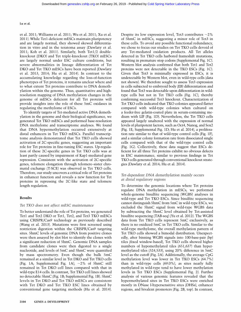
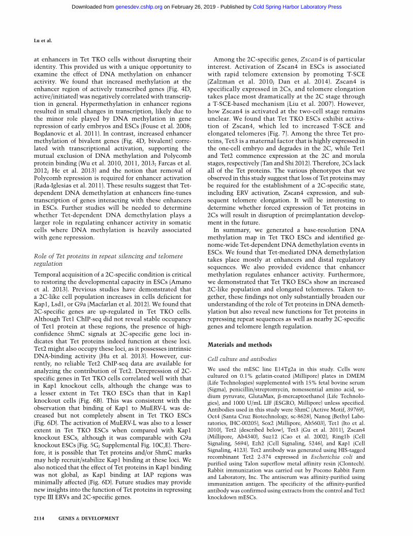
To better understand the role of Tet proteins, we generatedTet1 and Tet2 DKO or Tet1, Tet2, and Tet3 TKO mESCsusing CRISPR/Cas9 technology as previously described(Wang et al. 2013). Mutations were first screened usingrestriction digestion within the CRISPR/Cas9 targetingsites. 5hmC levels of genomic DNA from positive cloneswere then assayed by slot blot to identify the clones witha significant reduction of 5hmC. Genomic DNA samplesfrom candidate clones were then digested to a singlenucleotide, and levels of 5mC and 5hmC were quantifiedby mass spectrometry. Even though the bulk 5mCremained at a similar level in Tet DKO and Tet TKO cells(Fig. 1A; Supplemental Fig. 1A), ;2% of 5hmC stillremained in Tet DKO cell lines compared with that inwild-type E14 cells. In contrast, Tet TKO cell lines showedno detectable 5hmC (Fig. 1B; Supplemental Fig. 1B). 5hmClevels in Tet DKO and Tet TKO mESCs are consistentwith Tet DKO and Tet TKO ESC lines obtained byconventional gene targeting methods (Hu et al. 2014).
Despite its low expression level, Tet3 contributes ;2%of 5hmC in mESCs, suggesting a minor role of Tet3 inthese cells. To avoid any possible functional redundancy,we chose to focus our studies on Tet TKO cells devoid ofany Tet-mediated oxidation products. All Tet allelesdetected in Tet TKO cells harbored frameshift mutations,resulting in premature stop codons (Supplemental Fig. 1C).Western blot analysis confirmed that both Tet1 and Tet2proteins were not detectable in the TKO ESCs (Fig. 1C).Given that Tet3 is minimally expressed in ESCs, it isundetectable by Western blot, even in wild-type cells (datanot shown). We therefore sought to assess Tet3 expressionin cells subjected to embryoid body (EB) differentiation andfound that Tet3 was detectable upon differentiation in wild-type cells but not in Tet TKO cells (Fig. 1C), therebyconfirming successful Tet3 knockout. Characterization ofTet TKO cells indicated that TKO colonies appeared flattercompared with wild-type colonies when cultured ona feeder-free gelatin-coated plate in serum-containing me-dium with LIF (Fig. 1D). Nevertheless, the Tet TKO cellsappeared largely unaltered with the expression of normallevels of pluripotent factors, such as Oct4, Nanog, and Sox2(Fig. 1E; Supplemental Fig. 1D; Hu et al. 2014), a prolifera-tion rate similar to that of wild-type control cells (Fig. 1F),and a similar colony formation rate from dissociated singlecells compared with that of the wild-type control cells(Fig. 1G). Collectively, these data suggest that ESCs de-ficient for all three Tet proteins exhibit no obvious defectsin ESC maintenance, similar to previous findings in TetTKO cells generated through conventional knockout strate-gies (Dawlaty et al. 2014; Hu et al. 2014).
Tet-dependent DNA demethylation mainly occursat distal regulatory regions
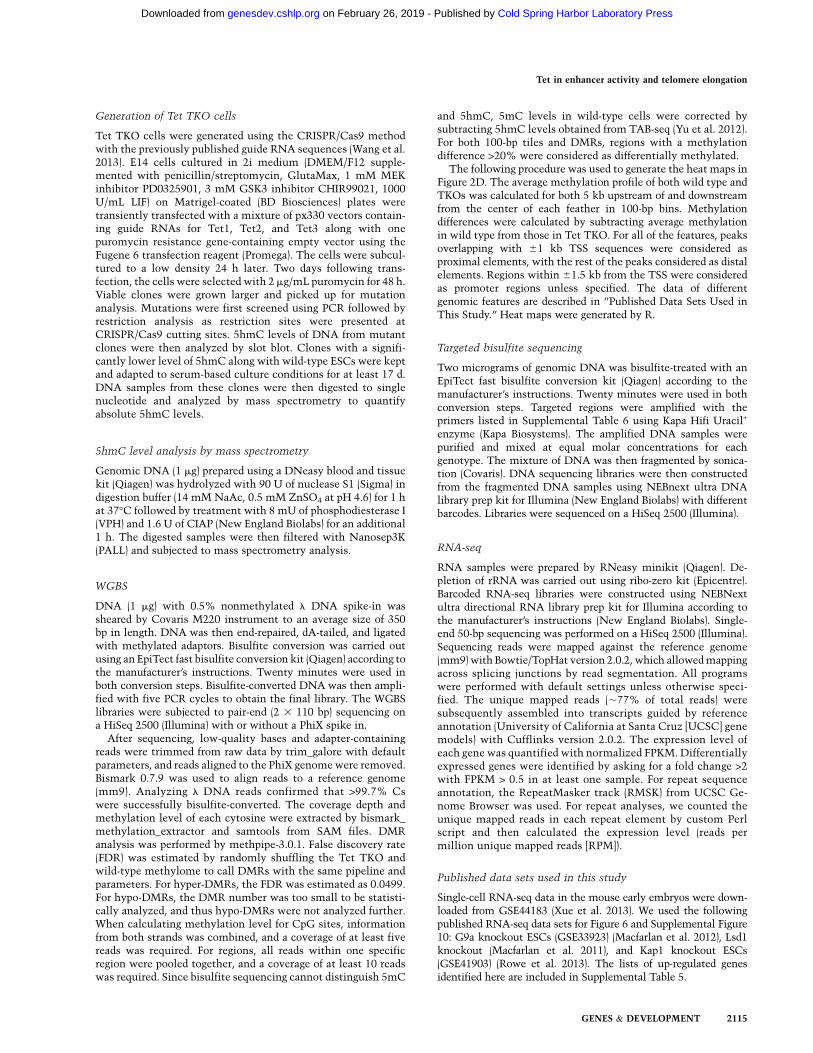
To determine the genomic locations where Tet proteinsregulate DNA methylation in mESCs, we performedwhole-genome bisulfite sequencing (WGBS) analyses inwild-type and Tet TKO ESCs. Since bisulfite sequencingcannot distinguish 5hmC from 5mC inwild-type ESCs, weexcluded the 5hmC signal from wild-type WGBS databy subtracting the 5hmC level obtained by Tet-assistedbisulfite sequencing (TAB-seq) (Yu et al. 2012). The WGBSdata from Tet TKO cells represent 5mC exclusively, asthere is no oxidized 5mC in Tet TKO cells. Similar to thewild-type methylome, the overall methylation pattern ofTet TKO cells showed a bimodal distribution. Unexpect-edly, after binning WGBS signals into 100-base-pair (bp)tiles (fixed window-based), Tet TKO cells showed highernumbers of hypomethylated tiles (651,637) than hyper-methylated tiles (524,533), using 20% difference in 5mClevel as the cutoff (Fig. 2A). Additionally, the average CpGmethylation level was lower in Tet TKO ESCs (64.7%)than in wild-type cells (69.0%), as sites nearly fullymethylated in wild-type tend to have lower methylationlevels in Tet TKO ESCs (Supplemental Fig. 2). Furtheranalysis of various genomic features revealed that thehypermethylated sites in Tet TKO ESCs were enrichedmostly in DNase I-hypersensitive sites (DHSs), enhancerregions, and bivalent promoters (Fig. 2B, top). In contrast,
Lu et al.
2104 GENES & DEVELOPMENT
Cold Spring Harbor Laboratory Press on February 26, 2019 - Published by genesdev.cshlp.orgDownloaded from
hypomethylated sites were not highly enriched for anygenomic features (Fig. 2B, bottom). With Methpipe, a hid-den Markov model (HMM; nonfixed window)-based algo-rithm for detecting differentially methylated regions(DMRs) (Song et al. 2013), we identified 20,109 hyper-methylated DMRs (hyper-DMRs) and only 315 hypometh-ylated DMRs (hypo-DMRs) in Tet TKO ESCs (Supplemen-tal Table 1). Taken together, these data indicate thathypermethylated CpGs tend to cluster together at specificgenomic loci with gene regulatory potentials (promoter,enhancers, and gene bodies). In contrast, hypomethylatedCpGs are randomly distributed across the genome.We found that 66% of hyper-DMRs overlapped with5fC/5caC DNA immunoprecipitation (DIP) sequencing(DIP-seq) peaks in TDG knockdown ESCs, and another14% of hyper-DMRs overlapped with 5hmC DIP-seqpeaks when not covered by 5fC/5caC peaks (Fig. 2C, left;Shen et al. 2013). Although the remaining 20% of hyper-DMRs did not overlap with 5hmC, 5fC, or 5caC peaks,there was significant 5hmC enrichment at these regionswhen looking into the high-confidence base-resolution5hmC data generated by TAB-seq analysis (Fig. 2C, right;Yu et al. 2012). Therefore, these data strongly suggestthat the majority of hyper-DMRs identified are targets ofTet-mediated oxidation-dependent DNA demethylationin mESCs.To further characterize the differential methylation in
Tet TKO cells, we calculated the average levels of 5mCchange within genomic features derived from publishedgenome-wide mapping data sets for 5mC oxidative de-rivatives, DNA-binding factors, and major histone modifi-cations (Fig. 2D). DNA hypermethylation was detected atthe center of 5fC, 5caC, and, to a lesser extent, 5hmCDIP-seq peaks (Fig. 2D, cyan color group; Shen et al. 2013)regardless of proximal or distal localization. Although 5fCand 5caC accumulated to high abundance, 5mC levels
remained similar to TDG knockdown cells (Shen et al.2013). Additionally, hypermethylation levels at the centerof 5hmC peaks were lower than those for 5fC and 5caC(Fig. 2D). These data therefore provide the first directevidence of 5fC and 5caC serving as demethylation in-termediates in cells.To determine whether 5hmCs serves as an intermedi-
ate of the DNA demethylation process or as a stableepigenetic mark, we analyzed the relationship betweenhypermethylated CpGs in Tet TKO cells and the pre-viously reported high-confidence 5hmC (Yu et al. 2012).This analysis revealed no clear correlation betweenhypermethylation in Tet TKO cells and high-confidence5hmC in wild-type ESCs. Indeed, a significant portion ofhypermethylated cytosines had no or a very low level of5hmC in wild-type ESCs (Supplemental Fig. 3A). Consis-tently, only 27% of the hypermethylated cytosines in TetTKO ESCs had a 5hmC signal in wild-type ESCs whencytosines with high coverage in both our WGBS dataand the TAB-seq data were analyzed (at least covered by10 reads per strand) (Supplemental Fig. 3B). These resultssuggest that stable 5hmC may serve as an epigeneticmark rather than as an intermediate of active DNAdemethylation. Hypermethylation was preferentially de-tected at binding sites of core transcription factors inESCs (Oct4, Nanog, Sox2, and Esrrb) (Chen et al. 2008;Marson et al. 2008) and histone marks (H3K4me1 andH3K27ac) associated with enhancers (Fig. 2D, yellowgroup; Meissner et al. 2008; Creyghton et al. 2010), themediator complex, and the cohesion complex (Fig. 2D,pink color group; Kagey et al. 2010). Furthermore, hyper-methylation was detected at binding sites of complexesinvolved in decommissioning active enhancers duringdifferentiation, such as the LSD1–NuRD complex (Fig.2D, red color group; Whyte et al. 2012). These features areall known to be associated with enhancers, indicating
Figure 1. Tet TKO ESCs are depleted of5hmC but retain ESC characteristics. (A,B)While 5mC level was not globally altered byTet TKO (A), 5hmC was undetectable (B) inTet TKO ESCs. Bars are 5mC and 5hmC levels(relative to unmodified C) in control and TKOmESCs as measured by mass spectrometry. (C)Western blot analysis demonstrated depletionof Tet1, Tet2, and Tet3 proteins in Tet TKOcells, with Suz12 serving as a loading controlfor all blots. ESC nuclear extracts were usedfor Tet1, Tet2, and Suz12 blots, while nuclearextracts from differentiated cells (EBs) wereused for the Tet3 and Suz12 blots. (D) Tet TKOmESCs exhibited relatively flatter morphologywhen compared with wild-type (WT) mESCs.(E) Pluripotent marker genes were expressednormally in Tet TKO mESCs. Data shown areimages of wild-type and Tet TKO mESCscostained with Oct4 and Nanog antibodies.Nuclei were visualized by DAPI staining. (F)Growth curves of wild-type and Tet TKO
mESCs. Data shown are mean of three biological replicates, with error bars representing standard deviation. (G) Tet TKO did not affectESC colony formation capacity. Data shown are mean single-cell colony formation rates of three biological replicates, with error barsrepresenting standard deviation.
Tet in enhancer activity and telomere elongation
GENES & DEVELOPMENT 2105
Cold Spring Harbor Laboratory Press on February 26, 2019 - Published by genesdev.cshlp.orgDownloaded from
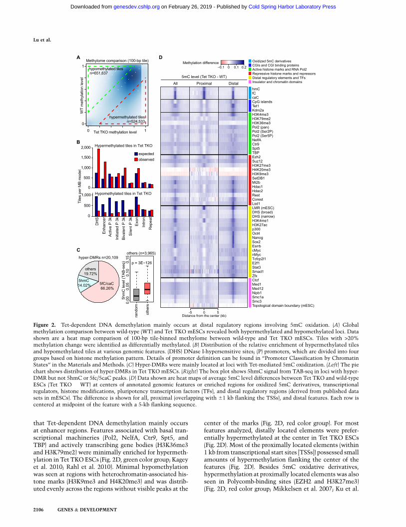
that Tet-dependent DNA demethylation mainly occursat enhancer regions. Features associated with basal tran-scriptional machineries (Pol2, NelfA, Ctr9, Spt5, andTBP) and actively transcribing gene bodies (H3K36me3and H3K79me2) were minimally enriched for hypermeth-ylation in Tet TKO ESCs (Fig. 2D, green color group; Kageyet al. 2010; Rahl et al. 2010). Minimal hypomethylationwas seen at regions with heterochromatin-associated his-tone marks (H3K9me3 and H4K20me3) and was distrib-uted evenly across the regions without visible peaks at the
center of the marks (Fig. 2D, red color group). For mostfeatures analyzed, distally located elements were prefer-entially hypermethylated at the center in Tet TKO ESCs(Fig. 2D). Most of the proximally located elements (within1 kb from transcriptional start sites [TSSs]) possessed smallamounts of hypermethylation flanking the center of thefeatures (Fig. 2D). Besides 5mC oxidative derivatives,hypermethylation at proximally located elements was alsoseen in Polycomb-binding sites (EZH2 and H3K27me3)(Fig. 2D, red color group; Mikkelsen et al. 2007; Ku et al.
Figure 2. Tet-dependent DNA demethylation mainly occurs at distal regulatory regions involving 5mC oxidation. (A) Globalmethylation comparison between wild-type (WT) and Tet TKO mESCs revealed both hypermethylated and hypomethylated loci. Datashown are a heat map comparison of 100-bp tile-binned methylome between wild-type and Tet TKO mESCs. Tiles with >20%methylation change were identified as differentially methylated. (B) Distribution of the relative enrichment of hypermethylated tilesand hypomethylated tiles at various genomic features. (DHS) DNase I-hypersensitive sites; (P) promoters, which are divided into fourgroups based on histone methylation pattern. Details of promoter definition can be found in ‘‘Promoter Classification by ChromatinStates’’ in the Materials and Methods. (C) Hyper-DMRs were mainly located at loci with Tet-mediated 5mC oxidization. (Left) The piechart shows distribution of hyper-DMRs in Tet TKO mESCs. (Right) The box plot shows 5hmC signal from TAB-seq in loci with hyper-DMR but not 5hmC or 5fc/5caC peaks. (D) Data shown are heat maps of average 5mC level differences between Tet TKO and wild-typeESCs (Tet TKO � WT) at centers of annotated genomic features or enriched regions for oxidized 5mC derivatives, transcriptionalregulators, histone modifications, pluripotency transcription factors (TFs), and distal regulatory regions (derived from published datasets in mESCs). The difference is shown for all, proximal (overlapping with 61 kb flanking the TSSs), and distal features. Each row iscentered at midpoint of the feature with a 5-kb flanking sequence.
Lu et al.
2106 GENES & DEVELOPMENT
Cold Spring Harbor Laboratory Press on February 26, 2019 - Published by genesdev.cshlp.orgDownloaded from
2008), indicating that bivalent promoters are the majortype of promoters regulated byTet-mediatedDNAdemeth-ylation. In summary, these results demonstrate that Tet-mediated DNA demethylation mainly occurs at distalregulatory elements.
Enhancers and promoters are the main targetsof Tet-mediated DNA demethylation
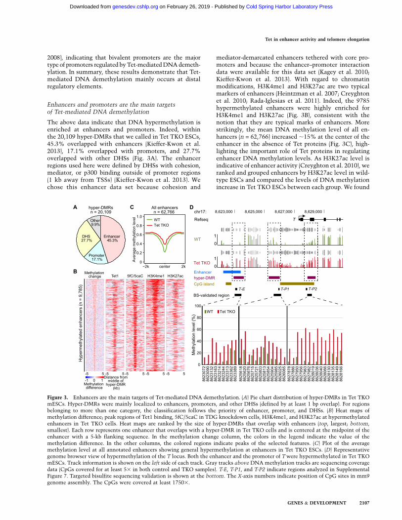
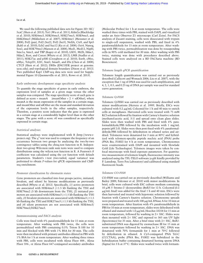
The above data indicate that DNA hypermethylation isenriched at enhancers and promoters. Indeed, withinthe 20,109 hyper-DMRs that we called in Tet TKO ESCs,45.3% overlapped with enhancers (Kieffer-Kwon et al.2013), 17.1% overlapped with promoters, and 27.7%overlapped with other DHSs (Fig. 3A). The enhancerregions used here were defined by DHSs with cohesion,mediator, or p300 binding outside of promoter regions(1 kb away from TSSs) (Kieffer-Kwon et al. 2013). Wechose this enhancer data set because cohesion and
mediator-demarcated enhancers tethered with core pro-moters and because the enhancer–promoter interactiondata were available for this data set (Kagey et al. 2010;Kieffer-Kwon et al. 2013). With regard to chromatinmodifications, H3K4me1 and H3K27ac are two typicalmarkers of enhancers (Heintzman et al. 2007; Creyghtonet al. 2010; Rada-Iglesias et al. 2011). Indeed, the 9785hypermethylated enhancers were highly enriched forH3K4me1 and H3K27ac (Fig. 3B), consistent with thenotion that they are typical marks of enhancers. Morestrikingly, the mean DNA methylation level of all en-hancers (n = 62,766) increased ;15% at the center of theenhancer in the absence of Tet proteins (Fig. 3C), high-lighting the important role of Tet proteins in regulatingenhancer DNA methylation levels. As H3K27ac level isindicative of enhancer activity (Creyghton et al. 2010), weranked and grouped enhancers by H3K27ac level in wild-type ESCs and compared the levels of DNA methylationincrease in Tet TKO ESCs between each group. We found
Figure 3. Enhancers are the main targets of Tet-mediated DNA demethylation. (A) Pie chart distribution of hyper-DMRs in Tet TKOmESCs. Hyper-DMRs were mainly localized to enhancers, promoters, and other DHSs (defined by at least 1 bp overlap). For regionsbelonging to more than one category, the classification follows the priority of enhancer, promoter, and DHSs. (B) Heat maps ofmethylation difference; peak regions of Tet1 binding, 5fC/5caC in TDG knockdown cells, H3K4me1, and H3K27ac at hypermethylatedenhancers in Tet TKO cells. Heat maps are ranked by the size of hyper-DMRs that overlap with enhancers (top, largest; bottom,smallest). Each row represents one enhancer that overlaps with a hyper-DMR in Tet TKO cells and is centered at the midpoint of theenhancer with a 5-kb flanking sequence. In the methylation change column, the colors in the legend indicate the value of themethylation difference. In the other columns, the colored regions indicate peaks of the selected features. (C) Plot of the averagemethylation level at all annotated enhancers showing general hypermethylation at enhancers in Tet TKO ESCs. (D) Representativegenome browser view of hypermethylation of the T locus. Both the enhancer and the promoter of Twere hypermethylated in Tet TKOmESCs. Track information is shown on the left side of each track. Gray tracks above DNA methylation tracks are sequencing coveragedata (CpGs covered for at least 53 in both control and TKO samples). T-E, T-P1, and T-P2 indicate regions analyzed in SupplementalFigure 7. Targeted bisulfite sequencing validation is shown at the bottom. The X-axis numbers indicate position of CpG sites in mm9genome assembly. The CpGs were covered at least 17503.
Tet in enhancer activity and telomere elongation
GENES & DEVELOPMENT 2107
Cold Spring Harbor Laboratory Press on February 26, 2019 - Published by genesdev.cshlp.orgDownloaded from
an inverse correlation between DNA methylation andH3K27ac levels as well as all 10 groups of enhancersexhibiting a significant increase in DNA methylation inTet TKO cells (Supplemental Fig. 4). Our analysis in-dicates that proximal features associated with promotersshow relatively lower hypermethylation at the shores ofthe center and that Polycomb-binding sites show hyper-methylation at the center of the elements (Fig. 2D).Indeed, bivalent promoters showed the largest averagemethylation increase among promoters (SupplementalFig. 5). For example, we detected a significant increasein DNAmethylation at both the promoter and one nearbyenhancer of T (also named Brachyury) in Tet TKO ESCs(Fig. 3D). The methylation change was validated bytargeted bisulfite sequencing of an independent batch ofsamples with at least 17503 coverage (Fig. 3D). Theaverage methylation level at active and initiated pro-moters also increased to a lesser extent, whereas silentpromoters were heavilymethylated even inwild-type cells,with levels comparable with those of repeat sequences(Supplemental Fig. 5). These results suggest a generalenrichment of Tet-mediated DNA demethylation at regu-latory sequences, especially at enhancers and promoterregions. Taken together, our data reveal the location ofgenome-wide Tet-dependent DNA demethylation inmESCs, which mainly include enhancers, promoters, andother distal regulatory elements.
Dual function of Tet-mediated DNA demethylationin transcriptional regulation
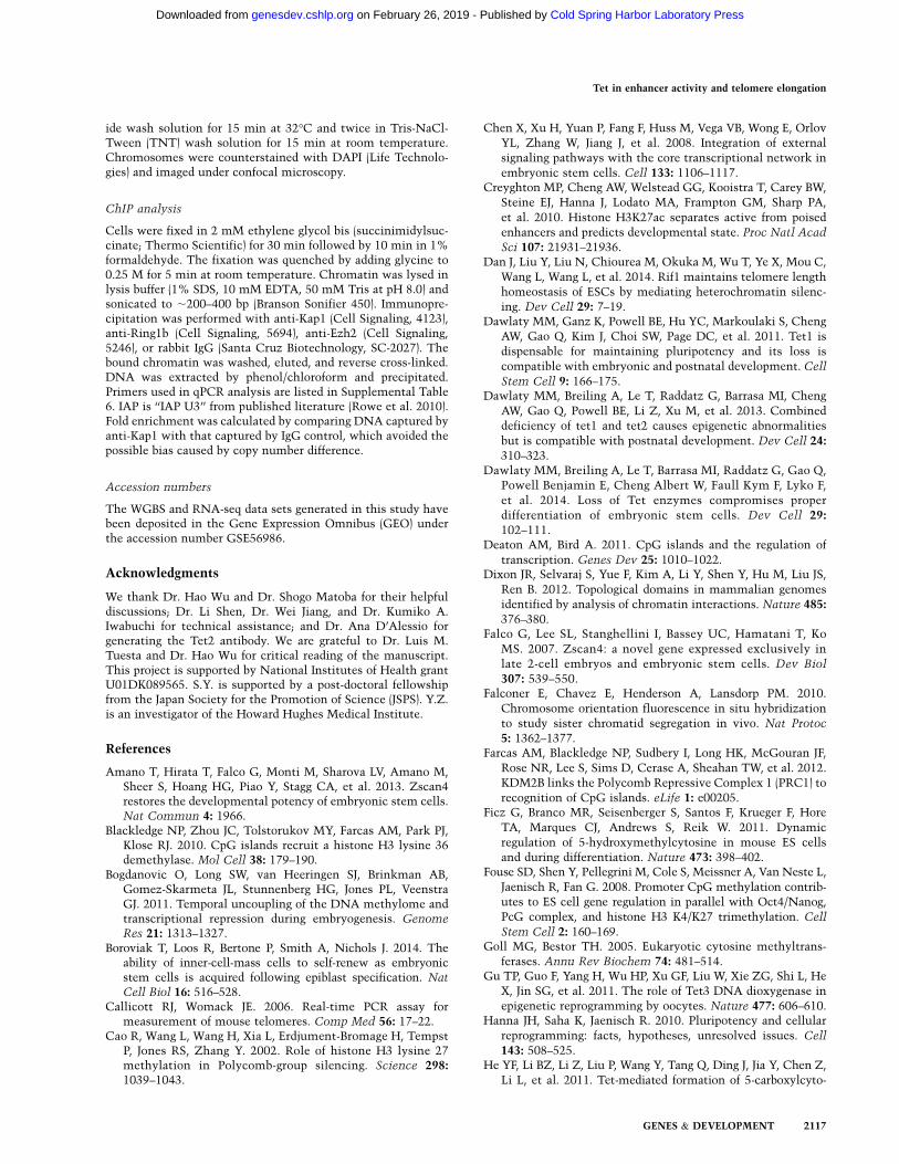
To examine the transcriptional effect of Tet TKO, weperformed RNA sequencing (RNA-seq) in wild-type andTet TKO ESCs and identified 735 up-regulated and 290down-regulated genes, respectively (fold change > 2, FPKM[fragments per kilobase of exon per million mappedfragments] > 0.5 in at least one sample) (Fig. 4A; Supple-mental Table 2). In order to analyze the effect of DNAhypermethylation on transcriptional change, we analyzedhypermethylated genes exhibiting hypermethylation ineither promoters or associated enhancers. Previous chro-matin interaction analysis by paired-end tag sequencing(ChIA-PET) analysis has experimentally identified pro-moter–enhancer interactions representing 3764 genes,among which 998 showed hypermethylation in at leastone enhancer in Tet TKO cells (Kieffer-Kwon et al. 2013).When compared with DNAmethylation change, we foundthat hypermethylated genes (3296 promoter hypermeth-ylated genes and 998 enhancer hypermethylated geneswith known enhancer–promoter interaction, of which 270genes were shared) overlapped significantly with differen-tially expressed genes (Fig. 4B; Supplemental Table 3),indicating that change in DNA methylation is likely oneof the primary causes of the expression change in Tet TKOESCs.To further examine this relationship, we analyzed
promoter methylation and enhancer methylation sepa-rately. Promoters can be classified into four groups basedon their associated histone modification (Whyte et al.2012). The four groups include (1) active promoters that
contain H3K4me3 and downstream H3K79me2/3, (2)initiated promoters that contain H3K4me3 only, (3) bi-valent promoters that contain H3K4me3 and H3K27me3,and (4) silent promoters that do not have H3K4me3.Analysis of the relationship between transcription andpromoter hypermethylation for each gene group revealedthat transcription from active promoters and initiatedpromoters decreased significantly when they were hyper-methylated (Fig. 4C; Supplemental Fig. 6). This is consis-tent with the notion that promoter methylation generallycorrelates with transcriptional repression (Klose and Bird2006). In contrast, hypermethylation of bivalent pro-moters associated with significant up-regulation of tran-scription (Fig. 4C), which is consistent with the previousfinding that increased DNA methylation at bivalentgenes positively correlates with increased transcription,possibly due to impaired recruitment of Polycomb re-pression complexes (PRCs) (Wu et al. 2011).Enhancers are generally hypomethylated when they are
bound by cell type-specific transcription factors, suggest-ing that active demethylation at active enhancers mightbe important for their activity (Stadler et al. 2011; Kieffer-Kwon et al. 2013; Ziller et al. 2013). However, thesestudies were carried out by comparing enhancer methyl-ation and enhancer activity in different cell types, makingthe correlations difficult to interpret. Tet TKO cells arelargely normal and exhibit aberrantmethylation at a largecohort of enhancers, which provide a unique opportunityfor studying the role of enhancer methylation in tran-scriptional regulation. We found that expression of activegenes and initiated genes was significantly decreasedwhen their associated enhancers were hypermethylated(Fig. 4D–F). In contrast, bivalent genes showed increasedexpression when their enhancers became hypermeth-ylated in the absence of Tet proteins (Fig. 4D). Activationof this group of genes might be caused by either methyl-ation-mediated repelling of PRCs similar to that observedat hypermethylated bivalent promoters or gene bodies(Wu et al. 2010, 2011), or Polycomb binding necessitatingnonmethylated CpG (Farcas et al. 2012; He et al. 2013;Wu et al. 2013). Clearly, these enhancers are modified byH3K27me3 in wild-type cells (Fig. 4G). Using T as anexample, we found that expression of Twas increased inTet TKO (Supplemental Fig. 7A). ChIP-qPCR (chromatinimmunoprecipitation [ChIP] coupled with quantitativePCR [qPCR]) analysis of hypermethylated regions withinpromoter and enhancer regions showed that binding ofboth Ring1B and Ezh2, core components of the PRC1 andPRC2 complexes, decreased upon hypermethylation(Supplemental Fig. 7B). The number of silent genes withhypermethylated enhancers and the number of silentgenes with hypermethylated promoters were small, sothe transcriptional changes associated with these hyper-methylation events were not statistically significant(Fig. 4C,D). Taken together, we found that hypermeth-ylation at promoters/enhancers of active/initiated genesis generally associated with gene repression, while theopposite is observed at bivalent gene promoters andassociated enhancers, possibly due to the negative effectof 5mC on the binding of Polycomb group proteins.
Lu et al.
2108 GENES & DEVELOPMENT
Cold Spring Harbor Laboratory Press on February 26, 2019 - Published by genesdev.cshlp.orgDownloaded from
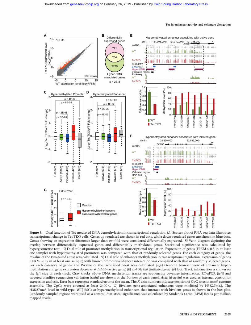
Figure 4. Dual function of Tet-mediated DNA demethylation in transcriptional regulation. (A) Scatter plot of RNA-seq data illustratestranscriptional change in Tet TKO cells. Genes up-regulated are shown in red dots, while down-regulated genes are shown in blue dots.Genes showing an expression difference larger than twofold were considered differentially expressed. (B) Venn diagram depicting theoverlap between differentially expressed genes and differentially methylated genes. Statistical significance was calculated byhypergeometric test. (C) Dual role of promoter methylation in transcriptional regulation. Expression of genes (FPKM > 0.5 in at leastone sample) with hypermethylated promoters was compared with that of randomly selected genes. For each category of genes, theP-value of the two-tailed t-test was calculated. (D) Dual role of enhancer methylation in transcriptional regulation. Expression of genes(FPKM > 0.5 in at least one sample) with known promoter–enhancer interaction was compared with that of randomly selected genes.For each category of genes, the P-value of the two-tailed t-test was calculated. (E,F) Genome browser view of enhancer hyper-methylation and gene expression decrease at Inhbb (active gene) (E) and Slc2a8 (initiated gene) (F) loci. Track information is shown onthe left side of each track. Gray tracks above DNA methylation tracks are sequencing coverage information. RT-qPCR (left) andtargeted bisulfite sequencing validation (right) are shown at the bottom of each panel. Actb (b-actin) was used as internal control forexpression analysis. Error bars represent standard error of the mean. The X-axis numbers indicate position of CpG sites in mm9 genomeassembly. The CpGs were covered at least 24003. (G) Bivalent gene-associated enhancers were modified by H3K27me3. TheH3K27me3 level in wild-type (WT) ESCs at hypermethylated enhancers that interact with bivalent genes is shown in the box plot.Randomly sampled regions were used as a control. Statistical significance was calculated by Student’s t-test. (RPM) Reads per millionmapped reads.
Tet in enhancer activity and telomere elongation
GENES & DEVELOPMENT 2109
Cold Spring Harbor Laboratory Press on February 26, 2019 - Published by genesdev.cshlp.orgDownloaded from
Increased 2C-like population in Tet TKO ESCs
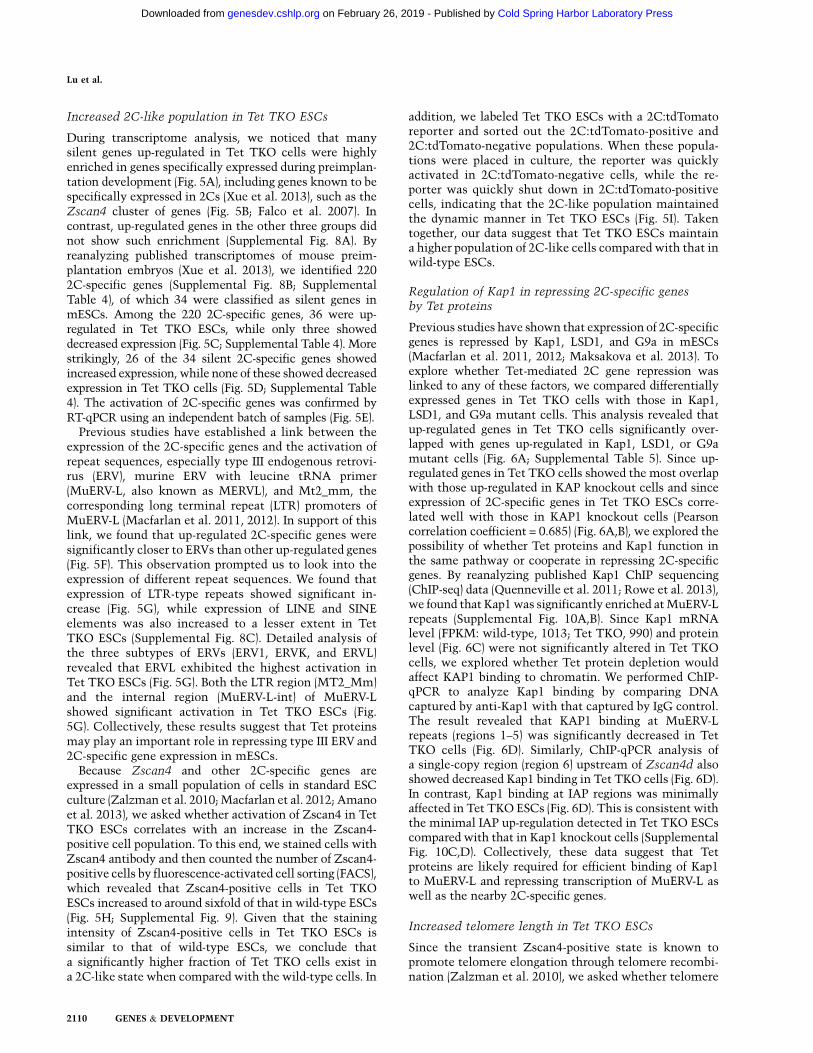
During transcriptome analysis, we noticed that manysilent genes up-regulated in Tet TKO cells were highlyenriched in genes specifically expressed during preimplan-tation development (Fig. 5A), including genes known to bespecifically expressed in 2Cs (Xue et al. 2013), such as theZscan4 cluster of genes (Fig. 5B; Falco et al. 2007). Incontrast, up-regulated genes in the other three groups didnot show such enrichment (Supplemental Fig. 8A). Byreanalyzing published transcriptomes of mouse preim-plantation embryos (Xue et al. 2013), we identified 2202C-specific genes (Supplemental Fig. 8B; SupplementalTable 4), of which 34 were classified as silent genes inmESCs. Among the 220 2C-specific genes, 36 were up-regulated in Tet TKO ESCs, while only three showeddecreased expression (Fig. 5C; Supplemental Table 4). Morestrikingly, 26 of the 34 silent 2C-specific genes showedincreased expression, while none of these showed decreasedexpression in Tet TKO cells (Fig. 5D; Supplemental Table4). The activation of 2C-specific genes was confirmed byRT-qPCR using an independent batch of samples (Fig. 5E).Previous studies have established a link between the
expression of the 2C-specific genes and the activation ofrepeat sequences, especially type III endogenous retrovi-rus (ERV), murine ERV with leucine tRNA primer(MuERV-L, also known as MERVL), and Mt2_mm, thecorresponding long terminal repeat (LTR) promoters ofMuERV-L (Macfarlan et al. 2011, 2012). In support of thislink, we found that up-regulated 2C-specific genes weresignificantly closer to ERVs than other up-regulated genes(Fig. 5F). This observation prompted us to look into theexpression of different repeat sequences. We found thatexpression of LTR-type repeats showed significant in-crease (Fig. 5G), while expression of LINE and SINEelements was also increased to a lesser extent in TetTKO ESCs (Supplemental Fig. 8C). Detailed analysis ofthe three subtypes of ERVs (ERV1, ERVK, and ERVL)revealed that ERVL exhibited the highest activation inTet TKO ESCs (Fig. 5G). Both the LTR region (MT2_Mm)and the internal region (MuERV-L-int) of MuERV-Lshowed significant activation in Tet TKO ESCs (Fig.5G). Collectively, these results suggest that Tet proteinsmay play an important role in repressing type III ERV and2C-specific gene expression in mESCs.Because Zscan4 and other 2C-specific genes are
expressed in a small population of cells in standard ESCculture (Zalzman et al. 2010;Macfarlan et al. 2012; Amanoet al. 2013), we asked whether activation of Zscan4 in TetTKO ESCs correlates with an increase in the Zscan4-positive cell population. To this end, we stained cells withZscan4 antibody and then counted the number of Zscan4-positive cells by fluorescence-activated cell sorting (FACS),which revealed that Zscan4-positive cells in Tet TKOESCs increased to around sixfold of that in wild-type ESCs(Fig. 5H; Supplemental Fig. 9). Given that the stainingintensity of Zscan4-positive cells in Tet TKO ESCs issimilar to that of wild-type ESCs, we conclude thata significantly higher fraction of Tet TKO cells exist ina 2C-like state when compared with the wild-type cells. In
addition, we labeled Tet TKO ESCs with a 2C:tdTomatoreporter and sorted out the 2C:tdTomato-positive and2C:tdTomato-negative populations. When these popula-tions were placed in culture, the reporter was quicklyactivated in 2C:tdTomato-negative cells, while the re-porter was quickly shut down in 2C:tdTomato-positivecells, indicating that the 2C-like population maintainedthe dynamic manner in Tet TKO ESCs (Fig. 5I). Takentogether, our data suggest that Tet TKO ESCs maintaina higher population of 2C-like cells compared with that inwild-type ESCs.
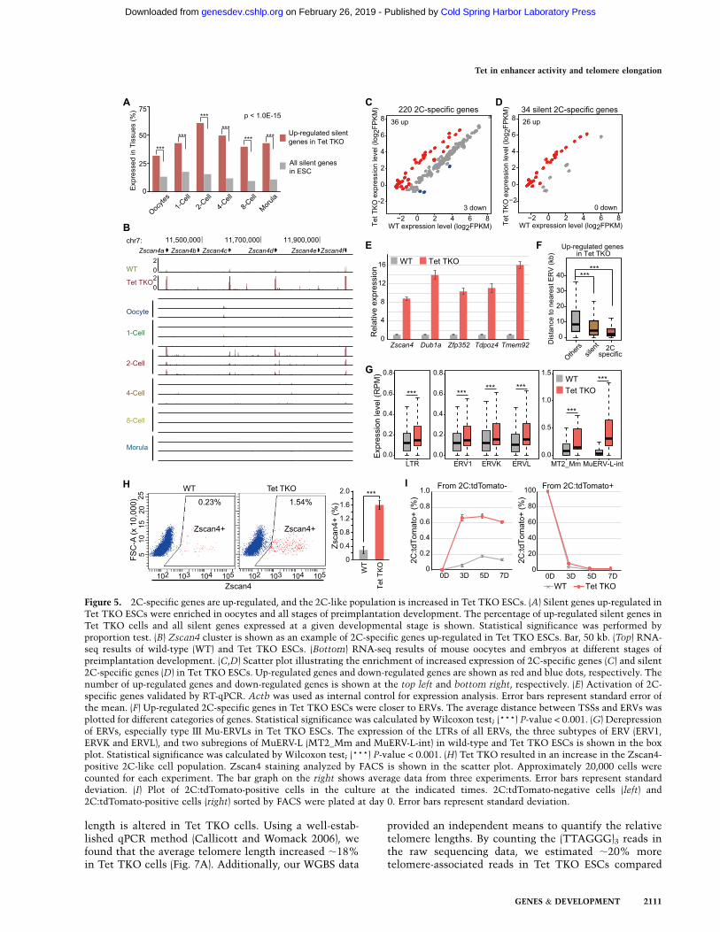
Regulation of Kap1 in repressing 2C-specific genesby Tet proteins
Previous studies have shown that expression of 2C-specificgenes is repressed by Kap1, LSD1, and G9a in mESCs(Macfarlan et al. 2011, 2012; Maksakova et al. 2013). Toexplore whether Tet-mediated 2C gene repression waslinked to any of these factors, we compared differentiallyexpressed genes in Tet TKO cells with those in Kap1,LSD1, and G9a mutant cells. This analysis revealed thatup-regulated genes in Tet TKO cells significantly over-lapped with genes up-regulated in Kap1, LSD1, or G9amutant cells (Fig. 6A; Supplemental Table 5). Since up-regulated genes in Tet TKO cells showed the most overlapwith those up-regulated in KAP knockout cells and sinceexpression of 2C-specific genes in Tet TKO ESCs corre-lated well with those in KAP1 knockout cells (Pearsoncorrelation coefficient = 0.685) (Fig. 6A,B), we explored thepossibility of whether Tet proteins and Kap1 function inthe same pathway or cooperate in repressing 2C-specificgenes. By reanalyzing published Kap1 ChIP sequencing(ChIP-seq) data (Quenneville et al. 2011; Rowe et al. 2013),we found that Kap1was significantly enriched atMuERV-Lrepeats (Supplemental Fig. 10A,B). Since Kap1 mRNAlevel (FPKM: wild-type, 1013; Tet TKO, 990) and proteinlevel (Fig. 6C) were not significantly altered in Tet TKOcells, we explored whether Tet protein depletion wouldaffect KAP1 binding to chromatin. We performed ChIP-qPCR to analyze Kap1 binding by comparing DNAcaptured by anti-Kap1 with that captured by IgG control.The result revealed that KAP1 binding at MuERV-Lrepeats (regions 1–5) was significantly decreased in TetTKO cells (Fig. 6D). Similarly, ChIP-qPCR analysis ofa single-copy region (region 6) upstream of Zscan4d alsoshowed decreased Kap1 binding in Tet TKO cells (Fig. 6D).In contrast, Kap1 binding at IAP regions was minimallyaffected in Tet TKO ESCs (Fig. 6D). This is consistent withthe minimal IAP up-regulation detected in Tet TKO ESCscompared with that in Kap1 knockout cells (SupplementalFig. 10C,D). Collectively, these data suggest that Tetproteins are likely required for efficient binding of Kap1to MuERV-L and repressing transcription of MuERV-L aswell as the nearby 2C-specific genes.
Increased telomere length in Tet TKO ESCs
Since the transient Zscan4-positive state is known topromote telomere elongation through telomere recombi-nation (Zalzman et al. 2010), we asked whether telomere
Lu et al.
2110 GENES & DEVELOPMENT
Cold Spring Harbor Laboratory Press on February 26, 2019 - Published by genesdev.cshlp.orgDownloaded from
length is altered in Tet TKO cells. Using a well-estab-lished qPCR method (Callicott and Womack 2006), wefound that the average telomere length increased ;18%in Tet TKO cells (Fig. 7A). Additionally, our WGBS data
provided an independent means to quantify the relativetelomere lengths. By counting the (TTAGGG)3 reads inthe raw sequencing data, we estimated ;20% moretelomere-associated reads in Tet TKO ESCs compared
Figure 5. 2C-specific genes are up-regulated, and the 2C-like population is increased in Tet TKO ESCs. (A) Silent genes up-regulated inTet TKO ESCs were enriched in oocytes and all stages of preimplantation development. The percentage of up-regulated silent genes inTet TKO cells and all silent genes expressed at a given developmental stage is shown. Statistical significance was performed byproportion test. (B) Zscan4 cluster is shown as an example of 2C-specific genes up-regulated in Tet TKO ESCs. Bar, 50 kb. (Top) RNA-seq results of wild-type (WT) and Tet TKO ESCs. (Bottom) RNA-seq results of mouse oocytes and embryos at different stages ofpreimplantation development. (C,D) Scatter plot illustrating the enrichment of increased expression of 2C-specific genes (C) and silent2C-specific genes (D) in Tet TKO ESCs. Up-regulated genes and down-regulated genes are shown as red and blue dots, respectively. Thenumber of up-regulated genes and down-regulated genes is shown at the top left and bottom right, respectively. (E) Activation of 2C-specific genes validated by RT-qPCR. Actb was used as internal control for expression analysis. Error bars represent standard error ofthe mean. (F) Up-regulated 2C-specific genes in Tet TKO ESCs were closer to ERVs. The average distance between TSSs and ERVs wasplotted for different categories of genes. Statistical significance was calculated by Wilcoxon test; (***) P-value < 0.001. (G) Derepressionof ERVs, especially type III Mu-ERVLs in Tet TKO ESCs. The expression of the LTRs of all ERVs, the three subtypes of ERV (ERV1,ERVK and ERVL), and two subregions of MuERV-L (MT2_Mm and MuERV-L-int) in wild-type and Tet TKO ESCs is shown in the boxplot. Statistical significance was calculated by Wilcoxon test; (***) P-value < 0.001. (H) Tet TKO resulted in an increase in the Zscan4-positive 2C-like cell population. Zscan4 staining analyzed by FACS is shown in the scatter plot. Approximately 20,000 cells werecounted for each experiment. The bar graph on the right shows average data from three experiments. Error bars represent standarddeviation. (I) Plot of 2C:tdTomato-positive cells in the culture at the indicated times. 2C:tdTomato-negative cells (left) and2C:tdTomato-positive cells (right) sorted by FACS were plated at day 0. Error bars represent standard deviation.
Tet in enhancer activity and telomere elongation
GENES & DEVELOPMENT 2111
Cold Spring Harbor Laboratory Press on February 26, 2019 - Published by genesdev.cshlp.orgDownloaded from
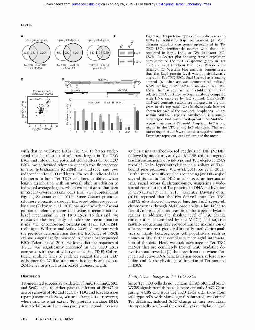
with that in wild-type ESCs (Fig. 7B). To better under-stand the distribution of telomere length in Tet TKOESCs and rule out the potential clonal effect of Tet TKOESCs, we performed telomere quantitative fluorescencein situ hybridization (Q-FISH) in wild-type and twoindependent Tet TKO cell lines. The result indicated thattelomeres in both Tet TKO cell lines exhibited widerlength distribution with an overall shift in addition toincreased average length, which was similar to that seenin Zscan4-overexpressing cells (Fig. 7C; SupplementalFig. 11; Zalzman et al. 2010). Since Zscan4 promotestelomere elongation through increased telomere recom-bination (Zalzman et al. 2010), we asked whether Zscan4promoted telomere elongation using a recombination-based mechanism in Tet TKO ESCs. To this end, wemeasured the frequency of telomere recombinationusing the chromosome orientation FISH (CO-FISH)technique (Williams and Bailey 2009). Consistent withthe previous demonstration that the frequency of T-SCEevents is significantly increased in Zscan4-overexpressedESCs (Zalzman et al. 2010), we found that the frequency ofT-SCE was significantly increased in Tet TKO ESCscompared with that of wild-type cells (Fig. 7D,E). Collec-tively, multiple lines of evidence suggest that Tet TKOcells enter the 2C-like state more frequently and acquire2C-like features such as increased telomere length.
Discussion
Tet-mediated successive oxidation of 5mC to 5hmC, 5fC,and 5caC leads to either passive dilution of 5hmC oractive removal of 5fC and 5caC by TDG and base excisionrepair (Pastor et al. 2013; Wu and Zhang 2014). However,where and to what extent Tet proteins mediate DNAdemethylation still remains poorly understood. Previous
studies using antibody-based methylated DIP (MeDIP)followed bymicroarray analysis (MeDIP–chip) or targetedbisulfite sequencing of wild-type and Tet1-depleted ESCsrevealed DNA hypermethylation at a cohort of Tet1-bound gene promoters (Wu et al. 2011; Xu et al. 2011).Furthermore, MeDIP-coupled sequencing (MeDIP-seq) ofseveral tissues in Tet DKO mice showed an increase of5mC signal across all chromosomes, suggesting a wide-spread contribution of Tet proteins in DNA methylationin vivo (Dawlaty et al. 2013). Recently, Dawlaty et al.(2014) reported that the EBs derived from Tet TKOmESCs also showed increased baseline 5mC across allchromosomes through MeDIP-seq analysis but failed toidentifymore distribution features of the hypermethylatedregions. In addition, the absolute level of 5mC changecould not be determined by the MeDIP, and targetedbisulfite sequencing only provided limited information ofselected promoter regions. Additionally, methylation anal-yses of highly heterogeneous cell populations, such astissues or EBs, further complicate meaningful interpreta-tion of the data. Here, we took advantage of Tet TKOmESCs that are completely free of 5mC oxidative de-rivatives and revealed (1) the exact locations where Tet-mediated active DNA demethylation occurs at base reso-lution and (2) the physiological function of Tet proteinsin ESCs.
Methylation changes in Tet TKO ESCs
Since Tet TKO cells do not contain 5hmC, 5fC, and 5caC,WGBS signals from these cells represent only 5mC. Com-paring WGBS data from Tet TKO ESCs with those fromwild-type cells with 5hmC signal subtracted, we definedTet deficiency-induced 5mC change at base resolution.Unexpectedly, we found the overall CpGmethylation level
Figure 6. Tet proteins repress 2C-specific genes andLTRs by facilitating Kap1 recruitment. (A) Venndiagram showing that genes up-regulated in TetTKO ESCs significantly overlap with those up-regulated in Kap1, Lsd1, or G9a knockout (KO)ESCs. (B) Scatter plot showing strong expressioncorrelation of the 220 2C-specific genes in TetTKO and Kap1 knockout ESCs. (cor) Pearson coef-ficiency. (C) Western blot analysis demonstratedthat the Kap1 protein level was not significantlyaltered in Tet TKO ESCs. Suz12 served as a loadingcontrol. (D) ChIP analysis demonstrated reducedKAP1 binding at MuERV-L elements in Tet TKOESCs. The relative enrichment is fold enrichment ofrelative DNA captured by Kap1 antibody comparedwith DNA captured by IgG control. ChIP-qPCR-analyzed genomic regions are indicated in the dia-gram in the top panel. One-kilobase scale bars areshown for each of the two loci. Amplicons 1–5 arewithin MuERV-L repeats. Amplicon 6 is a single-copy region that partly overlaps with the MuERV-Lrepeat upstream of Zscan4d. Amplicon IAP is oneregion in the LTR of the IAP elements. The pro-moter region of Actb was used as a negative control.Error bars represent standard error of the mean.
Lu et al.
2112 GENES & DEVELOPMENT
Cold Spring Harbor Laboratory Press on February 26, 2019 - Published by genesdev.cshlp.orgDownloaded from
in Tet TKO cells (64.7%) to be slightly lower than that inwild-type cells (69%). This is likely due to decreasedmethylation within highly methylated CpGs (Fig. 2A;Supplemental Fig. 2), such as those located in heterochro-matin regions (Fig. 2D, H3K9me3 and H4K20me3). Thissuggests that DNA methylation is not fully maintained inTet TKO ESCs. It would be interesting to investigate therelationship between 5mC oxidation and maintenance inthe future.Nevertheless, in Tet TKO cells, we found 60 times
more hyper-DMRs across the genome than hypo-DMRs,indicating that hypomethylated CpG sites are randomlydistributed, whereas hypermethylated CpG sites tend tocluster at specific genomic loci. Apart from a subset ofhypermethylated CpGs that are 5hmC-modified in wild-type ESCs, a significant fraction of hypermethylatedCpGs are not associated with stable 5hmC signalsdetected in wild-type cells. It appears that a cohort ofthese 5hmC-free hypermethylated CpGs resides within5fC/5caC peaks, suggesting that active demethylation(conversion of 5mC to C) and stable accumulation of5hmC modification may represent distinct molecularevents. Thus, genome-wide mapping of 5hmC may notbe sufficient to identify all cytosines undergoing activeDNA demethylation. Mapping 5fC/5caC accumulationin TDG-deficient cells at single-base resolution maycontribute to a better understanding of Tet and TDG-dependent active DNA demethylation.Previous studies have shown that Tet proteins are
required for the maintenance of low methylation statusat a subset of promoters in ESCs (Wu et al. 2011; Xu et al.2011; Dawlaty et al. 2014). In this study, we not only
provided a complete list of promoters undergoing activeDNA demethylation but also found that enhancer regionsare the most profound Tet-dependent active demethyla-tion loci. Proximal CpG islands colocalize with themajority of promoters in the mouse genome and aremostly unmethylated (Deaton and Bird 2011). We foundthat DNAmethylation increased significantly at the CGIshores but minimally at the center of CGIs (Fig. 2D),indicating that Tet proteins are not the major players inpreventing CGIs from methylation. The main players inpreventing CGI methylation remain undefined. En-hancers are generally lower in DNA methylation (Stadleret al. 2011; Kieffer-Kwon et al. 2013; Ziller et al. 2013).Here we provide evidence indicating that Tet-mediatedactive DNA demethylation is one of the major contrib-uting factors in maintaining this phenomenon. Sinceenhancers are well known to be cell type-specific, it willbe interesting to study the roles of Tet proteins inenhancer reconfiguration during cell fate transition.
DNA methylation at enhancers fine-tune enhanceractivity in ESCs
Previous studies have revealed that active enhancers aregenerally hypomethylated, suggesting that hypomethyla-tion may be required for enhancer activity (Stadler et al.2011; Kieffer-Kwon et al. 2013; Ziller et al. 2013). How-ever, this hypothesis has not been confirmed, as all of thestudies so far have compared enhancer methylation andactivity from different cell types and have not ruled outa possible secondary effect resulting from different cellidentities. Here, we found widespread hypermethylation
Figure 7. Tet TKO results in telomere elongationalong with high frequency of T-SCE. (A) Increasedtelomere length in Tet TKO ESCs. Average telomerelength was assessed by qPCR with a telomere repeat-specific primer set and normalized to a single-copycontrol primer set. (B) Increased telomere length inTet TKO ESCs as measured by WGBS reads. Thetable summarizes WGBS reads and uniquely mappedreads. (TTAGGG)3-containing reads were countedas telomere reads in the raw data and normalized toeither total sequencing reads or uniquely mappedreads. (C) Distribution histogram of relative telomerelength analyzed by telomere Q-FISH and TFL-TELOsoftware. (D,E) Increased T-SCE in Tet TKO cells.T-SCE events were measured by CO-FISH. (D) Repre-sentative images are shown. (E) Quantification ofchromosomes with T-SCE events are shown in the bargraph. Statistical significance was calculated by student’st-test; (*) P-value < 0.05; (***) P-value < 0.001.
Tet in enhancer activity and telomere elongation
GENES & DEVELOPMENT 2113
Cold Spring Harbor Laboratory Press on February 26, 2019 - Published by genesdev.cshlp.orgDownloaded from
at enhancers in Tet TKO cells without disrupting theiridentity. This provided us with a unique opportunity toexamine the effect of DNA methylation on enhanceractivity. We found that increased methylation at theenhancer region of actively transcribed genes (Fig. 4D,active/initiated) was negatively correlated with transcrip-tion in general. Hypermethylation in enhancer regionsresulted in small changes in transcription, likely due tothe minor role played by DNA methylation in generepression of early embryos and ESCs (Fouse et al. 2008;Bogdanovic et al. 2011). In contrast, increased enhancermethylation of bivalent genes (Fig. 4D, bivalent) corre-lated with transcriptional activation, supporting themutual exclusion of DNA methylation and Polycombprotein binding (Wu et al. 2010, 2011, 2013; Farcas et al.2012; He et al. 2013) and the notion that removal ofPolycomb repression is required for enhancer activation(Rada-Iglesias et al. 2011). These results suggest that Tet-dependent DNA demethylation at enhancers fine-tunestranscription of genes interacting with these enhancersin ESCs. Further studies will be needed to determinewhether Tet-dependent DNA demethylation plays alarger role in regulating enhancer activity in somaticcells where DNA methylation is heavily associatedwith gene repression.
Role of Tet proteins in repeat silencing and telomereregulation
Temporal acquisition of a 2C-specific condition is criticalto restoring the developmental capacity in ESCs (Amanoet al. 2013). Previous studies have demonstrated thata 2C-like cell population increases in cells deficient forKap1, Lsd1, or G9a (Macfarlan et al. 2012). We found that2C-specific genes are up-regulated in Tet TKO cells.Although Tet1 ChIP-seq did not reveal stable occupancyof Tet1 protein at these regions, the presence of high-confidence 5hmC signals at 2C-specific gene loci in-dicates that Tet proteins indeed function at these loci.Tet2might also occupy these loci, as it possesses intrinsicDNA-binding activity (Hu et al. 2013). However, cur-rently, no reliable Tet2 ChIP-seq data are available foranalyzing the contribution of Tet2. Derepression of 2C-specific genes in Tet TKO cells correlated well with thatin Kap1 knockout cells, although the change was toa lesser extent in Tet TKO ESCs than that in Kap1knockout cells (Fig. 6B). This was consistent with theobservation that binding of Kap1 to MuERV-L was de-creased but not completely absent in Tet TKO ESCs(Fig. 6D). The activation of MuERV-L was also to a lesserextent in Tet TKO ESCs when compared with Kap1knockout ESCs, although it was comparable with G9aknockout ESCs (Fig. 5G; Supplemental Fig. 10C,E). There-fore, it is possible that Tet proteins and/or 5hmC marksmay help recruit/stabilize Kap1 binding at these loci. Wealso noticed that the effect of Tet proteins in Kap1 bindingwas not global, as Kap1 binding at IAP regions wasminimally affected (Fig. 6D). Future studies may providenew insights into the function of Tet proteins in repressingtype III ERVs and 2C-specific genes.
Among the 2C-specific genes, Zscan4 is of particularinterest. Activation of Zscan4 in ESCs is associatedwith rapid telomere extension by promoting T-SCE(Zalzman et al. 2010; Dan et al. 2014). Zscan4 isspecifically expressed in 2Cs, and telomere elongationtakes place most dramatically at the 2C stage througha T-SCE-based mechanism (Liu et al. 2007). However,how Zscan4 is activated at the two-cell stage remainsunclear. We found that Tet TKO ESCs exhibit activa-tion of Zscan4, which led to increased T-SCE andelongated telomeres (Fig. 7). Among the three Tet pro-teins, Tet3 is a maternal factor that is highly expressed inthe one-cell embryo and degrades in the 2C, while Tet1and Tet2 commence expression at the 2C and morulastages, respectively (Tan and Shi 2012). Therefore, 2Cs lackall of the Tet proteins. The various phenotypes that weobserved in this study suggest that loss of Tet proteinsmaybe required for the establishment of a 2C-specific state,including ERV activation, Zscan4 expression, and sub-sequent telomere elongation. It will be interesting todetermine whether forced expression of Tet proteins in2Cs will result in disruption of preimplantation develop-ment in the future.In summary, we generated a base-resolution DNA
methylation map in Tet TKO ESCs and identified ge-nome-wide Tet-dependent DNA demethylation events inESCs. We found that Tet-mediated DNA demethylationtakes place mostly at enhancers and distal regulatorysequences. We also provided evidence that enhancermethylation regulates enhancer activity. Furthermore,we demonstrated that Tet TKO ESCs show an increased2C-like population and elongated telomeres. Taken to-gether, these findings not only substantially broaden ourunderstanding of the role of Tet proteins in DNA demeth-ylation but also reveal new functions for Tet proteins inrepressing repeat sequences as well as nearby 2C-specificgenes and telomere length regulation.
Materials and methods
Cell culture and antibodies
We used the mESC line E14Tg2a in this study. Cells werecultured on 0.1% gelatin-coated (Millipore) plates in DMEM(Life Technologies) supplemented with 15% fetal bovine serum(Sigma), penicillin/streptomycin, nonessential amino acid, so-dium pyruvate, GlutaMax, b-mercaptoethanol (Life Technolo-gies), and 1000 U/mL LIF (ESGRO, Millipore) unless specified.Antibodies used in this study were 5hmC (Active Motif, 39769),Oct4 (Santa Cruz Biotechnology, sc-8628), Nanog (Bethyl Labo-ratories, IHC-00205), Sox2 (Millipore, Ab5603), Tet1 (Ito et al.2010), Tet2 (described below), Tet3 (Gu et al. 2011), Zscan4(Millipore, Ab4340), Suz12 (Cao et al. 2002), Ring1b (CellSignaling, 5694), Ezh2 (Cell Signaling, 5246), and Kap1 (CellSignaling, 4123). Tet2 antibody was generated using HIS-taggedrecombinant Tet2 2-374 expressed in Escherichia coli andpurified using Talon superflow metal affinity resin (Clontech).Rabbit immunization was carried out by Pocono Rabbit Farmand Laboratory, Inc. The antiserum was affinity-purified usingimmunization antigen. The specificity of the affinity-purifiedantibody was confirmed using extracts from the control and Tet2knockdown mESCs.
Lu et al.
2114 GENES & DEVELOPMENT
Cold Spring Harbor Laboratory Press on February 26, 2019 - Published by genesdev.cshlp.orgDownloaded from
Generation of Tet TKO cells
Tet TKO cells were generated using the CRISPR/Cas9 methodwith the previously published guide RNA sequences (Wang et al.2013). E14 cells cultured in 2i medium (DMEM/F12 supple-mented with penicillin/streptomycin, GlutaMax, 1 mM MEKinhibitor PD0325901, 3 mM GSK3 inhibitor CHIR99021, 1000U/mL LIF) on Matrigel-coated (BD Biosciences) plates weretransiently transfected with a mixture of px330 vectors contain-ing guide RNAs for Tet1, Tet2, and Tet3 along with onepuromycin resistance gene-containing empty vector using theFugene 6 transfection reagent (Promega). The cells were subcul-tured to a low density 24 h later. Two days following trans-fection, the cells were selected with 2 mg/mL puromycin for 48 h.Viable clones were grown larger and picked up for mutationanalysis. Mutations were first screened using PCR followed byrestriction analysis as restriction sites were presented atCRISPR/Cas9 cutting sites. 5hmC levels of DNA from mutantclones were then analyzed by slot blot. Clones with a signifi-cantly lower level of 5hmC along with wild-type ESCs were keptand adapted to serum-based culture conditions for at least 17 d.DNA samples from these clones were then digested to singlenucleotide and analyzed by mass spectrometry to quantifyabsolute 5hmC levels.
5hmC level analysis by mass spectrometry
Genomic DNA (1 mg) prepared using a DNeasy blood and tissuekit (Qiagen) was hydrolyzed with 90 U of nuclease S1 (Sigma) indigestion buffer (14 mM NaAc, 0.5 mM ZnSO4 at pH 4.6) for 1 hat 37°C followed by treatment with 8 mU of phosphodiesterase I(VPH) and 1.6 U of CIAP (New England Biolabs) for an additional1 h. The digested samples were then filtered with Nanosep3K(PALL) and subjected to mass spectrometry analysis.
WGBS
DNA (1 mg) with 0.5% nonmethylated l DNA spike-in wassheared by Covaris M220 instrument to an average size of 350bp in length. DNA was then end-repaired, dA-tailed, and ligatedwith methylated adaptors. Bisulfite conversion was carried outusing an EpiTect fast bisulfite conversion kit (Qiagen) according tothe manufacturer’s instructions. Twenty minutes were used inboth conversion steps. Bisulfite-converted DNA was then ampli-fied with five PCR cycles to obtain the final library. The WGBSlibraries were subjected to pair-end (2 3 110 bp) sequencing ona HiSeq 2500 (Illumina) with or without a PhiX spike in.
After sequencing, low-quality bases and adapter-containingreads were trimmed from raw data by trim_galore with defaultparameters, and reads aligned to the PhiX genomewere removed.Bismark 0.7.9 was used to align reads to a reference genome(mm9). Analyzing l DNA reads confirmed that >99.7% Cswere successfully bisulfite-converted. The coverage depth andmethylation level of each cytosine were extracted by bismark_methylation_extractor and samtools from SAM files. DMRanalysis was performed by methpipe-3.0.1. False discovery rate(FDR) was estimated by randomly shuffling the Tet TKO andwild-type methylome to call DMRs with the same pipeline andparameters. For hyper-DMRs, the FDR was estimated as 0.0499.For hypo-DMRs, the DMR number was too small to be statisti-cally analyzed, and thus hypo-DMRs were not analyzed further.When calculating methylation level for CpG sites, informationfrom both strands was combined, and a coverage of at least fivereads was required. For regions, all reads within one specificregion were pooled together, and a coverage of at least 10 readswas required. Since bisulfite sequencing cannot distinguish 5mC
and 5hmC, 5mC levels in wild-type cells were corrected bysubtracting 5hmC levels obtained from TAB-seq (Yu et al. 2012).For both 100-bp tiles and DMRs, regions with a methylationdifference >20% were considered as differentially methylated.
The following procedure was used to generate the heat maps inFigure 2D. The average methylation profile of both wild type andTKOs was calculated for both 5 kb upstream of and downstreamfrom the center of each feather in 100-bp bins. Methylationdifferences were calculated by subtracting average methylationin wild type from those in Tet TKO. For all of the features, peaksoverlapping with 61 kb TSS sequences were considered asproximal elements, with the rest of the peaks considered as distalelements. Regions within 61.5 kb from the TSS were consideredas promoter regions unless specified. The data of differentgenomic features are described in ’’Published Data Sets Used inThis Study.’’ Heat maps were generated by R.
Targeted bisulfite sequencing
Two micrograms of genomic DNA was bisulfite-treated with anEpiTect fast bisulfite conversion kit (Qiagen) according to themanufacturer’s instructions. Twenty minutes were used in bothconversion steps. Targeted regions were amplified with theprimers listed in Supplemental Table 6 using Kapa Hifi Uracil+
enzyme (Kapa Biosystems). The amplified DNA samples werepurified and mixed at equal molar concentrations for eachgenotype. The mixture of DNA was then fragmented by sonica-tion (Covaris). DNA sequencing libraries were then constructedfrom the fragmented DNA samples using NEBnext ultra DNAlibrary prep kit for Illumina (New England Biolabs) with differentbarcodes. Libraries were sequenced on a HiSeq 2500 (Illumina).
RNA-seq
RNA samples were prepared by RNeasy minikit (Qiagen). De-pletion of rRNA was carried out using ribo-zero kit (Epicentre).Barcoded RNA-seq libraries were constructed using NEBNextultra directional RNA library prep kit for Illumina according tothe manufacturer’s instructions (New England Biolabs). Single-end 50-bp sequencing was performed on a HiSeq 2500 (Illumina).Sequencing reads were mapped against the reference genome(mm9)with Bowtie/TopHat version 2.0.2, which allowedmappingacross splicing junctions by read segmentation. All programswere performed with default settings unless otherwise speci-fied. The unique mapped reads (;77% of total reads) weresubsequently assembled into transcripts guided by referenceannotation (University of California at Santa Cruz [UCSC] genemodels) with Cufflinks version 2.0.2. The expression level ofeach gene was quantified with normalized FPKM. Differentiallyexpressed genes were identified by asking for a fold change >2with FPKM > 0.5 in at least one sample. For repeat sequenceannotation, the RepeatMasker track (RMSK) from UCSC Ge-nome Browser was used. For repeat analyses, we counted theunique mapped reads in each repeat element by custom Perlscript and then calculated the expression level (reads permillion unique mapped reads [RPM]).
Published data sets used in this study
Single-cell RNA-seq data in the mouse early embryos were down-loaded from GSE44183 (Xue et al. 2013). We used the followingpublished RNA-seq data sets for Figure 6 and Supplemental Figure10: G9a knockout ESCs (GSE33923) (Macfarlan et al. 2012), Lsd1knockout (Macfarlan et al. 2011), and Kap1 knockout ESCs(GSE41903) (Rowe et al. 2013). The lists of up-regulated genesidentified here are included in Supplemental Table 5.
Tet in enhancer activity and telomere elongation
GENES & DEVELOPMENT 2115
Cold Spring Harbor Laboratory Press on February 26, 2019 - Published by genesdev.cshlp.orgDownloaded from
We used the following published data sets for Figure 2D: 5fC/5caC (Shen et al. 2013); Tet1 (Wu et al. 2011); Kdm2a (Blackledgeet al. 2010); H3K4me3, H3K36me3, H3K27me3, H3K9me3, andH4K20me3 (Mikkelsen et al. 2007); H3K4me1 (Meissner et al.2008); Pol2 (pan), Pol2 (Ser2), Pol2 (Ser5), NelfA, Ctr9, and Spt5(Rahl et al. 2010); Ezh2 and Suz12 (Ku et al. 2008); Oct4, Nanog,Sox2, and H3K79me2 (Marson et al. 2008); Med1, Med12, Nipbl,Smc1a, Smc3, and TBP (Kagey et al. 2010); LSD1, Mi2b, Hdac1,Hdac2, Rest, and Corest (Whyte et al. 2012); LMR (Stadler et al.2011); H3K27ac and p300 (Creyghton et al. 2010); Esrrb, cMyc,nMyc, Tcfcp2l1, E2f1, Stat3, Smad1, and Zfx (Chen et al. 2008);CTCF (Shen et al. 2012); DHSs (ENCODE project at genome.ucsc.edu); and topological domain boundary (Dixon et al. 2012).Two published Kap1 ChIP-seq data sets were used for Supple-mental Figure 10 (Quenneville et al. 2011; Rowe et al. 2013).
Early embryonic development stage specificity analysis
To quantify the stage specificity of genes in early embryos, theexpression level of samples at a given stage versus the othersamples was compared. The stage specificity score of each gene isdefined as score = meanA � (meanOther + 2 3 sdOther), wheremeanA is the mean expression of the samples in a certain stage,and meanOther and sdOther are the mean and standard deviationof the expression levels in the other samples, respectively.Therefore, a positive score indicates that the gene is expressedin a certain stage at a considerably higher level than in the otherstages. The gene with a score >0 was considered as specificallyexpressed in certain stage.
Statistical analyses
Statistical analyses were implemented with R (http://www.r-project.org). The x2 test was used to compare the frequency of anoccurrence between two groups by analyzing the two-by-twocontingency tables using the chisq.test function in R. Indepen-dent two-group Wilcoxon rank sum tests were used to comparedistributions using the wilcox.test function in R. A Pearson’s Rcoefficient was calculated using the cor function with defaultparameters. Student’s t-test (two-tailed, equal variance) wasperformed to obtain P-values for qPCR experiments and ChIP-seq enrichment.
Promoter classification by chromatin states
Gene promoters are classified into four groups (active, initiated,bivalent, and silent) by histone modifications as previouslydescribed (Whyte et al. 2012). Specifically, (1) active promotersare associated with H3K4me3 (61.5 kb flanking the TSS) andH3K79me2 (5 kb downstream from the TSS), (2) initiated pro-moters are associated with H3K4me3 (61.5 kb flanking the TSS)only, (3) bivalent promoters are associated with H3K4me3 (61.5kb flanking the TSS) and H3K27me3 (61.5 kb flanking the TSS),and (4) silent promoters are not associated with H3K4me3/H3K79me2/H3K27me3.
Immunostaining and FACS analysis
Cells were fixed with 4% paraformaldehyde for 15 min at roomtemperature. After washing with PBS twice, the cells werepermeabilized with PBS containing 0.3% Triton X-100 for 10min and blocked with PBS with 1% BSA for 30 min. The cellswere then incubated with primary antibodies diluted in PBS with1% BSA for 1 h at room temperature. After washing three timeswith PBS, cells were incubated with Alexa Fluor 488-, AlexaFluor 594-, or Alexa Fluor 647-conjugated secondary antibodies
(Molecular Probes) for 1 h at room temperature. The cells werewashed three times with PBS, stained with DAPI, and visualizedunder an Axio Observer Z1 microscope (Carl Zeiss). For FACSanalysis of Zscan4 staining, cells were dissociated with trypsinto single-cell suspension, washed with PBS, and fixed in 4%paraformaldehyde for 15 min at room temperature. After wash-ing with PBS twice, permeabilization was done by resuspendingcells in 90% cold methanol for 30 min. After washing with PBStwice, staining was done with procedures described above.Stained cells were analyzed on a BD FACSaria machine (BDBiosciences).
Telomere length qPCR quantification
Telomere length quantification was carried out as previouslydescribed (Callicott and Womack 2006; Liu et al. 2007), with theexception that 1 ng of DNAwas used for each amplification, and4, 2, 1, 0.5, and 0.25 ng of DNA per sample was used for standardcurve generation.
Telomere Q-FISH
Telomere Q-FISH was carried out as previously described withminor modifications (Herrera et al. 1999). Briefly, ESCs werecultured with 0.2 mg/mL Colcemid for 1 h and 40 min to enrichcells at metaphases. Harvested cells were treated with 75 mMKCl solution followed by fixation with Carnoy’s fixative solution(methanol:acetic acid, 3:1) and spread onto clean glass slides.Slides were then washed with PBS and fixed with 4% para-formaldehyde/PBS followed by the incubation with 1 mg/mLpepsin (pH 2.0). Slides were then post-fixed with 4% paraformal-dehyde/PBS followed by dehydration in ethanol series and air-dried. Telomeres were denatured for 3 min at 80°C and hybrid-ized with telomere-specific peptide nucleic acid (PNA) probe(PNA Bio, F1002) for 4 h at room temperature. Chromosomeswere counterstained with DAPI and mounted with SlowfadeGold (Life Technologies). Telomere images were taken by con-focal microscope with fixed exposure parameters. For quantita-tivemeasurement of telomere length, fluorescence intensity wasanalyzed using the TFL-TELO software (a gift kindly provided byP. Lansdorp, Terry Fox Laboratory) and calibrated using standardfluorescent beads.
Telomere CO-FISH
CO-FISH was carried out as previously described (Williams andBailey 2009; Falconer et al. 2010) with minor modifications. Inbrief, cells were cultured with ESC culture medium containing10 mM 59-bromo-29-deoxyuridine (BrdU) for 12 h. Colcemid (0.2mg/mL final) was added for the final 1 h and 40 min. ESCs werethen harvested and treated with hypotonic solution followed byfixation with Carnoy’s fixative solution. Chromosome spreadswere prepared and treated with 100 mg/mL RNase A for 10min atroom temperature. After fixation with 4% paraformaldehyde inPBS for 10min at room temperature, slides were dehydrated withethanol and stained with 1.0 mg/mL Hoechst 33258 for 15 min atroom temperature, followed by washing in 23 SSC. Slides werethen mounted with 23 SSC and exposed to 365 nm UV light(Spectronics) for 35 min. After a brief rinse with 23 SSC, BrdU-substituted DNA was digested by exonuclease III for 15 min atroom temperature followed by washing in 23 SSC. DNA wasdenatured with 70% formamide for 1 min at 70°C followedby dehydration in ethanol. A Cy3-conjugated PNA Tel-C(CCCTAA)3 probe (PNA Bio, F1002) was hybridized in thehybridization buffer containing denatured herring sperm DNA(Sigma) for 3 h at 37°C. Slides were washed twice with formam-
Lu et al.
2116 GENES & DEVELOPMENT
Cold Spring Harbor Laboratory Press on February 26, 2019 - Published by genesdev.cshlp.orgDownloaded from
ide wash solution for 15 min at 32°C and twice in Tris-NaCl-Tween (TNT) wash solution for 15 min at room temperature.Chromosomes were counterstained with DAPI (Life Technolo-gies) and imaged under confocal microscopy.
ChIP analysis
Cells were fixed in 2 mM ethylene glycol bis (succinimidylsuc-cinate; Thermo Scientific) for 30 min followed by 10 min in 1%formaldehyde. The fixation was quenched by adding glycine to0.25 M for 5 min at room temperature. Chromatin was lysed inlysis buffer (1% SDS, 10 mM EDTA, 50 mM Tris at pH 8.0) andsonicated to ;200–400 bp (Branson Sonifier 450). Immunopre-cipitation was performed with anti-Kap1 (Cell Signaling, 4123),anti-Ring1b (Cell Signaling, 5694), anti-Ezh2 (Cell Signaling,5246), or rabbit IgG (Santa Cruz Biotechnology, SC-2027). Thebound chromatin was washed, eluted, and reverse cross-linked.DNA was extracted by phenol/chloroform and precipitated.Primers used in qPCR analysis are listed in Supplemental Table6. IAP is ‘‘IAP U3’’ from published literature (Rowe et al. 2010).Fold enrichment was calculated by comparing DNA captured byanti-Kap1 with that captured by IgG control, which avoided thepossible bias caused by copy number difference.
Accession numbers
The WGBS and RNA-seq data sets generated in this study havebeen deposited in the Gene Expression Omnibus (GEO) underthe accession number GSE56986.
Acknowledgments
We thank Dr. Hao Wu and Dr. Shogo Matoba for their helpfuldiscussions; Dr. Li Shen, Dr. Wei Jiang, and Dr. Kumiko A.Iwabuchi for technical assistance; and Dr. Ana D’Alessio forgenerating the Tet2 antibody. We are grateful to Dr. Luis M.Tuesta and Dr. Hao Wu for critical reading of the manuscript.This project is supported by National Institutes of Health grantU01DK089565. S.Y. is supported by a post-doctoral fellowshipfrom the Japan Society for the Promotion of Science (JSPS). Y.Z.is an investigator of the Howard Hughes Medical Institute.
References
Amano T, Hirata T, Falco G, Monti M, Sharova LV, Amano M,Sheer S, Hoang HG, Piao Y, Stagg CA, et al. 2013. Zscan4restores the developmental potency of embryonic stem cells.Nat Commun 4: 1966.
Blackledge NP, Zhou JC, Tolstorukov MY, Farcas AM, Park PJ,Klose RJ. 2010. CpG islands recruit a histone H3 lysine 36demethylase. Mol Cell 38: 179–190.
Bogdanovic O, Long SW, van Heeringen SJ, Brinkman AB,Gomez-Skarmeta JL, Stunnenberg HG, Jones PL, VeenstraGJ. 2011. Temporal uncoupling of the DNA methylome andtranscriptional repression during embryogenesis. Genome
Res 21: 1313–1327.Boroviak T, Loos R, Bertone P, Smith A, Nichols J. 2014. The
ability of inner-cell-mass cells to self-renew as embryonicstem cells is acquired following epiblast specification. Nat
Cell Biol 16: 516–528.Callicott RJ, Womack JE. 2006. Real-time PCR assay for
measurement of mouse telomeres. Comp Med 56: 17–22.Cao R, Wang L, Wang H, Xia L, Erdjument-Bromage H, Tempst
P, Jones RS, Zhang Y. 2002. Role of histone H3 lysine 27methylation in Polycomb-group silencing. Science 298:1039–1043.
Chen X, Xu H, Yuan P, Fang F, Huss M, Vega VB, Wong E, OrlovYL, Zhang W, Jiang J, et al. 2008. Integration of external
signaling pathways with the core transcriptional network inembryonic stem cells. Cell 133: 1106–1117.
Creyghton MP, Cheng AW, Welstead GG, Kooistra T, Carey BW,Steine EJ, Hanna J, Lodato MA, Frampton GM, Sharp PA,
et al. 2010. Histone H3K27ac separates active from poisedenhancers and predicts developmental state. Proc Natl Acad
Sci 107: 21931–21936.Dan J, Liu Y, Liu N, Chiourea M, Okuka M, Wu T, Ye X, Mou C,
Wang L, Wang L, et al. 2014. Rif1 maintains telomere lengthhomeostasis of ESCs by mediating heterochromatin silenc-ing. Dev Cell 29: 7–19.
Dawlaty MM, Ganz K, Powell BE, Hu YC, Markoulaki S, ChengAW, Gao Q, Kim J, Choi SW, Page DC, et al. 2011. Tet1 is
dispensable for maintaining pluripotency and its loss iscompatible with embryonic and postnatal development. Cell
Stem Cell 9: 166–175.Dawlaty MM, Breiling A, Le T, Raddatz G, Barrasa MI, Cheng
AW, Gao Q, Powell BE, Li Z, Xu M, et al. 2013. Combineddeficiency of tet1 and tet2 causes epigenetic abnormalities
but is compatible with postnatal development. Dev Cell 24:310–323.
Dawlaty MM, Breiling A, Le T, Barrasa MI, Raddatz G, Gao Q,Powell Benjamin E, Cheng Albert W, Faull Kym F, Lyko F,
et al. 2014. Loss of Tet enzymes compromises properdifferentiation of embryonic stem cells. Dev Cell 29:102–111.
Deaton AM, Bird A. 2011. CpG islands and the regulation of
transcription. Genes Dev 25: 1010–1022.Dixon JR, Selvaraj S, Yue F, Kim A, Li Y, Shen Y, Hu M, Liu JS,
Ren B. 2012. Topological domains in mammalian genomesidentified by analysis of chromatin interactions. Nature 485:
376–380.Falco G, Lee SL, Stanghellini I, Bassey UC, Hamatani T, Ko
MS. 2007. Zscan4: a novel gene expressed exclusively inlate 2-cell embryos and embryonic stem cells. Dev Biol
307: 539–550.Falconer E, Chavez E, Henderson A, Lansdorp PM. 2010.
Chromosome orientation fluorescence in situ hybridizationto study sister chromatid segregation in vivo. Nat Protoc
5: 1362–1377.Farcas AM, Blackledge NP, Sudbery I, Long HK, McGouran JF,
Rose NR, Lee S, Sims D, Cerase A, Sheahan TW, et al. 2012.KDM2B links the Polycomb Repressive Complex 1 (PRC1) torecognition of CpG islands. eLife 1: e00205.
Ficz G, Branco MR, Seisenberger S, Santos F, Krueger F, Hore
TA, Marques CJ, Andrews S, Reik W. 2011. Dynamicregulation of 5-hydroxymethylcytosine in mouse ES cellsand during differentiation. Nature 473: 398–402.
Fouse SD, Shen Y, Pellegrini M, Cole S, Meissner A, Van Neste L,
Jaenisch R, Fan G. 2008. Promoter CpG methylation contrib-utes to ES cell gene regulation in parallel with Oct4/Nanog,PcG complex, and histone H3 K4/K27 trimethylation. Cell
Stem Cell 2: 160–169.Goll MG, Bestor TH. 2005. Eukaryotic cytosine methyltrans-
ferases. Annu Rev Biochem 74: 481–514.Gu TP, Guo F, Yang H, Wu HP, Xu GF, Liu W, Xie ZG, Shi L, He
X, Jin SG, et al. 2011. The role of Tet3 DNA dioxygenase inepigenetic reprogramming by oocytes. Nature 477: 606–610.
Hanna JH, Saha K, Jaenisch R. 2010. Pluripotency and cellular
reprogramming: facts, hypotheses, unresolved issues. Cell
143: 508–525.He YF, Li BZ, Li Z, Liu P, Wang Y, Tang Q, Ding J, Jia Y, Chen Z,
Li L, et al. 2011. Tet-mediated formation of 5-carboxylcyto-
Tet in enhancer activity and telomere elongation
GENES & DEVELOPMENT 2117
Cold Spring Harbor Laboratory Press on February 26, 2019 - Published by genesdev.cshlp.orgDownloaded from
sine and its excision by TDG in mammalian DNA. Science333: 1303–1307.
He J, Shen L, WanM, Taranova O, Wu H, Zhang Y. 2013. Kdm2bmaintains murine embryonic stem cell status by recruitingPRC1 complex to CpG islands of developmental genes. Nat
Cell Biol 15: 373–384.Heintzman ND, Stuart RK, Hon G, Fu Y, Ching CW, Hawkins
RD, Barrera LO, Van Calcar S, Qu C, Ching KA, et al. 2007.Distinct and predictive chromatin signatures of transcrip-tional promoters and enhancers in the human genome. NatGenet 39: 311–318.
Herrera E, Samper E, Martin-Caballero J, Flores JM, Lee HW,Blasco MA. 1999. Disease states associated with telomerasedeficiency appear earlier in mice with short telomeres.EMBO J 18: 2950–2960.
Hu L, Li Z, Cheng J, Rao Q, Gong W, Liu M, Shi YG, Zhu J,Wang P, Xu Y. 2013. Crystal structure of TET2–DNAcomplex: insight into TET-mediated 5mC oxidation. Cell
155: 1545–1555.Hu X, Zhang L, Mao SQ, Li Z, Chen J, Zhang RR, Wu HP, Gao J,
Guo F, Liu W, et al. 2014. Tet and TDG mediate DNAdemethylation essential for mesenchymal-to-epithelial tran-sition in somatic cell reprogramming. Cell Stem Cell 14:512–522.
Ito S, D’Alessio AC, Taranova OV, Hong K, Sowers LC, Zhang Y.2010. Role of Tet proteins in 5mC to 5hmC conversion, ES-cell self-renewal and inner cell mass specification. Nature
466: 1129–1133.Ito S, Shen L, Dai Q, Wu SC, Collins LB, Swenberg JA, He C,
Zhang Y. 2011. Tet proteins can convert 5-methylcytosineto 5-formylcytosine and 5-carboxylcytosine. Science 333:1300–1303.
Kagey MH, Newman JJ, Bilodeau S, Zhan Y, Orlando DA, vanBerkum NL, Ebmeier CC, Goossens J, Rahl PB, Levine SS,et al. 2010. Mediator and cohesin connect gene expressionand chromatin architecture. Nature 467: 430–435.
Kieffer-Kwon KR, Tang Z, Mathe E, Qian J, Sung MH, Li G,Resch W, Baek S, Pruett N, Grontved L, et al. 2013.Interactome maps of mouse gene regulatory domains revealbasic principles of transcriptional regulation. Cell 155: 1507–1520.
Klose RJ, Bird AP. 2006. Genomic DNA methylation: the markand its mediators. Trends Biochem Sci 31: 89–97.
Koh KP, Yabuuchi A, Rao S, Huang Y, Cunniff K, Nardone J,Laiho A, Tahiliani M, Sommer CA, Mostoslavsky G, et al.2011. Tet1 and Tet2 regulate 5-hydroxymethylcytosine pro-duction and cell lineage specification in mouse embryonicstem cells. Cell Stem Cell 8: 200–213.
Ku M, Koche RP, Rheinbay E, Mendenhall EM, Endoh M,Mikkelsen TS, Presser A, Nusbaum C, Xie X, Chi AS,et al. 2008. Genomewide analysis of PRC1 and PRC2occupancy identifies two classes of bivalent domains. PLoSGenet 4: e1000242.
Liu L, Bailey SM, Okuka M, Munoz P, Li C, Zhou L, Wu C,Czerwiec E, Sandler L, Seyfang A, et al. 2007. Telomerelengthening early in development. Nat Cell Biol 9: 1436–1441.
Macfarlan TS, Gifford WD, Agarwal S, Driscoll S, Lettieri K,Wang J, Andrews SE, Franco L, Rosenfeld MG, Ren B, et al.2011. Endogenous retroviruses and neighboring genes arecoordinately repressed by LSD1/KDM1A. Genes Dev 25:594–607.
Macfarlan TS, Gifford WD, Driscoll S, Lettieri K, Rowe HM,Bonanomi D, Firth A, Singer O, Trono D, Pfaff SL. 2012.Embryonic stem cell potency fluctuates with endogenousretrovirus activity. Nature 487: 57–63.
Maksakova IA, Thompson PJ, Goyal P, Jones SJ, Singh PB,Karimi MM, Lorincz MC. 2013. Distinct roles of KAP1,HP1 and G9a/GLP in silencing of the two-cell-specificretrotransposon MERVL in mouse ES cells. Epigenetics
Chromatin 6: 15.Marson A, Levine SS, Cole MF, Frampton GM, Brambrink T,
Johnstone S, Guenther MG, Johnston WK, Wernig M,Newman J, et al. 2008. Connecting microRNA genes tothe core transcriptional regulatory circuitry of embryonicstem cells. Cell 134: 521–533.
Meissner A, Mikkelsen TS, Gu H, Wernig M, Hanna J,Sivachenko A, Zhang X, Bernstein BE, Nusbaum C, JaffeDB, et al. 2008. Genome-scale DNA methylation maps ofpluripotent and differentiated cells. Nature 454: 766–770.
Mikkelsen TS, Ku M, Jaffe DB, Issac B, Lieberman E, GiannoukosG, Alvarez P, Brockman W, Kim TK, Koche RP, et al. 2007.Genome-wide maps of chromatin state in pluripotent andlineage-committed cells. Nature 448: 553–560.
Ooi SK, Bestor TH. 2008. The colorful history of active DNAdemethylation. Cell 133: 1145–1148.
Pastor WA, Aravind L, Rao A. 2013. TETonic shift: biologicalroles of TET proteins in DNA demethylation and transcrip-tion. Nat Rev Mol Cell Biol 14: 341–356.
Quenneville S, Verde G, Corsinotti A, Kapopoulou A, Jakobsson J,Offner S, Baglivo I, Pedone PV, Grimaldi G, Riccio A, et al.2011. In embryonic stem cells, ZFP57/KAP1 recognize ameth-ylated hexanucleotide to affect chromatin and DNA methyl-ation of imprinting control regions. Mol Cell 44: 361–372.
Rada-Iglesias A, Bajpai R, Swigut T, Brugmann SA, Flynn RA,Wysocka J. 2011. A unique chromatin signature uncoversearly developmental enhancers in humans. Nature 470:279–283.
Rahl PB, Lin CY, Seila AC, Flynn RA, McCuine S, Burge CB,Sharp PA, Young RA. 2010. c-Myc regulates transcriptionalpause release. Cell 141: 432–445.
Rossant J. 2008. Stem cells and early lineage development. Cell
132: 527–531.Rowe HM, Jakobsson J, Mesnard D, Rougemont J, Reynard S,
Aktas T, Maillard PV, Layard-Liesching H, Verp S, Marquis J,et al. 2010. KAP1 controls endogenous retroviruses inembryonic stem cells. Nature 463: 237–240.
Rowe HM, Kapopoulou A, Corsinotti A, Fasching L, MacfarlanTS, Tarabay Y, Viville S, Jakobsson J, Pfaff SL, Trono D. 2013.TRIM28 repression of retrotransposon-based enhancers isnecessary to preserve transcriptional dynamics in embryonicstem cells. Genome Res 23: 452–461.
Shen Y, Yue F, McCleary DF, Ye Z, Edsall L, Kuan S, Wagner U,Dixon J, Lee L, Lobanenkov VV, et al. 2012. A map of the cis-regulatory sequences in the mouse genome. Nature 488:116–120.
Shen L, Wu H, Diep D, Yamaguchi S, D’Alessio AC, Fung HL,Zhang K, Zhang Y. 2013. Genome-wide analysis reveals TET-and TDG-dependent 5-methylcytosine oxidation dynamics.Cell 153: 692–706.
Silva J, Smith A. 2008. Capturing pluripotency. Cell 132: 532–536.
Song Q, Decato B, Hong EE, Zhou M, Fang F, Qu J, Garvin T,Kessler M, Zhou J, Smith AD. 2013. A reference methylomedatabase and analysis pipeline to facilitate integrative andcomparative epigenomics. PLoS ONE 8: e81148.
Stadler MB, Murr R, Burger L, Ivanek R, Lienert F, Scholer A,van Nimwegen E, Wirbelauer C, Oakeley EJ, Gaidatzis D,et al. 2011. DNA-binding factors shape the mouse methyl-ome at distal regulatory regions. Nature 480: 490–495.
Tahiliani M, Koh KP, Shen Y, Pastor WA, Bandukwala H, BrudnoY, Agarwal S, Iyer LM, Liu DR, Aravind L, et al. 2009.
Lu et al.
2118 GENES & DEVELOPMENT
Cold Spring Harbor Laboratory Press on February 26, 2019 - Published by genesdev.cshlp.orgDownloaded from
Conversion of 5-methylcytosine to 5-hydroxymethylcyto-sine in mammalian DNA by MLL partner TET1. Science324: 930–935.
Tan L, Shi YG. 2012. Tet family proteins and 5-hydroxymethyl-cytosine in development and disease. Development 139:1895–1902.
Wang H, Yang H, Shivalila CS, Dawlaty MM, Cheng AW, ZhangF, Jaenisch R. 2013. One-step generation of mice carryingmutations in multiple genes by CRISPR/Cas-mediated ge-nome engineering. Cell 153: 910–918.
Whyte WA, Bilodeau S, Orlando DA, Hoke HA, Frampton GM,Foster CT, Cowley SM, Young RA. 2012. Enhancer decom-missioning by LSD1 during embryonic stem cell differenti-ation. Nature 482: 221–225.
Williams ES, Bailey SM. 2009. Chromosome orientation fluo-rescence in situ hybridization (CO-FISH). Cold Spring HarbProtoc 2009: pdb prot5269.
Williams K, Christensen J, Pedersen MT, Johansen JV, Cloos PA,Rappsilber J, Helin K. 2011. TET1 and hydroxymethylcyto-sine in transcription and DNA methylation fidelity. Nature473: 343–348.
Wu SC, Zhang Y. 2010. Active DNA demethylation: many roadslead to Rome. Nat Rev Mol Cell Biol 11: 607–620.
Wu H, Zhang Y. 2014. Reversing DNA methylation: mecha-nisms, genomics, and biological functions. Cell 156: 45–68.
Wu H, Coskun V, Tao J, Xie W, Ge W, Yoshikawa K, Li E, ZhangY, Sun YE. 2010. Dnmt3a-dependent nonpromoter DNAmethylation facilitates transcription of neurogenic genes.Science 329: 444–448.
Wu H, D’Alessio AC, Ito S, Xia K, Wang Z, Cui K, Zhao K, SunYE, Zhang Y. 2011. Dual functions of Tet1 in transcrip-tional regulation in mouse embryonic stem cells. Nature
473: 389–393.Wu X, Johansen JV, Helin K. 2013. Fbxl10/Kdm2b recruits
polycomb repressive complex 1 to CpG islands and regulatesH2A ubiquitylation. Mol Cell 49: 1134–1146.
Xu Y, Wu F, Tan L, Kong L, Xiong L, Deng J, Barbera AJ, Zheng L,Zhang H, Huang S, et al. 2011. Genome-wide regulation of5hmC, 5mC, and gene expression by Tet1 hydroxylase inmouse embryonic stem cells. Mol Cell 42: 451–464.
Xue Z, Huang K, Cai C, Cai L, Jiang CY, Feng Y, Liu Z, Zeng Q,Cheng L, Sun YE, et al. 2013. Genetic programs in humanand mouse early embryos revealed by single-cell RNAsequencing. Nature 500: 593–597.
Yu M, Hon GC, Szulwach KE, Song CX, Zhang L, Kim A, Li X,Dai Q, Shen Y, Park B, et al. 2012. Base-resolution analysis of5-hydroxymethylcytosine in the mammalian genome. Cell
149: 1368–1380.Zalzman M, Falco G, Sharova LV, Nishiyama A, Thomas M, Lee
SL, Stagg CA, Hoang HG, Yang HT, Indig FE, et al. 2010.Zscan4 regulates telomere elongation and genomic stabilityin ES cells. Nature 464: 858–863.
Ziller MJ, Gu H, Muller F, Donaghey J, Tsai LT, Kohlbacher O, DeJager PL, Rosen ED, Bennett DA, Bernstein BE, et al. 2013.Charting a dynamic DNAmethylation landscape of the humangenome. Nature 500: 477–481.
Tet in enhancer activity and telomere elongation
GENES & DEVELOPMENT 2119
Cold Spring Harbor Laboratory Press on February 26, 2019 - Published by genesdev.cshlp.orgDownloaded from
10.1101/gad.248005.114Access the most recent version at doi: originally published online September 15, 201428:2014, Genes Dev.
Falong Lu, Yuting Liu, Lan Jiang, et al. Role of Tet proteins in enhancer activity and telomere elongation
Material
Supplemental
http://genesdev.cshlp.org/content/suppl/2014/09/10/gad.248005.114.DC1
References
http://genesdev.cshlp.org/content/28/19/2103.full.html#ref-list-1
This article cites 71 articles, 12 of which can be accessed free at:
License
Commons Creative
.http://creativecommons.org/licenses/by-nc/4.0/at Creative Commons License (Attribution-NonCommercial 4.0 International), as described
). After six months, it is available under ahttp://genesdev.cshlp.org/site/misc/terms.xhtmlsix months after the full-issue publication date (see This article is distributed exclusively by Cold Spring Harbor Laboratory Press for the first
ServiceEmail Alerting
click here.right corner of the article or
Receive free email alerts when new articles cite this article - sign up in the box at the top
© 2014 Lu et al.; Published by Cold Spring Harbor Laboratory Press
Cold Spring Harbor Laboratory Press on February 26, 2019 - Published by genesdev.cshlp.orgDownloaded from


![Determination of Telomere Length by the Quantitative ... · Telomere intensity assessed by FISH using a PNA probe is known to correlate with telomere length [20]. Therefore, PNA probes](https://img.pdfslide.net/doc/110x75/5f2629add358ac5cd71a88d8/determination-of-telomere-length-by-the-quantitative-telomere-intensity-assessed.jpg)


![Research Paper MiR-185 targets POT1 to induce telomere ... · and induce telomere fragility, replication fork stalling, and telomere elongation [5, 6]. POT1 is a key protein linking](https://img.pdfslide.net/doc/110x75/603d50e8cb3cfc37ff77b2c6/research-paper-mir-185-targets-pot1-to-induce-telomere-and-induce-telomere-fragility.jpg)

![Intrarenal arteriosclerosis and telomere attrition ...€¦ · Telomere length is a well-established marker of biological age [4]. Although telomere length is partly heritable, there](https://img.pdfslide.net/doc/110x75/5f2629fb310cc83259516f06/intrarenal-arteriosclerosis-and-telomere-attrition-telomere-length-is-a-well-established.jpg)