Embed Size (px)
Citation preview

Review ArticleRole of the Hedgehog Signaling Pathway in Regulating theBehavior of Germline Stem Cells
Shiqin Li, Meng Wang, Yanghui Chen, Wei Wang, Junying Wu, Chengpeng Yu,Yuehui Zheng, and Zezheng Pan
Jiangxi Medical College, Nanchang University, Nanchang, China
Correspondence should be addressed to Zezheng Pan; [email protected]
Received 26 January 2017; Revised 11 May 2017; Accepted 21 May 2017; Published 13 August 2017
Academic Editor: Leonard M. Eisenberg
Copyright © 2017 Shiqin Li et al. This is an open access article distributed under the Creative Commons Attribution License, whichpermits unrestricted use, distribution, and reproduction in any medium, provided the original work is properly cited.
Germline stem cells (GSCs) are adult stem cells that are responsible for the production of gametes and include spermatogonial stemcells (SSCs) and ovarian germline stem cells (OGSCs). GSCs are located in a specialized microenvironment in the gonads called theniche. Many recent studies have demonstrated that multiple signals in the niche jointly regulate the proliferation and differentiationof GSCs, which is of significance for reproductive function. Previous studies have demonstrated that the hedgehog (Hh) signalingpathway participates in the proliferation and differentiation of various stem cells, including GSCs in Drosophila and malemammals. Furthermore, the discovery of mammalian OGSCs challenged the traditional opinion that the number of primaryfollicles is fixed in postnatal mammals, which is of significance for the reproductive ability of female mammals and thetreatment of diseases related to germ cells. Meanwhile, it still remains to be determined whether the Hh signaling pathwayparticipates in the regulation of the behavior of OGSCs. Herein, we review the current research on the role of the Hh signalingpathway in mediating the behavior of GSCs. In addition, some suggestions for future research are proposed.
1. Introduction
Stem cells have many possible applications based on theirability for differentiation. At present, advances in regenera-tive treatments with stem cells are progressing dramatically,bringing hope for individuals with certain complicated andrefractory diseases, such as maculopathy and nerve injury[1, 2]. GSCs are adult stem cells responsible for the produc-tion of gametes. The propermaintenance of GSCs is regulatedby various signals in the surrounding microenvironment (theniche). In recent years, the hedgehog (Hh) signaling pathwaywas identified as a relevant regulator of GSCs through itsdirect regulation of GSCs and indirect regulation of the nichesurrounding GSCs to affect GSCs. Because of their ability fordifferentiation, GSCs have possible applications in reproduc-tive medicine. Here, we review and, respectively, discuss theeffect of the Hh signaling pathway on the proliferation anddifferentiation of SSCs in Drosophila and male mammals,the migration of OGSCs from the ovarian surface epithelium(OSE) to the ovary cortex, and the development of OGSCs inthe ovary cortex of mammals.
2. Hedgehog Signaling Pathway
In 1980, whenNusslein-Volhard andWieschaus were screen-ing genes that affected the growth of fly larvae, they found thata certain mutated gene caused hedgehog-like spiked protru-sions on the ventral side of Drosophila embryos; thus, thegene was named Hh [3]. Afterwards, three genes, Shh, Dhh,and Ihh, homologous to Hh, were found in vertebrates. Theproteins encoded by all of the above genes can activate theHh signaling pathway, which can regulate the function ofstem cells, repair damaged cellular tissue, and maintain cellu-lar structure [4, 5].
The classical Hh signaling pathway is composed of theHh ligand, a membrane protein receptor complex of patched(Ptc) and smoothened (Smo) proteins, nuclear transcriptionfactors, and target genes. Hh encodes the Hh precursor pro-tein, which turns into the Hh ligand protein after self-cleavage and palmitoylation. The Hh ligand protein acts ontarget cells through paracrine or autocrine modes of actionwith the help of the dispatched (Disp) protein [6, 7].
HindawiStem Cells InternationalVolume 2017, Article ID 5714608, 9 pageshttps://doi.org/10.1155/2017/5714608

Interference hedgehog (iHOG; CDO is homologous to it inmammals) and brother of iHOG (BOI; BOC is homologousto it in mammals) are two transmembrane proteins in targetcells that can help to reinforce the interaction between Hhand Ptc [8, 9]. In the absence of Hh, Ptc represses the activityof Smo, which inhibits the transduction of Hh signaling viaSmo. In contrast, when Hh binds with Ptc, the inhibition ofSmo is relieved, which promotes the phosphorylation of thedownstream complex SuFu/Ci/Fu (in nonmammals) orSuFu/Gli/Kif7 (in mammals) and releases the nuclear tran-scription factors Ci or Gli (there are three types of Gli factorsin mammals: Gli1, Gli2, and Gli3). Subsequently, transcrip-tion factors enter the nucleus to directly regulate the tran-scription of target genes. In addition, it has been shownthat Hh signaling transduction requires the participation ofprimary cilium.
Generally, nuclear transcription factors are divided intotranscription activators (CiA/GliA) and transcription repres-sors (CiR/GliR) [10]. In mammals, the activities of the pro-teins Gli1 and Gli2 are similar, and they mainly function asa transcription factor as does GliA. However, Gli3 mostlyfunctions as a transcription repressor as does GliR. In theabsence of Hh ligand, Gli3 is restricted to a microtubule com-plex composed of Kif7 (kinesin family member 7), SuFu(suppressor of fused), CK1 (casein kinase 1), PKA (proteinkinase A), and Gsk3β (glycogen synthase kinase 3 beta) andis phosphorylated by PKA, GSK3β, and CK1. Then, Gli3 isrecognized by the E3 ubiquitin ligase βTrCP (beta-transdu-cin repeat-containing proteins). Meanwhile, the transcrip-tional activation region at the C-terminal end is removed,and Gli3 becomes a transcription repressor (GliR), whichenters the cell nucleus and inhibits the transcription of tar-get genes. When the Hh ligand is present, activated Smomediates the phosphorylation of SuFu/Gli/Kif7 complexesthat release Gli (mainly Gli1), which blocks the hydrolysisprocess of the transcriptional activation region at the C-terminal end. Then, the full-length Gli acts like GliA to pro-mote the transcription of target genes. It has been proventhat Hh, Gli1, Ptc, and Bmp are target genes for the classicalHh signaling pathway.
3. Overview of Germline Stem Cells (GSCs)
GSCs are restoring cells in the gonads that have the ability todifferentiate into germ cells. They exist in a microenviron-ment (niche) in the gonads that contain somatic cells. Thesesomatic cells cooperatively regulate the proliferation and dif-ferentiation of GSCs. GSCs are usually called spermatogonialstem cells (SSCs) in males [11] and ovarian germline stemcells (OGSCs) in females [12].
3.1. Spermatogonial Stem Cells (SSCs). SSCs in Drosophilagonads are located in a niche composed of SSCs, hub cells(HCs), and cyst stem cells (CySCs). InDrosophila, at the apexof the bilateral testicle, there is a cluster of HCs. One side ofthe cluster connects with the basement membrane of the tes-ticle; the other side is the binding site for SSCs and CySCs.Usually, there are six to nine SSCs on one side of the testicle,and each SSC is isolated by two irregular-shaped CySCs.
During spermatogenesis, SSCs divide asymmetrically intotwo daughter cells. One cell adheres to HCs to maintainself-renewal, while the other separates from the HCs andforms a gonialblast (GB), which enters the differentiationstage. CySCs divide into two cyst cells, which surround theGB and regulate its proliferation and differentiation [13–15].
In 1994, SSCs were first discovered in mammals in thebasement membrane of the mouse testis seminiferous tubule,and they account for 0.02%–0.03% of the total cells in the tes-tis [16]. They are located in a niche that is mainly composedof SSCs, Sertoli cells (SCs), Leydig cells (LCs), peritubularmyoid (PTM) cells, and other spermatogonia and spermaticcells in various differentiation phases. Spermatogonia in thetestes are classified into three types, including type A, typeintermediate, and type B. Type A can be subdivided into typeA single (As), type A paired (Apr), and type A aligned (Aal).Among them, type As has the lowest differentiation degreeand is currently widely recognized as SSCs [11, 17].
3.2. Ovarian Germline Stem Cells (OGSCs). OGSCs in Dro-sophila are located in a niche composed of cap cells (CpCs),terminal filament cells (TFCs), and escort cells (ECs). Theniche is located in the apex of the oviduct in the bilateral ger-marium. In general, there are two or three OGSCs in eachgermarium. They adhere to CpCs through the E-calciumadhesion protein. OGSCs can asymmetrically divide intotwo daughter cells: one cell turns into a new OGSC and con-tinues to adhere to CpCs while the other one separates itselffrom CpCs and proceeds to becoming an oocyte latterly[18, 19]. Furthermore, Zou et al. [20] first segregated a typeof cell from the postnatal mouse ovarian surface epithelium(OSE) in 2009. These isolated cells could coexpress the germcell-specific marker Mvh and the stem cell markers OCT-4and SSEA-1 [21, 22], and they are able to differentiate intooocyte-like cells in vitro. This type of cell is known as anOGSC. Then, in 2012,White et al. [23] further isolated mitot-ically active germ cells from the ovaries of reproductive-agewomen that could generate oocytes in vitro and in vivo.Afterwards, in 2014, Dunlop et al. [24] purified OGSCs fromthe adult and bovine ovarian cortex. Hence, OGSCs in mam-mals especially in humans probably have great significancefor the reproductive ability of female mammals and treat-ment of diseases related to germ cells. However, OGSCs arepresent in small amounts in the ovary. Thus, further studieson the molecular regulatory mechanism of OGSC behaviorare especially crucial.
4. Effect of the Hedgehog Signaling Pathway onSSCs
4.1. Effect of the Hedgehog Signaling Pathway on DrosophilaSSCs. In Drosophila, the hedgehog signaling pathway regu-lates the proliferation and differentiation of SSCs via an indi-rect mechanism. Although SSCs are closely conglutinatedwith HCs, SSCs cannot express Ptc and Smo and fail toreceive Hh signaling from HCs. Furthermore, the Hh ligandoriginating from HCs directly binds with Ptc in CySCs,which results in the upregulation of the transcription oftarget genes, including Hh and Bmp (bone morphogenetic
2 Stem Cells International
protein) [15]. The increased expression of Hh participates inthe positive feedback of Hh signaling pathway activity inCySCs. Moreover, BMPs specifically activate the BMP signal-ing pathway in SSCs, which inhibits the transcription of bag-of-marbles (Bam), which encodes Bam (differentiation-asso-ciated factor), to sustain the undifferentiated status of SSCs[25]. Hence, Hh signaling in the niche is an indirect signalthat helps to sustain the proper number of SSCs. An overac-tivated or insufficiently activated Hh signaling pathway inCySCs would result in a decrease in SSCs. When the Hh sig-naling pathway is overactivated in CySCs, the number ofSSCs sharply decreases probably because of the competitionof abnormal proliferous CySCs. However, when the Hh sig-naling pathway is insufficiently activated, the number ofCySCs decreases, and they fail to form the proper BMP con-centration in the niche, which negatively affects the regener-ation of SSCs [26]. Hence, controlling Hh signalingaccurately in the SSC niche helps maintain SSCs, and it likelybenefits the Drosophila spermatogenic cycle.
4.2. Hedgehog Signaling Pathway Regulates the Proliferationand Differentiation of Mammalian SSCs. In mammals, the
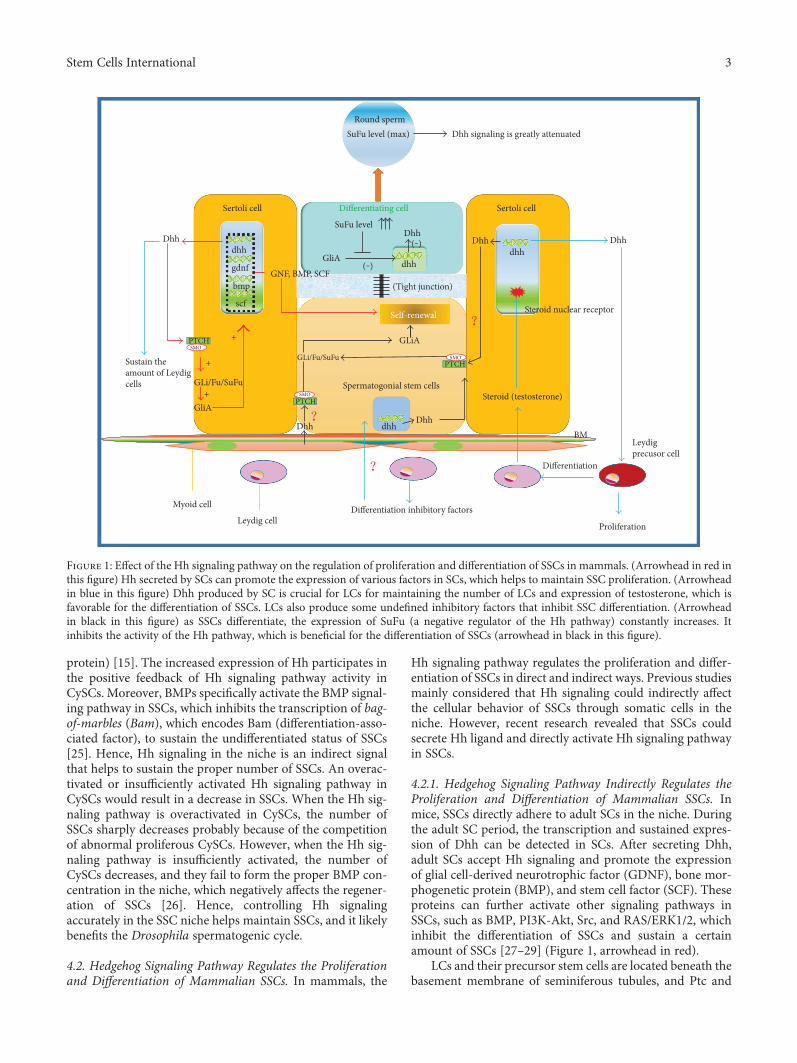
Hh signaling pathway regulates the proliferation and differ-entiation of SSCs in direct and indirect ways. Previous studiesmainly considered that Hh signaling could indirectly affectthe cellular behavior of SSCs through somatic cells in theniche. However, recent research revealed that SSCs couldsecrete Hh ligand and directly activate Hh signaling pathwayin SSCs.
4.2.1. Hedgehog Signaling Pathway Indirectly Regulates theProliferation and Differentiation of Mammalian SSCs. Inmice, SSCs directly adhere to adult SCs in the niche. Duringthe adult SC period, the transcription and sustained expres-sion of Dhh can be detected in SCs. After secreting Dhh,adult SCs accept Hh signaling and promote the expressionof glial cell-derived neurotrophic factor (GDNF), bone mor-phogenetic protein (BMP), and stem cell factor (SCF). Theseproteins can further activate other signaling pathways inSSCs, such as BMP, PI3K-Akt, Src, and RAS/ERK1/2, whichinhibit the differentiation of SSCs and sustain a certainamount of SSCs [27–29] (Figure 1, arrowhead in red).
LCs and their precursor stem cells are located beneath thebasement membrane of seminiferous tubules, and Ptc and
Sustain theamount of Leydigcells
Leydig cellDifferentiation inhibitory factors
Differentiation
Proliferation
Leydigprecusor cell
Myoid cell
GliA+
+
+
GLi/Fu/SuFu
PTCH
PTCH
PTCH
SMO
SMO
SMO
GLi/Fu/SuFu
GLiA
Self-renewal
(Tight junction)
Dhh
Sertoli cell
Dhh signaling is greatly attenuated
Sertoli cell
Dhh
(‒) dhh
SuFu level
Differentiating cell
Dhh
Steroid nuclear receptor
Steroid (testosterone)Spermatogonial stem cells
DhhDhh
dhhBM
dhhDhh
Round spermSuFu level (max)
gdnf
dhh
bmp
scf
GNF, BMP, SCFGliA
(‒)
Figure 1: Effect of the Hh signaling pathway on the regulation of proliferation and differentiation of SSCs in mammals. (Arrowhead in red inthis figure) Hh secreted by SCs can promote the expression of various factors in SCs, which helps to maintain SSC proliferation. (Arrowheadin blue in this figure) Dhh produced by SC is crucial for LCs for maintaining the number of LCs and expression of testosterone, which isfavorable for the differentiation of SSCs. LCs also produce some undefined inhibitory factors that inhibit SSC differentiation. (Arrowheadin black in this figure) as SSCs differentiate, the expression of SuFu (a negative regulator of the Hh pathway) constantly increases. Itinhibits the activity of the Hh pathway, which is beneficial for the differentiation of SSCs (arrowhead in black in this figure).
3Stem Cells International
Smo are on the surface of precursor stem cells. Dhh secretedby SCs affects the differentiation of Leydig lineage cells andestablishment of the mature LC system, which is of great sig-nificance to the growth and development of SSCs [30, 31].First, Dhh secreted by SCs is essential for the secretion of ste-roid hormones such as testosterone. Testosterone is indis-pensable in the process of spermatogenesis as it promotesthe differentiation of SSCs into spermatids. Moreover, thereare testosterone receptors in SCs. The hyposecretion oftestosterone results in developmental defects in SCs, whichnegatively affect SSC differentiation [32]. Second, Dhh pro-motes the differentiation of LC precursor stem cells into adultLCs, and it is worth emphasizing that activated Hh signalingis a prerequisite for this differentiation process [33]. Third,research shows that there is primary cilium in poorly differ-entiated LCs in human adult testes [34]. However, the ciliumwill disappear when LCs develop into mature cells, whichprobably explains why LC precursor stem cells are sensitiveto Hh signaling. Moreover, it also illustrates that Dhhsecreted by SCs is important in establishing a mature LC sys-tem, which is ultimately favorable to SSC differentiation [35].In addition, it has been reported that some undefined prod-ucts seem to contribute to the inhibition of spermatogonialdifferentiation in irradiated rats [33]. Hence, during the pro-cess of SSC proliferation and differentiation, the balancebetween SCs and LCs regulated by Hh signaling is crucial[36] (Figure 1, arrowhead in blue).
4.2.2. Hedgehog Signaling Pathway Directly Regulates theProliferation and Differentiation of Mammalian SSCs. In2014, Sahin et al. [37] found that the transcription of theHh pathway components Hh, Ptc, Smo, and Gli1 could be
detected in an environment that solely contains undifferenti-ated SSCs. Moreover, SSCs did not differentiate and the num-ber of SSCs increased in the first several months. Thus, theyproposed that SSCs could maintain self-renewal before dif-ferentiation through an autocrine loop of Hh signaling.Meanwhile, SuFu is a negative regulatory factor in the Hhpathway, and it cannot be detected in earlier differentiationperiods of SSCs. However, when SSCs develop into maturespermatids, the expression of SuFu constantly increases andinhibits Gli transcriptional activity, which results in the sup-pression of Dhh signaling in advanced circular sperm cells[38–40]. Therefore, as SSCs differentiate, their Hh signalingactivity continuously decreases. The reason why SuFu is notexpressed in earlier periods remains unknown. In addition,SSCs are the direct target cells of Hh ligand, which suggeststhat Dhh secreted by SCs perhaps could directly regulatethe proliferation of SSCs. However, the regulatory effect ofDhh produced by SCs or PMCs on SSCs needs to be exploredfurther (Figure 1, arrowhead in black).
5. Effect of the Hedgehog Signaling Pathway onOGSCs
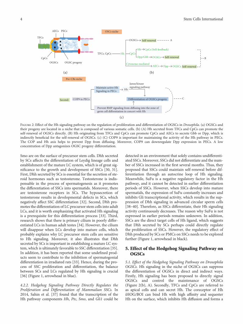
5.1. Effect of the Hedgehog Signaling Pathway on DrosophilaOGSCs. Hh signaling in the niche of OGSCs can suppressthe differentiation of OGSCs in direct and indirect ways.Firstly, Hh signaling has been proposed to directly signalOGSCs and control the maintenance of OGSCs(Figure 2(b), A). Secondly, TFCs and CpCs are referred toas apical cells and can secret Hh. The coreceptor of HhiHOG/BOI can bind Hh with high affinity and sequesterHh on the surface, which inhibits Hh diffusion and forms a
AECs PECs
TFCs
CpCsOGSC progenyOGSCs
(a)
TFCs, CpCs
OGSCs
Self-renewal
Self-renewal
Self-renewal
OSCs niche
A
B
Hh CpCsHh
Dpp/Gbb OGSCs
Cytoneme-mediatedAECs Dpp OGSCs
CpCs (Self-feedback)
(b)
Maintain active HhSignaling in PECs
Second:
Pre‒CBs niche
COP9 in PECs
Janus/kinasesignaling pathway
CDifferentiation of OGSCs progeny
Prevent BMP signaling from diffusing into the zone ofgerm cell diffrentiation in concert with Hh
Hh
(c)
Figure 2: Effect of the Hh signaling pathway on the regulation of proliferation and differentiation of OGSCs in Drosophila. (a) OGSCs andtheir progeny are located in a niche that is composed of various somatic cells. (b) (A) Hh secreted from TFCs and CpCs can promote theself-renewal of OGSCs directly. (B) Hh originating from TFCs and CpCs can promote CpCs and AECs to secrete Gbb or Dpp, which isindirectly beneficial for the self-renewal of OGSCs. (c) (C) COP9 is important for maintaining the activity of the Hh pathway in PECs.The COP and Hh axis helps to prevent Dpp from diffusing. Moreover, COP9 can downregulate Dpp expression in PECs. A lowconcentration of Dpp antagonizes OGSC progeny differentiation.
4 Stem Cells International

positive feedback of Hh pathway in apical cell. As targetgenes of Hh pathway, the expression of Hh increased [41,42]. Then, CpCs deliver Hh signaling to AECs (ECs locatedin the anterior part of the germarium) and promote the tran-scription of the target genes Decapentaplegic (Dpp) and glassbottom boat (Gbb) (both belong to the Bmp family), in AECs[27, 43]. Hh secreted by CpCs also directly enhances the tran-scription of Dpp in CpCs. Eventually, BMP (Dpp) signalingin the OGSCs niche is intensified. As a result, Dpp suppressesthe expression of Bam and inhibits the differentiation ofOGSCs. As OGSCs differentiate, the expression of Fused(Fu), a positive regulator in the Hh pathway, is constantlyincreased.Moreover, Fumediates the ubiquitination and pro-teolysis of thickveins (Tkv, a BMP receptor) in OGSCs, whichis beneficial for the differentiation of OGSCs [19, 44–47](Figure 2(b), B).
When OGSC progeny separates from CpCs, PEC- (ECslocated in the posterior part of the germarium-) derived butnot CpC-derived Hh starts to participate in the differentia-tion process. It was discovered that maintaining the activa-tion of the Hh signaling pathway and secretion of Hhligand in PECs requires the presence of COP9 (also knownas the CSN complex) in ECs [48]. Moreover, the COP9-Hhaxis in PECs can partly prevent the diffusing of Dpp andpromote the differentiation of OGSC progeny. In addition,JAK/STAT signaling promotes Dpp expression, whereas Hhsignaling from PECs suppresses Dpp expression by antago-nizing JAK/STAT signaling, which favors the differentiationof OGSC progeny. Therefore, it seems that Hh protein inthe niche promotes the differentiation of OGSC progeny viasuppressing the transcription of Dpp in a non-Hh signalingmechanism [49] (Figure 2(c), C).
5.2. Effect of the Hedgehog Signaling Pathway on MammalianOGSCs. OGSCs in mammals are located in a single layer ofepithelial cells with tight junctions in the OSE. In almost allspecies, it is not possible to observe obvious OGSC divisionin the normal OSE, and most research concerning OGSCfunction involves OGSC transplantation experiments. Thus,it is more reasonable to discuss the effect of Hh signalingpathway on OGSCs in normal OSE or in the ovary cortexafter transplantation [50–52]. Moreover, it will be interestingto determine whether artificially altering the activity of Hhpathway will benefit ovum regeneration.
5.2.1. Effect of the Hedgehog Signaling Pathway on theMigration of OGSCs in the OSE. We proposed a hypothesisthat the special structure of the OSE restricts the activity ofOGSCs, but Hh signaling could help OGSCs migrate intothe ovary cortex. Under natural conditions, the outside ofthe OSE is covered by the peritoneum, and the underside ofthe OSE adjoins the ovary cortex [53–55]. However, there isa layer of dense-constructed ovary tunica albuginea betweenthe OSE and the ovary cortex. Restricted by this physiologicalstructure, OGSCs seem to be isolated in an area that is distantfrom the ovary cortex, which contains various somatic cellssuch as granulosa cells (GCs), theca cells (TCs), and mesen-chymal cells (MCs). Thus, in the normal ovary, it is difficultto predict how Hh signaling regulates the proliferation,
differentiation, or any other cellular behavior of OGSCsthrough other somatic cells. Epithelial cells in the OSE areequipped with both epithelial and mesenchymal phenotypes.They can undergo epithelial-mesenchymal transition (EMT),which changes the tight junctions between epithelial cellsinto looser junctions between mesenchymal cells. Previousstudies revealed that Hh signaling pathway participates inEMT in other tissues [56–58]. However, Gli1 transcriptionis not observed in the OSE, seemingly indicating that Hhpathway is not activated in the OSE in the normal state.However, the Hh signaling pathway participates in themigration of epithelial ovarian cancer cells into the ovarycortex. This suggests that the Hh pathway is possibly relatedto the migration of OGSCs into the ovary cortex. OnceOGSCs shift to the cortex, they enter into an entirely differentenvironment that contains various biosignals that probablypromote the proliferation or differentiation of OGSCs(Figure 3(b)).
5.2.2. Effect of the Hedgehog Signaling Pathway on theProliferation and Differentiation of OGSCs. It is not clearwhether somatic cells in the ovary cortex regulate the func-tion of OGSCs through the Hh signaling pathway. Duringthe initial forming period of the mammalian fetal ovary, thetranscription levels of Hh, Ptc, and Gli are very low [59].After birth, the primordial oocyte of the primary folliclesecretes the differentiation factor GDF9 (growth differentia-tion factor 9) [60], inducing granulosa cells to secrete Dhhand Ihh. Then, Dhh and Ihh continue to induce progenitorTCs to differentiate into TCs to promote follicle development[61–64]. In the ovary, GCs are the main source of Hh protein,while Ptc is the most abundant on the surface of TCs. Ptcnot only binds with Hh but also works in preventing Hhsignaling from diffusing [63, 65]. Hence, Ptc in TCs distrib-uted around the outer sphere of the follicle seems to helpprevent Hh from diffusing into nearby follicles or interstitialcells. Thus, when we injected OGSCs into the ovary cortex,they came into direct contact with the somatic cells(Figure 3(c)). In 2015, a study reported that cancer stemcells of ovary cancer are very likely a malignant transformedproduct of OGSCs [66]. Furthermore, a previous studyfound that there is a BMP4-Hh-positive feedback loopbetween CSCs (cancer stem cells) of ovary cancer and CA-MACs (cancer-associated mesenchymal stem cells), whichenhances the proliferation of CSCs. Meanwhile, CSCsproduce Hh to activate the BMP signaling pathway in CA-MSCs [67]. Then, the overexpression of BMP4 suppressesCSCs to differentiate, resulting in the overexpression ofHh. Although CSCs can secret Hh, it is not clear whetherCSCs have the ability to secrete Hh before or after cancera-tion. Until now, no study has mentioned whether OGSCscan produce Hh. Normal MSCs (mesenchymal stem cells)can also produce BMPs but at lower levels compared withCA-MACs, so it is unclear whether OGSCs in the normalovarian stroma together with MACs form a positive feed-back loop like the BMP4-Hh feedback loop between CSCsand CA-MACs and promote the proliferation of OGSCs(Figures 3(a) and 3(d)). Furthermore, Park et al. [68] foundthat BMP4 could promote the differentiation of OGSCs into
5Stem Cells International

oocytes via Smad1/5/8 signaling in mice. This result wascounter to the assumption that BMP4 helps to sustain theself-renewal of OGSCs. However, the result may dependon the dosage of BMP. Therefore, we conclude that theproliferation or differentiation of OGSCs varies with differ-ent concentrations of BMP4 conducted by Hh signalingfrom OGSCs. This resembles the dual regulation inDrosophila when Hh signaling stimulates other cells tosecrete BMP protein.
Can somatic cells in the ovary regulate OGSC develop-ment as in Drosophila? Grieve et al. [51] found that stage-specific granulosa cells could induce the expression ofoocyte-specific genes in embryonic stem cells under cocul-ture conditions. He proposed that somatic cells are importantfor facilitating the differentiation of stem cells into functionaloocytes. GCs can secrete the Hh ligand, but it is not clearwhether the Hh pathway is involved in the proliferationand differentiation of OGSCs under coculture with OGSCs.However, it would perhaps be more convincing if we useOGSCs to replace the oogonia in primary follicles and thenobserve follicle development. A developing follicle is a goodmodel for studying the relationship between GCs, Hh, andOGSCs. However, it is not clear whether Ptc and Smo are
in OGSCs, which if so would indicate that altering Hh path-way activity would regulate the proliferation of OGSCs.
6. Perspective
According to the current research progress in this area, thefollowing questions need to be thoroughly addressed in thenear future.
Currently, many studies have indicated that the Hh sig-naling pathway has a strong effect on the proliferation anddifferentiation of SSCs. What we are mostly interested iswhether we can artificially alter the self-renewal or differenti-ation process via regulating Hh signaling in the SSC niche,thereby improving the reproductive ability of animals. Forexample, some animals with azoospermia cannot produce asufficient number of sperm and lose their reproductive abilitybecause of the dysfunction of SSCs or the low number ofSSCs. A sufficient number of SSCs is a prerequisite for a nor-mal spermatogenic ability in males. In 2014, it was deter-mined in testicular biopsies that the ectopic expression ofShh results in the absence of spermatocytes and increasednumbers of LCs in the testes [69]. We discussed in this articlethat LCs have dual roles in regulating SSC physiology. Hence,
Malignant transform
High density of Ptc on the thecacell surface may help to con�ne theHhs in the zone of developingfollicle which prevent Hhs disfusingto other regions
Normal mesenchymal stem cell (MAC)
Ovary cancer-assosiated mesenchymalstem cells (CA-MAC)
Mesenchymal cells (MAC)
Ovary cancer cells
Ovary cancer stem cell (CSC)
Oocyte
Mature granular cells
�eca cells
Ovarain surface epithelial
BMP4
Ovary cortex
(a)
(d)
(c)
(b)
Tunica albuginea
Peritoneum
Self-renewal
(+)
(+)
cells (ECs)
Ovarian germline stem cell
Fusiform pregranular cells
Hh ligand
Ptc
�eca progenitor cell
Pregranulose cell
Base membrane (BM)
Secondary follicle
GDF9
Primary folliclePrimitive follicle
?
EMT Migration
BMP4
Figure 3: Effect of the Hh signaling pathway on the behavior of OGSCs in mammals. (a) It is not clear whether OGSCs in the ovarian surfaceepithelium (OSE) secrete Hh. (b) It is thought that the EMT could help OGSCs migrate to the ovary cortex from the OSE. (c) Ptc in TCs assistsin preventing the Hh ligand from diffusing to other developing follicles and the interstitial stroma. (d) CSCs of ovarian cancer are likely to bethe malignant transformation products of OGSCs. There is a BMP4-Hh feedback loop between CSCs and CA-MACs that promotes CSCproliferation and maintains the CSC number. It has not been determined whether there is a similar BMP4-Hh feedback loop betweenOGSCs and MSCs.
6 Stem Cells International

the proper regulation of Hh signaling in the niche is vital tomaintain the number of SSCs. In addition, apart from prolif-eration and differentiation, apoptosis is also an importantphysiological process of SSCs that could impact the numberof SSCs. Moreover, the Hh signaling pathway possibly takespart in antiapoptosis, but there has been no relevant study.In Drosophila, the JAK-STAT signaling pathway, whosefunctions overlap with those of the Hh signaling pathway,enhances the proliferation of SSCs and the overexpressionof the antiapoptosis protein DIAPI in cells, which helps sus-tain the activity of SSCs [70]. Thus, the Hh pathway probablyfunctions in the antiapoptosis process. When SSCs differenti-ate, the increase in the SuFu level inhibits the activity of theHh pathway. While there is little SuFu in SSCs, it is notknown whether any substance other than SuFu antagonizesectopic Hh signaling to maintain the proper number of SSCs.In short, regardless of what promotes the proliferation,differentiation, or antiapoptosis of SSCs via altering Hhsignaling pathway activity, these results all provide somenew ideas for clinical therapeutic methods for treatingazoospermia or other diseases related to SSCs.
OGSCs are considered one of the possible cells in animalsthat can generate oocytes. Regulating OGSC function can beused to help those who wish to lengthen their reproductivelife span or to treat animals or humans with germ celldysfunction to restore their reproductive ability. Whetherchanging the Hh signaling pathway in OGSCs can affect theformation of primordial follicles in mammals and whetherany effects on the proliferation and differentiation of OGSCsare caused by Hh signaling regulation still require furtherinvestigation. Previous studies have shown that Hh signalingcan influence the development of TCs and hinder ovulationin mammals [71], but there has been no report on the effectof Hh signaling on OGSCs and primordial follicle formation.However, other signaling pathways, such as Hippo-YAP,Notch, and WNT [72–74], closely related to the Hh signalingpathway, have been found to be involved in regulating theproliferation and differentiation of OGSCs, as well asinfluencing the formation and development of mammalianprimordial follicles. For example, Ci [75], a component ofthe Hh pathway, suppresses the activity of the Hippo signal-ing pathway kinase cascade inDrosophila ovary somatic cells,which eventually promotes the differentiation of OGSCs.Meanwhile, the Hippo-YAP signaling pathway has a negativeregulatory effect on the generation and development ofmammalian follicles [76], which probably indicates that theactivation of the Hh pathway can enhance the growth anddevelopment of mammalian follicles via regulating thebehavior of OGSCs.
Conflicts of Interest
The authors declare that there is no conflict of interestsregarding the publication of this paper.
Acknowledgments
The authors thank all of the members in their lab. This workwas supported by the National Natural Science Foundation
of China (no. 81660245 and no. 81360100), the Excellence555 Engineering of JiangXi Province, and the Natural ScienceFoundation of JiangXi Province (no. 20142BAB205069).
References
[1] P. Song, Y. Inagaki, Y. Sugawara, and N. Kokudo, “Perspec-tives on human clinical trials of therapies using iPS cells inJapan: reaching the forefront of stem-cell therapies,” BioscienceTrends, vol. 7, no. 3, p. 157, 2013.
[2] A. S. Bryukhovetskiy and I. S. Bryukhovetskiy, “Effectivenessof repeated transplantations of hematopoietic stem cells inspinal cord injury,” World Journal of Transplantation, vol. 5,no. 3, pp. 110–128, 2015.
[3] C. Nusslein-Volhard and E. Wieschaus, “Mutations affectingsegment number and polarity in Drosophila,” Nature,vol. 287, no. 30, p. 795801, 1980.
[4] P. W. Ingham and A. P. McMahon, “Hedgehog signaling inanimal development: paradigms and principles,” Genes &Development, vol. 15, no. 23, pp. 3059–3087, 2001.
[5] C.-c. Hui and S. Angers, “Gli proteins in development anddisease,” Annual Review of Cell and Developmental Biology,vol. 27, pp. 513–537, 2011.
[6] J. A. Buglino and M. D. Resh, “Hhat is a palmitoylacyltransfer-ase with specificity for N-palmitoylation of sonic hedgehog,”The Journal of Biological Chemistry, vol. 283, no. 32,pp. 22076–22088, 2008.
[7] T. Kawakami, T. Kawcak, Y. J. Li, W. Zhang, Y. Hu, and P. T.Chuang, “Mouse dispatched mutants fail to distributehedgehog proteins and are defective in hedgehog signaling,”Development, vol. 129, no. 18, pp. 5753–5765, 2002.
[8] T. Tenzen, B. L. Allen, F. Cole, J. S. Kang, R. S. Krauss, and A.P. McMahon, “The cell surface membrane proteins Cdo andBoc are components and targets of the hedgehog signalingpathway and feedback network in mice,” Developmental Cell,vol. 10, no. 5, pp. 647–656, 2006.
[9] S. Yao, L. Lum, and P. Beachy, “The ihog cell-surface proteinsbind hedgehog and mediate pathway activation,” Cell, vol. 125,no. 2, pp. 343–357, 2006.
[10] C. C. Hui and S. Angers, “Gli proteins in development and dis-ease,” Annual Review of Cell and Developmental Biology,vol. 27, p. 513537, 2011.
[11] S. S. Ram, B. T. Ozanna, C. Chhavi, and S. X. Hou, “Spermato-gonial stem cells, infertility and testicular cancer,” Journal ofCellular & Molecular Medicine, vol. 15, no. 3, pp. 468–483,2011.
[12] Z. Pan, M. Sun, X. Liang et al., “The controversy, challenges,and potential benefits of putative female germline stem cellsresearch in mammals,” Stem Cells International, vol. 2016,Article ID 1728278, 2016.
[13] L. J. Greenspan, M. D. Cuevas, and E. Matunis, “Genetics ofgonadal stem cell renewal,” Annual Review of Cell & Develop-mental Biology, vol. 31, no. 31, p. 291, 2015.
[14] A. Spradling, M. T. Fuller, R. E. Braun, and S. Yoshida, “Germ-line stem cells,” Cold Spring Harbor Perspectives in Biology,vol. 3, no. 3, article 002642, 2011.
[15] E. L. Matunis, R. R. Stine, and C. M. De, “Recent advances inDrosophila male germline stem cell biology,” Spermatogenesis,vol. 2, no. 3, pp. 137–144, 2012.
[16] R. L. Brinster and J. W. Zimmermann, “Spermatogenesisfollowing male germ-cell transplantation,” Proceedings of the
7Stem Cells International
National Academy of Sciences of the United States of America,vol. 91, no. 24, pp. 11298–11302, 1994.
[17] Z. Chen, Z. Li, and Z. He, “Plasticity of male germline stemcells and their applications in reproductive and regenerativemedicine,” Asian Journal of Andrology, vol. 17, no. 3,pp. 367–372, 2014.
[18] F. J. King, A. Szakmary, D. N. Cox, and H. Lin, “Yb, modulatesthe divisions of both germline and somatic stem cells throughpiwi-, and hh- mediated mechanisms in the Drosophilaovary,” Molecular Cell, vol. 7, no. 3, pp. 497–508, 2001.
[19] L. Xia, X. Zheng, W. Zheng et al., “The niche-dependent feed-back loop generates a BMP activity gradient to determine thegermline stem cell fate,” Current Biology, vol. 22, no. 6,pp. 515–521, 2012.
[20] K. Zou, Z. Yuan, Z. Yang et al., “Production of offspring from agermline stem cell line derived from neonatal ovaries,” NatureCell Biology, vol. 11, no. 5, pp. 631–636, 2009.
[21] Z. Pan, M. Sun, J. Li et al., “The expression of markers relatedto ovarian germline stem cells in the mouse ovarian surfaceepithelium and the correlation with Notch signaling pathway,”Cellular Physiology & Biochemistry, vol. 37, no. 6, pp. 2311–2322, 2015.
[22] S. Parvari, M. Abbasi, N. Abbasi et al., “Stem cell isolation by amorphology-based selection method in postnatal mouseovary,” Archives of Medical Science, vol. 11, no. 3, pp. 670–678, 2015.
[23] Y. A. White, D. C. Woods, Y. Takai, O. Ishihara, H. Seki,and J. L. Tilly, “Oocyte formation by mitotically active germcells purified from ovaries of reproductive-age women,”Nature Medicine, vol. 18, no. 3, pp. 413–421, 2012.
[24] C. E. Dunlop, R. A. Bayne, M. McLaughlin, E. E. Telfer, andR. A. Anderson, “Isolation, purifi cation, and culture ofoogonial stem cells from adult human and bovine ovariancortex,” Lancet, vol. 383, 2017.
[25] M. Michel, A. P. Kupinski, I. Raabe, and C. Bökel, “Hh signal-ling is essential for somatic stem cell maintenance in theDrosophila testis niche,” Development, vol. 139, no. 15,p. 2663, 2012.
[26] Z. Zhang, C. Pan, and Y. Zhao, “Hedgehog in the Drosophilatestis niche: what does it do there?” Protein & Cell, vol. 4,no. 9, pp. 650–655, 2013.
[27] M. Inaba, Y. M. Yamashita, and M. Buszczak, “Keeping stemcells under control: new insights into the mechanisms thatlimit niche-stem cell signaling within the reproductive sys-tem,” Molecular Reproduction & Development, vol. 83, no. 8,pp. 675–683, 2016.
[28] X. Meng, M. Lindahl, M. E. Hyvönen et al., “Regulation of cellfate decision of undifferentiated spermatogonia by GDNF,”Science, vol. 287, no. 5457, pp. 1489–1493, 2000.
[29] J. M. Oatley and R. L. Brinster, “The germline stem cell nicheunit in mammalian testes,” Physiological Reviews, vol. 92,no. 2, pp. 577–595, 2012.
[30] L. J. Martin, “Cell interactions and genetic regulation thatcontribute to testicular Leydig cell development and differenti-ation,” Molecular Reproduction & Development, vol. 83, no. 6,pp. 470–487, 2016.
[31] R. Hazra, M. Jimenez, R. Desai, D. J. Handelsman, andC. M. Allan, “Sertoli cell androgen receptor expressionregulates temporal fetal and adult Leydig cell differentiation,function, and population size,” Endocrinology, vol. 154, no. 9,p. 3410, 2013.
[32] S. Y. Park, M. Tong, and J. L. Jameson, “Distinct roles forsteroidogenic factor 1 and desert hedgehog pathways in fetaland adult Leydig cell development,” Endocrinology, vol. 148,no. 8, pp. 3704–3710, 2007.
[33] I. Barsoum and H. H. C. Yao, “Redundant and differentialroles of transcription factors Gli1 and Gli2 in the developmentof mouse fetal Leydig Cells1,” Biology of Reproduction, vol. 84,no. 5, pp. 894–899, 2011.
[34] M. B. Nygaard, K. Almstrup, L. Lindbæk, S. T. Christensen,and T. Svingen, “Cell context-specific expression of primarycilia in the human testis and ciliary coordination of hedgehogsignalling in mouse Leydig cells,” Scientific Reports, vol. 5,pp. 1–14, 2015.
[35] H. L. Franco and H. H. Yao, “Sex and hedgehog: roles of genesin the hedgehog signaling pathway in mammalian sexualdifferentiation,” Chromosome Research, vol. 20, no. 1,pp. 247–258, 2012.
[36] G. Shetty, W. Zhou, C. C. Weng, S. H. Shao, and M. L.Meistrich, “Leydig cells contribute to the inhibition of sper-matogonial differentiation after irradiation of the rat,”Andrology, vol. 18, no. 3, pp. 479–491, 2005.
[37] Z. Sahin, A. Szczepny, M. L. EA et al., “Dynamic hedgehog sig-nalling pathway activity in germline stem cells,” Andrology,vol. 2, no. 2, p. 267, 2014.
[38] J. A. Mäkelä, V. Saario, S. Bourguiba-Hachemi et al., “Hedge-hog signalling promotes germ cell survival in the rat testis,”Reproduction, vol. 142, no. 5, pp. 711–721, 2011.
[39] T. L. Kroft, J. Patterson, J. Won Yoon et al., “GLI1 localizationin the germinal epithelial cells alternates between cytoplasmand nucleus: upregulation in transgenic mice blocks spermato-genesis in pachytene,” Biology of Reproduction, vol. 65, no. 6,p. 1663, 2001.
[40] A. Szczepny, G. R. Hime, and K. L. Loveland, “Expression ofhedgehog signalling components in adult mouse testis,” Devel-opmental Dynamics, vol. 235, no. 11, p. 3063, 2006.
[41] T. R. Hartman, D. Zinshteyn, H. K. Schofield, E. Nicolas, A.Okada, and A. M. O'Reilly, “Drosophila Boi limits hedgehoglevels to suppress follicle stem cell proliferation,” Journal ofCell Biology, vol. 191, no. 5, pp. 943–952, 2010.
[42] T. R. Hartman, T. I. Strochlic, Y. Ji, D. Zinshteyn, and A. M.O'Reilly, “Diet controls Drosophila follicle stem cell prolifera-tion via hedgehog sequestration and release,” Journal of CellBiology, vol. 201, no. 5, pp. 741–757, 2013.
[43] P. Rojasríos, I. Guerrero, and A. Gonzálezreyes, “Cytoneme-mediated delivery of hedgehog regulates the expression ofbone morphogenetic proteins to maintain germline stem cellsin Drosophila,” PLoS Biology, vol. 10, no. 4, article e1001298,2012.
[44] W. Lu, M. O. Casanueva, A. P. Mahowald, M. Kato, D.Lauterbach, and E. L. Ferguson, “Niche-associated activationof rac promotes the asymmetric division of Drosophilafemale germline stem cells,” PLoS Biology, vol. 10, no. 7,article e1001357, 2012.
[45] L. Xia, S. Jia, S. Huang et al., “The fused/Smurf complex con-trols the fate of Drosophila germline stem cells by generatinga gradient BMP response,” Cell, vol. 143, no. 6, p. 978, 2010.
[46] S. Eliazer and M. Buszczak, “Finding a niche: studies from theDrosophila ovary,” Stem Cell Research & Therapy, vol. 2, no. 6,pp. 1–8, 2011.
[47] K. Narbonne-Reveau, F. Besse, C. Lamour-Isnard, D. Busson,and A. M. Pret, “Fused, regulates germline cyst mitosis and
8 Stem Cells International

differentiation during Drosophila, oogenesis,” Mechanisms ofDevelopment, vol. 123, no. 3, pp. 197–209, 2006.
[48] T. Lu, S. Wang, Y. Gao et al., “COP9-hedgehog axis regulatesthe function of the germline stem cell progeny differentiationniche in the Drosophila ovary,” Development, vol. 142,no. 24, pp. 4242–4252, 2015.
[49] Z. Liu, G. Zhong, P. C. Chai et al., “Coordinated niche-associated signals promote germline homeostasis in the Dro-sophila ovary,” Journal of Cell Biology, vol. 211, no. 2, p. 469,2015.
[50] I. Virantklun, “Postnatal oogenesis in humans: a review ofrecent findings,” Stem Cells & Cloning Advances & Applica-tions, vol. 8, pp. 49–60, 2015.
[51] K. M. Grieve, M. McLaughlin, C. E. Dunlop, E. E. Telfer, andR. A. Anderson, “The controversial existence and functionalpotential of oogonial stem cells,” Maturitas, vol. 82, no. 3,p. 278, 2015.
[52] O. Celik, E. Celik, I. Turkcuoglu, E. Yilmaz, Y. Simsek, and B.Tiras, “Germline cells in ovarian surface epithelium of mam-malians: a promising notion,” Reproductive Biology and Endo-crinology, vol. 10, no. 1, pp. 1–8, 2012.
[53] W. J. Murdoch and A. C. Mcdonnel, “Roles of the ovariansurface epithelium in ovulation and carcinogenesis,” Repro-duction, vol. 123, no. 123, pp. 743–750, 2002.
[54] E. Lengyel, “Ovarian cancer development and metastasis,”American Journal of Pathology, vol. 177, no. 3, pp. 1053–1064, 2010.
[55] U. Urzúa, S. Ampuero, K. F. Roby, G. A. Owens, and D. J.Munroe, “Dysregulation of mitotic machinery genes precedesgenome instability during spontaneous pre-malignant trans-formation of mouse ovarian surface epithelial cells,” BMCGenomics, vol. 17, no. 8, p. 728, 2016.
[56] S. Lamouille, J. Xu, and R. Derynck, “Molecular mechanismsof epithelial-mesenchymal transition,” Nature Reviews Molec-ular Cell Biology, vol. 15, no. 3, pp. 178–196, 2014.
[57] A. Omenetti, A. Porrello, Y. Jung et al., “Hedgehog signalingregulates epithelial-mesenchymal transition during biliaryfibrosis in rodents and humans,” Journal of Clinical Investiga-tion, vol. 118, no. 10, pp. 3331–3342, 2008.
[58] B. Davidson, C. G. Tropé, and R. Reich, “Epithelial–mesenchy-mal transition in ovarian carcinoma,” Frontiers in Oncology,vol. 2, p. 33, 2012.
[59] N. C. Bingham, K. L. Parker, J. S. Jorgensen, and H. H. Yao,“Activation of the hedgehog pathway in the mouse fetal ovaryleads to ectopic appearance of fetal Leydig cells and femalepseudohermaphroditism,” Developmental Biology, vol. 329,no. 1, p. 96, 2009.
[60] F. C. de Castro, M. H. Cruz, and C. L. Leal, “Role of growthdifferentiation factor 9 and bone morphogenetic. Protein 15in ovarian function and their importance in mammalianfemale fertility—a review,” Asian-Australasian Journal ofAnimal Sciences, vol. 29, no. 8, pp. 1065–1074, 2016.
[61] M. E. Pepling, “Hedgehog signaling in follicle development,”Biology of Reproduction, vol. 86, no. 6, p. 173, 2012.
[62] L. J. Spicer, S. Sudo, P. Y. Aad et al., “The hedgehog-patchedsignaling pathway and function in the mammalian ovary: anovel role for hedgehog proteins in stimulating proliferationand steroidogenesis of theca cells,” Reproduction, vol. 138,no. 2, pp. 329–339, 2009.
[63] M. C. Russell, R. G. Cowan, R. M. Harman, A. L. Walker, andS. M. Quirk, “The hedgehog signaling pathway in the mouseovary,” Biology of Reproduction, vol. 77, no. 2, p. 226, 2007.
[64] Y. Ren, R. G. Cowan, F. F. Migone, and S. M. Quirk, “Overac-tivation of hedgehog signaling alters development of the ovar-ian vasculature in mice,” Biology of Reproduction, vol. 86, no. 6,p. 174, 2012.
[65] Y. Chen and G. Struhl, “Dual roles for patched in sequesteringand transducing hedgehog,” Cell, vol. 87, no. 87, pp. 553–563,1996.
[66] O. B. Ozakpinar, A. M.Maurer, and D. Ozsavci, “Ovarian stemcells: From basic to clinical applications,” World Journal ofStem Cells, vol. 7, no. 4, pp. 757–768, 2015.
[67] L. G. Coffman, Y. J. Choi, K. McLean, B. L. Allen, M. P. diMagliano, and R. J. Buckanovich, “Human carcinoma-associated mesenchymal stem cells promote ovarian cancerchemotherapy resistanceviaa BMP4/HH signaling loop,”Oncotarget, vol. 7, no. 6, pp. 6916–6932, 2016.
[68] E. S. Park, D. C. Woods, and J. L. Tilly, “Bone morphogeneticprotein 4 (BMP4) promotes mammalian oogonial stem celldifferentiation via Smad1/5/8 signaling,” Fertility & Sterility,vol. 100, no. 5, p. 1468, 2013.
[69] S. Zou, Y. Wang, T. Chen et al., “Ectopic expression of sonichedgehog in a cryptorchid man with azoospermia: a casereport,” The Journal of International Medical Research,vol. 42, no. 2, pp. 589–597, 2014.
[70] S. Hasan, P. Hétié, and E. L. Matunis, “Niche signalingpromotes stem cell survival in the Drosophila testis via theJAK-STAT target DIAP1,” Developmental Biology, vol. 404,no. 1, pp. 27–39, 2015.
[71] Y. Ren, R. G. Cowan, R. M. Harman, and S. M. Quirk,“Dominant activation of the hedgehog signaling pathway inthe ovary alters theca development and prevents ovulation,”Molecular Endocrinology, vol. 23, no. 5, p. 711, 2009.
[72] S. Klusza and W. M. Deng, “At the crossroads of differentia-tion and proliferation: precise control of cell-cycle changesby multiple signaling pathways in Drosophila follicle cells,”BioEssays, vol. 33, no. 2, pp. 124–134, 2011.
[73] C. P. Zhang, J. L. Yang, J. Zhang et al., “Notch signaling isinvolved in ovarian follicle development by regulatinggranul-osa cell proliferation,” Endocrinology, vol. 152, no. 6,pp. 2437–2447, 2011.
[74] C. L. Chen, X. F. Fu, L. Q. Wang et al., “Primordial follicleassembly was regulated by Notch signaling pathway in themice,” Molecular Biology Reports, vol. 41, no. 3, pp. 1891–1899, 2014.
[75] C. Li, L. Kan, Y. Chen et al., “Ci antagonizes Hippo signaling inthe somatic cells of the ovary to drive germline stem cell differ-entiation,” Cell Research, vol. 25, no. 10, p. 1152, 2015.
[76] K. Kawamura, Y. Cheng, N. Suzuki et al., “Hippo signaling dis-ruption and Akt stimulation of ovarian follicles for infertilitytreatment,” Proceedings of the National Academy of Sciencesof the United States of America, vol. 110, no. 43, pp. 17474–17479, 2013.
9Stem Cells International

Submit your manuscripts athttps://www.hindawi.com
Hindawi Publishing Corporationhttp://www.hindawi.com Volume 2014
Anatomy Research International
PeptidesInternational Journal of
Hindawi Publishing Corporationhttp://www.hindawi.com Volume 2014
Hindawi Publishing Corporation http://www.hindawi.com
International Journal of
Volume 201
Hindawi Publishing Corporationhttp://www.hindawi.com Volume 2014
Molecular Biology International
GenomicsInternational Journal of
Hindawi Publishing Corporationhttp://www.hindawi.com Volume 2014
The Scientific World JournalHindawi Publishing Corporation http://www.hindawi.com Volume 2014
Hindawi Publishing Corporationhttp://www.hindawi.com Volume 2014
BioinformaticsAdvances in
Marine BiologyJournal of
Hindawi Publishing Corporationhttp://www.hindawi.com Volume 2014
Hindawi Publishing Corporationhttp://www.hindawi.com Volume 2014
Signal TransductionJournal of
Hindawi Publishing Corporationhttp://www.hindawi.com Volume 2014
BioMed Research International
Evolutionary BiologyInternational Journal of
Hindawi Publishing Corporationhttp://www.hindawi.com Volume 2014
Hindawi Publishing Corporationhttp://www.hindawi.com Volume 2014
Biochemistry Research International
ArchaeaHindawi Publishing Corporationhttp://www.hindawi.com Volume 2014
Hindawi Publishing Corporationhttp://www.hindawi.com Volume 2014
Genetics Research International
Hindawi Publishing Corporationhttp://www.hindawi.com Volume 2014
Advances in
Virolog y
Hindawi Publishing Corporationhttp://www.hindawi.com
Nucleic AcidsJournal of
Volume 2014
Stem CellsInternational
Hindawi Publishing Corporationhttp://www.hindawi.com Volume 2014
Hindawi Publishing Corporationhttp://www.hindawi.com Volume 2014
Enzyme Research
Hindawi Publishing Corporationhttp://www.hindawi.com Volume 2014
International Journal of
Microbiology