Embed Size (px)
Citation preview
Role of the midbrain dopaminergic system in modulationof vocal brain activation by social context
Erina Hara,1 Lubica Kubikova,2,3 Neal A. Hessler1 and Erich D. Jarvis2
1Laboratory for Vocal Behaviour Mechanisms, RIKEN Brain Science Institute, Wako-shi, Japan2Department of Neurobiology, Duke University Medical Center, Durham, NC 27710, USA3Department of Endocrinology and Ethology, Institute of Animal Biochemistry and Genetics SAS, Moyzesova 61, 90028 Ivanka priDunaji, Slovakia
Keywords: birdsong, dopamine, immediate early gene, sexual motivation, vocal production
Abstract
In a well-studied model of social behaviour, male zebra finches sing directed song to court females and undirected song, usedpossibly for practice or advertisement. Although the two song types are similar, the level of neural activity and expression of theimmediate early gene egr-1 are higher during undirected than during directed singing in the lateral part of the basal ganglia songnucleus AreaX (LAreaX) and its efferent pallial song nuclei lateral magnocellular nucleus of the anterior nidopallium (LMAN) andthe robust nucleus of the arcopallium (RA). As social interactions are dependent on brain motivation systems, here we test thehypothesis that the midbrain ventral tegmental area–substantia nigra pars compacta (VTA–SNc) complex, which provides astrong dopaminergic input to LAreaX, is a source of this modulation. Using egr-1 expression, we show that GABAergicinterneurons in VTA–SNc are more active during directed courtship singing than during undirected singing. We also found thatunilateral removal of VTA–SNc input reduced singing-dependent gene expression in ipsilateral LAreaX during both socialcontexts but it did not eliminate social context differences in LAreaX. In contrast, such lesions reduced and eliminated the socialcontext differences in efferent nuclei LMAN and RA, respectively. These results suggest that VTA–SNc is not solely responsiblefor the social context gene regulation in LAreaX, but that VTA–SNc input to LAreaX enhances the singing-regulated geneexpression in this nucleus and, either through LAreaX or through direct projections to LMAN and RA, VTA–SNc is necessary forcontext-dependent gene regulation in these efferent nuclei.
Introduction
Songbirds possess the rare trait of vocal learning, which is controlledby a discrete neural network that consists of a vocal motor pathway[(HVC, a letter based name) fi robust nucleus of the arcopallium(RA) fi 12th nucleus, tracheosyringeal part (nXIIts) and otherbrainstem neurons; see Fig. 1A] involved in song production, and avocal pallial–basal ganglia loop [lateral magnocellular nucleus of theanterior nidopallium (LMAN) fi lateral AreaX of the striatum(LAreaX) fi dorsal lateral nucleus of the medial thalamus(DLM) fi LMAN] involved in song learning and modification(Fig. 1A; reviewed in Jarvis, 2004). Nuclei of these pathways showremarkable differences in activation depending upon the social contextin which male zebra finches produce their songs; males sing astereotyped directed song to females during courtship and a slightlymore variable undirected song that is thought to be used for practice oradvertisement (Sossinka & Bohner, 1980; Zann, 1996; Jarvis et al.,1998; Kao et al., 2005). During undirected singing (UD), the level ofneural activity and expression of the immediate early gene (IEG) egr-1(also known as ZENK; Mello et al., 1992) are high throughout bothvocal pathways, whereas during female-directed singing (FD), activityand ⁄ or egr-1 expression are low in the lateral part of the vocal pallial–
basal ganglia loop (LAreaX and LMAN) and in RA of the motorpathway (Jarvis et al., 1998; Hessler & Doupe, 1999). Because thelevel of egr-1 expression in HVC, the primary song system input toboth LAreaX and RA, was not dependent on social context, it has beenproposed that the social context-dependent modulation of these vocalnuclei may be controlled by midbrain motivation-related brain areas(Jarvis et al., 1998; Hessler & Doupe, 1999). The suggested candidateareas included the ventral tegmental area (VTA)–substantia nigra parscompacta (SNc) complex, which sends dopaminergic input predom-inantly into the striatal vocal nucleus AreaX (Fig. 1A; Lewis et al.,1981) and the locus coeruleus (LoC), which sends norepinephrineinput predominantly into pallial vocal nuclei HVC, RA and themagnocellular nucleus of the nidopallium (MAN); (Mello et al., 1998;Appeltants et al., 2002). These hypotheses are supported by severalrecent studies. In zebra finches, removal of all brain norepinephrineneurons causes the level of singing-induced egr-1 expression to beequally high in LAreaX in both social contexts (Castelino & Ball,2005). Activity of most VTA neurons is modulated more duringdirected than during UD (Yanagihara & Hessler, 2006); similarly,dopamine levels in LAreaX are higher during directed than during UD(Sasaki et al., 2006). Studies in other songbird species have foundsinging-associated increases in IEG expression in VTA in other socialcontexts, such as during territorial singing in song sparrows (Maney &Ball, 2003) and during the breeding season singing in starlings (Riterset al., 2004; Heimovics & Riters, 2005). Here, we tested whether the
Correspondence: Dr E. D. Jarvis or N. A. Hessler, as above.Email: [email protected] or [email protected]
Received 8 December 2006, revised 16 April 2007, accepted 19 April 2007
European Journal of Neuroscience, Vol. 25, pp. 3406–3416, 2007 doi:10.1111/j.1460-9568.2007.05600.x
ª The Authors (2007). Journal Compilation ª Federation of European Neuroscience Societies and Blackwell Publishing Ltd
VTA–SNc complex modulates vocal pathway nuclei function and theirsocial context differences. We report that VTA–SNc has high egr-1expression during FD but that VTA–SNc enhances the singing-drivengene activation of LAreaX in both social contexts, and that VTA–SNcis required, possibly via LAreaX, for the social context differences inefferent vocal nuclei.
Materials and methods
Animals
We used adult male zebra finches (> 90 days old), bred at the RIKENInstitute and Duke University Medical Center. All experiments wereperformed according to the RIKEN BSI guidelines and were approvedby the RIKEN Animal Experiments Committee and the DukeUniversity Animal Care and Use Committee.
VTA–SNc gene expression experiments
Behaviour
For the gene expression experiment (Fig. 1B–C), strong adult malesingers were selected from our aviaries. To select these males, a cagewith multiple females was placed next to a cage with multiple males.Males that started to sing immediately to the females and continued todo so for at least 10 min were chosen. They were then housedindividually in sound-attenuation chambers (75 · 27.5 · 28.8 cm)and singing behaviour was recorded in the next or subsequentmornings using Avisoft Recorder (Avisoft Bioacoustics, Berlin,Germany) or Sound Analysis Pro (http://ofer.sci.ccny.cuny.edu/html/sound_analysis.html; Tchernichovski et al., 2004). Birds were dividedinto four groups. These were, when including brain sections ofsimilarly treated birds from a previous study (Jarvis et al., 1998): (i)FD (n ¼ 14 total, three from this study); (ii) UD (n ¼ 10 total, threefrom this study); (iii) silent with a female (SF; n ¼ 4 from this study);and (iv) silent alone (SA; n ¼ 5 total, three from this study). Eachsession lasted 45 min. For the FD group, a female was placed in thebox with the male, separated by a cage wall barrier, on the night beforethe recording session. Males were presented with one female at firstand then 10–15 min later a second female was introduced so that twofemales were present in the cage at the same time. We then replacedone of the two females within the 45-min time period, so that therewere again two females in the cage to maintain high levels of FD.Only males that directed at least 98% of their songs to females wereused; we determined this by observing them on a TV monitor. Maleswho did not sing were classified as SF birds. For UD, males remainedalone during the recording session; males who did not sing wereclassified as SA birds. If a male did not sing within the first 2 h of themorning and we did not take it as a silent control, or if he did not singthe required amount of songs within the first 45 min of singing startedin a 3-h period, then we repeated the behavioural experiment for twomore days until the bird reached criterion; if not, we switched him foranother bird. For all singing groups, brains were collected only if thebirds performed 20 or more song bouts during the 45-min singingsession; this is sufficient to induce high egr-1 mRNA levels in vocalnuclei (Jarvis & Nottebohm, 1997).
In situ hybridization and immunocytochemistry (ICC)
Birds were killed by decapitation and brains were removed, frozen in atissue block mold with OCT Tissue-Tek compound (Sakura FineTechnical) and stored at )80 �C. Brain sections were cut at 12 lmthickness onto silanated glass slides. The previously collected brains
(Jarvis et al., 1998) had been cut in the sagittal plane and the recentones were cut in the frontal plane; all recent sections were cut on asimilar model cryostat, Microm HM560 (Zeiss), to that used in theprevious study. All sections were then processed for radioactive in situhybridization with a zebra finch egr-1 riboprobe (Wada et al., 2006) orwith a riboprobe containing the full-length egr-1 coding region(isolated by Dr Osceola Whitney, Duke University Medical Center)using a previously described procedure (Wada et al., 2004).ICC was performed on alternate sections that were fixed with 4%
paraformaldehyde for 10 min and rinsed three times with PBS.Sections were incubated in 5% normal donkey serum for 1 h at 4 �C,and then overnight at 4 �C with a mouse anti-tyrosine hydroxylase(TH) antibody (1 : 5000 or 1 : 2500; Chemicon, Temecula, CA, USA)to locate VTA and SNc (Lewis et al., 1981; Reiner et al., 2004) or arabbit anti-aromatase antibody (1 : 2500; a kind gift from Dr ArtArnold) to locate the ventromedial nucleus of the hypothalamus(VMN; Supplementary material, Fig. S1; Saldanha et al., 2000). Weincluded VMN in the gene expression analyses because its egr-1expression was very prominent in the FD animals and it is directlyadjacent to VTA–SNc. Sections were then rinsed three times with PBSand incubated in a mixture of secondary anti-mouse and anti-rabbitantibodies conjugated with Alexa 488 (green) or Alexa 568 (red;1 : 200; Molecular Probes) for 2 h at room temperature, rinsed threetimes in PBS, and coverslipped with Vectashield DAPI solution(Vector Laboratories).
Gene expression quantification
Using the TH and aromatase staining on adjacent sections to locateVTA–SNc and VMN (Supplementary Fig. S1), we quantified therelative levels of egr-1 mRNA expression from the in situ hybridi-zations following a previously described procedure (Jarvis et al.,1998). We also quantified the expression in LAreaX and striatumimmediately caudal (in sagittal sections) or lateral (in frontal sections)to LAreaX as control regions expected to show and not show egr-1regulation by singing, respectively. In brief, two pictures per brainregion from the same hemisphere were taken with a Leica DMXRAmicroscope using a 40· objective (290 · 210 lm area), and Spot IIIcamera (Diagnostic Instruments) and software (Molecular Dynamics).Silver grains over 100–130 cells per field (depending upon region)were counted using the density slice and analyse functions of ScionImage software (NIH). Background counts on the glass slides weresubtracted from the silver grain counts over the brain regions. In caseswhere we processed in situ hybridizations on different days, weincluded several brains that overlapped between the two differentdays, and used expression in selected brain regions (AreaX and VTA)to normalize between hybridizations on different days. The resultantvalues for each brain region of each bird were averaged across all birdsto obtain one value per brain region per group. To assess differencesbetween groups (FD, UD, SF and SA) in each brain region, a one-wayanova followed by a post hoc Holm–Sidak test was conducted.
Colocalization of egr-1 in different neuron types of VTA–SNc
For the retrograde tracer and egr-1 double-labelling experiment(Fig. 1Da), five strongly singing males were anaesthetized with1–3% isoflurane and placed in a stereotaxic apparatus. A retrogradetracer (0.2 lL fluorescent latex microspheres; Lumafluor) was pres-sure-injected either unilaterally or bilaterally into LAreaX using aHamilton syringe (head angle 45�, injection location � 5.1 mm rostraland 1.8 mm lateral to the posterior divergence of the central sinus atthe border of the forebrain and cerebellum, 3.15 mm below the brainsurface). After 5–7 days to allow for bird recovery and tracer
Midbrain dopamine modulation of singing-driven gene expression 3407
ª The Authors (2007). Journal Compilation ª Federation of European Neuroscience Societies and Blackwell Publishing LtdEuropean Journal of Neuroscience, 25, 3406–3416
transport, the males were placed individually in the sound-attenuationchambers. Males were presented with females as in the experimentabove. After they produced 30 or more bouts of FD in 45 min, themales were killed by decapitation. We chose 30 bouts as the lowerlimit because we found that this amount of singing was sufficient toinduce high levels of both egr-1 mRNA and protein in VTA–SNc.Brains were frozen in OCT compound and cut at 12 lm. In situhybridizations were performed with a riboprobe against egr-1 asdescribed above, except alcohols and xylenes were not used in order toprevent removal of the tracer (Jarvis et al., 1998).For ICC double-labelling of cells in VTA–SNc (Fig. 1, Db and c),
four males were presented with females as described above. After 30or more bouts of FD in 45 min, the males were anaesthetized withequithesin and perfused with 4% paraformaldehyde. Brains wereremoved, sunk overnight in 30% sucrose in PBS, and frozen in a tissueblock mold with OCT. Frontal 35-lm-thick sections were cut on acryostat, free-floated in PBS and stored at )20 �C in a mixture of 30%sucrose and 30% ethylene glycol in PBS. Selected sections were rinsedthree times with PBS, incubated in 5% normal donkey serum for 1 h at4 �C, and then 48 h at 4 �C in combinations of primary antibodies:rabbit anti-egr-1 (1 : 200; Santa Cruz Biotechnology) with mouse anti-TH (1 : 5000 or 1 : 2500; Chemicon) or with mouse anti-GABA(1 : 500; Sigma). Sections were then rinsed three times with PBS, andincubated in a mixture of anti-rabbit and anti-mouse secondaryantibodies conjugated with Alexa 488 or Alexa 568 for 2 h at roomtemperature, rinsed three times with PBS, mounted onto silanated glassslides and coverslipped with Vectashield DAPI solution. The GABAantibody has been previously shown to be highly specific for GABA inzebra finch brain (Pinaud et al., 2004). Although in that studyperfusion with a paraformaldehyde–glutaraldehyde mixture increasedthe intensity of labelling for GABA, we found similar staining withand without glutaraldehyde in the perfusate.
Quantification of colocalized neurons
For the retrograde tracer-injected birds, pictures of VTA–SNc weretaken using a 63· objective (182 · 132 lm area) under brightfield(egr-1) and then red fluorescent (tracer) settings; the two images werethen merged using Spot III software. Cells that contained a cluster offive or more silver grains were counted as egr-1-expressing. Thenumber of cells expressing egr-1 alone, tracer alone, and both weremanually counted in at least two separate merged images per section(n ¼ 2 or 3 sections per bird). For ICC double-labelling of egr-1 (red)and TH or GABA (green), a similar procedure was used in whichpictures of VTA–SNc in the red and green florescent channels weretaken using a 40· objective (290 lm · 210 lm area) and a cooledCCD camera (cool SNAP ES Olympus microscope and video camera;Nippon Roper, Tokyo, Japan; detection of GABA required using acamera sensitive to low-level signals). The two images were thenmerged, and single-labelled and double-labelled cells were countedmanually. To determine statistical differences between groups of egr-1double-labelled cells, anova with post hoc Holm–Sidak test wasconducted, using average data from individual sections as individualvariables.
VTA–SNc lesion experiments
Surgery
Birds were anaesthetized with 1–3% isoflurane and placed in astereotaxic apparatus. For VTA–SNc lesions (n ¼ 24) we used0.004% (low concentration, n ¼ 5) or 0.4% (high concentration,n ¼ 19) of 6-hydroxydopamine (6-OHDA; Sigma), dissolved in 0.2%
ascorbic acid in sterile saline. The 6-OHDA solutions were injectedunilaterally to VTA and SNc using a pressure injector (Nanoject II;Drummond Scientific). 6-OHDA is a neurotoxin that selectivelydestroys catecholaminergic neurons (Nagatsu & Ichinose, 1999). Tolocate VTA and SNc, we used a double-barrelled glass micropipette(two barrels, one filament, 1.2 · 150 mm; A-M Systems Inc.), withone barrel containing a silver wire electrode in 1 m NaCl and the otherbarrel containing 6-OHDA solution separated from the Nanojectpressure plunger by mineral oil. We used activity detected by theelectrode to locate the dorsal–ventral position of VTA and SNc; inanaesthetized rats, the region of VTA–SNc has higher spontaneousactivity than surrounding areas (Dr Regina Correlli, University ofNorth Carolina, personal communication). We found a similar activitypattern in zebra finches, so we used the pattern as a guide to inject intoVTA–SNc. To target the entire VTA–SNc, we made injections intoanterior and posterior locations. For the anterior location (VTA), fourinjections of 50.6 nL each were made in two dorsal–ventral sites (D-V5.8–6.45 mm range; R-C 2.1 mm, M-L 0.4 mm; two injections persite). Because the posterior location (SNc) is larger, we made four tonine injections at two or three dorsal–ventral sites (D-V 5.8–6.3 mmrange; R-C 1.7 mm, M-L 0.8 mm; two or three injections per site).After surgery, birds were placed in the sound-attenuation chamber torecover and to record their songs.
Behaviour, in situ hybridization and ICC
Pre-surgery UD and FD behaviour of the 24 male birds wereobserved and recorded for at least 45 min with Sound Analysis Proand a video camera inside the sound-attenuation chambers. After 5–7 days from surgery, the males were assigned to one of threebehavioural groups: FD for 45 min (n ¼ 11), UD for 45 min(n ¼ 9), and SA controls (n ¼ 4). The males were then killed bydecapitation and brains were processed for egr-1 in situ hybridizationas described above. To evaluate the lesion efficiency, alternatesections were incubated with mouse anti-TH (Chemicon) or rabbitanti-dopamine beta-hydroxylase (DBH) antibody (1 : 1000, Chem-icon; Mello et al., 1998), followed by a secondary antibodyconjugated with Alexa 488. TH is an enzyme required for dopaminesynthesis and is expressed at high levels in the VTA–SNc neuronsthat project to the striatum (Nagatsu & Ichinose, 1999), including toAreaX (Lewis et al., 1981). It is also present in other catecholam-inergic neurons that synthesize norepinephrine. DBH is an enzymethat converts dopamine to norepinephrine (Nagatsu & Ichinose,1999) and is expressed at high levels in the LoC, which provides astrong norepinephrine input to the pallium, including to RA, andweak input to AreaX (Mello et al., 1998; Appeltants et al., 2002).Somata that are TH-positive and DBH-negative, as is the case inVTA–SNc, are referred to as dopaminergic, following standardpractices (Bayer & Pickel, 1991; Liprando et al., 2004).
Quantification
To determine lesion sizes, pictures of VTA–SNc and LoC were takenfrom the intact and lesioned hemispheres, using a 40· objective withthe Olympus microscope and video camera. Using the TH staining, theareas of VTA–SNc and LoC in intact and lesion sides were measuredwith Neurolucida (Microbrightfield). The area of the remaining VTA–SNc on the lesion side was divided by the area of the VTA–SNc on theintact side to obtain a ratio that reflected lesion size. The samemeasurement was made for LoC. To quantify gene expression levels,pictures of silver grains for egr-1 in vocal nuclei were taken with a 63·objective and analysed as described above for the gene expressionexperiments. Simple regression analyses were used to determine the
3408 E. Hara et al.
ª The Authors (2007). Journal Compilation ª Federation of European Neuroscience Societies and Blackwell Publishing LtdEuropean Journal of Neuroscience, 25, 3406–3416
proportional relationship of egr-1 increases in vocal nuclei betweenthe intact and VTA–SNc-lesioned or sham hemispheres. Multipleregressions were used to determine whether there were significant
differences in this relationship between the VTA–SNc-lesioned andsham-operated birds. To determine the effects of VTA–SNc lesions onsocial context differences in vocal nuclei independent of singing
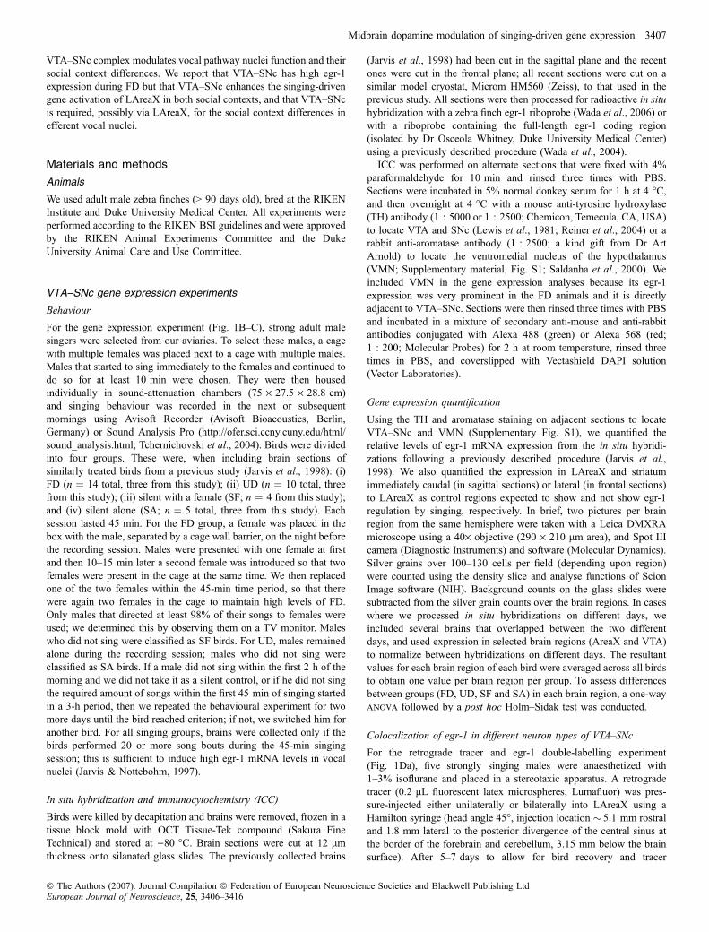
Fig. 1. Social context-dependent gene expression. (A) Schematic diagram of songbird brain showing the song system and VTA–SNc, which sends a strongdopaminergic projection to LAreaX and weaker projections to HVC and RA. Black solid arrows and yellow nuclei, vocal motor pathway; grey arrows and greennuclei, vocal pallial–basal ganglia–thalamic loop; dashed arrows, connections between the two vocal pathways. (B) Egr-1 mRNA expression (white, silver grains)in sagittal brain sections (counterstained with cresyl violet) from animals that sang similar amounts (50 and 70 motifs) of (a, c and e) UD and (b, d and f) FD song.Camera lucida drawings on the left highlight the VMN, VTA and SNc regions; there is no distinct boundary between VTA and SNc. Sections in (a)–(d) are cut in thesagittal plane, and (e) and (f) in the coronal plane. NIII, third cranial nerve. (C) Quantification of egr-1 expression. FD, n ¼ 14; UD, n ¼ 10; SF, n ¼ 4; SA, n ¼ 5.*P < 0.05, ***P < 0.0001, one-way anova, Holm–Sidak post hoc test. (D) Cell types with FD-driven egr-1 expression in VTA–SNc. Arrows, single-labelled cells;arrowheads, double-labelled cells. (a) Retrograde tracer backfilled from LAreaX to VTA neurons (red) and egr-1 mRNA expression (white silver grains). (b)Expression of TH (green-labelled cytoplasm) and egr-1 protein (red-labelled nuclei). Only one double-labelled cell can be seen in this image. (c) GABA (green-labelled cytoplasm) and egr-1 protein (red-labelled nuclei); inset in lower left shows a 3· higher magnification of a double-labelled neuron. (d) Percentage of egr-1positive neurons that were double-labelled with tracer from LAreaX (n ¼ 11 sections), TH (n ¼ 10 sections) or GABA (n ¼ 7 sections) in VTA–SNc [out of45–314 neurons across all sections in all birds (n ¼ 3–5) for each single-labelled category]. ***P < 0.0001, one-way anova, Holm–Sidak post hoc test. Error barsfor all panels represent SEM. Scale bars, 250 lm (B), 50 lm (D).
Midbrain dopamine modulation of singing-driven gene expression 3409
ª The Authors (2007). Journal Compilation ª Federation of European Neuroscience Societies and Blackwell Publishing LtdEuropean Journal of Neuroscience, 25, 3406–3416
amount, we calculated ratios of egr-1 expression in LAreaX, RA andLMAN, each relative to HVC, using a previously described approach(Jarvis et al., 1998). We used HVC as the normalizing brain regionbecause it does not show changes in egr-1 expression as a result ofsocial context (Jarvis et al., 1998) or VTA–SNc lesions (see Results).The ratio measure not only eliminates the amount of singing as avariable but it also serves as a robust internal bird and hemisphericcontrol. To determine statistical differences in ratios between groupswe used unpaired t-tests.
Song quantification for 6-OHDA-treated animals
To quantify potential lesion effects on singing behaviour wecompared singing rate, syllable similarity and sequence stereotopybefore and after the unilateral VTA–SNc lesions. To quantifysinging rate we calculated the mean number of song motifsproduced per minute in the 45-min singing sessions before and afterlesions. To measure syllable similarity, we calculated the meansimilarity score (% similarity and mean accuracy) with SoundAnalysis Pro from 10 aligned comparisons of representative songmotifs before and after lesions; the Sound Analysis Pro similarityfunction is an aggregate measure of five features: frequencymodulation, entropy, continuity, duration and pitch (Tchernichovskiet al., 2000). For the syllable similarity score, we discovered thatwe could not directly compare the similarity values of pre- andpost-lesion songs due to technical limitations of slight changes inrecording conditions (variations in microphone quality and attenu-ation box acoustics) before and after surgery. Therefore, tonormalize against this technical variable we computed the ratiosof the mean percentage syllable similarity of pre- vs. post-lesiondivided by pre- vs. pre-lesion for lesioned and sham-operated birds.To measure sequence stereotopy, we used the equations (sequencelinearity and consistency) in Scharff & Nottebohm (1991) asmodified in Foster & Bottjer (2001). First we scored the sequenceof a minimum of 30 song motifs per bird before and after surgery.We did not have presurgery song for three of the sham-treated birdsand therefore these birds were not included in the analysis. Songsequence linearity was calculated as the number of syllable typesper motif minus 1, divided by the number of different transitionsbetween syllables. A group of song motifs with no variability inpattern would produce a linearity score of 1.0. Sequence consis-tency for each pair of syllables of each motif was calculated bycalculating syllables across all song motifs as the fraction of themost common subsequent syllable transition type among alltransition types for that syllable. Then the average sequenceconsistency across all syllables was calculated for each bird. Theterminal syllable of song motifs was not included in the consistencycalculation, and introductory syllables were excluded from bothcalculations. A single stereotypy score was calculated for eachbird by averaging the linearity and consistency scores. A highlystereotyped sequence has a score close to 1.0. In order to quantifythe similarity of this measure between the two recording sessions foreach bird (e.g. pre- vs. post-lesion), we calculated the ratio of thestereotypy score before and after surgery. To determine statisticaldifferences in all three measures above we used the t-test.
Results
To determine a possible role of VTA–SNc in the modulation of vocalsystem nuclei function by social context, we performed two types ofexperiments: (i) descriptive experiments relating singing behaviourwith VTA–SNc egr-1 expression; and (ii) manipulation experiments
studying the effect that VTA–SNc lesions have on vocal nuclei egr-1expression.
VTA–SNc gene expression experiments
Social context during singing altered egr-1 expression in VTA–SNc
We first measured egr-1 mRNA expression levels in VTA–SNc aftersinging in different social contexts, using the expression changes invocal nuclei as our control measures. As expected (Jarvis et al., 1998),egr-1 mRNA expression in LAreaX was �5· higher after UD thanafter FD (Fig. 1, Ba and b, Ca). In contrast, we found that in the VTA(Fig. 1, Bc and d) as well as in the adjacent SNc (Fig. 1, Be and f), egr-1 mRNA expression was �2· higher after FD than after UD (Fig. 1,Cb). There was no consistent difference between VTA and SNc(P ¼ 0.93; average ± SEM VTA : SNc ratio, 0.95 ± 0.16, n ¼ 3 FDanimals, frontal sections), and thus we have presented our results withthe two regions combined as VTA–SNc. We also noted robustdifferences in a rostrally adjacent nucleus, the VMN, in which, as inVTA–SNc, egr-1 expression was higher after FD (Fig. 1, Bc and d,1Cc). Although we did not study the role of VMN with lesions in thisstudy, the descriptive expression results in this paragraph apply toVMN as well. The egr-1 increases in VTA–SNc (as well as VMN)were specific to FD, as the level of egr-1 expression in males thatcould see females but did not sing (SF) was similar to that of maleswho were silent alone and did not sing (SA; Fig. 1, Cb and c). Theexpression changes in VTA–SNc and LAreaX were not a result ofgeneral changes in brain gene regulation as, for example, the egr-1expression in the striatum adjacent to LAreaX was not significantlydifferent between the behavioural conditions (Fig. 1, Cd). Therelationship of VTA–SNc egr-1 expression and song production wasnot present as it was for LAreaX. In LAreaX, the level of egr-1expression was linearly proportional to the number of song motifsproduced during UD (r2 ¼ 0.795, P < 0.0001, n ¼ 10 UD and n ¼ 5SA; simple regression), as expected (Jarvis & Nottebohm, 1997),whereas in VTA–SNc there was no detectable correlation during FD(r2 ¼ 0.173, P ¼ 0.0862; n ¼ 13 FD and n ¼ 5 SA). The levels ofegr-1 expression between VTA–SNc and LAreaX were also notcorrelated (r2 ¼ 0.025 and 0.25, P ¼ 0.14 and 0.9 for UD and FD,respectively; simple regressions). Together, these results suggest thatthe high levels of egr-1 expression in VTA–SNc were stimulated by acombination of singing and courtship.
Singing-associated egr-1 was expressed in GABAergic neuronsof VTA–SNc
We next assessed which neuronal type in VTA–SNc expressed highlevels of egr-1 during FD. Zebra finch VTA–SNc is thought to containmainly two neuron types: type 1 neurons which are dopaminergic,express TH and project to the striatum, including to AreaX, and type 2neurons, which are inhibitory, presumably GABAergic, and arethought to synapse locally (Lewis et al., 1981; Gale & Perkel, 2006).In order to identify type 1 neurons, the dopaminergic projectionneurons, we retrogradely labelled them by injecting a tracer intoLAreaX and elicited FD as described in the methods. We found that< 5% of egr-1 expressing neurons in VTA–SNc were LAreaX-projecting neurons (Fig. 1, Da and d). As this low number of double-labelled egr-1 neurons in VTA–SNc could be due to the possibilitythat the localized tracer injections labelled a limited number ofdopaminergic projection neurons, we further performed double-labelICC with antibodies against egr-1 and TH. We found that only � 2.0%of the egr-1-positive neurons were TH-positive (Fig. 1, Db and d). Totest whether the remaining egr-1 cells were of type 2 neurons in VTA–
3410 E. Hara et al.
ª The Authors (2007). Journal Compilation ª Federation of European Neuroscience Societies and Blackwell Publishing LtdEuropean Journal of Neuroscience, 25, 3406–3416

SNc, we performed egr-1 double-labelling with a marker of inhibitoryinterneurons, GABA. We found that nearly half of the egr-1-positiveneurons were GABA-positive and that this was significantly more thanthe number of egr-1 neurons colocalized with the tracer or TH (Fig. 1,Dc and d). These results suggest that, during FD, egr-1 expressionis induced mainly in local inhibitory GABAergic interneurons ofVTA–SNc.
VTA–SNc lesion experiments
VTA–SNc was required for normal singing-driven egr-1 expressionin LAreaX, LMAN and RA
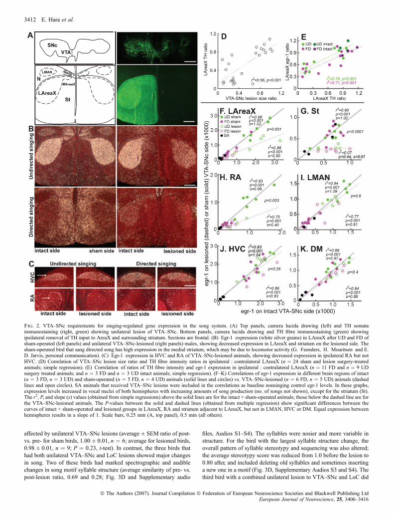
To directly test whether VTA–SNc influences the singing-regulatedegr-1 expression in LAreaX, we performed unilateral lesions using6-OHDA and compared singing-regulated egr-1 expression in theipsilateral and contralateral vocal nuclei (Fig. 2A–C). We used THstaining in VTA–SNc to assess lesion size ratios. Similar to birds thatdid not undergo surgery (n ¼ 3 FD, n ¼ 3 UD), birds that received thelow-concentration dose of 6-OHDA (n ¼ 3 FD, n ¼ 2 UD) and thosewith lesions dorsal to VTA–SNc (n ¼ 2 FD, n ¼ 2 UD) had VTA–SNc lesion size ratios at or near 1.0 (0.8–1.2; remaining unlesionedpart : intact side). The latter two groups were thus designated sham-operated controls (n ¼ 5 FD, n ¼ 4 UD of the 24 birds that underwentsurgery). The remaining birds that received the high-concentrationdose of 6-OHDA in VTA–SNc (n ¼ 6 FD, n ¼ 5 UD, n ¼ 4 SA) hadlesion size ratios that ranged from 0 to 0.7. Proportional to VTA–SNclesion size ratios was a reduction in TH ratios in the ipsilateral : con-tralateral LAreaX and surrounding striatum (Fig. 2A and D).Proportional to the reduction in the TH ratios in ipsilateral : contra-lateral LAreaX was a concomitant reduction of the egr-1 expressionratios in ipsilateral : contralateral LAreaX after both UD and FD(Fig. 2B and E). The level of the singing-driven egr-1 expression inipsilateral LAreaX was � 50% lower than in the intact contralateralhemisphere (Fig. 2F; compare the solid line of intact animals,slope ¼ 1.02, with the dashed line of the VTA–SNc-lesioned animals,slope ¼ 0.5). The level of egr-1 expression was also reduced in thesurrounding striatum (Fig. 2B and G) though, unlike in vocal nuclei,the level of striatal expression was not related to the amount of singing(r2 ¼ 0.083, P ¼ 0.217, UD; r2 ¼ 0.145, P ¼ 0.09, FD). The TH andegr-1 expression ratios in ipsilateral : contralateral LAreaX of sham-operated birds were not significantly different from birds that did notundergo surgery (P > 0.11, t-test, n ¼ 9 shams, n ¼ 4 intacts),indicating that the above results are specific to the VTA–SNc lesiongroup.
Similar to LAreaX, the VTA–SNc lesions also resulted in a 40%lower singing-driven egr-1 expression in ipsilateral song nucleus RArelative to the intact contralateral hemisphere (Fig. 2C and H). Incontrast, there was no consistent effect of VTA–SNc lesions on egr-1expression in LMAN (Fig. 2I) or HVC (Fig. 2C and J), nuclei thatproject to both LAreaX and RA (Fig. 1A). We noted that expression inmedial AreaX, which sits directly below medial MAN, may have beenaffected (Fig. 2A and B), but we did not assess this quantitatively dueto difficulty in locating these small nuclei. TH expression in RA wasslightly, but significantly, reduced by lesions of VTA–SNc (averageratio 0.89 ± 0.1, P < 0.001, paired t-test between hemispheres,n ¼ 16 lesioned animals). However, unlike in LAreaX, the THreduction in RA was not linearly proportional to the amount of egr-1reduction (r2 ¼ 0.31, P ¼ 0.4, UD; r2 ¼ )0.25, P ¼ 0.44, FD). Thereduced egr-1 in RA did not appear to reflect a gross reduction inoverall activity levels in RA during singing, as its efferent target vocalnucleus of the midbrain, the dorsal medial nucleus of the midbrain
(DM; Fig. 1A), still showed singing-driven egr-1 expression that wassimilar on the two sides of the brain (Fig. 2K).To determine whether the observed changes in egr-1 and ⁄ or TH
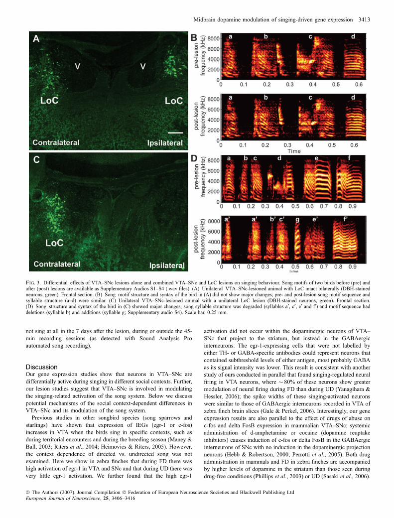
expression in LAreaX and RA were specific to the removal ofdopaminergic neurons and not to the removal by 6-OHDA of adjacentcatecholaminergic cell groups, such as the more caudally located LoC,we reacted adjacent sections with an antibody against DBH. We foundthat of the 16 VTA–SNc-lesioned birds, 10 had intact LoC (Fig. 3A),three had very small lesions (DBH intensity ratios of 0.7–0.8,lesion : intact side), and three (two UD singers and one SA) had largeLoC lesions (ratios �0.1; Fig. 3C). Unlike the VTA–SNc lesions(Fig. 2E, F and H), there was no relationship between the degree ofLoC damage and the level of egr-1 or TH expression in ipsilateralLAreaX or RA (P > 0.05, r2 ¼ 0.189–0.04).
VTA–SNc was required for social context-dependent differencesin LMAN and RA
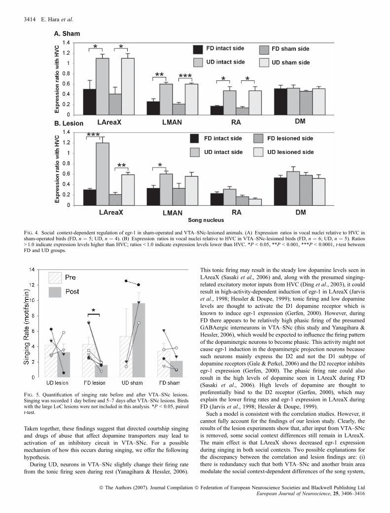
The above analyses assess relative differences between hemispheresbut do not assess either each hemisphere independently or socialcontext differences. Based upon the overlap in values of the VTA–SNc-lesioned hemispheres in LAreaX (Fig. 2F, open circles), RA(Fig. 2H) and LMAN (Fig. 2I) it is possible that social context-dependent egr-1 regulation was modified in these nuclei, but theseresults are also influenced by singing amount. To determine theeffects of social context independent of singing amount, we createdratios of egr-1 expression in LAreaX, RA and LMAN relative toHVC (see Materials and Methods). As expected (Jarvis et al., 1998),in the VTA–SNc sham-operated birds, the LAreaX : HVC,LMAN : HVC and RA : HVC egr-1 expression ratios in bothhemispheres (sham and intact) were lower after FD than after UD(Fig. 4A), and there was no social context difference in theDM : HVC ratios (Fig. 4A). In the VTA–SNc-lesioned birds,although the LAreaX : HVC expression ratio was reduced relativeto sham-lesioned birds (compare lesioned side of Fig. 4B with shamside of Fig. 4A; P ¼ 0.002, t-test), a significant social context-dependent difference remained in LAreaX between FD and UD inthe VTA–SNc-lesioned hemisphere (Fig. 4B, patterned bars). Incontrast, there was a reduction in the social context differences inLMAN of the VTA–SNc-lesioned hemisphere (Fig. 4B). Thereduction was due to a heterogeneous result of higher bilateralexpression in LMAN in some of the FD lesioned birds and lowerbilateral expression in some of the UD birds, such that the UD andFD expression values were more similar and were no longersignificantly different (Figs 2I and 4B). In RA there was anelimination of the social context differences in both the VTA–SNc-lesioned and intact hemispheres (Fig. 4B). The elimination appearedto be due to reduced egr-1 expression during UD (Fig. 4B). In DM,there were still high expression levels in both social contexts(Fig. 4B). Thus the effect of the VTA–SNc lesions on social context-dependent gene regulation appears to be specific to LMAN and RA.
Lesion effects on singing behaviour
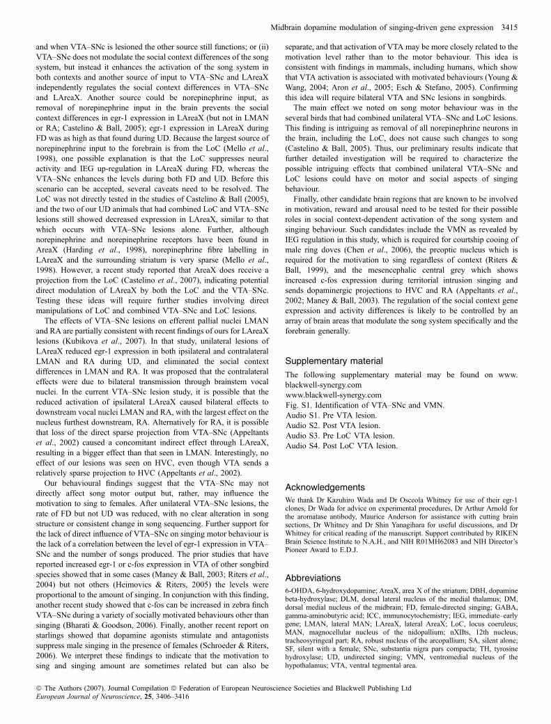
We next investigated whether the unilateral VTA–SNc lesions affectedsinging behaviour. Following unilateral lesions, the singing rate wassignificantly decreased when the males produced FD but not whenthey produced UD (Fig. 5). Relative to sham controls, songs of birdswith unilateral lesions only in VTA–SNc did not have noticeablealterations in syllable structure (average ± SEM similarity of pre- vs.post-lesion ratio for sham birds, 0.93 ± 0.01, n ¼ 6; average forVTA–SNc lesion birds, 0.93 ± 0.01, n ¼ 9; P ¼ 0.75, t-test). Therelative stereotypy of sequential syllable production was also not
Midbrain dopamine modulation of singing-driven gene expression 3411
ª The Authors (2007). Journal Compilation ª Federation of European Neuroscience Societies and Blackwell Publishing LtdEuropean Journal of Neuroscience, 25, 3406–3416
affected by unilateral VTA–SNc lesions (average ± SEM ratio of post-vs. pre- for sham birds, 1.00 ± 0.01, n ¼ 6; average for lesioned birds,0.98 ± 0.01, n ¼ 9; P ¼ 0.23, t-test). In contrast, the three birds thathad both unilateral VTA–SNc and LoC lesions showed major changesin song. Two of these birds had marked spectrographic and audiblechanges in song motif syllable structure (average similarity of pre- vs.post-lesion ratio, 0.69 and 0.28; Fig. 3D and Supplementary audio
files, Audios S1–S4). The syllables were nosier and more variable instructure. For the bird with the largest syllable structure change, theoverall pattern of syllable stereotypy and sequencing was also altered;the average stereotypy score was reduced from 1.0 before the lesion to0.80 after, and included deleting old syllables and sometimes insertinga new one in a motif (Fig. 3D, Supplementary Audios S3 and S4). Thethird bird with a combined unilateral lesion to VTA–SNc and LoC did
Fig. 2. VTA–SNc requirements for singing-regulated gene expression in the song system. (A) Top panels, camera lucida drawing (left) and TH somataimmunostaining (right, green) showing unilateral lesion of VTA–SNc. Bottom panels, camera lucida drawing and TH fibre immunostaining (green) showingipsilateral removal of TH input to AreaX and surrounding striatum. Sections are frontal. (B) Egr-1 expression (white silver grains) in LAreaX after UD and FD ofsham-operated (left panels) and unilateral VTA–SNc-lesioned (right panels) males, showing decreased expression in LAreaX and striatum on the lesioned side. Thesham-operated bird that sang directed song has high expression in the medial striatum, which may be due to locomotor activity (G. Feenders, H. Mouritsen and E.D. Jarvis, personal communication). (C) Egr-1 expression in HVC and RA of VTA–SNc-lesioned animals, showing decreased expression in ipsilateral RA but notHVC. (D) Correlation of VTA–SNc lesion size ratio and TH fibre intensity ratios in ipsilateral : contralateral LAreaX (n ¼ 24 sham and lesion surgery-treatedanimals; simple regression). (E) Correlation of ratios of TH fibre intensity and egr-1 expression in ipsilateral : contralateral LAreaX (n ¼ 11 FD and n ¼ 9 UDsurgery treated animals; and n ¼ 3 FD and n ¼ 3 UD intact animals; simple regression). (F–K) Correlations of egr-1 expression in different brain regions of intact(n ¼ 3 FD, n ¼ 3 UD) and sham-operated (n ¼ 5 FD, n ¼ 4 UD) animals (solid lines and circles) vs. VTA–SNc-lesioned (n ¼ 6 FD, n ¼ 5 UD) animals (dashedlines and open circles). SA animals that received VTA–SNc lesions were included in the correlations as baseline nonsinging control egr-1 levels. In these graphs,expression levels increased in vocal nuclei of both hemispheres with increasing amounts of song production (no. of songs not shown), except for the striatum (St).The r2, P, and slope (s) values (obtained from simple regressions) above the solid lines are for the intact + sham-operated animals; those below the dashed line are forthe VTA–SNc-lesioned animals. The P-values between the solid and dashed lines (obtained from multiple regression) show significant differences between thecurves of intact + sham-operated and lesioned groups in LAreaX, RA and striatum adjacent to LAreaX, but not in LMAN, HVC or DM. Equal expression betweenhemispheres results in a slope of 1. Scale bars, 0.25 mm (A, top panel), 0.5 mm (all others).
3412 E. Hara et al.
ª The Authors (2007). Journal Compilation ª Federation of European Neuroscience Societies and Blackwell Publishing LtdEuropean Journal of Neuroscience, 25, 3406–3416
not sing at all in the 7 days after the lesion, during or outside the 45-min recording sessions (as detected with Sound Analysis Proautomated song recording).
DiscussionOur gene expression studies show that neurons in VTA–SNc aredifferentially active during singing in different social contexts. Further,our lesion studies suggest that VTA–SNc is involved in modulatingthe singing-related activation of the song system. Below we discusspotential mechanisms of the social context-dependent differences inVTA–SNc and its modulation of the song system.
Previous studies in other songbird species (song sparrows andstarlings) have shown that expression of IEGs (egr-1 or c-fos)increases in VTA when the birds sing in specific contexts, such asduring territorial encounters and during the breeding season (Maney &Ball, 2003; Riters et al., 2004; Heimovics & Riters, 2005). However,the context dependence of directed vs. undirected song was notexamined. Here we show in zebra finches that during FD there washigh activation of egr-1 in VTA and SNc and that during UD there wasvery little egr-1 activation. We further found that the high egr-1
activation did not occur within the dopaminergic neurons of VTA–SNc that project to the striatum, but instead in the GABAergicinterneurons. The egr-1-expressing cells that were not labelled byeither TH- or GABA-specific antibodies could represent neurons thatcontained subthreshold levels of either antigen, most probably GABAas its signal intensity was lower. This result is consistent with anotherstudy of ours conducted in parallel that found singing-regulated neuralfiring in VTA neurons, where � 80% of these neurons show greatermodulation of neural firing during FD than during UD (Yanagihara &Hessler, 2006); the spike widths of these singing-activated neuronswere similar to those of GABAergic interneurons recorded in VTA ofzebra finch brain slices (Gale & Perkel, 2006). Interestingly, our geneexpression results are also parallel to the effect of drugs of abuse onc-fos and delta FosB expression in mammalian VTA–SNc; systemicadministration of d-amphetamine or cocaine (dopamine reuptakeinhibitors) causes induction of c-fos or delta FosB in the GABAergicinterneurons of SNc with no induction in the dopaminergic projectionneurons (Hebb & Robertson, 2000; Perrotti et al., 2005). Both drugadministration in mammals and FD in zebra finches are accompaniedby higher levels of dopamine in the striatum than those seen duringdrug-free conditions (Phillips et al., 2003) or UD (Sasaki et al., 2006).
Fig. 3. Differential effects of VTA–SNc lesions alone and combined VTA–SNc and LoC lesions on singing behaviour. Song motifs of two birds before (pre) andafter (post) lesions are available as Supplementary Audios S1–S4 (.wav files). (A) Unilateral VTA–SNc-lesioned animal with LoC intact bilaterally (DBH-stainedneurons, green). Frontal section. (B) Song motif structure and syntax of the bird in (A) did not show major changes; pre- and post-lesion song motif sequence andsyllable structure (a–d) were similar. (C) Unilateral VTA–SNc-lesioned animal with a unilateral LoC lesion (DBH-stained neurons, green). Frontal section.(D) Song structure and syntax of the bird in (C) showed major changes; song syllable structure was degraded (syllables a¢, c¢, e¢ and f¢) and motif sequence haddeletions (syllable b) and additions (syllable g; Supplementary audio S4). Scale bar, 0.25 mm.
Midbrain dopamine modulation of singing-driven gene expression 3413
ª The Authors (2007). Journal Compilation ª Federation of European Neuroscience Societies and Blackwell Publishing LtdEuropean Journal of Neuroscience, 25, 3406–3416
Taken together, these findings suggest that directed courtship singingand drugs of abuse that affect dopamine transporters may lead toactivation of an inhibitory circuit in VTA–SNc. For a possiblemechanism of how this occurs during singing, we offer the followinghypothesis.During UD, neurons in VTA–SNc slightly change their firing rate
from the tonic firing seen during rest (Yanagihara & Hessler, 2006).
This tonic firing may result in the steady low dopamine levels seen inLAreaX (Sasaki et al., 2006) and, along with the presumed singing-related excitatory motor inputs from HVC (Ding et al., 2003), it couldresult in high-activity-dependent induction of egr-1 in LAreaX (Jarviset al., 1998; Hessler & Doupe, 1999); tonic firing and low dopaminelevels are thought to activate the D1 dopamine receptor which isknown to induce egr-1 expression (Gerfen, 2000). However, duringFD there appears to be relatively high phasic firing of the presumedGABAergic interneurons in VTA–SNc (this study and Yanagihara &Hessler, 2006), which would be expected to influence the firing patternof the dopaminergic neurons to become phasic. This activity might notcause egr-1 induction in the dopaminergic projection neurons becausesuch neurons mainly express the D2 and not the D1 subtype ofdopamine receptors (Gale & Perkel, 2006) and the D2 receptor inhibitsegr-1 expression (Gerfen, 2000). The phasic firing rate could alsoresult in the high levels of dopamine seen in LAreaX during FD(Sasaki et al., 2006). High levels of dopamine are thought topreferentially bind to the D2 receptor (Gerfen, 2000), which mayexplain the lower firing rates and egr-1 expression in LAreaX duringFD (Jarvis et al., 1998; Hessler & Doupe, 1999).Such a model is consistent with the correlation studies. However, it
cannot fully account for the findings of our lesion study. Clearly, theresults of the lesion experiments show that, after input from VTA–SNcis removed, some social context differences still remain in LAreaX.The main effect is that LAreaX shows decreased egr-1 expressionduring singing in both social contexts. Two possible explanations forthe discrepancy between the correlation and lesion findings are: (i)there is redundancy such that both VTA–SNc and another brain areamodulate the social context-dependent differences of the song system,
Fig. 4. Social context-dependent regulation of egr-1 in sham-operated and VTA–SNc-lesioned animals. (A) Expression ratios in vocal nuclei relative to HVC insham-operated birds (FD, n ¼ 5; UD, n ¼ 4). (B) Expression ratios in vocal nuclei relative to HVC in VTA–SNc-lesioned birds (FD, n ¼ 6; UD, n ¼ 5). Ratios> 1.0 indicate expression levels higher than HVC; ratios < 1.0 indicate expression levels lower than HVC. *P < 0.05, **P < 0.001, ***P < 0.0001, t-test betweenFD and UD groups.
Fig. 5. Quantification of singing rate before and after VTA–SNc lesions.Singing was recorded 1 day before and 5–7 days after VTA–SNc lesions. Birdswith the large LoC lesions were not included in this analysis. *P < 0.05, pairedt-test.
3414 E. Hara et al.
ª The Authors (2007). Journal Compilation ª Federation of European Neuroscience Societies and Blackwell Publishing LtdEuropean Journal of Neuroscience, 25, 3406–3416
and when VTA–SNc is lesioned the other source still functions; or (ii)VTA–SNc does not modulate the social context differences of the songsystem, but instead it enhances the activation of the song system inboth contexts and another source of input to VTA–SNc and LAreaXindependently regulates the social context differences in VTA–SNcand LAreaX. Another source could be norepinephrine input, asremoval of norepinephrine input in the brain prevents the socialcontext differences in egr-1 expression in LAreaX (but not in LMANor RA; Castelino & Ball, 2005); egr-1 expression in LAreaX duringFD was as high as that found during UD. Because the largest source ofnorepinephrine input to the forebrain is from the LoC (Mello et al.,1998), one possible explanation is that the LoC suppresses neuralactivity and IEG up-regulation in LAreaX during FD, whereas theVTA–SNc enhances the levels during both FD and UD. Before thisscenario can be accepted, several caveats need to be resolved. TheLoC was not directly tested in the studies of Castelino & Ball (2005),and the two of our UD animals that had combined LoC and VTA–SNclesions still showed decreased expression in LAreaX, similar to thatwhich occurs with VTA–SNc lesions alone. Further, althoughnorepinephrine and norepinephrine receptors have been found inAreaX (Harding et al., 1998), norepinephrine fibre labelling inLAreaX and the surrounding striatum is very sparse (Mello et al.,1998). However, a recent study reported that AreaX does receive aprojection from the LoC (Castelino et al., 2007), indicating potentialdirect modulation of LAreaX by both the LoC and the VTA–SNc.Testing these ideas will require further studies involving directmanipulations of LoC and combined VTA–SNc and LoC lesions.
The effects of VTA–SNc lesions on efferent pallial nuclei LMANand RA are partially consistent with recent findings of ours for LAreaXlesions (Kubikova et al., 2007). In that study, unilateral lesions ofLAreaX reduced egr-1 expression in both ipsilateral and contralateralLMAN and RA during UD, and eliminated the social contextdifferences in LMAN and RA. It was proposed that the contralateraleffects were due to bilateral transmission through brainstem vocalnuclei. In the current VTA–SNc lesion study, it is possible that thereduced activation of ipsilateral LAreaX caused bilateral effects todownstream vocal nuclei LMAN and RA, with the largest effect on thenucleus furthest downstream, RA. Alternatively for RA, it is possiblethat loss of the direct sparse projection from VTA–SNc (Appeltantset al., 2002) caused a concomitant indirect effect through LAreaX,resulting in a bigger effect than that seen in LMAN. Interestingly, noeffect of our lesions was seen on HVC, even though VTA sends arelatively sparse projection to HVC (Appeltants et al., 2002).
Our behavioural findings suggest that the VTA–SNc may notdirectly affect song motor output but, rather, may influence themotivation to sing to females. After unilateral VTA–SNc lesions, therate of FD but not UD was reduced, with no clear alteration in songstructure or consistent change in song sequencing. Further support forthe lack of direct influence of VTA–SNc on singing motor behaviour isthe lack of a correlation between the level of egr-1 expression in VTA–SNc and the number of songs produced. The prior studies that havereported increased egr-1 or c-fos expression in VTA of other songbirdspecies showed that in some cases (Maney & Ball, 2003; Riters et al.,2004) but not others (Heimovics & Riters, 2005) the levels wereproportional to the amount of singing. In conjunction with this finding,another recent study showed that c-fos can be increased in zebra finchVTA–SNc during a variety of socially motivated behaviours other thansinging (Bharati & Goodson, 2006). Finally, another recent report onstarlings showed that dopamine agonists stimulate and antagonistssuppress male singing in the presence of females (Schroeder & Riters,2006). We interpret these findings to indicate that the motivation tosing and singing amount are sometimes related but can also be
separate, and that activation of VTA may be more closely related to themotivation level rather than to the motor behaviour. This idea isconsistent with findings in mammals, including humans, which showthat VTA activation is associated with motivated behaviours (Young &Wang, 2004; Aron et al., 2005; Esch & Stefano, 2005). Confirmingthis idea will require bilateral VTA and SNc lesions in songbirds.The main effect we noted on song motor behaviour was in the
several birds that had combined unilateral VTA–SNc and LoC lesions.This finding is intriguing as removal of all norepinephrine neurons inthe brain, including the LoC, does not cause such changes to song(Castelino & Ball, 2005). Thus, our preliminary results indicate thatfurther detailed investigation will be required to characterize thepossible intriguing effects that combined unilateral VTA–SNc andLoC lesions could have on motor and social aspects of singingbehaviour.Finally, other candidate brain regions that are known to be involved
in motivation, reward and arousal need to be tested for their possibleroles in social context-dependent activation of the song system andsinging behaviour. Such candidates include the VMN as revealed byIEG regulation in this study, which is required for courtship cooing ofmale ring doves (Chen et al., 2006), the preoptic nucleus which isrequired for the motivation to sing regardless of context (Riters &Ball, 1999), and the mesencephalic central grey which showsincreased c-fos expression during territorial intrusion singing andsends dopaminergic projections to HVC and RA (Appeltants et al.,2002; Maney & Ball, 2003). The regulation of the social context geneexpression and activity differences is likely to be controlled by anarray of brain areas that modulate the song system specifically and theforebrain generally.
Supplementary material
The following supplementary material may be found on www.blackwell-synergy.comwww.blackwell-synergy.comFig. S1. Identification of VTA–SNc and VMN.Audio S1. Pre VTA lesion.Audio S2. Post VTA lesion.Audio S3. Pre LoC VTA lesion.Audio S4. Post LoC VTA lesion.
Acknowledgements
We thank Dr Kazuhiro Wada and Dr Osceola Whitney for use of their egr-1clones, Dr Wada for advice on experimental procedures, Dr Arthur Arnold forthe aromatase antibody, Maurice Anderson for assistance with cutting brainsections, Dr Whitney and Dr Shin Yanagihara for useful discussions, and DrWhitney for critical reading of the manuscript. Support contributed by RIKENBrain Science Institute to N.A.H., and NIH R01MH62083 and NIH Director’sPioneer Award to E.D.J.
Abbreviations
6-OHDA, 6-hydroxydopamine; AreaX, area X of the striatum; DBH, dopaminebeta-hydroxylase; DLM, dorsal lateral nucleus of the medial thalamus; DM,dorsal medial nucleus of the midbrain; FD, female-directed singing; GABA,gamma-aminobutyric acid; ICC, immunocytochemistry; IEG, immediate–earlygene; LMAN, lateral MAN; LAreaX, lateral AreaX; LoC, locus coeruleus;MAN, magnocellular nucleus of the nidopallium; nXIIts, 12th nucleus,tracheosyringeal part; RA, robust nucleus of the arcopallium; SA, silent alone;SF, silent with a female; SNc, substantia nigra pars compacta; TH, tyrosinehydroxylase; UD, undirected singing; VMN, ventromedial nucleus of thehypothalamus; VTA, ventral tegmental area.
Midbrain dopamine modulation of singing-driven gene expression 3415
ª The Authors (2007). Journal Compilation ª Federation of European Neuroscience Societies and Blackwell Publishing LtdEuropean Journal of Neuroscience, 25, 3406–3416
References
Appeltants, D., Ball, G.F. & Balthazart, J. (2002) The origin of catecholami-nergic inputs to the song control nucleus RA in canaries. Neuroreport, 13,649–653.
Aron, A., Fisher, H., Mashek, D.J., Strong, G., Li, H. & Brown, L.L. (2005)Reward, motivation, and emotion systems associated with early-stage intenseromantic love. J. Neurophysiol., 94, 327–337.
Bayer, V.E. & Pickel, V.M. (1991) GABA-labeled terminals form proportion-ally more synapses with dopaminergic neurons containing low densities oftyrosine hydroxylase-immunoreactivity in rat ventral tegmental area. BrainRes., 559, 44–55.
Bharati, I.S. & Goodson, J.L. (2006) Fos responses of dopamine neurons tosociosexual stimuli in male zebra finches. Neuroscience, 143, 661–670.
Castelino, C.B. & Ball, G.F. (2005) A role for norepinephrine in the regulationof context-dependent ZENK expression in male zebra finches (Taeniopygiaguttata). Eur. J. Neurosci., 21, 1962–1972.
Castelino, C.B., Diekamp, B. & Ball, G.F. (2007) Noradrenergic projections tothe song control nucleus Area X of the medial striatum in male zebra finches(Taeniopygia guttata). J. Comp. Neurol., 502, 544–562.
Chen, G., Bonder, E.M. & Cheng, M.F. (2006) Lesion-induced neurogenesis inthe hypothalamus is involved in behavioral recovery in adult ring doves.J. Neurobiol., 66, 537–551.
Ding, L., Perkel, D.J. & Farries, M.A. (2003) Presynaptic depression ofglutamatergic synaptic transmission by D1-like dopamine receptor activationin the avian basal ganglia. J. Neurosci., 23, 6086–6095.
Esch, T. & Stefano, G.B. (2005) The neurobiology of love. Neuro. Endocrinol.Lett., 26, 175–192.
Foster, E.F. & Bottjer, S.W. (2001) Lesions of a telencephalic nucleus in malezebra finches: Influences on vocal behavior in juveniles and adults.J. Neurobiol., 46, 142–165.
Gale, S.D. & Perkel, D.J. (2006) Physiological properties of zebra finch ventraltegmental area and substantia nigra pars compacta neurons. J. Neurophysiol.,96, 2295–2306.
Gerfen, C.R. (2000) Molecular effects of dopamine on striatal-projectionpathways. Trends Neurosci., 23, S64–S70.
Harding, C.F., Barclay, S.R. & Waterman, S.A. (1998) Changes incatecholamine levels and turnover rates in hypothalamic, vocal control,and auditory nuclei in male zebra finches during development. J. Neurobiol.,34, 329–346.
Hebb, M.O. & Robertson, H.A. (2000) Identification of a subpopulation ofsubstantia nigra pars compacta gamma-aminobutyric acid neurons that isregulated by basal ganglia activity. J. Comp. Neurol., 416, 30–44.
Heimovics, S.A. & Riters, L.V. (2005) Immediate early gene activity in songcontrol nuclei and brain areas regulating motivation relates positively tosinging behavior during, but not outside of, a breeding context. J. Neurobiol.,65, 207–224.
Hessler, N.A. & Doupe, A.J. (1999) Social context modulates singing-relatedneural activity in the songbird forebrain. Nat. Neurosci., 2, 209–211.
Jarvis, E.D. (2004) Learned birdsong and the neurobiology of human language.Ann. NY Acad. Sci., 1016, 749–777.
Jarvis, E.D. & Nottebohm, F. (1997) Motor-driven gene expression. Proc. NatlAcad. Sci. USA, 94, 4097–4102.
Jarvis, E.D., Scharff, C., Grossman, M.R., Ramos, J.A. & Nottebohm, F. (1998)For whom the bird sings: context-dependent gene expression. Neuron, 21,775–788.
Kao, M.H., Doupe, A.J. & Brainard, M.S. (2005) Contributions of an avianbasal ganglia-forebrain circuit to real-time modulation of song. Nature, 433,638–643.
Kubikova, L., Turner, E.A. & Jarvis, E.D. (2007) The pallial–basal gangliapathway modulates the behaviorally-driven gene expression of the motorpathway. Eur. J. Neurosci., 25, 2145–2160.
Lewis, J.W., Ryan, S.M., Arnold, A.P. & Butcher, L.L. (1981) Evidence for acatecholaminergic projection to area X in the zebra finch. J. Comp. Neurol.,196, 347–354.
Liprando, L.A., Miner, L.H., Blakely, R.D., Lewis, D.A. & Sesack, S.R. (2004)Ultrastructural interactions between terminals expressing the norepinephrinetransporter and dopamine neurons in the rat and monkey ventral tegmentalarea. Synapse, 52, 233–244.
Maney, D.L. & Ball, G.F. (2003) Fos-like immunoreactivity in catecholami-nergic brain nuclei after territorial behavior in free-living song sparrows.J. Neurobiol., 56, 163–170.
Mello, C.V., Pinaud, R. & Ribeiro, S. (1998) Noradrenergic system of the zebrafinch brain: immunocytochemical study of dopamine-beta-hydroxylase.J. Comp. Neurol., 400, 207–228.
Mello, C.V., Vicario, D.S. & Clayton, D.F. (1992) Song presentation inducesgene expression in the songbird forebrain. Proc. Natl Acad. Sci. USA, 89,6818–6822.
Nagatsu, T. & Ichinose, H. (1999) Molecular biology of catecholamine-related enzymes in relation to Parkinson’s disease. Cell. Mol. Neurobiol.,19, 57–66.
Perrotti, L.I., Bolanos, C.A., Choi, K.H., Russo, S.J., Edwards, S., Ulery, P.G.,Wallace, D.L., Self, D.W., Nestler, E.J. & Barrot, M. (2005) DeltaFosBaccumulates in a GABAergic cell population in the posterior tail of theventral tegmental area after psychostimulant treatment. Eur. J. Neurosci., 21,2817–2824.
Phillips, P.E., Stuber, G.D., Heien, M.L., Wightman, R.M. & Carelli, R.M.(2003) Subsecond dopamine release promotes cocaine seeking. Nature, 422,614–618.
Pinaud, R., Velho, T.A., Jeong, J.K., Tremere, L.A., Leao, R.M., vonGersdorff, H. & Mello, C.V. (2004) GABAergic neurons participate in thebrain’s response to birdsong auditory stimulation. Eur. J. Neurosci., 20,1318–1330.
Reiner, A., Perkel, D.J., Bruce, L., Butler, A.B., Csillag, A., Kuenzel, W.,Medina, L., Paxinos, G., Shimizu, T., Striedter, G.F., Wild, M., Ball, G.F.,Durand, S., Gunturkun, O., Lee, D.W., Mello, C.V., Powers, A., White, S.A.,Hough, G., Kubikova, L., Smulders, T.V., Wada, K., Dugas-Ford, J.,Husband, S. & Yamamoto, K., YuJ., Siang, C. & Jarvis, E.D. (2004) Revisednomenclature for avian telencephalon and some related brainstem nuclei.J. Comp. Neurol., 473, 377–414.
Riters, L.V. & Ball, G.F. (1999) Lesions to the medial preoptic area affectsinging in the male European starling (Sturnus vulgaris). Hormones Behav.,36, 276–286.
Riters, L.V., Teague, D.P., Schroeder, M.B. & Cummings, S.E. (2004) Vocalproduction in different social contexts relates to variation in immediate earlygene immunoreactivity within and outside of the song control system. Behav.Brain Res., 155, 307–318.
Saldanha, C.J., Tuerk, M.J., Kim, Y.H., Fernandes, A.O., Arnold, A.P. &Schlinger, B.A. (2000) Distribution and regulation of telencephalicaromatase expression in the zebra finch revealed with a specific antibody.J. Comp. Neurol., 423, 619–630.
Sasaki, A., Sotnikova, T.D., Gainetdinov, R.R. & Jarvis, E.D. (2006) Socialcontext-dependent singing-regulated dopamine. J. Neurosci., 26, 9010–9014.
Scharff, C. & Nottebohm, F. (1991) A comparative study of the behavioraldeficits following lesions of various parts of the zebra finch song system:implications for vocal learning. J. Neurosci., 11, 2896–2913.
Schroeder, M.B. & Riters, L.V. (2006) Pharmacological manipulations ofdopamine and opioids have differential effects on sexually motivated song inmale European starlings. Physiol. Behav., 88, 575–584.
Sossinka, R. & Bohner, J. (1980) Song types in the zebra finch (Poephilaguttata castanotis). Z. Tierpsychol., 53, 123–132.
Tchernichovski, O., Lints, T.J., Deregnaucourt, S., Cimenser, A. & Mitra, P.P.(2004) Studying the song development process: rationale and methods. Ann.NY Acad. Sci., 1016, 348–363.
Tchernichovski, O., Nottebohm, F., Ho, C.E., Pesaran, B. & Mitra, P.P. (2000)A procedure for an automated measurement of song similarity. Anim. Behav.,59, 1167–1176.
Wada, K., Howard, J.T., McConnell, P., Whitney, O., Lints, T., Rivas, M.V.,Horita, H., Patterson, M.A., White, S.A., Scharff, C., Haesler, S., Zhao, S.,Sakaguchi, H., Hagiwara, M., Shiraki, T., Hirozane-Kishikawa, T., Skene, P.,Hayashizaki, Y., Carninci, P. & Jarvis, E.D. (2006) A molecularneuroethological approach for identifying and characterizing a cascade ofbehaviorally regulated genes. Proc. Natl Acad. Sci. USA, 103, 15212–15217.
Wada, K., Sakaguchi, H., Jarvis, E.D. & Hagiwara, M. (2004) Differentialexpression of glutamate receptors in avian neural pathways for learnedvocalization. J. Comp. Neurol., 476, 44–64.
Yanagihara, S. & Hessler, N.A. (2006) Modulation of singing-related activity inthe songbird ventral tegmental area by social context. Eur. J. Neurosci., 24,3619–3627.
Young, L.J. & Wang, Z. (2004) The neurobiology of pair bonding. Nat.Neurosci., 7, 1048–1054.
Zann, R.A. (1996) The Zebra Finch: A Synthesis of Field and LaboratoryStudies. [Chapter 10: Vocalizations.] Oxford University Press, New York, pp.196–247.
3416 E. Hara et al.
ª The Authors (2007). Journal Compilation ª Federation of European Neuroscience Societies and Blackwell Publishing LtdEuropean Journal of Neuroscience, 25, 3406–3416