Embed Size (px)
Citation preview
Role of Thylakoid ATP/ADP Carrier in Photoinhibitionand Photoprotection of Photosystem IIin Arabidopsis1[W][OA]
Lan Yin, Bjorn Lundin2, Martine Bertrand, Markus Nurmi, Katalin Solymosi, Saijaliisa Kangasjarvi,Eva-Mari Aro, Benoıt Schoefs, and Cornelia Spetea*
Division of Molecular Genetics, Department of Physics, Chemistry, and Biology, Linkoping University, 581 83Linkoping, Sweden (L.Y., B.L., C.S.); National Institute for Marine Sciences and Techniques, Cnam, 50103Cherbourg-Octeville cedex, France (M.B.); Department of Plant Anatomy, Eotvos University, 1117 Budapest,Hungary (K.S.); Department of Biochemistry and Food Chemistry, University of Turku, 20014 Turku, Finland(M.N., S.K., E.-M.A.); and UMR Plante-Microbe-Environnement, INRA-1088/CNRS-5184/Universite deBourgogne, 21065 Dijon cedex, France (B.S.)
The chloroplast thylakoid ATP/ADP carrier (TAAC) belongs to the mitochondrial carrier superfamily and supplies thethylakoid lumen with stromal ATP in exchange for ADP. Here, we investigate the physiological consequences of TAACdepletion in Arabidopsis (Arabidopsis thaliana). We show that the deficiency of TAAC in two T-DNA insertion lines does notmodify the chloroplast ultrastructure, the relative amounts of photosynthetic proteins, the pigment composition, and thephotosynthetic activity. Under growth light conditions, the mutants initially displayed similar shoot weight, but lower whenreaching full development, and were less tolerant to high light conditions in comparison with the wild type. Theseobservations prompted us to study in more detail the effects of TAAC depletion on photoinhibition and photoprotection of thephotosystem II (PSII) complex. The steady-state phosphorylation levels of PSII proteins were not affected, but the degradationof the reaction center II D1 protein was blocked, and decreased amounts of CP43-less PSII monomers were detected in themutants. Besides this, the mutant leaves displayed a transiently higher nonphotochemical quenching of chlorophyllfluorescence than the wild-type leaves, especially at low light. This may be attributed to the accumulation in the absence ofTAAC of a higher electrochemical H+ gradient in the first minutes of illumination, which more efficiently activatesphotoprotective xanthophyll cycle-dependent and independent mechanisms. Based on these results, we propose that TAACplays a critical role in the disassembly steps during PSII repair and in addition may balance the trans-thylakoid electrochemicalH+ gradient storage.
In plants, the chloroplast thylakoid membrane is thesite of light-driven photosynthetic reactions coupled toATP synthesis. There are four major protein complexesinvolved in these reactions, namely, PSI, PSII, the
cytochrome b6 f, and the H+-translocating ATP synthase(for review, see Nelson and Ben-Shem, 2004). Thephotosystems and the cytochrome b6 f complex alsocontain redox components and pigments bound toprotein subunits. Their synthesis, assembly, optimalfunction, and repair during normal development andstress require a number of transport and regulatorymechanisms. In this context, the water-oxidizing PSIIcomplex composed of more than 25 integral andperipheral proteins attracts special attention since itsreaction center D1 subunit is degraded and replacedmuch faster than the other subunits under excess andeven growth light conditions (for review, see Aro et al.,2005). Thus, the D1 protein turnover is the major eventin the repair cycle of the PSII complex and occurssubsequently to the inactivation of PSII electron trans-port. D1 degradation is most likely performed bythylakoid FtsH and Deg proteases, operating on bothsides of the thylakoid membrane (Lindahl et al., 2000;Haussuhl et al., 2001; Silva et al., 2003; Kapri-Pardeset al., 2007). The PSII repair cycle is regulated byreversible phosphorylation of several core subunits(Tikkanen et al., 2008).
1 This work was supported by grants from the Swedish ResearchCouncil, the Swedish Research Council for Environment, Agricul-ture, and Space Planning (Formas), the Graduate Research Schoolin Genomics and Bioinformatics (C.S.), the Academy of Finland (toE.-M.A.), the French Ministere de l’Education Nationale de l’En-seignement Superieur et de la Recherche, and the Institut Nationalde la Recherche Agronomique, the Centre National de la RechercheScientifique (B.S.).
2 Present address: Graduate School of Natural Science and Tech-nology, Okayama University, 700–8530 Okayama, Japan.
* Corresponding author; e-mail [email protected] author responsible for distribution of materials integral to the
findings presented in this article in accordance with the policydescribed in the Instructions for Authors (www.plantphysiol.org) is:Cornelia Spetea ([email protected]).
[W] The online version of this article contains Web-only data.[OA] Open Access articles can be viewed online without a sub-
scription.www.plantphysiol.org/cgi/doi/10.1104/pp.110.155804
666 Plant Physiology�, June 2010, Vol. 153, pp. 666–677, www.plantphysiol.org � 2010 American Society of Plant Biologists www.plantphysiol.orgon April 4, 2018 - Published by Downloaded from
Copyright © 2010 American Society of Plant Biologists. All rights reserved.
ATP is produced as a result of the light-drivenphotosynthetic reactions in the thylakoid membraneand mainly is utilized in the carbon fixation reactionsoccurring in the soluble stroma. Besides this, ATP alsodrives several energy-dependent processes occurringon the stromal side of the thylakoid membrane, in-cluding phosphorylation, folding, import, and deg-radation of proteins. Furthermore, experimentalevidence for ATP transport across the thylakoid mem-brane and nucleotide metabolism inside the lumenalspace has been reported (Spetea et al., 2004; for review,see Spetea and Thuswaldner, 2008; Spetea and Schoefs,2010). The protein responsible for the thylakoid ATPtransport activity has been identified in Arabidopsis(Arabidopsis thaliana) as the product of the At5g01500gene and functionally characterized in Escherichia colias an ATP/ADP exchanger (Thuswaldner et al., 2007).This protein is homologous to the extensively studiedbovine mitochondrial ADP/ATP carrier and thereforehas been named thylakoid ATP/ADP carrier (TAAC).In the same report, it has been demonstrated thatTAAC transports ATP from stroma to lumen in ex-change for ADP, as based on radioactive assays us-ing thylakoids isolated from Arabidopsis wild-typeplants and a T-DNA insertion knockout line (namedtaac). Furthermore, TAAC was shown to be mainlyexpressed in photosynthetic tissues with an up-regulation during greening, senescence, and stress(e.g. high light) conditions, implying a physiologicalrole during thylakoid biogenesis and turnover.The ATP translocated by TAAC across the thylakoid
membrane is converted to GTP by the lumenal nucle-oside diphosphate kinase III; GTP can then be boundand hydrolyzed to GDP and inorganic phosphate bythe PsbO protein, a lumenal extrinsic subunit of thePSII complex (Spetea et al., 2004; Lundin et al., 2007a).The anion transporter 1 from Arabidopsis has beenproposed to export to the stroma the phosphate gen-erated during nucleotide metabolism in the thylakoidlumen (Ruiz Pavon et al., 2008). Between the two PsbOisoforms in Arabidopsis, it has recently been reportedthat PsbO2 plays an essential role in D1 proteinturnover during high light stress and that it has ahigher GTPase activity than PsbO1 (Lundin et al.,2007b, 2008; Allahverdiyeva et al., 2009). The precisemechanism of PsbO2-mediated PSII repair is notknown. Nevertheless, the requirement of GTP forefficient proteolytic removal of the D1 protein duringrepair of photoinactivated PSII was previously re-ported (Spetea et al., 1999). Furthermore, it has beenproposed that the PsbO2 type of PSII complexes un-dergo more efficient repair. This has been attributed tothe PsbO2-mediated GTPase activity that inducesPsbO2 release from the complex, thus facilitating thenext steps in the repair process, namely, dissociation ofthe CP43 subunit and proteolysis of the D1 subunit(Lundin et al., 2007b, 2008).TAAC may represent the missing link between ATP
synthesis on the stromal side of the thylakoid mem-brane and nucleotide-dependent reactions in the lu-
menal space. The taac mutant provides an interestingtool to study whether there are any regulatory net-works between the activity of TAAC and PSII repair.Based on phenotypic characterization of two differentT-DNA insertion lines of the TAAC gene, we report inthis article that the PSII repair cycle is malfunctioningin the absence of TAAC and that the thermal photo-protection is faster activated during light stress.
RESULTS
PSII Activity under Growth Light Conditions
The taac knockout mutant, initially characterized byThuswaldner et al. (2007), was obtained from theInstitut National de la Recherche Agronomique Public-Lines collection (FLAG_443D03, Wassilewskija [Ws]ecotype) and is shown here to contain a single T-DNAinsertion (Supplemental Fig. S1). A second homozy-gous taac mutant identified from Syngenta T-DNAinsertion lines collection (SAIL_209_A12, Columbiaecotype) is described in Supplemental Figure S2.
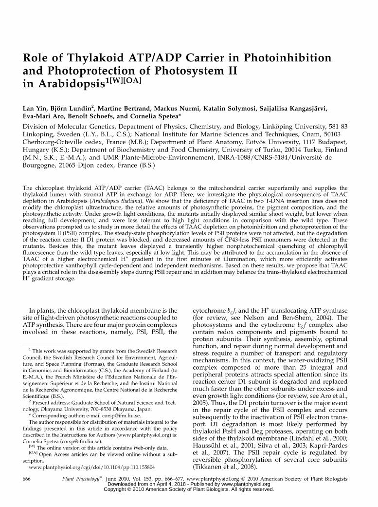
At an irradiance of 120 mmol photons m22 s21 (GL),the taac mutants grew initially with the same rate asthe wild-type plants of the respective ecotype, but thenslower and had significantly less (20%) leaf biomasscompared to the wild type (Fig. 1, A and B; Supple-mental Fig. S3, A and B). When examined with elec-tron microscopy, structurally normal chloroplastscontaining starch grains and thylakoid membraneswith typical stacked grana were found in all types ofplants (Fig. 1C; Supplemental Fig. S3C). Plastid lengthand width ranged in all samples between 5 to 6 and2 to 3 mm, respectively. The studied plastid profilescontained 58 to 61 grana on average, and there was nolarge difference between taac and wild-type plants inthis respect. Similarly, no large difference was found inheight (number of appressed thylakoid lamellae pergranum) or length of the grana between the mutantsand their respective ecotypes. This indicates that de-pletion of TAAC does not influence chloroplast andthylakoid ultrastructure under average, nonstressfulconditions.
The chlorophyll (Chl) content of whole leaves (ex-pressed per leaf fresh weight or per leaf surface unit)and Chl a/b ratio were not significantly affected by thedeficiency of TAAC in the mutant compared to thewild type (Table I; Supplemental Table S1). The max-imum quantum yield of PSII photochemistry was thesame in wild-type and taac leaves (Fv/Fm = 0.8). Similarsteady-state rates of oxygen evolution were measuredin thylakoid membranes in the presence of the PSIIacceptor phenyl-p-benzoquinone. Nevertheless, addi-tion of NH4Cl has resulted in significantly higher ratesof oxygen evolution in the mutant compared with thewild type. The observed difference is consistent withthe existence in the mutant of an overall higher ca-pacity for PSII electron transport per unit Chl. Astimulatory effect of NH4Cl on the electron transport
Role of Thylakoid ATP/ADP Carrier in Arabidopsis
Plant Physiol. Vol. 153, 2010 667 www.plantphysiol.orgon April 4, 2018 - Published by Downloaded from
Copyright © 2010 American Society of Plant Biologists. All rights reserved.
in isolated thylakoid membranes has previously beenattributed to an increased dissipation of H+ gradientacross thylakoids and uncoupled photophosphoryla-tion (McCarty, 1980; Thomasset et al., 1984). Therefore,our results may indicate a higher H+ gradient existingacross the thylakoid membrane in the mutant com-pared to the wild type. The activities of the whole
linear electron transport chain were indistinguishable,based on the rates of oxygen consumption in thepresence of methyl viologen (and sodium azide; TableI; Supplemental Table S1). Furthermore, no significantdifferences were found in the rates of cyclic electrontransport based on the calculated halftime (t1/2) for theP700
+ re-reduction in darkness (Table I; SupplementalTable S1). Finally, the rates of CO2 fixation were similarin taac and wild-type leaves (Supplemental Fig. S4)and are therefore not able to explain the reducedbiomass of the mutant.
The leaves of wild-type and taac plants exhibited atypical O-J-I-P fluorescence induction curve (Supple-mental Fig. S5; Strasser et al., 1996; Tsimilli-Michaelet al., 2000). The shape of the curve, the valuesobtained for the relative variable fluorescence at theJ-step (FJ) between 0.36 and 0.47, and the initial slope(M0) between 0.78 and 1.26 are typical for healthyplants (Tsimilli-Michael et al., 2000). The analysis ofthe O-J-I-P curves allows the determination of the fateof the absorbed energy (parameter ABS): part of theexcitation energy is dissipated in the antenna of thelight-harvesting complex II (LHCII; parameter DI0) asheat and/or fluorescence, and the remaining is chan-neled to the reaction centers (parameter TR0). There,the energy is converted to redox energy by reducingthe first stable quinone electron acceptor QA, while thereaction center (RC) Chl P680 is oxidized, thus creatingan electron transport (parameter ET0) within the pho-tosynthetic apparatus (Supplemental Fig. S6). Table IIcompares the PSII behavior in wild-type and taacleaves using energy fluxes expressed per RC of thesample. The ABS/RC parameter was shown to be ameasure of the antenna size (Strasser et al., 1996). Theobtained similar values in taac and the wild type arein line with the absence of significant difference inthe Chl a/b ratio (Table I) and the only slight modi-fication in the Chl and xanthophyll levels (see be-low). Furthermore, calculations of the flux of energyreaching the RCII (TR0) and of the flux of energydissipated as heat and/or fluorescence in the an-tenna, when the first excitons closed all the RCII (DI0)indicated no significant differences between the twotypes of plants (Table II). Taken together, these dataindicate a similar distribution of energy fluxes at PSIIlevel.
Figure 1. Comparison of the taac mutant with wild-type Arabidopsisplants of the same Ws ecotype. A, The photographs of representativeplants were taken at an age of 42 d of growth using a hydroponic systemat an irradiance of 120 mmol photons m22 s21. B, Plot of shoot weight6SD as a function of plant age (n = 10). The difference in the weight issignificant at 6 and 8 weeks of growth (Student’s t test P , 0.05). C,Transmission electron microscopy images, showing chloroplast ultra-structure, were recorded at an age of 28 d of growth. Bar = 2 mm.
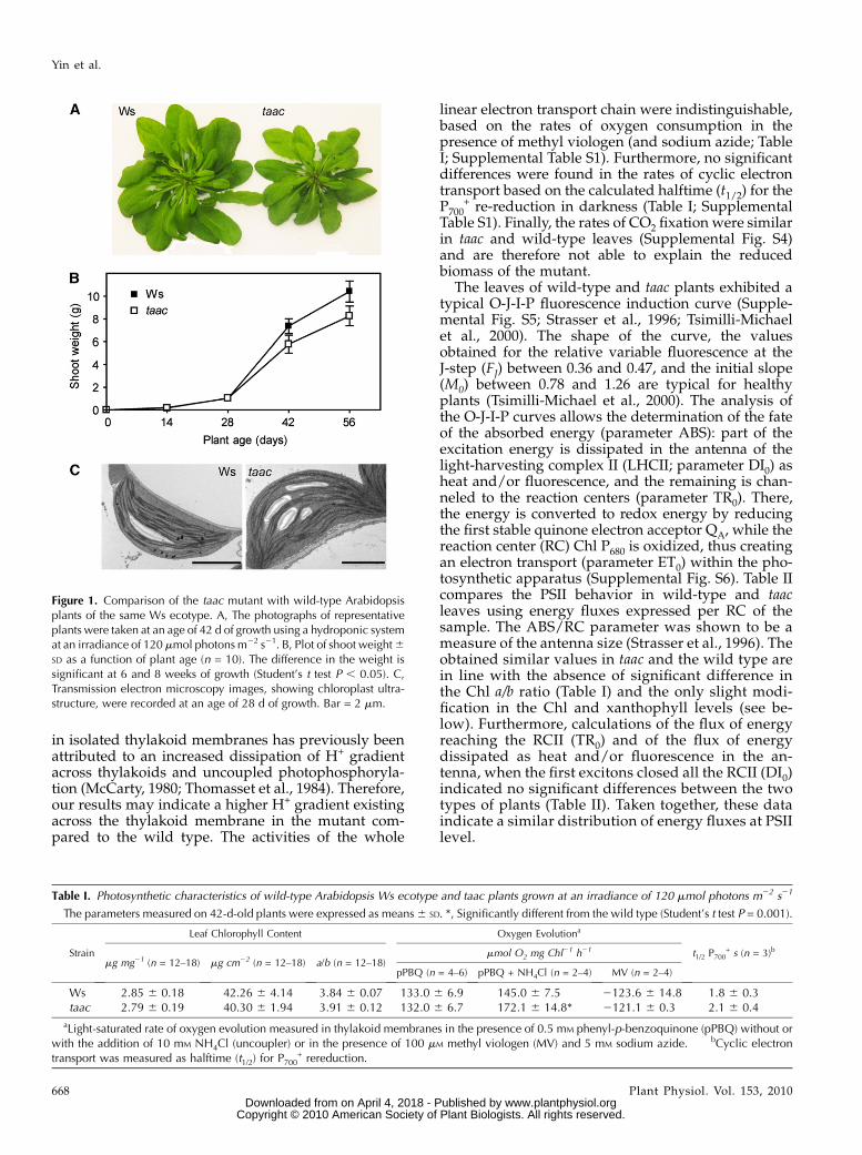
Table I. Photosynthetic characteristics of wild-type Arabidopsis Ws ecotype and taac plants grown at an irradiance of 120 mmol photons m22 s21
The parameters measured on 42-d-old plants were expressed as means6 SD. *, Significantly different from the wild type (Student’s t test P = 0.001).
Strain
Leaf Chlorophyll Content Oxygen Evolutiona
t1/2 P700+ s (n = 3)b
mg mg21 (n = 12–18) mg cm22 (n = 12–18) a/b (n = 12–18)mmol O2 mg Chl21 h21
pPBQ (n = 4–6) pPBQ + NH4Cl (n = 2–4) MV (n = 2–4)
Ws 2.85 6 0.18 42.26 6 4.14 3.84 6 0.07 133.0 6 6.9 145.0 6 7.5 2123.6 6 14.8 1.8 6 0.3taac 2.79 6 0.19 40.30 6 1.94 3.91 6 0.12 132.0 6 6.7 172.1 6 14.8* 2121.1 6 0.3 2.1 6 0.4
aLight-saturated rate of oxygen evolution measured in thylakoid membranes in the presence of 0.5 mM phenyl-p-benzoquinone (pPBQ) without orwith the addition of 10 mM NH4Cl (uncoupler) or in the presence of 100 mM methyl viologen (MV) and 5 mM sodium azide. bCyclic electrontransport was measured as halftime (t1/2) for P700
+ rereduction.
Yin et al.
668 Plant Physiol. Vol. 153, 2010 www.plantphysiol.orgon April 4, 2018 - Published by Downloaded from
Copyright © 2010 American Society of Plant Biologists. All rights reserved.
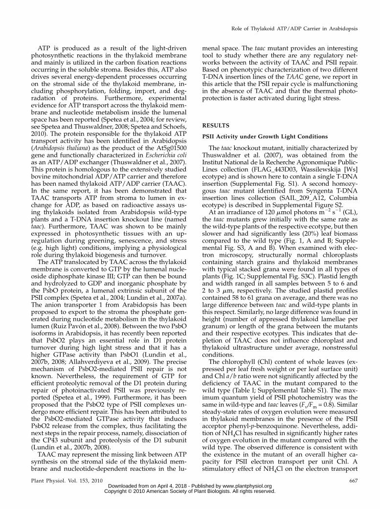
Nonphotochemical quenching of Chl fluorescence(NPQ) is an important photoprotective mechanismminimizing the photooxidative damage to the photo-synthetic apparatus (for review, see Horton et al.,2008). To test whether the deficiency of TAAC affectsphotoprotection, the kinetics of NPQ formation wererecorded for 20 min at photosynthetic active radiation(PAR) of 300, 600, and 1,250 mmol m22 s21 in the twotaac mutants and the respective background plants(Fig. 2; Supplemental Fig. S3C). The initial phase wastransient with NPQ reaching a maximum 120 to 200 safter the first saturating pulse, as described by D’Haeseet al. (2004). Nevertheless, the amplitude of this phaseand the rate of NPQ formation were higher in taacmutants at the three tested light intensities. The secondphase during the remaining illumination period of 20min was slow and displayed no significant differencesin the levels of NPQ between the mutants and therespective wild-type ecotype. The induced NPQ com-ponent was rapidly reversible and decayed during thedark period with similar kinetics and extent in thewild-type and taac plants (Fig. 2). Taken together, theseresults indicate that the mutants are more efficient inactivating the formation of NPQ. Although of differentecotypes, the SAIL_209_A12 mutant showed pheno-types identical with FLAG_443D03 (Supplemental Fig.S3), chosen for detailed physiological and biochemicalcharacterization (see below).
PSII Activity under High Light Conditions
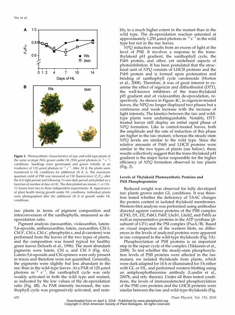
The wild-type Ws and FLAG 443D03 taac plantswere grown under GL conditions for 28 d and thenunder high light (HL; 950 mmol photons m22 s21)conditions for additional 28 d, maintaining the diurnalcycle. Leaf samples were collected in the course oftheir growth to measure the maximum quantum yieldof PSII photochemistry (Fv/Fm). Similar values (0.8)during day 0 of HL growth, followed by a slowdecrease until day 4 (0.72), were measured in bothtypes of plants (Fig. 3A). This parameter continued todecrease until day 7 in the mutant (0.62), whereas atthe same time point the wild-type plants almost fullyrestored their PSII activity (0.78). The mutant fully
restored the activity only on day 14 (0.8). Taken to-gether, the pattern observed for Fv/Fm indicates anenhanced susceptibility to high light for the mutantcompared to the wild type. The slower recovery of PSIIactivity was at the expense of plant growth since themutant displayed poor health, as shown in Figure 3Bphotos, taken after 28 d of HL conditions.
Leaf Pigment Analysis and Xanthophyll Cycle
The amplitude of the initial rapid phase of NPQformation depends on the accumulation of H+ in thethylakoid lumen and on the pigment and proteincomposition of the antenna (Horton et al., 2008).Acidification of the lumen leads to activation ofviolaxanthin de-epoxidase, which converts violaxan-thin to anteraxanthin and finally to zeaxanthin as partof the xanthophyll cycle (for review, see Hieber et al.,2000; Horton et al., 2008). To investigate the mecha-nism behind the faster response of the mutant when itcomes to NPQ (Fig. 2), we analyzed the wild-type and
Table II. Comparison of energy fluxes in the wild-type (Ws) andtaac plants
Fast kinetics of fluorescence induction were recorded, and variousenergy fluxes were calculated per RC, as described in SupplementalMaterials and Methods S1. ABS, absorption; TR0, trapping; DI0,dissipation; ET0, electron transport. Experiments are means 6 SD of10 to 11 replicates. No significant differences were found between thetwo types of plants (Student’s t test P $ 0.07).
Energy Flux Ws taac
ABS/RC 2.815 6 0.065 2.607 6 0.074TR0/RC 2.198 6 0.270 2.125 6 0.520DI0/RC 0.624 6 0.192 0.483 6 0.024ET0/RC 1.182 6 0.044 1.182 6 0.097
Figure 2. Kinetics of NPQ formation. Chlorophyll fluorescence ofleaves detached from 16-h dark-adapted wild-type (Ws) and taac plantswas recorded during a 20-min exposure to light (white bar) of 300, 600,or 1,250 mmol photons m22 s21, followed by 15-min recovery indarkness (black bar). Saturating pulses (1.0 s) of actinic light wereapplied to determine the maximum fluorescence yield Fm or Fm’. NPQwas calculated from fluorescence data as (Fm 2 Fm’)/Fm’ and plotted 6SD as a function of illumination time (n = 7–10).
Role of Thylakoid ATP/ADP Carrier in Arabidopsis
Plant Physiol. Vol. 153, 2010 669 www.plantphysiol.orgon April 4, 2018 - Published by Downloaded from
Copyright © 2010 American Society of Plant Biologists. All rights reserved.
taac plants in terms of pigment composition andinterconversion of the xanthophylls, measured as de-epoxidation ratio.
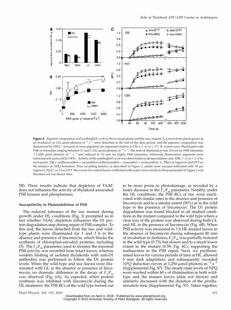
Pigment analysis (neoxanthin, violaxanthin, lutein-5,6-epoxide, antheraxanthin, lutein, zeaxanthin, Chl b,Chl b’, Chl a, Chl a’, pheophytin a, and b-carotene) wasperformed from the leaves of the two types of plants,and the composition was found typical for healthygreen leaves (Schoefs et al., 1996). The most abundantpigments were lutein, Chl a, and Chl b (Fig. 4A).Lutein-5,6-epoxide and Chl epimers were only presentin traces and therefore were not quantified. Generally,the pigments were slightly but less abundant in thetaac than in the wild-type leaves. At a PAR of 120 mmolphotons m22 s21, the xanthophyll cycle was onlyweakly activated in both the wild type and mutant,as indicated by the low values of the de-epoxidationratio (Fig. 4B). As PAR intensity increased, the xan-thophyll cycle was progressively activated, and nota-
bly, to a much higher extent in the mutant than in thewild type. The de-epoxidation reaction saturated atapproximately 1,250 mmol photons m22 s21 in the wildtype but not in the taac leaves.
NPQ induction results from an excess of light at thelevel of PSII. It involves a response to the trans-thylakoid pH gradient, the xanthophyll cycle, thePsbS protein, and other, yet undefined aspects ofphotoinhibition. It has been postulated that the struc-tural unit of NPQ consists of LHCII proteins and thePsbS protein and is formed upon protonation andbinding of xanthophyll cycle carotenoids (Hortonet al., 2008). Therefore, it was of great interest to ex-amine the effect of nigericin and dithiothreitol (DTT),the well-known inhibitors of the trans-thylakoidpH gradient and of violaxanthin de-epoxidation, re-spectively. As shown in Figure 4C, in nigericin-treatedleaves, the NPQ no longer displayed two phases but acontinuous and weak increase with the increase oflight intensity. The kinetics between the taac and wild-type plants were undistinguishable. Notably, DTT-treated leaves still display an initial rapid phase ofNPQ formation. Like in control-treated leaves, boththe amplitude and the rate of induction of this phaseare higher in the taac mutant, whereas the steady-stateNPQ levels are similar to the wild type. Since therelative amounts of PsbS and LHCII proteins weresimilar in the two types of plants (see below), theseresults collectively suggest that the trans-thylakoid pHgradient is the major factor responsible for the higherefficiency of NPQ formation observed in taac plants(Fig. 2).
Levels of Thylakoid Photosynthetic Proteins and
PSII Phosphoproteins
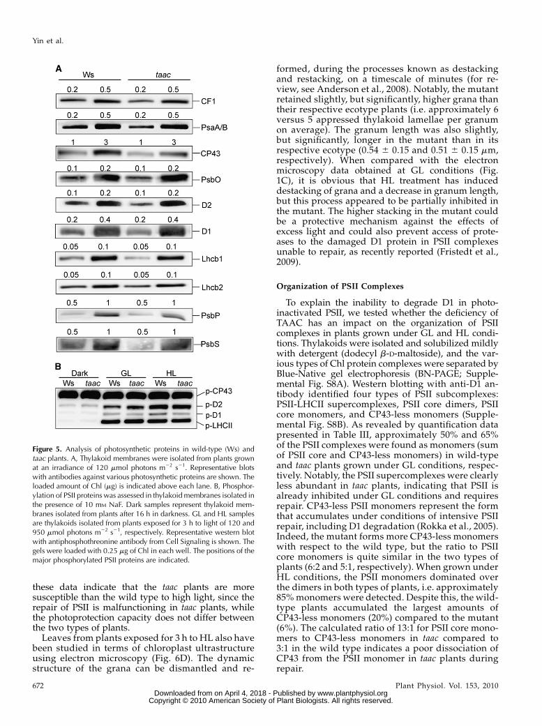
Reduced weight was observed for fully developedtaac plants grown under GL conditions. It was there-fore tested whether the deficiency of TAAC changesthe protein content in isolated thylakoid membranes.Western-blot analysis was performed using antibodiesdirected against various proteins of the PSII complex(CP43, D1, D2, PsbO, PsbP, Lhcb1, Lhcb2, and PsbS) aswell as representative proteins in the ATP synthase (b-subunit of CF1) and the PSI complex (PsaA/B). Basedon visual inspection of the western blots, no differ-ences in the levels of analyzed proteins were apparentin taac compared to the wild-type thylakoids (Fig. 5A).
Phosphorylation of PSII proteins is an importantstep in the repair cycle of the complex (Tikkanen et al.,2008). To test whether the steady-state phosphoryla-tion levels of PSII proteins were affected in the taacmutant, we isolated thylakoids from plants, whichwere dark adapted for 16 h or illuminated for 3 h eitherwith GL or HL, and performed western blotting usingan antiphosphothreonine antibody (Lundin et al.,2007b, and refs. therein). Under all three tested condi-tions, the levels of immunodetected phosphorylationof the PSII core proteins and the LHCII proteins weresimilar between the taac andwild-type thylakoids (Fig.
Figure 3. Photosynthetic characteristics of taac and wild-type plants ofthe same ecotype (Ws) grown under HL (950 mmol photons m22 s21)conditions. Seedlings were germinated and grown initially at anirradiance of 120 mmol photons m22 s21. After 28 d, the plants weretransferred in HL conditions for additional 28 d. A, The maximumquantum yield of PSII was measured as Chl fluorescence (Fv/Fm) afterthe 8-h light period and following 15-min dark period and plotted as afunction of number of days in HL. The data plotted are means6 SD (10–12 leaves from two to three independent experiments). B, Appearanceof plant health during growth under HL conditions. Individual plantswere photographed after the additional 28 d of growth under HLconditions.
Yin et al.
670 Plant Physiol. Vol. 153, 2010 www.plantphysiol.orgon April 4, 2018 - Published by Downloaded from
Copyright © 2010 American Society of Plant Biologists. All rights reserved.
5B). These results indicate that depletion of TAACdoes not influence the activity of thylakoid-associatedPSII kinases and phosphatases.
Susceptibility to Photoinhibition of PSII
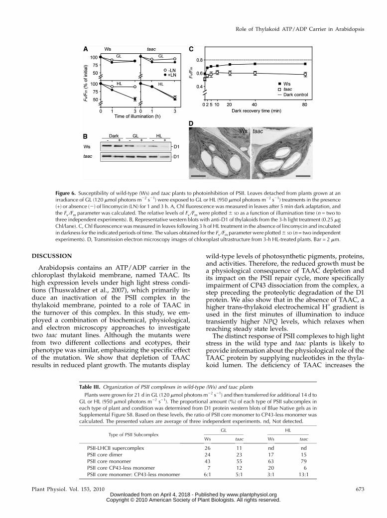
The reduced tolerance of the taac mutant duringgrowth under HL conditions (Fig. 3) prompted us totest whether TAAC depletion influences the D1 pro-tein degradation step during repair of PSII complex. Tothis end, the leaves detached from the taac and wild-type plants were illuminated for 1 and 3 h in theabsence and presence of lincomycin, which blocks thesynthesis of chloroplast-encoded proteins, includingD1. The Fv/Fm parameter, used to monitor the maximalPSII activity, was recorded from intact leaves, whereaswestern blotting of isolated thylakoids with anti-D1antibodies was performed to follow the D1 proteinlevels. When the wild-type and taac leaves were illu-minated with GL in the absence or presence of linco-mycin, no dramatic difference in the decay of Fv/Fmwas observed (Fig. 6A). As expected, when proteinsynthesis was inhibited with lincomycin during theHL treatment, the PSII RCs of the wild type turned out
to be more prone to photodamage, as revealed by afaster decrease in the Fv/Fm parameter. Notably, underthe HL conditions, the PSII RCs of taac were inacti-vated with similar rates in the absence and presence oflincomycin and to a similar extent (50%) as in the wildtype in the presence of lincomycin. The D1 proteindegradation was found blocked in all studied condi-tions in the mutant compared to the wild type where aclear loss of the protein was observed during both GLand HL in the presence of lincomycin (Fig. 6B). WhenPSII activity was measured in 3 h HL-treated leaves inthe absence of lincomycin during subsequent 80 minof incubation in darkness, Fv/Fm was partially restoredin the wild type (0.73), but slower and to a much lowerextent in the mutant (0.59; Fig. 6C), supporting themalfunction in the PSII repair. Next, we preillumi-nated leaves for various periods of time at HL, allowed5 min dark adaptation, and subsequently recordedNPQ induction curves at 1,250 mmol photons m22 s21
(Supplemental Fig. S7). The steady-state levels of NPQwere reached within 60 s of illumination in both wild-type and the mutant leaves (data not shown) andsimilarly decreased with the duration of the preillu-mination time (Supplemental Fig. S7). Taken together,
Figure 4. Pigment composition and xanthophyll cycle in Ws ecotype plants and the taacmutant. A, Leaves from plants grown atan irradiance of 120 mmol photons m22 s21 were detached at the end of the dark period, and the pigment composition wasdetermined by HPLC. Amounts of main pigments are expressed relative to Chl a 6 SD (n = 17). B, Leaves were illuminated withPAR of intensities ranging between 53 and 1,952 mmol photons m22 s21. The time of illumination was 20 min for PAR intensities,1,000 mmol photons m22 s21 and reduced to 10 min for higher PAR intensities. Following illumination, pigments wereextracted and analyzed by HPLC. Activity of the xanthophyll cycle was determined as de-epoxidation ratio (DR)6 SD (n = 3–4 foreach point). DR = (antheraxanthin + zeaxanthin)/(antheraxanthin + zeaxanthin + violaxanthin). C, Effect of nigericin and DTTonthe kinetics of NPQ formation. Prior recording kinetics as described in Figure 2, plants were vacuum infiltrated with 50 mM
nigericin (NGC) or 5 mM DTT. The curves for control leaves (infiltrated with water) were similar to those presented in Figure 2 andtherefore are not shown here.
Role of Thylakoid ATP/ADP Carrier in Arabidopsis
Plant Physiol. Vol. 153, 2010 671 www.plantphysiol.orgon April 4, 2018 - Published by Downloaded from
Copyright © 2010 American Society of Plant Biologists. All rights reserved.
these data indicate that the taac plants are moresusceptible than the wild type to high light, since therepair of PSII is malfunctioning in taac plants, whilethe photoprotection capacity does not differ betweenthe two types of plants.
Leaves from plants exposed for 3 h to HL also havebeen studied in terms of chloroplast ultrastructureusing electron microscopy (Fig. 6D). The dynamicstructure of the grana can be dismantled and re-
formed, during the processes known as destackingand restacking, on a timescale of minutes (for re-view, see Anderson et al., 2008). Notably, the mutantretained slightly, but significantly, higher grana thantheir respective ecotype plants (i.e. approximately 6versus 5 appressed thylakoid lamellae per granumon average). The granum length was also slightly,but significantly, longer in the mutant than in itsrespective ecotype (0.54 6 0.15 and 0.51 6 0.15 mm,respectively). When compared with the electronmicroscopy data obtained at GL conditions (Fig.1C), it is obvious that HL treatment has induceddestacking of grana and a decrease in granum length,but this process appeared to be partially inhibited inthe mutant. The higher stacking in the mutant couldbe a protective mechanism against the effects ofexcess light and could also prevent access of prote-ases to the damaged D1 protein in PSII complexesunable to repair, as recently reported (Fristedt et al.,2009).
Organization of PSII Complexes
To explain the inability to degrade D1 in photo-inactivated PSII, we tested whether the deficiency ofTAAC has an impact on the organization of PSIIcomplexes in plants grown under GL and HL condi-tions. Thylakoids were isolated and solubilized mildlywith detergent (dodecyl b-D-maltoside), and the var-ious types of Chl protein complexes were separated byBlue-Native gel electrophoresis (BN-PAGE; Supple-mental Fig. S8A). Western blotting with anti-D1 an-tibody identified four types of PSII subcomplexes:PSII-LHCII supercomplexes, PSII core dimers, PSIIcore monomers, and CP43-less monomers (Supple-mental Fig. S8B). As revealed by quantification datapresented in Table III, approximately 50% and 65%of the PSII complexes were found as monomers (sumof PSII core and CP43-less monomers) in wild-typeand taac plants grown under GL conditions, respec-tively. Notably, the PSII supercomplexes were clearlyless abundant in taac plants, indicating that PSII isalready inhibited under GL conditions and requiresrepair. CP43-less PSII monomers represent the formthat accumulates under conditions of intensive PSIIrepair, including D1 degradation (Rokka et al., 2005).Indeed, the mutant forms more CP43-less monomerswith respect to the wild type, but the ratio to PSIIcore monomers is quite similar in the two types ofplants (6:2 and 5:1, respectively). When grown underHL conditions, the PSII monomers dominated overthe dimers in both types of plants, i.e. approximately85%monomers were detected. Despite this, the wild-type plants accumulated the largest amounts ofCP43-less monomers (20%) compared to the mutant(6%). The calculated ratio of 13:1 for PSII core mono-mers to CP43-less monomers in taac compared to3:1 in the wild type indicates a poor dissociation ofCP43 from the PSII monomer in taac plants duringrepair.
Figure 5. Analysis of photosynthetic proteins in wild-type (Ws) andtaac plants. A, Thylakoid membranes were isolated from plants grownat an irradiance of 120 mmol photons m22 s21. Representative blotswith antibodies against various photosynthetic proteins are shown. Theloaded amount of Chl (mg) is indicated above each lane. B, Phosphor-ylation of PSII proteins was assessed in thylakoid membranes isolated inthe presence of 10 mM NaF. Dark samples represent thylakoid mem-branes isolated from plants after 16 h in darkness. GL and HL samplesare thylakoids isolated from plants exposed for 3 h to light of 120 and950 mmol photons m22 s21, respectively. Representative western blotwith antiphosphothreonine antibody from Cell Signaling is shown. Thegels were loaded with 0.25 mg of Chl in each well. The positions of themajor phosphorylated PSII proteins are indicated.
Yin et al.
672 Plant Physiol. Vol. 153, 2010 www.plantphysiol.orgon April 4, 2018 - Published by Downloaded from
Copyright © 2010 American Society of Plant Biologists. All rights reserved.
DISCUSSION
Arabidopsis contains an ATP/ADP carrier in thechloroplast thylakoid membrane, named TAAC. Itshigh expression levels under high light stress condi-tions (Thuswaldner et al., 2007), which primarily in-duce an inactivation of the PSII complex in thethylakoid membrane, pointed to a role of TAAC inthe turnover of this complex. In this study, we em-ployed a combination of biochemical, physiological,and electron microscopy approaches to investigatetwo taac mutant lines. Although the mutants werefrom two different collections and ecotypes, theirphenotype was similar, emphasizing the specific effectof the mutation. We show that depletion of TAACresults in reduced plant growth. The mutants display
wild-type levels of photosynthetic pigments, proteins,and activities. Therefore, the reduced growth must bea physiological consequence of TAAC depletion andits impact on the PSII repair cycle, more specificallyimpairment of CP43 dissociation from the complex, astep preceding the proteolytic degradation of the D1protein. We also show that in the absence of TAAC, ahigher trans-thylakoid electrochemical H+ gradient isused in the first minutes of illumination to inducetransiently higher NPQ levels, which relaxes whenreaching steady state levels.
The distinct response of PSII complexes to high lightstress in the wild type and taac plants is likely toprovide information about the physiological role of theTAAC protein by supplying nucleotides in the thyla-koid lumen. The deficiency of TAAC increases the
Figure 6. Susceptibility of wild-type (Ws) and taac plants to photoinhibition of PSII. Leaves detached from plants grown at anirradiance of GL (120 mmol photons m22 s21) were exposed to GL or HL (950 mmol photons m22 s21) treatments in the presence(+) or absence (2) of lincomycin (LN) for 1 and 3 h. A, Chl fluorescence was measured in leaves after 5 min dark adaptation, andthe Fv /Fm parameter was calculated. The relative levels of Fv /Fm were plotted6 SD as a function of illumination time (n = two tothree independent experiments). B, Representative western blots with anti-D1 of thylakoids from the 3-h light treatment (0.25 mgChl/lane). C, Chl fluorescence was measured in leaves following 3 h of HL treatment in the absence of lincomycin and incubatedin darkness for the indicated periods of time. The values obtained for the Fv /Fm parameter were plotted6 SD (n = two independentexperiments). D, Transmission electron microscopy images of chloroplast ultrastructure from 3-h HL-treated plants. Bar = 2 mm.
Table III. Organization of PSII complexes in wild-type (Ws) and taac plants
Plants were grown for 21 d in GL (120 mmol photons m22 s21) and then transferred for additional 14 d toGL or HL (950 mmol photons m22 s21). The proportional amount (%) of each type of PSII subcomplex ineach type of plant and condition was determined from D1 protein western blots of Blue Native gels as inSupplemental Figure S8. Based on these levels, the ratio of PSII core monomer to CP43-less monomer wascalculated. The presented values are average of three independent experiments. nd, Not detected.
Type of PSII SubcomplexGL HL
Ws taac Ws taac
PSII-LHCII supercomplex 26 11 nd ndPSII core dimer 24 23 17 15PSII core monomer 43 55 63 79PSII core CP43-less monomer 7 12 20 6PSII core monomer: CP43-less monomer 6:1 5:1 3:1 13:1
Role of Thylakoid ATP/ADP Carrier in Arabidopsis
Plant Physiol. Vol. 153, 2010 673 www.plantphysiol.orgon April 4, 2018 - Published by Downloaded from
Copyright © 2010 American Society of Plant Biologists. All rights reserved.
susceptibility of PSII to photoinhibition (Figs. 3 and 6).The inhibition of D1 protein degradation results in themalfunctioning of the repair cycle. The site of mal-function was localized at the step when CP43 disso-ciates from damaged PSII, since a relatively lowproportion of CP43-less monomers were detected inthe mutant compared with the wild type, and mostcomplexes were found as intact core monomers (TableIII; Supplemental Fig. S8).
Interestingly, the phenotype of a T-DNA insertionline lacking the PsbO2 protein (psbo2) resembles taacwith respect to PSII repair during HL stress. The psbo2mutant displays smaller size and reduced HL stresstolerance, impaired D1 protein degradation, and pre-dominance of the core monomer over the CP43-lessmonomer (Lundin et al., 2007b, 2008). This raises thequestion whether the poor dissociation of the CP43subunit from the PSII core monomer observed in taacand psbo2 plants is due to a cross talk between TAACand PsbO2 in performing ATP transport and GTPase-mediated signaling, resulting in efficient turnover ofthe D1 protein. The following lines of experimentalevidence support this hypothesis. (1) D1 protein deg-radation following PSII photoinactivation is a GTP-dependent process (Spetea et al., 1999). (2) PsbO is aGTPase and dissociates from PSII complex in a light-and GTP-dependent manner (Lundin et al., 2007a).PsbO is present in PSII core monomers but not inCP43-less monomers (Rokka et al., 2005), indicatingthat its dissociation preceeds that of CP43. (3) Arabi-dopsis PsbO2 is a better GTPase than PsbO1. Itselimination leads to accumulation of PSII core mono-mers and impaired repair cycle (Lundin et al., 2007b,2008). (4) TAAC depletion leads to a reduced trans-thylakoid ATP transport and conversion to GTP in thelumen (Thuswaldner et al., 2007) as well as to accu-mulation of PSII core monomers and impaired repaircycle (this study). Interaction of TAAC with PSIIproteins is not documented, and its function is mostlikely to supply nucleotides to the lumen for PsbO2-mediated signaling of PSII disassembly.
Generally, the short-term photosynthetic acclima-tion of plants to high light includes reversible phos-phorylation of thylakoid membrane proteins and thedissipation of excess excitation energy as heat. Basedon our results, it appears that the former is functioningoptimally in taac mutants, whereas the latter is moreefficiently activated compared to the wild type. ThePSII proteins were found similarly phosphorylated intaac and the wild type in both dark- and light-adaptedplants (Fig. 5B). A similar phosphorylation patternwas observed in the previously characterized psbo2mutant (Lundin et al., 2007b). This would imply thatthe responsible protein kinases and/or phosphatasesrequire neither lumenal nucleotides supplied byTAAC nor GTPase-mediated signaling.
Regarding NPQ, the taac mutants were distinguish-able from the respective wild-type ecotypes partic-ularly in terms of rate and amplitude of the initialrapid phase of NPQ formation and also in terms of
activation of violaxanthin de-epoxidase (Figs. 2 and 4;Supplemental Fig. S3D). Notably, psbo2 plants displaysimilar NPQ induction and capacity as the wild type(Allahverdiyeva et al., 2009). This implies that theNPQ pattern observed in taac plants is not related tothe impaired GTP-mediated PSII repair cycle duringphotoinhibition, but rather a physiological conse-quence of TAAC depletion.
Two relevant questions are whether and by whichmechanism the deficiency of TAAC leads to a transientacidification of the lumen in the first minutes ofillumination. In vivo measurements in wild-type andtaac plants did not reveal significant differences in theelectrochemical H+ gradient (A. Kanazawa, D.M.Kramer, and C. Spetea, unpublished data). The cellularhomeostasis, similar to that of the wild type, is mostlikely reached in the mutant in order to maintainsteady and balanced electrochemical gradient acrossthe thylakoid membrane. Nevertheless, there are sev-eral lines of evidence to support the establishment of atransiently higher transthylakoid H+ gradient in themutant: (1) an enhanced stimulatory effect of NH4Clon the rate of oxygen evolution from isolated thyla-koids; (2) faster and higher amplitude of the initialrapid phase ofNPQ formation in dark-adapted leaves,but not in preilluminated leaves (this pattern is pre-served in DTT-treated, but not in nigericin-treated,leaves); (3) a higher de-epoxidation ratio and, thus, amore active violaxanthin de-epoxidase. The steady-state NPQ levels are indistinguishable between thewild type and mutants and are lowered drasticallyby nigericin and to a lower extent by treatment withDTT. Therefore, we cannot exclude that an importantfraction of the observed NPQ is zeaxanthin indepen-dent. This will support previous and recent observa-tions about zeaxanthin-independent NPQ mechanisms(Finazzi et al., 2004; Li et al., 2009; Lambrev et al.,2010). Thus, the transient accumulation of higherelectrochemical H+ gradient in the mutants more effi-ciently activates photoprotective xanthophyll cycle-dependent and -independent mechanisms comparedto the wild type.
We do not understand at present stage the mecha-nism by which absence of TAAC leads to the above-discussed effects on H+ gradient and photoprotection.An attractive possibility would be that trans-thylakoidadenine nucleotide exchange by TAAC is electrogenic(ATP42/ADP32), as demonstrated for the mitochon-drial AAC-catalyzed transport (Gropp et al., 1999).Although AAC and TAAC share a similar structureand antiport mechanism (Thuswaldner et al., 2007),the electrogenic properties of TAAC activity dissipat-ing a fraction of the H+ gradient in the wild-typeplants, while accumulating it in the taac mutants,remain to be investigated.
The results presented and discussed here supportthe previously proposed significant role of the thyla-koid lumen in metabolism and cellular signaling. Thethylakoid lumen is a multifunctional cellular compart-ment, as based on the large variety of proteins iden-
Yin et al.
674 Plant Physiol. Vol. 153, 2010 www.plantphysiol.orgon April 4, 2018 - Published by Downloaded from
Copyright © 2010 American Society of Plant Biologists. All rights reserved.
tified biochemically and by large-scale proteomics(Spetea and Thuswaldner, 2008). The TAAC proteinand activity renders this view evenmore complex withrespect to the regulation of PSII repair and photo-protection during light stress.
MATERIALS AND METHODS
Plant Material and Growth Conditions
The taac mutant is a T-DNA insertion knockout Arabidopsis (Arabidopsis
thaliana) mutant for the TAAC gene (At5g01500; FLAG_443D03, Ws ecotype)
obtained from PublicLines at Institut National de la Recherche Agronomique
(http://dbsgap.versailles.inra.fr/publiclines/). PCR analysis was performed
to confirm the homozygosity of the mutant, whereas the absence of the TAAC
protein was verified by western blotting, as described (Thuswaldner et al.,
2007). The Southern-blot analysis of HindIII-digested mutant performed with
a T-DNA-specific probe demonstrated that the FLAG line contained only one
T-DNA insert (Supplemental Fig. S1). The details of the Southern blotting are
given in Supplemental Materials and Methods S1.
Arabidopsis seeds for the SAIL_209_A12 T-DNA insertion line for the
TAAC gene (Columbia ecotype; Supplemental Fig. S2A) were produced by
Syngenta (McElver et al., 2001) and ordered at the Salk SIGnAL T-DNA
express Arabidopsis Genemapping tool (http://www.signal.salk.edu/cgi-bin/
tdnaexpress). PCR and RT-PCR analyses of this mutant are given in Sup-
plemental Materials and Methods S1. Although of different ecotypes, the
SAIL_209_A12 mutant showed visible phenotypes identical with FLAG_
443D03, chosen for detailed physiological and biochemical characterization.
Characterization of SAIL_209_A12 is presented in Supplemental Table S1 and
Supplemental Figure S4.
The two mutants and wild-type Arabidopsis of respective ecotype were
grown hydroponically at an irradiance of 120 mmol photons m22 s21 (GL) at
22�C with 8-h-light/16-h-dark cycles and relative humidity 70% for 42 d
unless otherwise indicated. For HL stress experiments, 28-d-old GL plants
were grown at an irradiance of 950 mmol photons m22 s21 (HL) for an
additional 28 d. For pigment analysis, plants were grown on soil under GL
conditions for 15 to 17 d before use. For analysis of PSII subcomplexes, plants
were grown on soil for 21 d under GL conditions and then grown for
additional 14 d under either GL or HL conditions.
Chlorophyll Fluorescence, Oxygen Evolution,
P700 Oxidoreduction, and CO2 Fixation
Chl fluorescence at room temperature was measured using a pulsed-
amplitude fluorometer either dual PAM-100 (for the data presented in Table II
and Supplemental Fig. S5) or model PAM-210 (Walz; for the data presented in
Figs. 2, 3, and 6 and Supplemental Fig. S3). The maximum quantum yield of
PSII photochemistry (Fv/Fm) was determined as a ratio of variable fluores-
cence (Fv) to maximal fluorescence (Fm) measured from attached leaves dark
adapted for 15 min.
Fast kinetics of Chl fluorescence induction were recorded, and the obtained
O-J-I-P transient was analyzed according to the JIP-test (Strasser et al., 1996).
Using the fluorescence parameters, the energy fluxes per RC were calculated.
For details and equations, see SupplementalMaterials andMethods S1. All the
measurements were performed on attached leaves dark adapted for 15 min.
During HL growth, the maximum quantum yield of PSII photochemistry
was determined as Chl fluorescence (Fv/Fm) in leaves harvested after the 8-h
light period followed by 15 min of dark adaptation. For determination ofNPQ,
slow kinetics of Chl fluorescence induction were recorded in leaves detached
from 16 h dark-adapted plants, exposed for 20 min to PAR of 300, 600, and
1,250 mmol photons m22 s21 followed by 15 min in darkness. Saturating pulses
(1.0 s) of actinic light were applied to determine Fm or Fm’. Where indicated,
16-mm leaf discs were vacuum infiltrated with 5 mM DTT or 50 mM nigericin
(Ruban and Horton, 1995; Johnson et al., 2008). The NPQ parameter was
calculated using the equation NPQ = (Fm 2 Fm’)/Fm’.
Steady-state oxygen evolution was measured with a Clark-type electrode
(Chlorolab 2 system; Hansatech) in isolated thylakoid membranes using high-
intensity red LED light (LH11/2R, 1,250 mmol photons m22 s21) at 22�C and in
the presence of 0.5 mM phenyl-p-benzoquinone as electron acceptor from PSII
or 100 mM methyl viologen and 5 mM NaN3 to measure the whole electron
transport chain. To uncouple the thylakoids, 10 mM NH4Cl was added to the
reaction mixture prior the measurement of oxygen evolution.
P700 redox changes were measured from intact leaves with JTS-10 (Bio-
logic). P700 was oxidized by a far-red LED (720 nm) and computed as DI/I700 =
DI/I820nm2 0.8xDI/I880nm. The kinetics of P700+ rereduction in the dark was
fitted by a single exponential term and the half-lifetimes calculated via t1/2 =
ln 2 T.
CO2 fixation was measured from intact leaves using an open gas portable
photosynthesis system (LI-6400; LI-COR) as described in Supplemental Ma-
terials and Methods S1.
Electron Microscopy
The plants used for ultrastructural investigations were 28 d old. The leaves
were directly fixed at least 3 h after the start of the light phase and prepared for
electronmicroscopy as described by Abdelkader et al. (2007). The ultrathin (70
nm) sections of the leaf CSs were investigated by a Hitachi 7100 transmission
electron microscope at 75-kVaccelerating voltage. From all samples at least 35
different plastids were studied (chosen randomly from at least 100 plastids),
and representative pictures are shown. Calculations of plastid size and
number of grana per plastid profile were done on 35 representative plastids.
The granum height (number of appressed thylakoid lamellae) and length were
calculated from analyzing 230 to 330 grana in each strain. Granum length was
defined and measured at the middle of the granum.
Pigment Analysis
Chl content of whole leaves was determined by extraction in 96% ethanol
and spectrophotometry according to Lichtenthaler and Wellburn, (1983). For
determination of the xanthophyll cycle activity, the leaves were illuminated
for 20 min with PAR of various intensities ranging between 53 and 1,952 mmol
photons m22 s21. Because long and intense illumination triggers pigment
photodestruction, the time of illumination was reduced to 10 min for PAR
intensity higher than 1,000 mmol m22 s21, as recommended by Niyogi et al.
(1998). When the illumination period was completed, Chl and carotenoids
were extracted from detached leaves (Schoefs, 2002) and analyzed by HPLC
(Darko et al., 2000). Details are provided in Supplemental Materials and
Methods S1. The xanthophyll cycle or de-epoxidation ratio was measured as
(antheraxanthin + zeaxanthin)/(antheraxanthin + zeaxanthin + violaxanthin).
Thylakoid Preparation
Thylakoid membranes were isolated from 16-h dark-adapted plants and
purified as described (Noren et al., 1999). In some experiments aimed at
studying the steady-state level of phosphorylation of PSII proteins, thylakoid
membranes were isolated in the presence of 10 mM NaF (a general inhibitor of
protein phosphatases) from plants that were dark adapted and exposed for 3 h
to GL or HL. Chl concentration was determined spectrophotometrically in
80% acetone according to Porra et al. (1989).
Short-Term Light Treatment
In experiments aimed to study the effect of inhibition of chloroplast protein
synthesis, detached leaves were treated with 2 mM lincomycin in darkness
overnight. The leaves together with the media were then transferred to petri
dishes and exposed to GL or HL for 1 and 3 h at 22�C. Chl fluorescence was
measured in control and lincomycin-treated leaves after 5 min of dark
adaptation. For quantification of D1 protein levels, the leaves were frozen in
liquid nitrogen and stored at 280�C until isolation of thylakoid membranes.
Where indicated, Chl fluorescence was recorded in 3 h HL-treated leaves
during a subsequent period of recovery in darkness.
SDS-PAGE, BN-PAGE, and Western Blotting
Thylakoid proteins were separated by SDS-PAGE using 14% (w/v) acryl-
amide gels with 6 M urea. Following electrophoresis and electroblotting,
various photosynthetic proteins were immunodetected using specific anti-
bodies and the ECL-Plus detection system (GE Healthcare). Antibodies were
raised in rabbit against PsaA/B, CP43, D2, D1, PsbO, and PsbP proteins from
spinach (Spinacia oleracea). Antibodies against the CF1 b-subunit of ATP-
synthase, Lhcb1, Lhcb2, and PsbS proteins were purchased from Agrisera.
Role of Thylakoid ATP/ADP Carrier in Arabidopsis
Plant Physiol. Vol. 153, 2010 675 www.plantphysiol.orgon April 4, 2018 - Published by Downloaded from
Copyright © 2010 American Society of Plant Biologists. All rights reserved.
Where indicated, rabbit anti-phosphothreonine antibodies fromCell Signaling
were used.
For determination of PSII subcomplex distribution, thylakoid membranes
were isolated and solubilized as described (Lundin et al., 2008). The Chl
protein complexes were separated by BN-PAGE, and the PSII complexes
immunodetected with the anti-D1 antibody.
Statistical Analysis
The mean and SD were calculated for each data set, where appropriate.
Standard error bars were plotted except where smaller than the symbol size.
Where appropriate, the Student’s t test was used to identify the difference
between the wild type and the mutant.
Supplemental Data
The following materials are available in the online version of this article.
Supplemental Figure S1. Southern blot of the EcoRI-digested
FLAG_443D03 taac mutant DNA probed with a radiolabeled T-DNA
fragment.
Supplemental Figure S2. Screening for homozygous Arabidopsis SAIL_
209_A12 taac mutant.
Supplemental Figure S3. Characterization of the SAIL_209_A12 taac
knockout mutant.
Supplemental Figure S4. CO2 fixation of the taac mutant and wild-type
Arabidopsis Ws ecotype.
Supplemental Figure S5. Fast fluorescence kinetics of taac mutant and
wild-type Arabidopsis Ws ecotype.
Supplemental Figure S6.A highly simplified model presenting the energy
fluxes per RC in photosystem II.
Supplemental Figure S7. Steady-state levels of nonphotochemical quench-
ing in high-light stressed leaves.
Supplemental Figure S8.Analysis by Blue Native gel electrophoresis (BN-
PAGE) of protein complexes from wild-type and taac plants.
Supplemental Table S1. Photosynthetic characteristics of wild-type Arab-
idopsis of Columbia ecotype (Col) and SAIL_209_A12 taac plants.
Supplemental Materials and Methods S1.
ACKNOWLEDGMENTS
We thank the Institut National de la Recherche Agronomique (Station de
Genetique, Versailles, France) and the Arabidopsis Biological Resource Cen-
ter (The Ohio State University, Columbus, OH) for providing the T-DNA
insertion lines FLAG_443D03 and SAIL_209_A12, respectively. We thank
Csilla Jonas (Eotvos University, Budapest) for her skillful assistance with the
electron microscopy sample preparation and Yagut Allahverdiyeva (Univer-
sity of Turku, Turku, Finland) for help with the cyclic electron transport
measurements. We also thank the anonymous reviewers for their constructive
comments and experiment suggestions.
Received March 5, 2010; accepted March 28, 2010; published March 31, 2010.
LITERATURE CITED
Abdelkader AF, Aronsson H, Solymosi K, Boddi B, Sundqvist C (2007)
High salt stress induces swollen prothylakoids in dark-grown wheat
and alters both prolamellar body conversion and reformation after
irradiation. J Exp Bot 58: 2553–2564
Allahverdiyeva Y, Mamedov F, Holmstrom M, Nurmi M, Lundin B,
Styring S, Spetea C, Aro EM (2009) Comparison of the electron
transport properties of the psbo1 and psbo2 mutants of Arabidopsis
thaliana. Biochim Biophys Acta 1787: 1230–1237
Anderson JM, Chow WS, De Las Rivas J (2008) Dynamic flexibility in the
structure and function of photosystem II in higher plant thylakoid
membranes: the grana enigma. Photosynth Res 98: 575–587
Aro EM, Suorsa M, Rokka A, Allahverdiyeva Y, Paakkarinen V, Saleem A,
Battchikova N, Rintamaki E (2005) Dynamics of photosystem II: a pro-
teomic approach to thylakoid protein complexes. J Exp Bot 56: 347–356
Darko E, Schoefs B, Lemoine Y (2000) Improved liquid chromatograpic
method for the analysis of photosynthetic pigments of higher plants.
J Chromatogr 876: 111–116
D’Haese D, Vandermeiren K, Caubergs RJ, Guisez Y, De Temmerman L,
Horemans N (2004) Non-photochemical quenching kinetics during the
dark to light transition in relation to the formation of antheraxanthin
and zeaxanthin. J Theor Biol 227: 175–186
Finazzi G, Johnson GN, Dall’Osto L, Joliot P, Wollman FA, Bassi R (2004)
A zeaxanthin-independent nonphotochemical quenching mechanism
localized in the photosystem II core complex. Proc Natl Acad Sci USA
101: 12375–12380
Fristedt R, Willig A, Granath P, Crevecoeur M, Rochaix JD, Vener AV
(2009) Phosphorylation of photosystem II controls functional macro-
scopic folding of photosynthetic membranes in Arabidopsis. Plant Cell
21: 3950–3964
Gropp T, Brustovetsky N, Klingenberg M, Muller V, Fendler K, Bamberg
E (1999) Kinetics of electrogenic transport by the ADP/ATP carrier.
Biophys J 177: 714–726
Haussuhl K, Andersson B, Adamska I (2001) A chloroplast DegP2 prote-
ase performs the primary cleavage of the photodamaged D1 protein in
plant photosystem II. EMBO J 20: 713–722
Hieber AD, Bugos RC, Yamamoto HY (2000) Plant lipocalins: violaxanthin
de-epoxidase and zeaxanthin epoxidase. Biochim Biophys Acta 1482:
84–91
Horton P, Johnson MP, Perez-Bueno ML, Kiss AZ, Ruban AV (2008)
Photosynthetic acclimation: does the dynamic structure and macro-
organisation of photosystem II in higher plant grana membranes reg-
ulate light harvesting states? FEBS J 275: 1069–1079
Johnson MP, Davison PA, Ruban AV, Horton P (2008) The xanthophyll
cycle pool size controls the kinetics of non-photochemical quenching in
Arabidopsis thaliana. FEBS Lett 582: 262–266
Kapri-Pardes E, Naveh L, Adam Z (2007) The thylakoid lumen protease
Deg1 is involved in the repair of photosystem II from photoinhibition in
Arabidopsis. Plant Cell 19: 39–47
Lambrev PH, Nilkens M, Miloslavina M, Jahns P, Holzwarth AR (2010)
Kinetic and spectral resolution of multiple nonphotochemical quench-
ing components in Arabidopsis leaves. Plant Physiol 152: 1611–1624
Li Z, Ahn TK, Avenson TJ, Ballottari M, Cruz JA, Kramer DM, Bassi R,
Fleming GR, Keasling JD, Niyogi KK (2009) Lutein accumulation in the
absence of zeaxanthin restores nonphotochemical quenching in the
Arabidopsis thaliana npq1 mutant. Plant Cell 21: 1798–1812
Lichtenthaler HK,Wellburn AR (1983) Determinations of total carotenoids
and chlorophylls a and b of leaf extracts in different solvents. Biochem
Soc Trans 603: 591–592
Lindahl M, Spetea C, Hundal T, Oppenheim AB, Adam Z, Andersson B
(2000) The thylakoid FtsH protease plays a role in the light-induced
turnover of the photosystem II D1 protein. Plant Cell 12: 419–431
Lundin B, Hansson M, Schoefs B, Vener AV, Spetea C (2007b) The
Arabidopsis PsbO2 protein regulates dephosphorylation and turnover
of the photosystem II reaction centre D1 protein. Plant J 49: 528–539
Lundin B, NurmiM, Rojas-Stuetz M, Aro EM, Adamska I, Spetea C (2008)
Towards understanding the functional difference between the two PsbO
isoforms in Arabidopsis thaliana—insights from phenotypic analyses of
psbo knockout mutants. Photosynth Res 98: 405–414
Lundin B, Thuswaldner S, Shutova T, Eshaghi S, Samuelsson G, Barber J,
Andersson B, Spetea C (2007a) Subsequent events to GTP binding by
the plant PsbO protein: structural changes, GTP hydrolysis and disso-
ciation from the photosystem II complex. Biochim Biophys Acta 1767:
500–508
McCarty RE (1980) Delineation of the mechanism of ATP synthesis in
chloroplasts: use of uncouplers, energy transfer inhibitors, and modi-
fiers of coupling factor 1. Methods Enzymol 69: 719–728
McElver J, Tzafrir I, Aux G, Rogers R, Ashby C, Smith K, Thomas C,
Schetter A, Zhou Q, Cushman MA, et al (2001) Insertional mutagenesis
of genes required for seed development in Arabidopsis thaliana. Genetics
159: 1751–1763
Nelson N, Ben-Shem A (2004) The complex architecture of oxygenic
photosynthesis. Nat Rev Mol Cell Biol 5: 971–982
Yin et al.
676 Plant Physiol. Vol. 153, 2010 www.plantphysiol.orgon April 4, 2018 - Published by Downloaded from
Copyright © 2010 American Society of Plant Biologists. All rights reserved.
Noren H, Svensson P, Andersson B (1999) Auxiliary photosynthetic
functions of Arabidopsis thaliana—studies in vitro and in vivo. Biosci
Rep 19: 499–509
Niyogi KK, Grossman AR, Bjorkman O (1998) Arabidopsis mutants define
a central role for the xanthophyll cycle in the regulation of photosyn-
thetic energy conversion. Plant Cell 10: 1121–1134
Porra RJ, Thompson WA, Kriedemann PE (1989) Determination of accu-
rate extinction coefficients and simultaneous-equations for assaying
chlorophyll a and chlorophyll b extracted with 4 different solvents—
verification of the concentration of chlorophyll standards by atomic
absorption spectroscopy. Biochim Biophys Acta 975: 384–394
Rokka A, Suorsa M, Saleem A, Battchikova N, Aro EM (2005) Synthesis
and assembly of thylakoid protein complexes: multiple assembly steps
of photosystem II. Biochem J 388: 159–168
Ruban AV, Horton P (1995) An investigation of the sustained component of
nonphotochemical quenching of chlorophyll fluorescence in isolated
chloroplasts and leaves of spinach. Plant Physiol 108: 721–726
Ruiz Pavon L, Lundh F, Lundin B, Mishra A, Persson BL, Spetea C (2008)
Arabidopsis ANTR1 is a thylakoid Na(+)-dependent phosphate trans-
porter: functional characterization in Escherichia coli. J Biol Chem 283:
13520–13527
Schoefs B (2002) Chlorophyll and carotenoid analysis in food products.
Properties of the pigments and methods of analysis. Trends Food Sci
Technol 13: 361–371
Schoefs B, Lemoine Y, Bertrand M (1996) Reversed-phase high-perfor-
mance liquid chromatography separation of photosynthetic pigments
and their precursors. Am Biotechnol Lab 14: 18–22
Silva P, Thompson E, Bailey S, Kruse O, Mullineaux CW, Robinson C,
Mann NH, Nixon PJ (2003) FtsH is involved in the early stages of
repair of photosystem II in Synechocystis sp PCC 6803. Plant Cell 15:
2152–2164
Spetea C, Hundal T, Lohmann F, Andersson B (1999) GTP bound to
chloroplast thylakoid membranes is required for light-induced, multi-
enzyme degradation of the photosystem II D1 protein. Proc Natl Acad
Sci USA 96: 6547–6552
Spetea C, Hundal T, Lundin B, Heddad M, Adamska I, Andersson B
(2004) Multiple evidence for nucleotide metabolism in the chloroplast
thylakoid lumen. Proc Natl Acad Sci USA 101: 1409–1414
Spetea C, Schoefs B (2010) Solute transporters in plant thylakoid
membranes—key players during photosynthesis and stress. Commun
Integr Biol 3: 1–8
Spetea C, Thuswaldner S (2008) Update in nucleotide-dependent pro-
cesses in plant chloroplasts. In B Schoefs, ed, Plant Cell Compartments:
Selected Topics. Research Signpost, Kerala, India, pp 105–149
Strasser RJ, Eggenberg P, Strasser BJ (1996) How to work without stress
but with fluorescence. Bull Soc R Sci Liege 65: 330–349
Thomasset B, Barbotin JN, Thomas D (1984) The effects of oxygen
solubility and high concentrations of salts on photosynthetic electron
transport in chloroplast membranes. Biochem J 218: 539–545
Thuswaldner S, Lagerstedt JO, Rojas-Stutz M, Bouhidel K, Der C,
Leborgne-Castel N, Mishra A, Marty F, Schoefs B, Adamska I, Persson
BL, Spetea C (2007) Identification, expression, and functional analyses
of a thylakoid ATP/ADP carrier from Arabidopsis. J Biol Chem 282:
8848–8859
Tikkanen M, Nurmi M, Kangasjarvi S, Aro EM (2008) Core protein
phosphorylation facilitates the repair of photodamaged photosystem II
at high light. Biochim Biophys Acta 1777: 1432–1437
Tsimilli-Michael M, Eggenberg P, Biro B, Koves-Oechy K, Voros I,
Strasser RJ (2000) Synergetic and antagonistic effects of arbuscular
mycorrhizal fungi and Azospirillum and Rhizobium nitrogen-fixers on the
photosynthetic activity of Alfalfa, probed by the polyphasic chlorophyll
a fluorescence transient O-J-I-P. Appl Soil Ecol 15: 169–182
Role of Thylakoid ATP/ADP Carrier in Arabidopsis
Plant Physiol. Vol. 153, 2010 677 www.plantphysiol.orgon April 4, 2018 - Published by Downloaded from
Copyright © 2010 American Society of Plant Biologists. All rights reserved.


![Role of Suspended Sediments and Mixing in Reducing Photoinhibition … · 2013. 12. 24. · photoinhibition [36]. Φ. et. natu- rally decreases with increased light intensity, but](https://img.pdfslide.net/doc/110x75/608243142a03b357173446ee/role-of-suspended-sediments-and-mixing-in-reducing-photoinhibition-2013-12-24.jpg)