Embed Size (px)
Citation preview
Journal of Sedimentary Research, 2013, v. 83, 1130–1146
Research Article
DOI: 10.2110/jsr.2013.85
ROOT CALCRETES AND URANIUM-BEARING SILCRETES AT SEDIMENTARY DISCONTINUITIES INTHE MIOCENE OF THE MADRID BASIN (TOLEDO, SPAIN)
M. ANGELES BUSTILLO,1 CHLOE PLET,1 AND ANA M. ALONSO-ZARZA2,3
1Departamento de Geologıa, Museo Nacional de Ciencias Naturales, CSIC, c/ Jose Gutierrez Abascal no. 2, 28006 Madrid, Spain2Departamento de Petrologıa y Geoquımica, Facultad de Geologicas, c/ Jose Antonio Novais, Universidad Complutense de Madrid, 28040 Madrid, Spain
3Instituto de Geociencias (UCM, CSIC), c/ Jose Antonio Novais, 28040 Madrid, Spain
ABSTRACT: This paper reports a detailed study of the calcrete and silcrete profiles in the Miocene detrital deposits in thewestern area of Madrid, at the boundary of two main sedimentary units. The aims of this work were to better understand thepedogenic and diagenetic environments in which these profiles formed and to determine the cause(s) of their enrichment inuranium. Calcrete and silcrete duricrusts are characteristic features of closed continental basins in semiarid climates; this paperdiscusses the significance of duricrusts as indicators of important change in such basins.
The detailed macromorphological, micromorphological, and geochemical study of three duricrust profiles revealed thesequence of pedogenic, vadose, and groundwater processes responsible for their formation. During the first stage of theirdevelopment, carbonate laminae formed a white ‘‘grill-like’’ structure within the detrital parent materials. The microstructureand macrostructure of the carbonate, which includes alveolar septal structures and needle-fiber calcite, indicates the importantrole of roots and their associated microorganisms in calcrete formation. Early silicification occurred in the pedogenic-vadoseenvironment affecting the detrital parent material, roots, and calcretes, forming an early silcrete defined by opaline glaebulesand silica rhizoliths. The detailed preservation of the cells in the silicified roots denotes the early replacement of root organicmatter. The green or green-yellowish fluorescence of the silicified root structures under short-wavelength UV shows theirpreferential enrichment in uranium. Calcitization and silicification coexisted in the pedogenic vadose environment, leading toseveral reversible replacements of calcite and silica. Later, the rise of the water table promoted silicification under phreaticconditions, as indicated by the good preservation of the texture of the detrital host rocks and calcretes. Other silcrete textures,such as ovoidal opaline accumulations, intraclasts produced by autobrecchification, and vadose silica cements, indicate latervadose environments, and consequently variations in the water table.
The geochemical features of the calcretes and silcretes (major, minor, and rare earth elements) were inherited from theirparent materials. The rare-earth-element patterns of some silcretes show them to have a positive Ce anomaly, suggesting thatoxidizing conditions reigned during their formation. The good correlation between silica and uranium suggests that the silicaphases acquired uranium through the direct silicification of roots that had fixed uranium from organic matter.
This study shows that calcrete–silcrete duricrusts provide detailed information regarding the processes occurring in semiaridcontinental basins. In the studied basin, roots played a key role in both the development of the duricrust profiles and theirenrichment in uranium. These duricrusts provide important information for understanding the overall stratigraphy of thestudied basin and its large-scale sequential evolution.
INTRODUCTION
Calcretes are calcium carbonate accumulations produced in a variety ofsoils (and paleosols) via pedogenic processes. They also appear as surfacesediments where carbonate precipitation takes places in shallow vadoseand phreatic meteoric water (Alonso-Zarza and Wright 2010). Aridisols,Vertisols, Mollisols, and Alfisols (Soil Survey Staff 1975) are the mostusual soils to contain calcretes (Wright and Tucker 1991), in which theycommonly develop in the B or C horizon as illuvial concentrations. Inpaleosol classifications, calcrete-bearing paleosols are termed Aridisols(Retallack 1993), Calcisols (Mack et al. 1993), or Paleoaridisols(Nettleton et al. 2000).
Silcretes are silica accumulations formed by chemical precipitation aspart of pedogenic or nonpedogenic processes at or near the Earth’s
surface. Pedogenic silcretes may form in a vadose environment throughintermittent phases of leaching, infiltration, and illuviation alternatingwith evaporation (Thiry and Milnes1991). In contrast, the formation ofgroundwater silcretes is related to silica transport driven by fluctuatingwater tables or lateral groundwater flows (Thiry et al. 1988; Thiry 1997).Far fewer studies have been published on silcretes than on calcretes, andeven fewer on duricrusts, in which calcrete and silcrete interfinger. Silica–carbonate associations form three main types of silcrete–calcreteintergrade duricrust (Nash and Shaw 1998): 1) those with secondarysilica occurring within the primary calcareous accumulations, 2) thosewith secondary carbonate occurring within the primary silica accumula-tions, and 3) crusts where the precipitation of carbonate and silicaappears to have been contemporaneous. The study of the relationshipsbetween the textures and structures of calcite and silica minerals (opaline
Published Online: December 2013
Copyright E 2013, SEPM (Society for Sedimentary Geology) 1527-1404/13/083-1130/$03.00
phases, moganite, and quartz) throws light on the various processes ofsilicification and calcification involved in formation of intergradeduricrusts (Bustillo 2010). Interpretations can be complex, since laterdiagenetic processes mask the primary pedogenic features and because theopaline phases may undergo aging (Thiry and Millot 1987; Lynne et al.2005), i.e., successive dissolution–precipitation–recrystallization stagesthat turn opal into moganite and/or quartz, altering the primary texturesand mineralogy of these phases.
Calcrete and silcrete duricrusts associate in semiarid, closed continentalbasins (Watts 1980; Armenteros et al. 1995; Ringrose et al. 2009; Perez-Jimenez 2010). Such associations are indicators of the prevailingsedimentary regimes, vegetation, and climate, but they also reveal majorchanges in the position and chemistry of the water table (Watts 1980;Bustillo and Alonso-Zarza 2007). Most studies on calcretes and silcreteshave been petrological or have examined their stable isotopes (carbon andoxygen); few have determined their major-element, minor-element, rare-earth-element (REE), and uranium concentrations (Ramakrishnan andTiwari 1998; Ringrose et al. 2009) despite their possible hosting ofuranium and gold deposits (McQueen 2006; Liu and Jaireth 2011).Differences in the concentrations of certain elements in the parentmaterial, calcretes, and silcretes can provide clues to the mechanisms andenvironments of formation of these duricrusts (Kampunzu et al. 2007). Ofspecial importance is the concentration of uranium in siliceous rhizocre-tions, which suggests a role for vegetation (especially its roots; Dusenkovet al. 1997; Ebbs et al. 1998; Laroche et al. 2005) in the uptake andaccumulation of this element (Kabata-Pendias and Pendias 2001).
The present paper reports a detailed petrographic and geochemicalstudy of three calcrete–silcrete duricrusts in the western part of theMadrid Basin. The aims of the study were: 1) to characterize the mainpetrographic and geochemical signatures of these duricrusts, and todetermine whether their geochemical compositions help in their interpre-tation, 2) to determine the environments and conditions of formation ofthese terrestrial deposits, and 3) to determine the main processes thatcontributed to the accumulation of uranium in these duricrusts. The resultsmay help us better understand the sequence of pedogenic–diagenetic(vadose and groundwater) processes that act in distal alluvial-fan areas insemiarid, closed basins. They also indicate the processes that operated inthese basins during stages of low sedimentation rates that preceded themajor progradations of the alluvial-fan deposits, such as those observed atthe boundary between the Lower and Intermediate Units.
GEOLOGICAL SETTING
The Madrid Basin (Fig 1), which is bounded by reverse and normalfaults, is infilled by Cenozoic continental deposits ranging from Paleogeneto Pliocene in age. Among these, its Miocene sediments (300 m thick)have been divided into three stratigraphic units, formally defined as theLower, Intermediate, and Upper Units (Junco and Calvo 1983; Alonso-Zarza at al. 2004; and many others) (Fig. 2). The distribution ofsedimentary environments and facies shows an irregular concentricpattern, especially in the Lower and Intermediate Units. Coarse alluvialdetrital deposits are found close to the margins of the basin, graduallypassing into finer clastic sediments (sands and mudstones). In the morecentral areas of the basin, lacustrine sediments (both carbonates andevaporites) were deposited. Vertical changes can be seen within eachMiocene unit, with clastic deposits situated preferentially at the base andcarbonates and evaporites at the top. The distinction of the Miocene unitsfrom one another is based upon observable lithological differences invertical sections and the recognition of sedimentary discontinuities(paleokarstic surfaces, thick calcretes, silcretes, erosional and/or minorangular disconformities) (Canaveras et al. 1996; Rodrıguez-Aranda et al.2002; Perez-Jimenez 2010). The sedimentary discontinuities are overlain
by (usually) clastic deposits of the following unit, a consequence of theprogradation of the alluvial deposits coming from the basin margins.
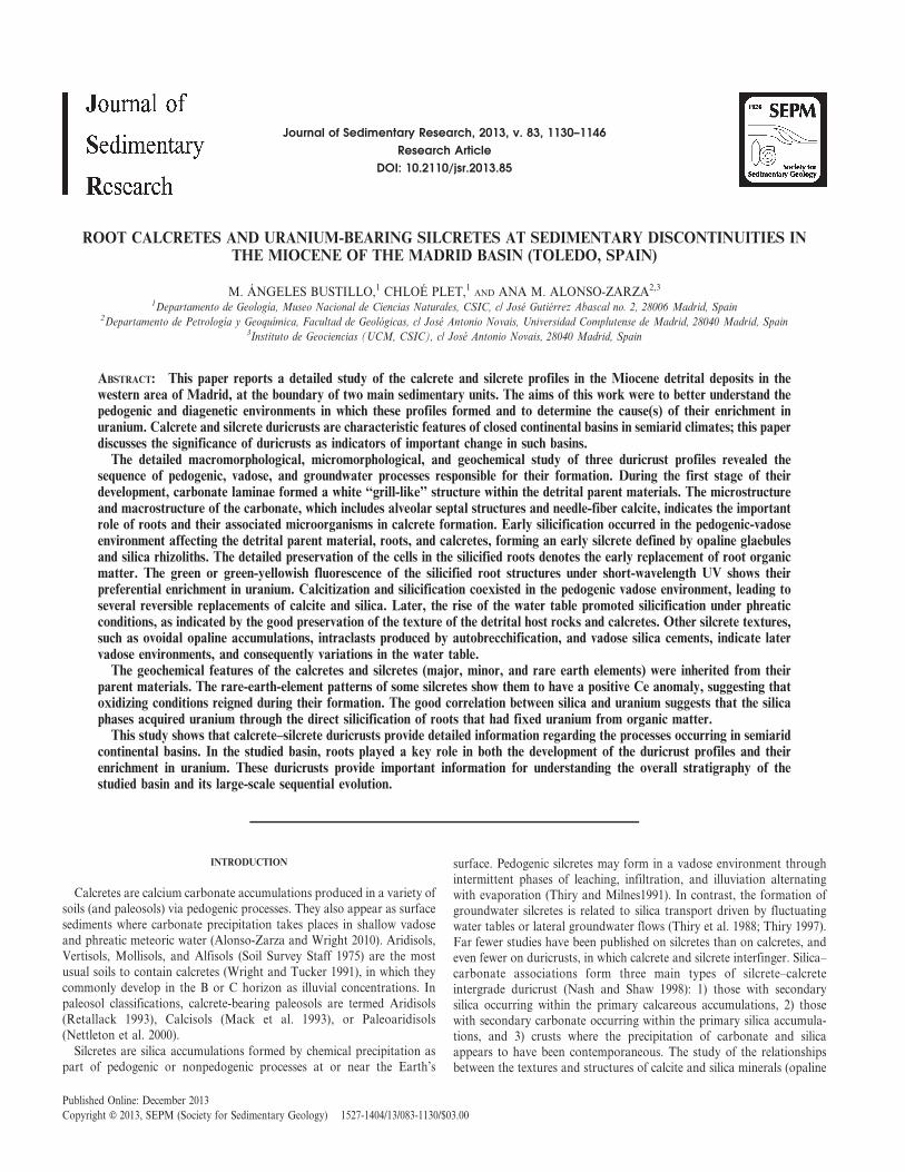
The present study area is located in the western part of the basin (Fig. 1),in the Province of Toledo, near the village of Torrijos. The deposits studiedcorrespond to the top of the Lower Miocene Unit (Fig. 2). These depositsare middle Aragonian in age (Lopez Olmedo et al. 2004); this iscorroborated by fossils at the paleontological sites of Torrijos (Aguirre etal. 1982). The Miocene Lower Unit, in the central part of the basin, iscomposed predominantly of evaporite facies, which grade laterally intoreddish-green mudstones containing anhydrite and/or gypsum nodules,and then into coarser clastic materials that were deposited in alluvialenvironments (Ordonez et al. 1991). In the western part of the basin,however, the deposits of the Lower Unit consist mainly of arkoses andassociated sandy clays supplied by the granitic and high-grade metamor-phic rocks of the Central System and the Toledo Mountains. The proximalalluvial fan facies consist of coarse conglomerates with an arkosic matrix;paleosols are very rare. The alluvial facies coalesce distally in this westernpart of the basin and consist of mudstones with associated calcretes and/orcarbonate pond deposits (Alonso-Zarza et al. 2002; Lopez Olmedo et al.2004). These carbonates are commonly silicified and occur mostly at thetop of the Lower Unit, and are the focus of the present work. They areoverlain by the detrital alluvial deposits of the Intermediate Miocene Unit.
MATERIALS AND METHODS
Samples of silcretes, calcretes, and parent materials were studied usingconventional mineralogical, petrographic, and geochemical techniques.
FIG. 1.—Location of the study area, shown in a paleogeographic sketch of theMadrid Basin at the time of the deposition of the topmost Lower Miocene Unit.The black area is the city of Madrid.
ROOT CALCRETES AND URANIUM-BEARING SILCRETES 1131J S R

The basic mineralogical and petrographic study involved transmitted-light-microscopy inspection of thin sections under polarized light. PowderX-ray diffraction (XRD) patterns were obtained from pressed powdermounts using a Philips semiautomatic PW 1710 diffractometer withmonochromatized CuKa radiation. Clay mineralogy was determined inoriented, glycol-solvated aggregates heated to 550uC. Uranium-bearingsilica zones were identified under short-wave UV light.
Appropriate areas for analysis by Micro-Raman spectroscopy wereselected by optical microscopy. A Thermo Fisher Raman microscope,which has a point-and-shoot Raman capability of 1 mm spatial resolution,was used with a 532 nm laser source. Variations in the moganite/quartzratio were obtained from the intensity ratios of the main symmetricstretching–bending vibrations of moganite (502 cm21) and alpha-quartz(465 cm21), multiplied by 100 (RM/Q). This method provides a relativemeasure of the moganite content in relation to quartz (Bustillo et al.2012).
Scanning electron microscopy (SEM) observations were made using aFEI INSPECT microscope, working at 30 kv and a distance of 10 mm,operating in high-vacuum mode and using secondary electrons andbackscatter detectors. The instrument used was equipped with an OxfordANALYTICAL-INCA X-ray energy-dispersive system (EDS). Cathode-luminescence SEM (CL-SEM) images and spectra were obtained using a
MONOCL3 Gatan apparatus. The excitation for CL measurements wasprovided by a 25 kV electron beam. The capability of combining CL withback-scattered electron (BSE) and secondary electron (SE) detection, orwith EDS microanalysis, allowed features of the rocks to be correlated withtheir uranium content.
Precise uranium determinations were obtained by SEM using a FEIQUANTA 200 machine equipped with a wavelength dispersive spec-trometry (WDS). The operating conditions were 30 kV with a workingdistance of 10 mm.
Major, minor, and REE elements were determined by Acme AnalyticalLaboratories (Vancouver, Canada) using inductively coupled emissionspectrometry (ICP-ES) and inductively coupled plasma mass spectrometry(ICP-MS). Loss on ignition (LOI) was determined by weight differenceafter ignition at 1000uC at the same laboratories. The analytical methodsand detection limits used can be found at http://www.acmelab.com/.
PROFILES STUDIED
Three carbonate–silica profiles were studied (termed Goya, Golf, andCastillo), corresponding to the topmost part of the Lower Miocene Unit.The Goya profile is located outside of the village of Val de SantoDomingo, in a recent road cutting made for the Goya Factory. The Golf
FIG. 2.—Stratigraphy of the study areashowing the location of the studied calcrete–silcrete profiles at the top of the Lower MioceneUnit. Modified from Alonso-Zarza et al. (2002).
1132 M.A. BUSTILLO ET AL. J S R
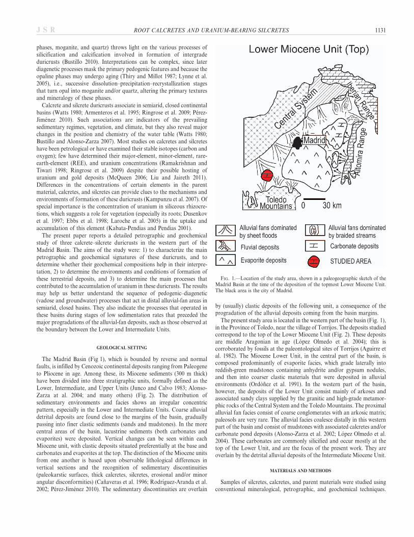
profile is located in the nearby entrance to the Fuensalida golf course, andthe Castillo profile, at the top of the hill on which the Barcience Castle sits(Fig. 3). All these profiles developed on detrital deposits. The maximumdistance between any two of these profiles is about 7 km.
The Goya Profile
Macromorphology.—This profile is about 2.5 m thick and is composedof three parts: detrital parent material, calcrete and silcrete duricrusts.The 80-cm-thick parent-material outcrop is a reddish-brown, loose, sandymudstone, interbedded with sand lenses. The number of these lensesincreases toward the top. Carbonates are absent at the base, but theybecome increasingly common towards the upper part as fine carbonatelaminae, giving rise to a gradual transition from the detrital parentmaterial to the calcrete horizon.
The calcrete is composed of three horizons. The lower horizon includesscarce white powdery carbonate nodules and very thin carbonate laminae,slightly inclined with respect to the stratification of the detrital sediments.The intermediate level forms a grill-like horizon about 50 cm thick. Itincludes horizontal laminae, 2–4 cm thick, connected by vertical veins ofthe same dimensions. The laminae are composed of powdery, soft,whitish carbonate. In the upper horizon, the carbonate laminae arenumerous and coalescent.
The top of the profile is harder and shows intense silicification. Themean thickness is 60 cm, varying laterally. This silicified horizon is beige tobrown in color with a vitreous luster. At its base it is composed of laminaethat correspond to the silicification of the laminar calcrete. Above, thesilicification preserves many features of argillaceous sands with partly
silicified carbonate laminae. Some spherical, ellipsoidal or irregular smallsilica accumulations (, 1 cm) are visible in the general mass.
Mineralogy and Petrology.—The parent material is composed mainly ofdetrital grains (quartz 40%, feldspar 30%, and mica 5–10%), dioctahedralsmectite (around 20%), and minor amounts of iron oxides. Whenobserved in thin section, samples show irregular, subparallel, flattenedpatterns of variable orientation corresponding to the smectite.
The carbonate horizons are composed of calcite (around 90%), minoramounts of quartz (, 10%), and traces of feldspar, mica, and smectites.The carbonate laminae of the grill-like structures and of the laminar levelare composed of a carbonate groundmass and relicts of sandy mudstones.The carbonate groundmass is composed of micritic nodules of differingsize (0.20–2 mm), plus small quartz, feldspars, and mica detrital grains,with the porosity filled by calcite spar cement. The detrital grains haverounded or angular shapes, in some cases are corroded by micrite. Thesegrains are 1 mm wide and up to 0.8 mm across. Isolated opaline roots,with the cellular structure preserved, can be seen in the relicts of thedetrital parent material and in the carbonate groundmass (Fig. 4A). Inaddition, local gray patches (up to 0.5 mm long) composed of needle-fibercalcite crystals (NFC) can be seen (Fig. 4B). According to Verrecchia andVerrecchia (1994), two types of NFC exist: 1) straight needles with acentral cavity, known as MA1 rods, and 2) undulating needles, known asMB rods. Both types were seen in the present work. The surface of theneedles is coated by microrods (about 100 nm across and 1 mm long) andsparse coccoids (Fig. 4C). The microrods are organized into ovoid masseswith a central spherical hole 1–5 mm in diameter. Locally, some fungalfilaments are visible (100 mm long), their surfaces also covered in
FIG. 3.—Measured sections and field images of the Goya, Golf, and Castillo profiles. All correspond to the same stratigraphic position (top of the Lower MioceneUnit). The numbers 1, 2, and 3 correspond to the parts described in the text. The hammer is 33 cm long.
ROOT CALCRETES AND URANIUM-BEARING SILCRETES 1133J S R

FIG. 4.—Goya profile. Optical microscopy (plane-parallel light) and SEM photographs. The optical-microscope images correspond to thin sections takenperpendicular to the surface of the beds. A) Silica rhizoliths (SR) within detrital host sediments. The root tissues are neatly preserved (red arrow). B) SEM image. Mass ofneedle-fiber calcite (NFC) crystals within grumelar micrite (GM). C) Detailed view of NFC crystals coated by microrods (red arrow), including some spherical bacterialbodies less than 1 mm in diameter (blue arrow). D) Relicts of the detrital parent material (quartz and feldspar grains in a clay matrix) within the opaline mass (OP). E)Horizontal silica rhizoliths (SR) within an opaline mass with ovoids (red arrows). F) Pendant vadose opaline and quartz cements (red arrows) within silica rhizoliths.
1134 M.A. BUSTILLO ET AL. J S R

microrods. EDS analyses of the NFC suggests proportions of carbonattributable to organic matter. The C/Ca ratio of an analyzed standardinorganic calcite was around 0.09, whereas for the studied NFC it wasabout 2.55.
The silcretes reproduce, in part, the fabric of the parent material, andinclude relicts of argillaceous sands, sandy mudstones, and somecarbonate laminae. The XRD results show the main silica phases to beopal CT (45 to 60%) and quartz (35%), with small amounts (up to 15%) ofclays and feldspars. Some opal A may be masked within the opal CTpeak. Moganite associated with quartz was identified by micro-Ramananalysis revealing a ratio of a moganite (502 cm21) to alphaquartz(465 cm21) (RM/Q) of between 28 and 44 (Fig. 5).
Silicification of the detrital parent materials generated a groundmasscontaining spherical and ellipsoid opaline accumulations, parent materialrelicts (Fig. 4D), and silica rhizoliths (Fig. 4E). Locally, the disorganizedstriated texture of the smectites and many features of the detrital parentmaterials are preserved. Some siliceous pendant cements are also present(Fig. 4F). Horizontal and vertical rhizoliths appear as fine white silicatubes in the brown opal. Locally they show numerous remains of rootcells. In longitudinal section these often appear elongated (about 50 mmlong by 10 mm across) in the direction of the rhizoliths. Somerhizocretions show a structure with: 1) an interior composed of grayishopaline root cells along with cryptocrystalline-to-microcrystalline quartzmosaics and, locally, length-slow chalcedony (generally the cellularstructure is better preserved in the opaline part), and 2) a fine outer layerof clear opal with no visible cellular structure or detrital grains.
SEM images show root cells in some cases associated with short NFCcrystals (about 6–10 mm long). The small size of the latter may be due topreferential dissolution of their ends. EDS analyses of the silicified rootsrevealed the presence of organic matter (atomic Si/C ratio 5 1.03).
The Golf Profile
Macromorphology.—The profile is composed of detrital parentmaterial, calcrete duricrust, and silcrete duricrust. The parent materialis composed of reddish-brown sands with very little pedogenicmodification; it contains some centimeter-size carbonate nodules. Thecalcrete (80 cm thick) is composed of sparse carbonate laminae withnodules at its base, but its top is composed entirely of amalgamatedcarbonate laminae. The silcrete has two parts: 1) a lower laminar part (upto 30 cm thick) showing an alternation of whitish opal and darker, hardercrystalline quartz zones about 10 cm thick, and 2) an upper part (up to90 cm thick) composed of a whitish-beige silica groundmass showingnodular shapes.
Mineralogy and Petrology.—The parent material is composed mainly ofdetrital grains (quartz 70% and feldspar 20%), traces of hematite, andsmectites.
In the calcrete, the carbonate laminae are composed of up to 95%calcite, with minor amounts of quartz and feldspar. Under the opticalmicroscope they show a micritic groundmass with sparite cements incracks (Fig. 6A), and in some places quartz cements filling voids. Thescarce detrital grains are highly corroded. The micritic groundmass andthe cracks contain many alveolar septal structures (Fig. 6A) and somecalcified root cells. Locally there are opal and cryptocrystalline-to-microcrystalline quartz root structures (Fig. 6B). In general, root cells arebetter preserved in opal. Some silica rhizoliths appear cracked andfragmented, probably due to dehydration. The cracks are filled withcalcite, indicating that calcretization took place. Later, the opaline rootstructure was transformed into microquartz in the central part of therhizolith fragments (Fig. 6C). Remains of plant tissue, preserved as opal,can be seen in the outer part.
The silcrete level shows variable degrees of silicification. The lower partis the richest in silica, with 15% opal and up to 85% quartz. The moganite,identified by micro-Raman analysis, has RM/Q values of between 30 and50. Quartz zones show some areas of homogeneous cryptocrystalline-to-microcrystalline quartz mosaics, and others with mosaics of varied crystalsize and length-fast chalcedony. Ovoids and laminations can be observedunder crossed-polarized light. The presence of cracks in the cryptocrystalline-to-microcrystalline zones, and the close association between the opal andquartz areas, suggest that the quartz is the result of opal recrystallization.Some carbonate micrite remains and partially silicified pendant carbonatecements can be recognized (Fig. 6D). Detrital grains are absent in this level.The upper level of the profile is a partially silicified calcrete (calcite 65%,silica 35%) with micrite ellipsoid accumulations (average 1 mm in diameter)and root structures. Cryptocrystalline-to-microcrystalline quartz mosaicsand length-fast chalcedony are the main products of silicification. Opalinepatches with detrital grains can be seen in the micrite; these are ellipsoidal,irregular, or angular in shape, and are cut by many micritic veins. Many ofthese patches show external parts corroded by micrite. In these areas,transformation of the opal into quartz can be seen.
Some silicified roots with preserved cellular structures are composed ofcryptocrystalline-to-microcrystalline quartz mosaics and, locally, length-slow chalcedony. The SEM images show the cellular structure to bepreserved in calcite with various degrees of silicification (Fig. 6E) andshort NFC crystals in the root trace, as in the Goya profile (Fig. 6F).
The Castillo Profile
Macromorphology.—This profile is 1.40 m thick and has three levels.The lower level (30 cm thick) is composed of either soft whitish orpartially silicified carbonate horizons, and contains relicts of clays andcalcified root hairs. The middle level (1 m thick) is massive and intenselysilicified (Fig. 3), and of variable color (white, cream, brown, or even
FIG. 5.—Raman spectra obtained from a cryptocrystalline–microcrystallinequartz area of the silcrete, where the moganite peak (Mo) appears next to thequartz peak (Qz).
ROOT CALCRETES AND URANIUM-BEARING SILCRETES 1135J S R

1136 M.A. BUSTILLO ET AL. J S R

bluish). Spherical, ellipsoidal, and irregular small silica accumulations,intraclasts, and cemented horizontal cracks are common. In some places,this level shows silicified nodular structures. The upper level (30 cm thick)is a laminar calcrete composed of 2-cm-thick carbonate laminae withsparse silica nodules.
Mineralogy and Petrology.—The lower horizon is composed mostly ofcalcite (. 90%) with scarce detrital grains (quartz and feldspar) andtraces of clays. Under optical microscopy its micritic groundmass appearshighly porous (largely the porosity left by root networks). Sparite is themost common cement. Locally, argillaceous glaebules with detrital grainsare crossed by micrite-filled cracks. The upper part of this lower horizonis silicified (up to 75%) and consists of quartz (up to 55%) and opal CT(up to 20%). Opal is found in discontinuous levels with quartz. The quartzoccurs in voids (mosaic quartz and length-fast chalcedony cements), but itis sometimes intermixed with the opal in the groundmass as recrystalliza-tion textures (aging). Some horizontal levels of opal and cryptocrystalline-to-microcrystalline quartz mosaics contain root structures silicified aslength-slow chalcedony; no carbonate precursor is visible. Locally, calcifiedroots can be seen, but their cells are more poorly preserved than in silicifi-ed roots. Some of the silica accumulations are broken by cracks filledby micrite, which also etch the opal (Fig. 7A), indicative of a latercalcretization stage.
The middle massive level is composed mainly of opal CT (25 to 50%),quartz (30 to 70%), and relicts of calcite and feldspar. Micro-Ramanspectrometry showed moganite to also be abundant (RM/Q 20–90depending on the area). The cryptocrystalline-to-microcrystalline quartzzones show the highest RM/Q values.
Thin sections showed this middle level to be composed of an opalinegroundmass, either very light in color and poor in detrital grains, ordarker (brown or grayish) with more grains. Some areas are richer inmicrite containing many rounded or angular detrital grains, in someinstances corroded at their edges. Root structures appear as gray-brownopal with some preserved cell walls. The uppermost part of this massivesilica level contains no carbonates, but it does contain floating detritalgrains. It also includes silicified intraformational sandy mudstone clasts(Fig. 7B). The number of detrital grains in these clasts is larger than in theopaline groundmass.
Quartz cementation varies from microcrystalline mosaics to length-fastchalcedony (a normal systematic organization for silica cements betweenthe walls and centers of voids), the result of decreasing amounts ofdissolved silica in the pore water. Locally, the infills of some voids arecomplex and show a base of silicified laminated micrite overlain bylength-fast chalcedony cements. These infillings are texturally similar tothose of carbonate pseudomicrokarst (Fig. 7C, D).
SEM images show some cavities to contain thin opaline cements (2–3 mm thick) that are very different from the groundmass. These cementsare composed of silica microspheres (up to 200 nm size) and lack carbon(as determined by EDS). The opaline cements are made up of lepisphereswith a carbon chemical signal (atomic Si/C ratio 5 4.9). Some lepispheres,seen individually and under higher magnification, appear to be composedof an assemblage of nanospheres (which might be an association of more
elongated shapes). Larger and more homogeneous branching filaments arecoated and trapped by these lepispheres (Fig. 7E, F).
The upper level is composed mainly of calcite (. 85%) with smallamounts of quartz (10%) and opal (, 5%). The groundmass is composedof micrite, with cracks filled by sparite but in some cases also by length-fast chalcedony. Opaline glaebules are interspersed among the micrite.These angular or rounded glaebules are crossed and etched by manymicrite veins. While some root networks are observed locally, cellstructures are very rare or absent. Some silica vadose cements are alsofound.
GEOCHEMISTRY
The geochemical compositions of 23 bulk samples representing allmaterial types were analyzed (Tables 1, 2, and 3 as supplementarymaterial, see Acknowledgments).
Major Elements
The considerable parent-material variation in SiO2 (49–74%), Al2O3
(9.3–16.1%), Fe2O3 (2.7–5.2%), and CaO (1.32–10.97%) depends on theamount and composition of the detrital sediment, and on the presence ofminor amounts of calcite.
The calcrete CaO content varies from 35.26–42.74%. The SiO2 content(15.42–27.98%) correlates with that of Al2O3 (1.69–3.78%), indicatingthat both are sourced from detrital parent-material components, withminor contributions from silica accumulations. In the silcretes, SiO2
varies between 80.83% and 97.07%, whereas sil-calcrete intergrades haveless SiO2 (42.20–77.56%) and more CaO (9.89–29.61%).
In the silcrete and sil-calcrete intergrades, the coefficient of correlationfor silica with other major elements is negative (r , 20.98), since SiO2 isthe product of a silicification process; it therefore replaces othercomponents. The correlation of Al2O3 with other major elements(Fig. 8A, e.g., see Fe2O3 and Al2O3) is overall strong and positive,reflecting the composition of the initial detrital components.
Minor Elements
With the exception of U and Sr, the minor-element contents of thedetrital parent material (Ba, Be, Co, Cs, Ga, Hf, Nb, Rb, Sn, Ta, Th, V,W, and Zr) are higher than those of the duricrust. The correlation matrixindicates a good positive correlation of Al2O3 (generally r . 0.80) withmost minor elements, indicating that the minor elements (except for Uand Sr) come from detrital minerals of the parent material.
In the duricrusts, the correlation between U and SiO2 is good(0.84 , r , 0.96) (Fig 8B). The U shows a negative correlation withAl2O3 (up to 20.82) and with CaO (up to 20.95), suggesting that it isassociated with the opal and quartz. The good positive correlation of Srwith CaO (0.80 , r , 0.97) (Fig. 8C) is due to the substitution of Ca bySr in the carbonate horizons. In the calcretes, the amount of Sr variesbetween 320 and 461 ppm.
The content of metals depends on the nature of the rock. While detritalparent material is relatively rich in all metals, these are less common in the
r
FIG. 6.—Golf profile. Optical microscopy (cross-polarized light) and SEM photographs. The optical-microscope images correspond to thin sections takenperpendicular to the surface of the beds. A) Micritic groundmass with sparite cements. The cracks contain calcified alveolar septal structures (arrows). The black zones arepores. B) Microquartz rhizoliths (SR) within a micritic calcrete with desiccation cracks (yellow arrow). C) Part of a large silica rhizolith cut by a network of cracks infilledwith calcite. The red arrows indicate areas where calcite clearly cuts the silica rhizolith. The fragments of the silica root structure (FR) show an external part of the opal;the internal parts have been transformed into microquartz by ageing. The high-magnification image shows the structure of the plant tissue in the external opaline area.D) Partially silicified vadose pendant calcite cements showing residual calcite layers (CA) and quartz (QZ) layers. E) SEM image of silica rhizoliths (SR) within a mass ofcarbonate crystals (CA). F) SEM image of short NFC within the opaline mass of a rhizolith.
ROOT CALCRETES AND URANIUM-BEARING SILCRETES 1137J S R

1138 M.A. BUSTILLO ET AL. J S R

silcretes, with the exception of Au (, 0.5 to 87.3 ppm). The goodcorrelation (0.93 , r , 1) of the metal values (with the exception of Auand Hf) with Al2O3 and Fe2O3 indicates that they are present in detritalgrains and clays within the duricrust. Au does not correlate well with anyelement, indicating its sporadic detrital origin.
Rare Earth Elements
The results of REE analyses were normalized using the composition ofNorth American shales (NASC index) (Gromet et al. 1984).
REE contents vary widely, with greater anomalies recorded for the detritalparent-material than for the duricrusts (Fig. 9). In the duricrusts, only weak,negative correlations between REE and SiO2 and CaO exist. However, totalREE correlates well with Al2O3 (Fig. 8D), and with Fe2O3 (0.80 , r , 1),indicating that the REE are located in relicts of detrital parent materials. Inthe Castillo profile, the values are lower (r 5 0.73), possibly because thesamples include silcrete lithoclasts of different chemical composition.
In the Goya profile (Fig. 9A), the Eu anomaly of the silcretes isnegative, but smaller (Eu/Eu* 5 0.57) than in the detrital parent material(Eu/Eu* around 0.64). A weak positive anomaly for Ce is seen in thesesilcretes, something not seen for the detrital parent material or thesilicified roots (see Goya sample 8-clear), which show a flat pattern.
In the Golf profile (Fig. 9B), the calcrete Ce and Eu anomalies arenegative. Silcrete REE concentrations are low and their patterns nearlyflat, with a weak, negative anomaly for Eu and a weak positive anomalyfor Ce.
In the Castillo profile (Fig. 9C), the correlations are not as strong as inthe other profiles. Only Sm and Eu correlate well with Al2O3 and Fe2O3
(0.80 , r , 0.89), possibly because they are located solely in feldspars.The highest REE values were recorded in the duricrust samples (calcretesand some silcretes) with the largest numbers of detrital grains. The Eunegative anomaly of the Castillo profile is very clear, while its Ce anomalyis weak and variable. The REE content of the intermediate massive silicalevel is uniformly very small (the curves are nearly flat), with theexception of the Eu negative anomaly (Fig. 8C).
Uranium: Localization and Study
Under short-wavelength UV, many of the samples show green orgreen-yellowish fluorescence, indicating the possible presence of uranium.Many areas of fluorescence are associated with vertical or horizontalsilicified root structures (Fig. 10A, B). Indeed, in some cases more intensefluorescence is seen at the center of the root structure. In other cases,however, the fluorescence is restricted to fine silicified levels, which inhand samples appear more crystalline (Fig. 10C, D). Geochemicalanalyses of bulk silcretes and calcrete–silcrete intergrades showed theuranium content to vary from 6.2 ppm to 97.5 ppm (Table 2). Thecoefficient of correlation of SiO2 with uranium is high (. 0.9) suggestingthe latter to be located in the silica phases and not in the carbonates. Thecoefficient of correlation of CaO with uranium is 2 0.84, r , 20.94.The optical-microscopy observations made indicate the fluorescent areasto be concentrated in zones of cryptocrystalline-to-microcrystalline quartzmosaics. In root structures, the cryptocrystalline-to-microcrystalline quartzdisplays features corresponding to recrystallization (aging) and to theremains of opal with well-preserved root cell structures. Other fluorescent
zones correspond to areas of aging; here the quartz shows striatedbirefringence, and Micro-Raman analysis indicates relatively largeamounts of moganite.
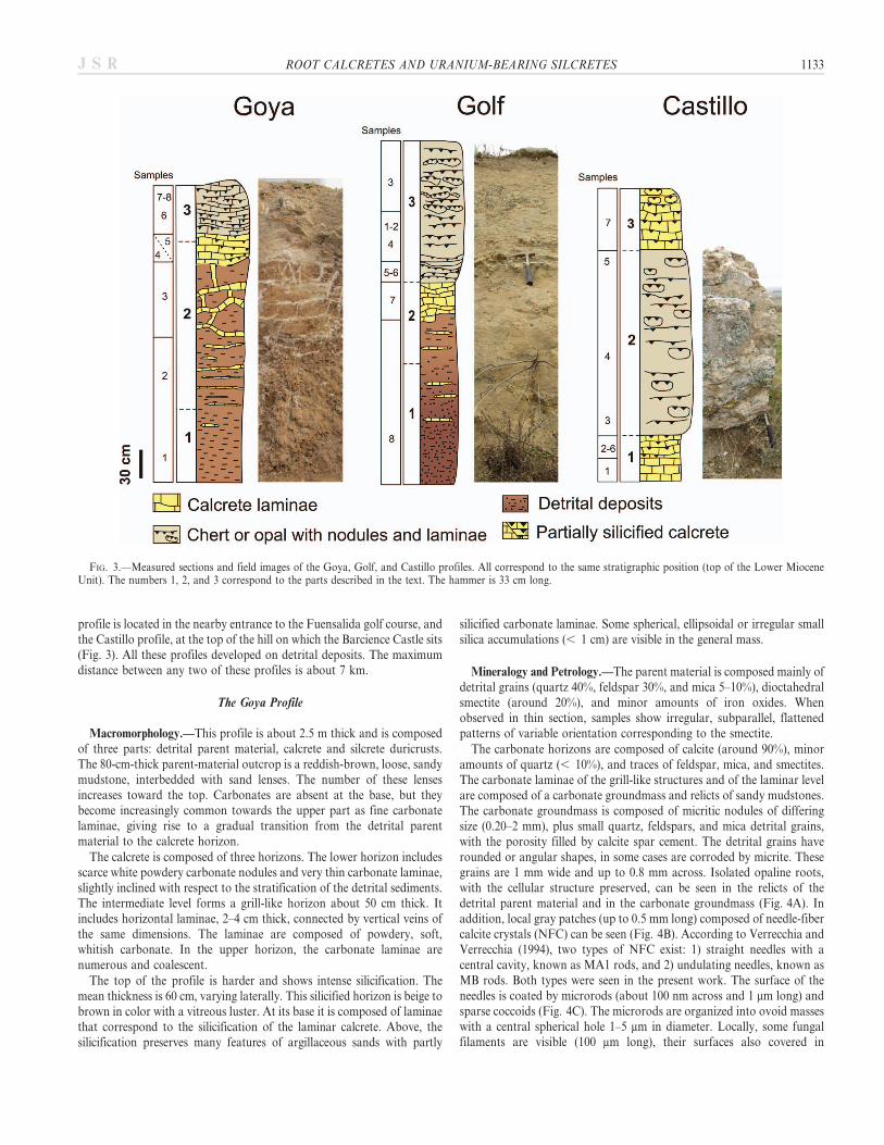
The main Micro-Raman spectroscopy uranium peaks defined byPointurier and Marie (2010) were not seen in the phases studied. Only asmall number of opaline rims showed low-intensity peaks, at 228–237 cm21, 334–350 cm21 (broad band), 412–414 cm21, and 781–784 cm21.Peaks around 237 cm21 and around 335 cm21 have been recorded insome UO2 samples (Manara and Renker 2003). The 412 cm21 (Piltch etal. 2009; Pointurier and Marie 2010) and the 780 cm21 peaks have beenfound in some uranyl chemical compounds (Sarsfield and Helliwell 2004;Frost and Cejka 2009), but for the present samples these peaks appearonly weakly, making uranium identification difficult. The CL-SEMstudies for uranium yielded more conclusive results. Analyses of thenonfluorescent zones showed spectra with various broad emission bandsaround 320, 380, 440, 460, and 650 nm (Fig. 11A) corresponding to opaland quartz (Stevens-Kalceff et al. 1997; Gotze el al. 2001). Other silicazones have two major peaks around 505 nm and 525 nm, and two minorpeaks at 545 and 570 that overlap the silica emission bands (Fig. 11B).These four peaks are attributed to the presence of uranyl ions (Billard andGeipel 2008) in silica minerals. The general shape of the spectra(Fig. 11B) coincides with those of silica gel containing uranium (Cheng2010). In some cases, silica bands are dominant and can mask the mainpotential uranium peaks; in other instances the main uranium peaks areso intense that the silica signal is masked (Fig. 10C). CL fluorescenceintensity can vary due to the number of uranyl ions, but also because offactors such as particle and pore size or water content in the silica phases(Cheng 2010). In general, spectral analyses show a greater intensity of themajor uranium peaks in cryptocrystalline-to-microquartz areas than inopal areas. The CL panchromatic images showed differences ofluminescence associated with rock textural features such as ovoidmorphologies or groundmass. The SEM with WDS analyses detectedvery small quantities of uranium (between 0.004 and 0.017 in atomicweight %) in some areas of the silicified roots. It is therefore very probablethat uranium is dispersed in the opal and quartz, and does not formindependent minerals.
DISCUSSION
Calcretes
The three profiles studied differ from classic calcrete profiles (Estebanand Klappa 1983). The main difference is the lack of well-developednodular horizons below the laminar ones; such nodular horizons arecommonly described in thick calcrete profiles (Melendez et al. 2011).Following the classification of Retallack (1988), the soil profiles can besaid to be only weakly developed, and it is difficult to classify themaccording to the morphological classifications of calcic soils (Gile et al.1965; Machette 1985) or Calcisols (Mack et al. 1993). It is also difficult todetermine whether they are part of an A or B horizon of a soil since theyoccur in different positions within the soil profiles. Mack and James(1992) refer to them as Ak horizons since they occur in the uppermosthorizon of a soil and contain carbonate, while Wright et al. (1995) refer toKrh horizons. In two of the profiles studied (Golf and Goya), the horizonbelow the laminar zone is made up of a framework of horizontal and/or
r
FIG. 7.—Castillo profile. Optical microscopy and SEM photographs. The optical- microscopy images correspond to thin sections taken perpendicular to the surface ofthe beds. A) The parent material includes opal angular clasts (OP) that are cut by a dense network of calcite veins (cross-polarized light). B) A silicified intraformationalargillaceous sandy clast (LT) in the opaline mass (plane-parallel light). C) The base is a silicified vadose carbonate silt (VS), the middle part corresponds to a new anderosional silicified carbonate vadose layer that also includes opaline intraclasts (OI) such as root tissues (arrow). The upper part it is a quartz cement (QZ) composedmainly of length-fast chalcedony (plane-parallel light). D) Same as Part C (cross-polarized light). E) SEM image of opal lepispheres with organic filaments (red arrow).F) Detailed view of Part E.
ROOT CALCRETES AND URANIUM-BEARING SILCRETES 1139J S R

vertical carbonate laminae intercalated within the parent material (Fig. 3).In the Castillo profile the initial calcrete structure has disappeared due tosilicification. The organization of the macrofabric and microfabric of thecarbonates is characteristic of incipient calcrete profiles the formation ofwhich is mostly driven by root activity (Alonso-Zarza 1999). Verycommonly, calcification of horizontal root mats results in the formationof isolated, thick laminar calcretes, but in other cases thin laminar calcretesare isolated within the parent material (Alonso-Zarza and Wright 2010).The calcification of vertical root systems has also been described(Rossinsky et al. 1992; Alonso Zarza and Jones 2007). In both the Golfand the Goya profiles, the mostly horizontal root systems were connectedvertically to form a grill-like framework. All these calcretes can beunderstood as rootcretes (Jones 1992) or rhizogenic calcretes (Wright et al.1995).
Evidence of root and associated microorganism activity can be seen inthe carbonate laminae. Alveolar septal structures attributed to thecalcification of root structures, in association with fungi (Wright 1986),are very common, along with calcified root cells, such as those describedby Jaillard et al. (1991) and Kosir (2004). NFC coated with microrods iscommonly found in modern and recent carbonate soils (Verrecchia andVerrecchia 1994), which is indicative of either direct fungal biominer-alization (MA rods) or of physicochemical mineralization of decayed
fungal-sheath organic matter (Verrecchia and Verrecchia 1994; Bajnocziand Kovacs-Kis 2006).
The REE and minor-element contents of the calcretes reflect mainlythe composition of the parent material, with the exception of Sr, which isassociated with previous carbonate phases. Calcrete enrichment in Sr ispossibly due to the replacement of Ca by Sr in carbonates, aconsequence of the strong affinity of Sr for Ca (both have similarchemical properties) (Morse and Mackenzie 1990; Capo and Chadwick1999; Dart et al. 2007). This explains the strong correlation between Srand Ca in the studied profiles. Ringrose et al. (2009) suggest that suchenrichments may be the product of Sr-enriched plant uptake within thecalcrete level. The shape of calcrete REE patterns does not differ greatlyfrom those of the siliciclastic parent material, but the concentration ofREE elements is different, indicating a common source for the REE.Relicts of detrital components included in the calcretes are the likelycause of the negative Eu anomaly.
Silcretes
The silcretes formed by the extensive replacement of detrital parentmaterials and calcretes. The main characteristics of the observed silcretesare the partial preservation of: 1) the textures, structures, and components
FIG. 8.—Binary diagrams of major-elements, minor-elements and total rare-earth-element concentrations for the Goya, Golf, and Castillo profiles. A) Fe2O3 vs.Al2O3. B) U vs. SiO2. C) Sr vs. CaO. D) Al2O3 vs. SREE. Major-elements are in % weight; minor-elements and rare-earth-elements are in ppm.
1140 M.A. BUSTILLO ET AL. J S R

of the detrital parent materials, 2) the macrostructure and microstructureand some components of the calcrete (some detrital grains, relicts of clays,laminar and root structures, glaebules, vadose cements, microkarstscements, etc.), and 3) the presence of silicified root structures.
The silicification of detrital parent material and the calcretes initiallyyielded opal. The replacement of calcite by silica (which is very common)is driven mainly by variations in pH. Maliva and Siever (1989) reportedthree mechanisms for the replacement of carbonates by silica: (1) theproduction of CO2 by the decomposition of organic matter, or the
introduction of CO2 into the water present by biological activity, resultingin a local lowering of the pH and thus affecting calcite solubility andinducing silica precipitation (Siever 1962; Knoll 1985; among others) (pHvalues of , 9 favor the dissolution of calcite and the precipitation ofsilica whereas higher values induce the opposite, promoting thecalcification of silcretes; Nash and Shaw 1998), (2) oxidation of hydrogensulfide, reducing the pH at oxic–anoxic boundaries (Clayton 1986), and(3) the dissolution of calcite and the precipitation of silica via the mixingof marine and continental waters (Knauth 1979). Part of the groundwaterin the mixing zone can be simultaneously supersaturated in quartz andopal, and undersaturated in calcite. In continental semiarid environments,the same mechanism can occur at the top of the groundwater, where moresaline percolating waters mix with less salty groundwater. In the studiedduricrusts, mechanisms 1 and 3 may have caused the silicification seen.
The mechanism of silicification of detrital parent material has beenstudied less extensively than that affecting limestone. However, it involvesa progressive loss of Al in the clay-mineral structures and other aluminumsilicates. Any structure remaining after the destruction of the octahedralsheets of the clay minerals must become totally disorganized before anynew recrystallization into silica, and this transition could occur onlythrough a local dissolution–recrystallization process (Elssas et al. 2000).The problem is to explain the evacuation of Al after the replacement ofclay minerals and other aluminosilicates. Summerfield (1983) proposesthe gradual replacement of clay minerals by silica through the removal ofAl where the pH is below 4, implying a high rate of organic activity withabundant production of humic acids. However, it would be difficult forsuch pH values to exist in the studied environments, except in areas ofroot decomposition.
The source of silica in groundwater silcretes is sometimes difficult tofind. During the Miocene, the Madrid Basin was closed and had an aridor semiarid climate. In semiarid closed basins and near-surfaceenvironments, variations in the water table might give rise to complexreplacements. Some silica might come from the alteration of siliclasticgrains, but much more may come from the formation of calcretes. Duringcalcrete formation, siliciclastic grains are replaced by calcite, and thegroundwater, enriched in silica, will migrate across the correspondingalluvial fan towards the lake margins. This source of silica has beenrecognized for several types of silcrete formed in arid environments(Summerfield 1982; Khalaf 1988; Armenteros et al. 1995). A great deal ofsilica concentration and precipitation may also occur when waterevaporates from plants (Sommer et al. 2006). McCarthy and Ellery(1995) showed the accumulation of silica in the distal reaches of theOkavango Fan (Botswana) to be due to (1) phytoliths mixed with thesubstrate by illuviation and bioturbation, and (2) the precipitation of fine-grained amorphous silica from the groundwater induced by thetranspiration of rooted plants, plus increases in salinity.
The aging process promotes the transition opalRmoganiteRquartz.Consequently, part of the opal aged to cryptocrystalline-to-microcrystal-line quartz mosaics, moganite and small-sized (up to 40 m) length-fastchalcedony. Due to the inhibition of aging caused by clay minerals(Bustillo 1982), silcretes that form directly on argillaceous sands containmore opal than quartz; this is not the case for silcretes that replacecalcretes. The aging of opal, which is common in surface silcretes (Thiryand Millot 1987), erases part of the original opal textures.
Root cellular structures are better preserved in silica (opal andcryptocrystalline-to-microcrystalline quartz) than in calcite, indicatingthat some roots were directly silicified without first passing through acarbonate phase. Length-slow chalcedony occurs only in roots, whereaschalcedony is length-fast in the silicified calcrete groundmass. Length-slow chalcedony, which indicates a basic pH or evaporitic setting (Folkand Pittman 1971), can be taken as evidence that silicification of roottissue occurred in a microenvironment different from that of calcretesilicification.
FIG. 9.—NASC-normalized (Gromet el al. 1984) REE plots for samples fromthe Goya, Golf, and Castillo profiles. Detrital parent materials: Goya-1, 2, and 3and Golf-8 (brown). Calcretes: Golf-7 and Castillo-1 and 7 (blue). Calc-silcretes:Golf-1, 3, 4, and Castillo-2 (light green). Silcretes: Goya-5, 6, 7, and 8 dark, Golf-2,5, 6, and Castillo-3, 4, 5, 6 (intense green). Silica rhizolith: Goya-8 clear.
ROOT CALCRETES AND URANIUM-BEARING SILCRETES 1141J S R

Premortem or postmortem silicification of roots is possible. Owen et al.(2008) suggest that the close association of opal with roots indicates thatplants contribute to their own silicification through evapotranspiration,which would locally increase the silica concentration. Silica providesmechanical strength, resistance to toxicity, and fluid loss (Epstein 2001).Silica is taken up through the roots as monosilicic acid (H4SiO4) and iscommonly precipitated as opal-A. The absorption of silica is likely tocontinue as long as the silica concentration does not inhibit growth. Ifconcentrations of dissolved silica become very high, the silica andassociated salts might have a potentially lethal effect on plantmetabolism.
In addition, in pedogenic environments permeated by root networks,pervasive but incomplete decomposition of plant tissue may lead to thepost-mortem silicification of root cells (Knoll 1985). Such decompositionimpacts the microenvironment, leading to a local increase in CO2 coupledwith a reduction in pH.
The studied silcretes are made up of parent material, calcrete relicts, amassive groundmass with spherical, ellipsoidal, or irregular opalineaccumulations, and intraclasts. All of these features are common insilicification occurring in surface environments with interplay betweenphreatic and vadose environments (Bustillo and Alonso-Zarza 2007).Some of the opaline accumulations and intraclasts are replaced by micritein cracks and/or at the outer edge. Such replacement by calcite indicatesthat some opal accumulated before or during calcrete development.
With the exception of uranium, these silcretes are deficient in elementsfound in the relicts of parent materials and calcretes. The REE patternsof the silcretes are nearly flat, but in some cases negative Eu anomaliesare related to detrital parent material and calcrete relicts. Ce anomaliesare more variable. A positive Ce anomaly is present in the Goya profilebut not in the detrital deposits of the section (Fig. 9). In the Golf profile,small Ce-negative anomalies occur in the REE patterns of somecalcretes, while small Ce-positive anomalies occur in those of some
FIG. 10.—A) Hand sample of silcrete from the Goya profile, formed by the silicification of detrital parent material. The brown parts are the silicified detrital deposits;the white parts are mainly opaline rhizocretions (red arrows). B) Image of Part A under short- wavelength UV light. The greener fluorescent areas correspond mainly torhizocretions. C) Hand sample of silcrete from the Golf profile, formed by the silicification of calcrete. The laminar structure is composed of partially silicified calcreteand cryptocrystalline–microcrystalline quartz zones (MQ). D) Image of PartC under short-wavelength UV light. Zones with green fluorescence are mainlycryptocrystalline–microcrystalline quartz.
1142 M.A. BUSTILLO ET AL. J S R
silcretes (Fig. 9). Ce-positive anomalies are due to a change in valency,from Ce+3 to Ce+4 (immobile form) under oxidizing conditions, and theoxidized Ce occurring in cerianite (CeO2) is accumulated in higherconcentrations than other REEs. Positive Ce anomalies tend to form atthe tops of weathering profiles in lateritic soils (Braun et al. 1990).Kampunzu et al. (2007) reported duricrusts in the Moshawengh dryvalleys of the Kalahari in which calcretes had negative Ce anomalies,while in the cal-silcretes the Ce anomaly was positive, possibly indicating
that the silcretes formed from residual solutions after the calcreteprecipitated.
Sequences of Processes and Environments
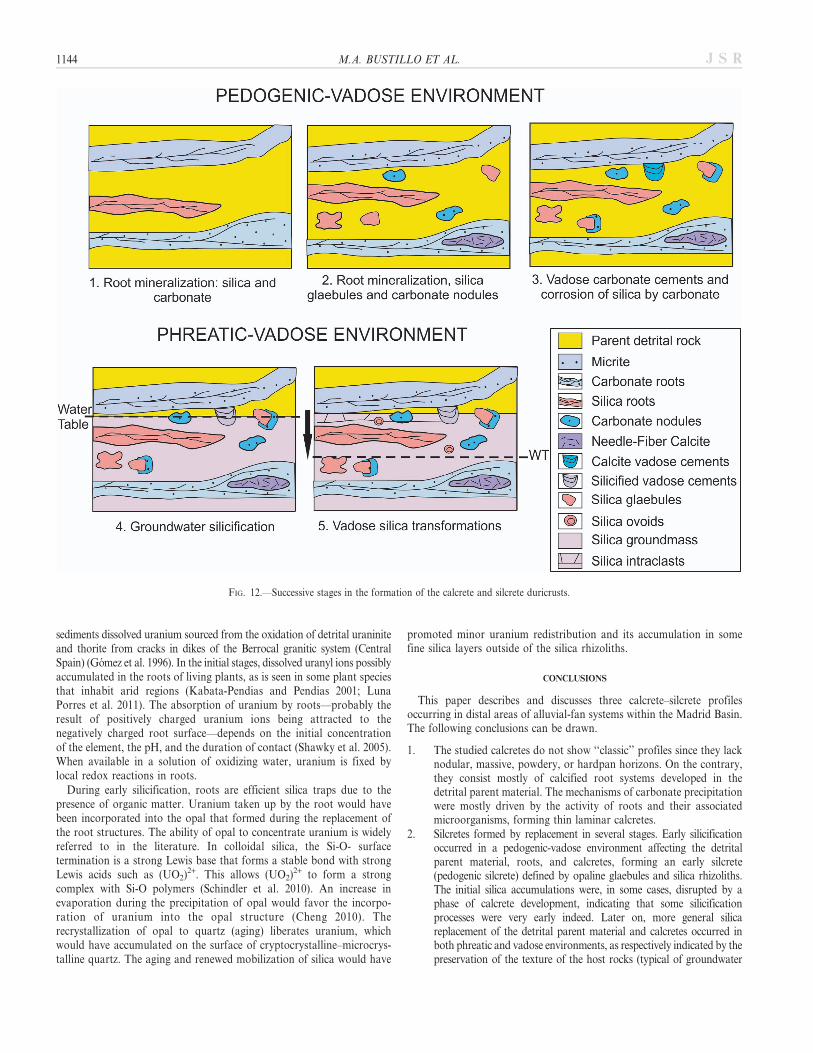
The processes in the alluvial fans responsible for the formation of thestudied profiles include pedogenic, vadose and groundwater processes(Fig. 12).
Pedogenic-Vadose Environment.—Features such as glaebules, nodules,rhizocretions, NFC, and laminar structures indicate that calcrete formedin a pedogenic vadose environment. This is also indicated by the pendantvadose cements.
In the initial stage (stage 1, Fig. 12,) the mineralization of roots bysilica and calcite occurred. Later on (stage 2, Fig. 12), the formation ofcalcite nodules and the progressive calcification of roots accounted forthe formation of the thin laminar calcretes. Isolated silica glaebulesformed by replacement of the carbonate nodules and laminae, and ofthe detrital parent material. New silica rhizoliths may also have formedat this stage. Both types of silica accumulation (rhizoliths and glaebules)would lead to the formation of a pedogenic silcrete (Dixon andMcLaren 2009). The micrite corrosion of opaline glaebules and thependant vadose cements indicate a new stage of calcretization (stage 3,Fig. 12).
Phreatic Environment.—The calcretes and incipient silcretes underwentintense silicification to generate the laterally continuous thick layers andlenticular silica levels. This groundwater silicification affected the detritalrocks of the alluvial fans, the calcretes forming therein (stage 4, Fig. 12),and also the incipient pedogenic silcrete (silicified roots and sparse silicaglaebules), reproducing the features of the host rocks, as seen in manygroundwater silcretes (Thiry and Ribet 1999; Nash et al. 2004). Thegroundwater must have contained and transported large amounts ofdissolved silica. According to Nash and Ullyott (2007), silicificationoccurs at the water table, or near groundwater outflow zones, with water-table fluctuations exerting a strong control over silicification. Within thegroundwater fluctuation zone, variations in pH due to recharge caused bymeteoric water percolating through the soil capillary zone, plus surfaceevaporation, would have favored opal precipitation (Arakel et al. 1989).During dry periods a lowering of the water table would have led to thelocalization of silicified layers and lenticular levels in a vadoseenvironment. New silica textures (spherical or ellipsoidal opalineaccumulations, intraclasts produced by autobrecchification, etc.), andvadose silica cements formed, highlighting the interplay between phreaticand vadose environments (stage 5, Fig. 12).
The studied calcrete–silcrete intergrades were favored by a period ofvery reduced or absent sedimentation in the basin that allowed pedogenicand diagenetic (vadose and phreatic) modification of the primary detritalsediments. It is not easy to say how long this period lasted, but it wasprobably some hundreds of thousands of years (Bustillo 2010). Thelandscape was probably very stable over this period.
All the features and processes described herein are linked to asedimentary discontinuity recognized in most of the Madrid Basin. Thisdiscontinuity is the boundary between the Lower and IntermediateMiocene Units (Fig. 2). In the eastern and southern areas of the basin, alarge paleokarst developed on evaporite deposits, whereas in thenortheastern and western areas, thick palustrine deposits, calcretes, andsilcretes record the discontinuity. In short, these duricrusts and theirintegrades can also be used as indicators of significant sedimentarydiscontinuities in continental basins.
The Concentration of Uranium.—The detrital siliciclastic strata of alluvialfans are permeable. In the present basin, groundwater circulating through the
FIG. 11.—Cathode-luminescence SEM spectra. A) Nonfluorescent silica zoneswith broad emission bands (Si) around 320, 380, 440, and 460 nm, and a weaksignal at 650 nm. B) Two major uranium peaks around 505 nm and 525 nm, andtwo minor uranium peaks at 545 and 570, overlap the broad emission silica spectrabands (Si). C) The major uranium peaks are so intense that the silica signal (Si) isrelatively weak.
ROOT CALCRETES AND URANIUM-BEARING SILCRETES 1143J S R
sediments dissolved uranium sourced from the oxidation of detrital uraniniteand thorite from cracks in dikes of the Berrocal granitic system (CentralSpain) (Gomez et al. 1996). In the initial stages, dissolved uranyl ions possiblyaccumulated in the roots of living plants, as is seen in some plant speciesthat inhabit arid regions (Kabata-Pendias and Pendias 2001; LunaPorres et al. 2011). The absorption of uranium by roots—probably theresult of positively charged uranium ions being attracted to thenegatively charged root surface—depends on the initial concentrationof the element, the pH, and the duration of contact (Shawky et al. 2005).When available in a solution of oxidizing water, uranium is fixed bylocal redox reactions in roots.
During early silicification, roots are efficient silica traps due to thepresence of organic matter. Uranium taken up by the root would havebeen incorporated into the opal that formed during the replacement ofthe root structures. The ability of opal to concentrate uranium is widelyreferred to in the literature. In colloidal silica, the Si-O- surfacetermination is a strong Lewis base that forms a stable bond with strongLewis acids such as (UO2)2+. This allows (UO2)2+ to form a strongcomplex with Si-O polymers (Schindler et al. 2010). An increase inevaporation during the precipitation of opal would favor the incorpo-ration of uranium into the opal structure (Cheng 2010). Therecrystallization of opal to quartz (aging) liberates uranium, whichwould have accumulated on the surface of cryptocrystalline–microcrys-talline quartz. The aging and renewed mobilization of silica would have
promoted minor uranium redistribution and its accumulation in somefine silica layers outside of the silica rhizoliths.
CONCLUSIONS
This paper describes and discusses three calcrete–silcrete profilesoccurring in distal areas of alluvial-fan systems within the Madrid Basin.The following conclusions can be drawn.
1. The studied calcretes do not show ‘‘classic’’ profiles since they lacknodular, massive, powdery, or hardpan horizons. On the contrary,they consist mostly of calcified root systems developed in thedetrital parent material. The mechanisms of carbonate precipitationwere mostly driven by the activity of roots and their associatedmicroorganisms, forming thin laminar calcretes.
2. Silcretes formed by replacement in several stages. Early silicificationoccurred in a pedogenic-vadose environment affecting the detritalparent material, roots, and calcretes, forming an early silcrete(pedogenic silcrete) defined by opaline glaebules and silica rhizoliths.The initial silica accumulations were, in some cases, disrupted by aphase of calcrete development, indicating that some silicificationprocesses were very early indeed. Later on, more general silicareplacement of the detrital parent material and calcretes occurred inboth phreatic and vadose environments, as respectively indicated by thepreservation of the texture of the host rocks (typical of groundwater
FIG. 12.—Successive stages in the formation of the calcrete and silcrete duricrusts.
1144 M.A. BUSTILLO ET AL. J S R

silcretes) and by the characteristic textures of the vadose silcretes seen,e.g., ovoidal opaline accumulations, intraclasts produced by auto-brecchification, and vadose silica cements. Calcrete–silcrete intergradestherefore formed under pedogenic-vadose and phreatic conditions.
3. Geochemical features of calcretes and silcretes are inherited fromrelicts of the parent materials. However, the Ce anomalies of somesilcretes are different from those of the parent materials. That theseCe anomalies are sometimes positive suggests oxidizing conditionsduring silcrete formation, possibly due to residual solutions leftbehind after the main calcretization process.
4. The good correlation between silica and uranium suggests that thelatter is located in the silica phases and not in the carbonates.Uranium-bearing silcretes initially acquired the element through thedirect silicification of roots that fixed it from organic matter.Subsequent uranium mobilization was due mainly to the aging ofopal to quartz. U-bearing siliceous rhizoliths indicate the importantrole of plants in uranium fixation in surface environments.
5. The location of these integrades at the boundary between the Lowerand Intermediate Miocene Units indicates that similar overlappingsof pedogenic and phreatic duricrust profiles can be used as a markerof important sedimentary discontinuities in other continental basins.
ACKNOWLEDGMENTS
The authors are grateful to Laura Tormo, Marta Furio, and Alberto JorgeGarcıa of the laboratory of ‘‘Non-destructive analytical techniques,’’ MuseoNacional de Ciencias Naturales, for assistance with the CL-SEM-EDS-WDSand Micro-Raman analyses. This work was funded by Projects CGL2011-27826-CO2-01 and -02 from the Spanish Ministry of Science and Innovation.We would like to thank reviewers Dr. Medard Thiry, Dr. Julia Kahmann-Robinson, and Dr. Dennis Terry and Associate Editors Dr. Stacy Atchleyand Dr. Paul McCarthy for their elaborated and detailed reviews, whichgreatly improved the manuscript. We also wish to thank James MacEachernfor helpful editorial comments.
We thank James Cerne, Adrian Burton, and John B. Southard forassistance with the English manuscript.
Tables 1–3 are available from JSR’s Data Archive: http://sepm.org/pages.aspx?pageid5229.
REFERENCES
AGUIRRE, E., ALBERDI, M.T., JIMENEZ, E., MARTIN-ESCORZA, C., MORALES, J., SESE, C.,AND SORIA, D., 1982, Torrijos: nueva fauna con Hispanotherium de la cuenca mediadel Tajo: Acta Geologica Hispanica, v. 1, p. 39–61.
ALONSO-ZARZA, A.M., 1999, Initial stages of laminar calcrete formation by roots:examples from the Neogene of central Spain: Sedimentary Geology, v. 126, p. 177–191.
ALONSO-ZARZA, A.M., AND JONES, B.J., 2007, Root calcrete formation on Quaternarykarstic surfaces of Grand Cayman: Geologica Acta, v. 5, p. 77–88.
ALONSO-ZARZA, A.M., AND WRIGHT, V.P., 2010, Palustrine carbonates, in Alonso-Zarza,A.M., and Tanner, L.H., eds., Carbonates in Continental Settings: Facies,Environments and Processes: Amsterdam, Elsevier, Developments in Sedimentology,v. 61, p. 225–267.
ALONSO-ZARZA, A.M., ARMENTEROS, I., BRAGA, J.C., MUNOZ, A., PUJALTE, V., RAMOS,E., AGUIRRE, J., ALONSO-GAVILAN, G., ARENAS, C., BACETA, J.I., CARBALLEIRA, J.,CALVO, J.P., CORROCHANO, A., FORNOS, J.J., GONZALEZ, A., LUZON, A., MARTıN, J.M.,PARDO, G., PAYROS, A., PEREZ, A., POMAR, L., RODRıGUEZ, J.M., AND VILLENA, J.,2002, Tertiary, in Gibbons, W., and Moreno T., eds., The Geology of Spain: TheGeological Society, London, p. 293–334.
ALONSO-ZARZA, A.M., CALVO, J.P., SILVA, P.G., AND TORRES, T., 2004, Cuenca del Tajo,in Vera, J.A., ed., Geologıa de Espana: Madrid, Sociedad Geologica de Espana andInstituto Geologico y Minero de Espana, p. 556–563.
ARAKEL, A.V., JACOBSON, G., SALEHI, M., AND HILL, C.M., 1989, Silicification of calcretein paleodrainage basins of the Australian arid zone: Australian Journal of EarthSciences, v. 36, p. 73–89.
ARMENTEROS, I., BUSTILLO, M.A., AND BLANCO, J., 1995, Pedogenic and groundwaterprocesses in a closed Miocene basin (northern Spain): Sedimentary Geology, v. 99,p. 17–36.
BAJNOCZI, B., AND KOVACS-KIS, V., 2006, Origin of pedogenic needle-fiber calciterevealed by micromorphology and stable isotope composition: a case study of aQuaternary paleosol from Hungary: Chemie der Erde, v. 66, p. 203–212.
BILLARD, I., AND GEIPEL, G., 2008, Luminescence analysis of actinides: instrumentation,applications, quantification, future trends, and quality assurance: Fluorescence, v. 5,p. 465–492.
BRAUN, J., PAGEL, M., MULLER, J., BILLONG, P., MICHARD, A., AND GUILLET, B., 1990,Cerium anomalies in lateritic profiles: Geochimica et Cosmochimica Acta, v. 54, p.781–795.
BUSTILLO, M.A., 1982, Aging features in inorganic continental opals: EstudiosGeologicos, v. 38, p. 335–344.
BUSTILLO, M.A., 2010, Silicification of continental carbonates, in Alonso-Zarza, A.M.,and Tanner, L.H., eds., Carbonates in Continental Settings: Processes, Facies andApplications: Amsterdam, Elsevier, Developments in Sedimentology, v. 62, p. 153–174.
BUSTILLO, M.A., AND ALONSO-ZARZA, A.M., 2007, Overlapping of pedogenesis andmeteoric diagenesis in distal alluvial and shallow lacustrine deposits in the MadridMiocene Basin, Spain: Sedimentary Geology, v. 198, p. 255–271.
BUSTILLO, M.A., PEREZ-JIMENEZ, J.L., ALONSO-ZARZA, A.M., AND FURIO, M., 2012,Moganite in the chalcedony varieties of continental cherts (Miocene, Madrid BasinSpain): Spectroscopy Letters, v. 45, p. 109–113.
CANAVERAS, J.C., SANCHEZ-MORAL, S., CALVO, J.P., HOYOS, M., AND ORDONEZ, S., 1996,Dedolomites associated with karstic features, an example of early dedolomitization inlacustrine sequences from the Tertiary Madrid Basin, Central Spain: Carbonates andEvaporites, v. 11, p. 85–103.
CAPO, R.C., AND CHADWICK, O.A., 1999, Sources of strontium and calcium in desert soiland calcrete: Earth and Planetary Science Letters, v. 170, p. 61–72.
CHENG C, 2010, Optical measurement of environmental uranium using porous silicamaterials [Dissertation, Degree of Doctor]: Virginia Commonwealth University,Taiwan. http://hdl.handle.net/10156/3074.digarchive.library.vcu.edu
CLAYTON, C.J., 1986, The chemical environment of flint formation in Upper Cretaceouschalks, in Sieveking, G.G., and Hart, M.B., eds., The Scientific Study of Flint andChert: Cambridge, U.K., Cambridge University Press, p. 43–54.
DART, R.C., BAROVICH, K.M., CHITTLEBOROUGH, D.J., AND HILL, S.M., 2007, Calcium inregolith carbonates of central and southern Australia: its source and implications forthe global carbon cycle: Palaeogeography, Palaeoclimatology, Palaeoecology, v. 249,322–334.
DIXON, J.C., AND MCLAREN, S.J., 2009, Duricrust, in Parson, A.J., and Abrahams, A.D.,eds., Geomorphology of Desert Environments: Berlin, Springer, p. 123–151.
DUSENKOV, S., VASUDEV, D., KAPULNIK, Y., GLEBA, D., FLEISHER, D., TING, K.C., AND
ENSLEY, B., 1997, Removal of Uranium from water using terrestrial plants:Environmental Science and Technology, v. 31, p. 3468–3474.
EBBS, D.S., BRADI, D.J., AND KOCHIAN, L.V., 1998, Role of uranium speciation in theuptake and translocation of uranium by plants: Journal of Experimental Botany, v.49, p. 1183–1190.
ELSASS, F., DUBROEUCQ, D., AND THIRY, M., 2000, Diagenesis of silica minerals from clayminerals in volcanic soils of Mexico: Clay minerals, v. 35, p. 477–489.
EPSTEIN E, 2001, Silicon in plants: facts vs. concepts: Studies in Plant Science, v. 8,p. 1–15.
ESTEBAN, M., AND KLAPPA, C.F., 1983, Subaerial exposure environments, in Scholle,P.A., Bebout, D.G., and Moore, C.H., eds., Carbonate Depositional Environments:American Association of Petroleum Geologists, Memoir 33, p. 1–96.
FOLK, R., AND PITTMAN, S., 1971, Length-slow chalcedony: a new testament for vanishedevaporates: Journal of Sedimentary Petrology, v. 41, p. 1045–1058.
FROST, R.L., AND CEJKA, J., 2009, Raman spectroscopic study of the uranyl phosphatemineral dumontite Pb2 [(UO2)3 O2 (PO4)2]. 5H2O: Journal of Raman Spectroscopy, v.40, p. 591–594.
GILE, L.H., PETERSON, F.F., AND GROSSMAN, R.B., 1965, The K horizon: a masterhorizon of carbonate accumulation: Soil Science, v. 97, p. 74–82.
GOMEZ, P., TURRERO, M.J., PENA, J., GIMENO, M.J., CRESPO, M.T., GORDIENKO, F.B.,MARTINEZ, F., REYES, E., RIVAS, P., AND IVANOVICH, M., 1996, Procesos de interaccionagua–roca y comportamiento del uranio en el sistema granıtico de El Berrocal(Espana): Geogaceta, v. 20, p. 1626–1629.
GOTZE, J., PLOTZE, M., AND HABERMANN, D., 2001, Origin, spectral characteristics andpractical applications of the cathodoluminescence (CL) of quartz. A review:Mineralogy and Petrology, v. 71, p. 225–250.
GROMET, L.P., HASKIN, L.A., KOROTEV R.L., AND DYMEK, R.F., 1984, The ‘‘NorthAmerican shale composite’’: its compilation, major and trace element characteristics:Geochimica et Cosmochimica Acta, v. 48, p. 2469–2482.
JAILLARD, B., GUYON, A., AND MAURIN, A.F., 1991, Structure and composition ofcalcified roots and their identification in calcareous soils: Geoderma, v. 50, p. 197–210.
JONES, B., 1992, Construction of spar calcite crystals around spores: Journal ofSedimentary Petrology, v. 62, p. 1054–1057.
JUNCO, F., AND CALVO, J.P., 1983, Cuenca de Madrid, in Geologıa de Espana, LibroHomenaje a J.M. Rıos: Madrid, Instituto Geologico y Minero de Espana, v. 2, p. 534–542.
KABATA-PENDIAS, A., AND PENDIAS, H., 2001, Trace elements in soils and plants:Washington, CRC Press, 413 p.
KOSIR, A., 2004, Microcodium revisited: root calcification products of terrestrial plantson carbonate-rich substrates: Journal of Sedimentary Research, v. 74, p. 845–857.
KAMPUNZU, A.B., RINGROSE, S., HUNTSMAN-MAPILA, P., HARRIS, C., VINK, B.W., AND
MATHESON, W., 2007, Origins and palaeo-environments of Kalahari duricrusts in theMoshaweng dry valleys (Botswana) as detected by major and trace elementcomposition : Journal of African Earth Sciences, v. 48, p. 199–221.
KHALAF, F.I., 1988, Petrography and diagenesis of silcrete from Kuwait, Arabian Gulf:Journal of Sedimentary Petrology, v. 58, p. 1014–1022.
ROOT CALCRETES AND URANIUM-BEARING SILCRETES 1145J S R

KNAUTH, L.P., 1979, A model for the origin of chert in limestone: Geology, v. 7, p. 274–277.
KNOLL, A.H., 1985, Exceptional preservation of photosynthetic organisms in sicilifiedcarbonates and silicified peats: Royal Society of London, PhilosophicalTransactions B, v. 311, p. 111–122.
LAROCHE, L., HENNER, P., CAMILLERI, V., MORELLO, M., AND GARNIER-LAPLACE, J., 2005,Root uptake of uranium by a higher plant model (Phaseolus vulgaris), bioavailabilityfrom soil solution: Radioprotection, v. 40, p. 33–39.
LIU, S., AND JAIRETH, S., 2011, Exploring for calcrete-hosted uranium deposits in thePaterson region, Western Australia: AusGeo News, v. 103, p. 1–5.
LOPEZ OLMEDO, F., DIAZ DE NEIRA, A., MARTIN SERRANO, A., CALVO, J.P., MORALES, J.,AND PELAEZ-CAMPOMANES, P., 2004, Unidades estratigraficas en el registro sedimen-tario Neogeno del sector occidental de la Cuenca de Madrid: Sociedad Geologica deEspana, Revista, v. 17, p. 87–101.
LUNA PORRES, M.Y., ALARCON HERRERA, M.T., SILVA SAENZ, M., RENTERıA VILLALOBOS,M., RODRıGUEZ VILLA, M.A., HERRERA PERAZA, E., REYES CORTES, M., AND MONTERO
CABRERA, M.E., 2011, Baccharis salicifolia development in the presence of highconcentrations of uranium in the arid environment of San Marcos, Chihuahua:Revista Mexicana de Fısica, v. 57, p. 40–43.
LYNNE, B.Y., CAMPBELL, K.A., MOORE, J.N., AND BROWNE, P.R.L., 2005, Diagenesis of1900-year-old siliceous sinter (opal-A to quartz) at Opal Mound, Roosevelt HotSprings, Utah, U.S.A.: Sedimentary Geology, v. 179, p. 249–278.
MACHETTE, M.N., 1985, Calcic soils of southwestern United States, in Weide, D.L., ed.,Soil and Quaternary Geology of the Southwestern United States: Geological Societyof America, Special Paper 203, p. 1–21.
MACK, G.H., AND JAMES, W.C., 1992, Calcic paleosols of the Plio-Pleistocene CampRice and Palomas Formations, southern Rio Grande rift, US: Sedimentary Geology,v. 77, p. 89–109.
MACK, G.H., JAMES, W.C., AND MONGER, H.C., 1993, Classification of paleosols:Geological Society of America, Bulletin, v. 105, p. 129–136.
MALIVA, R.G., AND SIEVER, R., 1989, Nodular chert formation in carbonate rocks:Journal of Geology, v. 97, p. 421–433.
MANARA, D., AND RENKER, B., 2003, Raman spectra of stoichiometric andhyperstoichiometric uranium dioxide: Journal of Nuclear Materials, v. 321, p. 233–237.
MCCARTHY, T.S., AND ELLERY, W.N., 1995, Sedimentation on the distal reaches of theOkavango Fan, Botswana, and its bearing on calcrete and silcrete (ganister)formation: Journal of Sedimentary Research, v. 65, p. 77–90.
MCQUEEN, K.G., 2006, Calcrete geochemistry in the Cobar–Girilambone Region, NewSouth Wales: Australia, Cooperative Research Centre for Landscape Environmentsand Mineral Exploration, Open File Report 200.
MELENDEZ, A., ALONSO-ZARZA, A.M., AND SANCHO, C., 2011, Multi-story calcreteprofiles developed during the initial stages of the configuration of the Ebro Basin’sexorrheic fluvial network: Geomorphology, v. 134, p. 232–248.
MORSE, W.M., AND MACKENZIE, F.T., 1990, Geochemistry of Sedimentary Carbonates:Amsterdam, Elsevier, Developments in Sedimentology, v. 48, 707 p.
NASH, D.J., AND SHAW P.A., 1998, Silica and carbonate relationships in silcrete–calcreteintergrade duricrusts from the Kalahari of Botswana and Namibia: Journal of AfricanEarth Sciences v. 27, p. 11–25.
NASH, D.J., AND ULLYOTT, J.S., 2007, Silcrete, in Nash, D., and McLaren, S.J. eds.,Geochemical Sediments and Landscapes: Oxford U.K., Blackwell, p. 95–143.
NASH D.J., MCLAREN S.J., AND WEBB, J.A., 2004, Petrology, geochemistry andenvironmental significance of silcrete–calcrete intergrade duricrusts at Kang Panand Tswaane, central Kalahari, Botswana: Earth Surface Processes and Landforms, v.29, p. 1559–1582.
NETTLETON, W.D., OLSON, C.G., AND WYSOCKI, D.A., 2000, Paleosol classification:problems and solutions: Catena, v. 41, p. 61–92.
ORDONEZ, S., CALVO, J.P., GARCIA DEL CURA, M.A., ALONSO-ZARZA, A.M., AND HOYOS,M., 1991, Sedimentology of sodium sulphate deposits and special clays from theTertiary Madrid Basin (Spain), in Anadon, P., Cabrera, L., and Kelts, K., eds.,Lacustrine Facies Analysis: International Association of Sedimentologists, SpecialPublication, 13, p. 39–55.
OWEN, R.B., RENAUT, R.W., SCOTT, J.J., JONES, B., AND ASHLEY, G.M., 2008,Mineralogy and origin of rhizoliths on the margins of saline, alkaline Lake Bogoria,Kenya Rift Valley: Sedimentary Geology, v. 203, p. 143–163.
PEREZ-JIMENEZ, J.L., 2010, Sedimentologıa, silicificaciones y otros procesos diageneticosen las unidades Intermedia y Superior del Mioceno de la Cuenca de Madrid (zonasNE, NW y W) [Degree of Doctor, Facultad de Geologicas]: Universidad ComplutenseMadrid, 336 p. ISBN 978-84-694-9538-4.
PILTCH, M.S., GRAY, P.C., COOLEY, J.C., AND MANLEY, M., 2009, Surface-enhancedRaman scattering in insulating materials by artificial plasmon production: applicationto uranium compounds: Symposium on Correlated Electron Materials: PhilosophicalMagazine, v. 89, p. 1947–1951.
POINTURIER, F., AND MARIE, O., 2010, Identification of the chemical forms of uraniumcompounds in micrometer-size particles by means of micro-Raman spectrometry andscanning electron microscope: Spectrochimica Acta, Part B: Atomic Spectroscopy, v.65, p. 797–804.
RAMAKRISHNAN, D., AND TIWARI, K.C., 1998, REE chemistry of arid zone calcreteprofiles, a case study from the Thar Desert, India: Turkish Journal of Earth Sciences,v. 7, p. 97–103.
RETALLACK, G.J., 1988, Field recognition of paleosols, in Reinhart, J., and Sigleo, W.R.,eds., Paleosols and Weathering through Geologic Time: Principles and Applications:Geological Society of America, Special Paper 216, p. 1–20.
RETALLACK, G.J., 1993, Classification of paleosols: discussion: Geological Society ofAmerica, Bulletin, v. 105, p. 1635–1637.
RINGROSE, S., HARRIS, C., HUNTSMAN-MAPILA, P., VINK, B.W., DISKINS, S., VANDERPOST,C., AND MATHESON, W., 2009, Origins of strandline duricrusts around theMakgadikgadi Pans (Botswana Kalahari) as deduced from their chemical and isotopecomposition: Sedimentary Geology, v. 219, p. 262–279.
RODRIGUEZ-ARANDA, J.P., CALVO, J.P., AND SANZ-MONTERO, M.E., 2002, LowerMiocene gypsum palaeokarst in the Madrid Basin (central Spain): dissolutiondiagenesis, morphological relicts and karst end-products: Sedimentology, v. 49,p. 1385–1400.
ROSSINSKY, V., WANLESS, H.R., AND SWART, P.K., 1992, Penetrative calcretes and theirstratigraphic implications: Geology, v. 20, p. 331–334.
SARSFIELD, M.J., AND HELLIWELL, M., 2004, Extending the chemistry of the uranylion: Lewis acid coordination to a U-O oxygen: American Chemical Society, Journal,v. 126, p. 1036–1037.
SCHINDLER, M., FAYEK, M., AND HAWTHORNE, F.C., 2010, Uranium-rich opal from theNopal I uranium deposit, Pena Blanca, Mexico: evidence for the uptake andretardation of radionuclides: Geochimica et Cosmochimica Acta, v. 74, p. 187–202.
SHAWKY, S., ABDEL GELEEL, M., AND ALY, A., 2005, Sorption of uranium by non-livingwater hyacinth roots: Journal of Radionalytical and Nuclear Chemistry, v. 265, p. 81–84.
SIEVER, R., 1962, Silica solubility 0u–200uC and the diagenesis of siliceous sediments:Journal of Geology, v. 70, p. 127–150.
SOIL SURVEY STAFF, 1975, Soil Taxonomy, a Basic System of Soil Classification forMaking and Interpreting Soil Surveys: U.S. Department of Agriculture, Handbook,436 p.
SOMMER, M., KACZOREK, D., KUZYAKOV, Y., AND BREUER, J., 2006, Silicon pools andfluxes in soils and landscapes, a review: Journal of Plant Nutrion Soil Science, v. 169,p. 310–329.
STEVENS-KALCEFF, M.A., PHILLIPS, M.R., MOON, A.R., AND SMALLWOOD, A., 1997,Cathodoluminescence microanalysis of natural hydrated amorphous SiO2; opal:Physics and Chemistry of Minerals, v. 24, p. 131–138.
SUMMERFIELD, M.A., 1982, Distribution, nature and probable genesis of silcrete in aridand semi-arid Southern Africa, in Yaalon, D.H., ed., Aridic Soils and GeomorphicProcesses: Catena Supplement 1, Braunschweig, p. 37–65.
SUMMERFIELD, M.A., 1983, Geochemistry of weathering profile silcretes, southern CapeProvince, South Africa, in Wilson, R.C.L., ed., Residual Deposits: Surface andRelated Weathering Processes and Materials: Geological Society of London, SpecialPublication 11, p. 167–178.
THIRY, M., 1997, Continental silicifications: a review, in Paquet, H., and Clauer, N.,eds., Soils and Sediments: Mineralogy and Geochemistry: Berlin, Springer, p. 191–221.
THIRY M, AND MILLOT G, 1987, Mineralogical forms of silica and their sequence offormation in silcretes: Journal of Sedimentary Petrology, v. 57, p. 343–352.
THIRY, M., AND MILNES, A.R., 1991, Pedogenic and groundwater silcretes at StuartCreek Opal Field, South Australia: Journal of Sedimentary Petrology, v. 61, p. 111–127.
THIRY, M., AND RIBET, I., 1999, Groundwater silicification in Paris basin limestones:fabrics, mechanisms, and modeling: Journal of Sedimentary Research, v. 69, p. 171–183.
THIRY, M., BERTRAND-AYRAULT, M., AND GRISONI, J.C., 1988, Ground-watersilicification and leaching in sands: example of Fontainebleau Sand (Oligocene) inthe Paris Basin: Geological Society of America, Bulletin, v. 100, p. 1283–1290.
VERRECCHIA, E.P., AND VERRECCHIA, K.E., 1994, Needle-fiber calcite: a critical reviewand a proposed classification: Journal of Sedimentary Research, v. 64, p. 650–664.
WATTS, N.L., 1980, Quaternary pedogenic calcretes from the Kalahari (southernAfrica): mineralogy, genesis and diagenesis: Sedimentology, v. 27, p. 661–686.
WRIGHT, V.P., 1986, The role of fungal biomineralization in the formation of earlyCarboniferous soil fabrics: Sedimentology, v. 33, p. 831–838.
WRIGHT, V.P., AND TUCKER, M.E., 1991, Calcretes: an introduction, in Wright, V.P., andTucker, M.E., eds., International Association of Sedimentologits, Reprint Series:Oxford U.K., Blackwell Scientific Publications, p. 1–22.
WRIGHT, V.P., PLATT, N.H., MARRIOT, S.B., AND BECK, V.H., 1995, A classification ofrhizogenic (root-formed) calcretes, with examples from the Upper Jurassic–LowerCarboniferous of Spain and Upper Cretaceous of southern France: SedimentaryGeology, v. 100, p. 143–158.
Received 15 October 2012; accepted 6 September 2013.
1146 M.A. BUSTILLO ET AL. J S R


![Western Uranium Corporation [Type text]western-uranium.com/media/Western Uranium Corp...2015, Western Uranium acquired Black Range Minerals Ltd to acquire additional uranium assets](https://img.pdfslide.net/doc/110x75/5e9e2fdc39245c320521c248/western-uranium-corporation-type-textwestern-uranium-corp-2015-western-uranium.jpg)