Embed Size (px)
Citation preview
REVIEWARTICLE
Root taxa identification in plant mixtures – currenttechniques and future challenges
Boris Rewald & Catharina Meinen &
Michael Trockenbrodt & Jhonathan E. Ephrath &
Shimon Rachmilevitch
Received: 13 October 2011 /Accepted: 1 February 2012 /Published online: 16 March 2012# The Author(s) 2012. This article is published with open access at Springerlink.com
AbstractBackground Studying root biomass, root system dis-tribution and belowground interactions is essential forunderstanding the composition of plant communities,the impact of global change, and terrestrial biogeo-chemistry. Most soil samples and minirhizotron pic-tures hold roots of more than one species or plantindividual. The identification of taxa by their rootswould allow species-specific questions to be posed;information about root affiliation to plant individualscould be used to determine intra-specific competition.Scope Researchers need to be able to discern planttaxa by roots as well as to quantify abundances in mixedroot samples. However, roots show less distinctive
features that permit identification than abovegroundorgans. This review discusses the primary use of avail-able methods, outlining applications, shortcomings andfuture developments.Conclusion Methods are either non-destructive, e.g.visual examination of root morphological criteria insitu, or require excavated and excised root samples.Among the destructive methods are anatomical keys,chemotaxonomic approaches and molecular markers.While some methods allow for discerning the rootsystems of individual plants, others can distinguishroots on the functional group or plant taxa level;methods such as IR spectroscopy and qPCR allowfor quantifying the root biomass proportion of specieswithout manual sorting.
Keywords Anatomy and morphology .
Chemotaxonomy. IR Spectroscopy .Molecularmarkers . Root biomass . Root taxa determination
Introduction
The location and activity of plant roots play importantecological roles, affecting processes such as competi-tion for water and nutrients, direct plant-plant interac-tions, dynamics of mycorrhizal fungi and rhizospherebacteria, and biogeochemistry of soils (de Kroon et al.2003; Reynolds et al. 2003; Schenk 2006) and maytherefore strongly influence ecosystem responses toglobal change.
Plant Soil (2012) 359:165–182DOI 10.1007/s11104-012-1164-0
Responsible Editor: Alexia Stokes.
B. Rewald (*) : J. E. Ephrath : S. RachmilevitchFrench Associates Institute for Agriculture andBiotechnology of Drylands, Blaustein Institutes for DesertResearch, Ben-Gurion University of the Negev,Campus Sede Boker, Midreshet Ben Gurion,Beersheba 84990, Israele-mail: [email protected]
C. MeinenDepartment of Crop Sciences, Division of Agronomy,Georg-August-University Göttingen,Göttingen, Germany
M. TrockenbrodtFaculty of Forestry, National University of Laos,Vientiane, Lao PDR

The lack of knowledge on rhizosphere processes–as compared to aboveground organs–is partiallycaused by the inaccessibility of root systems, “thehidden half” of plants (Eshel and Beeckman 2012).Standard methods for studying the abundance, distri-bution and dynamics of root systems are either tocollect (a series of) soil samples or to determine rootgrowth and distribution via picture analyses (e.g., viaminirhizotron systems; Rewald and Ephrath 2012).Because spatially segregated root systems are fore-most limited to resource-poor environments such asarid and semi-arid areas, roots of more than one plantindividual are commonly found in close proximity(Schenk et al. 1999; Rewald and Leuschner 2009;Jones et al. 2011).
Plant taxa can be commonly identified from above-ground criteria, such as flower morphology and, with-in local communities, usually from leaf shapes;however, roots show less distinctive features that per-mit identification. While individual roots are some-times followed back to the stem for identification(Murphy et al. 2009) or whole root systems or plotsare excavated by digging (Brisson and Reynolds1994), most studies addressing rooting patterns orrhizosphere processes are hampered by the lack ofbroadly applicable methods for discerning plant taxaby their underground parts. Sound abilities to identifyspecies-specific root distributions in time and spacewould strongly facilitate research in belowgroundcommunity ecology, including studies on invasiveplants, mixed forests, and agricultural systems (e.g.,Lopez-Zamora et al. 2004; Rewald and Leuschner2009). Spatial presence/absence scores, gained by be-lowground taxa identification, are only part of theinformation needed. Because root system functionsare strongly related to (fine) root densities (Barber1995), the belowground proportions of species haveto be quantified. The quantitative analysis of species’root distribution will improve our understanding ofplant carbon allocation and competition in general,and, in addition, has practical implications for irriga-tion and fertilizer placement in mixed cropping sys-tems. Affiliating root systems to plant individuals iseven more complex than taxa differentiation; however,data about root system overlapping in mono-specificagricultural systems and tree plantations are needed toquantify intra-specific competition.
To overcome technological limitations in the rela-tively young scientific discipline of “root research”,
several methods–ranging from anatomical and mor-phological keys to labelling approaches, from infraredabsorbance spectra and other chemotaxonomic meth-ods to molecular markers–have been developed. Thetechniques to identify plant individuals, functionalgroups or taxa by their underground parts and/or todetermine the root proportion of species are summa-rized in this review to help facilitate their use in futurestudies.
Anatomical and morphological criteria
Tree species are routinely identified by using the ana-tomical characteristics of stem wood. Identifying plantspecies based on their root anatomy imposes challengesbecause knowledge of root anatomy is comparativelylimited, and important anatomical characteristics candiffer between stem and root. For example, wood ofUlmus glabra is ring-porous in the stem, semi-ring-porous in roots that are in close proximity to the stem,and diffuse-porous in more distal roots (Fig. 1;Trockenbrodt et al. 2001). Thus, root identificationrequires specific anatomical keys; however, studies onthe comparative anatomy of (woody) roots are relativelyrare (e.g., Riedel 1937; Prakash 1972). In 1987, Cutler etal. published a root anatomy key for 280 tree and shrubspecies from Northern and Central Europe which hasbecome the standard reference for woody root anatomy.To date, a similarly comprehensive anatomical key forherbaceous roots is missing. However, based on theirstudy of the root anatomy of trees, shrubs and herba-ceous plants from an Acer saccharum forest, Brundrettand Kendrick (1988) observed that “there was no diffi-culty in determining the identity of roots [by uniquecombinations of anatomical features]”.
Commonly used characteristics for anatomical rootkeys are based on xylem traits (e.g., conduit types,xylem vessel distribution, type of perforation plates,pit types, tyloses), parenchyma and sclerenchymaanatomy (e.g., septated fibres, type and position ofaxial parenchyma, numbers of cells in wood rays),and the appearance and abundance of specific cellcontents (e.g., crystalline silicic acid, calcium oxalate;Cutler et al. 1987; Trockenbrodt et al. 2001). Forexample, anatomical characteristics relevant for theidentification of Populus tremula are the presence ofsecondary phloem fibres and sclereids, and the wedge-shaped dilatation of phloem rays in root bark (Fig. 2a).
166 Plant Soil (2012) 359:165–182

In contrast, sclereid formation is pronounced, but nosecondary phloem fibres are formed in Betula pendularoot bark (Fig. 2b). While seminal roots of grasses,fibrous roots of herbs and ephemeral fine roots ofwoody species are more difficult to identify becausethey show no or minor secondary growth and, thus, lesspronounced tissue differentiation than woody roots, rootanatomy has been found to be a useful tool for speciesidentification in a range of non-woody species (e.g.,
Wang et al. 2003, abstract; Basconsuelo et al. 2011).For example, Brundrett and Kendrick (1988) found thestructure of the exodermis and epidermis as well as largeintercellular spaces to be sufficient characteristics for theidentification of small diameter roots (0.1–2 mm) ofherbaceous plants as well as of shrubs and trees in adiverse forest ecosystem.
Due to the destructive nature and the time consumingpreparation of cross sections (embedding, staining, etc.),anatomical keys are foremost suitable to determine thetaxa of a limited number of excised root segments (butsee Brundrett and Kendrick 1988). Ideally, anatomicalcharacteristics used for root species differentiation mustbe stable under different environments and during ontog-eny and are mirrored in the aboveground anatomy foreasy taxa affiliation. However, because anatomical roottraits can change significantly between environments andwithin a root system (e.g., Huang and Fry 1998; Rewaldet al. 2011), a comparison of cross sections with knownroot samples from the vicinity and establishment of localkeys is required.
Root morphology
A less time consuming technique, allowing for manualsorting of larger root biomass samples, uses generalmorphological root traits (“gross morphology”) to de-termine the taxa. Because larger root fragments usuallyhold more referable criteria than small segments, exces-sive fragmentation of roots (e.g. by using overly smallaugers or an abrasive rinsing procedure) needs to beavoided. Finér et al. (1997) identified tree species bycomparing key criteria such as root colour, odour,
Fig. 1 Anatomical differences between stem and root woodmakespecific root identification keys necessary. In Ulmus glabra, thexylem vessel distribution changes from ring-porous to diffuse-porous. a ring-porous stem; b semi-ring-porous root at 0.8 m
distance to stem; c diffuse-porous root at 7.60 m distance to stem(Trockenbrodt et al. 2001; images reproduced with friendly per-mission by Patzer Verlag, Berlin)
Fig. 2 The anatomical characteristics of young root barks rele-vant for species identification. Populus tremula a – formation ofsecondary phloem fibres and sclereids (yellow), wedge-shapeddilatation of phloem rays (blue); Betula pendula b – pronouncedsclereid formation (whitish-pink), no secondary phloem fibresformed (Trockenbrodt et al. 2001; images reproduced withfriendly permission by Patzer Verlag, Berlin)
Plant Soil (2012) 359:165–182 167

resilience (to breakage), type of mycorrhiza, existenceof root hairs or resin, and/or woody structure. Meinen(2008) compiled a root identification key for ten decid-uous Central European tree species based on colour andgross morphological criteria. The core of this identifica-tion technique is the examination of the root surfacestructure under a dissecting microscope (4–40×), takingthe exodermal cell structure and peridermis character-istics (e.g., furrows or dead periderm layers) into specialconsideration. For example, the rhizodermis of Larixdecidua and Prunus avium fine roots (≤2 mm in diam-eter) can be reliably used for taxa differentiation (Fig. 3).The roots of Larix decidua are dark brown to reddish,and the surface features thin longitudinal furrows; incontrast, Prunus avium roots are dark brown to beigewith rectangular-shaped cells visible in the beige-coloured areas. Furthermore, the gross morphology ofroot branches, including the root diameter, branchingpattern, root tip density and the morphotype of mycor-rhizal root tips, is known to differ between species(Agerer 1988; Yanai et al. 2008; Leva et al. 2009;
Meinen et al. 2009a). However, although grossmorphology-based criteria have been widely used ondiverse root mixtures–for example, Meinen et al.(2009b) discerned up to eight tree species and herba-ceous roots in a deciduous old-growth forest inThuringia, Germany–species-specific criteria have yetto be documented in a comprehensive key. An examplefor a gross morphological key allowing the discernmentof Fagus sylvatica and Quercus petraea fine roots canbe found in Table 1 (Hölscher et al. 2002, modified).
Gross morphological criteria cannot only be used todistinguish the roots of woody species. Wardle andPeltzer (2003) used general appearance and colourcriteria to distinguish Lolium perenne roots from thoseof dicotyledonous species, “enabling separation ofroots into those of component species with a reason-able level of accuracy”. Roots of Triticum spp., Zeamays, and Vicia faba were distinguished by colour,texture and rooting pattern by Li et al. (2006); forexample, the roots of Triticum spp. (C3 plant) weredescribed as yellowish and hairy as compared withthose of Zea mays (C4 plant), which had smoothsurfaces and were of a white colour. Most previousstudies comparing root biomass and distribution ofnon-woody plants used grass/herb or C3 grass/C4 grassmixtures, possibly leveraging the larger differences inroot morphology between more distantly related plantspecies. However, Vandenkoornhuyse et al. (2003)“identified, [and] separated” roots of Agrostis capilla-ris, Festuca rubra, and Poa pratensis from semi-natural grassland by morphological criteria (seeRidgway et al. 2003); sorting was confirmed byTrnL intron amplification (see below). Leva et al.(2009) used root colour, hair abundance, diameter,branching pattern and tensile strength to distinguishroots of eight grasses from the Patagonian steppe.Thus, morphological keys can be reliably used todiscern even seminal roots in diverse swards; howev-er, many experimental set-ups are limited to a speciesnumber of two (e.g., Huber-Sannwald et al. 1998),possibly allowing for more unambiguous discrimina-tion of roots and reducing the time required fortraining.
The use of colour as a distinction criterion wasfound to be a valuable method to study belowgroundinteractions in natural habitats and mixed croppingsystems (e.g., Tosti and Thorup-Kristensen 2010);however, root colour might be subject to change underdifferent environmental conditions, requiring caution
Fig. 3 Larix decidua a and Prunus avium b fine roots, illustratinghow the differences among root surface structures can be used forthe identification of fine root tree taxa. The fine roots of Larixdecidua have a dark-brown to reddish colour and a surface featur-ing thin longitudinal furrows and a rather filamentary structure. Incontrast, Prunus avium fine roots are pied-coloured (i.e. darkreddish-brown to beige) with cell shapes clearly visible in beige-coloured areas (C. Meinen and B. Rewald, unpublished results)
168 Plant Soil (2012) 359:165–182

in using colour as a sole criterion. The use of colourtables and the three variables of hue, brightness andsaturation have been suggested to make colour esti-mation less arbitrary (Leva et al. 2009).
A main drawback of gross morphological keys isthe plasticity of root morphology under changing envi-ronments and with mycorrhization status (e.g., Tayloret al. 2008); thus, keys often need local adjustment.Furthermore, broad usage of gross morphological keysis limited by the time consuming hand sorting processof root segments and the extensive operator trainingrequired, making high throughput root identificationand biomass sorting by morphological criteria labourintensive. However, morphological keys are envi-sioned to continue to be used in future studies becausethe method compels by its very low set-up costs andthe possibilities to discern roots in situ (e.g. for manip-ulation of competitive neighbourhoods) or to use ex-cised root segments for further analyses (e.g., specificroot area, chemical composition, mycorrhization etc.;Rewald and Leuschner 2009).
Staining
When natural colour differences or morphologicalparameters are not sufficiently pronounced or rooting ofindividual plants should be mapped, root staining wassuggested (Böhm 1979). In 2003, Holzapfel and Alpertused dyes to relate root segments harvested by soil coringto either one of two Fragaria chiloensis plants. The dyeswere fed through cut leaf petioles; the plants had to besufficiently dry, and all leaves were cut prior to the dyeapplication. Similarly, Murakami et al. (2006) developeda technique to stain the root systems of several herba-ceous crop species via injecting a dye solution into the
clipped shoot. Using this technique, the root systems oftwo pot-grown Lycopersicon spp. plants have been clear-ly identified by using either red or blue dye (Fig. 4). Asreported by Holzapfel and Alpert (2003) and Murakamiet al. (2006), a highly desiccated soil is necessary prior tostaining, in order to induce sufficient downward flux ofthe dye. The direct staining of root systems is thought tobe most useful to distinguish the root systems of neigh-bouring plants in detail (Cahill et al. 2010); root biomass
Table 1 Gross morphological criteria for distinguishing fine roots of Fagus sylvatica and Quercus petraea trees from habitats inNorthern and Central Germany (after Hölscher et al. 2002, modified)
Criteria Fagus sylvatica Quercus petraea
Colour Reddish brown to orange brown Light brown to beige (rarely yellow)
Root surface structure Rough surface with longitudinalfurrows; dead, flaky periderm
First order roots with lateral furrows, partly with visible cellstructure; roots partly coated with shiny, transparent, deadperiderm layers
Branching structure Evenly ramified with numerousclustered root tips
Long, winding and less ramified first order branches.
Root slightly thickened at branching points
Mycorrhizal status Ectomycorrhizal fungi Ectomycorrhizal fungi
Fig. 4 The root systems of two Lycopersicon spp. plants,stained 53 days after sowing with either red or blue dye. ashows the distinguishably stained root systems after pot re-moval; b provides a close-up view of the stained roots inparallel adhesion (Murakami et al. 2006; images courtesy ofT. Murakami, National Agriculture Research Center forTohoku Region, Fukushima, Japan)
Plant Soil (2012) 359:165–182 169
can be quantified by manual sorting or colour-based image analyses (e.g., WinRhizo Pro, RegentInstruments, Canada). Cahill et al. (2010) reported that>90% of Abutilon theophrasti roots, growing under intra-specific competition in pots, could be attributed to a plantindividual by either stain colour and/or visible connectionto other roots of known ownership. However, the methodseems to be limited to small plants growing in dry soil toallow for sufficiently stained root systems.
Donaldson and Robinson (1971) and others appliedfluorescent dyes to soils where they were rapidlyabsorbed and transported to aboveground organs. Thistechnique, similar to isotope tracer applications (seebelow), could be a valuable tool to determine the affil-iation of (partially) excavated roots, as are available instudies using removable root windows or inflatableminirhizotron tubes, to individual plants if applied di-rectly to root segments. However, the potential range ofthe technique is restricted, i.e. large scale mapping ofroot system distribution is impossible, and the size ofeligible plants might be limited due to dilution effects.
Infrared spectroscopy and fluorescence
Infrared (IR) spectroscopy is a standard method in labsfor quantitative determination and identification of un-known substances; the IR spectrum ranges between thevisible and microwave region. Traditionally, the unitwavenumber (i.e. the inverse of the wavelength in cm)is used to describe the spectrum. The IR region can bedivided into near (wavenumber 12,500–4,000 cm−1),mid (4,000–400 cm−1), and far (400–5 cm−1) infrared.The principle of IR spectroscopy is irradiating a sampleand recording the spectral patterns; the chemical com-position of a sample determines the spectral print as afunction of wavenumber (Chalmers and Griffiths 2002;Günzler and Gremlich 2002). Thus, the spectral patternreveals information about the chemical composition of asample, e.g. the presence of -OH, -CH, and -NH bonds.Similarly, fluorescence spectroscopy uses a beam oflight, usually ultraviolet (UV) light (wavelength 230–400 nm), that excites electrons in certain compoundsand causes them to emit light of a lower energy (Sharmaand Schulman 1999). The differences in the chemicalcomposition of roots (see also below), detected by ab-sorption or transmission spectroscopy or fluorescenceintensity, can be utilized for taxa identification belowground.
Near infrared reflectance spectroscopy
Near infrared reflectance spectroscopy (NIRS) is arapid and cost-effective tool in constituent analysisand considered a valuable tool for ecological applica-tions (Foley et al. 1998). In agricultural studies, NIRSanalysis has been used to determine the abovegroundproportion of legumes in legume-grass mixtures(Shenk et al. 1979) and complex forage mixtures(Coleman et al. 1985). For studying roots, NIRS wasapplied first by Rumbaugh et al. (1988) to predict theroot biomass proportion of four grass species in binarymixtures with Medicago sativa. Artificially preparedMedicago-grass root mixtures with grass ratios from 0to 100% were recorded by NIRS, and root spectrawere correlated to corresponding root proportion(R200.92–0.99). Similar, Roumet et al. (2006) createdartificial root mixtures with combinations of two orthree species from greenhouses (Festuca arundinacea,Holcus lanatus, Lolium perenne) or from species col-lected in an old-field (Brachypodium phoenicoides,Bromus erectus, Picris hieracioides). These mixtureswere used to calibrate a NIRS model to predict speciesproportion (R00.97–0.99). Recently, the prediction ofthe species ratio in woody fine-root mixtures (Fagussylvatica, Quercus petraea, Picea abies, Pseudotsugamenziesii – with two, three, or four tree species plusherbal roots) was successfully demonstrated by Leiand Bauhus (2010). Even in samples with low rootabundance (<15%) of a specific species, NIRS modelspresented reasonable approximations of the biomassabundance.
Mid-infrared spectroscopy
Mid-infrared spectroscopy (MIRS) with an attenuatedtotal reflection (ATR) device can penetrate samples toa depth of a few μm, compared to NIRS with pene-tration depths of approximately 27–180 μm (Clarke etal. 2002). One advantage of MIRS-ATR, comparedwith NIRS, is the more structural spectra and displayof the “fingerprint region” (1,500–600 cm−1) which ishighly characteristic for specific substances and, con-sequently, beneficial for taxa identification (Skrabal2009). Today, Fourier transform infrared (FT IR) spec-troscopy is commonly used, replacing grating disper-sion IR spectrometers. FT IR spectroscopy offersmany advantages, such as short measuring times andhigh signal-to-noise ratios. Methodologically, FT IR
170 Plant Soil (2012) 359:165–182

spectroscopy records an interferogram (result of thereflected wave trains) which is converted by Fouriertransformation into an absorption or transmissionspectrum (Fig. 5).
Kim et al. (2004) demonstrated that FT IR spec-troscopy detects differences in the cell-wall composi-tion of leaves, reflecting the phylogenetic relationshipbetween the tested plants. Zhao et al. (2004) were ableto identify wheat varieties using FT IR spectra. Lately,a 100% correct discrimination of Pisum sativum andAvena sativa roots was achieved by FT MIR-ATRspectroscopy, independent of substrate, competitiveenvironment and root segment position (Naumann etal. 2010). In a recent experiment, Beta vulgaris ssp.,Brassica napus, Triticum aestivum, and Zea mayswere grown in a greenhouse for 6 weeks (C. Meinen,unpublished). Root segments, taken from the middlesection of a rootlet, were dried, and spectra werecollected by FT MIR-ATR spectroscopy (Fig. 5). Acluster analysis showed that the four species wereseparated in four clusters (Fig. 6). The inter-specificdifferences of the species were higher than the intra-specific heterogeneity; as expected, the differencesbetween monocotyledons and dicotyledons were morepronounced than within one group.
Up to now, IR spectroscopy is commonly used onexcavated, excised roots. Only drying and no/minimalgrinding is required for most IR spectroscopes without
an ATR device and makes the methods well suitablefor the analysis of large sample sizes. Because watercauses a strong absorbance which can cover the peaksof other components, it was recommended to dry thesample before recording FT IR-ATR spectra (Hsu1997). However, although fingerprint regions aremore distinct with dry and ground roots, FT IR-ATRallows for non-destructive measurements on freshroots (C. Meinen, unpublished). A non-destructiveapproach was tested by Nakaji et al. (2008) on hybridpoplar cuttings, using root windows in combinationwith visible (VIS) and near-infrared (NIR) reflectanceimages. While Nakaji et al. (2008) used the NIRtechnique to discriminate soil, leaf mould, and deadand living roots non-destructively, IR spectroscopytechniques have the potential to discriminate taxa insitu. Pierret (2008) suggested that multi-spectral im-aging systems–as used by Nakaji et al. (2008)–shouldbe included in future minirhizotron systems to allowfor species-specific measurements of root dynamics inplant mixtures. However, the main drawback of IRspectrometry is the exact calibration series needed todetermine species ratios. To create artificial root mix-tures, designated species should be grown in mono-cultures to ensure root affiliation to target species. Thisis laborious and time consuming, especially becausethe chemical composition of roots can change duringontogeny and under different abiotic and biotic
Fig. 5 Fourier transformmid-infrared spectroscopy-attenuated total reflection (FTMIR-ATR) spectra of Betavulgaris ssp., Brassica napus,Triticum aestivum and Zeamays. The plants were grownin a greenhouse for 6 weeks ina sand-compost mixture;samples were collected at themiddle section of a rootlet(mean, n09; C. Meinen,unpublished results)
Plant Soil (2012) 359:165–182 171

environments (e.g., by inoculation with rhizobacteria, ElZemrany et al. 2007). Hence, quantitative IR spectros-copy may be primarily applicable to studies where smallnumbers of well-known species tend to be examined,sufficient amounts of pure root material for calibrationcan be accessed and environmental gradients duringmeasurements are rather moderate (e.g., agri- or silvi-cultural ecosystems).
Fluorescence
Due to its sensitivity, simplicity and selectivity, UV-light induced fluorescence has been broadly used forchemical analyses of autofluorescing molecules(Sharma and Schulman 1999). Goodwin andKavanagh (1948) tested plant roots to evaluate theirfluorescence ability; 135 plant species showed fluo-rescence when irradiated with long-wave ultravioletlight. However, in situ root observations of Glycinemax showed that nutrient absorption and root elonga-tion rates were positively correlated to fluorescence
intensity (Dyer and Brown 1983) and that fluores-cence can be influenced by microbial colonisation(Gamalero et al. 2004). Thus, non-species-specificinfluences undermine the applicability of UV-light-induced autofluorescence to root classification.However, more advanced fluorescence spectroscopyapproaches (Sharma and Schulman 1999) are envi-sioned to lead to results as good as those gainedthrough IR spectroscopy techniques.
Avoiding problems with non-species-specific influ-ences on root fluorescence intensity, Faget et al.(2010) used transgenic Zea mays, expressing greenfluorescent protein (GFP), in combination with eitherits corresponding wild-type, Lolium multiflorum orGlycine max. GFP expressing and non-GFP plantscan be easily distinguished by the strong, green fluo-rescence of transgenic roots as compared with minorautofluorescence in wild-type roots (Fig. 7). The iden-tification of fluorescent roots on minirhizotron pic-tures allows observations of species-specific rootdistribution, root growth, and root system interaction
Fig. 6 Cluster analysis of FT MIR-ATR spectra (see Fig. 5)recorded from rootlets of Beta vulgaris ssp. (Bv), Brassicanapus (Bn), Triticum aestivum (Ta) and Zea mays (Zm). The
root segments of nine individual plants (i.e. numbers behindspecies abbreviation) were excised at the middle section of arootlet (C. Meinen, unpublished results)
172 Plant Soil (2012) 359:165–182

and interference in situ (Rewald and Ephrath 2012).The ability to mark only certain plant individuals in amonoculture would allow the use of the technique forthe determination of intra-specific interactions be-tween plant individuals. While the method is projectedto be of interest to a broad range of root researchers,GFP expression is most stable in model plants and thetransformation of a larger set of plant species is labo-rious. Hence, GFP fluorescence may be most applica-ble in studies where a low number of species tend tobe examined, e.g. in an agricultural context.
Chemical and biochemical analyses
Isotope discrimination and radioisotope labelling
Distinguishing root taxa by natural carbon isotopediscrimination relies on the biological principle thatC3 species discriminates more effectively than C4 spe-cies against the relatively rare isotopic form (13C) ofCO2 (e.g., Farquhar et al. 1989). This results in differ-ent 13C:12C isotope ratios (expressed as δ13C) in theroot tissues of C3 and C4 species, with C3 plantshaving more negative δ13C values. Using stable iso-tope discrimination is particularly useful for estimat-ing the proportion of C3 and C4 species in excisedsamples of visually indistinguishable, intermingledroot systems from intercropping systems or naturalplant communities (e.g., Ludlow et al. 1976; Svejcarand Boutton 1985; Wong and Osmond 1991; Gealyand Fischer 2010). For example, Eleki et al. (2005)
successfully segregated root systems of Zea mays andTrifolium ambiguum; Zea-Trifolium root mixtures withZea mays proportions from 1 to 100% were recorded tocorrelate δ13C values and the corresponding root pro-portions (Fig. 8). R2 was usually very high between theactual and the predicted root mass ratio, allowing for anaccurate determination of root biomass proportions inC3-C4 mixtures. Aboveground, leaf δ13C values werefound to differ even between closely related C3 species(e.g., Quercus spp., Williams and Ehleringer 2000). Ifthe same holds true for root δ13C values, this could beutilized for root taxa identification in C3 mixtures in thefuture. However, tissue δ13C varies strongly under dif-ferent environments, especially under different wateravailability, and responses can differ by species andunder competition (Rice et al. 1993; Kloeppel et al.1998), highlighting the need for costly δ13C standardcurves for different study years and species.
Differences between legumes and non-N-fixingplants in the ratios of stable nitrogen isotopes 15N to14N have been widely documented (e.g., Shearer et al.1983). Furthermore, foliar δ15N ratios of herbaceousand woody species are often not related to growthform or phenology, but a strong relationship existsbetween mycorrhizal status and plant δ15N. For exam-ple, Schmidt and Stewart (2003) found that Australianwoody species with ectomycorrhizal fungi had thelowest foliar δ15N, arbuscular species had intermediateδ15N values, and non-mycorrhizal Proteaceae had thehighest 15N proportion; similar differences were ob-served between AM and non-mycorrhizal herbaceous
Fig. 7 The roots of a genetically transformed Zea mays genotypeexpressing green fluorescent protein (GFP); the dark non-fluorescent root on the right side belongs to a non-GFP Zea maysvariety (Faget et al. 2009; image courtesy ofM. Faget,M. Liedgens,P. Stamp, P. Flutsch and J.M. Herrera, ETH, Zurich, Switzerland)
Fig. 8 The relationship between δ13C and the proportion of Zeamays roots in mixed samples containing known amounts ofexcised Zea mays and Trifolium ambiguum roots (after Eleki etal. 2005, modified)
Plant Soil (2012) 359:165–182 173
species. However, these differences, which result fromvariations in the abundance of 15N between atmo-spheric/soil pools of N, and the isotopic fractionation,occurring during N uptake, have rarely been exploitedto distinguish roots. One of the few studies was con-ducted by Corre-Hellou and Crozat (2005); they suc-cessfully used the differences in 15N abundance and Nconcentration between two legume species andHordeum vulgare to quantify root biomass propor-tions. Problems can arise if, for example, the legumeand the non-fixing plant differ in root distribution,temporal N uptake patterns, or preferences for soilN-forms. While this approach assumes there is notranslocation of N between plants and that δ15N valuesare homogeneously distributed within root systems, itis envisioned to facilitate root identification on taxa- or“mycorrhization type”-level in future studies. Thisholds especially true if combined approaches, usingmultiple stable isotopes, are used to separate the rootsof single species or of functional groups (Polley et al.1992; Dawson et al. 2000). Polley and colleagues(1992) were able to determine the root biomasses oflegumes and non-fixing C3 and C4 plants, in mixedgrass-scrubland communities, with differences in theratios of stable C and N isotopes, and C and N con-centration. For the four species tested, R2 for the actualand estimated root mass was 0.99, indicating thatcombined stable isotope signatures provide an expensivebut reliable and relatively fast method for estimating rootbiomass in mixed stands.
Besides using natural variances in isotope abundan-ces, several researchers have used radioactive isotopesto distinguish root species. Litav and Harper (1967)labelled one of two plants grown in mixture with 14Cvia leaf fumigation. After harvest, excised segmentswere checked for 14C presence by autoradiography,indicating the parental origin of the individual root.Similarly, Baldwin and Tinker (1972) and Fußeder(1986) used autoradiographic signatures of 32P, 33P,and 35S. Bookman and Mack (1982) successfully ap-plied a double-labelling approach using Cs and Rb todetermine the spatial distribution of the root systemsof Bromus tectorum and Poa pratensis. The uniformdistribution of isotopes in the root system is crucial forall labelling methods and requires, for example,knowledge about the movement of labels through soil,plant metabolism and root activity, including root ex-udation. The individual labelling of plants has advan-tages, especially with regard to overcoming the rather
small differences in natural isotope ratios and in allow-ing for intra-specific root distinguishing. However, theclear disadvantages of the radioactive labelling tech-nique include the hazardous potential and the inabilityto use the method in situ or in large scale studies.
Biochemical markers
Various products of secondary plant metabolism havebeen used extensively in botanical chemotaxonomicstudies. For example, plant wax alkanes and fattyalcohols have been found to show differences amongindividual species and have been used to determinespecies and plant organ composition above ground(Dove et al. 1996; Ali et al. 2005). Similarly, flavo-noids have been shown to be useful chemotaxonomicmarkers in certain plants, e.g. the Leguminosae; how-ever, characteristic leaf flavonoids used for taxa deter-mination are often absent or substituted in roots(Seneviratne and Harborne 1992; Hegnauer andHegnauer 2001). In a fast and relatively simple ap-proach, Caldwell et al. (1987, 1991) circumvented thisproblem by using the fluorescence intensity of rootextracts under UV light to distinguish Artemisia triden-tata and grass roots in soil cores (together with rootcolour criteria, see above). However, chemotaxonomicapproaches often require knowledge of root-specificmarker molecules which may underlie the infrequentuse of this technique for root taxa determination.
In a study on several grass species, Dawson et al.(2000) showed that n-alkanes (i.e. with different numb-ers of C atoms in the chain) were present in both shootand root tissues. Using a canonical variance analysis, theroots of all grass species could be separated using con-centrations of C26, C31 and C33-alkanes. Roumet et al.(2006) determined both n-alkanes and n-alcohol con-centrations in roots of five Poaceae and one Asteraceaespecies (Fig. 9). They found that n-alcohols predictedroot biomass more accurately than n-alkanes with onlymarginal improvements by joint analysis (r00.89–0.99). Similarly, n-alcohol signatures have been usedsuccessfully to quantify the root mass fractions in binarygrass mixtures (Soussana et al. 2005). Species-dependent analysis of either n-alkane or n-alcohol con-centrations shows great potential in quantifying the spe-cies composition of grass and herb root mixtures(Dawson et al. 2000; Dove and Bolger 2005) althoughits accuracy was found to be somewhat lower than theuse of NIR spectral analysis (see above; Roumet et al.
174 Plant Soil (2012) 359:165–182

2006). However, a disadvantage of the method, besidesrequiring the appropriate analytical facilities, is in thelarge amount of pure root material needed for calibra-tion. According to Roumet and colleagues (2006), thecalibration of each species required the preparation of>40 artificial mixtures in which the proportion of thespecies considered varied continuously; the amount ofpure reference samples needed per species was approx-imated as 15 g d.wt, which is difficult to harvest in situ.As with other methods, problemsmay arise from chang-ing chemical properties under different environmentalconditions (Soussana et al. 2005). However, the appli-cation of chemotaxonomic criteria is promising, espe-cially in plant communities with known speciescomposition. Future studies should evaluate if otherintrinsic secondary metabolites of roots, such as neutralcumarins (Nordby and Nagy 1981) or anthraquinones(Van Wyk et al. 1995), are similarly useful for reliableroot taxa and biomass determination in other genera.
Isozymes
Isozymes (or isoenzymes) are defined as the differentmolecular forms in which proteins may exist. Isozymesare a valuable tool for analysing gene variability, allow-ing for comparative studies of taxa, and are commonlyused to identify cultivars of scions and rootstocks (e.g.,Walker and Liu 1995). Evidence exists that isozymesmay even reflect specific root morphology (Lafitte et al.2001). While isozymes extracted from root tissue haverarely been used for taxonomical purpose (e.g., Buttonet al. 1976) and nowadays molecular markers are more
popular (see below), isozymes are still an inexpensivemarker system to identify low levels of genetic variationbetween taxa within one habitat; however, isozymes aresubject to changes under different environmental con-ditions and are inapplicable for the quantification ofroot systems or the determination of parental plantindividuals.
DNA-based techniques
Molecular methods, based on genomic differencesbetween species, circumvent the problem of changinganatomical, morphological and biochemical propertiesunder different environmental conditions. Followingthe pioneer work on the molecular identification ofwoody roots (Jackson et al. 1999), several protocols toidentify and quantify the roots of species were devel-oped in the past decade.
Techniques that allow taxa identification of root seg-ments make use of species-specific DNA sequences by1) sequencing the internal transcribed spacer (ITS) re-gion of ribosomal RNA genes without (Jackson et al.1999; Linder et al. 2000) or with species-specific primers(McNickle et al. 2008;Mommer et al. 2008), 2) sequenc-ing the large subunit of the ribulose-bisphosphate car-boxylase gene (rbcL) or other marker regions such asintergenic spacers (Kesanakurti et al. 2011; Jones et al.2011), or 3) analysing several marker regions (mostcommonly trnL) using restriction fragment length poly-morphisms (RFLP, Fig. 10; Bobowski et al. 1999;Brunner et al. 2001; Moore and Fields 2005) or
Fig. 9 The concentration of n-fatty alcohols a and n-alkanes b inthe roots of container-grown Festuca arundinacea (open bars) andHolcus lanatus (filled bars). The concentration is given relative to
organic matter (OM) content (mean + SE, n03; after Roumet et al.2006, modified)
Plant Soil (2012) 359:165–182 175

fluorescent fragment length polymorphisms (FFLP,Ridgway et al. 2003; Frank et al. 2010; Taggart et al.2011). Some studies combined sequencing and restric-tion digest approaches (e.g., Ridgway et al. 2003).
For all methods, DNA extraction and amplificationare crucial factors and often require modified extrac-tion protocols, intensive purification approaches and/or modified primer or polymerase volumes comparedto work on aboveground tissues, especially if polyphe-nolic and secondary compounds are present (e.g.,Brunner et al. 2001; Kesanakurti et al. 2011).Moreover, DNA extraction protocols have been opti-mized on the roots of dicotyledons but possibly needto be adjusted to monocotyledon species, followingobservations that they were less frequently detected inmixed species samples (Taggart et al. 2011).Alternatively, primers have to be adapted to increasethe successful amplification of monocot DNA inmixed samples.
Beside this, the various qualitative approaches havedifferent strengths and limitations. For example, ITSsequences may vary within species or individuals(Moore and Fields 2005 and references within), requir-ing a reference database of locally occurring species.Intraspecific polymorphisms and hybridization canmakespecies-level identification using ITS sequencing simi-larities impossible (Linder et al. 2000); they might alsoaffect RFLP analyses, although RFLP seems less vul-nerable than ITS-based techniques (Brunner et al. 2001).The FFLP approach by Taggart et al. (2011) and otherstried to circumvent these problems by using standardizedsequence-basedmarkers from the plastid genome, whosehomology is explicit-a process known as plant “DNAbarcoding” (CBOL Plant Working Group 2009).
Applying their approach to root samples of a highlydiverse, fescue grassland community, Taggart et al.(2011) were able to identify 80% of 95 species present(97% of 77 genera). Because the homology of fragmentlength-based markers was suggested to be harder todetermine, especially for distantly related taxa,Kesanakurti et al. (2011) sequenced the single-loci bar-code rcbL using standard primers on randomly chosenroot fragments from an old-field. Interestingly, their ap-proach discovered 19 out of 39 species detected above-ground and reported that ten additional taxa detectedbelow ground were not observed in the abovegroundplots. These results emphasise that using the globalspecies pool as a reference risks higher rates of falsepositives; however, if unknown root samples are com-pared only to species found above ground, as suggestedby Taggart and co-workers (2011), cryptic species willbe missed. Hence, the selection of the reference sequencedatabase highly influences the recovered species belowground. The accuracy of barcoding approaches can beincreased, and the risk of false positives decreased, byusing multiple barcode regions simultaneously (Kress etal. 2009). Recently, Jones et al. (2011) applied a multi-loci barcoding approach on single root segments of ahyper-diverse lowland tropical moist forest. In theirstudy, Jones et al. (2011) recovered 33 species (14% ofwoody species detected aboveground) from 12 soilcores, with an average of 4.6 species per soil core, bysequencing trnH-psba and rcbLa regions. However,while analysing multi-loci barcodes is highly accurate,it is also of limited use if highly diverse ecosystems arestudied and species information is not yet available inpublic gene databases or local DNA sequence referencelibraries as available on Barro Colorado Island (Jones etal. 2011). However, with commercial sequencing facili-ties becoming broadly available, throughput is increasedand costs are reduced constantly (Hudson 2008). Thus,when global DNA barcode libraries are available for awide range of plant species, barcoding techniques willbecome a widely applied method for high throughputroot identification, especially applicable on studies onroot system distribution.
Most techniques used to date analyse root segmentsseparately by manual, randomized subsampling fromroot mixtures; this not only requires an additional sort-ing step and means analysing huge sample numbers butalso increases the risk to lose species present in low rootdensities. Moore and Field (2005) were the first tosuccessfully developed a technique for identifying
Fig. 10 The agarose gel shows the separation of TaqI restrictionfragments of the PCR-amplified plastid trnL-introns of Fagussylvatica (Fs); Acer pseudoplatanus (Ap); Fraxinus excelsior(Fe); Picea abies (Pa); Abies alba (Aa) by electrophoresis (M,DNA marker bands; Brunner et al. 2001; image reproduced withfriendly permission of John Wiley & Sons Publisher, New York)
176 Plant Soil (2012) 359:165–182

species presence or absence in multi-species root sam-ples; digested ITS regions were shorter in Poaceae thanin Asteraceae, so size differences were sufficient todistinguish these taxonomic groups in mixtures.Applied to modern barcoding approaches, for example,this means that root identification by Sanger sequencingof individual root fragments needs to be replaced byparallel, “next-generation” sequencing (Hudson 2008)of mixed root samples in the future to exploit its fullpotential.
Using qualitative presence–absence scores of speciesto estimate relative species proportions in mixed rootsamples by DNA-based approaches would require theanalysis of a very large number of single root segments.Thus, Mommer et al. (2008) developed a real-time PCRmethod to quantify the relative contribution of twograsses and two forbs to root biomass in rinsed, mixedsamples. Mommer and colleagues designed species-specific primers, obtained from intersimple sequencerepeat (ISSR) analyses, to develop a quantitative RT–PCR protocol; the relationship between the percentageof fresh weight per species present and the estimatedpercentage using their qPCR method was strong andreproducible (R00.92–0.95; Fig. 11). However, individ-ual estimates had a relatively high confidence limit(approx. 10–15% deviation from the mean) for a given
species and relative fresh weight in mixture. Becauseroots of different species / genotypes, size, or viability(by age or stress) produce variable amounts of (extract-able) DNA independent of biomass, the estimation ofrelative root abundance can be biased (Mommer et al.2008; Fisk et al. 2010; Haling et al. 2011). For example,Riley et al. (2010) found higher DNA amounts in youngroots, suggesting that DNA assays reflect root functionrather than root dry weight. To determine more exactroot biomasses from amounts of absolute root DNA,additional calibration is required by quantifying rootmass after “traditional” root rinsing (Haling et al.2011). Mommer et al. (2008) suggested using relativeDNA abundances rather than absolute concentrationreadings. This also requires an accurate (multi-)speciesreference series similar to the calibration of quantitativeIR spectroscopy (see above); however, under the as-sumption that the DNA of each species in a sample isextracted with the same efficiency in root mixtures as inmonoculture (i.e. reference) samples, the absence of atruly quantitative DNA extraction is no longer a prob-lem. If additional internal standards are used to accountfor the variability between DNA extraction methods andDNA quality after storage (i.e. refrigerated, dried, etc.),the quantification of root proportions with qPCR meth-ods is envisioned to become a standard method, replac-ing manual sorting approaches especially in lowdiversity or well-known ecosystems which allow forthe establishment of sufficient calibration curves.
To date, DNA is most commonly extracted fromrinsed root systems; while the subsampling of rootsegments can lead to an underestimation of speciesrichness if not enough root fragments are sampled,rinsing of soil samples causes the potential loss of rootsegments, especially of thin, less sturdy roots. Riley etal. (2010) developed a method to quantify roots ofvarious grasses, legumes and some forbs directly fromsoil using qPCR with species-specific TaqMan®probes designed across the ITS region, avoiding pos-sible loss of (fine) root material by the rinsing process.For lyophilised roots of two Lolium spp., close corre-lations (R200.99) were found between root biomassand DNA amounts at low root densities ex situ (up to1.6 g roots kg soil−1). DNA of dead roots was found todegrade within days, but seed banks were found tolead to DNA detection in the absence of living roots(Riley et al. 2010). Thus, in the case of DNA extrac-tion from soil, the background DNA concentration, aswell as the influence of soil type on DNA extraction,
Fig. 11 Estimated percentage presence of Festuca rubra rootsin mixed samples by qPCR vs. actual percentage presence (freshweight). Festuca roots are in mixtures with Anthoxanthum odor-atum, Leucanthemum vulgare and Plantago lanceolata roots(after Mommer et al. 2008, modified)
Plant Soil (2012) 359:165–182 177
Tab
le2
Overviewon
mainmetho
dsableto
discernand/or
quantifyrootsof
species,taxo
nomicgrou
psor
plantind
ividualsin
mixed
soilsamples
orno
n-destructivelyinsitu.S
eetext
fordetails
andreferences
Metho
dHighest
levelof
resolutio
na
Mostqu
alified
root
typesand/or
species
Sam
ple
preparationb,
consum
ptivec
Local
adjustment
forqu
alitativ
eanalyses
Quantitativ
eanalyses
possible?
Calibratio
nfor
quantitative
analyses
Costs(set-up,
persample)
Handlingtim
e(few
samples,
manysamples)
Degreeof
training
requ
ired
Excavation
Individu
als
Coarseroots
Verydestructive,
noNo
For
coarse
roots
No
Low
,div.
High,
extrem
eLow
Anatomy
Species
Roo
tswith
second
ary
grow
thDestructiv
e,no
Local
key
No
–Mod
erate,low
Mod
erate,
very
high
High
Morph
olog
ySpecies
Roo
tsdifferingin
morph
olog
yDestructiv
e&
insitu,no
Local
key
Bymanual
sorting
No
Low
,low
Low
,high
Mod
erate
Staining
Individu
als
Small,no
n-woo
dyplants
Destructiv
e,no
No
Bymanual
sortingor
picture
analyses
No
Verylow,low
Mod
erate,high
Low
IRspectroscopy
Species
(varieties)
Well-kn
ownset
ofspecies
Destructiv
e&
insitu,partially
Calibratio
nseries
Yes
Extensive
calib
ratio
nHigh,
low
High,
low
Mod
erate
tohigh
GFPfluo
rescence
Individu
als
Mod
elplants
Insitu,no
No
Bypicture
analyses
No
High,
low
High,
low
Low
Natural
isotop
ediscrimination
Fun
ctional
grou
psC3vs
C4;legu
mes
vs.no
n-legu
mes
Destructiv
e,yes
Calibratio
nseries
Yes
Extensive
calib
ratio
nHigh,
high
High,
high
High
Isotop
elabelling
Individu
als
Smallplants
Destructiv
e,yes
No
No
–High,
high
High,
mod
erate
High
Chemical
compo
sitio
nSpecies
Distantly
relatedtaxa
Destructiv
e,yes
Calibratio
nseries
Yes
Extensive
calib
ratio
nMod
erate,low
High,
mod
erate
Mod
erate
tohigh
DNA-based
Species
All,
ifDNA
canbe
extracted&
amplified;
speciesavailablein
referencedatabases
Destructiv
e,yes
No
Yes
Extensive
calib
ratio
nMod
erate,
increasing
lylow
Mod
erate,low
Mod
erate
tohigh
aIndividu
als>
Species
>Fun
ctionalgrou
ps;bDestructive,e.g.
onexcisedroot
segm
ents;in
situ,i.e.no
n-destructive;
cconsum
tive,“no”
ifsecond
ary(e.g.anatom
ical
ormorph
olog
ical)analyses
arepo
ssible
onsamesample;
divdiverse
178 Plant Soil (2012) 359:165–182

needs to be determined in addition to the abovemen-tioned calibration series (Haling et al. 2011).
In summary, DNA-based approaches provide valuabletools for species identification and quantification asresults obtained with molecular techniques are not de-pendent on the environment or the trained eye of theresearcher. Multi-loci DNA barcoding approaches arethe most promising techniques to date for species identi-fication in diverse ecosystems. However, uncertaintiesstill remain in respect of false positives or missing spe-cies; hence, until DNA barcode libraries are available fora broad range of species, many molecular techniquesmay be more applicable to studies where a rather smallnumber of species will be examined. Quantification ofroot proportions or root biomass by qPCR will replacemanual root sorting in ecosystems with limited amountsof species, allowing for extensive calibration series onmonocultures and artificial root mixtures.
Conclusion
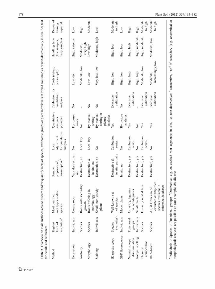
The available techniques for determining root speciesidentity differ broadly in their applicability to distin-guish and to quantify the root systems of functionalgroups, plant taxa and individual plants (Table 2).Because many root traits may vary with environmentalparameters, it seems likely that the search for the “holygrail” of an anatomy, morphology or secondarycompound-based root system taxonomy will remainunsuccessful. IR spectroscopy is the most promisingmethod for non-destructive root species identificationand quantification, in combination with (mini-)rhizo-tron analyses, in the future. Among the destructiveapproaches, multi-loci “barcode” sequencing is themost promising technique to determine species identi-ties, especially after reference libraries become broad-ly available, and qPCR approaches will ease rootquantification in ecosystems which allow for theestablishment of extensive reference curves. The in-creasing interest in belowground research is envi-sioned to bring these techniques from niche existenceto broad application. However, “classical” determina-tion criteria, such as exodermis morphology, will re-main important for now because they are notconsumptive and allow for “secondary” analyses (e.g.,determination of root branching structure, specific rootarea, anatomy, etc.) of the same segment. Further effortsare needed to facilitate the use and the development of
fast and reliable methods and to standardize protocolsand key criteria to enable easy comparisons betweenstudies.
Acknowledgement The authors wish to thank two anonymousreviewers for valuable comments on an earlier version of the man-uscript. B.R. acknowledges support by a postdoctoral fellowshipawarded by the Jacob Blaustein Center for Scientific Cooperation(BCSC), Israel.
Open Access This article is distributed under the terms of theCreative Commons Attribution License which permits any use,distribution, and reproduction in any medium, provided theoriginal author(s) and the source are credited.
References
Agerer R (1988) Colour atlas of ectomycorrhizae. EinhornVerlag, Schwäbisch Gemünd
Ali HAM, Mayes RW, Hector BL, Verma AK, Orskov ER(2005) The possible use of n-alkanes, long-chain fattyalcohols and long-chain fatty acids as markers in studiesof the botanical composition of the diet of free-rangingherbivores. J Agr Sci 143:85–95
Baldwin JP, Tinker PB (1972) Method for estimating lengthsand spatial patterns of two interpenetrating root systems.Plant Soil 37:209–213
Barber SA (1995) Soil nutrient bioavailability: a mechanisticapproach. Wiley, New York
Basconsuelo S, Grossoa M, Molina MG, Malpassia R, Kraus T,Bianco C (2011) Comparative root anatomy of papilionoidlegumes. Flora 206:799–807
Bobowski BR, Hole D, Wolf PG, Bryant L (1999) Identificationof roots of woody species using polymerase chain reaction(PCR) and restriction fragment length polymorphism(RFLP) analysis. Mol Ecol 8:485–491
Böhm W (1979) Methods of studying root systems. Springer,Berlin
Bookman PA, Mack RN (1982) Root interaction between Bromustectorum and Poa pratensis: a three-dimensional analysis.Ecology 63:640–646
Brisson J, Reynolds JF (1994) The effect of neighbors on rootdistribution in a creosotebush (Larrea tridentata) population.Ecol 75:1693–1702
Brundrett MC, Kendrick B (1988) The mycorrhizal status, rootanatomy, and phenology of plants in a Sugar maple forest.Can J For Res 66:1153–1173
Brunner I, Brodbeck S, Buchler U, Sperisen C (2001) Molecularidentification of fine roots of trees from the Alps: reliableand fast DNA extraction and PCR-RFLP analyses of plastidDNA. Mol Ecol 10:2079–2087
Button J, Vardi A, Spiegel-Roy P (1976) Root peroxidase iso-enzymes as an aid in Citrus breeding and taxonomy. TheorAppl Genet 47:119–123
Cahill JF, McNickle GG, Haag JJ, Lamb EG, Nyanumba SM,St. Clair CC (2010) Plants integrate information aboutnutrients and neighbors. Science 328:1657
Plant Soil (2012) 359:165–182 179

Caldwell MM, Richards JH, Manwaring JH, Eissenstat DM(1987) Rapid shifts in phosphate acquisition show di-rect competition between neighbouring plants. Nature327:615–616
Caldwell MM, Manwaring JH, Durham SL (1991) The micro-scale distribution of neighboring plant-roots in fertile soilmicrosites. Funct Ecol 5:765–772
CBOL Plant Working Group (2009) A DNA barcode for landplants. PNAS 106:12794–12797
Chalmers JM, Griffiths PR (2002) Handbook of vibrationalspectroscopy. Wiley, Chichester
Clarke FC, Hammond SV, Jee RD, Moffat AC (2002) Determi-nation of the information depth and sample size for theanalysis of pharmaceutical materials using reflectancenear-infrared microscopy. Appl Spectrosc 56:1475–1483
Coleman SW, Barton FE, Meyer RD (1985) The use of nearinfrared reflectance spectroscopy to predict species com-position of forage mixtures. Crop Sci 25:834–837
Corre-Hellou G, Crozat Y (2005) Assessment of root systemdynamics of species grown in mixtures under field condi-tions using herbicide injection and 15N natural abundancemethods: a case study with pea, barley and mustard. PlantSoil 276:177–192
Cutler DF, Rudall PJ, Gasson PE, Gale RMO (1987) Rootidentification manual of trees and shrubs. A guide to theanatomy of roots of trees and shrubs hardy in Britain andnorthern Europe. Chapman and Hall, London
Dawson LA, Mayes RW, Elston DA, Smart TS (2000) Roothydrocarbons as potential markers for determining speciescomposition. Plant Cell Environ 23:743–750
de Kroon H, Mommer L, Nishiwaki A (2003) Root competition:towards a mechanistic understanding. In: de Kroon H,Visser EJW (eds) Root ecology. Springer, Berlin, pp 215–235
Donaldson DE, Robinson TW (1971) Fluorescent dyes, theiruptake and translocation in plants. Water Resour Res7:692–696
Dove H, Bolger TP (2005) The potential for using the alkanesand long-chain alcohols of plant cuticular wax to distin-guish the contribution of different plant species to a mixedroot mass. In: O’Mara FP, Wilkins RJ, Mannetje L, LovettDK, Rogers PAM, Boland TM (eds) The XX internationalgrassland congress: offered papers. Academic Publishers,Wageningen, pp 646–647
Dove H, Mayes RW, Freer M (1996) Effects of species, plantpart, and plant age on the n-alkane concentrations in thecuticular wax of pasture plants. Aust J Agr Res 47:1333–1347
Dyer D, Brown DA (1983) Relationship of fluorescent intensityto ion uptake and elongation rates of soybean roots. PlantSoil 72:127–134
El Zemrany H, Czarnes S, Hallett PD, Alamercery S, Bally R,Jocteur Monrozier L (2007) Early changes in root charac-teristics of maize (Zea mays) following seed inoculationwith the PGPR Azospirillum lipoferum CRT1. Plant Soil291:109–118
Eleki K, Cruse RM, Albrecht KA (2005) Root segregation of C3
and C4 species using carbon isotope composition. Crop Sci45:879–882
Eshel A, Beeckman T (2012) Plant roots: the hidden half, 4thedn. CRC Press, New York
Faget M, Herrera JM, Stamp P, Aulinger-Leipner I, Frossard E,Liedgens M (2009) The use of green fluorescent protein asa tool to identify roots in mixed plant stands. Funct PlantBiol 36:930–937
Faget M, Liedgens M, Stamp P, Flütsch P, Herrera JM (2010) Aminirhizotron imaging system to identify roots expressingthe green fluorescent protein. Comput Electron Agr74:163–167
Farquhar GD, Ehleringer JR, Hubick KT (1989) Carbon isotopediscrimination and photosynthesis. Annu Rev Plant PhysiolPlant Mol Biol 40:503–537
Finér L, Messier C, De Grandpré L (1997) Fine-root dynamics inmixed boreal conifer - broad-leafed forest stands at differentsuccessional stages after fire. Can J For Res 27:304–314
Fisk MC, Yanai RD, Fierer N (2010) A molecular approach toquantify root community composition in a northern hard-wood forest - testing effects of root species, relative abun-dance, and diameter. Can J For Res 40:836–841
Foley WJ, McIlwee A, Lawler I, Aragones L, Woolnough AP,Berding N (1998) Ecological applications of near infraredreflectance spectroscopy − a tool for rapid, cost-effectiveprediction of the composition of plant and animal tissuesand aspects of animal performance. Oecologia 116:293–305
Frank DA, Pontes AW, Maine EM, Caruana J, Raina R, Raina S,Fridley JD (2010) Grassland root communities: speciesdistributions and how they are linked to abovegroundabundance. Ecology 91:3201–3209
Fußeder A (1986) Verteilung der Wurzelsysteme von Zea maysL. und Lupinus luteus L. in Mischkultur im Hinblick aufdie Konkurrenz um Phosphat und Kalium. Z PflanzBodenkunde 149:541–547
Gamalero E, Trotta A, Massa N, Copetta A, Martinotti MG,Berta G (2004) Impact of two fluorescent pseudomonadsand an arbuscular mycorrhizal fungus on tomato plantgrowth, root architecture and P acquisition. Mycorrhiza14:185–192
Gealy DR, Fischer AJ (2010) 13C discrimination: a stableisotope method to quantify root interactions between C3
rice (Oryza sativa) and C4 barnyardgrass (Echinochloacrus-galli) in flooded fields. Weed Sci 58:359–368
Goodwin RH, Kavanagh F (1948) Fluorescing substances inroots. Bull Torrey Bot Club 75:1–17
Günzler H, Gremlich HU (2002) IR spectroscopy – an introduction.Wiley-VCH, Weinheim
Haling R, Simpson R, McKay A, Hartley D, Lambers H, Ophel-Keller K, Wiebkin S, Herdina H, Riley I, Richardson A(2011) Direct measurement of roots in soil for single andmixed species using a quantitative DNA-based method.Plant Soil 348:123–137
Hegnauer R, Hegnauer M (2001) Chemotaxonomie der PflanzenXIb-2 - Leguminosae: Teil 3. Birkhäuser, Basel
Hölscher D, Hertel D, Leuschner C, Hottkowitz M (2002) Treespecies diversity and soil patchiness in a temperate broad-leaved forest with limited rooting space. Flora 197:118–125
Holzapfel C, Alpert P (2003) Root cooperation in a clonalplant: connected strawberries segregate roots. Oecologia134:72–77
Hsu SCP (1997) Infrared spectroscopy. In: Settler FA (ed) Hand-book of instrumental techniques for analytical chemistry.Prentice-Hall, New Jersey, pp 247–283
180 Plant Soil (2012) 359:165–182
Huang B, Fry JD (1998) Root anatomical, physiological, andmorphological responses to drought stress for tall fescuecultivars. Crop Sci 38:1017–1022
Huber-Sannwald E, Pyke DA, Caldwell MM, Durham S (1998)Effects of nutrient patches and root systems on the clonalplasticity of a rhizomatous grass. Ecol 79:2267–2280
Hudson ME (2008) Sequencing breakthroughs for genomicecology and evolutionary biology. Mol Ecol Res 8:3–17
Jackson RB, Moore LA, Hoffmann WA, Pockman WT, LinderCR (1999) Ecosystem rooting depth determined with cavesand DNA. PNAS 96:11387–11392
Jones FA, Erickson DL, Bernal MA, Bermingham E, Kress WJ,Herre EA, Muller-Landau HC, Turner BL (2011) The rootsof diversity: below ground species richness and rootingdistributions in a tropical forest revealed by DNA barcodesand inverse modeling. PLoS One 6:e24506
Kesanakurti PR, Fazekas AJ, Burgess KS, Percy DM,Newmaster SG, Graham SW, Barrett SCH, Hajibabaei M,Husband BC (2011) Spatial patterns of plant diversitybelow-ground as revealed by DNA barcoding. Mol Ecol20:1289–1302
Kim SW, Ban SH, Chung H, Cho S, Chung HJ, Choi PS, YooOJ, Liu JR (2004) Taxonomic discrimination of floweringplants by multivariate analysis of Fourier transform infraredspectroscopy data. Plant Cell Rep 23:246–250
Kloeppel BD, Gower ST, Treichel IW, Kharuk S (1998) Foliarcarbon isotope discrimination in Larix species and sympatricevergreen conifers: a global comparison. Oecologia114:153–159
Kress WJ, Erickson DL, Jones FA, Swenson NG, Perez R,Sanjur O, Bermingham E (2009) Plant DNA barcodesand a community phylogeny of a tropical forest dynamicsplot in Panama. PNAS 106:18621–18626
Lafitte HR, Champoux MC, McLaren G, O’Toole JC (2001)Rice root morphological traits are related to isozyme groupand adaptation. Field Crop Res 71:57–70
Lei P, Bauhus J (2010) Use of near-infrared reflectance spec-troscopy to predict species composition in tree fine-rootmixtures. Plant Soil 333:93–103
Leva PE, Aguiar MR, Oesterheld M (2009) Underground ecologyin a Patagonian steppe: root traits permit identification ofgraminoid species and classification into functional types. JArid Environ 73:428–434
Li L, Sun JH, Zhang FS, Guo TW, Bao XG, Smith FA, SmithSE (2006) Root distribution and interactions between inter-cropped species. Oecologia 147:280–290
Linder CR, Moore LA, Jackson RB (2000) A universal molec-ular method for identifying underground plant parts tospecies. Mol Ecol 9:1549–1559
Litav M, Harper JL (1967) A method for studying spatial relation-ships between the root systems of two neighbouring plants.Plant Soil 26:389–392
Lopez-Zamora I, Comerford NB, Muchovej RM (2004) Rootdevelopment and competitive ability of the invasive spe-cies Melaleuca quinquenervia (Cav.) S.T. Blake in theSouth Florida flatwoods. Plant Soil 263:239–247
Ludlow MM, Troughton JH, Jones RJ (1976) A technique for deter-mining the proportion of C3 and C4 species in plant samplesusing stable natural isotope’s of carbon. J Agr Sci 87:625–632
McNickle GG, Cahill JF, Deyholos M (2008) A PCR-basedmethod for the identification of the roots of 10 co-
occurring grassland species in mesocosm experiments.Botany 86:485–490
Meinen C (2008) Fine root dynamics in broad-leaved deciduousforest stands differing in tree species diversity. Dissertation,University of Göttingen, Germany
Meinen C, Hertel D, Leuschner C (2009a) Biomass and mor-phology of fine roots in temperate broad-leaved forestsdiffering in tree species diversity: is there evidence ofbelow-ground overyielding? Oecologia 161:99–111
Meinen C, Hertel D, Leuschner C (2009b) Root growth andrecovery in temperate broad-leaved forest stands differingin tree species diversity. Ecosystems 12:1103–1116
Mommer L, Wagemaker CAM, de Kroon H, Ouborg NJ (2008)Unravelling below-ground plant distributions: a real-timepolymerase chain reaction method for quantifying speciesproportions in mixed root samples. Mol Ecol Res 8:947–953
Moore LA, Field CB (2005) A technique for identifying theroots of different species in mixed samples using nuclearribosomal DNA. J Veg Sci 16:131–134
Murakami T, Shimano S, Kaneda S, Nakajima M, Urashima Y,Miyoshi N (2006) Multicolor staining of root systems inpot culture. Soil Sci Plant Nutr 52:618–622
Murphy MT, McKinley A, Moore TR (2009) Variations in above-and below-ground vascular plant biomass and water table ona temperate ombrotrophic peatland. Botany 87:845–853
Nakaji T, Noguchi K, Oguma H (2008) Classification of rhizo-sphere components using visible-near infrared spectralimages. Plant Soil 310:245–261
Naumann A, Heine G, Rauber R (2010) Efficient discriminationof oat and pea roots by cluster analysis of Fourier transforminfrared (FT IR) spectra. Field Crop Res 119:78–84
Nordby HE, Nagy S (1981) Chemotaxonomic study of neutralcoumarins in roots of Citrus and Poncirus by thin-layer,gas-liquid and high-performance liquid chromatographicanalyses. J Chromatogr A 207:21–28
Pierret A (2008) Multi-spectral imaging of rhizobox systems:new perspectives for the observation and discrimination ofrhizosphere components. Plant Soil 310:263–268
Polley HW, Johnson HB, Mayeux HS (1992) Determination ofroot biomasses of three species grown in a mixture: usingstable isotopes of carbon and nitrogen. Plant Soil 142:97–106
Prakash N (1972) Root-wood anatomy of some tropical eco-nomic plants. Notes Jodrell Laboratory. Royal Bot GardensKew 7:1–19
Rewald B, Ephrath JE (2012) Minirhizotron technique. In: EshelA, Beeckman T (eds) Plant roots: the hidden half, 4th edn.CRC Press, New York
Rewald B, Leuschner C (2009) Belowground competition in abroad-leaved temperate mixed forest: pattern analysis andexperiments in a four-species stand. Eur J For Res 128:387–398
Rewald B, Leuschner C, Wiesman Z, Ephrath JE (2011) Influenceof salinity on root hydraulic properties of three olive varieties.Plant Biosys 145:12–22
Reynolds HL, Packer A, Bever JD, Clay K (2003) Grassrootsecology: plant-microbe-soil interactions as drivers of plantcommunity structure and dynamics. Ecology 84:2281–2291
Rice KJ, Gordon DR, Hardison JL, Welker JM (1993) Pheno-typic variation in seedlings of a keystone tree species(Quercus douglasii) - the interactive effects of acorn sourceand competitive environment. Oecologia 96:537–547
Plant Soil (2012) 359:165–182 181

Ridgway KP, Duck JM, Young JPW (2003) Identification ofroots from grass swards using PCR-RFLP and FFLP of theplastid trnL (UAA) intron. BMC Ecol 3:8
Riedel H (1937) Bau und Leistungen des Wurzelholzes. GebrüderBorntraeger, Leipzig
Riley IT, Wiebkin S, Hartley D, McKay AC (2010) Quantifica-tion of roots and seeds in soil with real-time PCR. PlantSoil 331:151–163
Roumet C, Picon-Cochard C, Dawson LA, Joffre R, Mayes R,Blanchard A, Brewer MJ (2006) Quantifying species com-position in root mixtures using two methods: near-infraredreflectance spectroscopy and plant wax markers. New Phytol170:631–638
Rumbaugh MD, Clark DH, Pendery BM (1988) Determina-tion of root mass ratios in alfalfa-grass mixtures usingnear infrared reflectance spectroscopy. J Range Manage41:488–490
Schenk HJ (2006) Root competition: beyond resource depletion.J Ecology 94:725–739
Schenk HJ, Callaway RM, Mahall BE (1999) Spatial root seg-regation: are plants territorial? Adv Ecol Res 28:145–180
Schmidt S, Stewart GR (2003) δ15N values of tropical savannaand monsoon forest species reflect root specialisations andsoil nitrogen status. Oecologia 134:569–577
Seneviratne GI, Harborne JB (1992) Constitutive flavonoids andinduced isoflavonoids as taxonomic markers in the genusVigna. Biochemic Syst Ecol 20:459–467
Sharma A, Schulman SG (1999) Introduction to fluorescencespectroscopy. Wiley, New York
Shearer G, Kohl DH, Virginia RA, Bryan BA, Skeeters JL,Nilsen ET, Sharifi MR, Rundel PW (1983) Estimates ofN2-fixation from variation in the natural abundance of15N in Sonoran Desert ecosystems. Oecologia 56:365–373
Shenk JS, Westerhaus MO, Hoover MR (1979) Analysis offorages by infrared reflectance. J Dairy Sci 62:807–812
Skrabal PM (2009) Spektroskopie. Eine methodenübergreifendeDarstellung vom UV- bis zum NMR-Bereich. UTB, Zurich,Switzerland
Soussana JF, Teyssonneyre F, Picon-Cochard C, Dawson L(2005) A trade-off between nitrogen uptake and useincreases responsiveness to elevated CO2 in infrequentlycut mixed C3 grasses. New Phytol 166:217–230
Svejcar TJ, Boutton TW (1985) The use of stable carbon isotopeanalysis in rooting studies. Oecologia 67:205–208
Taggart JM, Cahill JF, McNickle GG, Hall JC (2011) Molecularidentification of roots from a grassland community usingsize differences in fluorescently labelled PCR amplicons ofthree cpDNA regions. Mol Ecol Res 11:185–195
Taylor JH, Waltenbaugh A, Shields M (2008) Impact of vesiculararbuscular mycorrhiza on root anatomy in Zea mays andLycopersicon esculentum. African J Agr Res 3:1–6
Tosti G, Thorup-Kristensen K (2010) Using coloured roots tostudy root interaction and competition in intercroppedlegumes and non-legumes. J Plant Ecol 3:191–199
TrockenbrodtM, Richter HG,Möller-Lindenhof Y, Dujesiefken D(2001) Identifizierung von Baumwurzeln - Möglichkeitender Bestimmung anhand der Holz und Rindenstruktur. Stadtund Grün 6:430–434
Van Wyk BE, Yenesew A, Dagne E (1995) Chemotaxonomicsignificance of anthraquinones in the roots of Asphodeloi-deae (Asphodelaceae). Biochem Syst Ecol 23:277–281
Vandenkoornhuyse P, Ridgway KP, Watson IJ, Fitter AH, YoungJPW (2003) Co-existing grass species have distinctive arbus-cular mycorrhizal communities. Mol Ecol 12:3085–3095
Walker MA, Liu L (1995) The use of isozymes to identify 60grapevine rootstocks (Vitis spp.). Am J Enol Viticult 46:299–305
Wang X, Qin M, Liang Z (2003) Microscopical identification ofroots and rhizome of six species of plants from Viola in theregion of Nanjing. Zhong Yao Cai 26:401–403, In Chinese,English abstract
Wardle DA, Peltzer DA (2003) Interspecific interactions andbiomass allocation among grassland plant species. Oikos100:497–506
Williams DG, Ehleringer JR (2000) Carbon isotope discriminationandwater relations of oak hybrid populations in southwesternUtah. West N Am Naturalist 60:121–129
Wong SC, Osmond CB (1991) Elevated atmospheric partial-pressure of CO2 and plant growth. 3. Interactions betweenTriticum aestivum (C3) and Echinochloa frumentacea (C4)during growth in mixed culture under different CO2, N-nutrition and irradiance treatments, with emphasis on be-lowground responses estimated using the δ13C value ofroot biomass. Aust J Plant Physiol 18:137–152
Yanai RD, Fisk MC, Fahey TJ, Cleavitt NL, Park BB (2008)Identifying roots of northern hardwood species: patternswith diameter and depth. Can J For Res 38:2862–2869
Zhao HR, Wang XY, Chen GH, Wen SM (2004) Identificationof wheat varieties by FT IR spectrum. Spectrosc SpectAnal 24:1338–1341, In Chinese, English abstract
182 Plant Soil (2012) 359:165–182