Embed Size (px)
Citation preview
Routine Leaflet Augmentation in the
Repair of CAVSDHani K. Najm MD, Msc, FRCSC, FRCS(UK) King Abdulaziz Cardiac CenterNational Guard Health AffairsRiyadh, Saudi Arabia

• The outcome of repair of complete AVSD is dependent on multiple factors such as: – The morphology of the lesion– Technique of repair– Perioperative care
• A better understanding of the morphology of CAVSD has impacted the outcome

Septum
Conduction systemvalves

I - Septal lesions
• ASD premium• VSD Inlet

• Decrease distance inlet-apex
• increase distance apex-outlet
• Aortic valve is elevated and deviated anteriorly
• LVOT narrowed, but rarely hemodynamically significant “gooseneck” LVOT

What is a Successful operation ?
• surgical repair is directed towards :1. closure of ASD, VSD2. avoidance of damage to the AV node and
bundle of His3. maintenance or creation of two competent,
non-stenotic AV valves

Evaluation
CAVSD
ASD LV LAVV
cleft
Place chordae
VSD
size
Papillary muscle

CAVSD Repair
• Single patch with leaflet division• Double patch with leaflet preservation• Single atrial patch with no VSD patch
(Australian)

Single Patch Technique

VSD ClosureDouble Patch Technique

• complete closure of “cleft” is recommended
• primary valve replacement should be avoided
• To prevent postoperative LVOTO– adequate sizing of interventricular patch– abnormal chordae to left-sided outlet septum– accessory valve tissue in outflow tract
LAVV Repair Techniques

LAVV repair techniques

Single Papillary Muscle
Parachute mitral valve
Valve composed of anterior and posterior leaflet without lateral i.e. bicuspid.
Split the muscle as far as possible.With partial closure of cleft on Hegar

Double orifice AV valve.
• Partial Closure of cleft with measurement

ASD Closure

Intermediate Form4 ASD +VSD (Small)4 Common AVV4 Chordae attach to base of the
cleft which lead to restricted leaflet motion
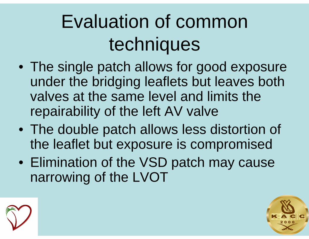
Evaluation of common techniques
• The single patch allows for good exposure under the bridging leaflets but leaves both valves at the same level and limits the repairability of the left AV valve
• The double patch allows less distortion of the leaflet but exposure is compromised
• Elimination of the VSD patch may cause narrowing of the LVOT

• All techniques do not address the deficient leaflet issue which may result in post operative LAVV regurgitation

Modified Operative Technique • Rt atriotomy, inspection of the defect
and evaluation of the common AV valve
• Both superior and inferior bridging leaflet are divided with inspection of the chordal attachment
• A patch of fresh autologus pericardium is accurately shaped
• The ventricular component is attached the crest of the ventricular septum

• The free edge is attached to the divided left AV valve leaflet at the same time approximating the base of the cleft
• The cleft is then closed• The LAVV is tested• The atrial patch is attached 3-5mm
away from the edge of the VSD patch together with approximating the RAVV. This part allows for better coaptation of the LAVV
• RA is then closed
Modified Operative Technique 2


Results From Jul 2000- Dec 2010No. Of children 148Mean age (mos) 9±1.5Median age (mos) 5.7Mean wt. (Kg) 6.7±0.8Pre-op banding 1Transitional type 15Coarcatation repair without banding 3Associated TOF/DORV 13DOLAVV 2Parachute LAVV 4

Amount Of AVSD’s Per Year
7 7
15
2018 18
25
15 15 16
8
0
5
10
15
20
25
30
2000 2001 2002 2003 2004 2005 2006 2007 2008 2009 2010

Age At Surgery
78
57
124 2 3 1 1 1 2 1 1 1
0
10
20
30
40
50
60
70
80
Infant<6/12
Infant>6/12
1 Yrs 2 Yrs 3 Yrs 4 Yrs 5 Yrs 6 Yrs 7 Yrs 8 Yrs 10 Yrs 11 Yrs 13 Yrs


Down’s Syndrome vs - Non Downs’
70
300
10
20
30
40
50
60
70
Down's Non Down's
Perc
enta
ge

Results
• Type of VSD Pacth – Gortex (PTFE) 5– Fresh autologus pericardium 143
• Atrial patch autologus pericardium 148• Additional LAVV repair 35%• TPT 85±24• Ischemic time 63±17

Other Procedures During repair• BTS Takedown 3• Coarctation 3• DORV Repair 4• Cor Triatriatum Repair 1• Pulmonary Artery DeBanding 1• Pulmonary Venous Stenosis Repair 1• Right Ventricle Outflow Tract Procedure 5• Right Ventricle Overhaul 15• TOF Transannular Patch 10• Baffle of LSVC to RA

Results
• Ventilatory support(days) 2±1• LCO 8• Renal failure requiring dialysis 2• ECMO support 2• Hospital stay 10±5• Early mortality 1 (0.7%)

Further Surgeries Required
• Early– Coarctation repair 1 – AVV Repair 1– Permanent Pacemaker 1
• Late – AVV Repair 6 (4%) – Mitral Valve Replacement 2 (1.3%)– PPM 1– PA stenting 1

Results
Follow up (mos) 55Late mortality 2Aneurysm of the VSD patch 1

Echocardiographic Results
• Last post operative LAVV regurgitation by TTE (24 lost to follow up)– None or trace 90– Mild 22– Moderate 11– Severe 2


Echocardiographic Results
• Residual VSD 1• Subaortic gradient 0• Clear evidence of LAVV augmentation by
the modified technique

• Between 1975 and 2006, • 312 patients underwent surgery for :
– complete AVSD (n 209; 67.0%), – partial AVSD (n 76; 24.4%), – or intermediate AVSD (n 27; 8.6%).
• Mean age was 2.4 +/- 3.9 years; • 142 patients (45.5%) were younger than 6
months. • Follow-up was 99.0% complete.

AVSD
• 26 in-hospital deaths (8.3%) • 6 late deaths (2.1% of 283).• Of the hospital survivors, 43 (14%) patients required a late
reoperation.

Methods• Between April 1997 and April 2007, • 93 consecutive patients underwent surgery for
biventricular correction of AVSD • Median age of 5.8 months (range, 9 days to 68.9 years). • Fifty-three patients had complete AVSD, • 6 patients had an intermediate type, • 29 patients had partial AVSD; • 4 patients had a complete AVSD with associated
tetralogy of Fallot, • 1 patient had a complete AVSD with double-outlet right
ventricle.

Results
• There was no in-hospital mortality. • There were 2 late deaths (2.2%). • Forty-three reoperations were performed
in 23 patients (24.7%), – 18 were for repair of significant left
atrioventricular valve regurgitation– 8 were mitral valve replacements.
• Seven patients (7.5%) required insertion of a permanent pacemaker.

Conclusions• Repair of AVSD depends on thorough knowledge and
evaluation of morphologic substrate• The use of this modified technique yields good results• This technique allows for good exposure, good LAVV
reconstruction and close to anatomical repair• The technique is in particular beneficial in deficient
leaflet tissue and may decrease the incidence of complete heart block
• This repair can be performed early in life with good results