Embed Size (px)
Citation preview

Routine Radiology of theTrauma Patient
Chantal VCA 440
Introduction

What to look for in the thorax
Pulmonary contusionsHemothorax
PneumomediastinumPneumothorax
Traumatic diaphragmatic hernia
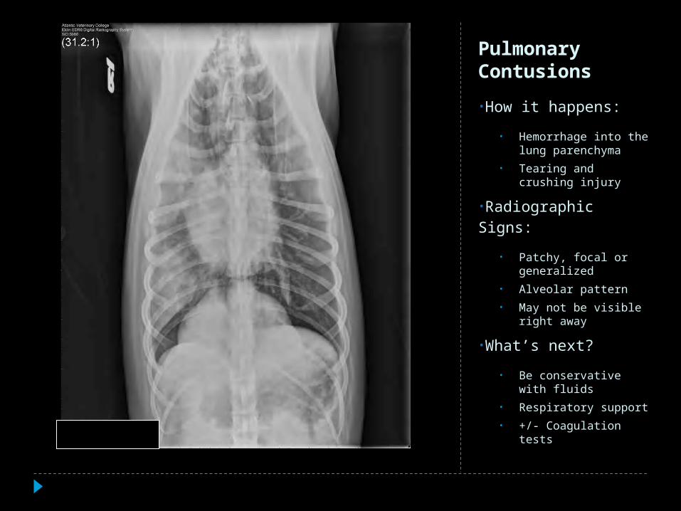
Pulmonary Contusions
•How it happens:
• Hemorrhage into the lung parenchyma
• Tearing and crushing injury
•Radiographic Signs:
• Patchy, focal or generalized
• Alveolar pattern
• May not be visible right away
•What’s next?
• Be conservative with fluids
• Respiratory support
• +/- Coagulation tests

Pulmonary Contusions

Hemothorax
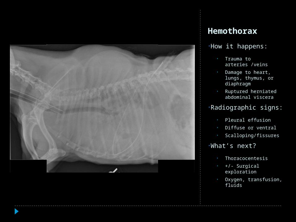
Hemothorax
•How it happens:
• Trauma to arteries /veins
• Damage to heart, lungs, thymus, or diaphragm
• Ruptured herniated abdominal viscera
•Radiographic signs:
• Pleural effusion
• Diffuse or ventral
• Scalloping/fissures
•What’s next?
• Thoracocentesis
• +/- Surgical exploration
• Oxygen, transfusion, fluids

Pneumomediastinum
•How it happens:
• Ruptured alveoli, trachea, or esophagus
• Tracheal avulsion
•Radiographic signs:
• Distinction of structures normally not seen
• Tracheal wall outlined
• SQ emphysema
•What’s next?
• Repair rents
• Monitor for progression

Pneumomediastinum

Pneumothorax

Pneumothorax
•How it happens:
• Chest wall rent
• Lung rupture
• Extension of pneumomediastinum
•Radiographic signs:
• Retracted lungs
• +/- collapse
• Raised heart
• +/-Small heart
• +/-flat caudal diaphragm
• +/-mediastinal shift
•What’s next?
• Thoracocentesis

Pneumothorax

Traumatic Diaphragmatic Hernia
•How it happens:
• Rapid increase in intra-abdominal pressure
• Rent in the muscular portion
•Radiographic signs:
• +/-Pleural effusion• +/-Gas filled loops, liver
stomach,spleen• +/-Loss of diaphragmatic
outline• +/-Asymmetric on VD/DV• +/-Missing viscera from
abdomen
•What’s next?
• Contrast study to definitively diagnose

The UpperGI

What to look for in the abdomen
Hemoperitoneum
Renal avulsion
Uroperitoneum
Traumatic hernias

Hemoperitoneum
•How it happens:
• Ruptured spleen
• Ruptured liver
• Disrupted vasculature
• Avulsed bladder
•Radiographic signs:
• Peritoneal effusion
• Focal or diffuse
• Decreased serosal detail
•What’s next?
• U/S
• Abdominocentesis

Hemoperitoneum

Renal Avulsion
•Radiographic signs:
• Focal decreased serosal detail
• Missing kidney
• Mass in caudal abdomen
•What’s next?
• U/S

Renal Avulsion

Renal Avulsion
Right Kidney Left (avulsed) Kidney

Uroperitoneum
•How it happens:
• Ruptured bladder
• Avulsed/torn ureter
• Urethral tear
•Radiographic signs:
• Diffuse decreased serosal detail
• Focal detail loss in the RPS
•What’s next?
• Abdominocentesis
• IVP (EU)
• Cystogram
• Urethrogram

Ruptured Bladder
The urethrocystogram
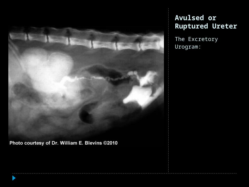
Avulsed or Ruptured Ureter
The Excretory Urogram:

Ruptured Urethra
The Urethrogram
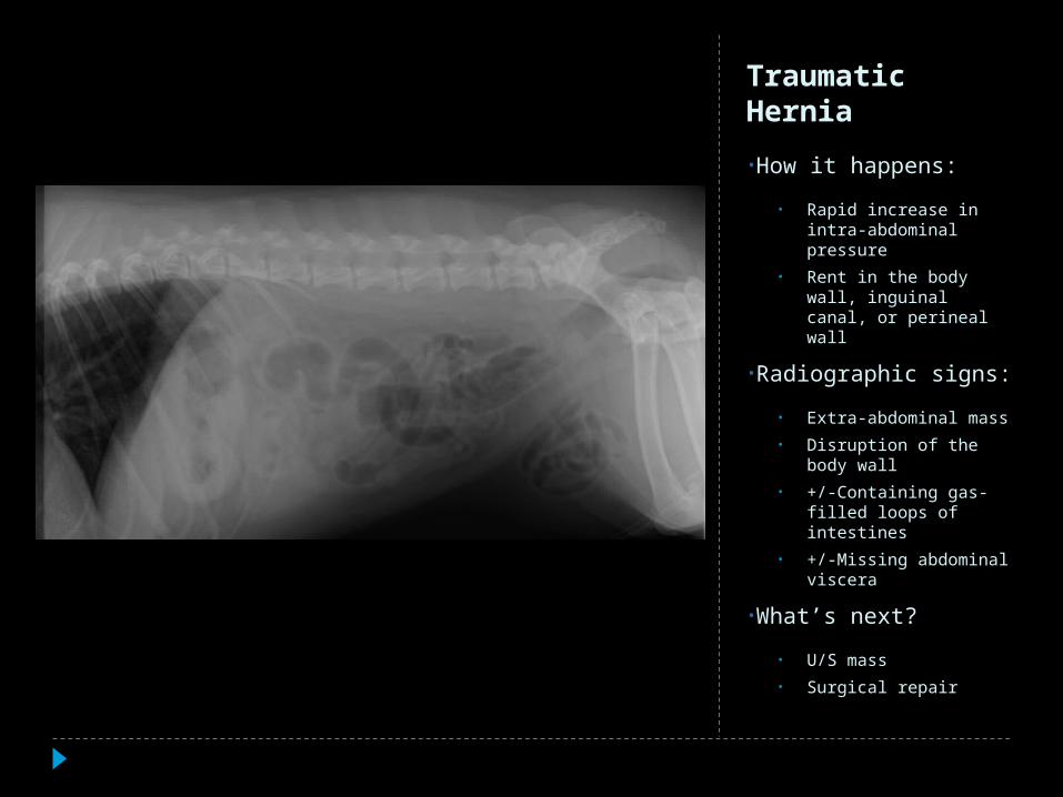
Traumatic Hernia
•How it happens:
• Rapid increase in intra-abdominal pressure
• Rent in the body wall, inguinal canal, or perineal wall
•Radiographic signs:
• Extra-abdominal mass
• Disruption of the body wall
• +/-Containing gas-filled loops of intestines
• +/-Missing abdominal viscera
•What’s next?
• U/S mass
• Surgical repair

Traumatic Hernia
In summary
•Breath
•Stabilize
•Don’t get dazzled by an impressive fracture
•Get ALL the info