Embed Size (px)
Citation preview
Articles
1066 www.thelancet.com Vol 380 September 22, 2012
Lancet 2012; 380: 1066–74
See Comment page 1036
Research Centre for Clinical and Community Practice
Innovation (Prof C M Rickard PhD,
Prof J Webster BA, Prof M C Wallis PhD, N Marsh BN,
J R Gowardman FCICM/FRACP, L Zhang PhD,
Prof M Whitby PhD), and National Health and Medical
Research Council Centre of Research Excellence in Nursing Interventions for Hospitalised
Patients (Prof C M Rickard, Prof J Webster, Prof M C Wallis,
N Marsh), Griffith Health Institute, Griffith University,
Nathan, QLD, Australia; Research and Development
Unit, Centre for Clinical Nursing (Prof C M Rickard,
Prof J Webster, N Marsh, A McClymont RN), and
Department of Intensive Care Medicine (J R Gowardman),
Royal Brisbane and Women’s Hospital, Herston, QLD,
Australia; Gippsland Medical School, Monash University,
Churchill, VIC, Australia (M R McGrail PhD); Princess
Alexandra Hospital, Woolloongabba, QLD, Australia
(Prof C M Rickard, V French RN, P Gallagher MBA,
Prof M Whitby); and Gold Coast Hospital, Southport,
QLD, Australia (Prof M C Wallis, L Foster MN)
Correspondence to: Prof Claire M Rickard,
Research Centre for Clinical and Community Practice Innovation,
Griffith Health Institute, Griffith University, Nathan,
QLD 4111, Australia [email protected]
Routine versus clinically indicated replacement of peripheral intravenous catheters: a randomised controlled equivalence trialClaire M Rickard, Joan Webster, Marianne C Wallis, Nicole Marsh, Matthew R McGrail, Venessa French, Lynelle Foster, Peter Gallagher, John R Gowardman, Li Zhang, Alice McClymont, Michael Whitby
SummaryBackground The millions of peripheral intravenous catheters used each year are recommended for 72–96 h replacement in adults. This routine replacement increases health-care costs and staff workload and requires patients to undergo repeated invasive procedures. The effectiveness of the practice is not well established. Our hypothesis was that clinically indicated catheter replacement is of equal benefit to routine replacement.
Methods This multicentre, randomised, non-blinded equivalence trial recruited adults (≥18 years) with an intravenous catheter of expected use longer than 4 days from three hospitals in Queensland, Australia, between May 20, 2008, and Sept 9, 2009. Computer-generated random assignment (1:1 ratio, no blocking, stratified by hospital, concealed before allocation) was to clinically indicated replacement, or third daily routine replacement. Patients, clinical staff, and research nurses could not be masked after treatment allocation because of the nature of the intervention. The primary outcome was phlebitis during catheterisation or within 48 h after removal. The equivalence margin was set at 3%. Primary analysis was by intention to treat. Secondary endpoints were catheter-related bloodstream and local infections, all bloodstream infections, catheter tip colonisation, infusion failure, catheter numbers used, therapy duration, mortality, and costs. This trial is registered with the Australian New Zealand Clinical Trials Registry, number ACTRN12608000445370.
Findings All 3283 patients randomised (5907 catheters) were included in our analysis (1593 clinically indicated; 1690 routine replacement). Mean dwell time for catheters in situ on day 3 was 99 h (SD 54) when replaced as clinically indicated and 70 h (13) when routinely replaced. Phlebitis occurred in 114 of 1593 (7%) patients in the clinically indicated group and in 114 of 1690 (7%) patients in the routine replacement group, an absolute risk difference of 0·41% (95% CI –1·33 to 2·15%), which was within the prespecified 3% equivalence margin. No serious adverse events related to study interventions occurred.
Interpretation Peripheral intravenous catheters can be removed as clinically indicated; this policy will avoid millions of catheter insertions, associated discomfort, and substantial costs in both equipment and staff workload. Ongoing close monitoring should continue with timely treatment cessation and prompt removal for complications.
Funding Australian National Health and Medical Research Council.
IntroductionUp to 70% of patients in acute care hospitals need a short peripheral intravenous catheter; about 200 million are used each year in the USA alone.1,2 Intravenous catheters frequently fail before the end of treatment because of irritation of the vein (phlebitis) with symptoms includ ing pain, swelling, redness, occlusion, and a palpable venous cord. Phlebitis necessitates catheter removal and replacement. Peripheral intravenous catheterrelated blood stream infection is a less frequent but serious compli cation, occuring in about 0·1% of intravenous catheters or 0·5 per 1000 catheter days.3
Intravenous catheters are often needed for a week or more, but have been recommended for regular removal and replacement with a new catheter in an attempt to decrease both phlebitis and infection. Replacement requires additional needlesticks for patients, increases work for clinical staff, and contributes to insertion of
intravenous catheters being the most common invasive medical procedure and therefore a substantial contributor to healthcare costs.2 Routine intravenous catheter replace ment no more frequently than every 72–96 hours is currently recommended for adults by the US Centers for Disease Control and Prevention (CDC).4 By contrast, the CDC recommends not to routinely replace intravenous catheters in children, or in highrisk catheters, such as arterial, haemodialysis, and central venous catheters, for which research has shown routine replacement does not prevent infection.4,5
Modern intravenous catheters are made of lowirritant materials and might not need routine replacement. Results of welldesigned observational studies have shown that longer dwell time increases daily phlebitis risk in a linear rather than exponential manner (ie, more intravenous catheter days overall increases risk, but later days of cannulation are not higher risk than
Articles
www.thelancet.com Vol 380 September 22, 2012 1067
earlier days).6–8 Thus, routine replacement of intravenous catheters might not benefit patients overall, since the same number of complications might be spread between larger numbers of devices. In 2007–10, results of four randomised controlled trials (n=200–755) supported clinically indicated removal of intravenous catheters as a safe alternative to routine replacement.9–12 Despite this evidence, concerns have remained about whether aban doning this established practice would increase blood stream infections.1 The 2011 CDC guidelines designate clinically indicated replacement of intravenous catheters as an unresolved issue, indicating that more research is needed.4
We aimed to understand the effect of extension of intravenous catheter dwelltime beyond 3 days with replacement of catheters only for clinical reasons. We postulated that patients who had intravenous catheters replaced when clinically indicated would have equivalent rates of phlebitis, and no difference in other complications, but reduced costs and number of catheter insertions, compared with patients with catheters removed every third day.
MethodsStudy design and participantsWe did this multicentre, nonblinded, randomised controlled equivalence trial in three universityaffiliated, government hospitals in Queensland, Australia (Royal Brisbane and Women’s Hospital, Herston; Princess Alexandra Hospital, Woolloongabba; and Gold Coast Hospital, Southport). Recruitment was from May 20, 2008, until the target sample size was achieved on Sept 9, 2009.
Research nurses screened medical and surgical units daily for participants. Patients aged at least 18 years with an intravenous catheter in place and expected treatment of longer than 4 days were eligible. Exclusion criteria were bloodstream infection, planned removal of intra venous catheter within 24 h, or intravenous catheter already in situ for more than 72 h. We permitted intravenous catheters inserted in any clinical area, including the emergency department and operating room. Intravenous catheters inserted in an emergency were not eligible (hospital policies required that these be replaced within 24 h). Intravenous catheters could be inserted by any nurse or doctor or by intravenous insertion teams (with no postinsertion care) in two of the hospitals. The ethics committee at each hospital and at Griffith University approved the protocol. We obtained written, informed consent from all participants before enrolment.
Randomisation and maskingPatients were randomly assigned to one of two treatment groups (simple randomisation with 1:1 ratio, no blocking, stratified by hospital). Random allocations were com putergenerated on a handheld device, at the point of each patient’s study entry, and thus were
concealed to patients, clinical staff, and research staff until this time. Patients and clinical staff could not be masked after allocation because of the nature of the intervention. Research nurses were similarly not masked because they had to allocate patients to the treatment group and monitor the integrity of the intervention. However, laboratory staff were masked for rating of all microbiological endpoints, and a masked, independent medical rater diagnosed catheterrelated infections and all bloodstream infections. We did blinded interrater reliability checks on a subset of phlebitis assessments.
ProceduresPatients in the clinically indicated group had their intravenous catheters removed only for completion of therapy, phlebitis, infiltration, occlusion, accidental removal, or suspected infection. Patients in the routine replacement group had their intravenous catheters replaced every third calendar day, unless clinical reasons made this impossible (eg, intravenous catheters failed before day 3, or patient unable to be recannulated). The day 3 resite occurred at about 72 h (48–96 h depending on insertion and removal times). Investigators and research nurses had no involvement in the decision to remove intravenous catheters, or to order cultures, and did not resite the catheters. Preinsertion skin disinfection was with 2% chlorhexidine in 70% ethanol. Insyte Autoguard 30 mm intravenous catheters (BD Medical, Franklin Lakes, NJ, USA) and Tegaderm 1624W trans parent dressings (3M, St Paul, MN, USA) were used, with dressings replaced weekly and when soiled or loose. Chlorhexidineimpregnated sponges were not used. We placed no restriction on products administered through the intravenous catheters. Patients generally entered the study with their first intravenous catheter,
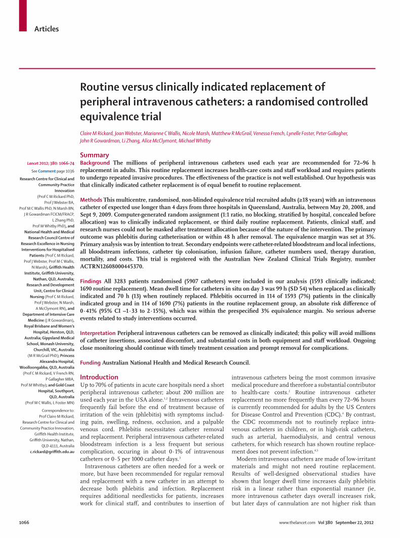
3379 assessed for eligibility
3283 randomised
1593 assigned clinically indicated removal
1690 assigned routine replacement on day 3
1593 included in analysis 1690 included in analysis
96 excluded for pre-existing bloodstream infection or intravenous catheter in situ for more than 72 h
1186 in situ on day 3 androutinely replaced
504 not in situ on day 3 or clinically replaced
1351 in situ on day 3 andclinically replaced
242 not in situ on day 3 or routinely replaced
Figure 1: Study profile of patient flow
Articles
1068 www.thelancet.com Vol 380 September 22, 2012
and all subsequent intravenous catheters (maximum five) for a course of treatment were included. An independent data and safety monitoring committee reviewed blinded data at two apriori defined intervals (n=1000 and n=2000), and recommended that the trial continue. Stopping rules were a greater than 2:1 ratio in either group for phlebitis or catheterrelated bloodstream infection.
The primary outcome was phlebitis during catheterisation or within 48 h after removal.8,13 Phlebitis was two or more of the following, present simultaneously: (1) patientreported pain or tenderness (on questioning, then palpation by the research nurse) with a severity of two or more on a tenpoint scale; (2) erythema, extending at least 1 cm from the insertion site; (3) swelling, extending at least 1 cm from the insertion site; (4) purulent discharge; or (5) palpable venous cord beyond the intravenous catheter tip. All items apart from patientreported pain or tenderness were rated by the research nurse after direct assessment of the patient, and review of clinical data. Phlebitis measures were repeated daily, and at 48 h after removal (by telephone if the patient had been discharged). A structured outcome assessment form was used and interrater reliability testing was done. A study manager visited each site at least monthly and was available to the research nurses at any time over the telephone. She audited study data for completeness and accuracy against hospital records and supervised the research nurses for compliance with study procedures. Monthly meetings were held by the investigators, research nurses, and the study manager to review progress and ensure consistency between sites.
Secondary endpoints were: (1) catheterrelated bloodstream infection, defined as positive blood culture from a peripheral vein; clinical signs of infection (ie, fever, chills, or hypotension); no other apparent source for the bloodstream infection except the intravenous catheter (in situ within 48 h of the bloodstream infection); and a colonised intravenous catheter tip culture with the same organism as identified in the blood;14 (2) allcause bloodstream infections, defined as any positive blood culture drawn from a peripheral vein while intravenous catheter in situ or for 48 h after removal; (3) local venous infection, defined as organisms grown from purulent discharge or vein segment with no evidence of associated bloodstream infection;14 (4) colon isation of intravenous catheter tip, with more than 15 colonyforming units15 (substudy of a convenience sample of 5% of patients, ie, catheters that were removed on days and times that the research nurses were available to take cultures); (5) infusion failure, defined as any premature removal of intravenous catheter before end of treatment, other than for routine replacement—includes phlebitis, infiltration, occlusion, accidental removal, and catheterrelated bloodstream infections; (6) number of intravenous catheters needed per patient for course of treatment; (7) overall duration of intravenous therapy (cumulative of all intravenous catheters) per patient (h); (8) costs per patient for the course of intravenous therapy, based on equipment required for insertion and removal of intravenous catheters12 with prices from negotiated hospital supply contract rate; and (9) mortality with intravenous catheter in situ or within 48 h of removal, collected from hospital records. Hospital costs were set at AU$25·13 for intravenous catheter plus administration set, burette, and fluid bag; $21·83 for intravenous catheter plus admin istration set and fluid bag; $12·73 for intravenous catheter plus end cap; and $0·37 gauze and tape for removal; and staff time (observed rates of 14·5 min per insertion and 4·5 min per removal at fixed industrial award wages for registered nurses of $32·93/h, junior [$45·96/h] and senior [$67·16/h] medical staff). Catheters inserted by intra venous insertion teams were costed at registered nurse rates; intravenous catheters inserted in an operating theatre or radiology suite were costed at senior medical officer rates, and other insertions were costed at junior medical officer to registered nurse rates in a 3:1 ratio (on the basis of internal observations). We did not include the cost of treating complications associated with intra venous catheters (since treatment typically consisted only of removal and replacement of the affected intravenous catheter, which was already accounted for in the cost calculations).
Statistical analysisWe used a twosided design to test equivalence between groups. The sample size was calculated to detect equivalence at 4% phlebitis11 (equivalence margin 3%) with 5% significance and more than 95% power. This
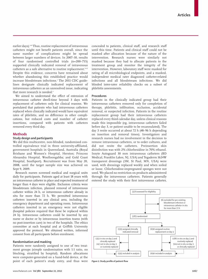
Clinically indicated (n=1593)
Routine replacement (n=1690)
Intravenous catheter dwell time (h)*
Mean (SD) 99 (54) 70 (13)
Median (IQR) 84 (64–118) 70 (57–77)
Age (years) 55·1 (18·6) 55·0 (18·4)
Men 1022 (64%) 1034 (61%)
Type of admission
Medical 292 (18%) 331 (20%)
Surgical 1301 (82%) 1359 (80%)
Comorbidities
None 387 (24%) 411 (24%)
One 350 (22%) 372 (22%)
Two or more 856 (54%) 907 (54%)
Present wound infection 256 (16%) 244 (14%)
Wound drain 95 (6%) 116 (7%)
Stoma 27 (2%) 37 (2%)
Data are mean (SD) or n (%). *Per protocol, N=1351 clinically indicated; N=1186 routine replacement.
Table 1: Baseline demographics and clinical characteristics of patients

Articles
www.thelancet.com Vol 380 September 22, 2012 1069
determined a total sample of 3000 patients, plus 300 to allow for attrition. Data were entered directly into a purposebuilt Microsoft Access (version 97) database. We used SPSS (version 18) and StataSE (version 10) for analyses.
Baseline characteristics of patients and catheters were described by group. The primary analysis was by intention to treat, including all patients (and all catheters) in their ran domised group. First, we calculated relative incidence rates of phlebitis and absolute rate differences per 100 catheters and per 100 patients, and we then used twosided Fisher’s exact test to assess equivalence in risk between groups. Second, we calculated hazard rates per 1000 catheter hours, with 95% CI, with a Cox proportional hazards model (assumptions were checked) to sum marise the effect of intervention per patient (includ ing all catheters per patient). Finally, we used KaplanMeier survival curves to compare rates of time until first phlebitis per patient between groups.
We also did a perprotocol analysis. Specifically, we analysed the first intravenous catheter per patient remaining in situ on day 3 that was treated as per the random allocation. Rates per 1000 days for this analysis consisted of aggregated rate comparisons (incident rate ratios) instead of hazard ratios (HR), we did not compare groups for survival from phlebitis beyond day 3, since in this analysis all intravenous catheters in the routine resite group were removed on day 3. We calculated intravenous catheter dwell times from this analysis since only first catheters were assessed prospectively as required for 4 days or more, and to estimate the difference in catheterisation that clinically indicated removal would achieve. We retrospectively assessed power to detect equivalence with the perprotocol analysis.
We compared patientlevel costs and the number of peripheral intravenous catheters used per patient with arithmetic means and the independent sample t test; we used bootstrapping with 1000 replications to calculate 95% CI for costs.16 We collected resourceuse data as how many catheters a patient had fitted and removed, and the costs associated with the insertion setting (eg, by staff in the operating theatre vs the medical and surgical wards). We compared overall treatment time (all intravenous catheters) between groups with the median, quartile, range, and MannWhitney test. We deemed p values less than 0·05 to be significant.
This trial is registered with the Australian New Zealand Clinical Trials Registry, number ACTRN12608000445370.
Role of the funding sourceThe sponsor had no involvement in the design and conduct of the study; collection, management, analysis, and interpretation of the data; and preparation, review, or approval of the report. The corresponding author had full access to study data and final responsibility in submitting the report for publication.
ResultsOf 3379 eligible patients, 3283 were enrolled, and no patient withdrew consent (figure 1). In total, we studied 5907 intravenous catheters and 17 412 catheter days (clinically indicated 8693 days; routine replacement 8719 days). Tables 1 and 2 show patient and catheter characteristics. Protocol adherence was 85% in the
Clinically indicated* (n=2692)
Routine replacement† (n=3215)
Inserted by
General clinical staff 1656 (62%) 1856 (58%)
Intravenous insertion service 1002 (38%) 1320 (42%)
Catheter gauge
≤18 430 (16%) 481 (15%)
20 1517 (57%) 1746 (54%)
≥22 736 (27%) 984 (31%)
Insertion in
Ward 2002 (74%) 2467 (77%)
Emergency 276 (10%) 305 (9%)
Operating theatre or radiology 361 (13%) 386 (12%)
Other 53 (2%) 57 (2%)
Skin integrity
Poor 116 (4%) 153 (5%)
Fair 767 (29%) 849 (26%)
Good 1809 (67%) 2213 (69%)
Vein quality
Poor 381 (14%) 499 (16%)
Fair 928 (35%) 1154 (36%)
Good 1383 (51%) 1562 (49%)
Insertion side
Left side 1390 (52%) 1616 (50%)
Right side 1301 (48%) 1599 (50%)
Insertion site
Cubital fossa 347 (13%) 394 (12%)
Hand 577 (21%) 726 (23%)
Inner forearm 277 (10%) 346 (11%)
Lower forearm 561 (21%) 662 (21%)
Mid forearm 442 (16%) 532 (17%)
Outer forearm 182 (7%) 164 (5%)
Wrist 69 (3%) 81 (3%)
Upper arm 201 (8%) 248 (8%)
Other 36 (1%) 62 (2%)
Prescribed treatment
Oral antibiotic 84 (3%) 88 (3%)
Intravenous antibiotic 1835 (68%) 2235 (70%)
Intravenous crystalloid 2668 (99%) 3180 (99%)
Intravenous potassium 222 (8%) 247 (8%)
Intravenous antipyretic 160 (6%) 158 (5%)
Intravenous cortisone 92 (3%) 73 (2%)
Other intravenous drugs 1158 (43%) 1327 (41%)
In some instances, total numbers are not 2692 or 3215 per group because of missing data. Some totals do not add to 100% because of rounding. *In 1593 patients. †In 1690 patients.
Table 2: Characteristics of peripheral intravenous catheters (per intravenous catheter analysis)

Articles
1070 www.thelancet.com Vol 380 September 22, 2012
clinically indicated group and 70% in the routine replacement group (figure 1). Of 1593 patients in the clinically indicated group, 1351 had 1844 intravenous catheters in place on day 3, and removed for clinical reasons; of 1690 patients randomised to routine replacement, 1186 had 1744 intravenous catheters removed on day 3. Intravenous catheter dwell times (per protocol) were 48–561 h in the clinically indicated group and 48–96 h in the routine replacement group.
Phlebitis interrater reliability testing across the three hospitals (248 blinded paired observations) showed 98% agreement, with a Cohen’s κ of 0·33. In the primary analysis, in both groups 7% of patients had phlebitis (table 3), with an absolute risk difference (ARD) of 0·41% (95% CI –1·33 to 2·15), which was within the predefined equivalence margin of 3%. Therefore we accepted the equivalence hypothesis. All comparisons of phlebitis occurrence between study groups were equivalent including per patient (p=0·64, table 3) and per 1000 catheter days (p=0·67, table 3), and on survival analysis (p=0·96, figure 2). The perprotocol analysis (n=2537) had consistent results with the primary analysis with ARD 0·70% (95% CI –0·88 to 2·28, table 3); this comparison had 90% power (p=0·05) to detect equiva lence (margin 3%) at the recorded occurrence of phlebitis of 5·5%.
No patient had a venous (local) infection and groups were equivalent for allcause bloodstream infections, and catheter colonisation. Only one patient had a catheterrelated bloodstream infection and this patient was in the routine replacement group. Overall, we identified 15 positive blood cultures from 13 patients (four patients in the clinically indicated group; nine patients in the routine replacement group [two patients had two separate bloodstream infections]). All blood stream infections in the clinically indicated group were Gram positive organisms, whereas Gram positive and negative organisms were similarly represented within the routine replacement group (table 4). In the substudy (n=298), intravenous catheter colonisation rates did not differ between groups (clinically indicated 13·0/1000 cath eter days
Clinically indicated (n=1593)
Routine replacement (n=1690)
Risk (95%CI) p value
Primary outcome, intention-to-treat analysis
Phlebitis per patient, n (%) 114 (7%) 114 (7%) RR 1·06 (0·83 to 1·36); ARD 0·41% (–1·33 to 2·15)
0·64
Phlebitis/1000 intravenous catheter days (95% CI) 13·08 (10·68–15·48) 13·11 (10·71–15·52) HR 0·94 (0·73 to 1·23) 0·67
Primary outcome, per-protocol analysis*
Phlebitis per patient 63/1351 (5%) 47/1186 (4%) RR 1·18 (0·81 to 1·70); ARD 0·70% (–0·88 to 2·28)
0·39
Phlebitis/1000 intravenous catheter days (95% CI) 11·4 (8·6–14·2) 13·8 (9·9–17·8) IRR 0·83 (0·56 to 1·23) 0·32
Secondary outcomes, n (n per 1000 intravenous catheter days)
Any infusion failure† 670 (76·9) 636 (73·2) HR 0·99 (0·89 to 1·11) 0·87
Infiltration 279 (32·0) 235 (27·0) HR 1·06 (0·89 to1·27) 0·51
Occlusion 344 (39·5) 344 (39·6) HR 0·92 (0·79 to 1·07) 0·92
Accidental removal 166 (19·0) 159 (18·3) HR 0·98 (0·79 to 1·23) 0·88
CRBSI‡ 0 (0) 1 (0·11) ·· ··
All BSI 4 (0·46) 9 (1·03) HR 0·46 (0·14 to 1·48) 0·19
Venous (local) infection‡ 0 0 ·· ··
Mortality, n (%)§ 4 (<1%) 4 (<1%) RR 1·06 (0·27 to 4·23) 0·93
ARD=absolute risk difference. BSI=bloodstream infection. CRBSI=catheter-related bloodstream infection. HR=hazard ratio. IRR=incident rate ratio. RR=relative risk. *First catheter per patient only. †Combined endpoint of phlebitis, infiltration, occlusion, accidental removal, and CRBSI. ‡Risk and p value inestimable because of 0 incidence in one or both groups. §In all cases, mortality was unrelated to intravenous catheter treatment.
Table 3: Study outcomes by treatment group (per-patient analysis)
Figure 2: Kaplan-Meier analysis of survival from phlebitis per patientIncludes all catheters per patient, log-rank p=0·96.
Number at riskClinical indication
Routine replacement
0 100 200 300 400 500 600
15901686
15931690
739743
230231
9190
4415
Cumulative time to first phlebitis episode (h)
0
0·1
0·2
0·3
0·4
0·5
0·6
0·7
0·8
0·9
1·0
Cum
ulat
ive
haza
rd
Clinical indicationRoutine replacement

Articles
www.thelancet.com Vol 380 September 22, 2012 1071
[eight of 143, 6% of intravenous catheters]; routine replacement 12·4/1000 catheter days [six of 155, 4% of intravenous catheters]; HR 1·05 [95% CI 0·32–3·68]). None of these colonised tips were associated with a bloodstream infection.
Rates of infiltration, occlusion, accidental removal, total infusion failure, and inhospital mortality were all equivalent between groups (table 3). The groups had equivalent overall duration of intravenous treatment; however, the clinically indicated group required significantly fewer intravenous catheters per patient, with significantly reduced hospital costs (both p<0·0001, table 5). No serious adverse events were related to the trial intervention.
DiscussionPhlebitis occurred in 7% of patients when intravenous catheters were removed when clinically indicated and when they were removed routinely every 3 days. The absolute difference was small (0·41%) and within the prestated 3% equivalence margin. We accepted the equivalence hypothesis and results were consistent across all analyses including per patient, per protocol, per catheter, and per 1000 catheter days. Likewise, study groups had equivalent occurrence of catheterrelated bloodstream infections and allcause bloodstream infections, with no local infections in either group.
Catheterrelated bloodstream infections were rare in our study at one per 3283 (0·03%) patients or one per 5907 (0·02%) catheters. This finding is reassuring, with no suggestion that clinically indicated replacement increased risk of bloodstream infection; this is a major
piece of evidence that routine removal is not warranted. Our results confirm the low occurrence of catheterrelated bloodstream infections in peripheral intravenous catheters identified in previous prospective studies of none in 2088,17 none in 1054,13 and none in 6538 intra venous catheters.18
Consideration of the pathogenesis of catheterrelated bloodstream infection might assist in understanding of our results. Such infections are initially related to the insertion procedure (poor hand hygiene or skin preparation) with later infections caused by colonisation along the skin tract, or contaminated hubs or fluids.19 Although routine replacement of intravenous catheters theoretically could reduce later infections, conversely it exposes the patient to the contamination risk of another insertion procedure. One study reported 16% of central catheter tips as already colonised immediately after insertion,20 and initial contamination might also be common for intravenous catheters. Our substudy rate of 5% tip colonisation with no associated bloodstream infections is consistent with results of previous studies showing
Study group Clinical signs of sepsis
Antimicrobial treatment started
Matched positive intravenous catheter tip culture
Other matched positive culture
Staphylococcus aureus Clinically indicated Yes Yes No No
S aureus Clinically indicated Yes Yes No No
Coagulase negative staphylococci Clinically indicated No No No No
S epidermidis Clinically indicated No No No No
Pseudomonas aeruginosa Routine replacement Yes Yes No Urine: Pseudomonas aeruginosa
Coagulase negative staphylococci Routine replacement Yes Yes No Wound: Coagulase negative staphylococci
S aureus Routine replacement Yes Yes No Wound: S aureus
Escherichia coli Routine replacement Yes Yes No No
Enterobacter cloacae* Routine replacement Yes Yes Enterobacter cloacae >100 CFU No
Escherichia coli† Routine replacement Yes Yes No Urine: Escherichia coli
Coagulase negative staphylococci† Routine replacement No No No No
Bacteroides fragilis‡ Routine replacement Yes Yes No No
Coagulase negative staphylococci‡ Routine replacement No No No No
Coagulase negative staphylococci Routine replacement Yes Yes No No
Klebsiella oxytoca Routine replacement Yes Yes No No§
CFU=colony forming units.*Only case of peripheral intravenous catheter-related bloodstream infection.14 †Two separate episodes from one patient. ‡Two separate episodes from one patient. §Intraperitoneal pus was identified in the operating theatre at the time of the bloodstream infection and was clinically suspected as the source.
Table 4: Microbiological and clinical information for 15 positive blood cultures from 13 patients
Clinically indicated (n=1593)
Routine replacement (n=1690)
Difference (95% CI) p value
Duration of therapy (h)* 98 (69–161) 96 (66–162) ·· 0·12
Intravenous catheters used 1·7 (1·0) 1·9 (1·2) 0·21 (0·13–0·29) <0·0001
Cost of therapy (AU$)† $61·66 ($39·46) $69·24 ($43·45) $7·58 ($4·78–10·38) <0·0001
Data are median (IQR) or mean (SD). *Cumulative of all intravenous catheters per patient. †2011 cost.
Table 5: Comparison of resource use and costs by treatment group (per patient analysis)

Articles
1072 www.thelancet.com Vol 380 September 22, 2012
colonisation as a poor predictor of infection.6,13 With the role of surface preconditioning and biofilm development now attaining increased prominence, new molecularbased diagnostics might offer improved insights.21
We recruited 3283 patients with intravenous catheters who were predicted by the research nurses at enrolment to require intravenous treatment for more than 4 days. However, this was a pragmatic clinical trial: we did not expect that all patients would actually have intravenous catheters remaining in situ for this length of time, and we recruited additional patients to allow for this attrition. Ultimately, only 2537 patients (77%) adhered to the protocol, and adherence was disproportionately lower in the routine replacement group than in the clinically indicated group (70% vs 85%) because for staff to always replace catheters on day 3 was more difficult than leaving them in situ in the clinically indicated group. The perprotocol analysis showed that even in patients with intravenous catheters in situ on day 3, and when clinically indicated or routine replacement was consistently applied, phlebitis was equivalent between groups, with adequate statistical power for this analysis.
Intravenous catheters are already frequently left in place beyond the currently recommended 72–96 h, typically as the result of a complex clinical judgment, rather than a policy violation.22,23 Prospective studies23–25 report that 21–62% of intravenous catheters remain in situ beyond the routine time for removal, and they are usually left for appropriate reasons—eg, treatment soon to be completed, poor veins, or no available staff to
cannulate.22–25 The CDC itself tempers its 72–96 h replacement recommendations with “if sites for venous access are limited and no evidence of phlebitis or infection is present, intravenous catheters can be left in place for longer”.14 Thus, a change to policies of clinically indicated removal of intravenous catheters might not be very far from the current realworld approach that occurs despite policies and recommendations.
Our data predict how intravenous catheter dwell times and numbers of intravenous catheters used would change with a clinically indicated replacement approach. Intravenous catheter use will be extended, on average, by a little more than 1 day, with the IQR showing that about a quarter will remain in use for longer than 5 days. The longest intravenous catheter duration we recorded was 3 weeks, without complications. Importantly, our study was not testing intravenous catheter dwell for short versus very lengthy periods per se (because we could not force intra venous catheters to remain in situ for set periods), but rather whether the policy of routine replacement reduces complications. Our data strongly suggest that routine replacement does not, but rather causes many unnecessary invasive procedures. The average reduction of 0·2 intravenous catheters per patient inserted in the clinically indicated group meant a number needed to treat of 5; that is, under a clinically indicated removal policy, one in every five patients will avoid an unnecessary procedure.
Because globally a high number of patients need intravenous catheters, clinically indicated replacement would have worldwide effects on healthcare costs each year. Of the 200 million catheters estimated to be inserted each year in the USA alone,1 if even 15% are needed for more than 3 days, then a change to clinically required replacement would prevent up to 6 million unnecessary intravenous catheter insertions, and would save about 2 million hours of staff time, and up to US$60 million in health costs each year for that country alone. These savings could then be redirected to other health interventions with better evidence of effectiveness than routine replacement.
Routine replacement of intravenous catheters has been practised for four decades, during which time catheter materials have changed from steel, polyvinylchloride, polyethylene, and tetrafluoroethylenehexafluoropropylene to more biocompatible polyurethane.13,26 This change might partly explain why intravenous catheters can now be tolerated for longer periods than previously and why our 7% phlebitis occurrence was lower than that reported with other catheter materials,6–8,13 but consistent with rates of 1–7% reported with polyurethane intravenous catheters.11,18,27–31 Our rates might have been somewhat lowered by the 40% of intravenous catheters placed by intravenous insertion teams, although our teams did no postinsertion care, the main factor by which such teams are believed to reduce complications.32 Our phlebitis occurrence was also affected by a strict
Panel: Research in context
Systematic reviewWe searched the Medline and CINAHL databases for studies comparing any duration of peripheral intravenous catheter replacement with any other duration of peripheral catheter replacement. Our search terms were “peripheral”, “intravenous”, “catheter/device/cannula”, “replacement”, “removal”, “resite”, “timing”, “duration”, “routine”, “phlebitis”, and “infection”. The search was not restricted by date or language. We also searched reference lists of articles identified by this strategy, and updated the search during write-up of our study. The date of the last search was May 14, 2012. Previous studies were smaller than ours and did not provide a conclusive answer as to whether clinically indicated replacement is equivalent to routine replacement. Some studies were limited to catheters inserted in particular groups of patients, and outcome measures were often composites. Our recent systematic review35 summarised six trials (including unpublished interim data from this trial) with a total of 3455 patients and showed no difference in phlebitis or bloodstream infection between routine and clinically indicated replacement groups. We concluded that a large trial focusing on phlebitis as the primary outcome was needed.
InterpretationOur study was powered to answer the question of equivalence in phlebitis for clinically indicated replacement versus routine third day replacement of peripheral intravenous catheters in general hospital patients. Bloodstream infections were confirmed as a rare complication in patients with peripheral catheters and did not differ between groups. Our results confirm previous smaller studies’ findings that clinically indicated peripheral intravenous catheter replacement is safe.
Articles
www.thelancet.com Vol 380 September 22, 2012 1073
definition whereby pain or tenderness was one criterion rather than two, and many criteria were quantitatively restricted, compared with other definitions. Similarly, our identification of catheterrelated bloodstream infections was affected by the stringent CDC definition; we required confirmed microbiological evidence of the intravenous catheter as the source of infection. Our findings might not be generalisable to settings where the incidence of phlebitis or catheterrelated bloodstream infection is high.
The major strengths of this study were the processes used to eliminate selection, allocation, and detection bias, and the large sample size with 100% followup for the primary endpoint. We included a broad range of hospital patients and did not restrict intravenous catheters by their use or by inserter. Most patients had several comorbidities and received veinirritant drugs such as intravenous antibiotics. Most catheters in our sample were inserted by medical and nursing staff, not intravenous teams, and we included those inserted in general wards, emergency, or operating departments. These factors promote generalisability to other acute, complex hospital populations. Our results do not apply to intravenous catheters inserted under emergency conditions, where aseptic insertion is not achieved because such insertions were not included in our study.
The study’s main limitation was its nonblinded design. Blinding was not possible because of the obvious nature of catheter placement to patients and staff. We considered using a second set of blinded research nurses for phlebitis assessments, but this procedure would be prohibitively expensive, in view of the large sample and daily measures. We recorded high phlebitis interrater agreement rates in a blinded substudy, suggesting our approach was acceptable. The phlebitis measures involved some subjectivity but that was reduced by use of a structured phlebitis instrument, continued training, and audit of research nurses. Patientreported ratings were only one item (pain or tenderness) on the fiveitem list, and because two items were required concurrently for diagnosis, patients’ perceptions alone were unable to affect phlebitis rates. Another limitation was our inability to culture all intravenous catheter tips because of restrictions of the hospital laboratories and budget. Instead we monitored all tips cultured on clinical indication, a method that produces much the same identification of catheterrelated bloodstream infec tions,3,19 and undertook a substudy. Additionally, we could not use catheterrelated bloodstream infec tion as the primary endpoint since rates with intravenous catheters are typically close to 0%.
Worries about phlebitis and bloodstream infections have sustained routine replacement policies, with these two complications generally the focus of intravenous catheter literature, yet our data show that the more frequent reasons for modern catheter failure are infiltration, occlusion, and accidental removal. In total, nearly 30% of intravenous catheters had some form of failure.
This finding is not indicative of particularly poor outcomes in our institutions—similarly high, and higher rates of up to 92%, have been reported in previous studies.27,28,33,34 Since routine replacement is ineffective, research attention should now focus on other interventions to reduce these complications. Improved survival of intravenous catheters for even small increments of time would further reduce the number of insertions, staff workloads, and costs. Improved insertion, securement, and flushing stategies could be key.
Our findings are consistent with previous smaller randomised controlled trials,9–12 and a systematic review showed no benefit of routine replacement for phlebitis or catheterrelated bloodstream infections (panel).35 Thus, much evidence now suggests that clinically indicated replacement is safe. Updated intravenous catheter policies should advocate clinically indicated removal— ie, to monitor and immediately remove intravenous catheters for complications or as soon as treatment is complete. The CDC guidelines already recommend clinically indicated replacement in chil dren,4 citing two nonrandomised studies (total n=589).14 Thus, despite a scarcity of large randomised controlled trials, paediatric patients are not subjected to routine replacement, perhaps because of the sensitivities of cannulating children. Our data support extension of these recom men dations, and this sensitivity, to the manage ment of adult patients. Insertion of an intravenous catheter is painful, requiring piercing of skin, tissue, and vein with a steel needle at least once, or several times for a difficult insertion. Investigation of patients’ per spectives of strategies for replacement of intravenous catheters has been recommended,25 but patients are presumably unlikely to want routine replacement since it has no proven benefit.ContributorsCR and JW did the scientific literature search, study design, and grant writing. CR recruited sites, wrote the first and final drafts of the report, and consulted with all authors about the article. MM, who is a statistician, analysed data and prepared figures. All authors contributed to data collection and substantially to interpretation and analysis of data and approved the final version of the report.
Conflicts of interestWe declare that we have no conflicts of interest.
AcknowledgmentsThe Australian National Health and Medical Research Council funded this study through the national competitive grants scheme. We received inkind support from Griffith University, Royal Brisbane and Women’s Hospital, Princess Alexandra Hospital, Gold Coast Hospital, and Monash University. We acknowledge the participants, and the clinical and research staff of the three hospitals. In particular, the research nurses: Kaye Sayed, Catherine Gale, and Wendy Jackman (Royal Brisbane and Women’s Hospital); Rebecca Kim, Jan Hine, Joanne Peters, and Mark Wood, (Princess Alexandra Hospital); Kenneth Boag, Tanya Clark, Tracey Patrick, Lyn Jenyns, Julie Pitman, and Melissa Medina (Gold Coast Hospital) for recruitment of patients and data collection. We also thank Nicolas Rossow for development and assistance with the database and David McMillan for microbiology advice. We thank Lukman Thalib and Damhnat McCann for advice on the grant proposal.
References1 Maki DG. Improving the safety of peripheral intravenous catheters.
BMJ 2008; 337: 122–23.

Articles
1074 www.thelancet.com Vol 380 September 22, 2012
2 Zingg W, Pittet D. Peripheral venous catheters: an underevaluated problem. Int J Antimicrob Agents 2009; 34: S38–42.
3 Maki DG, Kluger DM, Crnich CJ. The risk of bloodstream infection in adults with different intravascular devices: a systematic review of 200 published prospective studies. Mayo Clin Proc 2006; 81: 1159–71.
4 O’Grady NP, Alexander M, Burns LA, et al. Guidelines for the prevention of intravascular catheterrelated infections. Clin Infect Dis 2011; 52: e162–93.
5 Cook D, Randolph AG, Kernerman P, et al. Central venous catheter replacement strategies: a systematic review of the literature. Crit Care Med 1997; 25: 1417–24.
6 Bregenzer T, Conen D, Sakmann P, Widmer AF. Is routine replacement of peripheral intravenous catheters necessary? Arch Intern Med 1998; 158: 151–56.
7 Cornely OA, Bethe U, Pauls R, Waldschmidt D. Peripheral Teflon catheters: factors determining incidence of phlebitis and duration of cannulation. Infect Control Hosp Epidemiol 2002; 23: 249–53.
8 Grune F, Schrappe M, Basten J, Wenchel H, Tual E, Stutzer H. Phlebitis rate and time kinetics of short peripheral IV catheters. Infection 2004; 32: 30–32.
9 Rickard CM, McCann D, Munnings J, McGrail MR. Routine resite of peripheral intravenous devices every 3 days did not reduce complications compared with clinically indicated resite: a randomised controlled trial. BMC Med 2010; 8: 53.
10 Van Donk P, Rickard CM, McGrail MR, Doolan G. Routine replacement versus clinical monitoring of peripheral intravenous catheters in a regional hospital in the home program: a randomized controlled trial. Infect Control Hosp Epidemiol 2009; 30: 915–17.
11 Webster J, Clarke S, Paterson D, et al. Routine care of peripheral intravenous catheters versus clinically indicated replacement: randomised controlled trial. BMJ 2008; 337: a339.
12 Webster J, Lloyd S, Hopkins T, Osborne S, Yaxley M. Developing a Research base for Intravenous Peripheral cannula resites (DRIP trial). A randomised controlled trial of hospital inpatients. Int J Nurs Stud 2007; 44: 664–71.
13 Maki DG, Ringer M. Risk factors for infusionrelated phlebitis with small peripheral venous catheters. Ann Intern Med 1991; 114: 845–54.
14 O’Grady NP, Alexander M, Dellinger EP, et al. Guidelines for the prevention of intravascular catheterrelated infections. Infect Control Hosp Epidemiol 2002; 23: 759–69.
15 Maki DG, Weise CE, Sarafin HW. A semiquantitative culture method for identifying intravenouscatheterrelated infection. N Engl J Med 1977; 296: 1305–09.
16 Thompson SG, Barber JA. How should cost data in pragmatic randomised trials be analysed? BMJ 2000; 320: 1197–200.
17 Maki DG, Ringer M. Evaluation of dressing regimens for prevention of infection with peripheral intravenous catheters: gauze, a transparent polyurethane dressing, and an iodophortransparent dressing. JAMA 1987; 258: 2396–403.
18 Lee WL, Chen HL, Tsai TY, et al. Risk factors for peripheral intravenous catheter infection in hospitalized patients: a prospective study of 3165 patients. Am J Infect Control 2009; 37: 683–86.
19 Mermel LA, Allon M, Bouza E, et al. Clinical practice guidelines for the diagnosis and management of intravascular catheterrelated infection: 2009 update by the Infectious Diseases Society of America. Clin Infect Dis 2009; 49: 1–45.
20 Elliott TSJ, Moss HA, Tebbs SE, et al. Novel approach to investigate a source of microbial contamination of central venous catheters. Eur J Clin Microbiol Infect Dis 1997; 16: 210–13.
21 Zhang L, Gowardman J, Rickard C. Impact of microbial attachment on intravascular catheterrelated infections. Int J Antimicrob Agents 2011; 38: 9–15.
22 Schultz AA, Gallant P. Evidencebased quality improvement project for determining appropriate discontinuation of peripheral IV cannulas. Evid Based Nurs 2005; 8: 8.
23 Palese A, Cassone A, Kulla A, et al. Factors influencing nurses’ decisionmaking process on leaving in the peripheral intravascular catheter after 96 hours: a longitudinal study. J Infus Nurs 2011; 34: 319–26.
24 Bravery K, Dougherty L, Gabriel J, Kayley J, Malster M, Scales K. Audit of peripheral venous cannulae by members of an IV therapy forum. Br J Nurs 2006; 15: 1244–49.
25 Johansson M, Pilhammar E, Willman A. Nurses’ clinical reasoning concerning management of peripheral venous cannulae. J Clin Nurs 2009; 18: 366–75.
26 Gaukroger PB, Roberts JG, Manners TA. Infusion thrombophlebitis: a prospective comparison of 645 Vialon and Teflon cannulae in anaesthetic and postoperative use. Anaesth Intensive Care 1988; 16: 265–71.
27 BausoneGazda D, Lefaiver CA, Walters CA. A randomized controlled trial to compare the complications of 2 peripheral intravenous catheterstabilization systems. J Infus Nurs 2010; 33: 371–84.
28 Schears GJ. Summary of product trials for 10,164 patients: comparing an intravenous stabilizing device to tape. J Infus Nurs 2006; 29: 225–31.
29 Gallant P, Schultz AA. Evaluation of a visual infusion phlebitis scale for determining appropriate discontinuation of peripheral intravenous catheters. J Infus Nurs 2006; 29: 338–45.
30 Malach T, Jerassy Z, Rudensky B, et al. Prospective surveillance of phlebitis associated with peripheral intravenous catheters. Am J Infect Control 2006; 34: 308–12.
31 Powell J, Tarnow KG, Perucca R. The relationship between peripheral intravenous catheter indwell time and the incidence of phlebitis. J Infus Nurs 2008; 31: 39–45.
32 Soifer NE, Borzak S, Edlin BR, Weinstein RA. Prevention of peripheral venous catheter complications with an intravenous therapy team: a randomized controlled trial. Arch Intern Med 1998; 158: 473–77.
33 ChicoPadrón RM, CarriónGarcía L, DelleVedoveRosales L, et al. Comparative safety and costs of transparent versus gauze wound dressings in intravenous catheterization. J Nurs Care Qual 2011; 26: 371–76.
34 Smith B. Peripheral intravenous catheter dwell times. A comparison of 3 securement methods for implementation of a 96hour scheduled change protocol. J Infus Nurs 2006; 29: 14–17.
35 Webster J, Osborne S, Rickard C, Hall J. Clinicallyindicated replacement versus routine replacement of peripheral venous catheters. Cochrane Database Syst Rev 2010; 17: CD007798.

Comment
1036 www.thelancet.com Vol 380 September 22, 2012
Currently the US Centers for Disease Control and Prevention (CDC) state that peripheral catheters do not need to be replaced more frequently than every 72–96 h to reduce the risk of infection and phlebitis in adults.1 Although results from some observational studies have shown that the risk of phlebitis rises with increasing catheter dwell time,2–4 other studies have not confirmed this finding.5–8 Catheter replacement trials are frequently limited by study design and small sample size.6,8 Therefore, the study in The Lancet by Claire Rickard and colleagues,9 which compares intravenous catheter replacement in adults every 3 days with replacement when clinically indicated, is a major contribution to this debate. It is a large (3283 patients), multisite,
randomised trial with high quality methods, excellent enrolment (97%) and follow-up (100%), and broad inclusion criteria.
The investigators postulated that occurrence of phlebitis and other complications would be equivalent when intravenous catheters were replaced when clinically indicated compared with routine changes every third day. Indeed, the occurrence of the primary outcome of phlebitis was 7% in both groups (absolute risk difference 0·41%, 95% CI –1·33 to 2·15). Rickard and colleagues acknowledge that the non-masking of research nurses was a limitation that could have biased the recording of phlebitis. However, the high quality of this study provides a strong basis for their
Should intravenous catheters be replaced routinely?
Scie
nce
Phot
o Li
brar
y
See Articles page 1066

Comment
www.thelancet.com Vol 380 September 22, 2012 1037
conclusion that a fifth of patients will avoid unnecessary procedures when catheters are changed as clinically indicated. Indicative of the realities of a busy acute care setting, 30% of catheters in the routine change group were not changed as frequently as required. Although this nonadherence decreased the difference between groups in catheter dwell times, in view of the costs associated with routine catheter changes, the potential benefits of clinically indicated catheter changes are still very large indeed.3,6,10
Occurrence of phlebitis in this study was low compared with other reports3,6,7 but within the 1–7% occurrence reported with polyurethane intravenous catheters.3,4,11 Use of polyurethane catheters, insertion of 40% of catheters by an intravenous insertion team, and daily presence of research nurses at study sites might have contributed to this low level of phlebitis. Factors such as catheter material, insertion procedures, and personnel6,11–13 are associated with an increase in the rates of phlebitis and catheter-related infection. Therefore, the findings of this study might not be generalisable to settings in which different types of intravenous catheters and insertion and maintenance procedures are used.
Because the median catheter dwell time of 84 h (IQR 64–118) in the clinically indicated group is within the CDC guidelines of up to 96 h, some clinicians might argue that these findings are not enough to support a policy of clinically indicated catheter changes for intravenous therapy of more than 96 h. However, there was nothing to suggest an increase in the risk of phlebitis when catheters were used for longer periods. Additionally, no data suggested that patients who required catheters for longer periods were more prone to catheter-related bloodstream infection. A quarter had catheters in situ for more than 5 days with no evidence of infection, and only one person (who was in the routine change group) had a catheter-related bloodstream infection. Although catheter colon isation was 5%, this finding was not associated with blood-stream infection, confirming previous reports that cath-eter contamination is a poor predictor of infection.14 To prevent complications, Rickard and colleagues em-phasise that a policy of resiting of intravenous catheters as indicated must be accompanied by close monitoring and prompt removal of catheters at the completion of treatment and when complications occur.
A major finding of this study was the high proportion of catheter failures, at nearly 30%. The failure of catheters due to infiltration, occlusion, or accidental removal was far more frequent than phlebitis and infection. Therefore, future studies that identify means of prevention of such catheter failures might have even greater implications for cost, reduction of unnecessary invasive procedures, and staff workloads than the present findings.
This is a worthy paper describing a large scale, prag-matic, real-world trial that shows the potentially very large benefits in questioning of accepted practices. Clinically relevant studies such as this one are very important to improve evidence for clinical practice. Dis continuation of unnecessary practices is ever more important when clinical demands and health budgets continue to increase. Since routine replacement of intravenous catheters does not seem to decrease phlebitis and infection, future clinical practice should focus less on routine catheter changes and more on the resources, training, and education needed to ensure the highest level of care in the insertion, maintenance, and assessment of intravenous catheters.
*Donna Gillies, Elisabeth O’Riordan Western Sydney Local Health District, Westmead, NSW 2145, Australia (DG); and The Children’s Hospital at Westmead, Westmead, NSW, Australia (EO’R)[email protected]
We declare that we have no conflicts of interest.
1 O’Grady NP, Alexander M, Burns LA, et al. Guidelines for the prevention of intravascular catheter-related infections. Am J Infect Control 2011; 39 (4 suppl 1): S1–34.
2 Mestre Roca G, Berbel Bertolo C, Tortajada Lopez P, et al. Assessing the influence of risk factors on rates and dynamics of peripheral vein phlebitis: an observational cohort study. Medicina Clinica 2012; 139: 185–91.
3 Malach T, Jerassy Z, Rudensky B, et al. Prospective surveillance of phlebitis associated with peripheral intravenous catheters. Am J Infect Control 2006; 34: 308–12.
4 Powell J, Tarnow KG, Perucca R. The relationship between peripheral intravenous catheter indwell time and the incidence of phlebitis. J Infus Nurs 2008; 31: 39–45.
5 Sterba KG. Controversial issues in the care and maintenance of vascular access devices in the long-term/subacute care client. J Infus Nurs 2001; 24: 249–54.
6 Idvall E, Gunningberg L. Evidence for elective replacement of peripheral intravenous catheter to prevent thrombophlebitis: a systematic review. J Adv Nurs 2006; 55: 715–22.
7 Ho KH, Cheung DS. Guidelines on timing in replacing peripheral intravenous catheters. J Clin Nurs 2012; 21: 1499–506.
8 Webster J, Osborne S, Rickard C, Hall J. Clinically-indicated replacement versus routine replacement of peripheral venous catheters. Cochrane Database Syst Rev 2010; 3: CD007798.
9 Rickard CM, Webster J, Wallis MC, et al. Routine versus clinically indicated replacement of peripheral intravenous catheters: a randomised controlled equivalence trial. Lancet 2012; 380: 1066–74.

Comment
1038 www.thelancet.com Vol 380 September 22, 2012
10 Tolentino ACM, Takemoto ML, Fernandes RA, Passos RB, Cukier FN. Value in health. Routine replacement of peripheral intravenous catheter versus clinically indicated replacement: a cost comparison study from the public payer perspective. ISPOR 13th Annual European Congress; Prague, Czech Republic; Nov 6–9, 2010. A413–14.
11 Lee WL, Chen HL, Tsai TY, et al. Risk factors for peripheral intravenous catheter infection in hospitalized patients: a prospective study of 3165 patients. Am J Infect Control 2009; 37: 683–86.
12 Soifer NE, Borzak S, Edlin BR, Weinstein RA. Prevention of peripheral venous catheter complications with an intravenous therapy team: a randomized controlled trial. Arch Intern Med 1998; 158: 473–77.
13 Nassaji-Zavareh M, Ghorbani R. Peripheral intravenous catheter-related phlebitis and related risk factors. Singapore Med J 2007; 48: 733–36.
14 Gillies D, O’Riordan L, Wallen M, Morrison A, Rankin K, Nagy S. Optimal timing for intravenous administration set replacement. Cochrane Database Syst Rev 2005; 4: CD003588.