Embed Size (px)
Citation preview
Rp-cAMPS Prodrugs Reveal the cAMP Dependence ofFirst-Phase Glucose-Stimulated Insulin Secretion
Frank Schwede,* Oleg G. Chepurny,* Melanie Kaufholz, Daniela Bertinetti,Colin A. Leech, Over Cabrera, Yingmin Zhu, Fang Mei, Xiaodong Cheng,Jocelyn E. Manning Fox, Patrick E. MacDonald, Hans-G. Genieser,Friedrich W. Herberg, and George G. Holz
BIOLOG Life Science Institute (F.S., H.-G.G.), 28199 Bremen, Germany; Departments of Medicine(O.G.C., C.A.L., G.G.H.) and Pharmacology (G.G.H.), State University of New York, Upstate MedicalUniversity, Syracuse, New York 13210; Department of Biochemistry (M.K., D.B., F.W.H.), University ofKassel, 34132 Kassel, Germany; Eli Lilly and Company (O.C.), Indianapolis, Indiana 46225; Departmentof Integrative Biology and Pharmacology (Y.Z., F.M., X.C.), Texas Therapeutics Institute, The BrownFoundation Institute of Molecular Medicine, The University of Texas Health Science Center, Houston,Texas 77030; Department of Pharmacology and the Alberta Diabetes Institute (J.E.M.F., P.E.M.),University of Alberta, Edmonton, Canada AB T6G 2E1
cAMP-elevating agents such as the incretin hormone glucagon-like peptide-1 potentiate glucose-stimulated insulin secretion (GSIS) from pancreatic �-cells. However, a debate has existed since the1970s concerning whether or not cAMP signaling is essential for glucose alone to stimulate insulinsecretion. Here, we report that the first-phase kinetic component of GSIS is cAMP-dependent, asrevealed through the use of a novel highly membrane permeable para-acetoxybenzyl (pAB) esterprodrug that is a bioactivatable derivative of the cAMP antagonist adenosine-3�,5�-cyclic mono-phosphorothioate, Rp-isomer (Rp-cAMPS). In dynamic perifusion assays of human or rat islets, astep-wise increase of glucose concentration leads to biphasic insulin secretion, and under theseconditions, 8-bromoadenosine-3�,5�-cyclic monophosphorothioate, Rp-isomer, 4-acetoxybenzylester (Rp-8-Br-cAMPS-pAB) inhibits first-phase GSIS by up to 80%. Surprisingly, second-phase GSISis inhibited to a much smaller extent (�20%). Using luciferase, fluorescence resonance energytransfer, and bioluminescence resonance energy transfer assays performed in living cells, wevalidate that Rp-8-Br-cAMPS-pAB does in fact block cAMP-dependent protein kinase activation.Novel effects of Rp-8-Br-cAMPS-pAB to block the activation of cAMP-regulated guanine nucleo-tide exchange factors (Epac1, Epac2) are also validated using genetically encoded Epac biosensors,and are independently confirmed in an in vitro Rap1 activation assay using Rp-cAMPS and Rp-8-Br-cAMPS. Thus, in addition to revealing the cAMP dependence of first-phase GSIS from humanand rat islets, these findings establish a pAB-based chemistry for the synthesis of highly membranepermeable prodrug derivatives of Rp-cAMPS that act with micromolar or even nanomolar potencyto inhibit cAMP signaling in living cells. (Molecular Endocrinology 29: 988–1005, 2015)
Adenosine-3�,5�-cyclic monophosphorothioate, Rp-isomer (Rp-cAMPS) is a synthetic diastereomeric
phosphorothioate analog of naturally occurring cAMP,and it is commonly used in cyclic nucleotide research as anantagonist of cAMP-dependent protein kinase (PKA) ac-tivation (1). Rp-cAMPS competes with cAMP for binding
to the “A” and “B” cyclic nucleotide-binding domainslocated on PKA regulatory subunits, yet unlike cAMP, it
ISSN Print 0888-8809 ISSN Online 1944-9917Printed in USACopyright © 2015 by the Endocrine SocietyReceived October 16, 2014. Accepted May 8, 2015.First Published Online June 10, 2015
* F.S. and O.G.C. contributed equally to this work.Abbreviations: AKAR3, A-kinase activity reporter 3; AM-ester, acetoxymethyl ester; BRET,bioluminescence resonance energy transfer; CRE, cAMP response element; CREB, cAMPresponse element-binding protein; DMSO, dimethyl sulfoxide; Epac, exchange proteindirectly activated by cAMP; ESCA-AM, AM-ester of an Epac-selective cAMP analog; ESI-MS, electrospray ionization mass spectrometry; Ex-4, exendin-4; FBS, fetal bovine serum;FL, full length; FRET, fluorescence resonance energy transfer; GDP, guanosine 5�-diphosphate;GEF, guanine nucleotide exchange factor; GLP-1, glucagon-like peptide-1; GLP-1R, GLP-1receptor; GSIS, glucose-stimulated insulin secretion; IBMX, 3-isobutyl-1-methylxanthine;NMR, nuclear magnetic resonance; pAB, para-acetoxybenzyl; PKA, cAMP-dependent proteinkinase; RIP1, rat insulin 1 gene promoter; RIP1-CRE-Luc, RIP1-CRE luciferase assay.
O R I G I N A L R E S E A R C H
988 press.endocrine.org/journal/mend Mol Endocrinol, July 2015, 29(7):988–1005 doi: 10.1210/me.2014-1330
The Endocrine Society. Downloaded from press.endocrine.org by [${individualUser.displayName}] on 01 July 2015. at 07:17 For personal use only. No other uses without permission. . All rights reserved.
fails to promote PKA holoenzyme dissociation and resul-tant activation (1). For live-cell studies of PKA signaling,Rp-cAMPS can be introduced into cells by patch clampdialysis (2, 3) or by plasma membrane permeabilization(4, 5). However, Rp-cAMPS is a poor antagonist of PKAactivation when it is administered by the extracellularroute due to the fact that the negatively charged thiophos-phate moiety of Rp-cAMPS reduces its lipophilicity andmembrane permeability (1). Thus, it is imperative that anew cyclic nucleotide chemistry be identified, one thatwill allow the synthesis of highly membrane permeableanalogs of Rp-cAMPS.
Initial attempts to overcome the limitations of Rp-cAMPS involved the introduction of 8-bromo (8-Br) or8-(4-chlorophenylthio) (8-pCPT) substitutions on Rp-cAMPS to generate more lipophilic analogs such as Rp-8-Br-cAMPS and Rp-8-pCPT-cAMPS (6). However,these analogs were not optimal owing to their modestmembrane permeability. Subsequently, it was thoughtthat uncharged acetoxymethyl ester (AM-ester) prodrugderivatives of Rp-cAMPS might constitute a new class ofcAMP antagonist with high membrane permeability. Thisexpectation was based on the successful synthesis of AM-esters of cAMP and cGMP (7, 8). However, for the AM-ester of Rp-cAMPS, it soon became apparent that its use/application was complicated by an unexpected instabilityof the end product in which a significant amount of theagonist cAMP was generated spontaneously (Schultz C.and Schwede F., written communication).
We now report the synthesis of novel highly membranepermeable para-acetoxybenzyl (pAB) ester prodrug deriv-atives of Rp-cAMPS. These prodrugs include Rp-cAMPS-pAB, Rp-8-Br-cAMPS-pAB, and Rp-8-pCPT-cAMPS-pAB, each of which is quickly and efficiently bioactivatedby cytosolic esterases that are ubiquitously expressed inmammalian cells. Importantly, we find that these pro-drugs are useful tools for biological research, because theyexhibit reasonable hydrolytic stability while also actingwith micromolar or even nanomolar potency to disruptcAMP signaling in living cells. The effectiveness of suchpAB-based prodrugs as inhibitors of PKA activation isvalidated in assays of HEK cells expressing geneticallyencoded fluorescence resonance energy transfer (FRET)and bioluminescence resonance energy transfer (BRET)biosensors, or in a rat insulin 1 gene promoter (RIP1)-cAMP response element (CRE) luciferase assay (RIP1-CRE-Luc) that is specific for cAMP-stimulated geneexpression.
Using perifusion assays of biphasic insulin secretionfrom isolated human and rat islets of Langerhans, we alsoreport that the first-phase kinetic component of glucose-stimulated insulin secretion (GSIS) is nearly abrogated
during treatment of islets with Rp-8-Br-cAMPS-pAB.This finding resolves a decades-old controversy first ad-vanced by Charles et al (9) concerning whether or notglucose alone exerts cAMP-dependent actions to stimu-late insulin secretion (9–20). Equally important, we findthat Rp-8-Br-cAMPS-pAB antagonizes not only PKA ac-tivation, but also by the activation of cAMP-regulatedguanine nucleotide exchange factors (GEFs) designated asEpac1 and Epac2 and that are encoded by the RAPGEF3and RAPGEF4 genes, respectively. Thus, new findingspresented here concerning human and rat islets encouragea reinterpretation of previously published reports inwhich Rp-cAMPS was used as a specific inhibitor of PKA-regulated insulin secretion.
Materials and Methods
High-performance liquid chromatographyDetailed methods for HPLC are presented in Supplemental
Materials and Methods. All reagents were of analytical grade orHPLC grade available from commercial suppliers. Acetonitrile(CH3CN) and dimethyl sulfoxide (DMSO) were stored overactivated molecular sieves for at least 2 weeks before use. UVspectra were recorded with a Helios �-spectrometer (SpectronicUnicam). Mass spectra were obtained with an Esquire LC 6000spectrometer (Bruker Daltronics) in the electrospray ionizationmass spectrometry (ESI-MS) mode with 50% water/50% meth-anol as matrix. Nuclear magnetic resonance (NMR) spectrawere recorded with a Varian Inova 500 MHz (Agilent) by Deu-tero with tetramethylsilane and 85% phosphoric acid as stan-dards for 1H and 31P.
Synthesis of Rp-8-Br-cAMPS-pABSynthesis of 8-Br-cAMPS, Rp-isomer, 4-acetoxybenzyl ester
(Rp-8-Br-cAMPS-pAB) was performed as described by Schwedeet al (21) with some modifications. Rp-8-Br-cAMPS (50 �mol)and diisopropylethylammonium salt were suspended in 1000�L dried CH3CN in a 2-mL polypropylene reaction tube withscrew cap. After addition of 250 �mol (38.3 �L, 5 eq.) 4-(chlo-romethyl)phenyl acetate and 50 �mol (8.56 �L, 1 eq.) of diiso-propylethylamine, the reaction mixture was shaken in a MHL20 thermomixer (HLC Biotech) set at 25°C and 400 rpm. Prog-ress of pAB-ester formation was monitored by analytical HPLCwith 45% CH3CN as eluent. After completion of the reaction(18 h), volatile components were evaporated in a SpeedVacconcentrator under reduced pressure with oil pump vacuum.The residue was suspended in 300 �L CH3CN and was ex-tracted with hexane (3 � 1.5 mL) to remove unreacted chlorom-ethyl reagent. Purification was performed with preparativeHPLC using 40% CH3CN as eluent. Rp-8-Br-cAMPS-pAB(33.4 �mol) was obtained with a purity of 99.6% (by HPLC)(yield: 66.8%). Formula, C19H19BrN5O7PS (MW: 572.3). SeeSupplemental Materials and Methods for ESI-MS and NMRvalues.
doi: 10.1210/me.2014-1330 press.endocrine.org/journal/mend 989
The Endocrine Society. Downloaded from press.endocrine.org by [${individualUser.displayName}] on 01 July 2015. at 07:17 For personal use only. No other uses without permission. . All rights reserved.
Synthesis of Rp-cAMPS-pABSynthesis of adenosine-cAMPS, Rp-isomer, 4-acetoxybenzyl
ester (Rp-cAMPS-pAB) was performed in parallel reactions with2 � 100 �mol of Rp-cAMPS, diisopropylethylammonium salt,as described for Rp-8-Br-cAMPS-pAB. The reaction was com-pleted after 24 hours. Purification with preparative HPLC (35%CH3CN) and workup as described above led to 39 �mol Rp-cAMPS-pAB (purity: 99.4% [by HPLC]; yield: 19.5%). For-mula, C19H20N5O7PS (MW: 493.4). See Supplemental Materi-als and Methods for ESI-MS and NMR values.
Synthesis of Rp-8-pCPT-cAMPS-pABSynthesis of 8-(4-chlorophenylthio)adenosine-cAMPS, Rp-
isomer, 4-acetoxybenzyl ester (Rp-8-pCPT-cAMPS-pAB) wasperformed using Rp-8-pCPT-cAMPS (215 �mol), diisopropyl-ethylammonium salt, 240 �mol (36.9 �L, 1.1 eq.) 4-(chlorom-ethyl)phenyl acetate, 215 �mol (36.8 �L, 1 eq.) diisopropyleth-ylamine, 2800 �L dried CH3CN, and 200 �L dried DMSO wereadded to a 3.5-mL polypropylene reaction tube with screw cap.The reaction mixture was placed in a thermomixer (25°C/400rpm) for 21 hours. Workup and purification (45% CH3CN) ofthe crude reaction product was as described above for Rp-8-Br-cAMPS; 111.2 �mol Rp-8-pCPT-cAMPS-pAB was obtainedwith a purity of 99.3% (by HPLC) (yield: 51.7%). Formula,C25H23ClN5O7PS2 (MW: 636.1). See Supplemental Materialsand Methods for ESI-MS and NMR values.
Bioactivation of Rp-cAMPS-pAB estersHomogenates of insulin-secreting cell lines were prepared as
described in Supplemental Materials and Methods. Reactionconditions for analysis of Rp-cAMP-pAB ester bioactivation aredescribed in Supplemental Materials and Methods. Methodsconcerning the use of HPLC for analysis of Rp-cAMPS-pABester bioactivation end products are in Supplemental Materialsand Methods.
Cell cultureNormal HEK cells or HEK cells stably expressing the gluca-
gon-like peptide-1 (GLP-1) receptor (GLP-1R) (HEK-GLP-1Rcells) were from ATCC or Novo Nordisk A/S, respectively. HEKcells for BRET were from DSMZ (catalog no. ACC-305). HEKcell clones expressing A-kinase activity reporter 3 (AKAR3),Epac1, and Epac2 were generated by O.G. Chepurny (22, 23).DMEM with 25mM glucose was used for HEK cell culture.INS-1 cells (p78) from M. Asfari (Université Paris, France) werecultured in RPMI 1640 containing 11.1mM glucose (24). MIN6cells (p29) provided by J. Miyazaki (Osaka University, Japan)were cultured in DMEM containing 25mM glucose (25). Cul-tures were passaged once a week while maintained at 37°C in ahumidified incubator that was gassed with 5% CO2. Culturemedia and additives were from Life Technologies.
Luciferase reporter assaysThe luciferase assay using RIP1-CRE-Luc was performed as
described previously (26). RIP1-CRE-Luc generated by O.G.Chepurny consists of 4 multimerized nonpalindromic CREsfound within RIP1 and fused to the coding sequence of fireflyluciferase in pLuc-MCS (26). Transient transfections with thisplasmid were performed using Lipofectamine and Plus reagentaccording to the manufacturer’s protocol (Life Technologies).
For experiments, cells were exposed for 4 hours to serum-freemedium containing 0.1% BSA and test substances. Cells werelysed in Passive Lysis buffer (Promega), and lysates were assayedin triplicate for photoemissions using a luciferase assay kit (Pro-mega) and a FlexStation 3 microplate reader (Molecular De-vices). Experiments were performed 48 hours after transfection.
FRET reporter assaysHEK cell clones stably expressing AKAR3, Epac1, and
Epac2 FRET reporters (22, 23) were plated at 80% confluenceon 96-well clear bottom assay plates (Costar 3904) coated withrat tail collagen (RTC) (Collaborative Biomedical Products).HEK-GLP-1R cells (27) transduced for 16 hours with AKAR3virus (28) were cultured at a density of approximately 60 000cells/well under conditions in which the multiplicity of infectionwas equivalent to 25 viral particles per cell. The culture mediawas removed and replaced by 170 �L/well of a standard extra-cellular saline solution containing 11mM glucose and 0.1%BSA so that assays of FRET could be performed using a Flex-Station 3 microplate reader (22, 23). Excitation light was deliv-ered at 435/9 nm (455 nm cut-off), and emitted light was de-tected at 485/15 nm (cyan fluorescent protein) or 535/15 nm(yellow fluorescent protein). The emission intensities were theaverage of 12 excitation flashes for each time point per well.Test solutions dissolved in standard extracellular saline con-taining 0.1% DMSO were placed in V-bottom 96-well plates(Greiner Bio-One) and an automated pipetting procedure was usedto transfer 30 �L of each test solution to the assay plate containingcells. The cyan fluorescent protein to yellow fluorescent proteinemission ratio was calculated for each well, and the values for 8wells were averaged. The time course of the change of FRET ratiowas plotted after exporting data to Origin 8.0 (OriginLab).
BRET reporter assaysBRET assays used HEK cells cotransfected with a PKA reg-
ulatory subunit Rluc8 construct (hRI� or hRII�) and a GFP2-hC� catalytic subunit construct. This approach allows for theexpression of recombinant PKA holoenzymes with known sub-unit composition (29, 30). Transfection of HEK cells wasachieved using polyethyleneimine-coated microspheres (6�M,24 kDa, linear) from Polysciences, Inc. Transfected cells wereseeded on white 96-well Nunc plates (Thermo Scientific), andBRET assays were performed 48 hours after transfection. Onthe day of the assay, the HEK cells expressing recombinant PKAholoenzymes were rinsed twice with Dulbecco’s PBS (DPBS),after which they were treated for 20 minutes with DPBS con-taining the indicated test substances. The solution was thenreplaced with DPBS containing the same test substances plus aluciferase substrate (coelenterazine 400A, 5�M; BIOTRENDChemikalien). After an additional 15-minute incubation, theBRET emission ratio was determined using a POLARstarOmega microplate reader (BMG Labtech).
Rap1 activation assaysEpac-catalyzed Rap1 GTPase activation was monitored in
vitro using purified recombinant full-length (FL) Epac1 (human)and Epac2 (mouse) proteins in combination with purified C-ter-minal truncated Rap1b(1–167). These proteins were expressedin Escherichia coli and purified, as described previously (31).Epac1 or Epac2 GEF activity was monitored in vitro using
990 Schwede et al pAB Prodrug Derivatives of Rp-cAMPS Mol Endocrinol, July 2015, 29(7):988–1005
The Endocrine Society. Downloaded from press.endocrine.org by [${individualUser.displayName}] on 01 July 2015. at 07:17 For personal use only. No other uses without permission. . All rights reserved.
Rap1b(1–167) preloaded with a fluorescent guanosine 5�-diphosphate (GDP) analog of BODIPY (boron-dipyrromethene)from Life Technologies; catalog no. G-22360). Rap1b(1–167)loaded with BODIPY-GDP was then incubated with Epac1 orEpac2 in a buffer containing excess free GDP. BODIPY-GDPdissociates from Rap1b(1–167) and is replaced by GDP whenthe GEF activity of Epac1 or Epac2 is activated by cAMP, andthis dissociation is monitored as a decrease of BODIPY-GDPfluorescence (32). The assay was performed using 500nMRap1b(1–167) BODIPY GDP and 200nM Epac1 or Epac2 dis-solved in 50mM Tris-HCl (pH 7.5), 50mM NaCl, 5mM MgCl2,1mM dithiothreitol, and 50�M GDP and the indicated concen-trations of Rp-cAMPS analogs at room temperature using half-area 96-well plates (Corning Costar). A control vehicle solutionor 25�M cAMP dissolved in the vehicle solution was used tomonitor dissociation of BODIPY-GDP from Rap1b(1–167) un-der baseline conditions or cAMP-stimulated conditions, respec-tively. The exchange reaction was monitored using a Spectra-max M2 Plate Reader (Molecular Devices) with the excitationand emission wavelengths set at 485 and 515 nm, respectively.
Rat islet insulin secretion assaysSprague-Dawley rats fed a standard chow (TekLad Diet
2014; Harlan Laboratories) were housed in an Association forAssessment and Accreditation of Laboratory Animal Care-ac-credited facility at Lilly Research Laboratories. For 12-week-oldmale rats, the pancreas was surgically removed under conditionsof isofluorane anesthesia followed by cervical dislocation, asstipulated in an animal use protocol approved by the Eli LillyInstitutional Animal Care and Use Committee. After inflation ofthe pancreas with a Hank’s balanced salt solution (Life Tech-nologies, catalog no. 14175–103) containing collagenase (Vita-Cyte, LCC, catalog no. 005–1030), the pancreas was subjectedto collagenase digestion (14 min) to obtain islets. These isletswere cultured overnight in RPMI 1640 medium containing11.1mM glucose, 10% fetal bovine serum (FBS), glutamine(2mM GlutaMax; Life Technologies), and penicillin-streptomy-cin. Perifusion assays of secreted insulin were performed thenext day, as described previously (33). Briefly, 50 islets wereimmobilized on a P-4 gel matrix (Bio-Gel, Bio-Rad Laborato-ries) within perifusion chambers housed in a 37°C climate-con-trolled enclosure (Biorep Perifusion System). For each chamber,a peristaltic pump delivered HEPES-buffered saline solutioncontaining: 120mM NaCl, 4.8mM KCl, 2.5mM CaCl2, 1.2mMMgCl2, 10mM HEPES, 24mM NaHCO3, and 0.25% BSA at aflow rate of 100 �L/min. Perifusates were collected at 4°C usinga robotic fraction collector (BioRep) designed for 96-well plates.Insulin content in the perifusates was quantified by electrochem-ical luminescence detection using an MSD insulin assay kit(Meso Scale Discovery, catalog no. K152BZC). The amount ofsecreted insulin was normalized relative to islet DNA contentfor each chamber, as determined using a MagMax-96 DNAassay kit (Life Technologies).
Human islet insulin secretion assaysHuman islets were isolated from pancreata of 7 healthy
anonymous organ donors (age: 53.1 � 4.3 y; body mass index:29.7 � 2.1; 72% female) at the Alberta Diabetes Institute Islet-Core or the Clinical Islet Laboratory at the University of Alberta.The islets were cultured overnight in low-glucose (5.5mM) DMEM
with L-glutamine, 110 mg/L sodium pyruvate, 10% FBS, and 100U/mL penicillin/streptomycin. All studies were approved by theHuman Research Ethics Board (Pro00001754) at the University ofAlberta. All organ donors provided informed consent for use ofpancreatic tissue in medical research. Perifusion assays of secretedinsulin were performed as described above for rat islets except thatexperiments were performed 2–3 days after isolation.
Statistical analysesThe repeatability of findings was confirmed by performing
each experiment a minimum of 2 times. FRET, luciferase, andINS-1 cell insulin secretion assay data were evaluated for statis-tical significance by Student’s paired t test (Systat Software, Inc).Analysis of BRET data was performed using Tukey’s test andGraphPad Prism software. For studies of islet insulin secretion,statistical significance was evaluated using an ANOVA test fol-low by a Dunnett’s correction (rat islets), or a Bonferroni posthoc test (human islets). For all assays, P � .05 was considered tobe statistically significant.
Sources of reagentscAMP analogs (for a complete list with abbreviations, see
Supplemental Materials and Methods) were from the BIOLOGLife Science Institute. Forskolin, 3-isobutyl-1-methylxanthine(IBMX), exendin-4 (Ex-4), and 4-acetoxybenzyl alcohol (pAB)were from Sigma-Aldrich.
Results
Synthesis and metabolism of pAB prodrugderivatives of Rp-cAMPS
pAB derivatives of Rp-cAMPS were synthesized usinga reaction scheme (Supplemental Figure 1) analogous tothat first described for the synthesis of cAMP-AM andcGMP-AM esters (7, 8, 21). Based on theoretical consid-erations (34, 35), rapid bioactivation of Rp-cAMPS-based pAB derivatives is initiated with the hydrolysis ofthe terminal acyl group by cytosolic esterases, followed byspontaneous 1,4-elimination and hydrolysis in order togenerate 4-hydroxybenzyl alcohol and the unesterifiedRp-cAMPS analogs that act intracellularly as cAMP an-tagonists (Supplemental Figure 2A). However, it is alsopossible that these pAB derivatives of Rp-cAMPS willundergo nucleophilic attack at the central phosphorousatom in order to generate sulfur-free cAMP analogs thatwill act as agonists in assays of PKA activation (Supple-mental Figure 2B). To determine which of these 2 possibleroutes of conversion is of physiological relevance, we ex-amined bioactivation of Rp-8-pCPT-cAMPS-pAB in ly-sates of rat INS-1 cells, a cell line commonly used in stud-ies of cAMP-regulated insulin secretion (24). INS-1 celllysates were spiked with Rp-8-pCPT-cAMPS-pAB and in-cubated at 37°C for 15 minutes, after which HPLC anal-ysis demonstrated that esterase-catalyzed hydrolysis
doi: 10.1210/me.2014-1330 press.endocrine.org/journal/mend 991
The Endocrine Society. Downloaded from press.endocrine.org by [${individualUser.displayName}] on 01 July 2015. at 07:17 For personal use only. No other uses without permission. . All rights reserved.
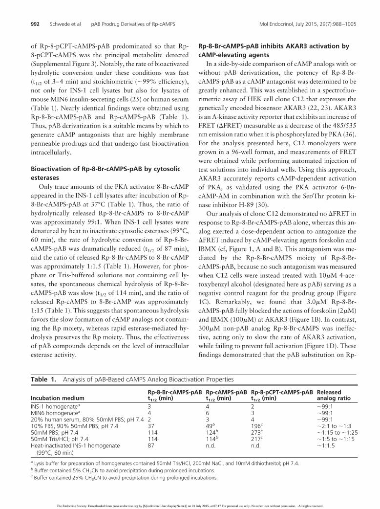
of Rp-8-pCPT-cAMPS-pAB predominated so that Rp-8-pCPT-cAMPS was the principal metabolite detected(Supplemental Figure 3). Notably, the rate of bioactivatedhydrolytic conversion under these conditions was fast(t1/2 of 3–4 min) and stoichiometric (�99% efficiency),not only for INS-1 cell lysates but also for lysates ofmouse MIN6 insulin-secreting cells (25) or human serum(Table 1). Nearly identical findings were obtained usingRp-8-Br-cAMPS-pAB and Rp-cAMPS-pAB (Table 1).Thus, pAB derivatization is a suitable means by which togenerate cAMP antagonists that are highly membranepermeable prodrugs and that undergo fast bioactivationintracellularly.
Bioactivation of Rp-8-Br-cAMPS-pAB by cytosolicesterases
Only trace amounts of the PKA activator 8-Br-cAMPappeared in the INS-1 cell lysates after incubation of Rp-8-Br-cAMPS-pAB at 37°C (Table 1). Thus, the ratio ofhydrolytically released Rp-8-Br-cAMPS to 8-Br-cAMPwas approximately 99:1. When INS-1 cell lysates weredenatured by heat to inactivate cytosolic esterases (99°C,60 min), the rate of hydrolytic conversion of Rp-8-Br-cAMPS-pAB was dramatically reduced (t1/2 of 87 min),and the ratio of released Rp-8-Br-cAMPS to 8-Br-cAMPwas approximately 1:1.5 (Table 1). However, for phos-phate or Tris-buffered solutions not containing cell ly-sates, the spontaneous chemical hydrolysis of Rp-8-Br-cAMPS-pAB was slow (t1/2 of 114 min), and the ratio ofreleased Rp-cAMPS to 8-Br-cAMP was approximately1:15 (Table 1). This suggests that spontaneous hydrolysisfavors the slow formation of cAMP analogs not contain-ing the Rp moiety, whereas rapid esterase-mediated hy-drolysis preserves the Rp moiety. Thus, the effectivenessof pAB compounds depends on the level of intracellularesterase activity.
Rp-8-Br-cAMPS-pAB inhibits AKAR3 activation bycAMP-elevating agents
In a side-by-side comparison of cAMP analogs with orwithout pAB derivatization, the potency of Rp-8-Br-cAMPS-pAB as a cAMP antagonist was determined to begreatly enhanced. This was established in a spectrofluo-rimetric assay of HEK cell clone C12 that expresses thegenetically encoded biosensor AKAR3 (22, 23). AKAR3is an A-kinase activity reporter that exhibits an increase ofFRET (�FRET) measurable as a decrease of the 485/535nm emission ratio when it is phosphorylated by PKA (36).For the analysis presented here, C12 monolayers weregrown in a 96-well format, and measurements of FRETwere obtained while performing automated injection oftest solutions into individual wells. Using this approach,AKAR3 accurately reports cAMP-dependent activationof PKA, as validated using the PKA activator 6-Bn-cAMP-AM in combination with the Ser/Thr protein ki-nase inhibitor H-89 (30).
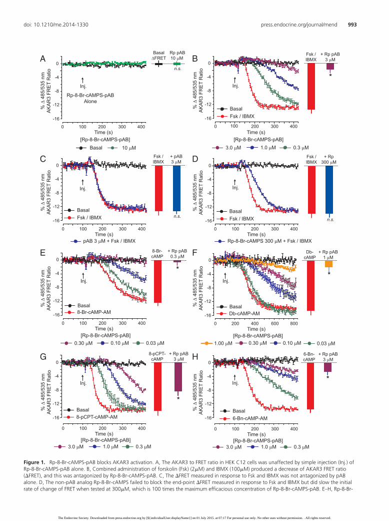
Our analysis of clone C12 demonstrated no �FRET inresponse to Rp-8-Br-cAMPS-pAB alone, whereas this an-alog exerted a dose-dependent action to antagonize the�FRET induced by cAMP-elevating agents forskolin andIBMX (cf, Figure 1, A and B). This antagonism was me-diated by the Rp-8-Br-cAMPS moiety of Rp-8-Br-cAMPS-pAB, because no such antagonism was measuredwhen C12 cells were instead treated with 10�M 4-ace-toxybenzyl alcohol (designated here as pAB) serving as anegative control reagent for the prodrug group (Figure1C). Remarkably, we found that 3.0�M Rp-8-Br-cAMPS-pAB fully blocked the actions of forskolin (2�M)and IBMX (100�M) at AKAR3 (Figure 1B). In contrast,300�M non-pAB analog Rp-8-Br-cAMPS was ineffec-tive, acting only to slow the rate of AKAR3 activation,while failing to prevent full activation (Figure 1D). Thesefindings demonstrated that the pAB substitution on Rp-
Table 1. Analysis of pAB-Based cAMPS Analog Bioactivation Properties
Incubation mediumRp-8-Br-cAMPS-pABt1/2 (min)
Rp-cAMPS-pABt1/2 (min)
Rp-8-pCPT-cAMPS-pABt1/2 (min)
Releasedanalog ratio
INS-1 homogenatea 3 4 2 �99:1MIN6 homogenatea 4 6 3 �99:120% human serum, 80% 50mM PBS; pH 7.4 2 3 4 �99:110% FBS, 90% 50mM PBS; pH 7.4 37 49b 196c �2:1 to �1:350mM PBS; pH 7.4 114 124b 273c �1:15 to �1:2550mM Tris/HCl; pH 7.4 114 114b 217c �1:5 to �1:15Heat-inactivated INS-1 homogenate
(99°C, 60 min)87 n.d. n.d. �1:1.5
a Lysis buffer for preparation of homogenates contained 50mM Tris/HCl, 200mM NaCl, and 10mM dithiothreitol; pH 7.4.b Buffer contained 5% CH3CN to avoid precipitation during prolonged incubations.c Buffer contained 25% CH3CN to avoid precipitation during prolonged incubations.
992 Schwede et al pAB Prodrug Derivatives of Rp-cAMPS Mol Endocrinol, July 2015, 29(7):988–1005
The Endocrine Society. Downloaded from press.endocrine.org by [${individualUser.displayName}] on 01 July 2015. at 07:17 For personal use only. No other uses without permission. . All rights reserved.
0 100 200 300 400Time (s)
A
Rp-8-Br-cAMPS-pABAlone
% Δ
485
/535
nm
AK
AR
3 FR
ET
Rat
io
0
-4
-8
-12
-16
Inj.
[Rp-8-Br-cAMPS-pAB] Basal
BasalΔFRET
Rp pAB10 μM
n.s.
C
pAB 3 μM + Fsk / IBMX
Fsk / IBMX Basal
Inj.
Fsk /IBMX
+ pAB3 μM
n.s.
% Δ
485
/535
nm
AK
AR
3 FR
ET
Rat
io
0
-4
-8
-12
-16
8-pCPT-cAMP-AM Basal
E
G
Inj.
Inj.
8-Br-cAMP-AM Basal
8-Br-cAMP
+ Rp pAB0.3 μM
*
8-pCPT-cAMP
+ Rp pAB3 μM
*
% Δ
485
/535
nm
AK
AR
3 FR
ET
Rat
io
0
-4
-8
-12
-16
% Δ
485
/535
nm
AK
AR
3 FR
ET
Rat
io
0
-4
-8
-12
-16
0.03 μM 0.10 μM 0.30 μM
0.3 μM 1.0 μM 3.0 μM
B
Fsk / IBMX Basal
Inj.
Fsk /IBMX
+ Rp pAB3 μM
*
D
Inj.
Rp-8-Br-cAMPS 300 μM + Fsk / IBMX
Fsk / IBMX Basal
Fsk /IBMX
+ Rp300 μM
n.s.
0 100 200 300 400Time (s)
% Δ
485
/535
nm
AK
AR
3 FR
ET
Rat
io
0
-4
-8
-12
-16
% Δ
485
/535
nm
AK
AR
3 FR
ET
Rat
io
0
-4
-8
-12
-16
0.3 μM 1.0 μM 3.0 μM[Rp-8-Br-cAMPS-pAB]
6-Bn-cAMP-AM Basal
F
H
Inj.
Inj.
Db-cAMP-AM Basal
0.03 μM0.10 μM0.30 μM1.00 μM
Db-cAMP
+ Rp pAB1 μM
*
6-Bn-cAMP
+ Rp pAB3 μM
*
% Δ
485
/535
nm
AK
AR
3 FR
ET
Rat
io
0
-4
-8
-12
-16
% Δ
485
/535
nm
AK
AR
3 FR
ET
Rat
io
0
-4
-8
-12
-16
0.3 μM 1.0 μM 3.0 μM
10 μM
0 100 200 300 400Time (s)
0 100 200 300 400Time (s)
0 100 200 300 400Time (s)
[Rp-8-Br-cAMPS-pAB]
0 200 400 600 800Time (s)
[Rp-8-Br-cAMPS-pAB]
0 100 200 300 400Time (s)
[Rp-8-Br-cAMPS-pAB]
0 100 200 300 400Time (s)
[Rp-8-Br-cAMPS-pAB]
Figure 1. Rp-8-Br-cAMPS-pAB blocks AKAR3 activation. A, The AKAR3 to FRET ratio in HEK C12 cells was unaffected by simple injection (Inj.) ofRp-8-Br-cAMPS-pAB alone. B, Combined administration of forskolin (Fsk) (2�M) and IBMX (100�M) produced a decrease of AKAR3 FRET ratio(�FRET), and this was antagonized by Rp-8-Br-cAMPS-pAB. C, The �FRET measured in response to Fsk and IBMX was not antagonized by pABalone. D, The non-pAB analog Rp-8-Br-cAMPS failed to block the end-point �FRET measured in response to Fsk and IBMX but did slow the initialrate of change of FRET when tested at 300�M, which is 100 times the maximum efficacious concentration of Rp-8-Br-cAMPS-pAB. E–H, Rp-8-Br-
doi: 10.1210/me.2014-1330 press.endocrine.org/journal/mend 993
The Endocrine Society. Downloaded from press.endocrine.org by [${individualUser.displayName}] on 01 July 2015. at 07:17 For personal use only. No other uses without permission. . All rights reserved.
8-Br-cAMPS afforded increased antagonist potency as aconsequence of increased membrane permeability.
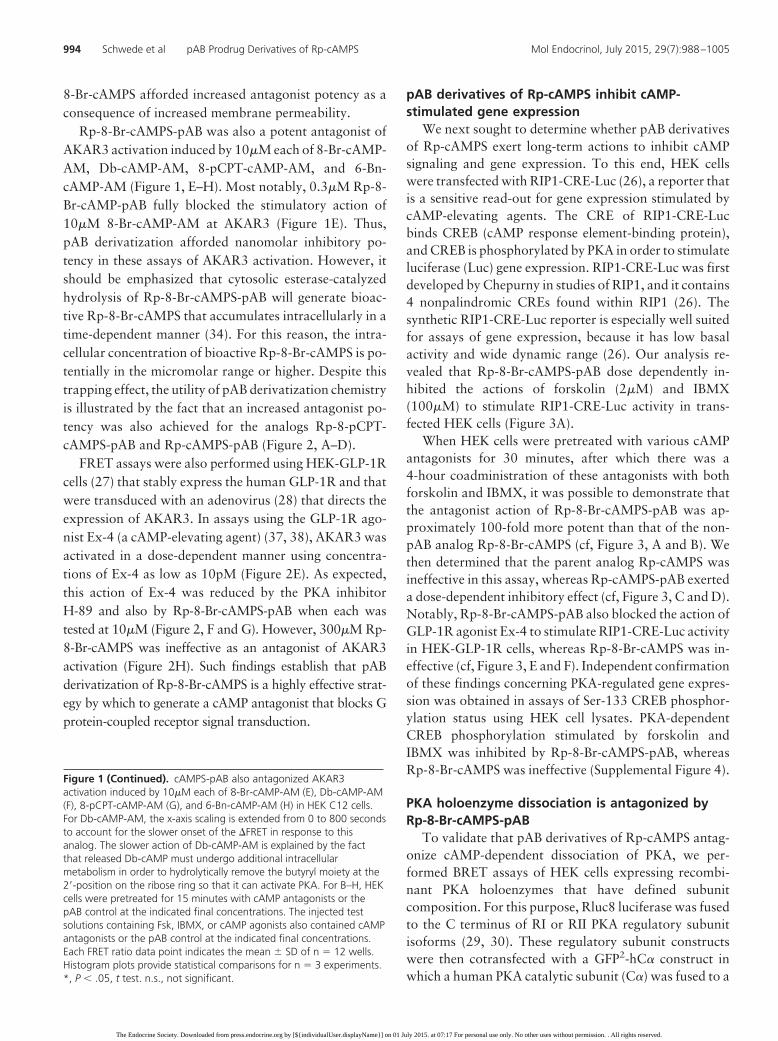
Rp-8-Br-cAMPS-pAB was also a potent antagonist ofAKAR3 activation induced by 10�M each of 8-Br-cAMP-AM, Db-cAMP-AM, 8-pCPT-cAMP-AM, and 6-Bn-cAMP-AM (Figure 1, E–H). Most notably, 0.3�M Rp-8-Br-cAMP-pAB fully blocked the stimulatory action of10�M 8-Br-cAMP-AM at AKAR3 (Figure 1E). Thus,pAB derivatization afforded nanomolar inhibitory po-tency in these assays of AKAR3 activation. However, itshould be emphasized that cytosolic esterase-catalyzedhydrolysis of Rp-8-Br-cAMPS-pAB will generate bioac-tive Rp-8-Br-cAMPS that accumulates intracellularly in atime-dependent manner (34). For this reason, the intra-cellular concentration of bioactive Rp-8-Br-cAMPS is po-tentially in the micromolar range or higher. Despite thistrapping effect, the utility of pAB derivatization chemistryis illustrated by the fact that an increased antagonist po-tency was also achieved for the analogs Rp-8-pCPT-cAMPS-pAB and Rp-cAMPS-pAB (Figure 2, A–D).
FRET assays were also performed using HEK-GLP-1Rcells (27) that stably express the human GLP-1R and thatwere transduced with an adenovirus (28) that directs theexpression of AKAR3. In assays using the GLP-1R ago-nist Ex-4 (a cAMP-elevating agent) (37, 38), AKAR3 wasactivated in a dose-dependent manner using concentra-tions of Ex-4 as low as 10pM (Figure 2E). As expected,this action of Ex-4 was reduced by the PKA inhibitorH-89 and also by Rp-8-Br-cAMPS-pAB when each wastested at 10�M (Figure 2, F and G). However, 300�M Rp-8-Br-cAMPS was ineffective as an antagonist of AKAR3activation (Figure 2H). Such findings establish that pABderivatization of Rp-8-Br-cAMPS is a highly effective strat-egy by which to generate a cAMP antagonist that blocks Gprotein-coupled receptor signal transduction.
pAB derivatives of Rp-cAMPS inhibit cAMP-stimulated gene expression
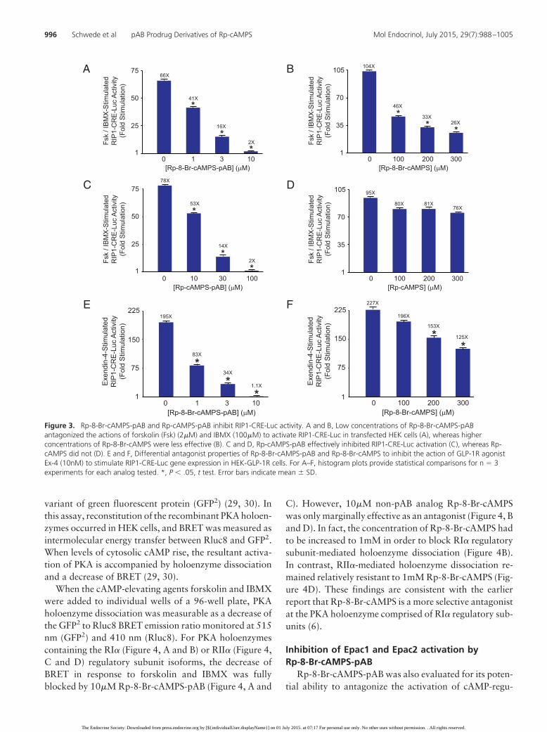
We next sought to determine whether pAB derivativesof Rp-cAMPS exert long-term actions to inhibit cAMPsignaling and gene expression. To this end, HEK cellswere transfected with RIP1-CRE-Luc (26), a reporter thatis a sensitive read-out for gene expression stimulated bycAMP-elevating agents. The CRE of RIP1-CRE-Lucbinds CREB (cAMP response element-binding protein),and CREB is phosphorylated by PKA in order to stimulateluciferase (Luc) gene expression. RIP1-CRE-Luc was firstdeveloped by Chepurny in studies of RIP1, and it contains4 nonpalindromic CREs found within RIP1 (26). Thesynthetic RIP1-CRE-Luc reporter is especially well suitedfor assays of gene expression, because it has low basalactivity and wide dynamic range (26). Our analysis re-vealed that Rp-8-Br-cAMPS-pAB dose dependently in-hibited the actions of forskolin (2�M) and IBMX(100�M) to stimulate RIP1-CRE-Luc activity in trans-fected HEK cells (Figure 3A).
When HEK cells were pretreated with various cAMPantagonists for 30 minutes, after which there was a4-hour coadministration of these antagonists with bothforskolin and IBMX, it was possible to demonstrate thatthe antagonist action of Rp-8-Br-cAMPS-pAB was ap-proximately 100-fold more potent than that of the non-pAB analog Rp-8-Br-cAMPS (cf, Figure 3, A and B). Wethen determined that the parent analog Rp-cAMPS wasineffective in this assay, whereas Rp-cAMPS-pAB exerteda dose-dependent inhibitory effect (cf, Figure 3, C and D).Notably, Rp-8-Br-cAMPS-pAB also blocked the action ofGLP-1R agonist Ex-4 to stimulate RIP1-CRE-Luc activityin HEK-GLP-1R cells, whereas Rp-8-Br-cAMPS was in-effective (cf, Figure 3, E and F). Independent confirmationof these findings concerning PKA-regulated gene expres-sion was obtained in assays of Ser-133 CREB phosphor-ylation status using HEK cell lysates. PKA-dependentCREB phosphorylation stimulated by forskolin andIBMX was inhibited by Rp-8-Br-cAMPS-pAB, whereasRp-8-Br-cAMPS was ineffective (Supplemental Figure 4).
PKA holoenzyme dissociation is antagonized byRp-8-Br-cAMPS-pAB
To validate that pAB derivatives of Rp-cAMPS antag-onize cAMP-dependent dissociation of PKA, we per-formed BRET assays of HEK cells expressing recombi-nant PKA holoenzymes that have defined subunitcomposition. For this purpose, Rluc8 luciferase was fusedto the C terminus of RI or RII PKA regulatory subunitisoforms (29, 30). These regulatory subunit constructswere then cotransfected with a GFP2-hC� construct inwhich a human PKA catalytic subunit (C�) was fused to a
Figure 1 (Continued). cAMPS-pAB also antagonized AKAR3activation induced by 10�M each of 8-Br-cAMP-AM (E), Db-cAMP-AM(F), 8-pCPT-cAMP-AM (G), and 6-Bn-cAMP-AM (H) in HEK C12 cells.For Db-cAMP-AM, the x-axis scaling is extended from 0 to 800 secondsto account for the slower onset of the �FRET in response to thisanalog. The slower action of Db-cAMP-AM is explained by the factthat released Db-cAMP must undergo additional intracellularmetabolism in order to hydrolytically remove the butyryl moiety at the2�-position on the ribose ring so that it can activate PKA. For B–H, HEKcells were pretreated for 15 minutes with cAMP antagonists or thepAB control at the indicated final concentrations. The injected testsolutions containing Fsk, IBMX, or cAMP agonists also contained cAMPantagonists or the pAB control at the indicated final concentrations.Each FRET ratio data point indicates the mean � SD of n � 12 wells.Histogram plots provide statistical comparisons for n � 3 experiments.*, P � .05, t test. n.s., not significant.
994 Schwede et al pAB Prodrug Derivatives of Rp-cAMPS Mol Endocrinol, July 2015, 29(7):988–1005
The Endocrine Society. Downloaded from press.endocrine.org by [${individualUser.displayName}] on 01 July 2015. at 07:17 For personal use only. No other uses without permission. . All rights reserved.
0 100 200 300 400Time (s)
A%
Δ 4
85/5
35 n
mA
KA
R3
FRE
T R
atio
0
-3
-6
-9
-12
B
C D
Inj.
Fsk / IBMX Basal
Inj.
Inj. Inj.
% Δ
485
/535
nm
AK
AR
3 FR
ET
Rat
io
0
-3
-6
-9
-12
% Δ
485
/535
nm
AK
AR
3 FR
ET
Rat
io
0
-3
-6
-9
-12
% Δ
485
/535
nm
AK
AR
3 FR
ET
Rat
io
0
-3
-6
-9
-12
+ Rp pAB3 μM
*
Fsk /IBMX
+ Rp100 μM
n.s.
Fsk /IBMX
+ Rp pAB100 μM
*
Fsk /IBMX
+ Rp300 μM
n.s.
Fsk /IBMX
0.3 μM 1.0 μM 3.0 μM[Rp-8-pCPT-cAMPS-pAB]
10 μM 30 μM 100 μM
10 μM 30 μM 100 μM 30 μM 100 μM 300 μM
Inj.
Ex-4
Ex-4+Rp pAB10 μM
*
0 100 200 300 400Time (s)
% Δ
485
/535
nm
AK
AR
3 FR
ET
Rat
io
0
-4
-8
-12
-16
1 μM 3 μM 10 μM[Rp-8-Br-cAMPS-pAB]
Inj.
Ex-4
Ex-4+Rp
300 μM.
% Δ
485
/535
nm
AK
AR
3 FR
ET
Rat
io
0
-4
-8
-12
-16
100 μM 200 μM 300 μM
Inj.
10 nMEx-4
30 pMEx-4
*
0 100 200 300 400Time (s)
% Δ
485
/535
nm
AK
AR
3 FR
ET
Rat
io
0
-4
-8
-12
-16[Exendin-4]
3 nM Ex-4
Ex-4Ex-4 +H-89
0 100 200 300 400Time (s)
% Δ
485
/535
nm
AK
AR
3 FR
ET
Rat
io
0
-4
-8
-12
-16
HG
FE
10 nM 300 pM 100 pM
30 pM 10 pM Vehicle
H-89vs. Ex-4
3 nM Ex-4 +10 μM H-89
Vehicle
0 100 200 300 400Time (s)
[Rp-8-pCPT-cAMPS]
Fsk / IBMX Basal
0 100 200 300 400Time (s)
[Rp-cAMPS-pAB]
0 100 200 300 400Time (s)
[Rp-cAMPS]
Fsk / IBMX Basal
Fsk / IBMX Basal
Inj.
Exendin-4 Basal
Exendin-4 Basal
0 100 200 300 400Time (s)
[Rp-8-Br-cAMPS]
*
n.s.
Figure 2. Additional cAMP antagonists for signal transduction research. A and B, Differential properties of Rp-8-pCPT-cAMPS-pAB (A) and Rp-8-pCPT-cAMPS (B) in the AKAR3 activation assay (95) using HEK C12 cells treated with forskolin (Fsk) (2�M) and IBMX (100�M). C and D,Differential actions of Rp-cAMPS-pAB (C) and Rp-cAMPS (D). E, Dose-dependent activation of AKAR3 by GLP-1R agonist exendin-4 (Ex-4). F and G,Validation that PKA inhibitor H-89 (10�M) and cAMP antagonist Rp-8-Br-cAMPS-pAB (1�M–10�M) reduce the action of Ex-4 (3nM) to activateAKAR3 in HEK-GLP-1R cells. H, Failure of Rp-8-Br-cAMPS (100�M–300�M) to block AKAR3 activation by Ex-4 (3nM) in HEK-GLP-1R cells. EachFRET ratio data point indicates the mean � SD of n � 12 wells. Histogram plots provide statistical comparisons for n � 3 experiments.*, P � .05, t test.
doi: 10.1210/me.2014-1330 press.endocrine.org/journal/mend 995
The Endocrine Society. Downloaded from press.endocrine.org by [${individualUser.displayName}] on 01 July 2015. at 07:17 For personal use only. No other uses without permission. . All rights reserved.
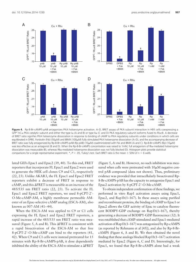
variant of green fluorescent protein (GFP2) (29, 30). Inthis assay, reconstitution of the recombinant PKA holoen-zymes occurred in HEK cells, and BRET was measured asintermolecular energy transfer between Rluc8 and GFP2.When levels of cytosolic cAMP rise, the resultant activa-tion of PKA is accompanied by holoenzyme dissociationand a decrease of BRET (29, 30).
When the cAMP-elevating agents forskolin and IBMXwere added to individual wells of a 96-well plate, PKAholoenzyme dissociation was measurable as a decrease ofthe GFP2 to Rluc8 BRET emission ratio monitored at 515nm (GFP2) and 410 nm (Rluc8). For PKA holoenzymescontaining the RI� (Figure 4, A and B) or RII� (Figure 4,C and D) regulatory subunit isoforms, the decrease ofBRET in response to forskolin and IBMX was fullyblocked by 10�M Rp-8-Br-cAMPS-pAB (Figure 4, A and
C). However, 10�M non-pAB analog Rp-8-Br-cAMPSwas only marginally effective as an antagonist (Figure 4, Band D). In fact, the concentration of Rp-8-Br-cAMPS hadto be increased to 1mM in order to block RI� regulatorysubunit-mediated holoenzyme dissociation (Figure 4B).In contrast, RII�-mediated holoenzyme dissociation re-mained relatively resistant to 1mM Rp-8-Br-cAMPS (Fig-ure 4D). These findings are consistent with the earlierreport that Rp-8-Br-cAMPS is a more selective antagonistat the PKA holoenzyme comprised of RI� regulatory sub-units (6).
Inhibition of Epac1 and Epac2 activation byRp-8-Br-cAMPS-pAB
Rp-8-Br-cAMPS-pAB was also evaluated for its poten-tial ability to antagonize the activation of cAMP-regu-
B
95XD
80X 81X
66X75
50
25
1
[Rp-8-Br-cAMPS-pAB] (μM)0 1 3 10
Fsk
/ IB
MX
-Stim
ulat
edR
IP1-
CR
E-L
uc A
ctiv
ity(F
old
Stim
ulat
ion)
A
41X
C
*
16X
*2X*
78X
75
50
25
1
[Rp-cAMPS-pAB] (μM)0 10 30 100
Fsk
/ IB
MX
-Stim
ulat
edR
IP1-
CR
E-L
uc A
ctiv
ity(F
old
Stim
ulat
ion) 53X
*
14X
*2X
*
104X105
70
35
1
[Rp-8-Br-cAMPS] (μM)0 100 200 300
Fsk
/ IB
MX
-Stim
ulat
edR
IP1-
CR
E-L
uc A
ctiv
ity(F
old
Stim
ulat
ion)
46X
* 33X
* 26X
*
105
70
35
1
[Rp-cAMPS] (μM)0 100 200 300
Fsk
/ IB
MX
-Stim
ulat
edR
IP1-
CR
E-L
uc A
ctiv
ity(F
old
Stim
ulat
ion) 76X
225
150
75
Exe
ndin
-4-S
timul
ated
RIP
1-C
RE
-Luc
Act
ivity
(Fol
d S
timul
atio
n)
F 227X
196X
[Rp-8-Br-cAMPS] (μM)0 100 200 300
1
225
150
75
1
[Rp-8-Br-cAMPS-pAB] (μM)
Exe
ndin
-4-S
timul
ated
RIP
1-C
RE
-Luc
Act
ivity
(Fol
d S
timul
atio
n)
E195X
0 1 3 10
83X
*34X
* 1.1X
*
153X
* 125X
*
Figure 3. Rp-8-Br-cAMPS-pAB and Rp-cAMPS-pAB inhibit RIP1-CRE-Luc activity. A and B, Low concentrations of Rp-8-Br-cAMPS-pABantagonized the actions of forskolin (Fsk) (2�M) and IBMX (100�M) to activate RIP1-CRE-Luc in transfected HEK cells (A), whereas higherconcentrations of Rp-8-Br-cAMPS were less effective (B). C and D, Rp-cAMPS-pAB effectively inhibited RIP1-CRE-Luc activation (C), whereas Rp-cAMPS did not (D). E and F, Differential antagonist properties of Rp-8-Br-cAMPS-pAB and Rp-8-Br-cAMPS to inhibit the action of GLP-1R agonistEx-4 (10nM) to stimulate RIP1-CRE-Luc gene expression in HEK-GLP-1R cells. For A–F, histogram plots provide statistical comparisons for n � 3experiments for each analog tested. *, P � .05, t test. Error bars indicate mean � SD.
996 Schwede et al pAB Prodrug Derivatives of Rp-cAMPS Mol Endocrinol, July 2015, 29(7):988–1005
The Endocrine Society. Downloaded from press.endocrine.org by [${individualUser.displayName}] on 01 July 2015. at 07:17 For personal use only. No other uses without permission. . All rights reserved.
lated GEFs Epac1 and Epac2 (39, 40). To this end, FRETreporters that incorporate FL Epac1 and Epac2 were usedto generate the HEK cell clones C9 and C1, respectively(22, 23). Unlike AKAR3, the FL Epac1 and Epac2 FRETreporters exhibit a decrease of FRET in response tocAMP, and this �FRET is measurable as an increase of the485/535 nm FRET ratio (22, 23). To activate the FLEpac1 and Epac2 FRET reporters, we used 8-pCPT-2�-O-Me-cAMP-AM, a highly membrane permeable AM-ester of an Epac-selective cAMP analog (ESCA-AM), alsoknown as 007-AM (41–44).
When the ESCA-AM was applied to C9 or C1 cellsexpressing the FL Epac1 and Epac2 FRET reporters, arapid increase of the 485/535 nm FRET ratio was mea-sured (Figure 5, A and B). This �FRET is consistent witha rapid bioactivation of the ESCA-AM so that free8-pCPT-2�-O-Me-cAMP can bind to the reporters (41,42). When C9 and C1 cells were instead pretreated for 15minutes with Rp-8-Br-cAMPS-pAB, it dose dependentlyinhibited the ability of the ESCA-AM to stimulate a �FRET
(Figure 5, A and B). However, no such inhibition was mea-sured when cells were pretreated with 10�M negative con-trol pAB compound (data not shown). Thus, preliminaryevidence was provided that intracellularly bioactivated Rp-8-Br-cAMPS-pAB has the capacity to antagonize Epac1 andEpac2 activation by 8-pCPT-2�-O-Me-cAMP.
To obtain independent confirmation of these findings, weperformed in vitro Rap1 activation assays using Epac1,Epac2, and Rap1b(1–167). In these assays using purifiedand recombinant proteins, the binding of cAMP to Epac1 orEpac2 allows the GEF activity of Epac to catalyze fluores-cent BODIPY-GDP exchange on Rap1b(1–167), therebygenerating a decrease of BODIPY-GDP fluorescence (32). Itwas established that cAMP-stimulated and Epac1-mediatedactivation of Rap1b(1–167) was antagonized by Rp-cAMPS(as reported by Rehmann et al [45]), and also by Rp-8-Br-cAMPS (Figure 6, A and B). We then obtained the novelfinding that these analogs blocked Rap1b(1–167) activationmediated by Epac2 (Figure 6, C and D). Interestingly, forEpac1, we found that Rp-8-Br-cAMPS alone had a weak
.12
.16
.20
.24
.28
B
DPBS DPBS+ Fsk
+IBMX
Rp10 μM
Rp10 μM+ Fsk
+IBMX
Rp1 mM
Rp1 mM+ Fsk
+IBMX
.12
.16
.20
.24
.28
A
pAB10 μM
pAB10 μM+ Fsk
+IBMX
Rp pAB10 μM
Rp pAB10 μM+ Fsk
+IBMX
Cα + RIα Cα + RIα
pAB10 μM
pAB10 μM+ Fsk
+IBMX
Rp pAB10 μM
Rp pAB10 μM+ Fsk
+IBMX
.07
.14
.21
.28
.35C Cα + RIIα
DPBS DPBS+ Fsk
+IBMX
Rp10 μM
Rp10 μM+ Fsk
+IBMX
Rp1 mM
Rp1 mM+ Fsk
+IBMX
.07
.14
.21
.28
.35D Cα + RIIα
**
*
* **
*
GFP
2 / R
Luc8
BR
ET
Rat
ioG
FP2
/ RLu
c8 B
RE
T R
atio
GFP
2 / R
Luc8
BR
ET
Rat
ioG
FP2
/ RLu
c8 B
RE
T R
atio
Figure 4. Rp-8-Br-cAMPS-pAB antagonizes PKA holoenzyme activation. A–D, BRET assays of PKA subunit interaction in HEK cells coexpressing aGFP2-hC� PKA catalytic subunit and either the type I� (A and B) or type II� (C and D) PKA regulatory subunit isoforms fused to Rluc8. A decreaseof BRET ratio signifies PKA holoenzyme dissociation in response to binding of cAMP to PKA regulatory subunits under conditions in which cells areequilibrated in DPBS. Forskolin (Fsk) (50�M) and IBMX (100�M) fully stimulated PKA holoenzyme dissociation (A–D), and the accompanying decrease ofBRET ratio was fully antagonized by Rp-8-Br-cAMPS-pAB (Rp pAB) (10�M) coadministered with Fsk and IBMX (A and C). Rp-8-Br-cAMPS (Rp) (10�M)was less effective as an antagonist (B and D). When the Rp-8-Br-cAMPS concentration was raised to 1mM, full antagonism of RI�-mediated holoenzymedissociation was measurable (B), whereas RII�-mediated holoenzyme dissociation was not fully blocked (D). Histogram plots provide statisticalcomparisons for a single representative experiment. *, P � .05, Tukey’s test. Each BRET ratio is the mean � SEM of n � 6 wells.
doi: 10.1210/me.2014-1330 press.endocrine.org/journal/mend 997
The Endocrine Society. Downloaded from press.endocrine.org by [${individualUser.displayName}] on 01 July 2015. at 07:17 For personal use only. No other uses without permission. . All rights reserved.
stimulatory effect before the addition of cAMP (Figure 6B,arrow). However, this was not the case for Epac2 (Figure6D). Collectively, these findings indicate that the pAB con-jugates of Rp-cAMPS can be used not only as inhibitors ofPKA activation but also as inhibitors of Epac1 and Epac2activation. Such findings have important implications for
the interpretation of previous studies using Rp-cAMPSanalogs.
Rp-8-Br-cAMPS-pAB inhibits GSIScAMP exerts PKA and Epac mediated actions to stim-
ulate insulin release from pancreatic �-cells (2, 3, 28, 46–
0
- 200
- 400
- 600
- 800
- 1000
Δ Fl
uore
scen
ce
0 10 20 30 40 50 60 70 80 90Time (min)
A
No Rp-cAMPS or cAMP
Rp-cAMPS + cAMP
cAMP
Rp-cAMPS
cAMP
Epac1 + Rap1b 0
- 200
- 400
- 600
- 800
- 1000
Δ Fl
uore
scen
ce
0 10 20 30 40 50 60 70 80 90Time (min)
B
No Rp-8-Br-cAMPS or cAMP
Rp-8-Br-cAMPS + cAMP
cAMP
Rp-8-Br-cAMPS
cAMP
Epac1 + Rap1b
0
- 200
- 400
- 600
- 800
- 1000
Δ Fl
uore
scen
ce
- 1200
0 10 20 30 40 50 60 70 80 90Time (min)
C
No Rp-cAMPS or cAMP
Rp-cAMPS + cAMP
cAMP
Rp-cAMPS
cAMP
Epac2 + Rap1b 0
- 200
- 400
- 600
- 800
- 1000
Δ Fl
uore
scen
ce
- 1200
0 10 20 30 40 50 60 70 80 90Time (min)
D
No Rp-8-Br-cAMPS or cAMP
Rp-8-Br-cAMPS + cAMP
cAMP
Rp-8-Br-cAMPS
cAMP
Epac2 + Rap1b
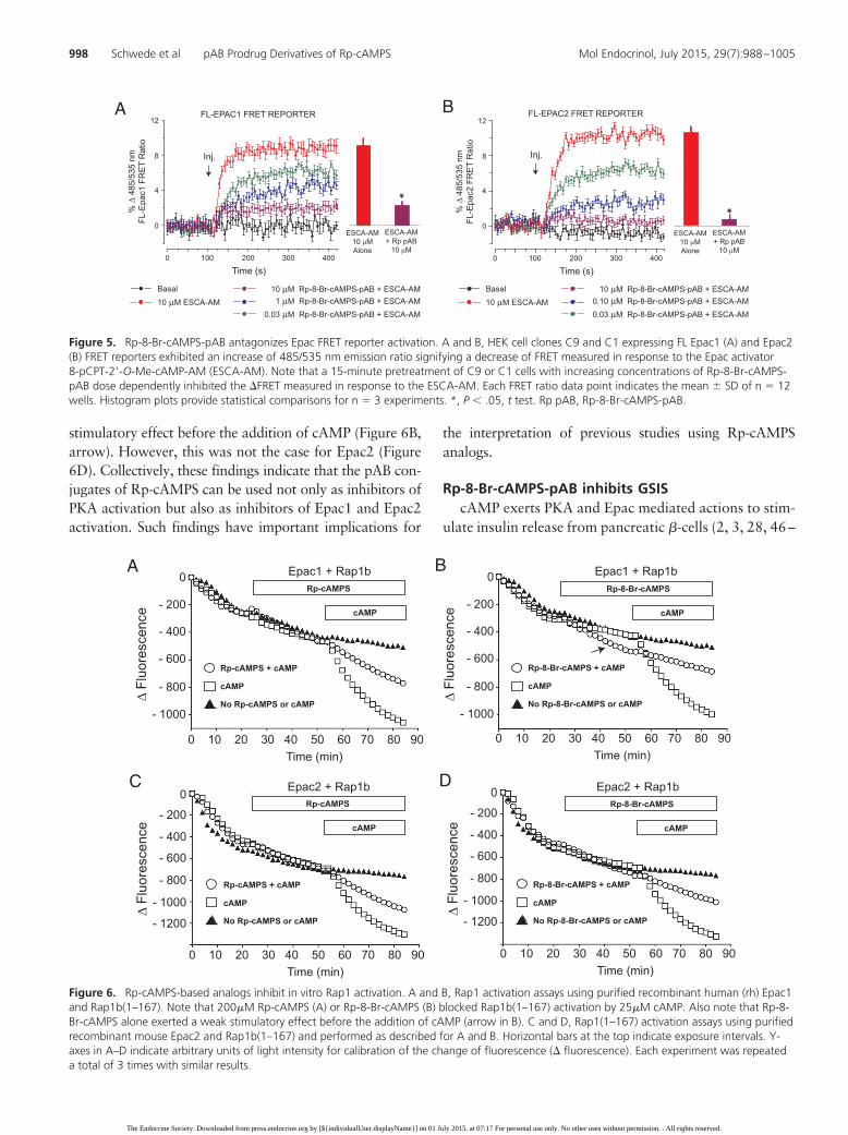
Figure 6. Rp-cAMPS-based analogs inhibit in vitro Rap1 activation. A and B, Rap1 activation assays using purified recombinant human (rh) Epac1and Rap1b(1–167). Note that 200�M Rp-cAMPS (A) or Rp-8-Br-cAMPS (B) blocked Rap1b(1–167) activation by 25�M cAMP. Also note that Rp-8-Br-cAMPS alone exerted a weak stimulatory effect before the addition of cAMP (arrow in B). C and D, Rap1(1–167) activation assays using purifiedrecombinant mouse Epac2 and Rap1b(1–167) and performed as described for A and B. Horizontal bars at the top indicate exposure intervals. Y-axes in A–D indicate arbitrary units of light intensity for calibration of the change of fluorescence (� fluorescence). Each experiment was repeateda total of 3 times with similar results.
Basal 10 μM Rp-8-Br-cAMPS-pAB + ESCA-AM 1 μM Rp-8-Br-cAMPS-pAB + ESCA-AM 0.03 μM Rp-8-Br-cAMPS-pAB + ESCA-AM
10 μM ESCA-AM
A
Inj.
% Δ
485
/535
nm
FL-E
pac1
FR
ET
Rat
io
0
4
8
12
0 100 200 300 400
Time (s)
ESCA-AM10 μMAlone
ESCA-AM+ Rp pAB
10 μM
*
FL-EPAC1 FRET REPORTER B
% Δ
485
/535
nm
FL-E
pac2
FR
ET
Rat
io
0
4
8
12
0 100 200 300 400
Time (s)
*
Inj.
FL-EPAC2 FRET REPORTER
Basal 10 μM Rp-8-Br-cAMPS-pAB + ESCA-AM 0.10 μM Rp-8-Br-cAMPS-pAB + ESCA-AM 0.03 μM Rp-8-Br-cAMPS-pAB + ESCA-AM
10 μM ESCA-AM
ESCA-AM10 μMAlone
ESCA-AM+ Rp pAB
10 μM
Figure 5. Rp-8-Br-cAMPS-pAB antagonizes Epac FRET reporter activation. A and B, HEK cell clones C9 and C1 expressing FL Epac1 (A) and Epac2(B) FRET reporters exhibited an increase of 485/535 nm emission ratio signifying a decrease of FRET measured in response to the Epac activator8-pCPT-2�-O-Me-cAMP-AM (ESCA-AM). Note that a 15-minute pretreatment of C9 or C1 cells with increasing concentrations of Rp-8-Br-cAMPS-pAB dose dependently inhibited the �FRET measured in response to the ESCA-AM. Each FRET ratio data point indicates the mean � SD of n � 12wells. Histogram plots provide statistical comparisons for n � 3 experiments. *, P � .05, t test. Rp pAB, Rp-8-Br-cAMPS-pAB.
998 Schwede et al pAB Prodrug Derivatives of Rp-cAMPS Mol Endocrinol, July 2015, 29(7):988–1005
The Endocrine Society. Downloaded from press.endocrine.org by [${individualUser.displayName}] on 01 July 2015. at 07:17 For personal use only. No other uses without permission. . All rights reserved.
58). However, assessment of how cAMP exerts its effect ishampered by the poor membrane permeability of avail-able pharmacological agents (59). To evaluate a potentialusefulness of Rp-8-Br-cAMPS-pAB for studies of insulinsecretion, pilot assays were performed using the rat in-sulinoma cell line INS-1. These assays demonstrated thatRp-8-Br-cAMPS-pAB inhibited the actions of glucose,forskolin, and IBMX to stimulate insulin secretion,whereas Rp-8-Br-cAMPS was ineffective (SupplementalFigure 5A). As expected, the expression of multiple PKAregulatory subunit isoforms in INS-1 cells was validated(Supplemental Figure 5B), and it was also possible to usea Kemptide phosphorylation assay to demonstrate thatextracellularly administered Rp-8-Br-cAMPS-pAB inhib-ited PKA activation in response to Db-cAMP-AM and6-Bnz-cAMP-AM (Supplemental Figure 5, C and D).
Although INS-1 cells are a convenient model systemfor in vitro assays, they have a limited usefulness due tothe fact that they are not fully differentiated but are in-stead transformed cells with properties not completely
identical to authentic �-cells (24). As an alternative toINS-1 cells, we performed perifusion assays of insulinsecretion from rat islets so that it could be determinedwhether Rp-8-Br-cAMPS-pAB altered first- and/or sec-ond-phase GSIS (20). It is known that rat islets are espe-cially well-suited for the study of biphasic GSIS, becausethey typically generate a fast transient first-phase increaseof insulin secretion that is easily distinguished from sec-ond-phase GSIS when using methods of rapid perifusion(60–62). Our primary goal was to evaluate the potentialrole of cAMP as a permissive factor, or a coupling factor,linking �-cell glucose metabolism to insulin secretion.This concept has remained controversial for over 40 years(9–20, 28, 48, 49, 58).
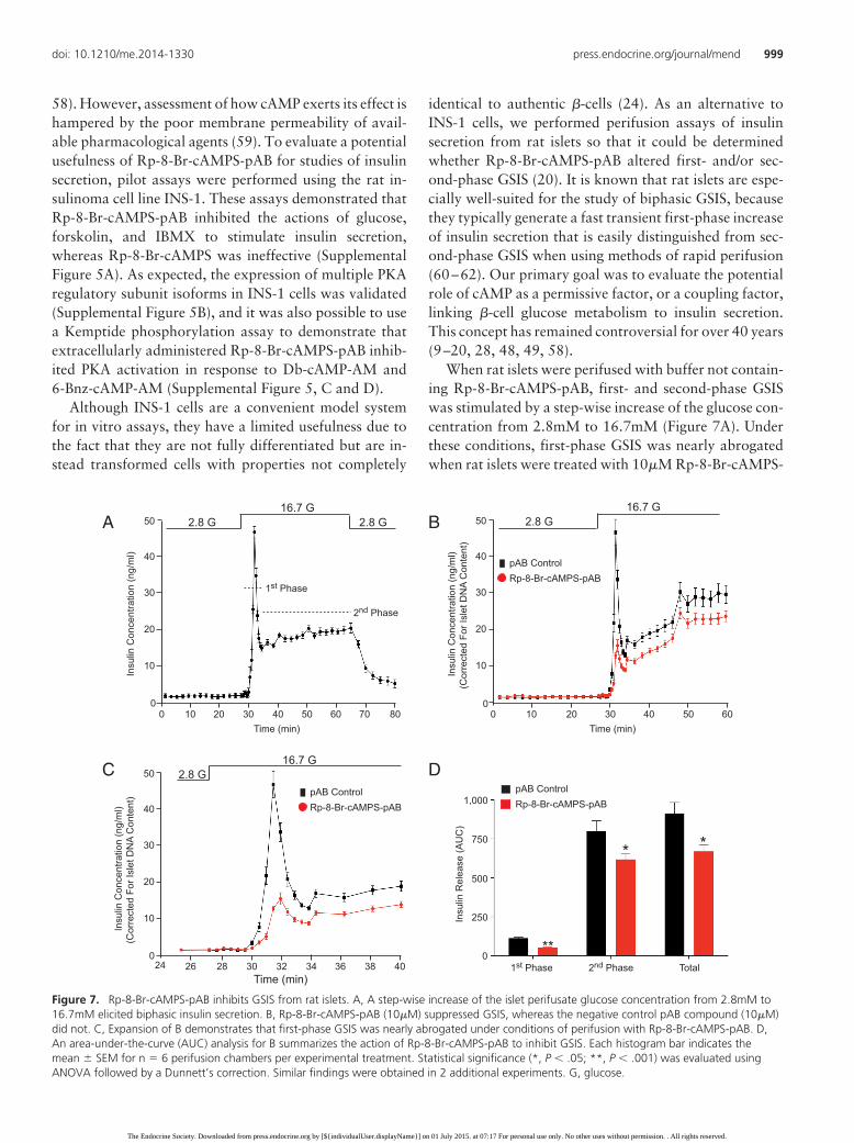
When rat islets were perifused with buffer not contain-ing Rp-8-Br-cAMPS-pAB, first- and second-phase GSISwas stimulated by a step-wise increase of the glucose con-centration from 2.8mM to 16.7mM (Figure 7A). Underthese conditions, first-phase GSIS was nearly abrogatedwhen rat islets were treated with 10�M Rp-8-Br-cAMPS-
10
20
30
40
50
0
10
20
30
40
50
0
10 20 30 40 50 600
26 28
Time (min)
Time (min)32 3430 38 4036
2.8 G16.7 G
2.8 G16.7 G
10
20
30
40
50
010 20 30 40 50 60 70 800
Time (min)
2.8 G 2.8 G16.7 G
Insu
lin C
once
ntra
tion
(ng/
ml)
A
Insu
lin C
once
ntra
tion
(ng/
ml)
(Cor
rect
ed F
or Is
let D
NA
Con
tent
)
B
C
pAB ControlRp-8-Br-cAMPS-pAB
pAB ControlRp-8-Br-cAMPS-pAB
1st Phase
2nd Phase
24
Insu
lin R
elea
se (A
UC
)
250
500
0
750
1,000
D
1st Phase 2nd Phase Total
pAB ControlRp-8-Br-cAMPS-pAB
**
* *
Insu
lin C
once
ntra
tion
(ng/
ml)
(Cor
rect
ed F
or Is
let D
NA
Con
tent
)
Figure 7. Rp-8-Br-cAMPS-pAB inhibits GSIS from rat islets. A, A step-wise increase of the islet perifusate glucose concentration from 2.8mM to16.7mM elicited biphasic insulin secretion. B, Rp-8-Br-cAMPS-pAB (10�M) suppressed GSIS, whereas the negative control pAB compound (10�M)did not. C, Expansion of B demonstrates that first-phase GSIS was nearly abrogated under conditions of perifusion with Rp-8-Br-cAMPS-pAB. D,An area-under-the-curve (AUC) analysis for B summarizes the action of Rp-8-Br-cAMPS-pAB to inhibit GSIS. Each histogram bar indicates themean � SEM for n � 6 perifusion chambers per experimental treatment. Statistical significance (*, P � .05; **, P � .001) was evaluated usingANOVA followed by a Dunnett’s correction. Similar findings were obtained in 2 additional experiments. G, glucose.
doi: 10.1210/me.2014-1330 press.endocrine.org/journal/mend 999
The Endocrine Society. Downloaded from press.endocrine.org by [${individualUser.displayName}] on 01 July 2015. at 07:17 For personal use only. No other uses without permission. . All rights reserved.
pAB (Figure 7, B and C). Surprisingly, the amplitude ofsecond-phase GSIS was only modestly reduced (Figure7B), and basal insulin secretion was unaffected (Supple-mental Figure 6A). Importantly, the inhibitory action ofthe Rp-8-Br-cAMPS-pAB prodrug was mediated by cyto-solic esterase-catalyzed generation of Rp-8-Br-cAMPS,because first-phase GSIS was retained under conditions inwhich islets were instead perifused with 10�M pAB com-pound serving as a negative control (Figure 7, B and C).These findings are summarized in a histogram plot thatquantifies insulin secretion using an area-under-the-curveanalysis for first- and second-phase GSIS (Figure 7D).
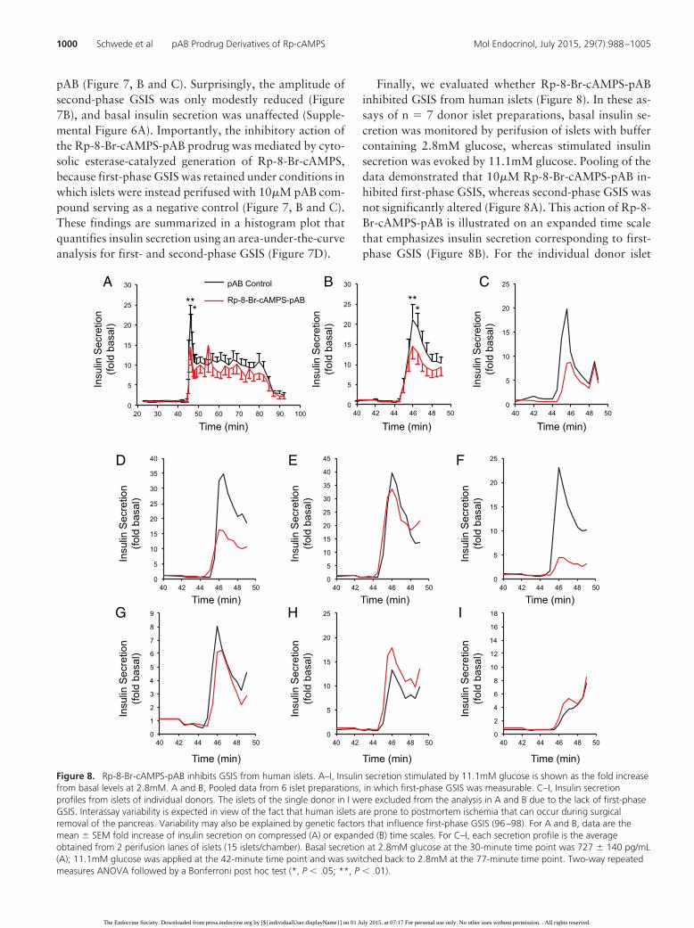
Finally, we evaluated whether Rp-8-Br-cAMPS-pABinhibited GSIS from human islets (Figure 8). In these as-says of n � 7 donor islet preparations, basal insulin se-cretion was monitored by perifusion of islets with buffercontaining 2.8mM glucose, whereas stimulated insulinsecretion was evoked by 11.1mM glucose. Pooling of thedata demonstrated that 10�M Rp-8-Br-cAMPS-pAB in-hibited first-phase GSIS, whereas second-phase GSIS wasnot significantly altered (Figure 8A). This action of Rp-8-Br-cAMPS-pAB is illustrated on an expanded time scalethat emphasizes insulin secretion corresponding to first-phase GSIS (Figure 8B). For the individual donor islet
0
5
10
15
20
25
40 42 44 46 48 50
0
5
10
15
20
25
30
35
40
40 42 44 46 48 50 0
5
10
15
20
25
30
35
40
45
40 42 44 46 48 50 0
5
10
15
20
25
40 42 44 46 48 50
0
1
2
3
4
5
6
7
8
9
40 42 44 46 48 50 0
5
10
15
20
25
40 42 44 46 48 50 0
2
4
6
8
10
12
14
16
18
40 42 44 46 48 50
0
5
10
15
20
25
30
40 42 44 46 48 50
Insu
lin S
ecre
tion
(fold
bas
al)
Insu
lin S
ecre
tion
(fold
bas
al)
Insu
lin S
ecre
tion
(fold
bas
al)
Insu
lin S
ecre
tion
(fold
bas
al)
Insu
lin S
ecre
tion
(fold
bas
al)
Insu
lin S
ecre
tion
(fold
bas
al)
Insu
lin S
ecre
tion
(fold
bas
al)
Insu
lin S
ecre
tion
(fold
bas
al)
Insu
lin S
ecre
tion
(fold
bas
al)
Time (min)
Time (min)Time (min)Time (min)
Time (min) Time (min) Time (min)
Time (min)Time (min)
*****
*
pAB Control
Rp-8-Br-cAMPS-pAB
A CB
IHG
FED
0
5
10
15
20
25
30
20 30 40 50 60 70 80 90 100
Figure 8. Rp-8-Br-cAMPS-pAB inhibits GSIS from human islets. A–I, Insulin secretion stimulated by 11.1mM glucose is shown as the fold increasefrom basal levels at 2.8mM. A and B, Pooled data from 6 islet preparations, in which first-phase GSIS was measurable. C–I, Insulin secretionprofiles from islets of individual donors. The islets of the single donor in I were excluded from the analysis in A and B due to the lack of first-phaseGSIS. Interassay variability is expected in view of the fact that human islets are prone to postmortem ischemia that can occur during surgicalremoval of the pancreas. Variability may also be explained by genetic factors that influence first-phase GSIS (96–98). For A and B, data are themean � SEM fold increase of insulin secretion on compressed (A) or expanded (B) time scales. For C–I, each secretion profile is the averageobtained from 2 perifusion lanes of islets (15 islets/chamber). Basal secretion at 2.8mM glucose at the 30-minute time point was 727 � 140 pg/mL(A); 11.1mM glucose was applied at the 42-minute time point and was switched back to 2.8mM at the 77-minute time point. Two-way repeatedmeasures ANOVA followed by a Bonferroni post hoc test (*, P � .05; **, P � .01).
1000 Schwede et al pAB Prodrug Derivatives of Rp-cAMPS Mol Endocrinol, July 2015, 29(7):988–1005
The Endocrine Society. Downloaded from press.endocrine.org by [${individualUser.displayName}] on 01 July 2015. at 07:17 For personal use only. No other uses without permission. . All rights reserved.
preparations, examples of the insulin secretion profilesare provided in Figure 8, C–I. Because a single islet prep-aration exhibited no first-phase GSIS (Figure 8I), thispreparation was excluded from the analysis presented inFigure 8, A and B. Importantly, Rp-8-Br-cAMPS-pABfailed to alter basal insulin secretion that was measuredunder conditions in which the perifusate contained2.8mM glucose (Supplemental Figure 6B).
Discussion
New evidence for cAMP-dependent actions ofglucose to stimulate insulin secretion
Over 40 years ago, it was reported that glucose metab-olism raises levels of cAMP in isolated islets (9–20).Charles et al (9) also reported that for rat islets, the timecourse of glucose-stimulated cAMP production matchedthat of first-phase GSIS. At that time, speculation existedconcerning a possible role for cAMP as a permissive fac-tor or a coupling factor linking glucose metabolism toinsulin secretion. However, these findings were largelyignored in later years because the ability of glucose tostimulate cAMP production was small, whereas a stron-ger effect was measured in response to cAMP-elevatingagents such as forskolin. Then, in 1990, Persaud et al (63)reported that rat islet GSIS was not blocked by Rp-cAMPS. Subsequently, Sato and Henquin investigated thenewly discovered “amplification” pathway of GSIS, andit too was found to be unaffected by Rp-cAMPS (64).When assessing these earlier findings concerning Rp-cAMPS, it is important to note that this analog has lowmembrane permeability (1). However, prolonged expo-sure of islets to a high concentration of Rp-cAMPS wasreported to block the action of forskolin to potentiateGSIS, whereas it failed to influence the action of glucosealone (63, 64). Harris et al (65) also reported that for ratislets, the secretagogue action of glucose was unaffected bysmall myristoylated synthetic peptides that are membranepermeable and that are derived from the larger endogenousprotein kinase A inhibitor peptide. Similarly, Lester et al (66)reported that rat islet GSIS was unaffected by synthetic pep-tides that disrupt the interaction of PKA regulatory subunitswith A-kinase anchoring proteins. Importantly, all suchstudies with rat islets were performed using methods ofstatic incubation, in which there was insufficient temporalresolution to detect first-phase GSIS.
We now report that rapid perifusion techniques reveala capacity of Rp-8-Br-cAMPS-pAB to nearly abrogatefirst-phase GSIS from human and rat islets. This fast com-ponent of GSIS is initiated by a “triggering” pathway of�-cell stimulus-secretion coupling in which depolariza-
tion-induced Ca2 influx stimulates the exocytosis of areadily-releasable pool of secretory granules (67–69).Based on findings presented here, glucose-stimulatedcAMP production might lead to PKA activation with re-sultant phosphorylation of SNARE (soluble NSF attach-ment protein receptor) complex-associated proteins suchas Snapin that facilitate exocytosis of the readily-re-leasable pool (52). Simultaneously, cAMP may act viaEpac proteins to promote Rap1 activation with conse-quent activation of a novel phospholipase C� that linkscAMP production to �-cell Ca2 influx, Ca2 mobiliza-tion, and protein kinase C activation (50, 51, 70–72).
When considering how glucose raises levels of cAMPin �-cells, Kasai and coworkers originally proposed thatglucose metabolism provides substrate ATP for adenylylcyclase-catalyzed cAMP production, PKA activation, andinsulin exocytosis (48, 49, 56). Tengholm (53) and Teng-holm and coworkers (73–77) more recently reported thatcompartmentalized cAMP production at the plasmamembrane allows glucose metabolism to raise levels ofcAMP, thereby promoting insulin secretion. The exactbiochemical mechanisms linking glucose metabolism tocAMP production remain debated because competing ev-idence exists for roles of transmembrane adenylyl cyclases(48, 49, 56, 73–82) and soluble adenylyl cyclase (adenylylcyclase 10; AC-10) (58, 83). Potentially, glucose metab-olism generates CO2 that is converted by carbonic anhy-drase to bicarbonate ion, which then activates solubleadenylyl cyclase to stimulate cAMP production (58, 83).Interestingly, metabolic compensation of �-cells underconditions of diet-induced obesity leads to a situation inwhich Epac2 plays an increasingly important role in GSIS,possibly by promoting the exocytosis of “restless new-comer” secretory granules (84–87).
New pAB-based prodrugs for cyclic nucleotideresearch
Here, we also address a fundamental limitation ofpresent-day cyclic nucleotide research, in which the anal-ysis of cAMP signaling is impeded by the lack of suitablecAMP antagonists that act with high potency when theyare administered extracellularly. Although (Rp)-phos-phorothioates of cAMP such as Rp-cAMPS are com-monly used as selective cAMP antagonists, their effective-ness for studies of living cells is compromised due to theirlow lipophilicity and poor membrane permeability (1).Using a pAB conjugation chemistry first reported byJessen et al (35) in studies of nucleoside diphosphates, wereport that pAB-ester prodrug derivatives of Rp-cAMPSact quickly to block PKA activation. Potentially, this pABconjugation chemistry can be applied to other small mol-
doi: 10.1210/me.2014-1330 press.endocrine.org/journal/mend 1001
The Endocrine Society. Downloaded from press.endocrine.org by [${individualUser.displayName}] on 01 July 2015. at 07:17 For personal use only. No other uses without permission. . All rights reserved.
ecules to achieve highly efficient prodrug deliveryintracellularly.
Because intracellular metabolism of Rp-8-Br-cAMPS-pAB by cytosolic esterases will generate 4-acetoxybenzylalcohol in addition to bioactive Rp-8-Br-cAMPS, futurestudies of other cell types should evaluate whether 4-ace-toxybenzyl alcohol is inert, toxic, or bioactive on its own.For the assays reported here, no such secondary effects areapparent, because we find that exposure of islets to 4-ace-toxybenzyl alcohol fails to alter basal levels of insulinsecretion, nor does it disrupt GSIS. Furthermore, expo-sure of HEK cells to 4-acetoxybenzyl alcohol fails to dis-rupt CRE-dependent gene transcription. However, po-tential off-target effects of 4-acetoxybenzyl alcohol inother cell types need to be determined because such effectsmight be cell type specific. The high potency of Rp-8-Br-cAMPS-pAB favors the use of low concentrations of thisantagonist, thereby minimizing any toxic effects. Indeed,we find that in assays of AKAR3 activity, the antagonistaction of Rp-8-Br-cAMPS-pAB is evident using concen-trations as low as 30nM (Figure 1E).
Conclusion
An interesting outgrowth of the present study is the dem-onstration that Rp-cAMPS and its related analog Rp-8-Br-cAMPS have the capacity to partially antagonize thecAMP-dependent activation of Rap1 in an in vitro Rap1activation assay that uses Epac1 or Epac2 as the cAMP-binding partners (Figure 6). Furthermore, Rp-8-Br-cAMPS weakly activates Epac1, as reported previously(88). Collectively, these findings reinforce the conceptthat analogs of Rp-cAMPS exert mixed agonist/antago-nist actions at Epac proteins (89). Furthermore, it is clearthat the pAB derivatives of Rp-cAMPS reported hereshould also be considered to be broad-spectrum antago-nists of PKA, Epac1, and Epac2 activation. Such findingshave important implications for the interpretation of pre-vious findings in which Rp-cAMPS was used as a specificantagonist of PKA activation. For example, intracellu-larly administered Rp-cAMPS was used in previous patchclamp studies of mouse islets to differentiate betweenPKA-dependent and PKA-independent actions of cAMPto stimulate insulin exocytosis (3, 49, 90). Using humanislets, a permissive role for PKA in support of GSIS wasalso explored through the use of Rp-8-pCPT-cAMPS(28). Because we find that Rp-cAMPS antagonizes Epac1and Epac2 activation, it could be that previous studiesunderestimated a role for Epac proteins as determinantsof insulin secretion. Intriguingly, there is known to exist acharacteristic loss of first-phase GSIS in early stages of
human type 2 diabetes mellitus (91–93). In these patients,first-phase GSIS can be restored rapidly upon administra-tion of the cAMP-elevating hormone GLP-1 (38, 94). Thechallenge now is to relate these clinical findings concern-ing type 2 diabetes mellitus to a possible dysfunction ofglucose-stimulated cAMP production and cAMP signal-ing in pancreatic �-cells.
Acknowledgments
We thank Irmtraud Hammerl-Witzel and Natascha Mumdy(University of Kassel) for assistance in the preparation of cellcultures for BRET assays; J.S. Bos and H. Rehmann (UniversityUtrecht) for providing Epac1, Epac2, and Rap1b(1–167) plas-mids; State University of New York Upstate student ParisaAfshari for pilot assays concerning Rp-8-Br-cAMPS-pAB; theHuman Organ Procurement and Exchange program and theTrillium Gift of Life Network for their efforts in obtaining hu-man organs for research; and Dr James Shapiro and Dr TatsuyaKin at the Clinical Islet Laboratory (University of Alberta) andDr James Lyon from the Alberta Diabetes Institute IsletCore forhuman islet isolations.
Author contributions: F.S. and H.-G. G. performed synthe-sis, purification, and metabolism of cyclic nucleotides; O.G.C.and C.A.L. conducted FRET, Luc, CREB, Kemptide, and INS-1insulin secretion assays. M.K., D.B., and F.W.H. conductedBRET and PKA immunoblot assays; O.C. conducted rat isletinsulin secretion assays. J.E.M.F. and P.E.M. conducted humanislet insulin secretion assays; Y.Z., F.M., and X.C. conductedRap1 activation assays; M.K., D.B., F.W.H., C.A.L., O.C., andX.C. edited the manuscript; G.G.H., F.S., and O.G.C. wrote themanuscript; and G.G.H. organized the project.
Address all correspondence and requests for reprints to:George G. Holz, PhD, Professor of Medicine and Pharmacol-ogy, State University of New York Upstate Medical University,IHP 4310 at 505 Irving Avenue, Syracuse, NY 13210. E-mail:[email protected].
This work was supported by the National Institutes ofHealth Grant R01-DK069575 (to G.G.H.) and by AmericanDiabetes Association Awards 7–12-BS-077 (to G.G.H.) and1–12-BS-109 (to C.A.L.). F.W.H. was supported by the Euro-pean Union FP7 Health Programme Contract 241481 (AFFIN-OMICS) and the Federal Ministry of Education and ResearchGrant FKZ 0316177F (No Pain). X.C. was supported by theNational Institutes of Health Grant R01-GM066170. P.E.M.was supported by a grant-in-aid from the Canadian DiabetesAssociation (OG-3–14-4565-PM). Human islet isolations werefunded in part by the Alberta Diabetes Foundation.
Disclosure Summary: O.G.C., M.K., D.B., C.A.L., O.C.,Y.Z., F.M., X.C., J.E.M.F., P.E.M., F.W.H., and G.G.H. havenothing to disclose. F.S. and H.-G.G. are employed by theBIOLOG Life Science Institute that sells cAMP analogs thatwere used in this study.
1002 Schwede et al pAB Prodrug Derivatives of Rp-cAMPS Mol Endocrinol, July 2015, 29(7):988–1005
The Endocrine Society. Downloaded from press.endocrine.org by [${individualUser.displayName}] on 01 July 2015. at 07:17 For personal use only. No other uses without permission. . All rights reserved.
References
1. Schwede F, Maronde E, Genieser H, Jastorff B. Cyclic nucleotideanalogs as biochemical tools and prospective drugs. PharmacolTher. 2000;87(2–3):199–226.
2. Renström E, Eliasson L, Rorsman P. Protein kinase A-dependentand -independent stimulation of exocytosis by cAMP in mouse pan-creatic B-cells. J Physiol. 1997;502(pt 1):105–118.
3. Eliasson L, Ma X, Renström E, et al. SUR1 regulates PKA-indepen-dent cAMP-induced granule priming in mouse pancreatic B-cells.J Gen Physiol. 2003;121(3):181–197.
4. Takuma T, Ichida T. Cyclic AMP antagonist Rp-cAMPS inhibitsamylase exocytosis from saponin-permeabilized parotid acini.J Biochem. 1991;110(2):292–294.
5. Branham MT, Mayorga LS, Tomes CN. Calcium-induced acro-somal exocytosis requires cAMP acting through a protein kinaseA-independent, Epac-mediated pathway. J Biol Chem. 2006;281(13):8656–8666.
6. Gjertsen BT, Mellgren G, Otten A, et al. Novel (Rp)-cAMPS ana-logs as tools for inhibition of cAMP-kinase in cell culture. BasalcAMP-kinase activity modulates interleukin-1 � action. J BiolChem. 1995;270(35):20599–20607.
7. Schultz C, Vajanaphanich M, Harootunian AT, Sammak PJ,Barrett KE, Tsien RY. Acetoxymethyl esters of phosphates, en-hancement of the permeability and potency of cAMP. J Biol Chem.1993;268(9):6316–6322.
8. Zhuo M, Hu Y, Schultz C, Kandel ER, Hawkins RD. Role of gua-nylyl cyclase and cGMP-dependent protein kinase in long-term po-tentiation. Nature. 1994;368(6472):635–639.
9. Charles MA, Fanska R, Schmid FG, Forsham PH, Grodsky GM.Adenosine 3�,5�-monophosphate in pancreatic islets: glucose-in-duced insulin release. Science. 1973;179(4073):569–571.
10. Grill V, Cerasi E. Activation by glucose of adenyl cyclase in pan-creatic islets of the rat. FEBS Lett. 1973;33(3):311–314.
11. Grill V, Cerasi E. Stimulation by D-glucose of cyclic adenosine3�:5�-monophosphate accumulation and insulin release in isolatedpancreatic islets of the rat. J Biol Chem. 1974;249:4196–4201.
12. Hellman B, Idahl LA, Lernmark A, Täljedal IB. The pancreatic�-cell recognition of insulin secretagogues: does cyclic AMP medi-ate the effect of glucose? Proc Natl Acad Sci USA. 1974;71(9):3405–3409.
13. Grill V, Cerasi E. Glucose-induced cyclic AMP accumulation in ratislets of Langerhans: preferential effect of the � anomer. FEBS Lett.1975;54:80–83.
14. Grill V, Asplund K, Hellerström C, Cerasi E. Decreased cyclic AMPand insulin response to glucose in isolated islets of neonatal rats.Diabetes. 1975;24:746–752.
15. Suzuki S, Oka H, Yasuda H, Yamashita K, Kaneko T, Oda T. Effectof glucose on adenosine 3�,5�-monophosphate levels in rat pancre-atic islets. Endocrinol Jpn. 1975;22:479–482.
16. Zawalich WS, Karl RC, Ferrendelli JA, Matschinsky F. M. Factorsgoverning glucose induced elevation of cyclic 3�5� AMP levels inpancreatic islets. Diabetologia. 1975;11:231–235.
17. Charles MA, Lawecki J, Pictet R, Grodsky GM. Insulin secretion.Interrelationships of glucose, cyclic adenosine 3:5-monophosphate,and calcium. J Biol Chem. 1975;250:6134–6140.
18. Grill V, Cerasi E. Enhancement by D2O of glucose-induced cyclicAMP accumulation in rat islets of Langerhans. FEBS Lett. 1976;68:165–169.
19. Rabinovitch A, Grill V, Renold AE, Cerasi E. Insulin release andcyclic AMP accumulation in response to glucose in pancreatic isletsof fed and starved rats. J Clin Invest. 1976;58:1209–1216.
20. Tsumura Y, Kobayashi K, Yoshida K, Kagawa S, Matsuoka A.Dynamics of insulin and cyclic adenosine 3�,5�-monophosphate re-lease from the perifused rat islets of Langerhans under a slow-risestimulation with D-glucose and its anomers. Endocrinol Jpn. 1979;26:245–253.
21. Schwede F, Brustugun OT, Zorn-Kruppa M, Døskeland SO,Jastorff B. Membrane-permeant, bioactivatable analogues ofcGMP as inducers of cell death in IPC-81 leukemia cells. BioorgMed Chem Lett. 2000;10:571–573.
22. Tsalkova T, Mei FC, Li S, et al. Isoform-specific antagonists ofexchange proteins directly activated by cAMP. Proc Natl Acad SciUSA. 2012;109(45):18613–18168.
23. Chen H, Tsalkova T, Chepurny OG, et al. Identification and char-acterization of small molecules as potent and specific EPAC2 an-tagonists. J Med Chem. 2013;56(3):952–962.
24. Asfari M, Janjic D, Meda P, Li G, Halban PA, Wollheim CB. Es-tablishment of 2-mercaptoethanol-dependent differentiated insu-lin-secreting cell lines. Endocrinology. 1992;130(1):167–178.
25. Miyazaki J, Araki K, Yamato E, et al. Establishment of a pancreatic� cell line that retains glucose-inducible insulin secretion: specialreference to expression of glucose transporter isoforms. Endocri-nology. 1990;127(1):126–132.
26. Chepurny OG, Holz GG. A novel cyclic adenosine monophosphateresponsive luciferase reporter incorporating a nonpalindromic cy-clic adenosine monophosphate response element provides optimalperformance for use in G protein coupled receptor drug discoveryefforts. J Biomol Screen. 2007;12(5):740–746.
27. Gromada J, Rorsman P, Dissing S, Wulff BS. Stimulation of clonedhuman glucagon-like peptide 1 receptor expressed in HEK 293 cellsinduces cAMP-dependent activation of calcium-induced calciumrelease. FEBS Lett. 1995;373(2):182–186.
28. Chepurny OG, Kelley GG, Dzhura I, et al. PKA-dependent poten-tiation of glucose-stimulated insulin secretion by Epac activator8-pCPT-2�-O-Me-cAMP-AM in human islets of Langerhans. Am JPhysiol Endocrinol Metab. 2010;298(3):E622–E633.
29. Prinz A, Diskar M, Erlbruch A, Herberg FW. Novel, isotype-spe-cific sensors for protein kinase A subunit interaction based on bio-luminescence resonance energy transfer (BRET). Cell Signal. 2006;18(10):1616–1625.
30. Chepurny OG, Bertinetti D, Diskar M, et al. Stimulation of prog-lucagon gene expression by human GPR119 in enteroendocrineL-cell line GLUTag. Mol Endocrinol. 2013;27(8):1267–1282.
31. Tsalkova T, Blumenthal DK, Mei FC, White MA, Cheng X. Mech-anism of Epac activation: structural and functional analyses ofEpac2 hinge mutants with constitutive and reduced activities. J BiolChem. 2009;284(35):23644–23651.
32. Courilleau D, Bisserier M, Jullian JC, et al. Identification of a tet-rahydroquinoline analog as a pharmacological inhibitor of thecAMP-binding protein Epac. J Biol Chem. 2012;287(53):44192–44202.
33. Cabrera O, Jacques-Silva MC, Speier S, et al. Glutamate is a positiveautocrine signal for glucagon release. Cell Metab. 2008;7(6):545–554.
34. Schultz C. Prodrugs of biologically active phosphate esters. BioorgMed Chem. 2003;11(6):885–898.
35. Jessen HJ, Schulz T, Balzarini J, Meier C. Bioreversible protectionof nucleoside diphosphates. Angew Chem Int Ed Engl. 2008;47(45):8719–8722.
36. Allen MD, Zhang J. Subcellular dynamics of protein kinase A ac-tivity visualized by FRET-based reporters. Biochem Biophys ResCommun. 2006;348(2):716–721.
37. Göke R, Fehmann HC, Linn T, et al. Exendin-4 is a high potencyagonist and truncated exendin-(9–39)-amide an antagonist at theglucagon-like peptide 1-(7–36)-amide receptor of insulin-secreting�-cells. J Biol Chem. 1993;268(26):19650–19655.
38. Holz GG, Chepurny OG. Glucagon-like peptide-1 synthetic ana-logs: new therapeutic agents for use in the treatment of diabetesmellitus. Curr Med Chem. 2003;10(22):2471–2483.
39. de Rooij J, Zwartkruis FJ, Verheijen MH, et al. Epac is a Rap1guanine-nucleotide-exchange factor directly activated by cyclicAMP. Nature. 1998;396(6710):474–477.
40. Kawasaki H, Springett GM, Mochizuki N, et al. A family of cAMP-
doi: 10.1210/me.2014-1330 press.endocrine.org/journal/mend 1003
The Endocrine Society. Downloaded from press.endocrine.org by [${individualUser.displayName}] on 01 July 2015. at 07:17 For personal use only. No other uses without permission. . All rights reserved.
binding proteins that directly activate Rap1. Science. 1998;282(5397):2275–2279.
41. Vliem MJ, Ponsioen B, Schwede F, et al. 8-pCPT-2�-O-Me-cAMP-AM: an improved Epac-selective cAMP analogue. Chembiochem.2008;9(13):2052–2054.
42. Chepurny OG, Leech CA, Kelley GG, et al. Enhanced Rap1 activa-tion and insulin secretagogue properties of an acetoxymethyl esterof an Epac-selective cyclic AMP analog in rat INS-1 cells: studieswith 8-pCPT-2�-O-Me-cAMP-AM. J Biol Chem. 2009;284(16):10728–10736.
43. Enserink JM, Christensen AE, de Rooij J, et al. A novel Epac-specific cAMP analogue demonstrates independent regulation ofRap1 and ERK. Nat Cell Biol. 2002;4(11):901–906.
44. Holz GG, Chepurny OG, Schwede F. Epac-selective cAMP analogs:new tools with which to evaluate the signal transduction propertiesof cAMP-regulated guanine nucleotide exchange factors. Cell Sig-nal. 2008;20(1):10–20.
45. Rehmann H, Schwede F, Døskeland SO, Wittinghofer A, Bos JL.Ligand-mediated activation of the cAMP-responsive guanine nucle-otide exchange factor Epac. J Biol Chem. 2003;278(40):38548–3856.
46. Kang G, Joseph JW, Chepurny OG, et al. Epac-selective cAMPanalog 8-pCPT-2�-O-Me-cAMP as a stimulus for Ca2-inducedCa2 release and exocytosis in pancreatic �-cells. J Biol Chem.2003;278(10):8279–8285.
47. Seino S, Shibasaki T. PKA-dependent and PKA-independent path-ways for cAMP-regulated exocytosis. Physiol Rev. 2005;85(4):1303–1342.
48. Hatakeyama H, Kishimoto T, Nemoto T, Kasai H, Takahashi N.Rapid glucose sensing by protein kinase A for insulin exocytosis inmouse pancreatic islets. J Physiol. 2006;570(pt 2):271–282.
49. Hatakeyama H, Takahashi N, Kishimoto T, Nemoto T, Kasai H.Two cAMP-dependent pathways differentially regulate exocytosisof large dense-core and small vesicles in mouse �-cells. J Physiol.2007;582(pt 3):1087–1098.
50. Leech CA, Chepurny OG, Holz GG. Epac2-dependent Rap1 acti-vation and the control of islet insulin secretion by glucagon-likepeptide-1. Vitam Horm. 2010;84:279–302.
51. Leech CA, Dzhura I, Chepurny OG, et al. Molecular physiology ofglucagon-like peptide-1 insulin secretagogue action in pancreatic �
cells. Prog Biophys Mol Biol. 2011;107(2):236–247.52. Song WJ, Seshadri M, Ashraf U, et al. Snapin mediates incretin
action and augments glucose-dependent insulin secretion. CellMetab. 2011;13(3):308–319.
53. Tengholm A. Cyclic AMP dynamics in the pancreatic �-cell. UpsJ Med Sci. 2012;117(4):355–369.
54. Kai AK, Lam AK, Chen Y, et al. Exchange protein activated bycAMP 1 (Epac1)-deficient mice develop �-cell dysfunction and met-abolic syndrome. FASEB J. 2013;27(10):4122–4135.
55. Henquin JC, Nenquin M. Activators of PKA and Epac distinctlyinfluence insulin secretion and cytosolic Ca2 in female mouse isletsstimulated by glucose and tolbutamide. Endocrinology. 2014;155(9):3274–3287.
56. Takahashi N, Kadowaki T, Yazaki Y, Ellis-Davies GC, MiyashitaY, Kasai H. Post-priming actions of ATP on Ca2-dependent exo-cytosis in pancreatic � cells. Proc Natl Acad Sci USA. 1999;96(2):760–765.
57. Leech CA, Dzhura I, Chepurny OG, Schwede F, Genieser HG, HolzGG. Facilitation of �-cell KATP channel sulfonylurea sensitivity by acAMP analog selective for the cAMP-regulated guanine nucleotideexchange factor Epac. Islets. 2010;2(2):72–81.
58. Holz GG, Leech CA, Chepurny OG. New insights concerning themolecular basis for defective glucoregulation in soluble adenylylcyclase knockout mice. Biochim Biophys Acta. 2014;1842(12 ptB):2593–2600.
59. Takahashi N, Nemoto T, Kimura R, et al. Two-photon excitation
imaging of pancreatic islets with various fluorescent probes. Dia-betes. 2002;51(suppl 1):S25–S28.
60. Ma YH, Wang J, Rodd GG, Bolaffi JL, Grodsky GM. Differences ininsulin secretion between the rat and mouse: role of cAMP. Eur JEndocrinol. 1995;132(3):370–376.
61. Waeber G, Calandra T, Roduit R, et al. Insulin secretion is regu-lated by the glucose-dependent production of islet � cell macro-phage migration inhibitory factor. Proc Natl Acad Sci USA. 1997;94(9):4782–4787.
62. Kawai J, Ohara-Imaizumi M, Nakamichi Y, et al. Insulin exocytosisin Goto-Kakizaki rat �-cells subjected to long-term glinide or sul-fonylurea treatment. Biochem J. 2008;412(1):93–101.
63. Persaud SJ, Jones PM, Howell SL. Glucose-stimulated insulin secre-tion is not dependent on activation of protein kinase A. BiochemBiophys Res Commun. 1990;173(3):833–839.
64. Sato Y, Henquin JC. The K-ATP channel-independent pathway ofregulation of insulin secretion by glucose: in search of the underly-ing mechanism. Diabetes. 1998;47(11):1713–1721.
65. Harris TE, Persaud SJ, Jones PM. Pseudosubstrate inhibition ofcyclic AMP-dependent protein kinase in intact pancreatic islets:effects on cyclic AMP-dependent and glucose-dependent insulin se-cretion. Biochem Biophys Res Commun. 1997;232(3):648–651.
66. Lester LB, Langeberg LK, Scott JD. Anchoring of protein kinase Afacilitates hormone-mediated insulin secretion. Proc Natl Acad SciUSA. 1997;94(26):14942–14947.
67. Henquin JC. Triggering and amplifying pathways of regulation ofinsulin secretion by glucose. Diabetes. 2000;49(11):1751–1760.
68. Henquin JC, Ishiyama N, Nenquin M, Ravier MA, Jonas JC. Sig-nals and pools underlying biphasic insulin secretion. Diabetes.2002;51(suppl 1):S60–S67.
69. Henquin JC. The dual control of insulin secretion by glucose in-volves triggering and amplifying pathways in �-cells. Diabetes ResClin Pract. 2011;93(suppl 1):S27–S31.
70. Dzhura I, Chepurny OG, Kelley GG, et al. Epac2-dependent mobi-lization of intracellular Ca2 by glucagon-like peptide-1 receptoragonist exendin-4 is disrupted in �-cells of phospholipase C-�knockout mice. J Physiol. 2010;588(pt 24):4871–4889.
71. Dzhura I, Chepurny OG, Leech CA, et al. Phospholipase C-� linksEpac2 activation to the potentiation of glucose-stimulated insulinsecretion from mouse islets of Langerhans. Islets. 2011;3(3):121–128.
72. Smrcka AV, Brown JH, Holz GG. Role of phospholipase C� inphysiological phosphoinositide signaling networks. Cell Signal.2012;24(6):1333–1343.
73. Dyachok O, Idevall-Hagren O, Sågetorp J, et al. Glucose-inducedcyclic AMP oscillations regulate pulsatile insulin secretion. CellMetab. 2008;8(1):26–37.
74. Idevall-Hagren O, Barg S, Gylfe E, Tengholm A. cAMP mediatorsof pulsatile insulin secretion from glucose-stimulated single �-cells.J Biol Chem. 2010;285(30):23007–23018.
75. Idevall-Hagren O, Jakobsson I, Xu Y, Tengholm A. Spatial controlof Epac2 activity by cAMP and Ca2-mediated activation of Ras inpancreatic � cells. Sci Signal. 2013;6(273):ra29.1–11, S1–S6.
76. Tian G, Sandler S, Gylfe E, Tengholm A. Glucose- and hormone-induced cAMP oscillations in �- and �-cells within intact pancreaticislets. Diabetes. 2011;60(5):1535–1543.
77. Tian G, Sol ER, Xu Y, Shuai H, Tengholm A. Impaired cAMPgeneration contributes to defective glucose-stimulated insulin secre-tion after long-term exposure to palmitate. Diabetes. 2015;64(3):904–915.
78. Landa LR Jr, Harbeck M, Kaihara K, et al. Interplay of Ca2 andcAMP signaling in the insulin-secreting MIN6 �-cell line. J BiolChem. 2005;280(35):31294–31302.
79. Fridlyand LE, Harbeck MC, Roe MW, Philipson LH. Regulation ofcAMP dynamics by Ca2 and G protein-coupled receptors in thepancreatic �-cell: a computational approach. Am J Physiol CellPhysiol. 2007;293(6):C1924–C1933.
1004 Schwede et al pAB Prodrug Derivatives of Rp-cAMPS Mol Endocrinol, July 2015, 29(7):988–1005
The Endocrine Society. Downloaded from press.endocrine.org by [${individualUser.displayName}] on 01 July 2015. at 07:17 For personal use only. No other uses without permission. . All rights reserved.
80. Holz GG, Heart E, Leech CA. Synchronizing Ca2 and cAMP os-cillations in pancreatic �-cells: a role for glucose metabolism andGLP-1 receptors? Focus on “regulation of cAMP dynamics by Ca2
and G protein-coupled receptors in the pancreatic �-cell: a compu-tational approach”. Am J Physiol Cell Physiol. 2008;294(1):C4–C6.
81. Takeda Y, Amano A, Noma A, Nakamura Y, Fujimoto S, InagakiN. Systems analysis of GLP-1 receptor signaling in pancreatic�-cells. Am J Physiol Cell Physiol. 2011;301(4):C792–C803.
82. Hodson DJ, Mitchell RK, Marselli L, et al. ADCY5 couples glucoseto insulin secretion in human islets. Diabetes. 2014;63(9):3009–3021.
83. Zippin JH, Chen Y, Straub SG, et al. CO2/HCO3- and calcium-
regulated soluble adenylyl cyclase as a physiological ATP sensor.J Biol Chem. 2013;288(46):33283–33291.
84. Song WJ, Mondal P, Li Y, Lee SE, Hussain MA. Pancreatic �-cellresponse to increased metabolic demand and to pharmacologicsecretagogues requires EPAC2A. Diabetes. 2013;62(8):2796–2807.
85. Holz GG, Chepurny OG, Leech CA. Epac2A makes a new impact in�-cell biology. Diabetes. 2013;62(8):2665–2666.
86. Shibasaki T, Takahashi H, Miki T, et al. Essential role of Epac2/Rap1 signaling in regulation of insulin granule dynamics by cAMP.Proc Natl Acad Sci USA. 2007;104(49):19333–19338.
87. Gaisano HY. Here come the newcomer granules, better late thannever. Trends Endocrinol Metab. 2014;25(8):381–388.
88. Christensen AE, Selheim F, de Rooij J, et al. cAMP analog mappingof Epac1 and cAMP kinase. Discriminating analogs demonstratethat Epac and cAMP kinase act synergistically to promote PC-12cell neurite extension. J Biol Chem. 2003;278(37):35394–35402.
89. Brown LM, Rogers KE, McCammon JA, Insel PA. Identification
and validation of modulators of exchange protein activated bycAMP (Epac) activity: structure-function implications for Epac ac-tivation and inhibition. J Biol Chem. 2014;289(12):8217–8230.
90. Skelin M, Rupnik M. cAMP increases the sensitivity of exocytosisto Ca2 primarily through protein kinase A in mouse pancreatic �cells. Cell Calcium. 2011;49(2):89–99.
91. Brunzell JD, Robertson RP, Lerner RL, et al. Relationships betweenfasting plasma glucose levels and insulin secretion during intrave-nous glucose tolerance tests. J Clin Endocrinol Metab. 1976;42(2):222–229.
92. Gerich JE. Is reduced first-phase insulin release the earliest detect-able abnormality in individuals destined to develop type 2 diabetes?Diabetes. 2002;51(suppl 1):S117–S121.
93. Seino S, Shibasaki T, Minami K. Dynamics of insulin secretion andthe clinical implications for obesity and diabetes. J Clin Invest.2011;121(6):2118–2125.
94. Nadkarni P, Chepurny OG, Holz GG. Regulation of glucose ho-meostasis by GLP-1. Prog Mol Biol Transl Sci. 2014;121:23–65.
95. Holz GG, Leech CA, Roe MW, Chepurny OG. High-throughputFRET assays for fast time-dependent detection of cyclic AMP inpancreatic � cells. In: Cheng X, ed. Cyclic Nucleotide Signaling,Boca Raton, FL: CRC Press; 2015:35–59.
96. Groenewoud MJ, Dekker JM, Fritsche A, et al. Variants of CD-KAL1 and IGF2BP2 affect first-phase insulin secretion during hy-perglycaemic clamps. Diabetologia. 2008;51(9):1659–1663.
97. Simonis-Bik AM, Eekhoff EM, de Moor MH, et al. Genetic influ-ences on the insulin response of the � cell to different secretagogues.Diabetologia. 2009;52(12):2570–2577.
98. ’t Hart LM, Simonis-Bik AM, Nijpels G, et al. Combined risk allelescore of eight type 2 diabetes genes is associated with reduced first-phase glucose-stimulated insulin secretion during hyperglycemicclamps. Diabetes. 2010;59(1):287–292.
doi: 10.1210/me.2014-1330 press.endocrine.org/journal/mend 1005
The Endocrine Society. Downloaded from press.endocrine.org by [${individualUser.displayName}] on 01 July 2015. at 07:17 For personal use only. No other uses without permission. . All rights reserved.