Embed Size (px)
Citation preview
doi:10.1136/jmg.40.8.609 2003;40;609-615 J. Med. Genet.
Bhattacharya, A R Webster, G E Holder, A C Bird, D E Bamiou and A J Hardcastle I Zito, S M Downes, R J Patel, M E Cheetham, N D Ebenezer, S A Jenkins, S S
infectionspigmentosa, impaired hearing, and sinorespiratory
mutation associated with retinitisRPGR
http://jmg.bmj.com/cgi/content/full/40/8/609Updated information and services can be found at:
These include:
References
http://jmg.bmj.com/cgi/content/full/40/8/609#otherarticles12 online articles that cite this article can be accessed at:
http://jmg.bmj.com/cgi/content/full/40/8/609#BIBLThis article cites 50 articles, 20 of which can be accessed free at:
Rapid responses
http://jmg.bmj.com/cgi/eletter-submit/40/8/609You can respond to this article at:
http://jmg.bmj.com/cgi/content/full/40/8/609#responses
One rapid response has been posted to this article, which you can access for free at:
serviceEmail alerting
top right corner of the article Receive free email alerts when new articles cite this article - sign up in the box at the
Notes
http://journals.bmj.com/cgi/reprintformTo order reprints of this article go to:
http://journals.bmj.com/subscriptions/ go to: Journal of Medical GeneticsTo subscribe to
on 15 September 2008 jmg.bmj.comDownloaded from
LETTERS TO JMG
Characterisation of deletions of the ZFHX1B region andgenotype-phenotype analysis in Mowat-Wilson syndromeC Zweier, I K Temple, F Beemer, E Zackai, T Lerman-Sagie, B Weschke, C E Anderson,A Rauch. . . . . . . . . . . . . . . . . . . . . . . . . . . . . . . . . . . . . . . . . . . . . . . . . . . . . . . . . . . . . . . . . . . . . . . . . . . . . . . . . . . . . . . . . . . . . . . . . . . . . . . . . . . . . . . . . . . . . . . . . . . . .
J Med Genet 2003;40:601–605
In 1998, Mowat et al1 delineated a syndrome with Hirsch-
sprung disease (HSCR) or severe constipation, micro-
cephaly, mental retardation, and a distinctive facial
appearance.1 Because two of the patients had a cytogenetically
visible deletion of 2q22-q23,1 2 and all patients were sporadic
cases, a contiguous gene syndrome or a dominant single gene
disorder involving this locus were suggested.1 Two similar
patients with cytogenetically balanced translocation
t(2;13)(q22;q22) and t(2;11)(q22.2;q21), respectively, allowed
Wakamatsu et al3 and Cacheux et al4 to narrow down the criti-
cal interval to 5 Mb and to one single gene respectively, which
led both groups independently to the detection of intragenic
mutations in the gene coding for Smad interacting protein-1
(formerly SIP1, now called zinc finger homeobox 1B
(ZFHX1B)) in patients with so called “syndromic HSCR”.
However, because HSCR is not an obligatory symptom and
patients with and without HSCR can be recognised by other
features, especially their distinct facial gestalt,5 6 we suggested
that “Mowat-Wilson syndrome” (MWS) is a more appropriate
name.6
Although the developmental ZFHX1B expression pattern
fully explains the clinical spectrum observed in patients with
Mowat-Wilson syndrome by haploinsufficiency of this gene
alone,5 7 Wakamatsu et al3 initially stated that their deletion
patient would have a more severe phenotype and therefore
would have a contiguous gene syndrome. Amiel et al8 reported
that the phenotype was similar in patients with “syndromic
HSCR” caused by mutations and cytogenetically non-visible
large scale deletions of the ZFHX1B locus, respectively, but the
deletion sizes were not delineated. We therefore analysed
deletion size and genotype-phenotype correlation in four new
patients with cryptic deletions of the ZFHX1B locus.
MATERIALS AND METHODSPatientsThe diagnosis of Mowat-Wilson syndrome was made in
patients 3 and 4 (fig 1C) because of HSCR and associated fea-
tures and in patients 1 and 2 because of mental retardation
associated with the distinct facial gestalt (fig 1A, B) in the
absence of HSCR. Clinical details are provided in table 1;
patient 2 will be described in more detail elsewhere.9
METHODSConventional chromosome analysis was performed from
cultivated peripheral blood cells after GTG and CBG banding
at a 550-850 band level10 according to standard protocols. FISH
analysis was performed with directly labelled BAC probes on
metaphase spreads as described previously.11 In patient 3 and
his parents, additional polymorphic markers D2S2184 (loca-
tion 147.5 Mb), D2S2335 (147.2 Mb), D2S2277 (148.3 Mb),
D2S2324 (148.6 Mb), D2S2275 (150.3 Mb), D2S2299 (152.2
Mb), D2S2241 (153.0 Mb), and a newly created marker from
the ZFHX1B locus were analysed with an ABI 310 capillary
sequencer as described previously.11 The ZFHX1B marker is
localised between bp 149003 and 149221 of BAC RP11-107E5
and was amplified with the following primers: ZFHX1Bms1f-
gctgcagtagttgcctttga and ZFHX1Bms1r-gtcctttcgaggtccagttg.
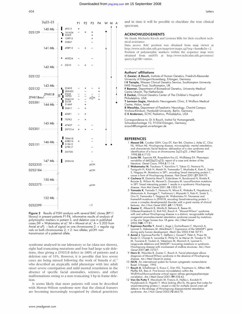
RESULTSResults of FISH and marker analysis are shown in fig 2. In
patient 3 the distal border of the deletion was determined
with polymorphic markers, which showed a distal breakpoint
between markers D2S2275 (150.3 Mb) and D2S2299 (152.2
Mb), and a paternal origin of the deletion. Deletions were of
differing sizes, approximately 300 kb in patient 4, 700 kb in
patient 2, 5 Mb in patient 1, and 11 Mb in patient 3. The
mothers of patients 2 and 4 and both parents of patient 3 were
available for FISH analysis, which showed normal results. The
phenotype observed in patient 3, with the largest deletion,
showed early seizures, hypoplastic big toes, and premature
death at the age of 4 months as additional features (table 1).
DISCUSSIONOur results indicate that deletion sizes and breakpoints in
Mowat-Wilson syndrome patients vary widely, ruling out a
true microdeletion syndrome with recurring breakpoints
mediated by low copy repeat regions. There was generally no
obvious correlation between the phenotype and the size of the
deletion and the phenotypic spectrum was similar to that
observed in patients with truncating mutations within
ZFHX1B (table 1). The only remarkable difference was noticed
Key points
• Mowat-Wilson syndrome (MWS) is a distinct multiplecongenital anomalies-mental retardation syndromecharacterised by severe mental retardation, recognis-able facial gestalt, pre- or postnatal microcephaly, andpostnatal growth retardation, as well as seizures (82%)and malformations such as Hirschsprung disease(67.6%), congenital heart defects (47%), and agenesisof the corpus callosum (35%), caused by mutations orlarge scale deletions of the ZFHX1B gene in 2q22.
• Deletion sizes and breakpoints in Mowat-Wilsonsyndrome patients vary widely from 300 kb to at least11 Mb, thus ruling out a true microdeletion syndrome.
• So far parental origin has only been determined in fourpatients and has always been paternal.
• In general, patients with deletions are very similar tothose with truncating mutations. There was nocorrelation between the phenotype and size of deletionup to 5 Mb. However, one patient with a larger deletion(∼11 Mb) had early seizures with a lethal course andhypoplasia of the big toes as additional features.
601
www.jmedgenet.com
on 15 September 2008 jmg.bmj.comDownloaded from
in patient 3 with the 11 Mb deletion, who presented with sei-
zures much earlier, had marked hypoplasia of the big toes, and
who died in early infancy. Thus, genes within the close vicin-
ity of the ZFXH1B gene seem not to be subject to gross
haploinsufficiency. Parental origin was only determined in
one of the present and three published patients,3 8 and was of
paternal origin in all cases investigated. As all investigated
patients had HSCR and congenital heart defects, it is not pos-
sible to draw any conclusion about these symptoms, but agen-
esis of the corpus callosum was present and absent in two
patients each, and thus shows no correlation with parental
origin of the deletion. Similarly, the early onset of seizures in
patient 3 is also not attributable to the parental origin of the
deletion.
The most frequently observed major malformation in
Mowat-Wilson syndrome is HSCR, which occurred in 21 of 30
(70 %) patients reported so far (table 1). As has been described
for patients with ZFHX1B truncating mutations, two of our
patients with deletions of approximately 700 kb and 5 Mb,
respectively, did not have HSCR, while the two with the small-
est and largest deletion (300 kb and 11 Mb deletions, respec-
tively) did have it. Thus, our results suggest that the manifes-
tation of HSCR is not influenced by deletion size. As ZFHX1Bknockout mice also do not exhibit HSCR,12 a non-allelic modi-
fier might contribute to the manifestation of HSCR. The high
rate of HSCR in humans is probably the result of recognition
bias, as in our cohort (four patients reported earlier6 and the
present four patients) HSCR occurs in only 50%. Less frequent
malformations include various congenital heart defects (for
example, septal defects, pulmonary stenosis, or atresia), agen-
esis of the corpus callosum, urogenital anomalies, talipes, and
strabismus.
Similarly, there is no difference in degree of mental retarda-
tion, facial appearance, and growth parameters. Regardless of
the underlying defect, which may be a truncating mutation in
ZFHX1B or a large scale deletion, psychomotor retardation is
severe with a mean walking age of 4-5 years and speech start-
ing at the age of 5-6 years, being restricted to single words.
Personality is generally happy and affectionate. Although
shortness of stature and low weight are characteristic in
school age children, birth measurements are usually normal
or even in the upper normal range. Only microcephaly was
already evident at birth in eight out of 19 patients with
reported measurements (table 1), and it has a tendency to
occur before the decline of body length in our patients. There-
fore, our findings do not support the initial statement by
Wakamatsu et al3 about the more severe phenotype in their
deletion patient. Nevertheless, severe cerebral atrophy re-
mains remarkable in this patient, but might be related to the
other translocation breakpoint on chromosome 13.
A seizure disorder with varying age of onset is a very com-
mon feature which is found in 82% of all 34 patients (table 1).
Severe neonatal seizures, however, have been reported only in
our patient 3 and a patient with a cytogenetically visible
deletion.2 Thus a gene(s) responsible for early seizures with a
lethal course and hypoplastic big toes might be located
between BAC RP11-207O14 at 145.3 Mb and marker D2S2299
at 152.2 Mb where at least one gene related to epilepsy,
CACNB4 (OMIM 601949), is known to be located. However,
detailed analysis in further patients is required for confirma-
tion of this putative association.
The characteristic facial appearance was evident in all
patients with deletion or truncating mutations and allows the
distinction between Mowat-Wilson syndrome and other types
of “syndromic HSCR” such as Goldberg-Shprintzen syn-
drome. The facial features are probably diagnosable in the
neonatal period in the presence of HSCR, but the sunken eyes,
broad, flared eyebrows, pointed nasal tip, short philtrum, and
upturned ear lobes become more obvious in early childhood.
Of 12 patients with the distinct facial gestalt of Mowat-Wilson
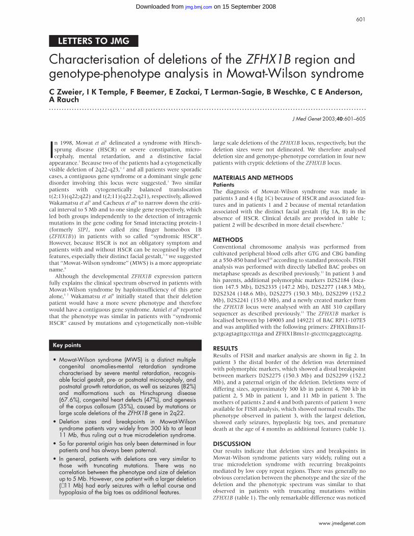
Figure 1 (Top row) Facial appearance of patient 1 in early childhood (A) and aged 8 years (B), and of patient 4 aged 10 years (C). (Bottomrow) Note similarity to patients with ZFHX1B point mutation reported elsewhere.6 (D, E) Patient with nt553-554insTG mutation aged 6 monthsand 6 years 10 months, respectively. (F) Patient with nt1892delA mutation aged 3 years 10 months.
602 Letters
www.jmedgenet.com
on 15 September 2008 jmg.bmj.comDownloaded from
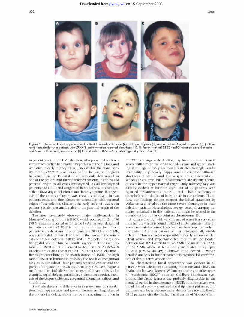
Table 1 Phenotype of previously published patients with Mowat-Wilson syndrome and mutation, deletion, or translocation breakpoint in the ZFHX1B gene and of the presentpatients
MWS patients with mutations(n=22)] 1 3-6 8 14 15 BP in ZFHX1B IVS2 4 15 Del2 Del1 Del3 5 16 Del 8 Pat 1 Pat 2 Pat 3 Pat 4
Gender 16 M, 6F 1 M 1 M 1 M 1 F 1 M, 3 F F M M MSporadic occurrence 22 + + + + + + + + +Birth length (centile) 5 × >90th 50th ? 50th ? ? ? 10th 5th 90th
4 × >50th4 × >10th1 × rd3
Birth weight (centile) 4 × >90th 90th 90th 50th 50th ? 50th 90th 60th 90th9 × 50th4 × >10th
OFC at birth 4 × >25th 50th <3rd 25th <3rd ? <3rd 3rd 20th ?(centile) 4 × 10th-25th
8 × <3rdAge at last investigation 13 mth-23 y 6 y 2y 6 mth 3 y 6 y ? 10 y 8 y 7mth 5 wks 13 yLength (centile) 1 × 75th <<3rd 3rd-10th 50th <3rd ? <10th 3rd 50th ?
2 × 10th-25th9 × <3rd
Weight (centile) 4 × <3rd <3rd 10th <3rd <3rd ? 3rd 10th 50th ?5 × 3rd-10th3 × <3rd
OFC (centile) 3 × 10th-50th <3rd <<3rd 3rd <<3rd ? <3rd <3rd 3rd 40th9 × <3rd
Microcephaly 18 + + + + 4 + + + -Severe mental retardation 22 + + + + 4 + + + +Walking at age 2-8 y 6 y - - - ? 6 y 9 y - 21⁄2 y
Average 4-5 yPersonality Predominantly happy ? ? Happy ? Happy Affectionate, happy Happy N/A Affectionate, happySeizures 16 Febrile Neonatal 2 y 11 mth In 4 7 y Childhood Neonatal 4 yHypotonia 7 + ? ? ? ? - + + ?
Spasticity in lower limbsPes planus with calcaneovalgus 6 + ? + ? ? - + Hypoplasia of big
toesWalks on tiptoes
Agenesis of corpus callosum 5 + ? + - 3 - + - +Cerebral atrophy MRI normal Colpocephaly
Hirschsprung disease 13 + + + + SS 4 2SS, 1LS - - + +Constipation 5 + -Congenital heart disease 7 - Ast, PDA, VSD, ASD - PDA 4 ASD - PFO, PS VSD, PSRenal anomalies 4 - - Duplex kidney - 0 - - - -Hypospadias 3 + - + ? 1 + Micro-phallus +Cryptorchidism 6 - ? + + -Cleft palate/lip 1 submucous - - - - 0 - - - -Ptosis 0 - - - - 0 - - - -Iris coloboma 0 - - - - 0 - - - -Strabismus 9 + - + + 3? - + ? +Hypertelorism 20 + - + + 2 of 2 + + + +Facial gestalt All MWS MWS MWS MWS MWS MWS MWS MWS MWS MWSDeletion or translocation of 2q22 t (2;11) (q22.2;2q21) del (2)(q22q23) del (2) (q21q23) 5 Mb Size? 5 Mb 700 kb 11 Mb 300 kbZFHX1B mutation (truncating) 1 × exon 3
1 × exon 51 × exon 71 × exon 102 × exon 616 × exon 8
BP: breakpoint; IVS2: intron 2 ; colpocephaly: enlargement of the occipital horns of the ventricular system owing to an underdevelopment of the white matter in the posterior cerebrum17; Ast: aortic stenosis; ASD: atrial septal defect; PDA: persistent ductusarteriosus; VSD: ventricular septal defect; PFO: persistent foramen ovale; PS: pulmonary stenosis; SS: short segment HSCR; LS: long segment HSCR; VUR: vesicoureteric reflux. In deletion patients age of onset of seizures is given.
Letters603
ww
w.jm
edgenet.com
on 15 Septem
ber 2008 jm
g.bmj.com
Dow
nloaded from
syndrome analysed in our laboratory so far (data not shown),
eight had truncating mutations and four had large scale dele-
tions, thus giving a ZFHX1B defect in 100% of patients and a
deletion rate of 33%. However, it is possible that less severe
cases are being missed following the work of Yoneda et al,13
who described an atypically mild phenotype with late adult
onset severe constipation and mild mental retardation in the
absence of specific facial anomalies, seizures, and other
malformations owing to a non-truncating 3 bp in frame dele-
tion.
It seems likely that more patients will soon be described
with Mowat-Wilson syndrome now that the clinical features
are becoming increasingly recognised by clinical geneticists
and in time it will be possible to elucidate the true clinical
spectrum.
ACKNOWLEDGEMENTSWe thank Michaela Kirsch and Leonora Bille for their excellent tech-nical assistance.Data access. BAC position was obtained from map viewer athttp://www.ncbi.nlm.nih.gov/mapview/maps.cgi?org=hum&chr=2.Position of polymorphic markers within the sequence map wereobtained from uniSTS at http://www.ncbi.nlm.nih.gov/entrez/query.fcgi?db=unists.
. . . . . . . . . . . . . . . . . . . . .Authors’ affiliationsC Zweier, A Rauch, Institute of Human Genetics, Friedrich-AlexanderUniversity of Erlangen-Nuremberg, Erlangen, GermanyI K Temple, Wessex Clinical Genetics Service, Southampton UniversityNHS Hospital Trust, Southampton, UKF Beemer, Department of Biomedical Genetics, University MedicalCentre Utrecht, The NetherlandsE Zackai, Clinical Genetics Center of The Children’s Hospital ofPhiladelphia, USAT Lerman-Sagie, Metabolic Neurogenetic Clinic, E Wolfson MedicalCentre, Holon, IsraelB Weschke, Department of Paediatric Neurology, Charité CampusVirchow-Klinikum, Humboldt University, Berlin, GermanyC E Anderson, SCHC Pediatrics, Philadelphia, USA
Correspondence to: Dr A Rauch, Institut für Humangenetik,Schwabachanlage 10, 91054 Erlangen, Germany;[email protected]
REFERENCES1 Mowat DR, Croaker GDH, Cass DT, Kerr BA, Chaitow J, Adès LC, Chia
NL, Wilson MJ. Hirschsprung disease, microcephaly, mental retardation,and characteristic facial features: delineation of a new syndrome andidentification of a locus at chromosome 2q22-q23. J Med Genet1998;35:617-23.
2 Lurie IW, Supovitz KR, Rosenblum-Vos LS, Wulfsberg EA. Phenotypicvariability of del(2)(q22-q23): report of a case and review of theliterature. Genet Couns 1994;5:11-14.
3 Wakamatsu N, Yasukazu Y, Kenichiro Y, Takao O, Nomura N,Taniguchi H, Kitoh H, Mutoh N, Yamanaka T, Mushiake K, Kato K, SontaS, Nagaya M. Mutations in SIP1, encoding Smad interacting protein-1,cause a form of Hirschsprung disease. Nat Genet 2001;27:369-70.
4 Cacheux V, Dastot-Le Moal F, Kääriäinen H, Bondurand N, Rintala R,Boissier B, Wilson M, Mowat D, Goossens M. Loss-of-function mutationsin SIP1 Smad interacting protein 1 results in a syndromic Hirschsprungdisease. Hum Mol Genet 2001;10:1503-10.
5 Yamada K, Yamada Y, Nomura N, Miura K, Wakako R, Hayakawa C,Matsumoto A, Kumagai T, Yoshimura I, Miyazaki S, Kato K, Sonta S,Ono H, Yamanaka T, Nagaya M, Wakamatsu N. Nonsense andframeshift mutations in ZFHX1B, encoding Smad-interacting protein 1,cause a complex developmental disorder with a great variety of clinicalfeatures. Am J Hum Genet 2001;69:1178-85.
6 Zweier C, Albrecht B, Mitulla B, Behrens R, Beese M,Gillessen-Kaesbach G, Rott H-D, Rauch A. “Mowat-Wilson” syndromewith and without Hirschsprung disease is a distinct, recognizable multiplecongenital anomalies-mental retardation syndrome caused by mutationsin the zinc finger homeo box 1B gene. Am J Med Genet2002;108:177-81.
7 Espinosa-Parrilla Y, Amiel J, Auge J, Encha-Razavi F, Munnich A,Lyonnet S, Vekemans M, Attie-Bitach T. Expression of the SMADIP1 geneduring early human development. Mech Dev 2002;114:187-91.
8 Amiel J, Espinosa-Parrilla Y, Steffann J, Gosset P, Pelet A, Prieur M,Boute O, Choiset A, Lacombe D, Philip N, Le Merrer M, Tanaka H, TillM, Touraine R, Toutain A, Vekemans M, Munnich A, Lyonnet S.Large-scale deletions and SMADIP1 truncating mutations in syndromicHirschsprung disease with involvement of midline structures. Am J HumGenet 2001;69:1370-7.
9 Horn D, Weschke B, Zweier C, Rauch A. Facial phenotype allowsdiagnosis of Mowat-Wilson syndrome in the absence of Hirschsprungdisease. Am J Med Genet (in press).
10 ISCN. An international system for human cytogenetic nomenclature.Basel: S Karger, 1995.
11 Rauch A, Schellmoser S, Kraus C, Dörr HG, Trautmann U, Altherr MR,Pfeiffer RA, Reis A. First known microdeletion within theWolf-Hirschhorn-syndrome critical region refines genotype-phenotypecorrelation. Am J Med Genet 2001;99:338-42.
12 Van De Putte T, Maruhashi M, Francis A, Nelles L, Kondoh H,Huylebroeck D, Higashi Y. Mice lacking zfhx1b, the gene that codes forsmad-interacting protein-1, reveal a role for multiple neural crest celldefects in the etiology of Hirschsprung disease-mental retardationsyndrome. Am J Hum Genet 2003;72:465-70.
Figure 2 Results of FISH analysis with several BAC clones (RP11library) in present patients P1-P4, informative results of analysis ofpolymorphic markers in patient 3, and deletion size in publishedcases (W = Wakamatsu et al,3 M = Mowat et al,1 A = S.203 fromAmiel et al8). – lack of signal on one chromosome 2; + regular sig-nals on both chromosomes 2; × 2: two alleles; pLOH: non-transmission of a paternal allele.
604 Letters
www.jmedgenet.com
on 15 September 2008 jmg.bmj.comDownloaded from
13 Yoneda M, Fujita T, Yamada Y, Yamada K, Fujii A, Inagaki T,Nakagawa H, Shimada A, Kishikawa M, Nagaya M, Azuma T,Kuriyama M, Wakamatsu N. Late infantile Hirschsprung disease-mentalretardation syndrome with a 3-bp deletion in ZFHX1B. Neurology2002;59:1637-40.
14 Garavelli L, Donadio A, Zanacca C, Banchini G, Della Giustina E,Bertani G, Albertini G, Del Rossi C, Zweier C, Rauch A, Zollino M, NeriG. Hirschsprung disease, mental retardation, characteristic facialfeatures, and mutation in the gene ZFHX1B (SIP1): confirmation of theMowat-Wilson syndrome. Am J Med Genet 2003;116A:385-8.
15 Kääriäinen H, Wallgren-Pettersson C, Clarke A, Pihko H, Taskinen H,Rintala R. Hirschsprung disease, mental retardation and dysmorphicfacial features in five unrelated children. Clin Dysmorphol2001;10:157-63.
16 Nagaya M, Kato J, Niimi N, Tanaka S, Wakamatsu N. Clinical featuresof a form of Hirschsprung’s disease caused by a novel geneticabnormality. J Pediatr Surg 2002;37:1117-22.
17 Hunter AGW. Brain. In: Stevenson RE, Hall JG, Goodman RM, eds.Human malformations and related anomalies. Vol II. Oxford: OxfordUniversity Press, 1993:73.
Molecular study of three cases ofodontohypophosphatasia resulting from heterozygosityfor mutations in the tissue non-specific alkalinephosphatase geneM Herasse, M Spentchian, A Taillandier, K Keppler-Noreuil, A N M FIiorito,J Bergoffen, R Wallerstein, C Muti, B Simon-Bouy, E Mornet. . . . . . . . . . . . . . . . . . . . . . . . . . . . . . . . . . . . . . . . . . . . . . . . . . . . . . . . . . . . . . . . . . . . . . . . . . . . . . . . . . . . . . . . . . . . . . . . . . . . . . . . . . . . . . . . . . . . . . . . . . . . .
J Med Genet 2003;40:605–609
Hypophosphatasia is an inherited disorder characterisedby defective bone and tooth mineralisation and defi-ciency of serum and bone alkaline phosphatase activity.
The bone symptoms are highly variable in their clinical expres-sion and range from stillbirths without mineralised bone topathological fractures developing only late in adulthood.1
Odontohypophosphatasia is characterised by premature exfo-liation of fully rooted primary teeth and/or severe dental caries,often not associated with abnormalities of the skeletalsystem.2 3 The anterior deciduous teeth are more likely to beaffected and the most frequently lost are the incisors.4 Dental xrays show reduced alveolar bone and enlarged pulp chambersand root canals.2 4 Although the only clinical feature is dentaldisease, biochemical findings are generally indistinguishablefrom those in patients with mild forms of hypophosphatasia(adult and childhood). While perinatal hypophosphatasia andinfantile hypophosphatasia are transmitted as an autosomalrecessive trait, both autosomal recessive and autosomal domi-nant transmission may be found in childhood, adult, andodontohypophosphatasia.3 5–9 The distinction between recessiveand dominant transmission may be difficult to determine con-clusively by using familial analysis because expression of thedisease is very variable, with parents of even severely affectedchildren showing no or extremely mild symptoms of thedisease.9 10
The tissue non-specific alkaline phosphatase (TNSALP) is aphosphomonoesterase anchored at its carboxyl terminus tothe plasma membrane by a phosphatidylinositol-glycanmoiety.11 The enzyme cleaves extracellular substratespyridoxal-5′-phosphate (PLP), phosphoethanolamine (PEA),and inorganic pyrophosphates (PPi). Its exact function inbone and dental mineralisation is still unclear but probablyinvolves hydrolysis of Ppi12 and perhaps mammalian specificactivities such as collagen and calcium binding.13
The TNSALP gene is localised on chromosome 1p36.114 andconsists of 12 exons distributed over 50 kb.15 More than 127distinct mutations have been described in the TNSALPgene,16–33 in a relatively small number of North American,Japanese, and European patients,34 indicating a very strongallelic heterogeneity in the disease. Most of them (82%) weremissense mutations. This variety of mutations results in vari-able clinical expression even among the severe or moderatetypes. We report here the study of TNSALP gene mutations in
three patients affected by odontohypophosphatasia and
provide evidence that heterozygosity may produce clinical
signs and symptoms that appear to be very variable in expres-
sion.
MATERIAL AND METHODSPatientsPatient 1The proband was a 9 year old boy affected by Down syndrome
and odontohypophosphatasia. Loss of seven deciduous teeth,
Key points
• Hypophosphatasia is an inherited disorder character-ised by defective bone mineralisation and deficiency oftissue non-specific alkaline phosphatase (TNSALP) activ-ity. We report here the molecular study of three cases ofodontohypophosphatasia where the disease was theresult of heterozygosity for TNSALP gene mutations.
• Three mutations were found, 323C>T (P91L), 346G>A(A99T), and 1240C>A (L397M). The mutation P91L hasnot been previously described and site directedmutagenesis experiments showed that it correspondedto a severe allele.
• In one family, the proband’s mother carried the mutationP91L and was affected. In the two other families, domi-nant transmission was more difficult to determine, owingto variable expression of the disease in carriers, rangingfrom hypophosphatasaemia only to periodontal diseaseassociated with multiple fractures.
• We show here that the convergence of clinical,biochemical, and molecular results may help to affirmthe dominant effect of TNSALP mutations. Analysis of a3D model of TNSALP indicated that residues affected bythese mutations were located near the active site or inthe mammalian specific crown domain, corroboratingthe functional effect of these mutations. This is consistentwith the dominant effect of these mutations and theallosteric properties of the enzyme.
Letters 605
www.jmedgenet.com
on 15 September 2008 jmg.bmj.comDownloaded from
mostly incisors, began at the age of 2 years. Serum alkaline
phosphatase was low (80 U/l, normal range >100). X rays
showed normal growth plate development and normal long
bones without evidence of fractures or rickets. The 39 year old
mother of the proband had lost her permanent teeth. Her
serum alkaline phosphatase level was low (18 U/l, normal
range 30-120). The father did not show any symptoms and
had a normal level of serum alkaline phosphatase (93 U/l,
normal range 30-120).
Patient 2The proband was a 2 year old male who has lost three teeth
and was referred to the genetics department by his dentist.
Serum alkaline phosphatase was low (64 U/l, normal range
100-320) and urinary phosphoethanolamine was high (583
µmol/g creatinine, normal range <350). The parents deny any
problem with multiple fractures, bowing of legs, or loss of
teeth but the 38 year old mother reported being affected by an
unusual number of dental cavities and having had numerous
treatments of dental root canals. Her serum ALP was low (29
U/l, normal range 47-137) while serum ALP of the proband’s
father was normal (66 U/l, 47-137). A second child was born in
July 2002. At nearly 7 months of age, this baby boy has not
shown any symptoms of hypophosphatasia to bring him to
clinical attention.
Patient 3The proband was a 2 year old boy diagnosed with hypophos-
phatasaemia. Serum alkaline phosphatase was repeatedly low
(84 and 86 U/l, normal range 104-345) and urinary
phosphoethanolamine was high (935 µmol/g creatinine,
normal range 108-533). He had very slight leg bowing and was
just below the 5th centile for height. The mother had a low
ALP level (32 U/l, normal range 30-107) and high PEA (138
µmol/g creatinine, normal range 20-100). The father showed
normal ALP and PEA levels. The proband’s 5 year old maternal
second cousin was diagnosed with hypophosphatasia because
of early loss of primary teeth and low serum ALP (92 U/l, nor-
mal range 108-317). Her skeletal survey and growth/stature
were normal for age. Her father had early loss of teeth, multi-
ple caries, and had had four fractures. Another second cousin
could not be tested but was reported to have lost her primary
teeth at the age of 2.
METHODSPrimer sequences of the 12 ALPL gene exons have been previ-
ously reported24 and allowed analysis of the whole coding
sequence, including intron-exon borders and untranslated
exons. PCR reactions were performed and analysed as
previously described.24 Site directed mutagenesis of the muta-
tion P91L was performed with the Quikchange Site Directed
Mutagenesis kit (Stratagene). Mutated and wild type plas-
mids were transiently transfected in COS-1 cells using the
Lipofectamine PLUS reagent (Life Technologies) according to
a methodology described previously.9 26 The mutations were
put into a 3D model of the TNSALP molecule13 by using the
molecular visualisation program RasMol (R. Sayle, Glaxo
Research and Development, Greenford).
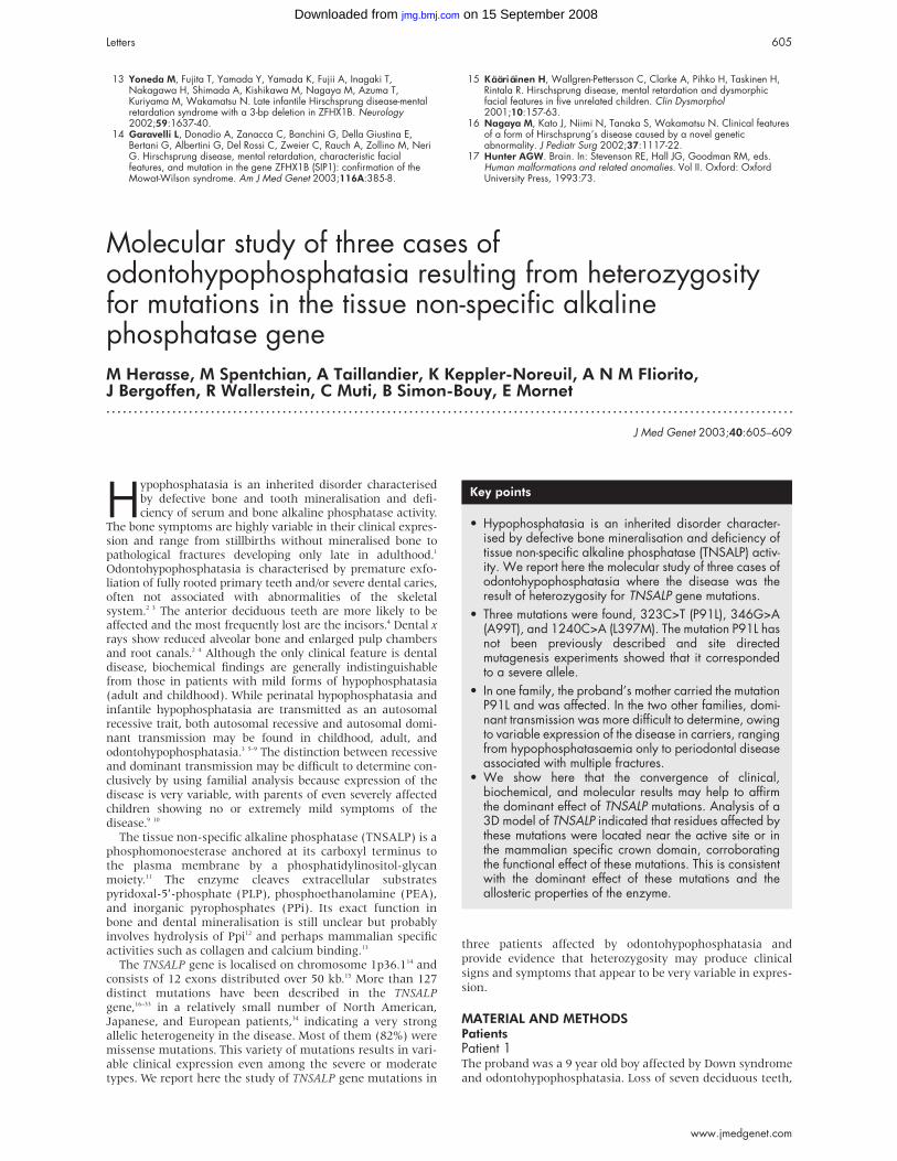
RESULTSPedigree data and mutation analysis results are shown in fig 1.
In family 1, the proband’s mother was affected by early loss of
teeth, suggesting that the disease could not be put down to the
proband’s Down syndrome condition only35 and that the
disease was dominantly inherited.
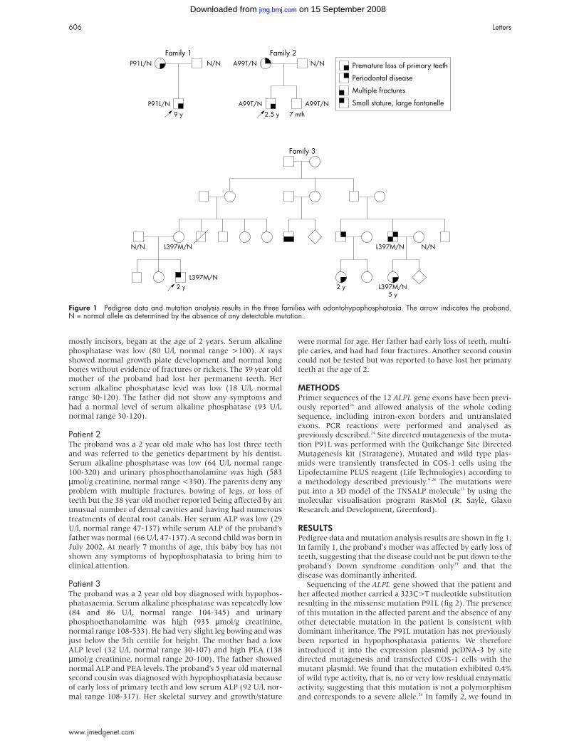
Sequencing of the ALPL gene showed that the patient and
her affected mother carried a 323C>T nucleotide substitution
resulting in the missense mutation P91L (fig 2). The presence
of this mutation in the affected parent and the absence of any
other detectable mutation in the patient is consistent with
dominant inheritance. The P91L mutation has not previously
been reported in hypophosphatasia patients. We therefore
introduced it into the expression plasmid pcDNA-3 by site
directed mutagenesis and transfected COS-1 cells with the
mutant plasmid. We found that the mutation exhibited 0.4%
of wild type activity, that is, no or very low residual enzymatic
activity, suggesting that this mutation is not a polymorphism
and corresponds to a severe allele.26 In family 2, we found in
Figure 1 Pedigree data and mutation analysis results in the three families with odontohypophosphatasia. The arrow indicates the proband.N = normal allele as determined by the absence of any detectable mutation.
606 Letters
www.jmedgenet.com
on 15 September 2008 jmg.bmj.comDownloaded from
the proband the heterozygous nucleotide substitution
346G>A resulting in the missense mutation A99T. The mother
reported being affected by dental problems. She showed a low
level of serum ALP and carried the mutation A99T, while the
father exhibited normal serum ALP and no ALPL gene muta-
tion. The second child, born in July 2002, was prenatally found
to be heterozygous for the A99T mutation. Although the
mother did not exhibit the typical signs of odontohypophos-
phatasia (loss of teeth), these results suggest that in this fam-
ily, heterozygosity for A99T resulted in clinical symptoms but
that the disease was minimally expressed in the mother. The
mutation A99T was previously described in a large family with
dominant odontohypophosphatasia8 and site directed muta-
genesis and transfections of COS cells previously showed that
A99T does not allow any significant in vitro residual activity
and shows a negative dominant effect.9 In family 3, the
proband did not show any symptoms of hypophosphatasia
and was referred to the genetics department because of hypo-
phosphatasaemia. We found the heterozygous substitution
1240C>A resulting in the missense mutation L397M of
maternal origin. Interestingly, the proband’s second cousin
and this cousin’s father were affected by odontohypophos-
phatasia and carried the L397M mutation found in the
proband. Exhaustive sequencing of the ALPL gene of the
affected cousin did not show evidence of any other mutation.
This suggests that in this family, heterozygotes for L397M may
be affected by the disease and that its expression was subject
to intrafamilial heterogeneity. The L397M mutation was
previously reported by Mumm et al,32 associated with the
D277A mutation, in a patient affected by perinatal hypophos-
phatasia. This suggests that, like P91L and A99T, L397M is a
severe allele. We finally concluded that the disease in these
three families resulted from heterozygosity for a severe hypo-
phosphatasia allele.
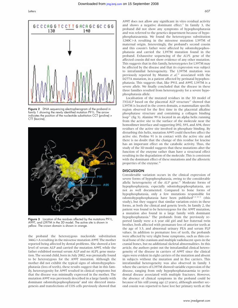
Localisation of the mutated residues in the 3D model of
TNSALP based on the placental ALP structure13 showed that
L397M is located in the crown domain, a mammalian specific
region observed for the first time in the placental alkaline
phosphatase structure and containing a collagen binding
loop13 (fig 3). Alanine 99 is located in an alpha helix running
from the active site to the surface of the molecule near the
homodimer interface and supporting D92, S93, and A94, three
residues of the active site involved in phosphate binding. By
disturbing this helix, mutation A99T could therefore affect the
active site. Proline 91 is in contact with the active site and
there is no doubt that the change of this residue for leucine
has an important effect on the catabolic activity. Thus, the
study of the 3D model suggests that these mutations alter the
function of the enzyme rather than have a structural effect
resulting in the degradation of the molecule. This is consistent
with the dominant effect of these mutations and the allosteric
properties of the enzyme.36
DISCUSSIONConsiderable variation occurs in the clinical expression of
severe forms of hypophosphatasia, owing to the considerable
allelic heterogeneity of the ALP gene.33 Moderate forms of
hypophosphatasia, especially odontohypophosphatasia, are
not as well documented. Compared to bone forms of
hypophosphatasia, only a few mutations responsible for
odontohypophosphatasia have been published8 22 28 31 (this
study), but they suggest that similar variation exists in these
forms, at both the clinical and genetic levels. In family 2, the
patient was found to be heterozygous for the A99T mutation,
a mutation also found in a large family with dominant
hypophosphatasia.8 The probands from the previously re-
ported family were a 6 year old girl and her fraternal twin
brother, both affected with premature loss of anterior teeth at
the age of 3.5, and abnormal urinary PEA and serum PLP
values. In addition to premature loss of teeth, the probands
were affected by very slight bone symptoms, such as thin cor-
tical bone of the cranium and multiple radiolucent spots in the
cranial bones, but no additional skeletal abnormalities. In this
article, the authors point out the intrafamilial clinical hetero-
geneity of the disease in carriers of A99T since the clinical
signs were evident in eight carriers of the mutation and absent
in subjects without the mutation and in five carriers. This
intrafamilial heterogeneity was also observed in family 3
where the carriers of L397M showed variable expression of the
disease, ranging from only hypophosphatasaemia to perio-
dontal disease associated with multiple fractures. However,
the absence of clinical symptoms in the proband could be
because of his still young age (2 years), although another sec-
ond cousin was reported to have lost her primary teeth at the
Figure 2 DNA sequencing electropherogram of the proband infamily 1 showing the newly identified mutation P91L. The arrowindicates the position of the nucleotide substitution CCT (proline) >CTT (leucine).
Figure 3 Location of the residues affected by the mutations P91L,A99T, and L397M in the 3D model. The active site is shown inyellow. The crown domain is shown in orange.
Letters 607
www.jmedgenet.com
on 15 September 2008 jmg.bmj.comDownloaded from
age of 2 (fig 1). Finally, this report confirms that moderate
forms of hypophosphatasia are also highly variable in their
clinical expression, owing to allelic heterogeneity but also to
other factors that remain to be determined, such as other
sequence variations in the ALPL gene, a trans effect of other
genes, or environmental factors.
Dominant transmission of moderate forms of hypophos-
phatasia has been documented in a few families.3 5–9 We report
here the case of one additional family with dominant odonto-
hypophosphatasia (family 1) and two others in which hetero-
zygotes for a TNSALP gene mutation show clinical symptoms,
however variable in expression. In our experience, we failed to
detect a second mutated allele in 18% of our hypophosphata-
sia patients, 70% of them being affected by moderate
(childhood, adult, or odonto-) hypophosphatasia (E Mornet,
unpublished data). In some of these patients, mutations of the
ALPL gene may have not been detected because of their loca-
tion in intronic or regulatory sequences, or because they
correspond to large deletions undetectable by the method-
ology routinely used here. In others, however, the disease may
be the result of heterozygosity and no other mutation needed
to be sought. Analysis of the transmission of the odontohypo-
phosphatasia phenotype, together with serum ALP level and
presence or absence of the mutation, may help to distinguish
between the two situations.
The mechanism of dominance remains unclear but prob-
ably involves interactions between monomers of the dimeric
structure that disturb the allosteric properties of TNSALP. We
and others have previously reported that some ALPL gene
mutations result in a dominant negative effect owing to com-
plete or partial inhibition of the normal monomer by the
mutated monomer in the dimeric molecule.7 9 Here, we show
that residues mutated in these families are localised in the
vicinity of functional regions such as the active site and the
crown domain, suggesting that they may have a functional
role. This is consistent with the expected localisation of muta-
tions resulting in an inhibitory effect.
. . . . . . . . . . . . . . . . . . . . .Authors’ affiliationsM Herasse, E Mornet, Laboratoire de Cytogénétique et GénétiqueMoléculaire Humaine, Université de Versailles Saint Quentin en Yvelines,Versailles, FranceM Spentchian, Laboratoire de Biochimie, Centre Hospitalier deVersailles, Versailles, FranceA Taillandier, C Muti, B Simon-Bouy, E Mornet, Laboratoire SESEP,Université de Versailles-Saint Quentin en Yvelines, Versailles, FranceK Keppler-Noreuil, Division of Medical Genetics, Department ofPediatrics, University of Iowa Hospitals and Clinics, Iowa City, IA, USAA N M Fiorito, J Bergoffen, Kaiser Permanente Genetics Department,5755 Cottle Road, Building 1, San Jose, CA 95123, USAR Wallerstein, Genetics Service, Hackensack University Medical Center,NJ 07601, USA
Correspondence to: Dr E Mornet, Laboratoire de Cytogénétique etGénétique Moléculaire Humaine Batiment Fermat, Université deVersailles-Saint Quentin en Yvelines, 45 avenue des Etats-Unis, 78035Versailles Cedex, France; [email protected]
REFERENCES1 Whyte MP. Hypophosphatasia and the role of alkaline phosphatase in
skeletal mineralization. Endocr Rev 1994;15:439-61.2 Brittain JM, Oldenburg TR, Burkes EJ Jr. Odontohypophosphatasia:
report of two cases. ASDC J Dent Child 1976;43:106-11.3 Eberle F, Hartenfels S, Pralle H, Kabisch A. Adult hypophosphatasia
without apparent skeletal disease: “odontohypophosphatasia” in fourheterozygote members of a family. Klin Wochenschr 1984;62:371-6.
4 Beumer J, Trowbridge HO, Silverman S Jr, Eisenberg E. Childhoodhypophosphatasia and the premature loss of teeth. A clinical andlaboratory study of seven cases. Oral Surg Oral Med Oral Pathol1973;35:631-40.
5 Whyte MP, Fallon MD, Murphy WA. Adult hypophosphatasia: clinical,laboratory and genetic investigation of a large kindred with review of theliterature. Medicine 1979;58:329-47.
6 Eastman JR, Bixler D. Clinical, laboratory, and genetic investigations ofhypophosphatasia: support for autosomal dominant inheritance withhomozygous lethality. J Craniofac Genet Dev Biol 1983;3:213-34.
7 Müller HL, Yamazaki M, Michigami T, Kageyama T, Schönau E,Schneider P, Ozono K. Asp361 mutant of alkaline phosphatase found inpatients dominantly inherited hypophosphatasia inhibits the activity of thewild-type enzyme. J Clin Endocrinol Metab 2000;85:743-47.
8 Hu JCC, Plaetke R, Mornet E, Zhang C, Sun X, Thomas HF, Simmer JP.Case report of a family with dominant hypophosphatasia. Eur J Oral Sci2000;108:189-94.
9 Lia-Baldini AS, Muller F, Taillandier A, Gibrat JF, Mouchard M, Robin B, Simon-Bouy B, Serre JL, Aylsworth AS, Bieth E, Delanote S, Freisinger P,Hu JCC, Krohn HP, Nunes ME, Mornet E. A molecular approach todominance in hypophosphatasia. Hum Genet 2001;109:99-108.
10 Moore CA, Curry CJR, Henthorn PS, Smith JA, Smith JC, O’Lague P,Coburn SP, Weaver DD, Whyte MP. Mild autosomal dominanthypophosphatasia: in utero presentation in two families. Am J Med Genet1999;86:410-15.
11 Jemmerson R, Low MG. Phosphatidylinositol anchor of HeLa cellalkaline phosphatase. Biochemistry 1987;26:5703-9.
12 Hessle L, Johnson KA, Anderson HC, Narisawa S, Sali A, Goding JW,Terkeltaub R, Millan JL. Tissue-nonspecific alkaline phosphatase andplasma cell membrane glycoprotein-1 are central antagonistic regulatorsof bone mineralization. Proc Natl Acad Sci U S A 2002;99:9445-9.
13 Mornet E, Stura E, Lia-Baldini AS, Stigbrand T, Ménez A, Le Du MH.Structural evidence for a functional role of human tissue non-specificalkaline phosphatase in bone mineralisation. J Biol Chem2001;276:31171-8.
14 Greenberg CR, Evans JA, McKendry-Smith S, Redekopp S, Haworth JC,Mulivor R, Chordiket BN. Infantile hypophosphatasia localization withinchromosome region 1p36.1.1-34 and prenatal diagnosis using linkedDNA markers. Am J Hum Genet 1990;46:286-92.
15 Weiss MJ, Ray K, Henthorn PS, Lamb B, Kadesch T, Harris H. Structureof the human liver/bone/kidney alkaline phosphatase gene. J Biol Chem1988;263:12002-10.
16 Weiss MJ, Cole DEC, Ray K, Whyte MP, Lafferty MA, Mulivor RA, HarrisH .A missense mutation in the liver/bone/kidney alkaline phosphatasegene causing a lethal form of hypophosphatasia. Proc Natl Acad SciUSA 1988;85:7666-9.
17 Henthorn PS, Raducha M, Fedde KN, Lafferty MA, Whyte MP. Differentmissense mutations at the tissue-nonspecific alkaline phosphatase genelocus in autosomal recessively inherited forms of mild and severehypophosphatasia. Proc Natl Acad Sci USA 1992;89:9924-8.
18 Orimo H, Haysshi Z, Watanabe A, Hirayama T, Hirayama T, ShimadaT. Novel missense and frameshift mutations in the tissue-nonspecificalkaline phosphatase gene in a Japanese patient with hypophosphatasia.Hum Mol Genet 1994;3:1683-4.
19 Ozono K, Yamagata M, Michigami T, Nakajima S, Sakai N, Cai G,Satomura K, Yasui N, Okada S, Nakayama M . Identification of novelmissense mutations (Phe310Leu and Gly439Arg) in a neonatal case ofhypophosphatasia. J Clin Endocrinol Metab 1996;81:4458-61.
20 Orimo H, Goseki-Sone M, Sato S, Shimada T. Detection of deletion1154-1156 hypophosphatasia mutation using TNSALP exonamplification. Genomics 1997;42:364-6.
21 Fukushi M, Amizuka N, Hoshi K, Ozawa H, Kumagai H, Omura S,Misumi Y, Ikehara Y, Oda K. Intracellular retention and degradation oftissue-nonspecific alkaline phosphatase with a Gly317Asp substitutionassociated with lethal hypophosphatasia. Biochem Biophys Res Commun1998;246:613-18.
22 Goseki-Sone M, Orimo H, Iimura T, Takagi Y, Watanabe H, Taketa K,Sato S, Mayanagi H, Shimada T, Oida S. Hypophosphatasia:identification of five novel missense mutations (G507A, G705A, A748G,T1155C, G1320A) in the tissue-nonspecific alkaline phosphatase geneamong Japanese patients. Hum Mutat Suppl 1998;1:S263-7.
23 Sugimoto N, Iwamoto S, Hoshimo Y, Kajii E. A novel missense mutationof the tissue-nonspecific alkaline phosphatase gene detected in a patientwith hypophosphatasia. J Hum Genet 1998;43:160-4.
24 Mornet E, Taillandier A, Peyramaure S, Kaper F, Muller F, Brenner R,Bussière P, Freisinger P, Godard J, Le Merrer M, Oury JF, Plauchu H,Puddu R, Rival JM, Superti-Furga A, Touraine RL, Serre JL, Simon-Bouy B.Identification of fifteen novel mutations in the tissue-nonspecific alkalinephosphatase (TNSALP) gene in European patients with severehypophosphatasia. Eur J Hum Genet 1998;6:308-14.
25 Taillandier A, Zurutuza L, Muller F, Simon-Bouy B, Serre JL, Bird L,Brenner R, Boute O, Cousin J, Gaillard D, Heidemann PH, Steinmann B,Wallot M, Mornet E. Characterization of eleven novel mutations (M45L,R119H, 544delG, G145 V, H154Y, C184Y, D289 V, 862+5A,1172delC, R411X, E459 K) in the tissue-nonspecific alkalinephosphatase (TNSALP) gene in patients with severe hypophosphatasia.Hum Mutat 1999;13:171-2.
26 Zurutuza L, Muller F, Gibrat JF, Taillandier A, Simon-Bouy B, Serre JL,Mornet E Correlations of genotype and phenotype in hypophosphatasia.Hum Mol Genet 1999;8:1039-46.
27 Mochizuki H, Saito M, Michigami T, Ohashi H, Koda N, Yamaguchi S.Severe hypercalcaemia and respiratory insufficiency associated withinfantile hypophosphatasia caused by two novel mutations of thetissue-nonspecific alkaline phosphatase gene. Eur J Pediatr2000;159:375-9.
28 Taillandier A, Cozien E, Muller F, Merrien Y, Bonnin E, Fribourg C,Simon-Bouy B, Serre JL, Bieth E, Brenner R, Cordier MP, De Bie S,Fellmann F, Freisinger P, Golembowski S, Hennekam RCM, Josifova D,Kerzin-Storrar L, Leporrier N, Zabot MT, Mornet E. Fifteen new mutations
608 Letters
www.jmedgenet.com
on 15 September 2008 jmg.bmj.comDownloaded from
(-195T, L-12X, 298-2G, T117 N, A159T, R229S, 997+2A, E274X,A331T, H364R, D389G, 1256delC, R433H, N461I, C472S) in thetissue-nonspecific alkaline phosphatase (TNSALP) gene in patients withhypophosphatasia. Hum Mutat 2000;15:293.
29 Orimo H, Girschick HJ, Goseki-Sone M, Ito M, Oda K, Shimada T.Mutational analysis and functional correlation with phenotype in Germanpatients with childhood-type hypophosphatasia. J Bone Miner Res2001;16:2313-19.
30 Watanabe H, Hashimoto-Uoshima M, Goseki-Sone M, Orimo H,Ishikawa I. A novel point mutation (C571T) in the tissue-non-specificalkaline phosphatase gene in a case of adult-type hypophosphatasia.Oral Dis 2001;7:331-5.
31 Taillandier A, Lia-Baldini AS, Mouchard M, Robin B, Muller F,Simon-Bouy B, Serre JL, Bera-Louville A, Bonduelle M, Eckhardt J,Gaillard D, Myhre AG, Kortge-Jung S, Larget-Piet L, Malou E, Sillence D,
Temple IK, Viot G, Mornet E. Twelve novel mutations in thetissue-nonspecific alkaline phosphatase gene (ALPL) in patients withvarious forms of hypophosphatasia. Hum Mutat 2001;18:83-4.
32 Mumm S, Jones J, Finnegan P, Henthorn PS, Podgornik MN, Whyte MP.Denaturing gradient gel electrophoresis analysis of the tissue nonspecificalkaline phosphatase isoenzyme gene in hypophosphatasia. Mol GenetMetab 2002;75:143-53.
33 The Tissue Nonspecific Alkaline Phosphatase Gene mutationsdatabase, http://www.sesep.uvsq.fr/Database.html
34 Mornet E. Hypophosphatasia: the mutations of the tissue-nonspecificalkaline phosphatase gene. Hum Mutat 2000;15:309-15.
35 Saxen L, Aula S. Periodontal bone loss in patients with Down’ssyndrome: a follow-up study. J Periodontol 1982;53:158-62.
36 Hoylaerts MF, Manes T, Millan JL. Mammalian alkaline phosphatasesare allosteric enzymes. J Biol Chem 1997;272:22781-7.
RPGR mutation associated with retinitis pigmentosa,impaired hearing, and sinorespiratory infectionsI Zito, S M Downes, R J Patel, M E Cheetham, N D Ebenezer, S A Jenkins,S S Bhattacharya, A R Webster, G E Holder, A C Bird, D E Bamiou, A J Hardcastle. . . . . . . . . . . . . . . . . . . . . . . . . . . . . . . . . . . . . . . . . . . . . . . . . . . . . . . . . . . . . . . . . . . . . . . . . . . . . . . . . . . . . . . . . . . . . . . . . . . . . . . . . . . . . . . . . . . . . . . . . . . . .
J Med Genet 2003;40:609–615
Retinitis pigmentosa (RP) is a progressive retinal degen-
eration that affects about 1 in 4000 of the population.1
Approximately 15-30% of patients with RP have X linked
retinitis pigmentosa (XLRP), which is the most severe form of
RP consistently manifesting early in life.2 3 Night blindness is
usually present in early childhood with loss of peripheral
visual fields and ultimately central vision, resulting in
registered blindness by the end of the third decade. Female
carriers display a broad spectrum of fundus appearances
ranging from normal to extensive retinal degeneration.4–6
XLRP is genetically heterogeneous with two major loci, RP2
(Xp11.23) and RP3 (Xp21.1). Both disease genes have now
been identified (respectively RP27 and RPGR8–10) with RP2mutations causing disease in approximately 15% of XLRP
families,11 12 while RPGR mutations are reportedly more
common, accounting for up to 75% of XLRP.10 Two other rare
loci for XLRP have also been described on Xp22 and
Xq26-27.13 141
Hong et al15 described the phenotype and pathology of an
RPGR knockout mouse model. They showed the subcellular
localisation of RPGR to the photoreceptor connecting cilia, and
in the absence of RPGR partial mislocalisation of essential
outer segment proteins. These data suggest a putative role for
RPGR in the retina, controlling movement of essential
proteins from the inner to the outer segment of photorecep-
tors via the connecting cilia. Several groups have recently
identified a retina specific RPGR interacting protein
(RPGRIP1).16–18 This protein also localises to the photoreceptor
connecting cilium and is thought to be a structural component
of the ciliary axoneme.18 Subsequent mutation screening in
patients suffering from retinal diseases has identified muta-
tions in RPGRIP1 as a cause of Leber congenital amaurosis.19 20
In this report, we present the phenotype of a family suffer-
ing from XLRP associated with hearing loss, sinusitis, and
chronic recurrent respiratory tract infections. To identify the
causative gene on the X chromosome, we performed haplotype
analysis with subsequent mutation screening of candidate
genes. The new phenotype described is associated with a
mutation in the RPGR gene, and highlights the significance of
RPGR protein kinociliary function in non-ocular tissue.
PATIENTS AND METHODSPatients and controlsAppropriate informed consent was obtained from the family
and control volunteers under investigation. An X linked form
of retinitis pigmentosa was established by pedigree analysis,
clinical examination, and ophthalmological tests. Blood sam-
ples were collected from each available member of the family
and from controls and DNA extracted using the Nucleon II Kit
(Scotlab Limited) according to the manufacturer’s instruc-
tions. Clinical characterisation included ophthalmic and
systemic history, visual field testing, and fundus examination.
In addition fundus photography was performed. Three
. . . . . . . . . . . . . . . . . . . . . . . . . . . . . . . . . . . . . . . . . . . . . . . . . . . . . . . . . . . . .
The first two authors contributed equally to this work
Key points
• We report a novel systemic phenotype associated withXLRP, with patients suffering from hearing loss, sinusitis,and chronic chest infections, suggesting a mutation in agene involved in ciliary function.
• The phenotype overlaps those described for primaryciliary dyskinesia and Usher syndrome.
• Genetic analysis of this family has identified a frameshiftmutation in exon 8 of the RPGR gene.
• A gene in close proximity to RPGR, TCTEL1, was alsoexamined for cSNPs as a potential phenotypic modifierlocus; none was detected.
• Our findings show that mutations in the RPGR gene areassociated with a complex phenotype broadening theclinical spectrum of disease, and provide supportingevidence for an essential ciliary function for RPGR in theretina and other tissues.
• RPGR and interacting partners involved in kinociliaryfunction in a variety of tissues may therefore representattractive candidate genes for other diseases, such asprimary ciliary dyskinesia or hearing loss.
Letters 609
www.jmedgenet.com
on 15 September 2008 jmg.bmj.comDownloaded from
subjects (fig 1, II.4, III.4, and IV.5) underwent electrophysi-
ological investigation; subject IV.3 was unable to attend
because of renal dialysis. Electro-oculographic responses
(EOG), full field electroretinography (ERG), and pattern elec-
troretinograms (PERG) were recorded to incorporate the
International Society for Clinical Electrophysiology of Vision
(ISCEV) standards.21–23 Hearing loss was assessed in one
affected male (III.8) by pure tone audiometry and systemic
disease history was provided by the patients’ physicians.
GenotypingMicrosatellite markers on the X chromosome were used to
generate haplotypes of affected, carrier, and unaffected mem-
bers of the family (primers and conditions available at http://
www.gdb.org/). Haplotypes were constructed assuming the
minimal number of recombination events. PCRs were carried
out in 10 µl reactions in the presence of 1 µCi α32P-dCTP, 0.5 U
Taq polymerase, 200 µmol/l each of dATP, dGTP, dTTP, and 20
µmol/l of dCTP, 50 pmol of each primer, 30 mmol/l Tris-HCl, pH
8.5, 50 mmol/l KCl, 1.5 mmol/l MgCl2, and 0.01% gelatine.
Amplification conditions were 95°C for five minutes, followed
by 35 cycles of denaturation at 95°C for 15 seconds, annealing
at the primer specific temperature for 15 seconds, and exten-
sion at 72°C for 30 seconds. A final extension followed for five
minutes at 72°C. Amplified products were mixed with 6 µl of
formamide sample buffer and 3 µl aliquots were electro-
phoresed in 6% denaturing polyacrylamide gels. Electrophore-
sis was carried out at a constant power of 90 W for between
two and five hours depending on fragment size. The gels were
then fixed in 10% methanol/10% acetic acid solution for five
minutes, dried onto Whatman paper, and analysed by autora-
diography.
Mutation screeningThe coding sequence and intron/exon boundaries for the RP2,
RPGR, and TCTE1L genes were amplified as described
previously.11 24 25 PCR products were examined by agarose gel
electrophoresis before sequencing. The TCTE1L gene was
amplified as described by Roux et al,25 except exon 1 primers
were redesigned (TCTE1L1F - TGAAGTGACGCCTGGCGTTG
and TCTE1L1R - AGAGGAAGGCGGGAGTGGG) and annealed
at 60°C. An aliquot of each amplification product (8 µl) was
then purified by the addition of 1 U shrimp alkaline
phosphatase (SAP, Amersham Life Science) and 1 U Exonucle-
ase I (United States Biochemical) in SAP buffer, and incubated
at 37°C for 30 minutes followed by 80°C for 15 minutes. Five µl
of the purified DNA sample was then used for cycle sequenc-
ing using Big Dye Terminator cycle sequencing kit following
the manufacturer’s instructions. Reactions were then electro-
phoresed on an ABI 373A automated sequencer.
RESULTSDuring the genetic and clinical analyses of families diagnosed
with XLRP, a family with additional systemic features was
identified. The four generation pedigree structure of this fam-
ily is shown in fig 1.
Clinical assessmentOphthalmic phenotypeAffected males manifested night blindness in early childhood,
had constricted visual fields by early teens, and were
registered as legally blind by 20 years of age. Gross fields to
confrontation in affected males IV.3 (aged 25) and IV.5 (aged
16) were less than 10°. Intraretinal bone spicule pigmentation
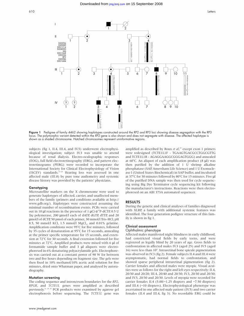
was observed in IV.3 (fig 2). Female subjects II.4 and III.4 were
asymptomatic, had normal fields to confrontation, and
showed sparse peripheral intraretinal pigmentation (fig 2).
Carrier females and affected males were myopic. Visual acui-
ties were as follows for the right and left eyes respectively: II.4,
20/30 and 20/20; III.4, 20/40 and 20/30; IV.3, 20/30 and 20/30;
and IV.5, 20/30 and 20/30. Levels of myopia were recorded for
carrier females II.4 (5.00/−1.25 diopters and −7.50 diopters)
and III.4 (−10 diopters). Electrophysiological phenotype was
ascertained in one affected male patient (IV.5) and two carrier
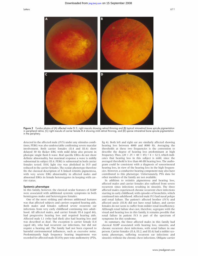
females (II.4 and III.4, fig 3). No recordable ERG could be
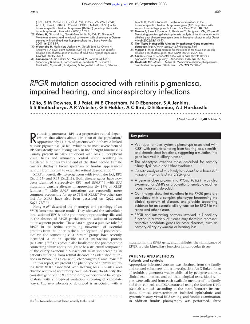
Figure 1 Pedigree of family 4462 showing haplotypes constructed around the RP2 and RP3 loci showing disease segregation with the RP3locus. The polymorphic variant detected within the RP2 gene is also shown and does not segregate with disease. The affected haplotype isshown as a shaded chromosome. Hatched chromosomes represent uninformative regions.
610 Letters
www.jmedgenet.com
on 15 September 2008 jmg.bmj.comDownloaded from
detected in the affected male (IV.5) under any stimulus condi-
tions; PERG was also undetectable confirming severe macular
involvement. Both carrier females (II.4 and III.4) show
delayed 30 Hz flicker ERG with mild delay also present in
photopic single flash b wave. Rod specific ERGs do not show
definite abnormality, but maximal response a wave is mildly
subnormal in subject III.4. PERG is subnormal in both carrier
females tested. EOG light rise was abolished in IV.5 and
reduced in the carrier females. The ocular phenotype therefore
fits the classical description of X linked retinitis pigmentosa,
with very severe ERG abnormality in affected males and
abnormal ERGs in female heterozygotes in keeping with car-
rier status.
Systemic phenotypeIn this family, however, the classical ocular features of XLRP
were associated with additional systemic symptoms in both
hemizygous males and heterozygous females.
One of the most striking and obvious additional features
was that affected subjects and carriers required hearing aids.
Both males and females suffered severe recurrent ear
infections from very early childhood continuing into adult-
hood. All affected males, and carrier female III.4 (aged 44),
had progressive hearing loss and required hearing aids.
Affected male I.1 (who had died) also had hearing loss and
was described as deaf. The exception is carrier female II.4
(aged 68), who had recurrent ear infections, but did not
require a hearing aid. The family had not been exposed to
harmful environmental influences, such as excessive noise.
Predominantly high frequency hearing impairment was
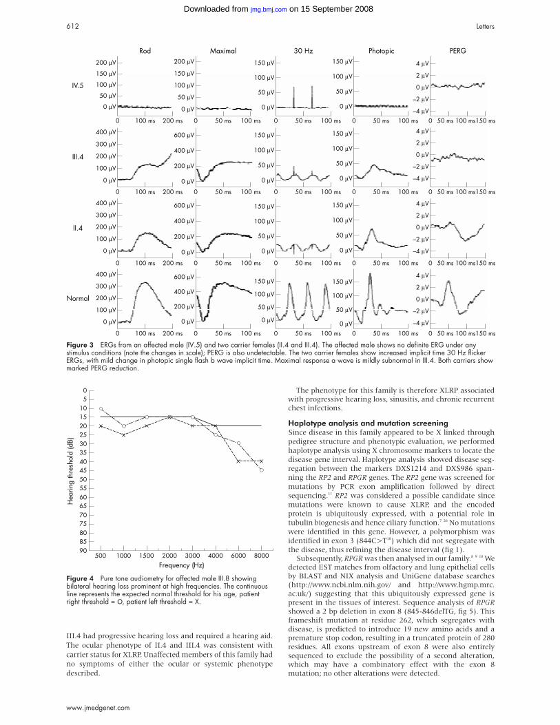
recorded for affected male III.8 by pure tone audiometry (PTA,
fig 4). Both left and right ear are similarly affected showinghearing loss between 4000 and 8000 Hz. Averaging thethresholds at these two frequencies is the convention todescribe the degree of hearing loss predominant at highfrequency. Thus, (20 + 25 + 40 + 45) ÷ 4 = 32.5, which indi-
cates that hearing loss in this subject is mild, since the
averaged threshold is less than 40 dB hearing loss. The audio-
gram could be consistent with a diagnosis of sensorineural
hearing loss, in view of the hearing loss in the high frequen-
cies. However, a conductive hearing component may also have
contributed to this phenotype. Unfortunately, PTA data for
other members of the family are not available.
In addition to retinitis pigmentosa and hearing loss,
affected males and carrier females also suffered from severe
recurrent sinus infections resulting in sinusitis. The three
affected males experienced chronic recurrent chest infections
starting in early childhood, with episodes of bronchitis, which
continued into adulthood. Affected male IV.3 had nasal polyps
and renal failure. The patient’s affected brother (IV.5) and
affected uncle (III.8) did not have renal failure, and carrier
females do not seem to suffer from milder renal insufficiency.
Although renal failure does not, therefore, segregate with the
visual and hearing loss in this family, it remains possible that
renal failure in patient IV.3 is part of the spectrum of
symptoms for this syndrome.
In summary, the three affected males in this family had
classical XLRP associated with hearing loss, sinusitis, and
chronic recurrent chest infections, with renal failure in one
person. Carrier females (II.4, III.2, and III.4) had a milder sys-
temic phenotype, suffering recurrent ear infections and
sinusitis without the chronic chest infections. Obligate carrier
Figure 2 Fundus photos of (A) affected male IV.3, right macula showing retinal thinning and (B) typical intraretinal bone spicule pigmentationin peripheral retina, (C) right macula of carrier female III.4 showing mild retinal thinning, and (D) sparse intraretinal bone spicule pigmentationin the periphery.
Letters 611
www.jmedgenet.com
on 15 September 2008 jmg.bmj.comDownloaded from
III.4 had progressive hearing loss and required a hearing aid.
The ocular phenotype of II.4 and III.4 was consistent with
carrier status for XLRP. Unaffected members of this family had
no symptoms of either the ocular or systemic phenotype
described.
The phenotype for this family is therefore XLRP associatedwith progressive hearing loss, sinusitis, and chronic recurrentchest infections.
Haplotype analysis and mutation screeningSince disease in this family appeared to be X linked through
pedigree structure and phenotypic evaluation, we performed
haplotype analysis using X chromosome markers to locate the
disease gene interval. Haplotype analysis showed disease seg-
regation between the markers DXS1214 and DXS986 span-
ning the RP2 and RPGR genes. The RP2 gene was screened for
mutations by PCR exon amplification followed by direct
sequencing.11 RP2 was considered a possible candidate since
mutations were known to cause XLRP, and the encoded
protein is ubiquitously expressed, with a potential role in
tubulin biogenesis and hence ciliary function.7 26 No mutations
were identified in this gene. However, a polymorphism was
identified in exon 3 (844C>T18) which did not segregate with
the disease, thus refining the disease interval (fig 1).Subsequently, RPGR was then analysed in our family.8 9 10 We
detected EST matches from olfactory and lung epithelial cellsby BLAST and NIX analysis and UniGene database searches(http://www.ncbi.nlm.nih.gov/ and http://www.hgmp.mrc.ac.uk/) suggesting that this ubiquitously expressed gene ispresent in the tissues of interest. Sequence analysis of RPGRshowed a 2 bp deletion in exon 8 (845-846delTG, fig 5). Thisframeshift mutation at residue 262, which segregates withdisease, is predicted to introduce 19 new amino acids and apremature stop codon, resulting in a truncated protein of 280residues. All exons upstream of exon 8 were also entirelysequenced to exclude the possibility of a second alteration,which may have a combinatory effect with the exon 8mutation; no other alterations were detected.
Figure 3 ERGs from an affected male (IV.5) and two carrier females (II.4 and III.4). The affected male shows no definite ERG under anystimulus conditions (note the changes in scale); PERG is also undetectable. The two carrier females show increased implicit time 30 Hz flickerERGs, with mild change in photopic single flash b wave implicit time. Maximal response a wave is mildly subnormal in III.4. Both carriers showmarked PERG reduction.
Figure 4 Pure tone audiometry for affected male III.8 showingbilateral hearing loss prominent at high frequencies. The continuousline represents the expected normal threshold for his age, patientright threshold = Ο, patient left threshold = X.
612 Letters
www.jmedgenet.com
on 15 September 2008 jmg.bmj.comDownloaded from
It is likely that the genetic background of subjects within
this pedigree contributes to the additional systemic pheno-
types observed. Since overlapping symptoms have been
observed in numerous XLRP patients and at least one other
XLRP pedigree (see Discussion), we hypothesised that a
predisposing locus could be closely linked to RPGR on the X
chromosome. The TCTE1L gene is approximately 500 kb distal
to RPGR and has been shown to be expressed in lung, trachea,
kidney, and brain, among other tissues and detects ESTs from
olfactory and lung epithelial cells and the organ of Corti25 28
(BLAST and UniGene searches at http://www.ncbi.nlm.
nih.gov/). The TCTE1L protein forms part of the cytoplasmic
dynein light chain of the microtubule motor complex, and
may be involved in tissue specific cargo binding activities since
other members of this protein family mediate specific interac-
tions, for example, with rhodopsin.29 30 The TCTE1L gene
therefore presented an attractive positional and functional
modifier locus for the phenotype described. To determine if a
linked locus could predispose subjects to susceptibility to the
systemic pathology observed, we screened the five exons of the
TCTE1L gene for cSNPs and no polymorphic variants were
detected in the affected males.
DISCUSSIONPhenotypic overlap with other syndromesThe systemic phenotype in this family has similarities with
those observed in immotile cilia syndrome (ICS1) or primary
ciliary dyskinesia (PCD, MIM 242650). PCD is a congenital
respiratory disease characterised by impaired mucociliary
clearance caused by cilia ultrastructural abnormalities.31 32
PCD patients suffer from chronic bronchiectasis and sinusitis,
usually associated with male infertility, but do not reportedly
suffer from recurrent ear infections or deafness.33 Approxi-
mately half of the patients with PCD also display situs inver-
sus (Kartagener syndrome, MIM 244400). One causative gene
for PCD has recently been identified, DNAI1, a dynein
intermediate chain gene on chromosome 9p13-21, with
mutations in this gene shown to cause axoneme ultrastruc-
tural abnormalities in two families.34 Genetic and phenotypic
heterogeneity are features of PCD, with a locus identified on
chromosome 5p14-15,34 35 and potential linkage to 11 other
chromosomal intervals.36 However, no linkage to the X
chromosome or association with RP and deafness has
previously been reported.
Usher syndrome is defined by an association of sen-
sorineural deafness with RP, with three distinct clinical
subtypes (I, II, and III) of variable severity and extensive
genetic heterogeneity.37 Usher syndrome is the most frequent
cause of sensorineural deafness accompanied by blindness,
although two of the causative genes have also been implicated
in isolated deafness38 or isolated retinitis pigmentosa.39
Although hearing loss in our family is associated with
retinitis pigmentosa, the mode of inheritance, nature, and
onset of hearing loss, and chronic infections leading to sinusi-
tis and bronchitis distinguish the phenotype in this family
from Usher syndrome types I, II, and III. Hearing loss in our
family appears relatively mild and progressive with a
sensorineural component, but the chronic infections suffered
by the patients may also contribute to an acquired conductive
hearing impairment. Unfortunately, we were not able to
record bone conduction thresholds in this family, so we
conclude that hearing loss is likely to be mixed, but not
proven. It is difficult to say whether the patients have any loss
of vestibular function without thorough neuro-otological
evaluation, but family members did not report any dizziness/
balance problems. The lack of reported symptoms may have
been because of the progressive nature of the condition which
allowed for vestibular compensation. The presence of an X
linked form of Usher-like phenotype has been suggested, but
no locus on the X chromosome has been described. In one
report, however, retinitis pigmentosa with deafness (described
as Usher syndrome) was associated with bronchiectasis and
immotile cilia syndrome, and the possibility of an X linked
mode of inheritance could not be excluded.40
The major sites of pathology in this new phenotype, causing
hearing loss and other disabling systemic abnormalities in
association with XLRP, suggest structural, degenerative, or
developmental kinociliary defects.
RPGR mutations and ciliary abnormalitiesSeveral lines of evidence support our findings that mutant
RPGR causes XLRP with associated generalised cilia abnor-
malities. In 1992, Van Dorp et al41 reported a family who
suffered from XLRP with associated susceptibility to respira-
tory infections in the majority of affected males. The patients
suffered from recurrent bronchitis and sinusitis, described as
indistinguishable from immotile cilia syndrome, but did not
suffer sterility or deafness. In a subsequent publication, a
mutation was identified in this family in the RPGR gene,
namely a G to T transversion at position +1 of the 5′ donor
splice site of intron 5, predicted to result in aberrant
splicing.42 This additional phenotype, reported by Van Dorp etal,41 overlaps the one described here with the exception of the
associated hearing loss, present in our family. Independent
studies describing the prevalence of deafness in association
with RP43 identified a group of patients that did not fit into
previously described clinical categories (that is, not Usher
syndrome). Hearing impairment in three families with XLRP
was reported by Rosenberg et al,43 and a mutation in RPGR had
previously been identified in one of these families.9 44 This
mutation is described as a 6.4 kb deletion which disrupts the
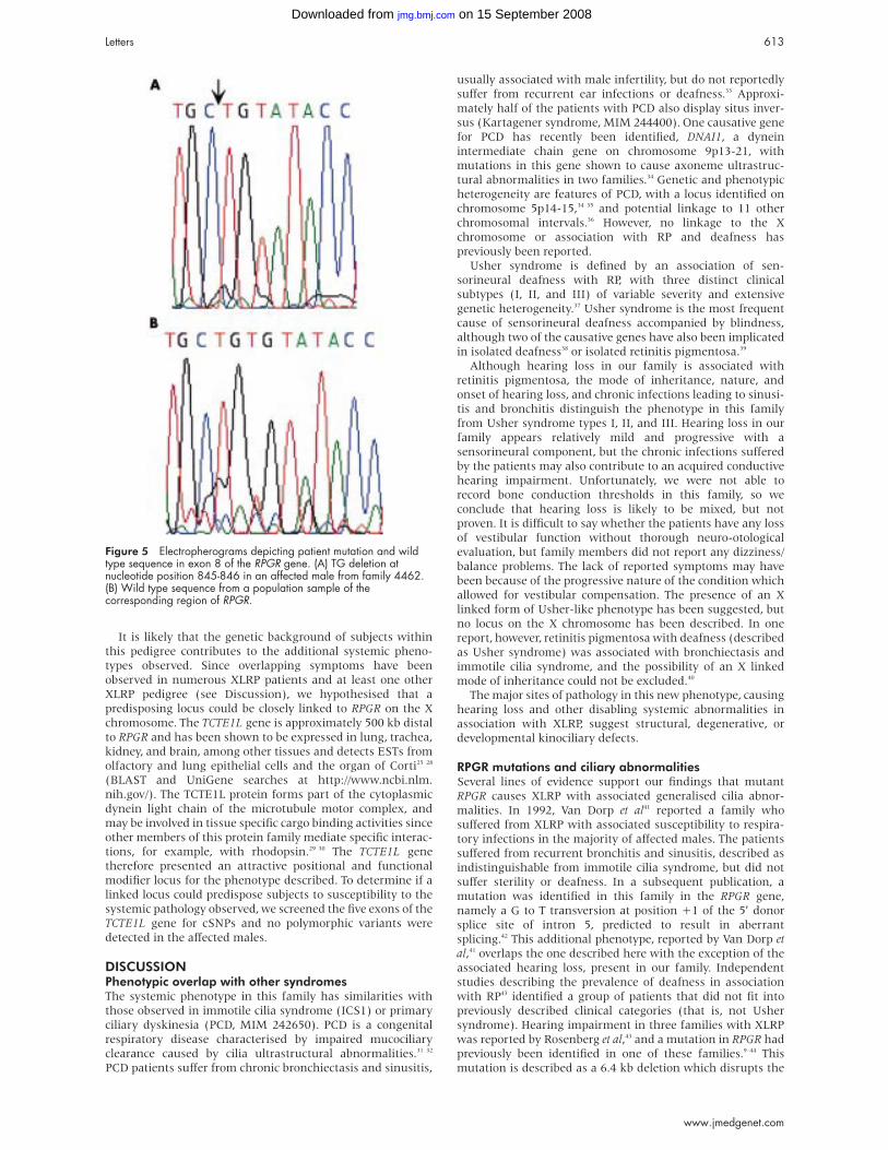
Figure 5 Electropherograms depicting patient mutation and wildtype sequence in exon 8 of the RPGR gene. (A) TG deletion atnucleotide position 845-846 in an affected male from family 4462.(B) Wild type sequence from a population sample of thecorresponding region of RPGR.
Letters 613
www.jmedgenet.com
on 15 September 2008 jmg.bmj.comDownloaded from
3′ end of RPGR removing the last six exons. Both affected
males and carrier females from this family had hearing
difficulties.43
Other studies have centred around examination of cilia in
patients with RP. Several reports examining nasal mucosa and
sperm in heterogeneous groups of patients suggest increased
incidence of abnormal cilia in XLRP patients.45–47 However, the
molecular basis for these observations remains undetermined.
Although the physiological role of RPGR in retina and other
tissues is not fully understood, compelling evidence for the
importance of RPGR in ciliary function comes from recent
studies of animal models and the identification of RPGR
interacting proteins.15–20 It is possible, therefore, that other cell
specific ciliary proteins exist in the lung, trachea, inner ear,
and nasal passages which bind RPGR, and that this
interaction may be compromised in the family described in
this report.
Mutation type and modifiers of phenotypeThe novel mutation we have identified results in partial loss of
the RCC1 domain (exon 8, 845-846delTG) and downstream
sequence. This protein truncation mutation occurs upstream
of many other protein truncation and missense mutations
previously reported to cause XLRP. It is unclear, however, why
the phenotype of this protein truncation mutation is different
from others reported to result in only an ocular phenotype.48
Perhaps persistent but milder systemic infections in other
families remain undetected or may not have been reported,
and exposure to infections and subsequent disease manifesta-
tion varies widely.
Mutations in the RPGR gene have also been detected in
families with X linked cone-rod dystrophy and X linked
macular degeneration (as opposed to the rod-cone degenera-
tion observed in classical XLRP), widening the clinical
spectrum associated with mutant RPGR and highlighting the
fact that other factors modulate the phenotype.49–51 The
factor(s) underlying the significant variability of the patho-
genic expression of RPGR remain to be identified.
The genotype at a particular locus may account for an inter-
individual susceptibility that can both increase or decrease the
risk to develop the pathology by modulating mechanisms
involved in the pathogenesis. We hypothesised that a closely
linked gene which segregated with the primary RPGRmutation could be acting as a modifier gene in this family,
since association with the symptoms described in this report
are more common than previously suspected. TCTE1L lies
approximately 500 kb distal to RPGR and presents an
interesting functional candidate which is expressed in the tis-
sues involved in the systemic disease associated with XLRP. No
cSNPs were identified. Predisposing SNPs may lie outside
those gene regions tested, and other loci on the autosomes can
not be excluded as predisposing factors; however, if autosomal
SNPs are involved in disease expression, they are predicted to
be common owing to the occurrence of disease in more than
one pedigree. It is now essential to collect a cohort of families
with these overlapping phenotypes to determine the factors
involved in disease expression.
Further evidence for this new syndrome being primarily an
RPGR gene disorder comes from colleagues who have
identified a family with an almost identical phenotype, XLRP,
hearing loss, and recurrent respiratory tract infections. On the
basis of our findings, they examined the RPGR gene and found
a missense mutation in exon 6 (Iannaccone et al, in
preparation). The data show that the families are unrelated
and that different mutations in RPGR can result in overlapping
phenotypes implicating ciliary dysfunction in a variety of tis-
sues. In addition, Iannaccone et al describe expression of RPGR
in human cochlea and bronchial and sinus epithelial lining.
Future studies towards unravelling the function of RPGR in
the retina will need to be expanded to include analyses of
multiple ciliated epithelial tissues. The identification of RPGR
binding partners within these tissues may identify other spe-
cific proteins capable of interacting with RPGR. It would be of
interest to evaluate the mouse and dog models of RPGRdisease15 52 with a view to examining structure/function and
development of the ciliated epithelium of the respiratory tract,
sinuses, and inner ear, for example, in addition to re-
evaluating the patients already described as harbouring RPGRmutations as a cause of XLRP.
SUMMARYIn conclusion, we describe a new phenotype of typical X linked
retinitis pigmentosa associated with hearing loss, chronic res-
piratory tract infections, and sinusitis caused by a mutation in
RPGR. The systemic phenotypes are predicted to be variable,
accentuated by repeated infections of the respiratory tract and
consequent upon impaired mucociliary clearance (as de-
scribed for PCD). Phenotypic variation between families may
be caused by RPGR mutation type, genetic background,
environmental effects, or a combination of these factors.
Additional families will need to be investigated for SNPs on
the X chromosome in proximity to RPGR to explore fully any
phenotypic modification caused by adjacent loci. RPGR and
interacting partners involved in kinociliary function in a vari-
ety of tissues may also represent attractive candidate genes for
other phenotypes such as primary ciliary dyskinesia or
isolated hearing loss.
ACKNOWLEDGEMENTSThe authors would like to thank the family for participating,
and The Wellcome Trust (AJH) and the British Retinitis
Pigmentosa Society for their support.
. . . . . . . . . . . . . . . . . . . . .Authors’ affiliationsI Zito, S M Downes, R J Patel, N D Ebenezer, S S Bhattacharya,A R Webster, A J Hardcastle, Division of Molecular Genetics, Instituteof Ophthalmology, UCL, London, UKS M Downes, S A Jenkins, A R Webster, G E Holder, A C Bird,Moorfields Eye Hospital, London, UKM E Cheetham, Division of Pathology, Institute of Ophthalmology, UCL,London, UKD E Bamiou, Department of Neuro-otology, National Hospital forNeurology and Neurosurgery, London, UK
Correspondence to: Dr A J Hardcastle, Division of Molecular Genetics,Institute of Ophthalmology, University College London, 11-43 Bath Street,London EC1V 9EL, UK; [email protected]
REFERENCES1 Heckenlively JR. Retinitis pigmentosa. Philadelphia: Lippincott,
1988:1-269.2 Bird AC. X-linked retinitis pigmentosa. Br J Ophthalmol
1975;59:177-99.3 Fishman GA, Farber MD, Derlacki DJ. X-linked retinitis pigmentosa.
Profile of clinical findings. Arch Ophthalmol 1988;106:369-75.4 Falls HF, Cotterman C. Choroidal degeneration. A sex-linked form in
which heterozygous women exhibit a tapetal-like reflex. Arch Ophthalmol1948;40:683-703.
5 Berson EL, Rosen JB, Simonoff EA. Electroretinographic testing as an aidin detection of carriers of X-chromosome linked retinitis pigmentosa. Am JOphthalmol 1979;87:460-8.
6 Merin S, Maggiano JM, Fishman GA. Retinal sensitivity under photopicconditions in patients with retinitis pigmentosa and carriers of the X-linkedrecessive form. Metab Ophthalmol 1978;2:225-9.
7 Schwahn U, Lenzner S, Dong J, Fell S, Hinzmann B, van Duijnhoven G,Kirschner R, Hernberg M, Bergen AAB, Rosenberg T, Pinckers ALG,Fundele R, Rosenthal A, Cremers FPM, Ropers HH, Berger W. Positionalcloning of the gene for X-linked retinitis pigmentosa 2. Nat Genet1998;19:327-32.
8 Meindl A, Dry K, Herrmann K, Manson F, Ciccodicola A, Edgar A,Carvalho MRS, Achatz H, Hellebrand H, Lennon A, Migliaccio C, PorterK, Zrenner E, Bird A, Jay M, Lorenz , Wittwer B, D’Urso M, Meitinger T,Wright A. A gene (RPGR) with homology to the RCC1 guanine nucleotideexchange factor is mutated in X-linked retinitis pigmentosa (RP3). NatGenet 1996;13:35-42.
9 Roepman R, van Duijnhoven G, Rosenberg T, Pinckers AJLG,Bleeker-Wagemakers LM, Bergen AAB, Post J, Beck A, Reinhardt R,Ropers HH, Cremers FPM, Berger W. Positional cloning of the gene for
614 Letters
www.jmedgenet.com
on 15 September 2008 jmg.bmj.comDownloaded from
X-linked retinitis pigmentosa 3: homology with theguanine-nucleotide-exchange factor RCC1. Hum Mol Genet1996;5:1035-41.
10 Vervoort R, Lennon A, Bird AC, Tulloch B, Axton R, Miano MG, MeindlA, Meitinger T, Ciccodicola A, Wright AF. Mutational hot spot within anew RPGR exon in X-linked retinitis pigmentosa. Nat Genet2000;25:462-6.
11 Hardcastle AJ, Thiselton DL, Van Maldergem L, Saha BK, Jay M, PlantC, Taylor R, Bird AC, Bhattacharya S. Mutations in the RP2 gene causedisease in 10% of familial XLRP assessed in this study. Am J Hum Genet1999;64:1210-15.
12 Mears AJ, Gieser L, Yan D, Chen C, Fahrner S, Hiriyanna S, Fujita R,Jacobson SG, Sieving PA, Swaroop A. Protein-truncation mutations in theRP2 gene in a North American cohort of families with X-linked retinitispigmentosa. Am J Hum Genet 1999;64:897-900.
13 Hardcastle AJ, Thiselton DL, Zito I, Ebenezer N, Mah T, Gorin M,Bhattacharya SS. Evidence for a new locus for X-linked retinitispigmentosa (RP23). Invest Ophthalmol Vis Sci 2000;41:2080-6.
14 Gieser L, Fujita R, Goring HHH, Ott J, Hoffman DR, Cideciyan AV,Birch DJ, Jacobson SG, Swaroop A. A novel locus (RP24) for X-linkedretinitis pigmentosa maps to Xq26-27. Am J Hum Genet1998;63:1439-47.
15 Hong DH, Pawlyk BS, Shang J, Sandberg MA, Berson EL, Li T. A retinitispigmentosa GTPase regulator (RPGR)-deficient mouse model for X-linkedretinitis pigmentosa (RP3). Proc Natl Acad Sci USA 2000;97:3649-54.
16 Boylan JP, Wright AF. Identification of a novel protein interacting withRPGR. Hum Mol Genet 2000;9:2085-93.
17 Roepman R, Bernoud-Hubac N, Schick DE, Maugeri A, Berger W,Ropers HH, Cremers FP, Ferreira PA. The retinitis pigmentosa GTPaseregulator (RPGR) interacts with novel transport-like proteins in the outersegments of rod photoreceptors. Hum Mol Genet 2000;9:2095-105.
18 Hong DH, Yue G, Adamian M, Li T. Retinitis pigmentosa GTPaseregulator (RPGR)-interacting protein is stably associated with thephotoreceptor ciliary axoneme and anchors RPGR to the connectingcilium. J Biol Chem 2001;276:12091-9.
19 Dryja TP, Adams SM, Grimsby JL, McGee TL, Hong DH, Li T,Andreasson S, Berson EL. Null RPGRIP1 alleles in patients with Lebercongenital amaurosis. Am J Hum Genet 2001;68:1295-8.
20 Gerber S, Perrault I, Hanein S, Barbet F, Ducroq D, Ghazi I,Martin-Coignard D, Leowski C, Homfray T, Dufier JL, Munnich A, KaplanJ, Rozet JM. Complete exon-intron structure of the RPGR-interactingprotein (RPGRIP1) gene allows the identification of mutations underlyingLeber congenital amaurosis. Eur J Hum Genet 2001;9:561-71.
21 Marmor MF, Zrenner E. Standard for clinical electroretinography (1999update). Doc Ophthalmol 1998;97:143-56.
22 Bach M, Hawlina M, Holder GE, Marmor M, Meigen T, Vaegan,Miyake Y. Standard for pattern electroretinography. Doc Ophthalmol2000;101:11-18.
23 Marmor MF, Zrenner E. Standard for clinical electro-oculography. DocOphthalmol 1993;85:115-124.
24 Zito I, Thiselton DL, Gorin MB, Stout JT, Plant C, Bird AC, BhattacharyaSS, Hardcastle AJ. Identification of novel RPGR (retinitis pigmentosaGTPase regulator) mutations in a subset of X-linked retinitis pigmentosafamilies segregating with the RP3 locus. Hum Genet 1999;105:57-62.
25 Roux AF, Rommens J, McDowell C, Anson-Cartwright L, Bell S,Schappert K, Fishman GA, Musarella M. Identification of a gene fromXp21 with similarity to the tctex-1 gene of the murine t complex. Hum MolGenet 1994;3:257-63.
26 Chapple JP, Hardcastle AJ, Grayson C, Spackman LA, Willison KR,Cheetham ME. Mutations in the N-terminus of the X-linked retinitispigmentosa protein RP2 interfere with the normal targeting of the proteinto the plasma membrane. Hum Mol Genet 2000;9:1919-26.
27 Thiselton DL, Zito I, Plant C, Jay M, Hodgson SV, Bird AC,Bhatttacharya SS, Hardcastle AJ. Novel frameshift mutations in the RP2gene and polymorphic variants. Hum Mutat 2000;15:580.
28 DiBella LM, Benashski SE, Tedford HW, Harrison A, Patel-King RS, KingSM. The Tctex1/Tctex2 class of dynein light chains. Dimerization,differential expression, and interaction with the LC8 protein family. J BiolChem 2001;276:14366-73.
29 King SM, Barbarese E, Dillman JF 3rd, Benashski SE, Do KT, Patel-KingRS, Pfister KK. Cytoplasmic dynein contains a family of differentiallyexpressed light chains. Biochemistry 1998;37:15033-41.
30 Tai AW, Chuang JZ, Bode C, Wolfrum U, Sung CH. Rhodopsin’scarboxy-terminal cytoplasmic tail acts as a membrane receptor forcytoplasmic dynein by binding to the dynein light chain Tctex-1. Cell1999;97:877-87.
31 Afzelius BA. A human syndrome caused by immotile cilia. Science1976;193:317-19.
32 Rossman CM, Forrest JB, Lee RM, Newhouse MT. The dyskinetic ciliasyndrome. Ciliary motility in immotile cilia syndrome. Chest1980;78:580-2.
33 Afzelius BA. The immotile-cilia syndrome: a microtubule-associateddefect. CRC Crit Rev Biochem 1985;19:63-87.
34 Pennarun G, Escudier E, Chapelin C, Bridoux AM, Cacheux V, RogerG, Clement A, Goossens M, Amselem S, Duriez B. Loss-of-functionmutations in a human gene related to Chlamydomonas reinhardtii dyneinIC78 result in primary ciliary dyskinesia. Am J Hum Genet1999;65:1508-19.
35 Omran H, Haffner K, Volkel A, Kuehr J, Ketelsen UP, Ross UH,Konietzko N, Wienker T, Brandis M, Hildebrandt F. Homozygositymapping of a gene locus for primary ciliary dyskinesia on chromosomep. and identification of the heavy dynein chain DNAH5 as a candidategene. Am J Respir Cell Mol Biol 2000;23:696-702.
36 Blouin JL, Meeks M, Radhakrishna U, Sainsbury A, Gehring C, Sail GD,Bartoloni L, Dombi V, O’Rawe A, Walne A, Chung E, Afzelius BA,Armengot M, Jorissen M, Schidlow DV, van Maldergem L, Walt H,Gardiner RM, Probst D, Guerne PA, Delozier-Blanchet CD, AntonarakisSE. Primary ciliary dyskinesia: a genome-wide linkage analysis revealsextensive locus heterogeneity. Eur J Hum Genet 2000;8:109-18.
37 Petit C. Usher syndrome: from genetics to pathogenesis. Ann RevGenomics. Hum Genet 2001;2:271-97.
38 Bork JM, Peters LM, Riazuddin S, Bernstein SL, Ahmed ZM, Ness SL,Polomeno R, Ramesh A, Schloss M, Srisailpathy CR, Wayne S, BellmanS, Desmukh D, Ahmed Z, Khan SN, Kaloustian VM, Li XC, Lalwani A,Riazuddin S, Bitner-Glindzicz M, Nance WE, Liu XZ, Wistow G, SmithRJ, Griffith AJ, Wilcox ER, Friedman TB, Morell RJ. Usher syndrome 1Dand nonsyndromic autosomal recessive deafness DFNB12 are caused byallelic mutations of the novel cadherin-like gene CDH23. Am J HumGenet 2001;68:26-37.
39 Rivolta C, Sweklo EA, Berson EL, Dryja TP. Missense mutation in theUSH2A gene: association with recessive retinitis pigmentosa withouthearing loss. Am J Hum Genet 2000;66:1975-8.
40 Bonneau D, Raymond F, Kremer C, Klossek JM, Kaplan J, Patte F. Ushersyndrome type I associated with bronchiectasis and immotile nasal ciliain two brothers. J Med Genet 1993;30:253-4.
41 Van Dorp DB, Wright AF, Carothers AD, Bleeker-Wagemakers EM. Afamily with RP3 type of X-linked retinitis pigmentosa: an association withciliary abnormalities. Hum Genet 1992;88:331-4.
42 Dry KL, Manson FDC, Lennon A, Bergen AA, VanDrop DB, Wright AF.Identification of a 5′ splice site mutation in the RPGR gene in a familywith X-linked retinitis pigmentosa (RP3). Hum Mutat 1999;13:141-5.
43 Rosenberg T, Haim M, Hauch AM, Parving A. The prevalence of Ushersyndrome and other retinal dystrophy-hearing impairment associations.Clin Genet 1997;51:314-21.
44 Roepman R, Bauer D, Rosenberg T, van Duijnhoven G, van de Vosse E,Platzer M, Rosenthal A, Ropers HH, Cremers FP, Berger W. Identificationof a gene disrupted by a microdeletion in a patient with X-linked retinitispigmentosa (XLRP). Hum Mol Genet 1996;5:827-33.
45 Arden GB, Fox B. Increased incidence of abnormal nasal cilia inpatients with retinitis pigmentosa. Nature 1979;279:534-6.
46 Fox B, Bull TB, Arden GB. Variations in ultrastructure of human nasalcilia including abnormalities found in retinitis pigmentosa. J Clin Pathol1980;33:327-35.
47 Hunter DG, Fishman GA, Kretzer FL. Abnormal axonemes in X-linkedretinitis pigmentosa. Arch Ophthalmol 1988;106:362-8.
48 Vervoort R, Wright AF. Mutations of RPGR in X-linked retinitispigmentosa (RP3). Hum Mutat 2002;19:486-500.
49 Yang Z, Peachey NS, Moshfeghi DM, Thirumalaichary S, Chorich L,Shugart YY, Fan K, Zhang K. Mutations in the RPGR gene cause X-linkedcone dystrophy. Hum Mol Genet 2002;11:605-11.
50 Demirci FY, Rigatti BW, Wen G, Radak AL, Mah TS, Baic CL, TraboulsiEI, Alitalo T, Ramser J, Gorin MB. X-linked cone-rod dystrophy (locusCOD1): identification of mutations in RPGR exon ORF15. Am J HumGenet 2002;70:1049-53.
51 Ayyagari R, Demirci FY, Liu J, Bingham EL, Stringham H, Kakuk LE,Boehnke M, Gorin MB, Richards JE, Sieving PA. X-linked recessiveatrophic macular degeneration from RPGR mutation. Genomics2002;80:166-71.
52 Zhang Q, Acland GM, Wu WX, Johnson JL, Pearce-Kelling S, Tulloch B,Vervoort R, Wright AF, Aguirre GD. Different RPGR exon ORF15mutations in Canids provide insights into photoreceptor celldegeneration. Hum Mol Genet 2002;11:993-1003.
Letters 615
www.jmedgenet.com
on 15 September 2008 jmg.bmj.comDownloaded from
Evidence of RPGRIP1 gene mutations associated withrecessive cone-rod dystrophyA Hameed, A Abid, A Aziz, M Ismail, S Q Mehdi, S Khaliq. . . . . . . . . . . . . . . . . . . . . . . . . . . . . . . . . . . . . . . . . . . . . . . . . . . . . . . . . . . . . . . . . . . . . . . . . . . . . . . . . . . . . . . . . . . . . . . . . . . . . . . . . . . . . . . . . . . . . . . . . . . . .
J Med Genet 2003;0:616–619
Cone-rod dystrophies (CRD) are forms of inherited retinal
dystrophy which characteristically lead to early impair-
ment of vision. An initial loss of colour vision (cone
mediated functions) and of visual acuity, usually from the first
or second decade of life, is followed by night blindness (largely
rod mediated) and loss of peripheral visual fields.1 CRD patients
suffer from severe photophobia and show reduced ERG
responses. In later life, vision may be reduced to a bare percep-
tion of light. CRD is a milder condition compared to Leber con-
genital amaurosis (LCA) which is the most severe form of all the
inherited retinal dystrophies and is diagnosed as bilateral
congenital blindness, with a diminished or absent electroretino-
gram (ERG). Cone-rod dystrophy loci have been mapped to
chromosomes 17q,2 19q,3 18q,4 17p13,5 6 6q,7 1q12,8 and 8p11.9
Mutations in the peripherin/RDS,10 CRX,11 12 and RetGC-I13 14 genes
have been shown to cause autosomal dominant CRD. Mutations
in the ATP binding cassette transporter rim protein (ABCR) gene
have been shown to be associated with autosomal recessive
CRD.15 Mutations in the CNGA3 gene encoding the α-subunit of
the cone photoreceptor cGMP gated channel have also been
reported to cause cone photoreceptor disorders.16
The RPGRIP1 protein (retinitis pigmentosa GTPase regulator
interacting protein 1, MIM 605446) is encoded by the gene
located on chromosome 14q11. It consists of 24 exons and the
predicted size of its protein product is 1259 amino acids. It is
expressed specifically in the rod and cone photoreceptors and is
a structural component of the ciliary axoneme. One of its func-
tions is to anchor the RPGR protein within the photoreceptor
connecting cilium.17 Recently, in an in vivo investigation of
RPGRIP1 function and its physical interaction, it has been
shown that RPGRIP1 is essential for RPGR function and is also
required for normal disk morphogenesis.18 Mutations in
RPGRIP1 have been reported to be a cause of LCA.19 20 Here we
report the first observation of the involvement of RPGRIP1 gene
mutations as a cause of CRD in four Pakistani families.
SUBJECTS AND METHODSWe studied 20 members of a two generation and 19 members
of a three generation consanguineous Pakistani families,
1CRD and 4CRD, respectively. The 1CRD family consists of
eight affected and 12 unaffected subjects and the 4CRD fam-
ily consists of eight affected and 11 unaffected subjects (fig
1A, B). One of the authors (A Aziz) clinically examined all the
patients and their unaffected family members. The deteriora-
tion in central vision and colour blindness was from an early
age in all the patients and there was a rapid loss of vision
between the ages of 14 and 16 years (visual acuity 1/60, 0.01).
Patients had also had severe photophobia since their
childhood. Fundoscopy showed a variable degree of fundus
granularity and macular degeneration. The affected subject
IV.7 (aged 14 years) has a characteristic macular bull’s eye
lesion in both her eyes. Full field flash ERG was used to meas-
ure functions of both cones and rods. Both photopic and sco-
topic full field ERG amplitudes were reduced, showing
involvement of both photoreceptor systems. However, among
the young patients, cone response was reduced more than that
of their rod response. Based on family history and clinical
diagnosis, the disease was classified as autosomal recessive
cone-rod dystrophy (arCRD).
For genetic analysis peripheral blood samples were collected
with informed consent from all members of the two families
and from 100 ethnically matched control subjects.
Linkage analysisGenomic DNA was extracted from whole blood using the
Nucleon II extraction kit (Scotlab Bioscience). DNA was
amplified using primers (Research Genetics) for polymorphic
microsatellite markers specific for known loci/genes associ-
ated with various retinal degenerations according to the con-
ditions described previously.8
Mutation detectionExon specific intronic primers were designed from the
genomic sequence of the RPGRIP1 gene (NM 020366). PCR
was performed in a 50 µl reaction volume using 1 µg of
genomic DNA. The resulting product was allowed to cool
slowly to room temperature to maximise the formation of
heteroduplexes. Heteroduplex analysis was performed using
an automated DHPLC instrumentation (WAVE DNA fragment
analysis system, Transgenomic, Crewe, UK). Sample prepara-
tion for heteroduplex analysis was carried out by denaturing
and reannealing of unpurified PCR products of the carriers
(heterozygotes). The temperature conditions required for the
successful resolution of heteroduplexes were obtained from
the website (http://insertion.stanford.edu/melt.html).
On identification of heteroduplex peaks in the carriers, all
family members were sequenced in the forward and reverse
directions using a commercially available kit (Big Dye, ABI)
and the products were analysed on an ABI Prism 377
automated DNA sequencer. Subsequently, six small families
with CRD (5, 6, 7, 8, 9, and 10CRDs) and two with LCA were
also included for mutation screening of the RPGRIP1 gene. To
exclude the possibility that the mutations are polymorphisms,
100 ethnically matched control samples were also screened for
heteroduplexes. The unpurified PCR products of the control
. . . . . . . . . . . . . . . . . . . . . . . . . . . . . . . . . . . . . . . . . . . . . . . . . . . . . . . . . . . . .
The first two authors contributed equally to this work.
Key points
x RPGRIP1 gene mutations have previously been reportedto cause Leber congenital amaurosis.
• Here we report two novel mutations in the RPGRIP1gene that cause cone-rod dystrophy in four Pakistanifamilies.
• A homozygous G to T point mutation was identified inexon 16 at nucleotide 2480 in all the affected membersof family 1CRD.
• In the affected members of another CRD family (4CRD),a G to T substitution was found in exon 13 at nucleotide1639.
• The same G 1639 T mutation was found in two moresmall families, 5CRD and 10CRD.
616 Letters
www.jmedgenet.com
on 15 September 2008 jmg.bmj.comDownloaded from
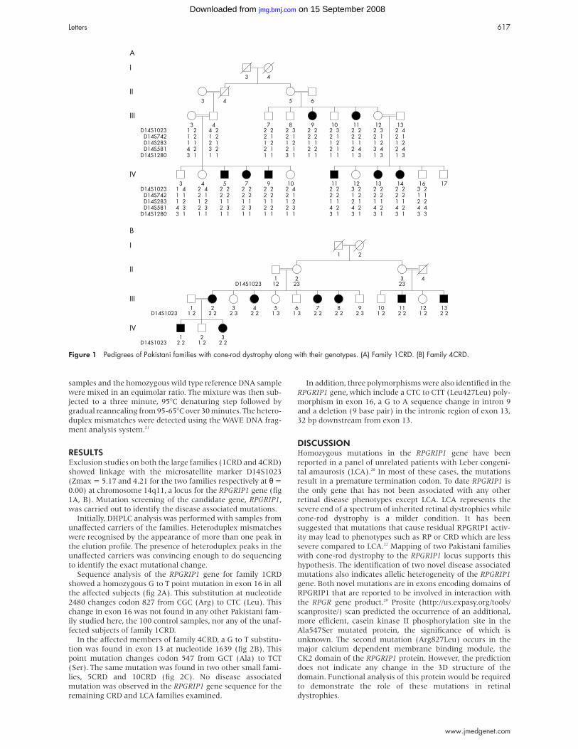
samples and the homozygous wild type reference DNA sample
were mixed in an equimolar ratio. The mixture was then sub-
jected to a three minute, 95°C denaturing step followed by
gradual reannealing from 95-65°C over 30 minutes.The hetero-
duplex mismatches were detected using the WAVE DNA frag-
ment analysis system.21
RESULTSExclusion studies on both the large families (1CRD and 4CRD)
showed linkage with the microsatellite marker D14S1023
(Zmax = 5.17 and 4.21 for the two families respectively at θ =
0.00) at chromosome 14q11, a locus for the RPGRIP1 gene (fig
1A, B). Mutation screening of the candidate gene, RPGRIP1,
was carried out to identify the disease associated mutations.
Initially, DHPLC analysis was performed with samples from
unaffected carriers of the families. Heteroduplex mismatches
were recognised by the appearance of more than one peak in
the elution profile. The presence of heteroduplex peaks in the
unaffected carriers was convincing enough to do sequencing
to identify the exact mutational change.
Sequence analysis of the RPGRIP1 gene for family 1CRD
showed a homozygous G to T point mutation in exon 16 in all
the affected subjects (fig 2A). This substitution at nucleotide
2480 changes codon 827 from CGC (Arg) to CTC (Leu). This
change in exon 16 was not found in any other Pakistani fam-
ily studied here, the 100 control samples, nor any of the unaf-
fected subjects of family 1CRD.
In the affected members of family 4CRD, a G to T substitu-
tion was found in exon 13 at nucleotide 1639 (fig 2B). This
point mutation changes codon 547 from GCT (Ala) to TCT
(Ser). The same mutation was found in two other small fami-
lies, 5CRD and 10CRD (fig 2C). No disease associated
mutation was observed in the RPGRIP1 gene sequence for the
remaining CRD and LCA families examined.
In addition, three polymorphisms were also identified in the
RPGRIP1 gene, which include a CTC to CTT (Leu427Leu) poly-
morphism in exon 16, a G to A sequence change in intron 9
and a deletion (9 base pair) in the intronic region of exon 13,
32 bp downstream from exon 13.
DISCUSSIONHomozygous mutations in the RPGRIP1 gene have been
reported in a panel of unrelated patients with Leber congeni-
tal amaurosis (LCA).20 In most of these cases, the mutations
result in a premature termination codon. To date RPGRIP1 is
the only gene that has not been associated with any other
retinal disease phenotypes except LCA. LCA represents the
severe end of a spectrum of inherited retinal dystrophies while
cone-rod dystrophy is a milder condition. It has been
suggested that mutations that cause residual RPGRIP1 activ-
ity may lead to phenotypes such as RP or CRD which are less
severe compared to LCA.22 Mapping of two Pakistani families
with cone-rod dystrophy to the RPGRIP1 locus supports this
hypothesis. The identification of two novel disease associated
mutations also indicates allelic heterogeneity of the RPGRIP1gene. Both novel mutations are in exons encoding domains of
RPGRIP1 that are reported to be involved in interaction with
the RPGR gene product.20 Prosite (http://us.expasy.org/tools/
scanprosite/) scan predicted the occurrence of an additional,
more efficient, casein kinase II phosphorylation site in the
Ala547Ser mutated protein, the significance of which is
unknown. The second mutation (Arg827Leu) occurs in the
major calcium dependent membrane binding module, the
CK2 domain of the RPGRIP1 protein. However, the prediction
does not indicate any change in the 3D structure of the
domain. Functional analysis of this protein would be required
to demonstrate the role of these mutations in retinal
dystrophies.
Figure 1 Pedigrees of Pakistani families with cone-rod dystrophy along with their genotypes. (A) Family 1CRD. (B) Family 4CRD.
Letters 617
www.jmedgenet.com
on 15 September 2008 jmg.bmj.comDownloaded from
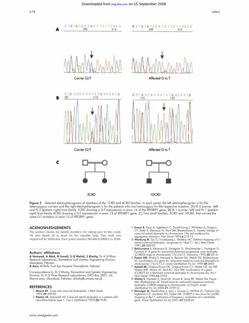
ACKNOWLEDGEMENTSThe authors thank the family members for taking part in this study.We also thank Dr Q Ayub for his valuable help. This work wassupported by Wellcome Trust grant number 063406/Z/2000/Z to SQM.
. . . . . . . . . . . . . . . . . . . . .Authors’ affiliationsA Hameed, A Abid, M Ismail, S Q Mehdi, S Khaliq, Dr A Q KhanResearch Laboratories, Biomedical and Genetic Engineering Division,Islamabad, PakistanA Aziz, Al-Shifa Trust Eye Hospital, Rawalpindi, Pakistan
Correspondence to: Dr S Khaliq, Biomedical and Genetic EngineeringDivision, Dr A Q Khan Research Laboratories, GPO Box 2891, 24Mauve area, Islamabad, Pakistan; [email protected]
REFERENCES1 Moore AT. Cone and cone-rod dystrophies. J Med Genet
1992;29:289-90.2 Klystra JA, Aylsworth AS. Cone-rod retinal dystrophy in a patient with
neurofibromatosis type 1. Can J Ophthalmol 1993;28:79-80.
3 Evans K, Fryer A, Inglehearn C, Duvall-Young J, Whittaker JL, GregoryCY, Butler R, Ebenezer N, Hunt DM, Bhattacharya S. Genetic linkage ofcone-rod retinal dystrophy to chromosome 19q and evidence forsegregation distortion. Nat Genet 1994;6:210-13.
4 Warburg M, Sjo O, Tranebjaerg L, Fledelius HC. Deletion mapping of aretinal cone-rod dystrophy: assignment to 18q211. Am J Med Genet1991;39:288-93.
5 Balciuniene J, Johansson K, Sandgren O, Wachtmeister L, Holmgren G,Forsman K. A gene for autosomal dominant progressive cone dystrophy(CORD5) maps to chromosome 17p12-p13. Genomics 1995;30:281-6.
6 Payne AM, Khaliq S, Hameed A, Bessant DA, Mehdi SQ, BhattacharyaSS. Localization of a gene for autosomal recessive cone-rod dystrophy tochromosome 17p12-13.3. Invest Ophthalmol Vis Sci 1999;40:S603.
7 Kelsell RE, Gregory-Evans GK, Gregory-Evans CY, Holder GE, Jay MR,Weber BHF, Moore AT, Bird AC, Hunt DM. Localization of a gene(CORD7) for a dominant cone-rod dystrophy to chromosome 6q. Am JHum Genet 1998;63:274-9.
8 Khaliq S, Hameed A, Ismail M, Anwar K, Leroy BP, Mehdi SQ, PayneAM, Bhattacharya SS. Novel locus for autosomal recessive cone-roddystrophy CORD8 mapping to chromosome 1q12-q24. InvestOphthalmol Vis Sci 2000;41:3709-12.
9 Danciger M, Hendrickson J, Lyon J, Toomes C, McHale JC, Fishman GA,Inglehearn CF, Jacobson SG, Farber DB. CORD9 a new locus for arCRD:mapping to 8p11, estimation of frequency, evaluation of a candidategene. Invest Ophthalmol Vis Sci 2001;42:2458-65.
Figure 2 Selected electropherograms of members of the 1CRD and 4CRD families. In each panel, the left electropherogram is for theheterozygous carriers and the right electropherogram is for the patients who are homozygous for the respective mutation. (A) III.4 (carrier, left)and IV.5 (patient, right) from family 1CRD showing a G-T transversion in exon 16 of the RPGRIP1 gene, (B) III.1 (carrier, left) and IV.1 (patient,right) from family 4CRD showing a G-T transversion in exon 13 of RPGRIP1 gene. (C) Two small families, 5CRD and 10CRD, that carried thesame G-T mutation in exon 13 of RPGRIP1 gene.
618 Letters
www.jmedgenet.com
on 15 September 2008 jmg.bmj.comDownloaded from
10 Nakazawa M, Kikawa, E, Chida Y, Tamai M. Asn244His mutation ofthe peripherin/RDS gene causing autosomal dominant cone-roddegeneration. Hum Mol Genet 1994;3:1195-6.
11 Freund CL, Gregory-Evans CY, Furukawa T, Papaioannou M, Looser J,Ploder L, Bellingham J, Ng D, Herbrick JAS, Duncan A, Scherer SW, TsuiLC, Loutradis-Anagnostou A, Jacobson SG, Cepko CL, Bhattacharya SS,McInnes RR. Cone-rod dystrophy due to mutations in a novelphotoreceptor-specific homeobox gene (CRX) essential for maintenanceof the photoreceptor. Cell 1997;91:543-53.
12 Swain PK, Chen SM, Wang QL, Affatigato LM, Coats CL, Brady KD,Fishman GA, Jacobson SG, Swaroop A, Stone E, Sieving PA, Zack DJ.Mutations in the cone-rod homeobox gene are associated with thecone-rod dystrophy photoreceptor degeneration. Neuron1997;19:1329-36.
13 Kelsell RE, Gregory-Evans K, Payne AM, Perrault I, Kaplan J, Yang RB,Garbers DL, Bird AC, Moore AT, Hunt DM. Mutations in the retinalguanylate cyclase (RETGC-1) gene in dominant cone dystrophy. Hum MolGenet 1998;7:1179-84.
14 Downes SM, Holder GE, Fitzke FW, Payne AM, Warren MJ,Bhattacharya SS, Bird AC. Autosomal dominant cone and cone-roddystrophy with mutations in the guanylate cyclase activator 1Agene-encoding guanylate cyclase activating protein-1. Arch Ophthalmol2001;119:96-105.
15 Cremers FPM, van de Pol DJR, van Driel M, den Hollander AI, vanHaren FJJ, Knoers NVAM, Tijmes N, Bergen AAB, Rohrschneider K,Blankenagel A, Pinckers AJLC, Deutman AF, Hoyng CB. Autosomalrecessive retinitis pigmentosa and cone-rod dystrophy caused by splicesite mutations in the Stargardt’s disease gene ABCR. Hum Mol Genet1998;7:355-62.
16 Wissinger B, Gamer D, Jagle H, Giorda R, Marx T, Mayer S, TippmannS, Broghammer M, Jurklies B, Rosenberg T, Jacobson SG, Sener EC,Tatlipinar S, Hoyng CB, Castellan C, Bitoun P, Andreasson S, Rudolph G,Kellner U, Lorenz B, Wolff G, Verellen-Dumoulin C, Schwartz M, CremersFP, Apfelstedt-Sylla E, Zrenner E, Salati R, Sharpe LT, Kohl S. CNGA3mutations in hereditary cone photoreceptor disorders. Am J Hum Genet2001;69:722-37.
17 Roepman R, Bernoud-Hubac N, Schick DE, Maugeri A, Berger W,Ropers HH, Cremers FP, Ferreira PA. The retinitis pigmentosa GTPaseregulator (RPGR) interacts with novel transport-like proteins in the outersegments of rod photoreceptors. Hum Mol Genet 2000;9:2095-105.
18 Zhao Y, Hong DH, Pawlyk B, Yue G, Adamian M, Grynberg M, GodzikA, Li T. The retinitis pigmentosa GTPase regulator (RPGR)-interactingprotein: Subserving RPGR function and participating in diskmorphogenesis. Proc Natl Acad Sci USA 2003;100:3965-70.
19 Gerber S, Perrault I, Hanein S, Barbet F, Ducroq D, Ghazi I,Martin-Coignard D, Leowski C, Homfray T, Dufier JL, Munnich A, KaplanJ, Rozet JM. Complete exon-intron structure of the RPGR-interactingprotein (RPGRIP1) gene allows the identification of mutations underlyingLeber congenital amaurosis. Eur J Hum Genet 2001;9:561-71.
20 Dryja TP, Adams SM, Grimsby JL, McGee TL, Hong DH, Li T,Andreasson S, Berson EL. Null RPGRIP1 alleles in patients with Lebercongenital amaurosis. Am J Hum Genet 2001;68:1295-8.
21 Underhill PA, Shen P, Lin AA, Jin L, Passarino G, Yang WH, KauffmanE, Bonne-Tamir B, Bertranpetit J, Francalacci P, Ibrahim M, Jenkins T,Kidd JR, Mehdi SQ, Seielstad MT, Wells RS, Piazza A, Davis RW,Feldman MW, Cavalli-Sforza LL, Oefner PJ. Y chromosome sequencevariation and the history of human populations. Nat Genet2000;26:358-61.
22 Cremers FP, van den Hurk JA, den Hollander AI. Molecular genetics ofLeber congenital amaurosis. Hum Mol Genet 2002;11:1169-76.
Geographical and ethnic variation of the 677C>T alleleof 5,10 methylenetetrahydrofolate reductase (MTHFR):findings from over 7000 newborns from 16 areas worldwideB Wilcken, F Bamforth, Z Li, H Zhu, A Ritvanen, M Redlund, C Stoll, Y Alembik, B Dott,A E Czeizel, Z Gelman-Kohan, G Scarano, S Bianca, G Ettore, R Tenconi, S Bellato,I Scala, O M Mutchinick, M A López, H de Walle, R Hofstra, L Joutchenko,L Kavteladze, E Bermejo, M L Martínez-Frías, M Gallagher*, J D Erickson*, S E Vollset*,P Mastroiacovo*, G Andria*, L D Botto*. . . . . . . . . . . . . . . . . . . . . . . . . . . . . . . . . . . . . . . . . . . . . . . . . . . . . . . . . . . . . . . . . . . . . . . . . . . . . . . . . . . . . . . . . . . . . . . . . . . . . . . . . . . . . . . . . . . . . . . . . . . . .
J Med Genet 2003;40:619–625
Since its biochemical characterisation in 19911 and itsgenetic identification in 1995,2 677C>T allele (T allele) ofthe 5,10 methylenetetrahydrofolate reductase (MTHFR)
gene has been a focus of increasing interest from researchersworld wide. The expanding spectrum of common conditionslinked with the 677C>T allele now includes certain adversebirth outcomes (including birth defects), pregnancy complica-tions, cancers, adult cardiovascular diseases, and psychiatricdisorders.3–8 Although several of these associations remainunconfirmed or controversial,4 their scope is such that itbecomes of interest to explore the geographical and ethnicdistribution of the allele and associated genotypes.9 Accurateinformation on such distribution can contribute to studies ofgene-disease associations (by providing reference populationdata) and population genetics (by highlighting geographicaland ethnic variations suggestive of evolutionary pressures),10
as well as help to evaluate health impact (by allowingestimates of population attributable fraction).
Current population data, however, show gaps and even forsome ethnic groups or large geographical areas (for example,
China) few data are available.3 Our aim was to supplement the
available data by collecting a large and diverse sample of new-
borns from different geographical areas and ethnic groups,
and to examine international variations in the distribution of
the 677C>T allele. We present findings relating to more than
7000 newborns from 16 areas around the world.
MATERIALS AND METHODSThe study was conducted under the auspices of the
International Clearinghouse for Birth Defect Monitoring Sys-
tems (ICBDMS) and was coordinated through its head office,
the International Center on Birth Defects (ICBD).
Sample selectionParticipating programmes, in consultation with the coordinat-
ing group, identified a population sampling approach that
would be simple yet minimise sampling bias with respect to
the MTHFR genotype. We made an explicit attempt to sample
systematically the newborn population. Details of each
programme’s approach are listed below, and further infor-
mation is available on request.
Generally, programmes chose one of two approaches. The
first approach used regional newborn screening programmes
as the source of samples. Typically, such an approach used a
. . . . . . . . . . . . . . . . . . . . . . . . . . . . . . . . . . . . . . . . . . . . . . . . . . . . . . . . . . . . .
Authors are listed by alphabetical order of country with coordinatinggroup and corresponding author last. *Coordinating group.
Letters 619
www.jmedgenet.com
on 15 September 2008 jmg.bmj.comDownloaded from
geographically defined birth population. In Atlanta, for exam-
ple, researchers visited the Georgia newborn screening
programme on different days over several weeks and selected
a 1 day collection of blood spots received by the laboratory
from children whose mothers resided in one of five counties in
Atlanta. Discussions with the director of the newborn screen-
ing programme indicated that day to day variability in the flow
of specimens from birth hospitals to the state laboratory was
negligible. The second approach relied on systematic sampling
directly from birth hospitals that were part of an established
network. In Spain, for example, staff collected specimens from
15 consecutive newborns at each hospital participating in the
ECEMC monitoring programme, which includes birth hospi-
tals from across Spain. Details for specific areas are
summarised below.
Australia, New South WalesSpecimens were obtained from the New South Wales newborn
screening programme, by selecting 100 consecutive newborn
screening cards on each of five consecutive days, excluding
repeat specimens, for a total of 500 specimens. All maternity
units in the state of New South Wales send their specimens to
the programme. Specimens consisted of blood remaining after
routine newborn screening tests had been completed.
Canada, AlbertaSpecimens were taken from consecutive newborns from the
provincial newborn screening programme in Alberta. Speci-
mens consisted of the remaining blood spots used by the
newborn screening programme. The first 100 specimens of the
month were collected each month for four months.
China, northern and southernUmbilical cord blood samples were collected from newborns
from major hospitals in 12 cities in China from March to
November 1998. One hundred consecutive samples were
requested from each hospital. The hospitals were in cities from
southern China (Wuhan, Nanjing, Guangzhou, and Chengdu)
and northern China (Yanbian, Urumchi, Changchun, Jinan,
Xi’an, Shenyang, Beijing, and Jilin). For homogeneity, only
newborns of Han ethnicity were included in the study.
Finland, HelsinkiSpecimens originated from newborns in the major maternity
hospital in Finland, at the Helsinki University Hospital. Sam-
pling was restricted to babies whose parents were both Finns.
Specimens consisted of the remainder of umbilical blood
specimens for hypothyroidism screening. The latter are
collected for every newborn in Finland.
France, StrasbourgSpecimens were taken from consecutive newborns from new-
born screening centres in Département du Bas-Rhin, whose
births are also covered by the Strasbourg Birth Defects Regis-
try. Specimens consisted of the remainder of blood spots used
by the newborn screening programme.
HungarySpecimens were taken from consecutive newborns. Specimens
were collected from the remainder of the blood spots from the
two newborn screening centres that operate in Hungary. For
twin pairs, only one of the pair, selected at random, was
included.
Israel, Tel AvivSpecimens were taken from consecutive newborns from one
major university hospital in Tel Aviv and consisted of blood
spots.
Italy, CampaniaSpecimens were taken from consecutive newborns at three
hospitals in Campania (two in Avellino, one in Benevento).
Specimens consisted of the remainder of blood spots used by
the newborn screening programme.
Italy, SicilySpecimens were taken from consecutive newborns from the
newborn screening programme in south east Sicily. Specimens
consisted of the remainder of blood spots used by the newborn
screening programme.
Italy, VenetoSpecimens were taken from consecutive newborns at one
hospital outside the town of Vicenza. The hospital was chosen
because it is an area hospital with 1200 births per year that has
good obstetric care but does not select for high risk pregnan-
cies. Specimens consisted of the remainder of blood spots used
by the newborn screening programme.
MexicoSpecimens were randomly selected from blood spots from
newborns born in hospitals that are part of the RYVEMCE
birth defect monitoring network in Mexico. Samples were
obtained from the remainder of the blood spot specimens col-
lected for hypothyroidism screening. Selection was stratified
to include equal numbers of males and females in the final
sample.
The Netherlands, northern regionSpecimens were randomly chosen from newborns whose
mothers resided in the northern Netherlands. Specimens con-
sisted of the remainder of blood spots used by the newborn
screening programme.
SpainSpecimens were taken from consecutive newborns in 67 hos-
pitals of the National Health Service throughout Spain.
Essentially all babies in Spain are born in such hospitals. These
hospitals are part of ECEMC (Spanish Collaborative Study of
Key points
• Our objectives were to characterise the geographicaland ethnic distribution of the 677C>T allele (T allele) ofthe MTHFR gene and its associated genotypes amongnewborns around the world, using newborn screeningprograms and birth hospitals. The participants were7130 newborns of different ethnicities from 16 areas inEurope, Asia, the Americas, the Middle East, and Aus-tralia.
• The distribution of the allele showed marked ethnic andgeographical variation The homozygous TT genotypewas particularly common in northern China (20%),southern Italy (26%), and Mexico (32%). There was alsosome evidence for geographical gradients in Europe(north to south increase) and China (north to southdecrease). The TT genotype frequency was low amongnewborns of African ancestry, intermediate amongnewborns of European origin, and high amongnewborns of American Hispanic ancestry. Areas at theextremes of the frequency distribution showed devia-tions from Hardy-Weinberg expectations (Helsinki,Finland, southern Italy, and southern China).
• This study, the largest to date, suggests the presence ofselective pressures leading to marked geographical andethnic variation in the frequency of the 677C>T allele.Geneticists can benefit from these reference data whenexamining links between the 677C>T allele and healthoutcomes in diverse populations.
620 Letters
www.jmedgenet.com
on 15 September 2008 jmg.bmj.comDownloaded from
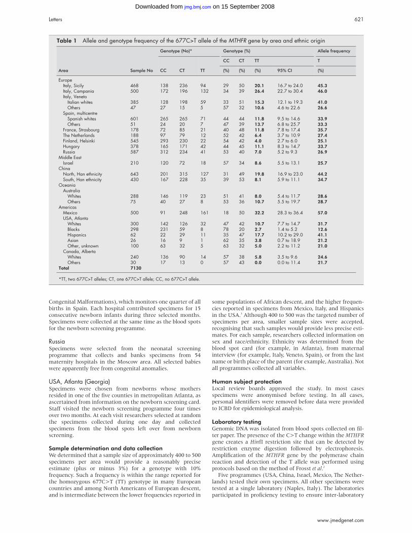
Congenital Malformations), which monitors one quarter of all
births in Spain. Each hospital contributed specimens for 15
consecutive newborn infants during three selected months.
Specimens were collected at the same time as the blood spots
for the newborn screening programme.
RussiaSpecimens were selected from the neonatal screening
programme that collects and banks specimens from 54
maternity hospitals in the Moscow area. All selected babies
were apparently free from congenital anomalies.
USA, Atlanta (Georgia)Specimens were chosen from newborns whose mothers
resided in one of the five counties in metropolitan Atlanta, as
ascertained from information on the newborn screening card.
Staff visited the newborn screening programme four times
over two months. At each visit researchers selected at random
the specimens collected during one day and collected
specimens from the blood spots left over from newborn
screening.
Sample determination and data collectionWe determined that a sample size of approximately 400 to 500
specimens per area would provide a reasonably precise
estimate (plus or minus 3%) for a genotype with 10%
frequency. Such a frequency is within the range reported for
the homozygous 677C>T (TT) genotype in many European
countries and among North Americans of European descent,
and is intermediate between the lower frequencies reported in
some populations of African descent, and the higher frequen-
cies reported in specimens from Mexico, Italy, and Hispanics
in the USA.3 Although 400 to 500 was the targeted number of
specimens per area, smaller sample sizes were accepted,
recognising that such samples would provide less precise esti-
mates. For each sample, researchers collected information on
sex and race/ethnicity. Ethnicity was determined from the
blood spot card (for example, in Atlanta), from maternal
interview (for example, Italy, Veneto, Spain), or from the last
name or birth place of the parent (for example, Australia). Not
all programmes collected all variables.
Human subject protectionLocal review boards approved the study. In most cases
specimens were anonymised before testing. In all cases,
personal identifiers were removed before data were provided
to ICBD for epidemiological analysis.
Laboratory testingGenomic DNA was isolated from blood spots collected on fil-
ter paper. The presence of the C>T change within the MTHFRgene creates a HinfI restriction site that can be detected by
restriction enzyme digestion followed by electrophoresis.
Amplification of the MTHFR gene by the polymerase chain
reaction and detection of the T allele was performed using
protocols based on the method of Frosst et al.2
Five programmes (USA, China, Israel, Mexico, The Nether-
lands) tested their own specimens. All other specimens were
tested at a single laboratory (Naples, Italy). The laboratories
participated in proficiency testing to ensure inter-laboratory
Table 1 Allele and genotype frequency of the 677C>T allele of the MTHFR gene by area and ethnic origin
Area Sample No
Genotype (No)* Genotype (%) Allele frequency
CC CT TT
CC CT TT T
(%) (%) (%) 95% CI (%)
EuropeItaly, Sicily 468 138 236 94 29 50 20.1 16.7 to 24.0 45.3Italy, Campania 500 172 196 132 34 39 26.4 22.7 to 30.4 46.0Italy, Veneto
Italian whites 385 128 198 59 33 51 15.3 12.1 to 19.3 41.0Others 47 27 15 5 57 32 10.6 4.6 to 22.6 26.6
Spain, multicentreSpanish whites 601 265 265 71 44 44 11.8 9.5 to 14.6 33.9Others 51 24 20 7 47 39 13.7 6.8 to 25.7 33.3
France, Strasbourg 178 72 85 21 40 48 11.8 7.8 to 17.4 35.7The Netherlands 188 97 79 12 52 42 6.4 3.7 to 10.9 27.4Finland, Helsinki 545 293 230 22 54 42 4.0 2.7 to 6.0 25.1Hungary 378 165 171 42 44 45 11.1 8.3 to 14.7 33.7Russia 587 312 234 41 53 40 7.0 5.2 to 9.3 26.9
Middle EastIsrael 210 120 72 18 57 34 8.6 5.5 to 13.1 25.7
ChinaNorth, Han ethnicity 643 201 315 127 31 49 19.8 16.9 to 23.0 44.2South, Han ethnicity 430 167 228 35 39 53 8.1 5.9 to 11.1 34.7
OceaniaAustralia
Whites 288 146 119 23 51 41 8.0 5.4 to 11.7 28.6Others 75 40 27 8 53 36 10.7 5.5 to 19.7 28.7
AmericasMexico 500 91 248 161 18 50 32.2 28.3 to 36.4 57.0USA, Atlanta
Whites 300 142 126 32 47 42 10.7 7.7 to 14.7 31.7Blacks 298 231 59 8 78 20 2.7 1.4 to 5.2 12.6Hispanics 62 22 29 11 35 47 17.7 10.2 to 29.0 41.1Asian 26 16 9 1 62 35 3.8 0.7 to 18.9 21.2Other, unknown 100 63 32 5 63 32 5.0 2.2 to 11.2 21.0
Canada, AlbertaWhites 240 136 90 14 57 38 5.8 3.5 to 9.6 24.6Others 30 17 13 0 57 43 0.0 0.0 to 11.4 21.7
Total 7130
*TT, two 677C>T alleles; CT, one 677C>T allele; CC, no 677C>T allele.
Letters 621
www.jmedgenet.com
on 15 September 2008 jmg.bmj.comDownloaded from
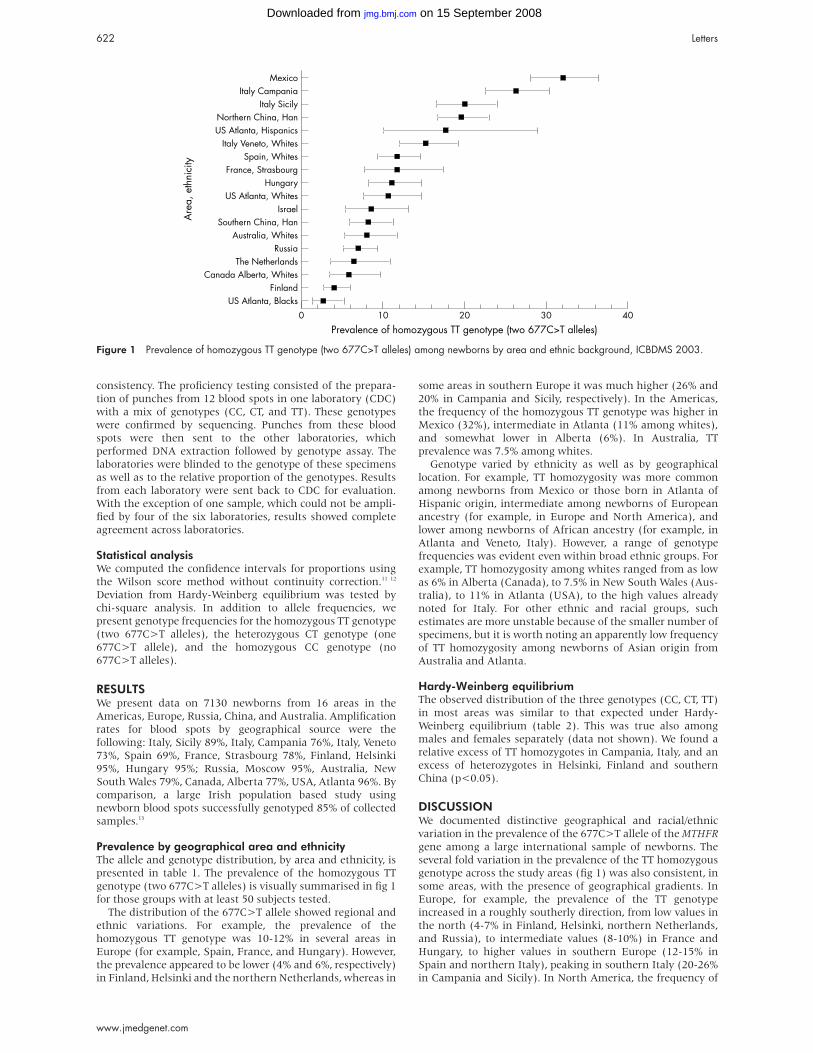
consistency. The proficiency testing consisted of the prepara-
tion of punches from 12 blood spots in one laboratory (CDC)
with a mix of genotypes (CC, CT, and TT). These genotypes
were confirmed by sequencing. Punches from these blood
spots were then sent to the other laboratories, which
performed DNA extraction followed by genotype assay. The
laboratories were blinded to the genotype of these specimens
as well as to the relative proportion of the genotypes. Results
from each laboratory were sent back to CDC for evaluation.
With the exception of one sample, which could not be ampli-
fied by four of the six laboratories, results showed complete
agreement across laboratories.
Statistical analysisWe computed the confidence intervals for proportions using
the Wilson score method without continuity correction.11 12
Deviation from Hardy-Weinberg equilibrium was tested by
chi-square analysis. In addition to allele frequencies, we
present genotype frequencies for the homozygous TT genotype
(two 677C>T alleles), the heterozygous CT genotype (one
677C>T allele), and the homozygous CC genotype (no
677C>T alleles).
RESULTSWe present data on 7130 newborns from 16 areas in the
Americas, Europe, Russia, China, and Australia. Amplification
rates for blood spots by geographical source were the
following: Italy, Sicily 89%, Italy, Campania 76%, Italy, Veneto
73%, Spain 69%, France, Strasbourg 78%, Finland, Helsinki
95%, Hungary 95%; Russia, Moscow 95%, Australia, New
South Wales 79%, Canada, Alberta 77%, USA, Atlanta 96%. By
comparison, a large Irish population based study using
newborn blood spots successfully genotyped 85% of collected
samples.13
Prevalence by geographical area and ethnicityThe allele and genotype distribution, by area and ethnicity, is
presented in table 1. The prevalence of the homozygous TT
genotype (two 677C>T alleles) is visually summarised in fig 1
for those groups with at least 50 subjects tested.
The distribution of the 677C>T allele showed regional and
ethnic variations. For example, the prevalence of the
homozygous TT genotype was 10-12% in several areas in
Europe (for example, Spain, France, and Hungary). However,
the prevalence appeared to be lower (4% and 6%, respectively)
in Finland, Helsinki and the northern Netherlands, whereas in
some areas in southern Europe it was much higher (26% and
20% in Campania and Sicily, respectively). In the Americas,
the frequency of the homozygous TT genotype was higher in
Mexico (32%), intermediate in Atlanta (11% among whites),
and somewhat lower in Alberta (6%). In Australia, TT
prevalence was 7.5% among whites.
Genotype varied by ethnicity as well as by geographical
location. For example, TT homozygosity was more common
among newborns from Mexico or those born in Atlanta of
Hispanic origin, intermediate among newborns of European
ancestry (for example, in Europe and North America), and
lower among newborns of African ancestry (for example, in
Atlanta and Veneto, Italy). However, a range of genotype
frequencies was evident even within broad ethnic groups. For
example, TT homozygosity among whites ranged from as low
as 6% in Alberta (Canada), to 7.5% in New South Wales (Aus-
tralia), to 11% in Atlanta (USA), to the high values already
noted for Italy. For other ethnic and racial groups, such
estimates are more unstable because of the smaller number of
specimens, but it is worth noting an apparently low frequency
of TT homozygosity among newborns of Asian origin from
Australia and Atlanta.
Hardy-Weinberg equilibriumThe observed distribution of the three genotypes (CC, CT, TT)
in most areas was similar to that expected under Hardy-
Weinberg equilibrium (table 2). This was true also among
males and females separately (data not shown). We found a
relative excess of TT homozygotes in Campania, Italy, and an
excess of heterozygotes in Helsinki, Finland and southern
China (p<0.05).
DISCUSSIONWe documented distinctive geographical and racial/ethnic
variation in the prevalence of the 677C>T allele of the MTHFRgene among a large international sample of newborns. The
several fold variation in the prevalence of the TT homozygous
genotype across the study areas (fig 1) was also consistent, in
some areas, with the presence of geographical gradients. In
Europe, for example, the prevalence of the TT genotype
increased in a roughly southerly direction, from low values in
the north (4-7% in Finland, Helsinki, northern Netherlands,
and Russia), to intermediate values (8-10%) in France and
Hungary, to higher values in southern Europe (12-15% in
Spain and northern Italy), peaking in southern Italy (20-26%
in Campania and Sicily). In North America, the frequency of
Figure 1 Prevalence of homozygous TT genotype (two 677C>T alleles) among newborns by area and ethnic background, ICBDMS 2003.
622 Letters
www.jmedgenet.com
on 15 September 2008 jmg.bmj.comDownloaded from
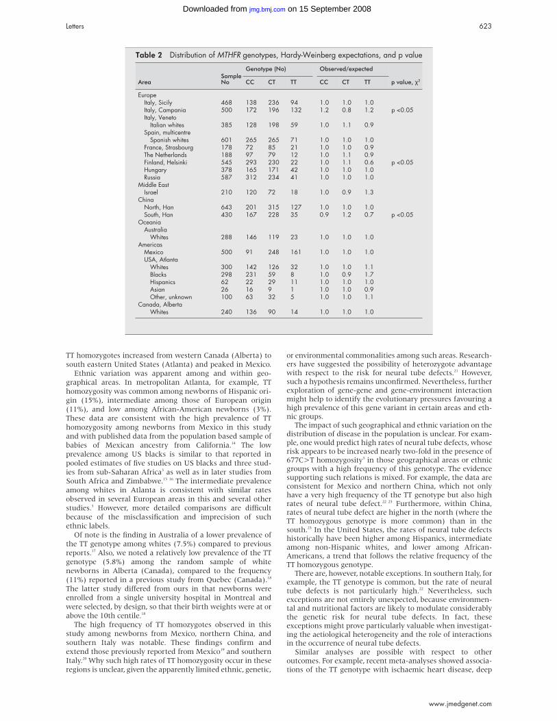
TT homozygotes increased from western Canada (Alberta) to
south eastern United States (Atlanta) and peaked in Mexico.
Ethnic variation was apparent among and within geo-
graphical areas. In metropolitan Atlanta, for example, TT
homozygosity was common among newborns of Hispanic ori-
gin (15%), intermediate among those of European origin
(11%), and low among African-American newborns (3%).
These data are consistent with the high prevalence of TT
homozygosity among newborns from Mexico in this study
and with published data from the population based sample of
babies of Mexican ancestry from California.14 The low
prevalence among US blacks is similar to that reported in
pooled estimates of five studies on US blacks and three stud-
ies from sub-Saharan Africa3 as well as in later studies from
South Africa and Zimbabwe.15 16 The intermediate prevalence
among whites in Atlanta is consistent with similar rates
observed in several European areas in this and several other
studies.3 However, more detailed comparisons are difficult
because of the misclassification and imprecision of such
ethnic labels.
Of note is the finding in Australia of a lower prevalence of
the TT genotype among whites (7.5%) compared to previous
reports.17 Also, we noted a relatively low prevalence of the TT
genotype (5.8%) among the random sample of white
newborns in Alberta (Canada), compared to the frequency
(11%) reported in a previous study from Quebec (Canada).18
The latter study differed from ours in that newborns were
enrolled from a single university hospital in Montreal and
were selected, by design, so that their birth weights were at or
above the 10th centile.18
The high frequency of TT homozygotes observed in this
study among newborns from Mexico, northern China, and
southern Italy was notable. These findings confirm and
extend those previously reported from Mexico19 and southern
Italy.20 Why such high rates of TT homozygosity occur in these
regions is unclear, given the apparently limited ethnic, genetic,
or environmental commonalities among such areas. Research-ers have suggested the possibility of heterozygote advantagewith respect to the risk for neural tube defects.21 However,such a hypothesis remains unconfirmed. Nevertheless, furtherexploration of gene-gene and gene-environment interactionmight help to identify the evolutionary pressures favouring ahigh prevalence of this gene variant in certain areas and eth-nic groups.
The impact of such geographical and ethnic variation on thedistribution of disease in the population is unclear. For exam-ple, one would predict high rates of neural tube defects, whoserisk appears to be increased nearly two-fold in the presence of677C>T homozygosity3 in those geographical areas or ethnicgroups with a high frequency of this genotype. The evidencesupporting such relations is mixed. For example, the data areconsistent for Mexico and northern China, which not onlyhave a very high frequency of the TT genotype but also highrates of neural tube defect.22 23 Furthermore, within China,rates of neural tube defect are higher in the north (where theTT homozygous genotype is more common) than in thesouth.23 In the United States, the rates of neural tube defectshistorically have been higher among Hispanics, intermediateamong non-Hispanic whites, and lower among African-Americans, a trend that follows the relative frequency of theTT homozygous genotype.
There are, however, notable exceptions. In southern Italy, forexample, the TT genotype is common, but the rate of neuraltube defects is not particularly high.22 Nevertheless, suchexceptions are not entirely unexpected, because environmen-tal and nutritional factors are likely to modulate considerablythe genetic risk for neural tube defects. In fact, theseexceptions might prove particularly valuable when investigat-ing the aetiological heterogeneity and the role of interactionsin the occurrence of neural tube defects.
Similar analyses are possible with respect to otheroutcomes. For example, recent meta-analyses showed associa-tions of the TT genotype with ischaemic heart disease, deep
Table 2 Distribution of MTHFR genotypes, Hardy-Weinberg expectations, and p value
AreaSampleNo
Genotype (No) Observed/expected
p value, χ2CC CT TT CC CT TT
EuropeItaly, Sicily 468 138 236 94 1.0 1.0 1.0Italy, Campania 500 172 196 132 1.2 0.8 1.2 p <0.05Italy, Veneto
Italian whites 385 128 198 59 1.0 1.1 0.9Spain, multicentre
Spanish whites 601 265 265 71 1.0 1.0 1.0France, Strasbourg 178 72 85 21 1.0 1.0 0.9The Netherlands 188 97 79 12 1.0 1.1 0.9Finland, Helsinki 545 293 230 22 1.0 1.1 0.6 p <0.05Hungary 378 165 171 42 1.0 1.0 1.0Russia 587 312 234 41 1.0 1.0 1.0
Middle EastIsrael 210 120 72 18 1.0 0.9 1.3
ChinaNorth, Han 643 201 315 127 1.0 1.0 1.0South, Han 430 167 228 35 0.9 1.2 0.7 p <0.05
OceaniaAustralia
Whites 288 146 119 23 1.0 1.0 1.0Americas
Mexico 500 91 248 161 1.0 1.0 1.0USA, Atlanta
Whites 300 142 126 32 1.0 1.0 1.1Blacks 298 231 59 8 1.0 0.9 1.7Hispanics 62 22 29 11 1.0 1.0 1.0Asian 26 16 9 1 1.0 1.0 0.9Other, unknown 100 63 32 5 1.0 1.0 1.1
Canada, AlbertaWhites 240 136 90 14 1.0 1.0 1.0
Letters 623
www.jmedgenet.com
on 15 September 2008 jmg.bmj.comDownloaded from
venous thrombosis, and perhaps stroke.24 Like neural tubedefects, these health outcomes are subject to interacting riskfactors and therefore the relation between genotype and out-come at a group level is likely to be complex. Nevertheless,researchers seeking to understand such relations might finddata such as these on the geographical and ethnic variation ofthe 677C>T allele helpful.
On a population level, the genotype distribution associatedwith the T allele was generally consistent with Hardy-Weinberg expectations. However, a few significant deviationsdid occur, mostly at the ends of the frequency spectrum. Anexcess of TT homozygotes was observed in southern Italy(Campania), where the allele was common, whereas thereverse was observed in Finland (Helsinki) where the allelewas uncommon. Though these two deviations from Hardy-Weinberg expectations could be the result of chance and mul-tiple statistical testing, they might also suggest the presence oflocal selective pressures.
In interpreting the findings of this study, one shouldconsider its strengths and limitations. Although we attemptedto draw unbiased, systematic samples of newborns fromdefined populations, sampling strategies varied across areas,and one cannot be certain that the efforts were always entirelysuccessful. We provide details on sampling procedures asguidance to readers who wish to use part or all of these data.Dealing with race and ethnicity was also a difficult butinescapable challenge. Classifications based on self report andparticularly on the birth place of the parent or last name areunsatisfactory to varying degrees. Thus, we present our data(table 1) either stratified in two groups (the main ethnic groupand all other groups combined), or present data only for themajor ethnic group (for example, Han Chinese). While thisapproach does not solve the difficulties entirely, it decreasesthe misclassification inherent in defining the many smallerethnic groups that coexist in many areas. Other limitations ofthis study include the lack of coverage from many areas of theworld, including most of Africa, the Middle East, LatinAmerica, and the Indian subcontinent.25
Another challenge of this study was addressing measure-ment error in genotyping. One might speculate, for example,that deviations in Hardy-Weinberg equilibrium may be theresult of genotyping errors. However, inter-laboratory consist-ency and quality control measures showed remarkable agree-ment among laboratories. In addition, the same laboratorythat assayed samples from areas showing deviations fromHardy-Weinberg equilibrium also assayed the samples frommany areas not showing such deviations, suggesting nosystematic laboratory error.
A strength of the study was the ability to assemble system-atically relatively large samples from newborns using explicitsampling protocols. Measures were also taken to ensure thereliability and comparability of genotypic data across laborato-ries, including quality control protocols that involved blindretesting of results and exchange of specimens.
Data from studies such as these can serve several purposes.Geneticists could find them useful when evaluating the distri-bution of genetic variation in human populations and its rolein genetic susceptibility to disease. For example, populationdata might help geneticists reassess controversial associationssuch as that between MTHFR genotypes and risk for Downsyndrome,26–29 for which the evidence favouring the associ-ation was largely derived from comparisons with convenientsamples of controls. As discussed previously, these geneticdata can help to interpret prevalence gradients of disease, suchas the well known geographical gradients of neural tubedefect occurrence. Similarly, huge amounts of data on otheroutcomes, such as other birth defects, pregnancy complica-tions, certain cancers, adult cardiovascular disease, andcertain psychiatric disorders,3–8 could be called upon tointerpret the prevalence gradients noted in this and otherstudies. Our data are offered as a contribution to such investi-gation.
Population data on the 677C>T variant might also help
population and public health geneticists assess the potential
impact of preventive measures based on environmental modi-
fications. For example, some adverse biochemical effects of the
thermolabile enzyme coded by the T allele, such as the
increase in total plasma homocysteine, appear to be reversible
by increasing the consumption of the B vitamin folic acid.30 If
the effect of folic acid varies by genotype, then the overall
impact in the population of fortification or supplementation
programmes might vary predictably once the genotype distri-
bution is known.
Finally, a practical outcome of this collaborative study was
to show the feasibility of conducting such genetic surveys
using existing networks of hospitals, birth defect registries,
and research institutions. Other research groups have
carefully selected and examined large and representative
samples of newborns from single states or countries (for
example, California14 and Ireland13) and generated genotype
prevalence data. We tried to expand such efforts to an
international scale, and suggest that, with appropriate
planning, such international networks can use their access
and experience in community based studies to provide core
data on the population distribution of common gene variants.
These data in turn can serve as the foundation for studies of
genetic variation and its role in increasing or decreasing
disease risk.
ACKNOWLEDGEMENTSThe study was supported in part by grant U50/CCU207141 from theUS Centers for Disease Control and Prevention. The work by GenerosoAndria (Napoli) was partially supported by grants No 97.03983,98.02936, and 99.2368 from CNR Rome. We also gratefully acknowl-edge the contribution of the following researchers: Australia: PaulLancaster; Canada (Alberta): Brian Lowry; China: Wanyin Shen, You-jun Gao, Fei Deng, Chunna He, Shuqin Zhang, Yabin Liu, GuangfengJu, Hua Dong, Zhongyue Yuan, Dongping Ye, Ping Bai, Yuqin Zhang, LiJin, Qian Gao; Hungary: László Tímár (National Centre of Health Pro-motion, Budapest); Israel: Paul Merlob (Beilinson Medical Center,Petah Tikva); Italy (Campania): Roberta Arsieri and Carmela Cafasso(Birth Defects Register of Campania); Italy (Napoli): RobertoBrancaccio and Anna Buoninconti (Department of Paediatrics,Federico II University, Napoli); Italy (Veneto): Luciano Marcazzo’(Paediatrics Unit, Arzignano Hospital, Vicenza); Mexico: Marcela Vela(Reproductive Health Agency of Ministry of Health); Russia(Moscow): Swetlana Kalinenkova (Neonatal screening programme);Spain: P Aparicio, F Ariza, I Arroyo, A Ayala, F Barranco, M Blanco, JM Bofarull, M J Calvo, R Calvo, A Cárdenes, S Castro, C Contessotto, MT Cortés, F Cucalón, J Egüés, M J Espinosa, V Felix, E Fernández, AFoguet, J M Gairi, E Galán, A García, M J García, M M García, J LGomar, H Gómez, F Gómez, J Gómez-Ullate, J González de Dios, PGutiérrez, F Hernández, H Huertas, N Jiménez-Muñoz, M MLertxundi, A Lara, J A López, E Mancebo, J J Marco, A Martínez, NMartínez, G Martínez, S Martínez, V Marugán, C Meipp, A Moral, MC Morales, A Moussallem, I Mújica, M J Oliván, L Paisán, M Pardo, APeñas, J L Pérez, I Puig, I Riaño, C Ribes, M J Ripalda, J Rosal, L Rota,J Rubio, A Sanchis, M Silveira, M E Suárez, J M de Tapia, M C Tauler,L Valdivia, M S Vázquez; USA (Atlanta, Georgia): Jennifer Rapier andVicki Brown (CDC), Muthukrishnan Ramachandran (Georgia PublicHealth Laboratory, Atlanta).
. . . . . . . . . . . . . . . . . . . . .
Authors’ affiliationsB Wilcken, Children’s Hospital at Westmead, Sydney, AustraliaF Bamforth, University of Alberta, Edmonton, CanadaZ Li, H Zhu, National Centre for Maternal and Infant Health, PekingUniversity, Beijing, ChinaA Ritvanen, The National Research and Development Centre for Welfareand Health, STAKES, FinlandM Redlund, Department of Paediatrics, Helsinki University CentralHospital, FinlandC Stoll, Y Alembik, B Dott, University of Strasbourg, Strasbourg, FranceA E Czeizel, Foundation for the Community Control of HereditaryDiseases, Budapest, HungaryZ Gelman-Kohan, Kaplan Medical Centre, Rehovot, IsraelG Scarano, Division of Medical Genetics, AO “G Rummo”, Benevento,and Birth Defects Registry of Campania, Italy
624 Letters
www.jmedgenet.com
on 15 September 2008 jmg.bmj.comDownloaded from
S Bianca, G Ettore, Sicily Congenital Malformation Registry, Sicily, ItalyR Tenconi, Department of Paediatrics, University of Padova, Veneto, ItalyS Bellato, Paediatrics Unit, Arzignano Hospital, Vicenza, Veneto, ItalyI Scala, G Andria, University Federico II, Naples, ItalyO M Mutchinick, M A López, Departamento de Genética, RYVEMCE,Instituto Nacional de Ciencias Médicas y Nutrición Salvador Zubirán,México, DFH de Walle, R Hofstra , University of Groningen, The NetherlandsL Joutchenki, L Kavteladze, Moscow Birth Defects Registry, Moscow,RussiaE Bermejo, ECEMC and Instituto de Salud Carlos III, Madrid, SpainM L Martínez-Frías, ECEMC, Instituto de Salud Carlos III, andUniversidad Complutense, Madrid, SpainM Gallagher, National Center on Environmental Health, Centers forDisease Control and Prevention, Atlanta, Georgia, USAJ D Erickson, L Botto, National Center on Birth Defects andDevelopmental Disabilities, Centers for Disease Control and Prevention,Atlanta, Georgia, USAS E Vollset, University of Bergen, Bergen, NorwayP Mastroiacovo, International Centre on Birth Defects, Rome, Italy
Correspondence to: Dr L D Botto, National Center on Birth Defects andDevelopmental Disabilities, Mailstop F-45, Centers for Disease Controland Prevention, 4770 Buford Highway NE, Atlanta, GA 30341, USA;[email protected]
REFERENCES1 Kang SS, Wong PW, Susmano A, Sora J, Norusis M, Ruggie N.
Thermolabile methylenetetrahydrofolate reductase: an inherited risk factorfor coronary artery disease. Am J Hum Genet 1991;48:536-45.
2 Frosst P, Blom HJ, Milos R, Goyette P, Sheppard CA, Matthews RG,Boers GJ, den Heijer M, Kluijtmans LA, van den Heuvel LP, et al. Acandidate genetic risk factor for vascular disease: a common mutation inmethylenetetrahydrofolate reductase. Nat Genet 1995;10:111-13.
3 Botto LD, Yang Q. 5,10-Methylenetetrahydrofolate reductase genevariants and congenital anomalies: a HuGE review. Am J Epidemiol2000;151:862-77.
4 Ueland PM, Hustad S, Schneede J, Refsum H, Vollset SE. Biological andclinical implications of the MTHFR C677T polymorphism. TrendsPharmacol Sci 2001;22:195-201.
5 Ray JG, Laskin CA. Folic acid and homocyst(e)ine metabolic defects andthe risk of placental abruption, pre-eclampsia and spontaneouspregnancy loss: a systematic review. Placenta 1999;20:519-29.
6 Ames BN. Cancer prevention and diet: help from single nucleotidepolymorphisms. Proc Natl Acad Sci USA 1999;96:12216-18.
7 Ames BN, Elson-Schwab I, Silver EA. High-dose vitamin therapystimulates variant enzymes with decreased coenzyme binding affinity(increased K(m)): relevance to genetic disease and polymorphisms. Am JClin Nutr 2002;75:616-58.
8 Rosenblatt DS. Methylenetetrahydrofolate reductase. Clin Invest Med2001;24:56-9.
9 Khoury MJ, Little J. Human genome epidemiologic reviews: thebeginning of something HuGE. Am J Epidemiol 2000;151:2-3.
10 Rosenberg N, Murata M, Ikeda Y, Opare-Sem O, Zivelin A, Geffen E,Seligsohn U. The frequent 5,10-methylenetetrahydrofolate reductaseC677T polymorphism is associated with a common haplotype in Whites,Japanese, and Africans. Am J Hum Genet 2002;70:758-62.
11 Vollset SE. Confidence intervals for a binomial proportion. Stat Med1993;12:809-24.
12 Brown LD, Cai TT, DanGupta A. Interval estimation for a binomialproportion. Stat Sci 2001;16:101-33.
13 Mills JL, Kirke PN, Molloy AM, Burke H, Conley MR, Lee YJ, Mayne PD,Weir DG, Scott JM. Methylenetetrahydrofolate reductase thermolabilevariant and oral clefts. Am J Med Genet 1999;86:71-4.
14 Shaw GM, Rozen R, Finnell RH, Wasserman CR, Lammer EJ. Maternalvitamin use, genetic variation of infant methylenetetrahydrofolatereductase, and risk for spina bifida. Am J Epidemiol 1998;148:30-7.
15 Rajkovic A, Mahomed K, Rozen R, Malinow MR, King IB, Williams MA.Methylenetetrahydrofolate reductase 677 C>T polymorphism, plasmafolate, vitamin B(12) concentrations, and risk of preeclampsia amongblack African women from Zimbabwe. Mol Genet Metabol2000;69:33-9.
16 Ubbink JB, Christianson A, Bester MJ, Van Allen MI, Venter PA, DelportR, Blom HJ, van der Merwe A, Potgieter H, Vermaak WJ. Folate status,homocysteine metabolism, and methylene tetrahydrofolate reductasegenotype in rural South African blacks with a history of pregnancycomplicated by neural tube defects. Metabolism 1999;48:269-74.
17 Wilcken DE, Wang XL, Sim AS, McCredie RM. Distribution in healthyand coronary populations of the methylenetetrahydrofolate reductase(MTHFR) C677T mutation. Arterioscler Thromb Vasc Biol1996;16:878-82.
18 Infante-Rivard C, Rivard GE, Yotov WV, Genin E, Guiguet M,Weinberg C, Gauthier R, Feoli-Fonseca JC. Absence of association ofthrombophilia polymorphisms with intrauterine growth restriction. N EnglJ Med 2002;347:19-25.
19 Mutchinick OM, Lopez MA, Luna L, Waxman J, Babinsky VE. Highprevalence of the thermolabile methylenetetrahydrofolate reductasevariant in Mexico: a country with a very high prevalence of neural tubedefects. Mol Genet Metabol 1999;68:461-7.
20 de Franchis R, Buoninconti A, Mandato C, Pepe A, Sperandeo MP, DelGado R, Capra V, Salvaggio E, Andria G, Mastroiacovo P. The C677Tmutation of the 5,10-methylenetetrahydrofolate reductase gene is amoderate risk factor for spina bifida in Italy. J Med Genet1998;35:1009-13.
21 Weitkamp LR, Tackels DC, Hunter AG, Holmes LB, Schwartz CE.Heterozygote advantage of the MTHFR gene in patients with neural-tubedefect and their relatives. Lancet 1998;351:1554-5.
22 International Clearinghouse for Birth Defects Monitoring Systems.Annual report 2001. Rome: International Centre for Birth Defects, 2001.
23 Moore CA, Li S, Li Z, Hong SX, Gu HQ, Berry RJ, Mulinare J, EricksonJD. Elevated rates of severe neural tube defects in a high-prevalence areain northern China. Am J Med Genet 1997;73:113-18.
24 Wald DS, Law M, Morris JK. Homocysteine and cardiovascular disease:evidence on causality from a meta-analysis. BMJ 2002;325:1202.
25 Mukherjee M, Joshi S, Bagadi S, Dalvi M, Rao A, Shetty K. A lowprevalence of the C677T mutation in the methylenetetrahydrofolatereductase gene in Asian Indians. Clin Genet 2002;61:155-9.
26 Hobbs CA, Sherman SL, Yi P, Hopkins SE, Torfs CP, Hine RJ, PogribnaM, Rozen R, James SJ. Polymorphisms in genes involved in folatemetabolism as maternal risk factors for Down syndrome. Am J HumGenet 2000;67:623-30.
27 O’Leary VB, Parle-McDermott A, Molloy AM, Kirke PN, Johnson Z,Conley M, Scott JM, Mills JL. MTRR and MTHFR polymorphism: link toDown syndrome? Am J Med Genet 2002;107:151-5.
28 James SJ, Pogribna M, Pogribny IP, Melnyk S, Hine RJ, Gibson JB, Yi P,Tafoya DL, Swenson DH, Wilson VL, Gaylor DW. Abnormal folatemetabolism and mutation in the methylenetetrahydrofolate reductasegene may be maternal risk factors for Down syndrome. Am J Clin Nutr1999;70:495-501.
29 Hassold TJ, Burrage LC, Chan ER, Judis LM, Schwartz S, James SJ,Jacobs PA, Thomas NS. Maternal folate polymorphisms and the etiologyof human nondisjunction. Am J Hum Genet 2001;69:434-9.
30 Rozen R. Genetic predisposition to hyperhomocysteinemia: deficiency ofmethylenetetrahydrofolate reductase (MTHFR). Thromb Haemost1997;78:523-6.
Letters 625
www.jmedgenet.com
on 15 September 2008 jmg.bmj.comDownloaded from
Small babies receive the cardiovascular protectiveapolipoprotein e2 allele less frequently than expectedC Infante-Rivard, E Lévy, G-E Rivard, M Guiguet, J-C Feoli-Fonseca. . . . . . . . . . . . . . . . . . . . . . . . . . . . . . . . . . . . . . . . . . . . . . . . . . . . . . . . . . . . . . . . . . . . . . . . . . . . . . . . . . . . . . . . . . . . . . . . . . . . . . . . . . . . . . . . . . . . . . . . . . . . .
J Med Genet 2003;40:626–629
Anewborn whose weight for gestational age and sex is lessthan expected, based on population standards, isconsidered as having intrauterine growth restriction
(IUGR); a cut off at less than the 10th centile is often used todefine IUGR. Causes of IUGR remain unclear although anumber of fetal and maternal risk factors have beenidentified.1 2 Increased early morbidity and mortality, as wellas, possibly, less than optimal neuropsychological develop-ment, have been reported as consequences of IUGR.2 3 In addi-tion, small size at birth has been associated with health prob-lems in adulthood such as coronary heart disease anddyslipidaemia.4 5 The association between restricted fetalgrowth and adult chronic diseases (often referred to as theBarker hypothesis) is now considered robust and possiblycausal.6
Apolipoprotein E (apoE) is one of the key regulators ofplasma lipid levels as it affects hepatic binding, uptake, andcatabolism of several classes of lipoproteins.7 The apolipopro-tein E gene (APOE) codes for the apoE protein; in animalmodels, underexpression of the APOE gene and lack of theapoE protein result in increased susceptibility toatherosclerosis,8 9 whereas gene overexpression displays anti-inflammatory, antiproliferative, and atheroprotectiveproperties.10 ApoE has also emerged as a central factor in vari-ous biological processes such as immunoregulation, control ofcell growth and differentiation,11 and brain development.12 The
three common allelic variants at the APOE locus (e2, e3, e4)
code for three major apoE protein isoforms (E2, E3, E4). These
isoforms differ from one another only by single amino acid
substitutions, yet these changes exhibit functional conse-
quences at both the cellular and molecular levels.13 14 In previ-
ous studies, children who carry the e4 allele and those who
carry the e2 allele have been shown to have, respectively,
higher and lower total cholesterol and low density lipoprotein
(LDL) cholesterol than those with the e3/e3 homozygous
genotype.15 16
Despite mounting evidence indicating the participation of
APOE polymorphisms in various developmental processes
involving cell growth and differentiation, atherosclerosis,
brain development, and other disorders, we found no report
on the relation between APOE polymorphisms and IUGR.
Given the data suggesting changes in cardiovascular disease
(CVD) risk with the different apo e alleles, and the data show-
ing that growth restricted babies are at higher risk of CVD, we
thought it justified to examine if there is or is not preferential
transmission of the apo e alleles from parents to IUGR cases.
METHODSStudy subjectsWe carried out a study of IUGR in relation to thrombophilic
polymorphisms.17 Cases were newborns whose birth weight
was below the 10th centile for gestational age and sex, based
on Canadian standards.18 All cases seen at our centre between
May 1998 and June 2000 who were born alive after the 24th
week of gestation and without severe congenital anomalies
were eligible for the study if the mother agreed to participate.
The project was approved by the Institutional Review Board of
the hospital. Informed consent was signed by the mother to
collect cord and maternal blood. During that period, 505 new-
borns met the criteria for cases and 493 participated in the
study (97.6%). In the original study, we also included controls,
which are not used in the present report. Midway through this
study, we started to collect buccal swabs from the fathers of
babies to be included in the final phase of the study. The goal
was to analyse case-parental trios (mother, father, newborn)
to test for association and linkage.19 Among the fathers
contacted, 86% provided genetic material. We genotyped 449
newborns and 440 mother-newborn pairs for APOE (89% of all
case pairs); there was enough DNA from the buccal swabs
remaining to genotype APOE in 194 fathers (78% of fathers
providing DNA). Genotyping of all three family members was
complete for 170 trios.
Laboratory investigationHuman genomic DNA was extracted from whole blood
samples (mothers and newborns) or from buccal swabs
(fathers), as previously described.17 Briefly, PCR reactions were
performed with a final reaction volume of 50 l, using 50-100
ng of DNA template per tube under the following conditions:
an initial DNA denaturation step at 94°C for three minutes
before adding the mixture containing the Taq DNA polymerase
enzyme; this was followed by a 40 cycle sequence of primer
annealing at 62°C for 30 seconds, extension at 72°C for one
minute, and denaturation at 94°C for 30 seconds with a 10
minute final extension step at 72°C. The PCR APOE primer
sequences were as follows: 5′ CGGGCACGGCTGTCCAAGGA 3′(forward) and 5′ CGGGCCCCGGCCTGGTACAC 3′ (reverse).20
Allele specific oligonucleotide hybridisation assays were
performed as described by others.21 PCR products were dena-
tured, divided into aliquots, and blotted onto nitrocellulose
membranes. Positive and negative controls were included on
each membrane. Specimens of family members were assigned
randomly to membranes. After hybridisation and washing,
the membranes were read using PhosphoImager (Molecular
Key points
• A newborn whose weight for gestational age and sex isless than expected, based on population standards, isconsidered as having intrauterine growth restriction(IUGR).
• The APOE gene has three common allelic variants (e2,e3, e4), which result in functional consequences at boththe cellular and molecular levels. The e2 allele has beenassociated with lower total cholesterol and low densitylipoprotein cholesterol.
• We studied the transmission of the three APOE allelesfrom heterozygous parents to newborns with IUGR andfound a significantly reduced transmission of allele e2.
• Because the e2 allele has been associated with a lowerrisk of cardiovascular disease, while babies born withgrowth restriction are reported to be at higher risk forsuch disease later in life, our data seem to reconcilethese two observations.
626 Letters
www.jmedgenet.com
on 15 September 2008 jmg.bmj.comDownloaded from
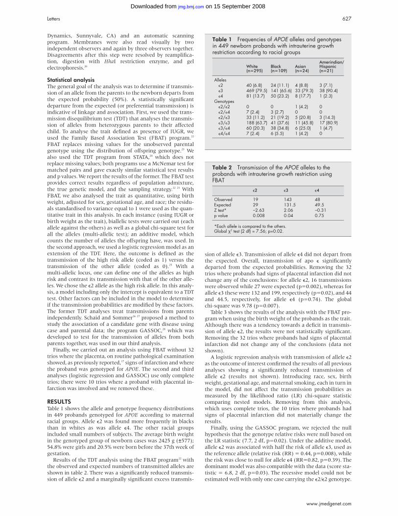
Dynamics, Sunnyvale, CA) and an automatic scanning
program. Membranes were also read visually by two
independent observers and again by three observers together.
Disagreements after this step were resolved by reamplifica-
tion, digestion with HhaI restriction enzyme, and gel
electrophoresis.20
Statistical analysisThe general goal of the analysis was to determine if transmis-
sion of an allele from the parents to the newborn departs from
the expected probability (50%). A statistically significant
departure from the expected (or preferential transmission) is
indicative of linkage and association. First, we used the trans-
mission disequilibrium test (TDT) that analyses the transmis-
sion of alleles from heterozygous parents to their affected
child. To analyse the trait defined as presence of IUGR, we
used the Family Based Association Test (FBAT) program.22
FBAT replaces missing values for the unobserved parental
genotype using the distribution of offspring genotype.23 We
also used the TDT program from STATA,24 which does not
replace missing values; both programs use a McNemar test for
matched pairs and gave exactly similar statistical test results
and p values. We report the results of the former. The FBAT test
provides correct results regardless of population admixture,
the true genetic model, and the sampling strategy.22 23 With
FBAT, we also analysed the trait as quantitative, using birth
weight, adjusted for sex, gestational age, and race; the residu-
als standardised to variance equal to 1 were used as the quan-
titative trait in this analysis. In each instance (using IUGR or
birth weight as the trait), biallelic tests were carried out (each
allele against the others) as well as a global chi-square test for
all the alleles (multi-allelic test); an additive model, which
counts the number of alleles the offspring have, was used. In
the second approach, we used a logistic regression model as an
extension of the TDT. Here, the outcome is defined as the
transmission of the high risk allele (coded as 1) versus the
transmission of the other allele (coded as 0).25 With a
multi-allelic locus, one can define one of the alleles as high
risk and contrast its transmission with that of the other alle-
les. We chose the e2 allele as the high risk allele. In this analy-
sis, a model including only the intercept is equivalent to a TDT
test. Other factors can be included in the model to determine
if the transmission probabilities are modified by these factors.
The former TDT analyses treat transmissions from parents
independently. Schaid and Sommer26 27 proposed a method to
study the association of a candidate gene with disease using
case and parental data; the program GASSOC,28 which was
developed to test for the transmission of alleles from both
parents together, was used in our third analysis.
Finally, we carried out an analysis using FBAT without 32
trios where the placenta, on routine pathological examination
showed, as previously reported,17 signs of infarction and where
the proband was genotyped for APOE. The second and third
analyses (logistic regression and GASSOC) use only complete
trios; there were 10 trios where a proband with placental in-
farction was involved and we removed these.
RESULTSTable 1 shows the allele and genotype frequency distributions
in 449 probands genotyped for APOE according to maternal
racial groups. Allele e2 was found more frequently in blacks
than in whites as was allele e4. The other racial groups
included small numbers of subjects. The average birth weight
in the genotyped group of newborn cases was 2425 g (±577);
54.8% were girls and 20.5% were born before the 37th week of
gestation.
Results of the TDT analysis using the FBAT program22 with
the observed and expected numbers of transmitted alleles are
shown in table 2. There was a significantly reduced transmis-
sion of allele e2 and a marginally significant excess transmis-
sion of allele e3. Transmission of allele e4 did not depart from
the expected. Overall, transmission of apo e significantly
departed from the expected probabilities. Removing the 32
trios where probands had signs of placental infarction did not
change any of the conclusions: for allele e2, 16 transmissions
were observed while 27 were expected (p=0.002), whereas for
allele e3 these were 132 and 199, respectively (p=0.02), and 44
and 44.5, respectively, for allele e4 (p=0.74). The global
chi-square was 9.78 (p=0.007).
Table 3 shows the results of the analysis with the FBAT pro-
gram when using the birth weight of the probands as the trait.
Although there was a tendency towards a deficit in transmis-
sion of allele e2, the results were not statistically significant.
Removing the 32 trios where probands had signs of placental
infarction did not change any of the conclusions (data not
shown).
A logistic regression analysis with transmission of allele e2
as the outcome of interest confirmed the results of all previous
analyses showing a significantly reduced transmission of
allele e2 (results not shown). Introducing race, sex, birth
weight, gestational age, and maternal smoking, each in turn in
the model, did not affect the transmission probabilities as
measured by the likelihood ratio (LR) chi-square statistic
comparing nested models. Removing from this analysis,
which uses complete trios, the 10 trios where probands had
signs of placental infarction did not materially change the
results.
Finally, using the GASSOC program, we rejected the null
hypothesis that the genotype relative risks were null based on
the LR statistic (7.7, 2 df, p=0.02). Under the additive model,
allele e2 was associated with half the risk of allele e3, used as
the reference allele (relative risk (RR) = 0.44, p=0.008), while
the risk was close to null for allele e4 (RR=0.82, p=0.39). The
dominant model was also compatible with the data (score sta-
tistic = 6.8, 2 df, p=0.03). The recessive model could not be
estimated well with only one case carrying the e2/e2 genotype.
Table 1 Frequencies of APOE alleles and genotypesin 449 newborn probands with intrauterine growthrestriction according to racial groups
White(n=295)
Black(n=109)
Asian(n=24)
Amerindian/Hispanic(n=21)
Allelese2 40 (6.8) 24 (11.1) 4 (8.8) 3 (7.1)e3 469 (79.5) 141 (65.6) 33 (79.3) 38 (90.4)e4 81 (13.7) 50 (23.2) 8 (17.7) 1 (2.3)
Genotypese2/e2 0 0 1 (4.2) 0e2/e4 7 (2.4) 3 (2.7) 0 0e2/e3 33 (11.2) 21 (19.2) 5 (20.8) 3 (14.3)e3/e3 188 (63.7) 41 (37.6) 11 (45.8) 17 (80.9)e3/e4 60 (20.3) 38 (34.8) 6 (25.0) 1 (4.7)e4/e4 7 (2.4) 6 (5.5) 1 (4.2) 0
Table 2 Transmission of the APOE alleles to theprobands with intrauterine growth restriction usingFBAT
e2 e3 e4
Observed 19 143 48Expected 29 131.5 49.5Z test* −2.63 2.06 −0.31p value 0.008 0.04 0.75
*Each allele is compared to the others.Global χ2 test (2 df) = 7.56; p=0.02.
Letters 627
www.jmedgenet.com
on 15 September 2008 jmg.bmj.comDownloaded from
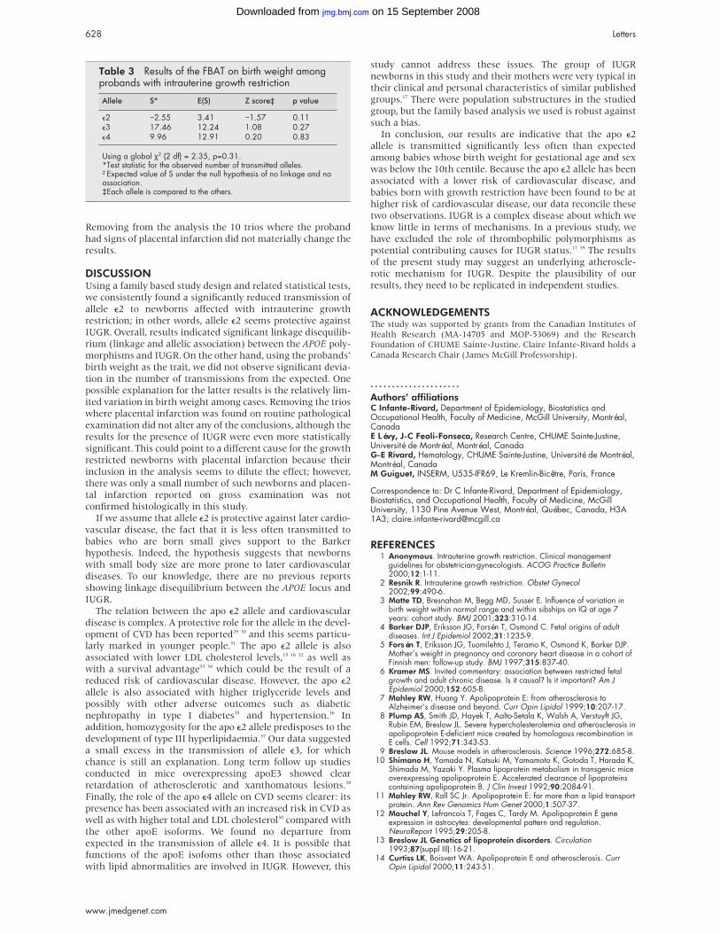
Removing from the analysis the 10 trios where the proband
had signs of placental infarction did not materially change the
results.
DISCUSSIONUsing a family based study design and related statistical tests,
we consistently found a significantly reduced transmission of
allele e2 to newborns affected with intrauterine growth
restriction; in other words, allele e2 seems protective against
IUGR. Overall, results indicated significant linkage disequilib-
rium (linkage and allelic association) between the APOE poly-
morphisms and IUGR. On the other hand, using the probands’
birth weight as the trait, we did not observe significant devia-
tion in the number of transmissions from the expected. One
possible explanation for the latter results is the relatively lim-
ited variation in birth weight among cases. Removing the trios
where placental infarction was found on routine pathological
examination did not alter any of the conclusions, although the
results for the presence of IUGR were even more statistically
significant. This could point to a different cause for the growth
restricted newborns with placental infarction because their
inclusion in the analysis seems to dilute the effect; however,
there was only a small number of such newborns and placen-
tal infarction reported on gross examination was not
confirmed histologically in this study.
If we assume that allele e2 is protective against later cardio-
vascular disease, the fact that it is less often transmitted to
babies who are born small gives support to the Barker
hypothesis. Indeed, the hypothesis suggests that newborns
with small body size are more prone to later cardiovascular
diseases. To our knowledge, there are no previous reports
showing linkage disequilibrium between the APOE locus and
IUGR.
The relation between the apo e2 allele and cardiovascular
disease is complex. A protective role for the allele in the devel-
opment of CVD has been reported29 30 and this seems particu-
larly marked in younger people.31 The apo e2 allele is also
associated with lower LDL cholesterol levels,15 16 32 as well as
with a survival advantage33 34 which could be the result of a
reduced risk of cardiovascular disease. However, the apo e2
allele is also associated with higher triglyceride levels and
possibly with other adverse outcomes such as diabetic
nephropathy in type I diabetes35 and hypertension.36 In
addition, homozygosity for the apo e2 allele predisposes to the
development of type III hyperlipidaemia.37 Our data suggested
a small excess in the transmission of allele e3, for which
chance is still an explanation. Long term follow up studies
conducted in mice overexpressing apoE3 showed clear
retardation of atherosclerotic and xanthomatous lesions.38
Finally, the role of the apo e4 allele on CVD seems clearer: its
presence has been associated with an increased risk in CVD as
well as with higher total and LDL cholesterol30 compared with
the other apoE isoforms. We found no departure from
expected in the transmission of allele e4. It is possible that
functions of the apoE isofoms other than those associated
with lipid abnormalities are involved in IUGR. However, this
study cannot address these issues. The group of IUGR
newborns in this study and their mothers were very typical in
their clinical and personal characteristics of similar published
groups.17 There were population substructures in the studied
group, but the family based analysis we used is robust against
such a bias.
In conclusion, our results are indicative that the apo e2
allele is transmitted significantly less often than expected
among babies whose birth weight for gestational age and sex
was below the 10th centile. Because the apo e2 allele has been
associated with a lower risk of cardiovascular disease, and
babies born with growth restriction have been found to be at
higher risk of cardiovascular disease, our data reconcile these
two observations. IUGR is a complex disease about which we
know little in terms of mechanisms. In a previous study, we
have excluded the role of thrombophilic polymorphisms as
potential contributing causes for IUGR status.17 39 The results
of the present study may suggest an underlying atheroscle-
rotic mechanism for IUGR. Despite the plausibility of our
results, they need to be replicated in independent studies.
ACKNOWLEDGEMENTSThe study was supported by grants from the Canadian Institutes ofHealth Research (MA-14705 and MOP-53069) and the ResearchFoundation of CHUME Sainte-Justine. Claire Infante-Rivard holds aCanada Research Chair (James McGill Professorship).
. . . . . . . . . . . . . . . . . . . . .Authors’ affiliationsC Infante-Rivard, Department of Epidemiology, Biostatistics andOccupational Health, Faculty of Medicine, McGill University, Montréal,CanadaE Lévy, J-C Feoli-Fonseca, Research Centre, CHUME Sainte-Justine,Université de Montréal, Montréal, CanadaG-E Rivard, Hematology, CHUME Sainte-Justine, Université de Montréal,Montréal, CanadaM Guiguet, INSERM, U535-IFR69, Le Kremlin-Bicêtre, Paris, France
Correspondence to: Dr C Infante-Rivard, Department of Epidemiology,Biostatistics, and Occupational Health, Faculty of Medicine, McGillUniversity, 1130 Pine Avenue West, Montréal, Québec, Canada, H3A1A3; [email protected]
REFERENCES1 Anonymous. Intrauterine growth restriction. Clinical management
guidelines for obstetrician-gynecologists. ACOG Practice Bulletin2000;12:1-11.
2 Resnik R. Intrauterine growth restriction. Obstet Gynecol2002;99:490-6.
3 Matte TD, Bresnahan M, Begg MD, Susser E. Influence of variation inbirth weight within normal range and within sibships on IQ at age 7years: cohort study. BMJ 2001;323:310-14.
4 Barker DJP, Eriksson JG, Forsén T, Osmond C. Fetal origins of adultdiseases. Int J Epidemiol 2002;31:1235-9.
5 Forsén T, Eriksson JG, Tuomilehto J, Teramo K, Osmond K, Barker DJP.Mother’s weight in pregnancy and coronary heart disease in a cohort ofFinnish men: follow-up study. BMJ 1997;315:837-40.
6 Kramer MS. Invited commentary: association between restricted fetalgrowth and adult chronic disease. Is it causal? Is it important? Am JEpidemiol 2000;152:605-8.
7 Mahley RW, Huang Y. Apolipoprotein E: from atherosclerosis toAlzheimer’s disease and beyond. Curr Opin Lipidol 1999;10:207-17.
8 Plump AS, Smith JD, Hayek T, Aalto-Setala K, Walsh A, Verstuyft JG,Rubin EM, Breslow JL. Severe hypercholesterolemia and atherosclerosis inapolipoprotein E-deficient mice created by homologous recombination inE cells. Cell 1992;71:343-53.
9 Breslow JL. Mouse models in atherosclerosis. Science 1996;272:685-8.10 Shimano H, Yamada N, Katsuki M, Yamamoto K, Gotoda T, Harada K,
Shimada M, Yazaki Y. Plasma lipoprotein metabolism in transgenic miceoverexpressing apolipoprotein E. Accelerated clearance of lipoproteinscontaining apolipoprotein B. J Clin Invest 1992;90:2084-91.
11 Mahley RW, Rall SC Jr. Apolipoprotein E: far more than a lipid transportprotein. Ann Rev Genomics Hum Genet 2000;1:507-37.
12 Mouchel Y, Lefrancois T, Fages C, Tardy M. Apolipoprotein E geneexpression in astrocytes: developmental pattern and regulation.NeuroReport 1995;29:205-8.
13 Breslow JL Genetics of lipoprotein disorders. Circulation1993;87(suppl III):16-21.
14 Curtiss LK, Boisvert WA. Apolipoprotein E and atherosclerosis. CurrOpin Lipidol 2000;11:243-51.
Table 3 Results of the FBAT on birth weight amongprobands with intrauterine growth restriction
Allele S* E(S)† Z score‡ p value
e2 −2.55 3.41 −1.57 0.11e3 17.46 12.24 1.08 0.27e4 9.96 12.91 0.20 0.83
Using a global χ2 (2 df) = 2.35, p=0.31.*Test statistic for the observed number of transmitted alleles.†Expected value of S under the null hypothesis of no linkage and noassociation.‡Each allele is compared to the others.
628 Letters
www.jmedgenet.com
on 15 September 2008 jmg.bmj.comDownloaded from
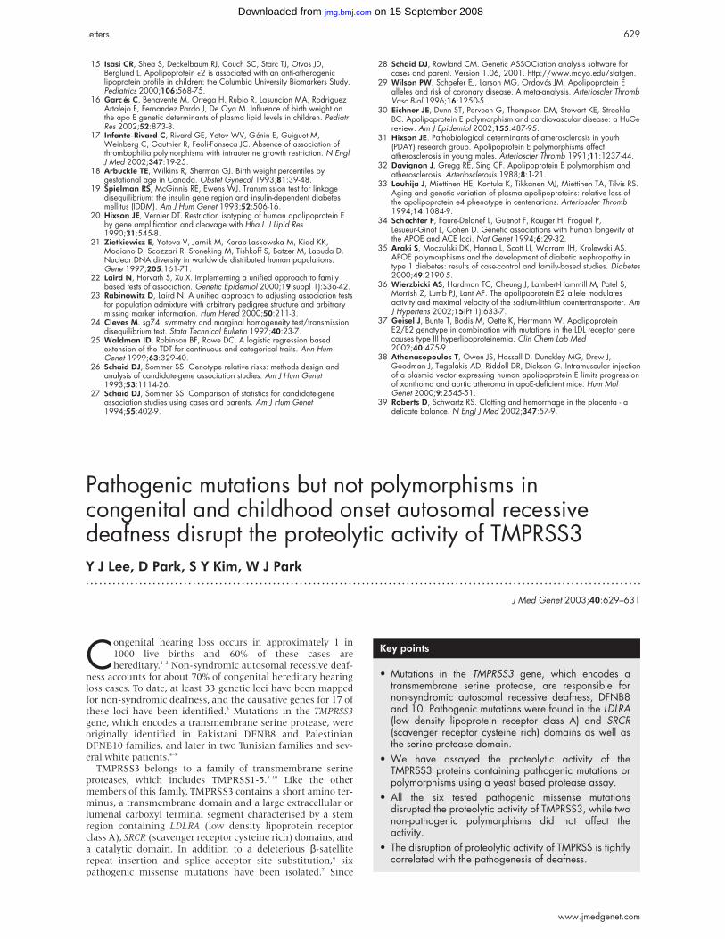
15 Isasi CR, Shea S, Deckelbaum RJ, Couch SC, Starc TJ, Otvos JD,Berglund L. Apolipoprotein e2 is associated with an anti-atherogeniclipoprotein profile in children: the Columbia University Biomarkers Study.Pediatrics 2000;106:568-75.
16 Garcés C, Benavente M, Ortega H, Rubio R, Lasuncion MA, RodriguezArtalejo F, Fernandez Pardo J, De Oya M. Influence of birth weight onthe apo E genetic determinants of plasma lipid levels in children. PediatrRes 2002;52:873-8.
17 Infante-Rivard C, Rivard GE, Yotov WV, Génin E, Guiguet M,Weinberg C, Gauthier R, Feoli-Fonseca JC. Absence of association ofthrombophilia polymorphisms with intrauterine growth restriction. N EnglJ Med 2002;347:19-25.
18 Arbuckle TE, Wilkins R, Sherman GJ. Birth weight percentiles bygestational age in Canada. Obstet Gynecol 1993;81:39-48.
19 Spielman RS, McGinnis RE, Ewens WJ. Transmission test for linkagedisequilibrium: the insulin gene region and insulin-dependent diabetesmellitus (IDDM). Am J Hum Genet 1993;52:506-16.
20 Hixson JE, Vernier DT. Restriction isotyping of human apolipoprotein Eby gene amplification and cleavage with Hha I. J Lipid Res1990;31:545-8.
21 Zietkiewicz E, Yotova V, Jarnik M, Korab-Laskowska M, Kidd KK,Modiano D, Scozzari R, Stoneking M, Tishkoff S, Batzer M, Labuda D.Nuclear DNA diversity in worldwide distributed human populations.Gene 1997;205:161-71.
22 Laird N, Horvath S, Xu X. Implementing a unified approach to familybased tests of association. Genetic Epidemiol 2000;19(suppl 1):S36-42.
23 Rabinowitz D, Laird N. A unified approach to adjusting association testsfor population admixture with arbitrary pedigree structure and arbitrarymissing marker information. Hum Hered 2000;50:211-3.
24 Cleves M. sg74: symmetry and marginal homogeneity test/transmissiondisequilibrium test. Stata Technical Bulletin 1997;40:23-7.
25 Waldman ID, Robinson BF, Rowe DC. A logistic regression basedextension of the TDT for continuous and categorical traits. Ann HumGenet 1999;63:329-40.
26 Schaid DJ, Sommer SS. Genotype relative risks: methods design andanalysis of candidate-gene association studies. Am J Hum Genet1993;53:1114-26.
27 Schaid DJ, Sommer SS. Comparison of statistics for candidate-geneassociation studies using cases and parents. Am J Hum Genet1994;55:402-9.
28 Schaid DJ, Rowland CM. Genetic ASSOCiation analysis software forcases and parent. Version 1.06, 2001. http://www.mayo.edu/statgen.
29 Wilson PW, Schaefer EJ, Larson MG, Ordovás JM. Apolipoprotein Ealleles and risk of coronary disease. A meta-analysis. Arterioscler ThrombVasc Biol 1996;16:1250-5.
30 Eichner JE, Dunn ST, Perveen G, Thompson DM, Stewart KE, StroehlaBC. Apolipoprotein E polymorphism and cardiovascular disease: a HuGereview. Am J Epidemiol 2002;155:487-95.
31 Hixson JE. Pathobiological determinants of atherosclerosis in youth(PDAY) research group. Apolipoprotein E polymorphisms affectatherosclerosis in young males. Arterioscler Thromb 1991;11:1237-44.
32 Davignon J, Gregg RE, Sing CF. Apolipoprotein E polymorphism andatherosclerosis. Arteriosclerosis 1988;8:1-21.
33 Louhija J, Miettinen HE, Kontula K, Tikkanen MJ, Miettinen TA, Tilvis RS.Aging and genetic variation of plasma apolipoproteins: relative loss ofthe apolipoprotein e4 phenotype in centenarians. Arterioscler Thromb1994;14:1084-9.
34 Schächter F, Faure-Delanef L, Guénot F, Rouger H, Froguel P,Lesueur-Ginot L, Cohen D. Genetic associations with human longevity atthe APOE and ACE loci. Nat Genet 1994;6:29-32.
35 Araki S, Moczulski DK, Hanna L, Scott LJ, Warram JH, Krolewski AS.APOE polymorphisms and the development of diabetic nephropathy intype 1 diabetes: results of case-control and family-based studies. Diabetes2000;49:2190-5.
36 Wierzbicki AS, Hardman TC, Cheung J, Lambert-Hammill M, Patel S,Morrish Z, Lumb PJ, Lant AF. The apolipoprotein E2 allele modulatesactivity and maximal velocity of the sodium-lithium countertransporter. AmJ Hypertens 2002;15(Pt 1):633-7.
37 Geisel J, Bunte T, Bodis M, Oette K, Herrmann W. ApolipoproteinE2/E2 genotype in combination with mutations in the LDL receptor genecauses type III hyperlipoproteinemia. Clin Chem Lab Med2002;40:475-9.
38 Athanasopoulos T, Owen JS, Hassall D, Dunckley MG, Drew J,Goodman J, Tagalakis AD, Riddell DR, Dickson G. Intramuscular injectionof a plasmid vector expressing human apolipoprotein E limits progressionof xanthoma and aortic atheroma in apoE-deficient mice. Hum MolGenet 2000;9:2545-51.
39 Roberts D, Schwartz RS. Clotting and hemorrhage in the placenta - adelicate balance. N Engl J Med 2002;347:57-9.
Pathogenic mutations but not polymorphisms incongenital and childhood onset autosomal recessivedeafness disrupt the proteolytic activity of TMPRSS3Y J Lee, D Park, S Y Kim, W J Park. . . . . . . . . . . . . . . . . . . . . . . . . . . . . . . . . . . . . . . . . . . . . . . . . . . . . . . . . . . . . . . . . . . . . . . . . . . . . . . . . . . . . . . . . . . . . . . . . . . . . . . . . . . . . . . . . . . . . . . . . . . . .
J Med Genet 2003;40:629–631
Congenital hearing loss occurs in approximately 1 in1000 live births and 60% of these cases arehereditary.1 2 Non-syndromic autosomal recessive deaf-
ness accounts for about 70% of congenital hereditary hearingloss cases. To date, at least 33 genetic loci have been mappedfor non-syndromic deafness, and the causative genes for 17 ofthese loci have been identified.3 Mutations in the TMPRSS3gene, which encodes a transmembrane serine protease, wereoriginally identified in Pakistani DFNB8 and PalestinianDFNB10 families, and later in two Tunisian families and sev-eral white patients.4–9
TMPRSS3 belongs to a family of transmembrane serineproteases, which includes TMPRSS1-5.5 10 Like the othermembers of this family, TMPRSS3 contains a short amino ter-minus, a transmembrane domain and a large extracellular orlumenal carboxyl terminal segment characterised by a stemregion containing LDLRA (low density lipoprotein receptorclass A), SRCR (scavenger receptor cysteine rich) domains, anda catalytic domain. In addition to a deleterious β-satelliterepeat insertion and splice acceptor site substitution,6 sixpathogenic missense mutations have been isolated.7 Since
Key points
• Mutations in the TMPRSS3 gene, which encodes atransmembrane serine protease, are responsible fornon-syndromic autosomal recessive deafness, DFNB8and 10. Pathogenic mutations were found in the LDLRA(low density lipoprotein receptor class A) and SRCR(scavenger receptor cysteine rich) domains as well asthe serine protease domain.
• We have assayed the proteolytic activity of theTMPRSS3 proteins containing pathogenic mutations orpolymorphisms using a yeast based protease assay.
• All the six tested pathogenic missense mutationsdisrupted the proteolytic activity of TMPRSS3, while twonon-pathogenic polymorphisms did not affect theactivity.
• The disruption of proteolytic activity of TMPRSS is tightlycorrelated with the pathogenesis of deafness.
Letters 629
www.jmedgenet.com
on 15 September 2008 jmg.bmj.comDownloaded from
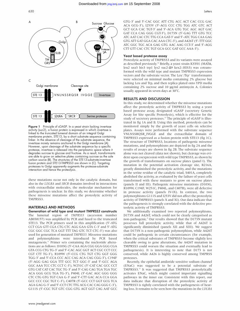
these mutations occur not only in the catalytic domain, but
also in the LDLRA and SRCR domains involved in interactions
with extracellular molecules, the molecular mechanism for
pathogenesis is unclear. In this study, we determine whether
these missense mutations affect the proteolytic activity of
TMPRSS3.
MATERIALS AND METHODSGeneration of wild type and mutant TMPRSS3 constructsThe lumenal region of TMPRSS3 (accession number
AB038157) was amplified by PCR and fused to the truncated
STE13. The PCR primers used in this amplification (5′-ATG
CCT CGA GTT CGA CTG CTC AGG GAA GTA C-3′ and 5′-ATG
CGC GGC CGC TCA GGT TTT TAG GTC TCT CTC-3′) was also
used for generation of mutated TMPRSS3. Missense mutations
and polymorphisms were introduced by PCR based
mutagenesis.11 Primer sets containing the nucleotide altera-
tions are as follows: D103G (5′-CAA AGA CGG GGA GGG CGA
GTA CCG CTG TG-3′ and 5′-CAC AGC GGT ACT CGC CCT CCC
CGT CTT TG-3′), R109W (5′-CCG CTG TGT CTG GGT GGG
TGG-3′ and 5′-CCA CCC ACC CAG ACA CAG CGG-3′), C194F
(5′-AGG GAG GGA TTT GCC TCT GGC-3′ and 5′-GCC AGA
GGC AAA TCC CTC CCT C-3′), W251C (5′-CAT CAC GCC CCT
GTG CAT CAT CAC TGC TG-3′ and 5′-CAG CAG TGA TGA TGC
ACA GGG GCG TGA TG-3′), P404L (5′-GAC AGC GGG GGG
CTC CTG GTG TGT CAA G-3′ and 5′-CTT GAC ACA CCA GGA
GCC CCC CGC TGT C-3′), C407R (5′-GCC CCT GGT GCG TCA
AGA GAG G-3′ and 5′-CCT CTC TTG ACG CAC CAG GGG C-3′),
G111S (5′-CGC TGT GTC CGG GTG AGT GGT CAG AAT GCC
GTG-3′ and 5′-CAC GGC ATT CTG ACC ACT CAC CCG GAC
ACA GCG-3′), I253V (5′-ACG CCC CTG TGG ATC GTC ACT
GCT GCA CAC TGT-3′ and 5′-ACA GTG TGC AGC AGT GAC
GAT CCA CAG GGG CGT-3′), D173N (5′-GAG TTT GTG TCC
ATC AAT CAC CTC TTG CCA GAT-3′ and 5′-ATC TGG CAA GAG
GTG ATT GAT GGA CAC AAA CTC-3′), and A426T (5′-TTT GGC
ATC GGC TGC ACA GAG GTG AAC AAG CCT-3′ and 5′-AGG
CTT GTT CAC CTC TGT GCA GCC GAT GCC AAA-3′).
Yeast based protease assayProteolytic activity of TMPRSS3 and its variants were assayed
as described previously.12 Briefly, a yeast strain KSY01 (MATα,
leu2 ura3 his3 trp1 lys2 suc2-∆9 kex2::HIS3) was cotrans-
formed with the wild type and mutant TMPRSS3 expression
vectors and the substrate vector. The Leu+/Trp+ transformants
were selected on minimal media containing 2% glucose but
lacking Leu and Trp, and then replica plated onto YPD media
containing 2% sucrose and 10 µg/ml antimycin A. Colonies
usually appeared in seven days at 30°C.
RESULTS AND DISCUSSIONIn this study, we determined whether the missense mutations
affect the proteolytic activity of TMPRSS3 by using a yeast
based protease assay, designated sGASP (secretory Genetic
Assay for Site specific Proteolysis), which is effective for the
study of secretory proteases.12 The principle of sGASP is illus-
trated in fig 1A and B. Using this method, proteolysis can be
monitored simply by the growth of yeast cells on selective
plates. Assays were performed with the substrate sequence
VNLNSSRQSR_IVGGE and the extracellular domain of
TMPRSS3 expressed as a fusion protein with STE13 (fig 1C).
The structure of TMPRSS3, locations of pathogenic missense
mutations, and polymorphisms are depicted in fig 2A and the
results of assays are shown in fig 2B. The substrate sequence
alone was not cleaved (data not shown), but cleavage was evi-
dent upon coexpression with wild type TMPRSS3, as shown by
the growth of transformants on sucrose plates (panel I). The
mutation in the potential activation cleavage site, R216A,
greatly diminished the proteolytic activity, and the mutations
in the serine residue of the catalytic triad, S401A, completely
abolished the activity, as evaluated by the failure of yeast cells
transformed with these mutants to grow on sucrose plates
(panels II and III). Pathogenic missense mutations (D103G,
R109W, C194F, W251C, P404L, and C407R), were all defective
in protease activity (panels IV-IX). In contrast, the two
polymorphisms G111S and I253V did not affect the proteolytic
activity of TMPRSS3 (panels X and XI). Our data indicate that
the pathogenesis is strongly correlated with the defective pro-
teolytic activity of TMPRSS3.
We additionally examined two reported polymorphisms,
D173N and A426T, which could not be clearly categorised as
non-pathogenic.9 Our results showed that the D173N mutant
possesses full proteolytic activity, while that of A426T is
significantly diminished (panels XII and XIII). We suggest
that D173N is a non-pathogenic polymorphism, while A426T
could be pathogenic in certain circumstances (for example,
when the critical substrates of TMPRSS3 become slightly less
cleavable owing to gene alterations, the A426T mutation in
TMPRSS3 could worsen the situation and eventually lead to
pathogenesis). It is interesting to note that D173 is not
conserved, while A426 is highly conserved among TMPRSS
proteases.
Recently, the epithelial amiloride sensitive sodium channel
(ENaC) was suggested to be a potential substrate of
TMPRSS3.13 It was suggested that TMPRSS3 proteolytically
activates ENaC, which might control important signalling
pathways in the inner ear. Consistent with this report, our
data indicate that disruption of the proteolytic activity of
TMPRSS3 is tightly correlated with the pathogenesis of hear-
ing loss. It remains to be seen how the mutations in the LDLRA
Figure 1 Principle of sGASP. In a yeast strain lacking invertaseactivity (suc2-), a fusion protein is expressed in which invertase islinked to the truncated lumenal domain of an integral Golgimembrane protein, STE13, by a short substrate sequence containinglinker. In the absence of cleavage of the substrate sequence, theinvertase moiety remains anchored to the Golgi membrane (A).However, upon cleavage of the substrate sequence by a specificprotease, invertase is released into the periplasmic space where itdegrades sucrose to glucose and fructose. As a result, transformantsare able to grow on selective plates containing sucrose as the solecarbon source (B). The structures of the STE13-substrate-invertasefusion protein and STE13-TMPRSS3 are shown in (C). Targetingproteases to Golgi apparently augments the protease-substrateinteraction and hence the proteolysis.
630 Letters
www.jmedgenet.com
on 15 September 2008 jmg.bmj.comDownloaded from
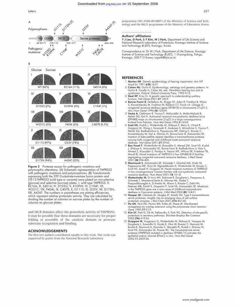
and SRCR domains affect the proteolytic activity of TMPRSS3.
It may be possible that these domains are necessary for proper
folding or assembly of the catalytic domain or protease
substrate recognition and binding.
ACKNOWLEDGEMENTSThe first two authors contributed equally to this work. This work wassupported by grants from the National Research Laboratory
programme (M1-0104-00-0007) of the Ministry of Science and Tech-nology and the BK21 programme of the Ministry of Education, Korea.
. . . . . . . . . . . . . . . . . . . . .Authors’ affiliationsY J Lee, D Park, S Y Kim, W J Park, Department of Life Science andNational Research Laboratory of Proteolysis, Kwangju Institute of Scienceand Technology (K-JIST), Kwangju, Korea
Correspondence to: Dr W J Park, Department of Life Science, KwangjuInstitute of Science and Technology (K-JIST), 1 Oryong-dong, Puk-gu,Kwangju, 500-712 Korea; [email protected]
REFERENCES1 Morton NE. Genetic epidemiology of hearing impairment. Ann NY
Acad Sci 1991;630:16-31.2 Cohen MJ, Gorlin R. Epidermiology, aetiology and genetics patterns. In:
Gorlin R, Toriello H, Cohen MJ, eds. Hereditary hearing loss and itssyndromes. Oxford: Oxford University Press, 1995:9-12.
3 Steel KP, Kros CJ. A genetic approach to understanding auditoryfunction. Nat Genet 2001;27:143-9.
4 Bonne-Tamir B, DeStefano AL, Briggs CE, Adair R, Franklyn B, WeissS, Korostishevsky M, Frydman M, Baldwin CT, Farrer LA. Linkage ofcongenital recessive deafness (gene DFNB10) to chromosome 21q22.3.Am J Hum Genet 1996;58:1254-9.
5 Veske A, Oehlmann R, Younus F, Mohyuddin A, Muller-Myhsok B,Mehdi SQ, Gal A. Autosomal recessive non-syndromic deafness locus(DFNB8) maps on chromosome 21q22 in a large consanguineouskindred from Pakistan. Hum Mol Genet 1996;5:165-8.
6 Scott HS, Kudoh J, Wattenhofer M, Shibuya K, Berry A, Chrast R,Guipponi M, Wang J, Kawasaki K, Asakawa S, Minoshima S, Younus F,Mehdi SQ, Radhakrishna U, Papasavvas MP, Gehrig C, Rossier C,Korostishevsky M, Gal A, Shimizu N, Bonne-Tamir B, Antonarakis SE.Insertion of beta-satellite repeats identifies a transmembrane proteasecausing both congenital and childhood onset autosomal recessivedeafness. Nat Genet 2001;27:59-63.
7 Ben-Yosef T, Wattenhofer M, Riazuddin S, Ahmed ZM, Scott HS, KudohJ, Shibuya K, Antonarakis SE, Bonne-Tamir B, Radhakrishna U, Naz S,Ahmed Z, Riazuddin S, Pandya A, Nance WE, Wilcox ER, Friedman TB,Morell RJ. Novel mutations of TMPRSS3 in four DFNB8/B10 familiessegregating congenital autosomal recessive deafness. J Med Genet2001;38:396-400.
8 Masmoudi S, Antonarakis SE, Schwede T, Ghorbel AM, Gratri M,Pappasavas MP, Drira M, Elgaied-Boulila A, Wattenhofer M, Rossier C,Scott HS, Ayadi H, Guipponi M. Novel missense mutations of TMPRSS3in two consanguineous Tunisian families with non-syndromic autosomalrecessive deafness. Hum Mutat 2001;18:101-8.
9 Wattenhofer M, Di Iorio MV, Rabionet R, Dougherty L, Pampanos A,Schwede T, Montserrat-Sentis B, Arbones ML, Iliades T,Pasquadibisceglie A, D’Amelio M, Alwan S, Rossier C, Dahl HH,Petersen MB, Estivill X, Gasparini P, Scott HS, Antonarakis SE. Mutationsin the TMPRSS3 gene are a rare cause of childhood nonsyndromicdeafness in Caucasian patients. J Mol Med 2002;80:124-31.
10 Hooper JD, Clements JA, Quigley JP, Antalis TM. Type II transmembraneserine proteases. Insights into an emerging class of cell surfaceproteolytic enzymes. J Biol Chem 2001;276:857-60.
11 Ho SN, Hunt HD, Horton RM, Pullen JK, Pease LR. Site-directedmutagenesis by overlap extension using the polymerase chain reaction.Gene 1989;77:51-9.
12 Kim SY, Park D, Oh M, Sellamuthu S, Park WJ. Detection of site-specificproteolysis in secretory pathways. Biochem Biophys Res Commun2002;296:419-24.
13 Guipponi M, Vuagniaux G, Wattenhofer M, Shibuya K, Vazquez M,Dougherty L, Scamuffa N, Guida E, Okui M, Rossier C, Hancock M,Buchet K, Reymond A, Hummler E, Marzella PL, Kudoh J, Shimizu N,Scott HS, Antonarakis SE, Rossier BC. The transmembrane serineprotease (TMPRSS3) mutated in deafness DFNB8/10 activates theepithelial sodium channel (ENaC) in vitro. Hum Mol Genet2002;11:2829-36.
Figure 2 Protease assays for pathogenic mutations andpolymorphic alterations. (A) Schematic representation of TMPRSS3with pathogenic mutations and polymorphisms. (B) Transformantsexpressing both the STE13-substrate-invertase fusion protein andSTE13-TMPRSS3 (wild type or variants) were plated on non-selective(glucose) and selective (sucrose) plates. I, wild type TMPRSS3; II,R216A; III, S401A; IV, D103G; V, R109W; VI, C194F; VII,W251C; VIII, P404L; IX, C407R; X, G111S; XI, I253V; XII, D173N;XIII, A426T. The numbers in parentheses are plating efficiencies,which represent relative proteolytic activity. They are calculated bydividing the number of colonies on sucrose plates by the number ofcolonies on glucose plates.
Letters 631
www.jmedgenet.com
on 15 September 2008 jmg.bmj.comDownloaded from
Heteroplasmy for the 1555A>G mutation in themitochondrial 12S rRNA gene in six Spanish familieswith non-syndromic hearing lossF J del Castillo, M Rodríguez-Ballesteros, Y Martín, B Arellano, J Gallo-Terán,C Morales-Angulo, R Ramírez-Camacho, M Cruz Tapia, J Solanellas,A Martínez-Conde, M Villamar, M A Moreno-Pelayo, F Moreno, I del Castillo. . . . . . . . . . . . . . . . . . . . . . . . . . . . . . . . . . . . . . . . . . . . . . . . . . . . . . . . . . . . . . . . . . . . . . . . . . . . . . . . . . . . . . . . . . . . . . . . . . . . . . . . . . . . . . . . . . . . . . . . . . . . .
J Med Genet 2003;40:632–636
Hearing impairment is the most prevalent sensory disor-der and genetic causes are thought to be responsible forover 60% of the cases in developed countries.1 Inherited
hearing impairment is highly heterogeneous from both theclinical and genetic points of view.1 2 It varies in age of onset,severity, and audiological characteristics, and it can be associ-ated or not with other clinical features (syndromic ornon-syndromic hearing impairment). Genetic transmissionincludes autosomal (dominant and recessive), X linked, andmaternal inheritance patterns. This unparalleled heterogen-eity is well illustrated by the fact that over 70 loci in thenuclear genome have been reported to be involved innon-syndromic hearing impairment, and about 30 genes havebeen isolated from their critical intervals.3 Furthermore, anumber of different mutations in several genes of themitochondrial genome are responsible for syndromic andnon-syndromic forms of hearing loss.4 5
Mutations responsible for maternally inherited non-syndromic hearing loss are so far confined to only two genes inthe mitochondrial genome. These include mutations7510T>C6 and 7511T>C7 in the tRNASer(UCN) gene, and1095T>C8 and 1555A>G9 in the gene for the 12S rRNA. Thislast mutation is responsible for a dual phenotype, since it alsoconfers increased susceptibility to the ototoxic action ofaminoglycoside antibiotics.9 Most of these mutations havebeen reported in a small number of families from severalcountries, with the exception of 1555A>G, which seems to bemore frequent than the others,10–13 although its real prevalenceremains to be determined in most populations. Remarkably, inSpain it accounts for about 15-20% of all familial cases ofnon-syndromic hearing loss, irrespective of their mode ofinheritance and age of onset14 15 (our unpublished results). Ina majority of these patients, the hearing loss is not attributableto aminoglycoside ototoxicity. A phylogenetic analysis ofmitochondrial DNA (mtDNA) haplogroups, performed on 50unrelated Spanish families, showed that the 1555A>G muta-tion could be caused by over 30 independent mutationalevents, occurring in mtDNA haplogroups which are commonin all European populations.16 These data indicate that thehigh detection rate of this mutation in Spain is not the resultof a single major founder event, at least with regard to themitochondrial genome. Given the high prevalence of the1555A>G mutation in Spain, and the possibility of preventingaminoglycoside ototoxicity in mutation carriers, its detectionhas become a priority in routine genetic testing. In contrast toother mutations in the mtDNA, which are frequentlyheteroplasmic,4 5 the 1555A>G mutation has been found inhomoplasmy in all but one of the families reported so far.17 Inthat study, the mutation was found in heteroplasmy in threesubjects, with the proportion of mutant copies (the “mutationload”) ranging from 85-94%. Here we report the genetic andclinical characterisation of six novel unrelated Spanishfamilies segregating the 1555A>G mutation in heteroplasmy,with a wide range of percentages of mutant copies in a total of19 subjects.
METHODSSubjectsFamilial cases of non-syndromic hearing loss were collected
with the only criterion of having at least two affected subjects.
Our collection procedure did not cause any other bias, such as
preferential selection of large pedigrees or compatibility with
maternal inheritance. A total of 649 unrelated Spanish fami-
lies were enrolled in the study. After getting informed consent,
peripheral blood samples were obtained from all participating
family members, and DNA extraction was performed by
standard procedures.
Mutation detectionScreening for the 1555A>G mutation was carried out by PCR
amplification of a 339 bp DNA fragment containing the muta-
tion site, followed by digestion with restriction endonuclease
HaeIII, as described previously.14 In the wild type allele, diges-
tion results in two fragments of 216 bp and 123 bp. The muta-
tion specifically creates a novel restriction site, and so
digestion results in three fragments (216 bp, 93 bp, and 30 bp).
Quantification of the mutation loadThe proportion of mutant copies was quantified by detection
of fluorescently labelled PCR products separated by capillary
electrophoresis. A 359 bp DNA fragment was amplified with
Key points
• Mutation 1555A>G in the 12S rRNA gene of the mito-chondrial genome is responsible for non-syndromichearing loss, as well as for increased susceptibility to theototoxicity of aminoglycoside antibiotics.
• In almost all the cases reported so far the mutation wasfound in homoplasmy. Here we report the clinical andgenetic characterisation of six Spanish families withsensorineural hearing loss, totalling 19 subjects withheteroplasmy for 1555A>G.
• The proportion of mutant copies ranged from 3.75-96.60%. Subjects carrying less than 20% of mutantcopies were asymptomatic or had a mild hearing loss,whereas heteroplasmic subjects with over 52% ofmutant copies suffered from moderate to severe hearingloss.
• Taking the six families together, there is a correlation ofthe mutation load with the severity of the hearing loss.However, when studying the families separately, thiscorrelation is confirmed in three of them and excluded inanother.
• Our study illustrates the difficulties in extracting generalprinciples from the analysis of the genotype-phenotypecorrelation regarding the 1555A>G mutation.
632 Letters
www.jmedgenet.com
on 15 September 2008 jmg.bmj.comDownloaded from
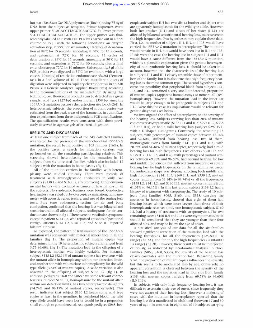
hot start FastStart Taq DNA polymerase (Roche) using 75 ng of
DNA from the subject as template. Primer sequences were:
upper primer 5′-AGACGTTAGGTCAAGGTG-3′; lower primer,
5′-GTTTAGCTCAGAGCGGTC-3′. The upper primer was fluo-
rescently labelled at 5′ with TET. PCR was carried out in a final
volume of 15 µl with the following conditions: an enzyme
activation step, at 95°C for six minutes; 10 cycles of denatura-
tion at 94°C for 15 seconds, annealing at 50°C for 15 seconds,
and extension at 72°C for 30 seconds; 13 cycles of
denaturation at 89°C for 15 seconds, annealing at 50°C for 15
seconds, and extension at 72°C for 30 seconds; plus a final
extension step at 72°C for 10 minutes. Subsequently, 8 µl of the
PCR product were digested at 37°C for two hours with a large
excess (10 units) of restriction endonuclease Alw26I (Fermen-
tas), in a final volume of 10 µl. Three microlitre aliquots of
digestion were subjected to capillary electrophoresis in an ABI
Prism 310 Genetic Analyzer (Applied Biosystems) according
to the recommendations of the manufacturer. By using this
technique, two fluorescent products can be detected in a given
sample, wild type (127 bp) and/or mutant (359 bp, since the
1555A>G mutation destroys the restriction site for Alw26I). In
heteroplasmic subjects, the proportion of mutant copies was
estimated from the peak areas of the fragments, in quantifica-
tion experiments from three independent PCR amplifications.
The quantification results were consistent with those previ-
ously observed in agarose gels after HaeIII digestion.
RESULTS AND DISCUSSIONAt least one subject from each of the 649 collected families
was tested for the presence of the mitochondrial 1555A>G
mutation, the result being positive in 105 families (16%). In
the positive cases, a search for mutation carriers was
performed on all the remaining participating relatives. This
screening showed heteroplasmy for the mutation in 19
subjects from six unrelated families, which also included 12
subjects with the mutation in homoplasmy (fig 1).
All of the mutation carriers in the families with hetero-
plasmy were studied clinically. There were records of
treatment with aminoglycoside antibiotics in only two
subjects (S138 I.2 and S160 I.2, streptomycin). Other environ-
mental factors were excluded as causes of hearing loss in all
the subjects. No syndromic features were found. Conductive
hearing loss was ruled out by otoscopic examination, tympano-
metry with acoustic reflex testing, and use of the tuning fork
tests. Pure tone audiometry, testing for air and bone
conduction, confirmed that the hearing loss was bilateral and
sensorineural in all affected subjects. Audiograms for air con-
duction are shown in fig 1. There were no vestibular symptoms
except in patient S141 I.2, who reported episodes of positional
vertigo. Patients S141 I.2, S141 II.3, and S338 II.1 reported
bilateral tinnitus.
As expected, the pattern of transmission of the 1555A>G
mutation was consistent with maternal inheritance in all the
families (fig 1). The proportion of mutant copies was
determined in the 19 heteroplasmic subjects and ranged from
3.75-96.60% (fig 1). The mutation load in the offspring of a
heteroplasmic mother was highly variable. For instance,
subject S338 I.2 (52.14% of mutant copies) has two sons with
the mutant allele in homoplasmy within our detection limits,
and another son with values close to homoplasmy for the wild
type allele (3.84% of mutant copies). A wide variation is also
observed in the offspring of subject S138 I.2 (fig 1). In
addition, pedigrees S160 and S068 have some relevant charac-
teristics. Subject S160 I.2, homoplasmic for the mutant allele
within our detection limits, has two heteroplasmic daughters
(94.74% and 96.15% of mutant copies, respectively). This
result indicates that subject S160 I.2 keeps some wild type
copies at least in the germline. In peripheral blood, the wild
type allele would have been lost or would be in a proportion
small enough to go undetected. As regards pedigree S068, het-
eroplasmic subject II.5 has two sibs (a brother and sister) whoare apparently homoplasmic for the wild type allele. However,both her brother (II.1) and a son of her sister (III.1) areaffected by bilateral sensorineural hearing loss, more severe inthe high frequencies. Two hypotheses may explain these data.First, I.2, the mother of subjects II.1, II.3, and II.5, would havecarried the 1555A>G mutation in heteroplasmy. The mutationwould remain in II.5, but would have been lost in II.1 and II.3.If this were the case, the hearing loss in subjects II.1 and III.1would have a cause different from the 1555A>G mutation,which is a plausible explanation given the genetic heterogen-eity of non-syndromic hearing loss. It should be taken intoaccount, however, that the characteristics of the hearing lossin subjects II.1 and III.1 closely resemble those of other mem-bers of the family, but it is also true that high frequency hear-ing loss is the most common type. The second hypothesis con-cerns the possibility that peripheral blood from subjects II.1,II.3, and III.1 contained a very small, undetected, proportionof mutant copies (apparent homoplasmy) or none at all (realhomoplasmy). However, the mutation load in the inner earwould be large enough to be pathogenic in subjects II.1 andIII.1. Were this the case, its implications would be relevant forgenetic diagnosis (see below).
We investigated the effect of heteroplasmy on the severity ofthe hearing loss. Subjects carrying less than 20% of mutantcopies were asymptomatic (S138 II.1 and II.2, S297 III.2, S338II.3 and II.4), or had a mild hearing loss (subject S297 IV.1,with a U shaped audiogram). Conversely, the remaining 13subjects, with percentages of mutant copies between 52.14%and 96.60%, suffered from hearing loss. Two of them,monozygotic twins from family S141 (II.1 and II.2) with70.93% and 64.48% of mutant copies, respectively, had a mildhearing loss for high frequencies. Five others (S068 II.5 andS138 II.3, II.4, II.5, and II.6), with percentages of mutant cop-ies between 69.78% and 96.60%, had normal hearing for lowand middle frequencies, but suffered from moderate or severehearing loss for high frequencies. In the remaining six cases,the audiogram shape was sloping, affecting both middle andhigh frequencies (S141 II.3, S160 II.1, and S338 I.2; mutantcopies ranging from 52.14% to 94.74%) or all the frequencies(S138 I.2, S141 I.2, and S160 II.3; mutant copies ranging from61.03% to 96.15%). In this last group, subject S138 I.2 had ahistory of treatment with streptomycin. The study of 10 sub-jects from families S068, S160, and S338, carrying themutation in homoplasmy, showed that eight of them hadhearing losses which were more severe than those of theirheteroplasmic relatives (only one homoplasmic subject, S160I.2, had a history of treatment with streptomycin). The tworemaining cases (S160 II.5 and II.6) were asymptomatic, but itshould be considered that they are younger than their fouraffected sibs, and may be below the age of onset.
A statistical analysis of our data for all the six familiesshowed significant correlation of the mutation load with thehearing thresholds, for all the frequencies (125-8000 Hzrange) (fig 2A), and for only the high frequencies (2000-8000Hz range) (fig 2B). However, these results must be interpretedcautiously, as indicated by intrafamilial analysis. In threefamilies (S068, S160, S338), the severity of the hearing lossclearly correlates with the mutation load. Regarding familyS141, the proportion of mutant copies influences the severity,but this seems to be modulated also by age. Conversely, noapparent correlation is observed between the severity of thehearing loss and the mutation load in four sibs from familyS138 with mutant copies ranging from 69.78% to 96.60%(II.3-II.6).
In subjects with only high frequency hearing loss, it wasdifficult to ascertain their age of onset, since frequently theywere not aware of their hearing loss. However, a majority ofcases with the mutation in heteroplasmy reported that thehearing loss first manifested in adulthood (between 17 and 50years of age). In contrast, in eight out of 10 subjects carrying
Letters 633
www.jmedgenet.com
on 15 September 2008 jmg.bmj.comDownloaded from
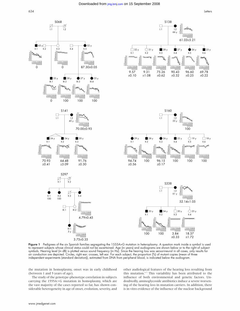
the mutation in homoplasmy, onset was in early childhood
(between 1 and 5 years of age).
The study of the genotype-phenotype correlation in subjects
carrying the 1555A>G mutation in homoplasmy, which are
the vast majority of the cases reported so far, has shown con-
siderable heterogeneity in age of onset, evolution, severity, and
other audiological features of the hearing loss resulting from
this mutation.4 5 This variability has been attributed to the
influence of both environmental and genetic factors. Un-
doubtedly, aminoglycoside antibiotics induce a severe worsen-
ing of the hearing loss in mutation carriers. In addition, there
is in vitro evidence of the influence of the nuclear background
Figure 1 Pedigrees of the six Spanish families segregating the 1555A>G mutation in heteroplasmy. A question mark inside a symbol is usedto represent subjects whose clinical status could not be ascertained. Age (in years) and audiograms are shown below or to the right of subjectsymbols. Hearing level (in dB) is plotted versus sound frequency (in Hz). Since the hearing loss was sensorineural in all cases, only results forair conduction are depicted. Circles, right ear; crosses, left ear. For each subject, the proportion (%) of mutant copies (mean of threeindependent experiments (standard deviation)), estimated from DNA from peripheral blood, is indicated below the audiogram.
634 Letters
www.jmedgenet.com
on 15 September 2008 jmg.bmj.comDownloaded from
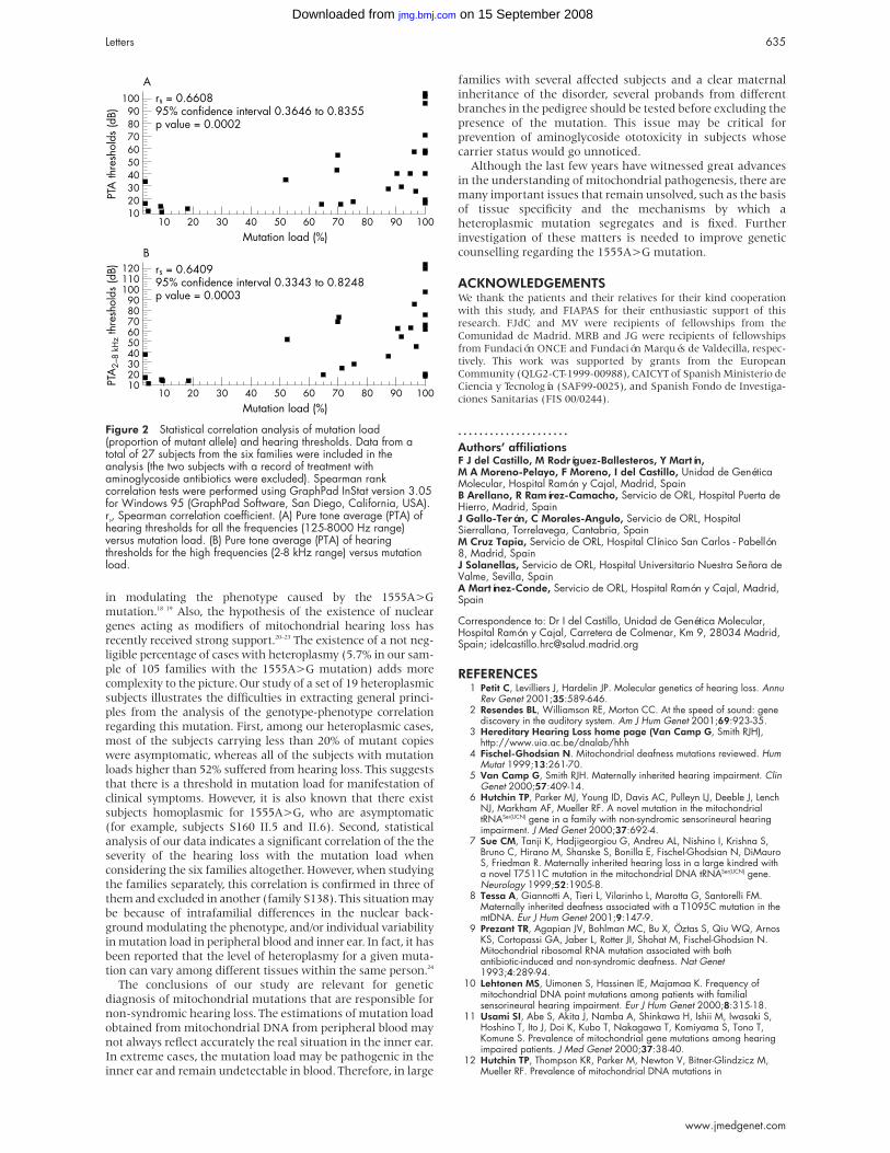
in modulating the phenotype caused by the 1555A>G
mutation.18 19 Also, the hypothesis of the existence of nuclear
genes acting as modifiers of mitochondrial hearing loss has
recently received strong support.20–23 The existence of a not neg-
ligible percentage of cases with heteroplasmy (5.7% in our sam-
ple of 105 families with the 1555A>G mutation) adds more
complexity to the picture. Our study of a set of 19 heteroplasmic
subjects illustrates the difficulties in extracting general princi-
ples from the analysis of the genotype-phenotype correlation
regarding this mutation. First, among our heteroplasmic cases,
most of the subjects carrying less than 20% of mutant copies
were asymptomatic, whereas all of the subjects with mutation
loads higher than 52% suffered from hearing loss. This suggests
that there is a threshold in mutation load for manifestation of
clinical symptoms. However, it is also known that there exist
subjects homoplasmic for 1555A>G, who are asymptomatic
(for example, subjects S160 II.5 and II.6). Second, statistical
analysis of our data indicates a significant correlation of the the
severity of the hearing loss with the mutation load when
considering the six families altogether. However, when studying
the families separately, this correlation is confirmed in three of
them and excluded in another (family S138). This situation may
be because of intrafamilial differences in the nuclear back-
ground modulating the phenotype, and/or individual variability
in mutation load in peripheral blood and inner ear. In fact, it has
been reported that the level of heteroplasmy for a given muta-
tion can vary among different tissues within the same person.24
The conclusions of our study are relevant for genetic
diagnosis of mitochondrial mutations that are responsible for
non-syndromic hearing loss. The estimations of mutation load
obtained from mitochondrial DNA from peripheral blood may
not always reflect accurately the real situation in the inner ear.
In extreme cases, the mutation load may be pathogenic in the
inner ear and remain undetectable in blood. Therefore, in large
families with several affected subjects and a clear maternal
inheritance of the disorder, several probands from different
branches in the pedigree should be tested before excluding the
presence of the mutation. This issue may be critical for
prevention of aminoglycoside ototoxicity in subjects whose
carrier status would go unnoticed.
Although the last few years have witnessed great advances
in the understanding of mitochondrial pathogenesis, there are
many important issues that remain unsolved, such as the basis
of tissue specificity and the mechanisms by which a
heteroplasmic mutation segregates and is fixed. Further
investigation of these matters is needed to improve genetic
counselling regarding the 1555A>G mutation.
ACKNOWLEDGEMENTSWe thank the patients and their relatives for their kind cooperationwith this study, and FIAPAS for their enthusiastic support of thisresearch. FJdC and MV were recipients of fellowships from theComunidad de Madrid. MRB and JG were recipients of fellowshipsfrom Fundación ONCE and Fundación Marqués de Valdecilla, respec-tively. This work was supported by grants from the EuropeanCommunity (QLG2-CT-1999-00988), CAICYT of Spanish Ministerio deCiencia y Tecnología (SAF99-0025), and Spanish Fondo de Investiga-ciones Sanitarias (FIS 00/0244).
. . . . . . . . . . . . . . . . . . . . .
Authors’ affiliationsF J del Castillo, M Rodríguez-Ballesteros, Y Martín,M A Moreno-Pelayo, F Moreno, I del Castillo, Unidad de GenéticaMolecular, Hospital Ramón y Cajal, Madrid, SpainB Arellano, R Ramírez-Camacho, Servicio de ORL, Hospital Puerta deHierro, Madrid, SpainJ Gallo-Terán, C Morales-Angulo, Servicio de ORL, HospitalSierrallana, Torrelavega, Cantabria, SpainM Cruz Tapia, Servicio de ORL, Hospital Clínico San Carlos - Pabellón8, Madrid, SpainJ Solanellas, Servicio de ORL, Hospital Universitario Nuestra Señora deValme, Sevilla, SpainA Martínez-Conde, Servicio de ORL, Hospital Ramón y Cajal, Madrid,Spain
Correspondence to: Dr I del Castillo, Unidad de Genética Molecular,Hospital Ramón y Cajal, Carretera de Colmenar, Km 9, 28034 Madrid,Spain; [email protected]
REFERENCES1 Petit C, Levilliers J, Hardelin JP. Molecular genetics of hearing loss. Annu
Rev Genet 2001;35:589-646.2 Resendes BL, Williamson RE, Morton CC. At the speed of sound: gene
discovery in the auditory system. Am J Hum Genet 2001;69:923-35.3 Hereditary Hearing Loss home page (Van Camp G, Smith RJH),
http://www.uia.ac.be/dnalab/hhh4 Fischel-Ghodsian N. Mitochondrial deafness mutations reviewed. Hum
Mutat 1999;13:261-70.5 Van Camp G, Smith RJH. Maternally inherited hearing impairment. Clin
Genet 2000;57:409-14.6 Hutchin TP, Parker MJ, Young ID, Davis AC, Pulleyn LJ, Deeble J, Lench
NJ, Markham AF, Mueller RF. A novel mutation in the mitochondrialtRNASer(UCN) gene in a family with non-syndromic sensorineural hearingimpairment. J Med Genet 2000;37:692-4.
7 Sue CM, Tanji K, Hadjigeorgiou G, Andreu AL, Nishino I, Krishna S,Bruno C, Hirano M, Shanske S, Bonilla E, Fischel-Ghodsian N, DiMauroS, Friedman R. Maternally inherited hearing loss in a large kindred witha novel T7511C mutation in the mitochondrial DNA tRNASer(UCN) gene.Neurology 1999;52:1905-8.
8 Tessa A, Giannotti A, Tieri L, Vilarinho L, Marotta G, Santorelli FM.Maternally inherited deafness associated with a T1095C mutation in themtDNA. Eur J Hum Genet 2001;9:147-9.
9 Prezant TR, Agapian JV, Bohlman MC, Bu X, Öztas S, Qiu WQ, ArnosKS, Cortopassi GA, Jaber L, Rotter JI, Shohat M, Fischel-Ghodsian N.Mitochondrial ribosomal RNA mutation associated with bothantibiotic-induced and non-syndromic deafness. Nat Genet1993;4:289-94.
10 Lehtonen MS, Uimonen S, Hassinen IE, Majamaa K. Frequency ofmitochondrial DNA point mutations among patients with familialsensorineural hearing impairment. Eur J Hum Genet 2000;8:315-18.
11 Usami SI, Abe S, Akita J, Namba A, Shinkawa H, Ishii M, Iwasaki S,Hoshino T, Ito J, Doi K, Kubo T, Nakagawa T, Komiyama S, Tono T,Komune S. Prevalence of mitochondrial gene mutations among hearingimpaired patients. J Med Genet 2000;37:38-40.
12 Hutchin TP, Thompson KR, Parker M, Newton V, Bitner-Glindzicz M,Mueller RF. Prevalence of mitochondrial DNA mutations in
Figure 2 Statistical correlation analysis of mutation load(proportion of mutant allele) and hearing thresholds. Data from atotal of 27 subjects from the six families were included in theanalysis (the two subjects with a record of treatment withaminoglycoside antibiotics were excluded). Spearman rankcorrelation tests were performed using GraphPad InStat version 3.05for Windows 95 (GraphPad Software, San Diego, California, USA).rs, Spearman correlation coefficient. (A) Pure tone average (PTA) ofhearing thresholds for all the frequencies (125-8000 Hz range)versus mutation load. (B) Pure tone average (PTA) of hearingthresholds for the high frequencies (2-8 kHz range) versus mutationload.
Letters 635
www.jmedgenet.com
on 15 September 2008 jmg.bmj.comDownloaded from

childhood/congenital onset non-syndromal sensorineural hearingimpairment. J Med Genet 2001;38:229-31.
13 Kupka S, Tóth T, Wróbel M, Zeiβler, Szyfter W, Szyfter K, NiedzielskaG, Bal J, Zenner HP, Sziklai I, Blin N, Pfister M. Mutation A1555G in the12S rRNA gene and its epidemiological importance in German,Hungarian and Polish patients. Hum Mutat 2002;19:308-9.
14 Estivill X, Govea N, Barceló A, Perelló E, Badenas C, Romero E, MoralL, Scozzari R, D’Urbano L, Zeviani M, Torroni A. Familial progressivesensorineural deafness is mainly due to the mtDNA A1555G mutationand is enhanced by treatment with aminoglycosides. Am J Hum Genet1998;62:27-35.
15 Sarduy M, del Castillo I, Villamar M, Romero L, Herraiz C, HernandezFJ, Tapia MC, Magariño C, Méndez del Castillo D, Menéndez-Alejo I,Ramírez-Camacho R, Arellano B, Morales C, Bellón J, Moreno F. Geneticstudy of mitochondrially inherited sensorineural hearing impairment ineight large families from Spain and Cuba. In: Stephens D, Read A,Martini A, eds. Developments in genetic hearing impairment. London:Whurr Publishers, 1998:121-5.
16 Torroni A, Cruciani F, Rengo C, Sellitto D, López-Bigas N, Rabionet R,Govea N, López de Munain A, Sarduy M, Romero L, Villamar M, delCastillo I, Moreno F, Estivill X, Scozzari R. The A1555G mutation in the12S rRNA gene of human mtDNA: recurrent origins and founder eventsin families affected by sensorineural deafness. Am J Hum Genet1999;65:1349-58.
17 El-Schahawi M, López de Munain A, Sarrazin AM, Shanske AL,Basirico M, Shanske S, DiMauro S. Two large Spanish pedigrees withnonsyndromic sensorineural deafness and the mtDNA mutation at nt1555 in the 12S rRNA gene: evidence of heteroplasmy. Neurology1997;48:453-6.
18 Guan MX, Fischel-Ghodsian N, Attardi G. Biochemical evidence fornuclear gene involvement in phenotype of non-syndromic deafnessassociated with mitochondrial 12S rRNA mutation. Hum Mol Genet1996;5:963-71.
19 Guan MX, Fischel-Ghodsian N, Attardi G. Nuclear backgrounddetermines biochemical phenotype in the deafness-associatedmitochondrial 12S rRNA mutation. Hum Mol Genet 2001;10:573-80.
20 Bykhovskaya Y, Estivill X, Taylor K, Hang T, Hamon M, Casano RA,Yang H, Rotter JI, Shohat M, Fischel-Ghodsian N. Candidate locus for anuclear modifier gene for maternally inherited deafness. Am J Hum Genet2000;66:1905-10.
21 Abe S, Kelley PM, Kimberling WJ, Usami SI. Connexin 26 gene (GJB2)mutation modulates the severity of hearing loss associated with the1555A>G mitochondrial mutation. Am J Med Genet 2001;103:334-8.
22 Bykhovskaya Y, Yang H, Taylor K, Hang T, Tun RY, Estivill X, CasanoRA, Majamaa K, Shohat M, Fischel-Ghodsian N. Modifier locus formitochondrial DNA disease: linkage and linkage disequilibrium mappingof a nuclear modifier gene for maternally inherited deafness. Genet Med2001;3:177-80.
23 Li X, Li R, Lin X, Guan MX. Isolation and characterization of the putativenuclear modifier gene MTO1 involved in the pathogenesis ofdeafness-associated mitochondrial 12S rRNA A1555G mutation. J BiolChem 2002;277:27256-64.
24 Matthews PM, Hopkin J, Brown RM, Stephenson JBP, Hilton-Jones D,Brown GK. Comparison of the relative levels of the 3243 (A>G) mtDNAmutation in heteroplasmic adult and fetal tissues. J Med Genet1994;31:41-4.
Uniparental disomy of chromosome 13q causinghomozygosity for the 35delG mutation in the geneencoding connexin26 (GJB2) results in prelingualhearing impairment in two unrelated Spanish patientsA Álvarez, I del Castillo, A Pera, M Villamar, M A Moreno-Pelayo, T Rivera,J Solanellas, F Moreno. . . . . . . . . . . . . . . . . . . . . . . . . . . . . . . . . . . . . . . . . . . . . . . . . . . . . . . . . . . . . . . . . . . . . . . . . . . . . . . . . . . . . . . . . . . . . . . . . . . . . . . . . . . . . . . . . . . . . . . . . . . . .
J Med Genet 2003;40:636–639
Inherited hearing impairment is a highly heterogeneousgroup of disorders with an overall incidence of about 1 in2000 newborns.1 In approximately 70% of cases, the
auditory impairment is not associated with other clinical fea-tures, that is, it is non-syndromic. The most frequent conditionis a severe or profound hearing loss of prelingual onset, whichis inherited mainly as an autosomal recessive trait.1 To date, 31different DFNB loci for autosomal recessive non-syndromichearing loss have been reported, and 16 genes have beenidentified.2 Among these loci, DFNB1 in 13q12 stands outbecause of its complexity and clinical relevance. It contains thegene GJB2, which encodes connexin26, a component of inter-cellular gap junctions. Mutations in the GJB2 gene are respon-sible for up to 50% of all cases of autosomal recessive hearingimpairment in most of the populations tested so far, with afrequent mutation (35delG) accounting for up to 86% of theGJB2 mutant alleles in white populations.3 4 However, not allthe DFNB1 mutations affect the GJB2 gene. Recently, severalresearch teams found a deletion in the 13q12 region which isfrequently inherited in double heterozygosity with mutantGJB2 alleles in affected subjects,5–7 but it was also found inhomozygosity.6 7 Molecular characterisation of this deletion,termed del(GJB6-D13S1830), showed that it encompasses 342kb and it does not affect the GJB2 gene, but it truncates thegene encoding connexin30 (GJB6), another gap junction pro-tein expressed in the inner ear.6 The existence of this deletionwas first suspected by the finding of inconsistencies in thesegregation of genetic markers distal to GJB2.
Here we report another inconsistency in the segregation of
markers in the 13q12 region in two unrelated cases of subjects
with prelingual hearing impairment. In these two cases, uni-
parental disomy of chromosome 13 caused homozygosity for
Key points
• Mutations in the gene encoding the gap junction proteinconnexin26 (DFNB1 locus on 13q12) are responsiblefor up to 50% of all cases of autosomal recessive hear-ing impairment in most populations, the 35delGmutation being the most frequent in white populations.
• Here we report two unrelated cases of homozygotes for35delG whose biological fathers were not carriers ofthe mutation. The study of the segregation ofpolymorphic genetic markers showed uniparental(maternal) disomy of chromosome 13, causing homozy-gosity for the mutation. In both cases, the disomicmaternal gamete may have resulted from non-disjunction of chromosome 13 in meiosis II.
• These two cases represent the first description ofUPD(13) with an abnormal phenotype, and they arealso the first cases of UPD resulting in non-syndromichearing impairment.
636 Letters
www.jmedgenet.com
on 15 September 2008 jmg.bmj.comDownloaded from
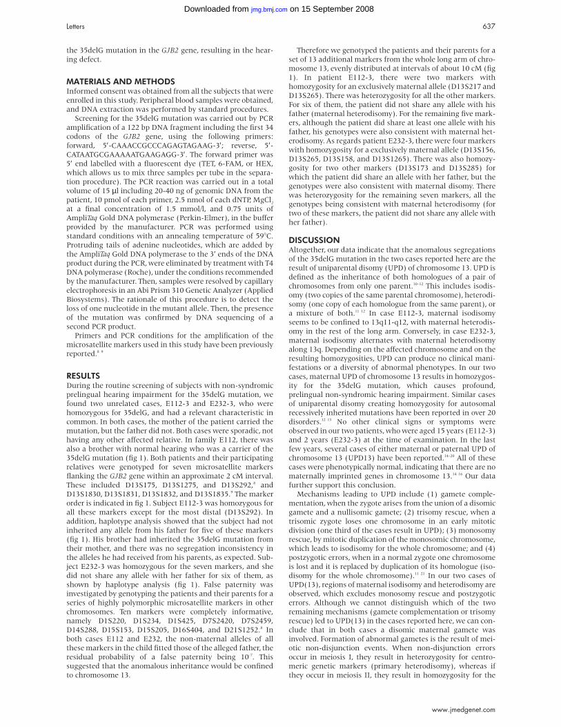
the 35delG mutation in the GJB2 gene, resulting in the hear-
ing defect.
MATERIALS AND METHODSInformed consent was obtained from all the subjects that were
enrolled in this study. Peripheral blood samples were obtained,
and DNA extraction was performed by standard procedures.
Screening for the 35delG mutation was carried out by PCR
amplification of a 122 bp DNA fragment including the first 34
codons of the GJB2 gene, using the following primers:
forward, 5′-CAAACCGCCCAGAGTAGAAG-3′; reverse, 5′-CATAATGCGAAAAATGAAGAGG-3′. The forward primer was
5′ end labelled with a fluorescent dye (TET, 6-FAM, or HEX,
which allows us to mix three samples per tube in the separa-
tion procedure). The PCR reaction was carried out in a total
volume of 15 µl including 20-40 ng of genomic DNA from the
patient, 10 pmol of each primer, 2.5 nmol of each dNTP, MgCl2
at a final concentration of 1.5 mmol/l, and 0.75 units of
AmpliTaq Gold DNA polymerase (Perkin-Elmer), in the buffer
provided by the manufacturer. PCR was performed using
standard conditions with an annealing temperature of 59°C.
Protruding tails of adenine nucleotides, which are added by
the AmpliTaq Gold DNA polymerase to the 3′ ends of the DNA
product during the PCR, were eliminated by treatment with T4
DNA polymerase (Roche), under the conditions recommended
by the manufacturer. Then, samples were resolved by capillary
electrophoresis in an Abi Prism 310 Genetic Analyzer (Applied
Biosystems). The rationale of this procedure is to detect the
loss of one nucleotide in the mutant allele. Then, the presence
of the mutation was confirmed by DNA sequencing of a
second PCR product.
Primers and PCR conditions for the amplification of the
microsatellite markers used in this study have been previously
reported.8 9
RESULTSDuring the routine screening of subjects with non-syndromic
prelingual hearing impairment for the 35delG mutation, we
found two unrelated cases, E112-3 and E232-3, who were
homozygous for 35delG, and had a relevant characteristic in
common. In both cases, the mother of the patient carried the
mutation, but the father did not. Both cases were sporadic, not
having any other affected relative. In family E112, there was
also a brother with normal hearing who was a carrier of the
35delG mutation (fig 1). Both patients and their participating
relatives were genotyped for seven microsatellite markers
flanking the GJB2 gene within an approximate 2 cM interval.
These included D13S175, D13S1275, and D13S292,8 and
D13S1830, D13S1831, D13S1832, and D13S1835.9 The marker
order is indicated in fig 1. Subject E112-3 was homozygous for
all these markers except for the most distal (D13S292). In
addition, haplotype analysis showed that the subject had not
inherited any allele from his father for five of these markers
(fig 1). His brother had inherited the 35delG mutation from
their mother, and there was no segregation inconsistency in
the alleles he had received from his parents, as expected. Sub-
ject E232-3 was homozygous for the seven markers, and she
did not share any allele with her father for six of them, as
shown by haplotype analysis (fig 1). False paternity was
investigated by genotyping the patients and their parents for a
series of highly polymorphic microsatellite markers in other
chromosomes. Ten markers were completely informative,
namely D1S220, D1S234, D1S425, D7S2420, D7S2459,
D14S288, D15S153, D15S205, D16S404, and D21S1252.8 In
both cases E112 and E232, the non-maternal alleles of all
these markers in the child fitted those of the alleged father, the
residual probability of a false paternity being 10-7. This
suggested that the anomalous inheritance would be confined
to chromosome 13.
Therefore we genotyped the patients and their parents for a
set of 13 additional markers from the whole long arm of chro-
mosome 13, evenly distributed at intervals of about 10 cM (fig
1). In patient E112-3, there were two markers with
homozygosity for an exclusively maternal allele (D13S217 and
D13S265). There was heterozygosity for all the other markers.
For six of them, the patient did not share any allele with his
father (maternal heterodisomy). For the remaining five mark-
ers, although the patient did share at least one allele with his
father, his genotypes were also consistent with maternal het-
erodisomy. As regards patient E232-3, there were four markers
with homozygosity for a exclusively maternal allele (D13S156,
D13S265, D13S158, and D13S1265). There was also homozy-
gosity for two other markers (D13S173 and D13S285) for
which the patient did share an allele with her father, but the
genotypes were also consistent with maternal disomy. There
was heterozygosity for the remaining seven markers, all the
genotypes being consistent with maternal heterodisomy (for
two of these markers, the patient did not share any allele with
her father).
DISCUSSIONAltogether, our data indicate that the anomalous segregations
of the 35delG mutation in the two cases reported here are the
result of uniparental disomy (UPD) of chromosome 13. UPD is
defined as the inheritance of both homologues of a pair of
chromosomes from only one parent.10–12 This includes isodis-
omy (two copies of the same parental chromosome), heterodi-
somy (one copy of each homologue from the same parent), or
a mixture of both.11 12 In case E112-3, maternal isodisomy
seems to be confined to 13q11-q12, with maternal heterodis-
omy in the rest of the long arm. Conversely, in case E232-3,
maternal isodisomy alternates with maternal heterodisomy
along 13q. Depending on the affected chromosome and on the
resulting homozygosities, UPD can produce no clinical mani-
festations or a diversity of abnormal phenotypes. In our two
cases, maternal UPD of chromosome 13 results in homozygos-
ity for the 35delG mutation, which causes profound,
prelingual non-syndromic hearing impairment. Similar cases
of uniparental disomy creating homozygosity for autosomal
recessively inherited mutations have been reported in over 20
disorders.12 13 No other clinical signs or symptoms were
observed in our two patients, who were aged 15 years (E112-3)
and 2 years (E232-3) at the time of examination. In the last
few years, several cases of either maternal or paternal UPD of
chromosome 13 (UPD13) have been reported.14–20 All of these
cases were phenotypically normal, indicating that there are no
maternally imprinted genes in chromosome 13.14 16 Our data
further support this conclusion.
Mechanisms leading to UPD include (1) gamete comple-
mentation, when the zygote arises from the union of a disomic
gamete and a nullisomic gamete; (2) trisomy rescue, when a
trisomic zygote loses one chromosome in an early mitotic
division (one third of the cases result in UPD); (3) monosomy
rescue, by mitotic duplication of the monosomic chromosome,
which leads to isodisomy for the whole chromosome; and (4)
postzygotic errors, when in a normal zygote one chromosome
is lost and it is replaced by duplication of its homologue (iso-
disomy for the whole chromosome).11 21 In our two cases of
UPD(13), regions of maternal isodisomy and heterodisomy are
observed, which excludes monosomy rescue and postzygotic
errors. Although we cannot distinguish which of the two
remaining mechanisms (gamete complementation or trisomy
rescue) led to UPD(13) in the cases reported here, we can con-
clude that in both cases a disomic maternal gamete was
involved. Formation of abnormal gametes is the result of mei-
otic non-disjunction events. When non-disjunction errors
occur in meiosis I, they result in heterozygosity for centro-
meric genetic markers (primary heterodisomy), whereas if
they occur in meiosis II, they result in homozygosity for the
Letters 637
www.jmedgenet.com
on 15 September 2008 jmg.bmj.comDownloaded from
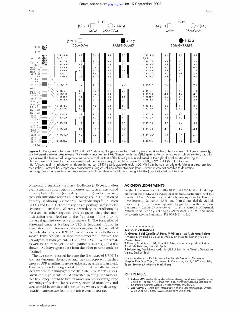
centromeric markers (primary isodisomy). Recombination
events can introduce regions of homozygosity in a situation of
primary heterodisomy (secondary isodisomy) and, conversely,
they can introduce regions of heterozygosity in a situation of
primary isodisomy (secondary heterodisomy).22 In both
E112-3 and E232-3, there are regions of primary isodisomy for
centromeric markers, whereas secondary heterodisomy is
observed in other regions. This suggests that the non-
disjunction event leading to the formation of the disomic
maternal gamete took place in meiosis II. The formation of
abnormal gametes leading to UPD is frequently found in
association with chromosomal rearrangements. In fact, all of
the published cases of UPD(13) were associated with Robert-
sonian translocations or isochromosomes.14–20 However, the
karyotypes of both patients E112-3 and E232-3 were normal,
as well as that of subject E232-1 (father of E232-3) (data not
shown). No karyotyping data from the other parents could be
obtained.
The two cases reported here are the first cases of UPD(13)
with an abnormal phenotype, and they also represent the first
cases of UPD resulting in non-syndromic hearing impairment.
They were found among a total of 115 unrelated affected sub-
jects who were homozygous for the 35delG mutation (1.7%).
Given the high incidence of inherited hearing impairment,
this frequency should be kept in mind when performing large
screenings of patients for recessively inherited mutations, and
UPD should be considered a possibility when anomalous seg-
regation patterns are found in routine genetic testing.
ACKNOWLEDGEMENTSWe thank the members of families E112 and E232 for their kind coop-eration in this study, and FIAPAS for their enthusiastic support of thisresearch. AA and MV were recipients of fellowships from the Fondo deInvestigaciones Sanitarias (BEFI) and from Comunidad de Madrid,respectively. This work was supported by grants from the EuropeanCommunity (QLG2-CT-1999-00988) (to FM), CAICYT of SpanishMinisterio de Ciencia y Tecnología (SAF99-0025) (to FM), and Fondode Investigaciones Sanitarias (FIS 00/0244) (to IdC).
. . . . . . . . . . . . . . . . . . . . .
Authors’ affiliationsA Álvarez, I del Castillo, A Pera, M Villamar, M A Moreno-Pelayo,F Moreno, Unidad de Genética Molecular, Hospital Ramón y Cajal,Madrid, SpainT Rivera, Servicio de ORL, Hospital Universitario Príncipe de Asturias,Alcalá de Henares, Madrid, SpainJ Solanellas, Servicio de ORL, Hospital Universitario Nuestra Señora deValme, Sevilla, Spain
Correspondence to: Dr F Moreno, Unidad de Genética Molecular,Hospital Ramón y Cajal, Carretera de Colmenar, Km 9, 28034 Madrid,Spain; [email protected]
REFERENCES1 Cohen MM, Gorlin RJ. Epidemiology, etiology, and genetic patterns. In:
Gorlin RJ, Toriello HV, Cohen MM, eds. Hereditary hearing loss and itssyndromes. Oxford: Oxford University Press, 1995:9-21.
2 Van Camp G, Smith RJH. Hereditary Hearing Loss Home page. WorldWide Web URL: http://www.uia.ac.be/dnalab/hhh
Figure 1 Pedigrees of families E112 and E232, showing the genotypes for a set of genetic markers from chromosome 13. Ages in years (y)are indicated between parentheses. The carrier status for the 35delG mutation in the GJB2 gene is shown below each subject symbol; wt, wildtype allele. The location of the genetic markers, as well as that of the GJB2 gene, is indicated to the right of a schematic drawing ofchromosome 13. Currently, the most centromeric sequence contig from chromosome 13 is NT_009917.11 (NCBI database,http://www.ncbi.nlm.nih.gov). In this contig, marker D13S1835 is approximately 1.55 Mb from the centromeric end. Alleles are representedby numbers. Vertical bars represent chromosomes. Regions of non-informativeness (that is, when it was not possible to determineunambiguously the parental chromosome from which an allele in a child was being inherited) are indicated by thin lines.
638 Letters
www.jmedgenet.com
on 15 September 2008 jmg.bmj.comDownloaded from
3 Rabionet R, Gasparini P, Estivill X. Molecular genetics of hearingimpairment due to mutations in gap junction genes encoding betaconnexins. Hum Mutat 2000;16:190-202.
4 Calvo J, Rabionet R, Gasparini P, Estivill X. Connexins and deafnessHome page. World Wide Web URL: http://www.crg.es/deafness
5 Lerer I, Sagi M, Ben-Neriah Z, Wang T, Levi H, Abeliovich D. A deletionmutation in GJB6 cooperating with a GJB2 mutation in trans innon-syndromic deafness: a novel founder mutation in Ashkenazi jews.Hum Mutat 2001;18:460.
6 del Castillo I, Villamar M, Moreno-Pelayo MA, del Castillo FJ, ÁlvarezA, Tellería D, Menéndez I, Moreno F. A deletion involving the connexin30 gene in nonsyndromic hearing impairment. N Engl J Med2002;346:243-9.
7 Pallares-Ruiz N, Blanchet P, Mondain M, Claustres M, Roux AF. Alarge deletion including most of GJB6 in recessive non syndromicdeafness: a digenic effect? Eur J Hum Genet 2002;10:72-6.
8 Dib C, Fauré S, Fizames C, Samson D, Drouot N, Vignal A, MillasseauP, Marc S, Hazan J, Seboun E, Lathrop M, Gyapay G, Morissette J,Weissenbach J. A comprehensive genetic map of the human genomebased on 5,264 microsatellites. Nature 1996;380:152-4.
9 Kibar Z, Lafrenière RG, Chakravarti A, Wang J, Chevrette M, DerKaloustian VM, Rouleau GA. A radiation hybrid map of 48 loci includingthe Clouston hidrotic ectodermal dysplasia locus in the pericentromericregion of chromosome 13q. Genomics 1999;56:127-30.
10 Engel E. A new genetic concept: uniparental disomy and its potentialeffect, isodisomy. Am J Med Genet 1980;6:137-43.
11 Robinson WP. Mechanisms leading to uniparental disomy and theirclinical consequences. BioEssays 2000;22:452-9.
12 Kotzot D. Complex and segmental uniparental disomy (UPD): review andlessons from rare chromosomal complements. J Med Genet2001;38:497-507.
13 Engel E. Uniparental disomies in unselected populations. Am J HumGenet 1998;63:962-6.
14 Slater H, Shaw JH, Dawson G, Bankier A, Forrest SM. Maternaluniparental disomy of chromosome 13 in a phenotypically normal child. JMed Genet 1994;31:644-6.
15 Slater H, Shaw JH, Bankier A, Forrest SM. UPD13: no indication ofmaternal or paternal imprinting of genes on chromosome 13. J MedGenet 1995;33:493.
16 Stallard R, Krueger S, James RS, Schwartz S. Uniparental isodisomy 13in a normal female due to transmission of a maternal t(13q13q). Am JMed Genet 1995;57:14-18.
17 Järvelä I, Savukoski M, Ämmälä P, Von Koskull H. Prenatally detectedpaternal uniparental chromosome 13 isodisomy. Prenat Diagn1998;18:1169-73.
18 Berend SA, Feldman GL, McCaskill C, Czarnecki P, Van Dyke DL,Shaffer LG. Investigation of two cases of paternal disomy 13 suggeststiming of isochromosome formation and mechanisms leading touniparental disomy. Am J Med Genet 1999;82:275-81.
19 Berend SA, Horvitz J, McCaskill C, Shaffer LG. Identification ofuniparental disomy following prenatal detection of Robertsoniantranslocations and isochromosomes. Am J Hum Genet2000;66:1787-93.
20 Soler A, Margarit E, Queralt R, Carrió A, Costa D, Gómez D, Ballesta F.Paternal isodisomy 13 in a normal newborn infant after trisomy rescueevidenced by prenatal diagnosis. Am J Med Genet 2000;90:291-3.
21 Kotzot D. Abnormal phenotypes in uniparental disomy (UPD):fundamental aspects and a critical review with bibliography of UPD otherthan 15. Am J Med Genet 1999;82:265-74.
22 Engel E. Uniparental disomy revisited: the first twelve years. Am J MedGenet 1993;46:670-4.
Letters 639
www.jmedgenet.com
on 15 September 2008 jmg.bmj.comDownloaded from
ECHO ................................................................................................................Genetic evidence of heterogeneity in intrahepatic cholestasis of pregnancyM Savander, A Ropponen, K Avela, N Weerasekera, B Cormand, M-L Hirvioja, S Riikonen,O Ylikorkala, A-E Lehesjoki, C Williamson and K Aittomäki
Background and aims: The aim of this study was to investigate the genetic aetiology of int-
rahepatic cholestasis of pregnancy (ICP) and the impact of known cholestasis genes (BSEP,
FIC1, and MDR3) on the development of this disease.
Patients and methods: Sixty nine Finnish ICP patients were prospectively interviewed for
a family history of ICP, and clinical features were compared in patients with familial ICP
(patients with a positive family history, n=11) and sporadic patients (patients with no known
family history of ICP, n=58). For molecular genetic analysis, 16 individuals from two
independently ascertained Finnish ICP families were genotyped for the flanking markers for
BSEP, FIC1, and MDR3.
Results: The pedigree structures in 16% (11/69) of patients suggested dominant inheritance.
Patients with familial ICP had higher serum aminotransferase levels and a higher recurrence
risk (92% v 40%). Both segregation of haplotypes and multipoint linkage analysis excluded
BSEP, FIC1, and MDR3 genes in the studied pedigrees. Additionally, the MDR3 gene, previously
shown to harbour mutations in ICP patients, was negative for mutations when sequenced in
four affected individuals from the two families.
Conclusions: These results support the hypothesis that the aetiology of ICP is heterogeneous
and that ICP is due to a genetic predisposition in a proportion of patients. The results of
molecular genetic analysis further suggest that the previously identified three cholestasis
genes are not likely to be implicated in these Finnish ICP families with dominant inheritance.
m Gut 2003;52:1025–1029
Please visit theJournal ofMedicalGenetics website[www.jmedgenet.com] for a link tothis full article.
640 Echo
www.jmedgenet.com
on 15 September 2008 jmg.bmj.comDownloaded from