Embed Size (px)
Citation preview

This journal is c The Royal Society of Chemistry 2013 Chem. Commun., 2013, 49, 2825--2827 2825
Cite this: Chem. Commun.,2013,49, 2825
Protein b-interfaces as a generic source of nativepeptide tectons†
Celine Valery,*a Rishi Pandeya and Juliet A. Gerrard*abcd
Motifs of 7–8 amino acids were designed from the b-continuous
interfaces of non-related homo-oligomeric proteins. These
peptides intrinsically self-assembled into nanoarchitectures in
water, while retaining some properties of their parent interfaces,
especially reversibility of assembly. These results reveal a novel
source of native peptide tectons.
Proteins commonly function as homo-oligomers, with many ofthese complexes able to dissociate and associate readilyin response to changing biological conditions.1 Such subtleequilibria rely on finely tuned protein–protein interfaces, whichprovide the necessary structural versatility through sets ofcomplementary non-covalent interactions.2 These proteinsubunits appear to be optimised by evolution to drive proteinself-assembly in a reversible and controlled manner.1,3 Wehypothesised that the corresponding isolated interfacesequences could serve as a source of native self-assemblingmotifs, or tectons, while retaining some of the parent interfaceproperties as they self-assemble.
Peptide tectons are defined as simple or minimal sequencesable to spontaneously self-associate into well-defined nanoscaleassemblies.4 For the past two decades, peptide chemistry hasfocused on exploring design principles and sources of suchmotifs, towards downstream applications in bionanotechnology.5
In the specific case of b-sheet-based motifs, tectons still remaindifficult to design outside a few discrete families of bioinspiredsequences, such as the cyclic D,L-peptides designed from thepore-forming gramicidin,6 the diphenylalanine motif inspired
from the high aromatic content in amyloid sequences,7 thefragments 16–22 from the Ab amyloid peptide,8,9 the lanreotidepeptide family designed as analogues of the self-assemblingpeptide hormone somatostatin-14,10,11 or the surfactant peptidesinspired from lipid structures.12 Novel generic sources of tectonswill therefore widen the current set of available sequences withwhich to explore specific applications. To design the peptides, wefocused on one of the simplest interface motifs, the b-continuousinterface, previously estimated to represent about 15% of thereported protein oligomeric interfaces.1 b-Continuous interfacesconsist of antiparallel non-covalent close contacts of two identicalb-strands from the two interacting protein units (Fig. 1).
From a survey of the entire protein data bank for the keyword ‘homodimer’ that resulted in >800 entries, we downsizedthe set of proteins to about 20 structures using the followingunbiased criteria: (i) selecting native homo-oligomers withb-continuous interfaces, while excluding structures withligands, to limit the complexity of the sets of interactionsinvolved; (ii) excluding fragmented and complex interfaces,the latter being defined as involving multiple different
Fig. 1 Protein homo-oligomers (pdb crystal structures for A–C, pdb solutionstructure for D, red: interface contacts according to the software PISA). (A)Peroxiredoxin III homo-dodecamer (1ZYE) (with zoom into the homodimerinterface); (B) b-lactoglobulin homodimer (2Q39); (C) diaminopimelate decar-boxylase homo-tetramer (2O0T) (with zoom into the tetramer interface); (D)umud’ homodimer (1I4V).
a Biomolecular Interaction Centre, University of Canterbury, Private Bag 4800,
Christchurch 8140, New Zealand. E-mail: [email protected],
[email protected]; Tel: +64 33642987b Riddet Institute, Massey University, Palmerston North, New Zealandc MacDiarmid Institute, University of Canterbury, New Zealandd Callaghan Innovation Research Limited, P.O. Box 31310, Lower Hutt 5040,
New Zealand
† Electronic supplementary information (ESI) available: Acknowledgements, fullexperimental details, PDB survey details, birefringence of peptide 3 samples, pHinfluence on peptide assemblies, TEM analysis of nanostructure reversibility. SeeDOI: 10.1039/c3cc39052g
Received 18th December 2012,Accepted 18th February 2013
DOI: 10.1039/c3cc39052g
www.rsc.org/chemcomm
ChemComm
COMMUNICATION
Ope
n A
cces
s A
rtic
le. P
ublis
hed
on 2
7 Fe
brua
ry 2
013.
Dow
nloa
ded
on 1
2/14
/202
1 9:
09:5
3 PM
. T
his
artic
le is
lice
nsed
und
er a
Cre
ativ
e C
omm
ons
Attr
ibut
ion
3.0
Unp
orte
d L
icen
ce.
View Article OnlineView Journal | View Issue
2826 Chem. Commun., 2013, 49, 2825--2827 This journal is c The Royal Society of Chemistry 2013
secondary structure motifs; (iii) selecting only one representativeinterface from families of proteins with high sequence homology,to ensure sequence variety and a valid test of the hypothesisamong different protein families. A statistical analysis performedon the interface sequences given by the PISA software for the finalset of structures revealed that 90% contained 11 or fewer residues,80% included at least one aromatic residue, all of the sequencescontained at least 30% of hydrophobic residues, and 15% ofcharged residues (ESI†). The finally selected capped sequences(both N-acetylated and C-amidated to conserve the net charge ofthe corresponding interfaces) were chosen as representative of theentire set of homodimers examined.
The selected peptides of 7–8 residues, respectively,correspond to the b-continuous interfaces of (i) the bovineperoxiredoxin III homodimer within the dodecameric ring,13
(ii) the bovine b-lactoglobulin homodimer,14,15 (iii) theMycobacterium tuberculosis diaminopimelate decarboxylasehomotetramer,16 and (iv) the E. coli umud’ protein homodimer17
(Fig. 1 and 2, Table 1).All the corresponding oligomers exist in solution.16,18–20 To
our knowledge, none of these peptides or similar sequenceshave been previously probed for assembly under any condition,
except for a b-lactoglobulin motif similar to peptide 2, reportedto assemble under denaturing conditions involving urea.21
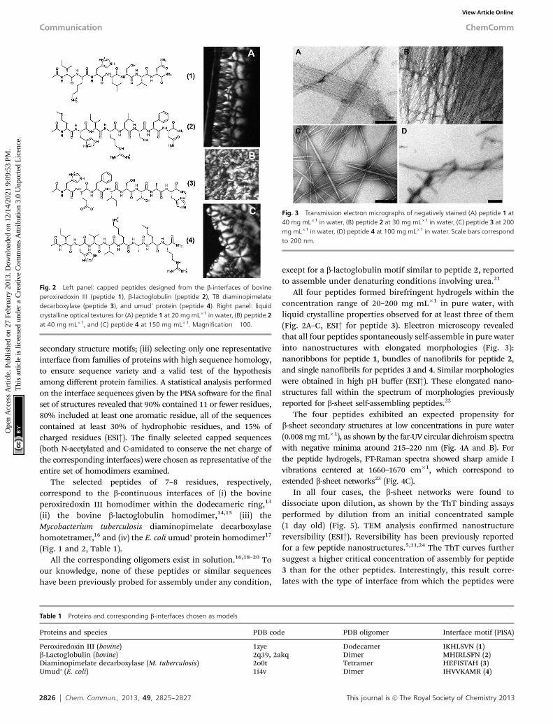
All four peptides formed birefringent hydrogels within theconcentration range of 20–200 mg mL�1 in pure water, withliquid crystalline properties observed for at least three of them(Fig. 2A–C, ESI† for peptide 3). Electron microscopy revealedthat all four peptides spontaneously self-assemble in pure waterinto nanostructures with elongated morphologies (Fig. 3):nanoribbons for peptide 1, bundles of nanofibrils for peptide 2,and single nanofibrils for peptides 3 and 4. Similar morphologieswere obtained in high pH buffer (ESI†). These elongated nano-structures fall within the spectrum of morphologies previouslyreported for b-sheet self-assembling peptides.22
The four peptides exhibited an expected propensity forb-sheet secondary structures at low concentrations in pure water(0.008 mg mL�1), as shown by the far-UV circular dichroism spectrawith negative minima around 215–220 nm (Fig. 4A and B). Forthe peptide hydrogels, FT-Raman spectra showed sharp amide Ivibrations centered at 1660–1670 cm�1, which correspond toextended b-sheet networks23 (Fig. 4C).
In all four cases, the b-sheet networks were found todissociate upon dilution, as shown by the ThT binding assaysperformed by dilution from an initial concentrated sample(1 day old) (Fig. 5). TEM analysis confirmed nanostructurereversibility (ESI†). Reversibility has been previously reportedfor a few peptide nanostructures.5,11,24 The ThT curves furthersuggest a higher critical concentration of assembly for peptide3 than for the other peptides. Interestingly, this result corre-lates with the type of interface from which the peptides were
Fig. 2 Left panel: capped peptides designed from the b-interfaces of bovineperoxiredoxin III (peptide 1), b-lactoglobulin (peptide 2), TB diaminopimelatedecarboxylase (peptide 3), and umud’ protein (peptide 4). Right panel: liquidcrystalline optical textures for (A) peptide 1 at 20 mg mL�1 in water, (B) peptide 2at 40 mg mL�1, and (C) peptide 4 at 150 mg mL�1. Magnification �100.
Table 1 Proteins and corresponding b-interfaces chosen as models
Proteins and species PDB code PDB oligomer Interface motif (PISA)
Peroxiredoxin III (bovine) 1zye Dodecamer IKHLSVN (1)b-Lactoglobulin (bovine) 2q39, 2akq Dimer MHIRLSFN (2)Diaminopimelate decarboxylase (M. tuberculosis) 2o0t Tetramer HEFISTAH (3)Umud’ (E. coli) 1i4v Dimer IHVVKAMR (4)
Fig. 3 Transmission electron micrographs of negatively stained (A) peptide 1 at40 mg mL�1 in water, (B) peptide 2 at 30 mg mL�1 in water, (C) peptide 3 at 200mg mL�1 in water, (D) peptide 4 at 100 mg mL�1 in water. Scale bars correspondto 200 nm.
Communication ChemComm
Ope
n A
cces
s A
rtic
le. P
ublis
hed
on 2
7 Fe
brua
ry 2
013.
Dow
nloa
ded
on 1
2/14
/202
1 9:
09:5
3 PM
. T
his
artic
le is
lice
nsed
und
er a
Cre
ativ
e C
omm
ons
Attr
ibut
ion
3.0
Unp
orte
d L
icen
ce.
View Article Online

This journal is c The Royal Society of Chemistry 2013 Chem. Commun., 2013, 49, 2825--2827 2827
designed. Peptides 1, 2 and 4 correspond to homo-dimericinterfaces, with Kd in the mM range for both the peroxiredoxin18
and b-lactoglobulin homodimers,19 but in the pm range for theumud’ homodimer.20 However, peptide 3 was designed from atetrameric interface, with a Kd dimer–tetramer still underdebate, although suggested in the mM range.16
The correlation between the peptide relative behaviour insolution and the type of oligomeric interface supports thatthese sequences are tectons with native intrinsic assemblyproperties derived from the parent interfaces. The resultspresented herein augur well for other peptide sequencesderived from protein interfaces providing a rich source ofuseful self-assembling tectons.
Notes and references1 I. M. A. Nooren and J. M. Thornton, J. Mol. Biol., 2003, 325, 991–1018.2 C. Chothia and J. Janin, Nature, 1975, 256, 705–708.3 S. Dey, A. Pal, P. Chakrabarti and J. Janin, J. Mol. Biol., 2010, 398,
146–160.4 E. H. C. Bromley, K. Channon, E. Moutevelis and D. N. Woolfson,
ACS Chem. Biol., 2008, 3, 38–50.5 C. Valery, F. Artzner and M. Paternostre, Soft Matter, 2011, 7,
9583–9594.6 M. R. Ghadiri, J. R. Granja, R. A. Milligan, D. E. McRee and
N. Khazanovich, Nature, 1993, 366, 324–327.7 M. Reches and E. Gazit, Science, 2003, 300, 625–627.8 K. Lu, J. Jacob, P. Thiyagarajan, V. P. Conticello and D. G. Lynn,
J. Am. Chem. Soc., 2003, 125, 6391–6393.9 I. W. Hamley, G. Cheng, V. Castelletto, S. Handschin and
R. Mezzenga, Chem. Commun., 2012, 48, 3757–3759.10 C. Valery, M. Paternostre, B. Robert, T. Gulik-Krzywicki,
T. Narayanan, J. C. Dedieu, G. Keller, M. L. Torres, R. Cherif-Cheikh,P. Calvo and F. Artzner, Proc. Natl. Acad. Sci. U. S. A., 2003, 100,10258–10262.
11 W. Van Grondelle, C. L. Iglesias, E. Coll, F. Artzner, M. Paternostre,F. Lacombe, M. Cardus, G. Martinez, M. Montes, R. Cherif-Cheikhand C. Valery, J. Struct. Biol., 2007, 160, 211–223.
12 S. Vauthey, S. Santoso, H. Y. Gong, N. Watson and S. G. Zhang, Proc.Natl. Acad. Sci. U. S. A., 2002, 99, 5355–5360.
13 Z. B. Cao, A. W. Roszak, L. J. Gourlay, J. G. Lindsay and N. W. Isaacs,Structure, 2005, 13, 1661–1664.
14 J. J. Adams, B. F. Anderson, G. E. Norris, L. K. Creamer andG. B. Jameson, J. Struct. Biol., 2006, 154, 246–254.
15 L. Vijayalakshmi, R. Krishna, R. Sankaranarayanan and M. Vijayan,Proteins: Struct., Funct., Bioinf., 2008, 71, 241–249.
16 S. Weyand, G. Kefala, D. Svergun and M. Weiss, J. Struct. Funct.Genomics, 2009, 10, 209–217.
17 A. E. Ferentz, G. C. Walker and G. Wagner, EMBO J., 2001, 20,4287–4298.
18 S. Barranco-Medina, J. J. Lazaro and K. J. Dietz, FEBS Lett., 2009, 583,1809–1816.
19 R. K. O. Apenten and D. Galani, Thermochim. Acta, 2000, 359,181–188.
20 S. M. Simon, F. J. R. Sousa, R. Mohana-Borges and G. C. Walkers,Proc. Natl. Acad. Sci. U. S. A., 2008, 105, 1152–1157.
21 D. Hamada, T. Tanaka, G. G. Tartaglia, A. Pawar, M. Vendruscolo,M. Kawamura, A. Tamura, N. Tanaka and C. M. Dobson, J. Mol. Biol.,2009, 386, 878–890.
22 A. Mitraki and E. Kasotakis, in Self-Assembled Peptide Nanostructures,ed. J. Castillo, L. Sasso and W. E. Svendsen, Pan Stanford Publish-ing, 2013.
23 S. Krimm and J. Bandekar, Adv. Protein. Chem., 1986, 38, 181–364.24 K. B. Andersen, J. Castillo-Leon, M. Hedstrom and W. E. Svendsen,
Nanoscale, 2011, 3, 994–998.
Fig. 4 Peptide conformation. CD spectra at 0.008 mg mL�1 in water of peptides1 and 2 (A), and peptides 3 and 4 (B). Panel C: FT-Raman amide I vibrations ofpeptides 1 and 2 at 100 mg mL�1 in water, peptides 3 and 4 at 200 mg mL�1.
Fig. 5 ThT binding assays upon dilution, for peptides 1 and 2 (A), and peptides3 and 4 (B). Fluorescence intensity values just after dilution (white scatters), and24 h after dilution (black scatters) of an initial sample equilibrated for 24 h(highest concentration).
ChemComm Communication
Ope
n A
cces
s A
rtic
le. P
ublis
hed
on 2
7 Fe
brua
ry 2
013.
Dow
nloa
ded
on 1
2/14
/202
1 9:
09:5
3 PM
. T
his
artic
le is
lice
nsed
und
er a
Cre
ativ
e C
omm
ons
Attr
ibut
ion
3.0
Unp
orte
d L
icen
ce.
View Article Online