Embed Size (px)
Citation preview

... rt \ . 401
The Effects of Hypertonic Saline (7.5%)/Dextran-70(HSD) on Human Red Cell Typing, Lysis, and Metabolism
in Vitro
Gerald L. Moore, James J. SummaryMichael A. Dubick, Mary E. Ledford
Barbara A. Ryan, Armando GonzalesCe Charles E. Wade
<C DIVISION OF MILITARY TRAUMAAN D
DIVISION OF BLOOD RESEARCH
<JC
I. 4w 4L
IUiEPP-~AN ARM Y INSTITUTE OF RESEARCH PRESID4) OF SAN FRANCISCO CALIFORNIA 94129

The Effects of iHypertonic Saline (7.5%)/Dextran-70 (HSD) on Human Red CellTyping, Lysis, and Metabolism in Vitro--GL Moore, JJ Summary, MA Dubick,ME Ledford, BA Ryan, A Gonzales, and CE Wade
Reproduction of this document in whole or in part is prohibited except with the 7crriza;3c.of the Commander, Letterman Army Institute of Research, Presidio of San Francisco,California 94129. Ilowever, the Defense Technical Information Center is authorized toreproduce the document for United States Government purposes.
)estrov this report khen it is no longer needed. Do not return to the originator.
Citation of trade names in this report does not constitute an official endorsement or approvalor the use of such items.
il-man Subjects participated in these studies after giving their free and informed voluntaryconsent. Investigators adhered to AR 70-25 and USAMRDC Reg 50-25 on the use ofvolunteers in research.
This material has been reviewed by Letterman Army Institute of Research andthere is no objection to its presentation and/or publication. The opinions orassertions contained herein are the private views of the author(s) and are not tobe construed as official or as reflecting the views of the Department of theArmy or the Department of Defense. (AR 360-5)
William C. Cole (d ate)COL, VCActing Commander
lhis document has been approved for public release and sale; its distribution is unlimited.

ERCURIT LASIICATION OF THIS PAGE
SForm Approved
REPORT DOCUMENTATION PAGE OMNo. 0704-0188
Ia. REPORT SECURITY CLASSIFICATION lb RESTRICTIVE MARKINGS
UNCLASSIFIED
2a. SECURITY CLASSIFICATION AUTHO, ITY 3. DISTRIBUTION /AVAILABILITY OF REPORT
Approved for public release, distribution2b. DECLASSIFICATION/DOWNGRADING SCHEDULE is unlimited.
4. PERFORMING ORGANIZATION REPORT NUMBER(S) 5. MONITORING ORGANIZATION REPORT NUMBER(S)
Institute Report No. 401
6a. NAME OF PERFORMING ORGANIZATION 6b. OFFICE SYMBOL 7a. NAME OF MONITORING ORGANIZATION
Div of Military Trauma and Div (If applicable)of Blooc_ Research ISGRD-ULT-M U.S._ArmmndI 6c. ADDRESS (City, Stare, and ZIP Code) 7b. ADDRESS (City, State, and ZIP Code)
Letterman Army Institute of Research Ft. DetrickPresidio of Sani Francisco, CA 94129-6800 Frederick, MD 21701-5012
8a NAME OF FUNDING/SPONSORING 8b. OFFICE SYMBOL 9. PROCUREMENT INSTRUMENT IDENTIFICATION NUMBERORGANIZATION (if applicable)
8c. ADDRESS (City, State, and ZIP Code) SOURCE OF FUNDING NUMBERSPROGRAM PROJECT TASK WORK UNITELEMENT NO. NO. NO. ACCESSION NO.
63807A 07D836 AX 1 087
11. TITLE (Include Security Classification)
(U) The Effects of Hypertonac Saline (7.5%)/Dextran-70 (HSD) on Human Red Cell Typing,Lysis, and Metabolism in vitro.
12. PERSONAL AUTHOR(S)
GL Moore, JJ SUmmary; MA Dubick, ME Ledford. " Rvan. A azales. and CP Wd13a. TYPE OF REPORT 13b. TIME COVERED 114. DATE OF REPORT (Year, Month, Day) 15. PAGE COUNTInstitute I FROM TO August 1989 12
16 SUPPLEMENTARY NOTATION
17. COSATI CODES 18. SUBJECT TERMS (:ontinue on reverse if necessary and identify by block number)FIELD GROUP SUB-GROUP (U) Hypertonic Saline. Dextran-70, Red Cell Typing,
Red Cell Lysis, Blood Storage (U)
19. ABSTRACT (Continue on reverse if necessary and identify by block number)
The introduction of a 7.5% hypertonic sal ine/6% Dextran-70 (HSD) solutioninto clinical trials for the treatment of hypcvolem;c states, and thepast concerns regarding possible interference of dextran with bloodserology, prompted us to investigate the effects of HSD on human red cel I
typing and stabi I ity. HSD was evaluated with fresh and 35-day storedCPDA-1 red cells from 12 healthy donors. A 1:5 mixture of HSD to bloodhad no effect on ABO, Rh, and MN typing in both fresh and stored blood.
HSD produced no significant lysis with fresh Cells and a minima levelwith stored blood. No evidence of metabolic or morphologic changes wereseen after HSD treatment. The results of this study suggest that clinicaluse of HSD for treatment of hemorrhagic snock wi I I not affect blood group
determinations or red cell stability from stored blood which may be
infused aiter %ne HSD treateo patient is transported to a hospital.
20 DISTRIBUTION/AVAILABILITY OF ABSTRACT 21 ABSTRACT SECURITY CLASSIFICATION
f3 IJNCLA-SIFi.O/NLIMITED C SAME AS RPT 0 DTIC USERS UNCLASSIFIED22a. NAME OF RESPONSIBLE INDIVIDUAL 22b TELEPHONE (Include Area Code) 22c. OFFICE SYMBOLWilliqm C (nlp CnT. C Ari-n C "rnripr (41" 5W1-5816 I qCRn-TT-MDD Form 1473, JUN 86 Previous editions are obsolete. SECURITY CLASSIFICATION OF THIS PAGE
UNCLASSIFIED
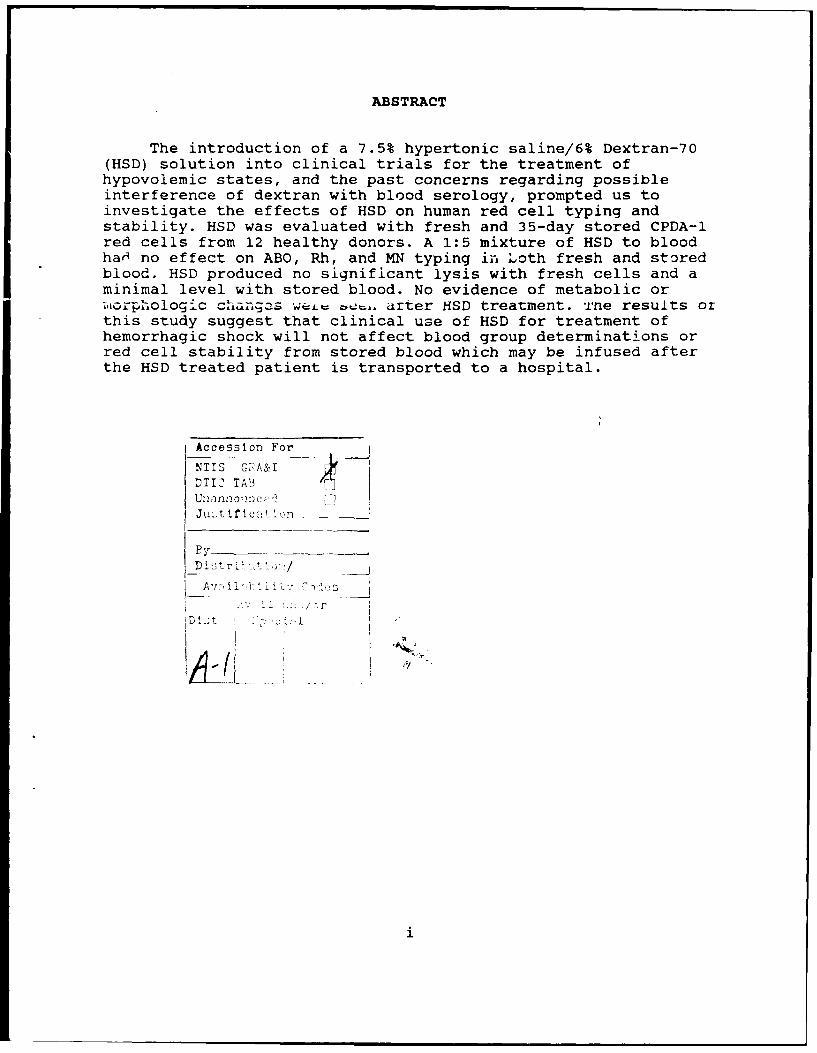
ABSTRACT
The introduction of a 7.5% hypertonic saline/6% Dextran-70(HSD) solution into clinical trials for the treatment ofhypovolemic states, and the past concerns regarding possibleinterference of dextran with blood serology, prompted us toinvestigate the effects of HSD on human red cell typing andstability. HSD was evaluated with fresh and 35-day stored CPDA-1red cells from 12 healthy donors. A 1:5 mixture of HSD to bloodhaH no effect on ABO, Rh, and MN typing in Loth fresh and storedblood. HSD produced no significant lysis with fresh cells and aminimal level with stored blood. No evidence of metabolic or,iaophologic chIangas viz= a drter HSD treatment. Tne results orthis study suggest that clinical use of HSD for treatment ofhemorrhagic shock will not affect blood group determinations orred cell stability from stored blood which may be infused afterthe HSD treated patient is transported to a hospital.
Accession For
NTIS GFA&IDTIC TABUn2annolrjcz-Ju:t if iIron -
By
D .i t r V : ,' /-,
A-i

The Effects of Hypertonic Saline(7.5%)/Dextran-70 (HSD) on HumanRed Cell Typing, Lysis, and Metabolism in vitro. -- Moore et al.
INTRODUCTION
In trauma scenarios initial treatment of hemorrhage andshock is frequently done with crystalloid solutions. Presentmedical doctrine calls for three volumes of saline or Ringer'ssolution per estimated volume of blood lost. More recently a 7.5%hypertonic saline / 6% Dextran-70 solution (HSD) has been showneffective in reducing volume requirements from 3 to 0.25 volumesfor treatment of hypoxia (1,2). Preliminary studies in humantrauma patients suggest that it may increase survival (3,4).
Previous experience with dextran has indicated that it mayinterfere with the ability to type and crossmatch red cells(5-7), presumably through i+s induction of rouleaux formation(7). Effects of dextran on typing or crossmatching are a directfunction of the dextran molecular weight and the quality of thestored red cells (6). Many Df the adverse effects are associatedwith dextran average molecular weights above 150,000 (9,10). Thiscould be a problem for the HSD resuscitated patient whosubsequently is transfused in the emergency room.
An additional problem has been the in vitro observation ofincreasing lysis when dog red cells were exposed to doses ofhypertonic saline solutions above 7.5% (8). While it is wellknown that dog red cells are more fragile than human, and readilylyse under slight stress, stored human blood is also well knownto increase its fragility during banked storage.
In this study we have examined the effects of HSD on bothfresh and 35 day stored human red cells to determine if HSDaltered the cell's metabolism, degree of lysis, or ability to betyped.
METHODS
Units of whole blood (450 ml each) were drawn withinformed consent from twelve healthy adult volunteers. The bloodwas collected into Fenwal CPDA-l bags according to AmericanAssociation of Blood Banks Standards (11). Volunteers were chosento insure that all ABO blood types were represented. Forty ml ofblood was removed for day zero analysis, and the remainder of theblood was stored at 3°C for 35 days, then re-analysed.
On day zero and day 35 the 40 ml aliquot of blood from eachdonor was divided into two 20 ml volumes in centrifuge tubes. To

2 -- Moore et al.
one tube was added 5 ml of isotonic saline containing 200 mg/dlof glucose (CTR). To the other paired tube was added 5 ml of theHSD test solution (HSD),which was prepared by Pharmacia AB,Uppsala, Sweden. Both tubes were capped, mixed, and incubated for30 min. in a 37°C water bath with remixing at 10 min. intervals.The tubes werc then centrifuged at 3400xg for 10 min. Thesupernatant was removed and saved for hemoglobin analysis. Thepacked cells were resuspended in 25 ml of isotonic saline plusglucose and centrifuged to wash out residual hypertonic salineand to return the cells to normal volume. These washed zells werethen suspended in an equal volume of isotonic saline andevaluated. The mixing ratio of 1:5 was chosen as a worse-casesituation. A typical 70 kg man has a blood volume of about 5000ml. If half of this volume were lost, then resuscitation would bedone with 250 ml of HSD, for a 1:10 ratio. Smaller people andlarger blood loss might reduce the ratio further, but 1:5 is themaximum ratio conceived for surviving patients.
During evaluation the cells were typed (11) and assayed forosmotic fragility (12), morphology (13), ATP (14), and 2,3-DPGconcentrations (14). The saline wash solution was saved forhemoglobin analysis (15). All bags were cultured at the end ofthe study to insure that they had remained sterile. Blood typingfor ABO, D(Rho) and MN antigens were performed using commercial(American Hospital Supply, Miami, FL) antisera and controls.Agglutination results were scored on the basis of noagglutination (N) to (+4). Statistical comparisons were madeusing the paired t-test in BMDP 3D. A 0.05 level of probabilitywas used.
RESULTS
Typical osmotic fragility curves for one donor are shown inFigure 1. HSD treatment of red cells causes a slight right shiftin the curve which is statistically but not clinicallysignificant. The osmotic fragility data was evaluated by linearregression of the linear, mid portion, of the curve (0.425 to0.525 percent saline) and testing for differences in the 50%lysis salt concentration, the slope of the regression, and thecorrelation coefficient (16). These data are summarized in Table1. Statistical differences were seen between CTR and HSD at bot ,time periods and between time periods with both CTR and HSD 50%lysis means. Similar statistical differences were seen betweenthe slopes of the regression lines, but no significantdifferences were seen among the correlation coefficients.
Red cell lysis was monitored by measuring both thesupernatant hemoglobin levels following 37°C incubation of thecells, and the isotonic saline wash solution subsequent to

Moore et al. -- 3
incubation. The data are summarized in Figure 2. There were nosignificant differences between the mean supernatant values atDay 0. Significant differences (control vs HSD) were seen betweenDay 0. washes, Day 35 supernatants, and Day 35 wash mens.
Figure 3 shows the mean values for red cell morphologyindex, ATP and 2,3-DPG. There were no significant differencesbetween CTR and HSD groups. The control results were typical ofprevious storage studies in CPDA-l (17).
No pseudoagglutination or rouleaux formation occurred in theblood of the 12 donors typed in this study, resulting in no ABO,D, or MN discrepancies in the fresh or stored red cells followingHSD Lceatment. The strength of the agglutinations (Table 2) wereidentical, except for one case.
DISCUSSION
In over 40 years of clinical use, primarily as a volumeexpander in the management of shock, dextran was occasionallyobserved to effect the behavior of red blood cells (7,9,18).Depending on the average molecular weight of the preparation,dextrans could decrease as well as increase red cell aggregation,even to the extent of promoting rouleaux formation (9,18,19).such concerns about possible pseudoagglutination induced bydextran prompted a number of investigators to evaluate theeffects of dextran on typing and crossmatching of blood.Following an extensive review of the literature, Gruber (9)reported that interference with red cell serology was observedonly with large molecular weight (.150,000 dalton) dextrans andnot with dextrans of 40-70,000 daltons. The present study, whichuses Dextran of 70,000 daltons in the HSD, shows no effect on redcell typing and thus confirms the earlier efforts. In addition wehave previously observed that infusion of HSD at therapeuticdoses to euvolemic or hemorrhaged rabbits and pigs (4) does notinduce significant rouleaux formation (20), suggesting that HSDinfusion would not effect red cell typing or crossmatcning. Thehypertonic saline component of HSD also does not effect typingwhich is consistent with the observation that red cell typing orcrossmatching is not affected by intravenous infusion ofelectrolyte solutions (21). This is also shown in thedeglycerolization of frozen-thawed red cells which are treatedwith 12% saline prior to washing with isotonic saline (22). Thefinal washed cells show no loss in ability to be typed orcrossmatched.
Red cells are good osmometers with great resistance toosmotic lysis. Treatment of thawed qlycerolized cells with 12%saline lyses only a small fraction of 1% of the cells (22). This

4 -- Moore et al.
resistance to lysis was also shown by Rocha and Silva for dog redcells which did not hemolyse until saline concentrations exceeded15% (8). The Day-0 arm of our study confirmed this resistance tolysis with freqh CPD banked blocd. These cells did shrink in thepresence of HSD and were somewhat difficult to resuspend inisotonic saline, but did not show evidence of rouleaux formation.When resuspended in isotonic saline the cells returned to normalsuspension behavior and had normal size and morphology. HSDtreatment did not alter the levels of ATP or 2,3-DPG in thecells, implying that this treatment did not affect red cellmetabolism by causing egress of metabolites, salt inactivation ofglycolytic enzymes, or general leakyness of the membranes. Storedred cells are more -rcne to lysis than fresh cells, as observedin our study between fresh and 35 day controls. HSD did cause asignificant increase in lysis of the 35 day stored red cells whencompared to controls. However, this lysis, which could equal 2 or3 percent of the cells, may not represent an increased decrementof therapeutic red cells because the 35 day cells would only havea 24 hour survivability of about 75 %. Thus the cells lysed byHSD may be the oldest cells which would lyse or be cleared anywayupon infusion. It is interesting that all of the HSD-inducedlysis we saw occurred in the wash, not when HSD was added to thecells. This suggests that lytic damage is a function of excesssalt leaving the red cells, but not entering them.
The MN antiaen system was measured because it is attached tothe membrane protein glycophorin A. This protein is involved withprotein 4.1 and band 3 protein in a mechanism, not yetunderstood, which seems to help bind together the majorstructural proteins in the membrane skeleton (23). Change inability to measure MN, which was not seen in our study, may beassociated with loss of membrane integrity.
The effects of HSD on fresh or stored banked red cells seemsto be limited to a slight amount of lysis after exposure to HSDand during return to isotonicity. This effect becomes somewhatmore pronounced as the blood ages and becomes more fragile, butis not of sufficient magnitude cause concern. No changes inability to measure red cell antigens was observed. Therefore, atthe doses used to treat hemorrhagic shock, it does not appearthat HSD poses a significant clinical problem with respect tocell typing or the stability of banked blood.
4

Moore et al. -- 5
REFERENCES
1. Kramer GC, English TP, Gunther RA, Holcroft JW.Physiological mechanisms of fluid resuscitation withhyperosmotic/hyperoncotic solutions. Perspectives in ShockResearch, Alan Liss, New York, 1989:311-320.
2. Kramer GC, Perron PR, Lindsey DC, Ho HS, Gunther RA, BoyleWA, Holcroft JW. Small-volume resuscitation with hypertonicsaline dextran solution. Surgery 1986;100:239-46.
3. Holcroft JW, Vassar MJ, Turner JE, Derlet RW, Kramer GC. 3%NaCI and 7.5% NaCl/Dextran 70 in the resuscitation ofseverely injured patients. Ann Surg 1987;206:279-88.
4. Holcroft JW, Vassar MJ, Perry CA, Ganaway WL, Kramer GC.Perspectives on clinical studies for hypertonic,saline/dextran solutions for the treatment of t7 aumaticshock. Brazilian J Med Biol Res 1989;22:291-3.
5. Roche JP, Dodelin RA, Bloom WL. Effect of dextran on bloodtyping and crossmatching. Blood 1952;7:373-5.
6. Marston NA. Crossmatching of blood in the presence ofdextran. Lancet 1954;2:688.
7. Wallace J. Crossmatching of Dlood in the presence ofdextran. Lancet 1954;2:761.
8. Rocha E, Silva R, Velasco IT, Porfirio MF. Hypertonicsaline resuscitation: saturated salt solutions are alsoeftective but induce heiaolysis in dogs. Circ. Shock1988;24:246.
9. Gruber UF. Blood replacement. Berlin, Springer-Verlag,1969.
10. Bartholomew JR, Bell WR, Kickter T, Williams GM. Aprospective study of the effect of dextran administration oncompatibility testing. Transfusion 1986;26:431-3.
11. American Association of Blood Banks Technical Manual, 9thEd, AABB Press, Arlington, VA, 1985.
12. Parpart, AK et al. The osmotic resistance of human redcells. J Clin Invest 1947;26:636-40.
13. Hogman CF, deVerdier C, Ericson A, Hedlund K. Studies on themechanism of human red cell loss of viability ri'i-ing storageat 4°C in vitro. Vox Sang 1985;48:257-68.
5

6 -- Moore et al.
14. Moore GL, Ledford ME, Unruh KA, Brummell MR. Red cellstorage in modified CPD-adenine. Transfusion1981;21:699-701.
15. Moore GL, Ledford ME, Merydith A. A micromodification ofthe Drabkin hemoglobin assay for measuring plasma hemoglobinin the range of 5 to 2000 mg/dl. Biochem Med1981;26:167-73.
16. Moore GL, Ledford ME, Gonzales A. Analysis of osmoticfragility curve data. MS in preparation.
17. Moore GL, Peck CC, Sohmer PR, Zuck TF. Properties of bloodstored in anticoagulant CPDA-l solution. Transfusion1981;21:135-8.
18. Hint H. Relationships between chemical and physicochemicalproperties of dextran and its pharmacological effects. IN:Derrick JR, Guest MM Eds. Dextrans, Current Concepts ofBasic Actions and Clinical Applications. Springfield,Charles C. Thomas, 1971;3-26.
19. Singh BA, Joseph KP. Erythrocyte sedimentation profilesunder gravitational field as determined by He-Ne laser. VIIInfluence of dextrans, albumin, and saline on cellularaggregation and sedimentation rates. Biorheology1987;24:53-61.
20. Ryan bA, Summary JJ, Dubick MA, Bowman PD, Wilson L, WadeCE. The hemostatic and hematogolic effects of hypertonicsaline (7.5%) Dextran-70 (HSD) in swine and rabbits. FASEBJ 1989;3:A1210.
21. DeViltorio AA. Plasma volume expanders and blood typing. JAm Med Assoc 1954;154:1398.
22. Valeri CR. Blood Banking and The Use of Frozen BloodProducts. Boca Raton: CRC Press, 1976.
23. Bennett V. The spectrin-actin junction of erythrocytemembrane skeletons. Biochem Biophys Acta 1989;988:107-21.

Moore et al. -- 7
120--.... •OTR-DO
100 "------- 0 HSD-DO• •........ A CTR-D35
) * * HSD-D35U80 .o 'o ;,\
'60
zLU ~'
a--40
20 O
0.0 0.2 0.4 0.6 0.8 1.0PERCENT NaCI
Figure 1: Typical osmotic fragility curve shown for one donor in
the study. Statistical data calculated on the linear drop portion
of the curve between salt % of 0.425 and 0.5, or 0.45 and 0.60.

8 -- Moore et al.
500 r-SUPER CTR
/ 400 - WASH CTR00 SUPER HSD
E WASH HSD0 300-00)
w200rULUlul_ 100
0 .. . ..
DAY DAY
Figure 2: Supernatant hemoglobin from lysed red cells. A
concentration of about 150 mg/dl would be equivalent to 1% lysis
of the red cells. Error bars are SD.

Moore et al. -- 9
120
100
80 <
L) 60 .LU
40 /
20 ,
0ATP 2,3-DPG MORPH-INDEX
CTR-DAY 0 HSD-DAY 0 E CTR-DAY 35 0 HSD-DAY 35
Figure 3: Red cell Morphology Index expressed as percent (100%is all biconcave disks) and red cell ATP and 2,3-DPG expressed aspercent ,if initial values. Day 35 2,3-DPG values are near zero.Error bars are SEM.

LO 0 LO 0
>1 +1 +1 +1(o N 'D 0
0
W, (N w (N
>1 +1 +1 +1(n
>4 IO
H 0
H- 0 H '.
H + to +1 +1
E- IL- H
zr 0- 'O
00
0
4(N LA en
It +1 +1 +1C 0 H- (N
r- H o LA
4E-4
H IA
1-4 1-icw tp v
00 4) a)H LA

Moore et al. -- 11
Table 2
The Effect of HSD1 and/or Storage on Red Blood Cell Typingfor ABO Groups and M/N Antigens*
Anti- Anti- Anti- Anti- Anti- Anti-Specimen # Type A B AB M N D
100 AB+ 4+ 4+ 4+ 3+ 3+ 3+102 B+ N 4+ 4+ 3+ 3+ 3+103 AB+ 2+(3+) 4+ 4+ 3+ 3+ 3+104 B+ N 4+ 4+ 3+ N 3+105 A+ 4+ N 4+ 3+ 3+ 3+106 0+ N N N 3+ N 3+107 A+ 4+ N 4+ 3+ N 3+108 0+ N N N 3+ 3+ 3+109 0+ N N N 3+ N 3+110 A+ 4+ N 4+ 3+ 3+ 3+ill B+ N 4+ 4+ 3+ 3+ 3+112 0+ N N N 3+ 3+ 3+
N No agglutination, NegativeHSD 7.5% Hypertonic Saline in 6% Dextran-70
* Identical results were obtained following 30 min incubationof normal saline or HSD with fresh blood at a ratio of 1:5(v/v). Further, repeating the incubations with whole bloodstored for 35days produced identical results as with freshblood.
Specimen # 103 (3+): Strength of agglutination increased after35 days in both the control and HSD incubation.
For the above specimens, all Rh and saline controls werenegative.

Moore etaL- 12
OFFICIAL DISTRIBUTION LISTCommanderUS Army Medical Research Commander
& Development Command US Army Medical BioengineeringATTN: SGRD-RMS/Mrs. Madigan Research & Development LaboratoryFort Detrick, MD 21701-5012 ATTN: Library
Fort Detrick, Bldg 568Defense Technical Information Center Frederick, MD 21701-5010A-1TN: DTIC/DDAB (2 copies)Cameron Station CommanderAlexandria, VA 22304-6145 US Army Research Institute
of Environmental MedicineOffice of Under Secretary of Defense ATTN: SGRD-UE-RSA
Research and Engineering Kansas StreetATTN: R&AT (E&LS), Room 3D129 Natick. MA 01760-5007The PentagonWashington, DC 20301-3080 Commander
US Army Research Institute ofDASG-AAFJML Surgical ResearchArmy/Air Force Joint Medical Library Fort Sam Houston, TX 78234-6200Offices of the Surgeons General5109 Leesburg Pike, Room 670 CommanderFalls Church, VA 22041-3258 US Army Research Institute of
Chemical DefenseHQ DA (DASG-ZXA) ATTN: SGRD-UV-AJWASH DC 20310-2300 Aberdeen Proving Ground, MD 21010-5425
Commandant CommanderAcademy of Health Sciences US Army Aeromedical ResearchUS Army LaboratoryATTN: HSHA-CDM Fort Rucker, AL 36362-5000Fort Sam Houston, TX 78234-6100
AIR FORCE Office of ScientificUniformed Services University of Research (NL)
Health Sciences Building 410. Room A217Office of Grants Management Boiling Air Force Base, DC 20332-64484301 Jones Bridge RoadBethesda, MD 20814-4799 USAF School of Aerospace Medicine
Document SectionUS Army Research Office USAFSAM/TSKDATTN: Chemical and Biological Brooks Air Force Base, TX 78235-5301
Sciences DivisionPO Box 12211 Head, Biological Sciences DivisionResearch Triangle Park, NC 27709-2211 OFFICE OF NAVAL RESEARCH
800 North Quincy StreetDirector Arlington, VA 22217-5000ATTN: SGRD-UWZ-LWalter Reed Army Institute of Research CommanderWashington, DC. 20307-5100 Naval Medical Command-02
Department of the NavyCommander
US Army Medical Research Institute Washington, DC 20372-5120
of Infectious DiseasesATITN: SGRD-ULZ-AFort Detrick. MD 21701-5011
CommanderUS Army Medical Bioengineering Researchand Development LaboratoryATTN: SGRD-UBG-MFort Detrick, Bldg 568Frederick, MD 21701-5010
4/89


![SHOCK[1] - Hypovolemic Shock](https://img.pdfslide.net/doc/110x75/58edc1bc1a28abae538b4711/shock1-hypovolemic-shock.jpg)