Embed Size (px)
Citation preview
Prophylactic removal of impacted third molars MTA
Page 1 of 140
This report was commissioned by the NIHR HTA Programme as
project number 15/69/16
Completed 15/02/2017
Prophylactic removal of impacted third molars
MT
A R
EP
OR
T
Prophylactic removal of impacted third molars MTA
Page 2 of 140
Title: Prophylactic removal of impacted third molars
Produced by: Liverpool Reviews and Implementation Group (LRiG), University of Liverpool
Authors: Hounsome, J, Pilkington, G, Mahon, J, Boland, A, Beale, S, Kotas, E, Dickson, R.
Correspondence to: Juliet Hounsome LRiG University of Liverpool Whelan Building The Quadrangle Brownlow Hill Liverpool L69 3GB [email protected] 0151 795 5441
Date completed: 15/02/2017
Source of funding: This report was commissioned by the NIHR HTA Programme as project number 15/69/16. Declared competing interests of the authors: None Acknowledgements: The authors would like to thank Tara Renton, Paul Coulthard, Rebecca Harris and Margaret Raison for clinical input and Joanne Fisher for assistance with data extraction. Rider on responsibility for report: The views expressed in this report are those of the authors and not necessarily those of the NIHR HTA Programme. Any errors are the responsibility of the authors. This report should be referenced as follows: Hounsome J, Pilkington G, Mahon J, Boland A, Beale S, Kotas, E, Dickson R, et al. Prophylactic removal of impacted third molars Health Technology Assessment. 2017; In press. Contributions of authors:
Juliet Hounsome Protocol development, clinical reviewer and writing of the report
Gerlinde Pilkington Protocol development, clinical reviewer and writing of the report
James Mahon Development of de novo economic model
Angela Boland Economic reviewer, checked economic model
Sophie Beale Economic reviewer, checked economic model
Eleanor Kotas Conducted clinical and economic searches
Rumona Dickson Screening of studies, protocol development and finalising of report
Data sharing statement All available data can be obtained by contacting the corresponding author.
Prophylactic removal of impacted third molars MTA
Page 3 of 140
SCIENTIFIC SUMMARY
Background
The four hindmost molars, known as third molars (3Ms) are the last teeth to erupt in the upper
(maxillary) and lower (mandibular) jaws, and this usually happens during young adulthood
between the ages of 18 and 24. Third molars can be either impacted or non-impacted, and an
impacted 3M (I3M) can be classed as erupted, partially erupted or unerupted. Impaction
occurs when the eruption of the tooth is blocked either by soft tissue (gum) or bone. Impacted
third molars can be potentially problematic to the individual by causing pain and disease;
however, many I3Ms are asymptomatic (trouble-free) and/or disease-free/pathology-free.
Impacted third molars may be associated with pathological changes such as infection
(pericoronitis), periodontal (gum) disease, dental caries, destruction of adjacent teeth, cysts
and tumours.
Treatment options for people with I3Ms include either surgical removal or standard care
without prophylactic removal of third molars.
The decision to remove or retain an I3M depends on whether it is asymptomatic and/or
pathology-free. Where there are pathological changes, current National Institute for Health
and Care Excellence (NICE) guidance states that the I3M should be removed. Even if an I3M
is pathology-free, the dentist may decide to remove the tooth to prevent future risk of
pathological changes; this is termed prophylactic removal.
Objectives
The remit of this review is to appraise the clinical and cost effectiveness of the prophylactic
removal of impacted mandibular 3Ms (IM3Ms) compared with standard care without
prophylactic removal.
Methods
Clinical effectiveness review
Five electronic databases were searched for: clinical trials (randomised and non-randomised),
observational studies, systematic reviews (SRs), decision analyses and UK costs. Studies
comparing the prophylactic removal of IM3Ms with standard care without prophylactic removal
or studies assessing the outcomes of either approach were considered. The outcomes of
interest were: pathology associated with retention of third molars, post-operative
complications following extraction, adverse effects of treatment and health-related quality of
life. Two reviewers independently screened all titles and/or abstracts including economic
Prophylactic removal of impacted third molars MTA
Page 4 of 140
evaluations, applied inclusion criteria to relevant publications and quality assessed the
included studies. The results of the data extraction and (clinical) quality assessment are
summarised in structured tables and as a narrative description. No meta-analysis or network
meta-analyses were undertaken.
Cost-effectiveness review
The search strategy developed for the clinical searches, with the addition of an economics
filter, was used to identify studies reporting the costs and benefits associated with
extracting/retaining I3Ms. As part of the search strategy, the NHS Economics Evaluation
Database (NHS EED), located within the Cochrane Library, and EconLit (EBSCO) were also
interrogated. Two reviewers independently screened all titles and/or abstracts, and applied
inclusion criteria to identify relevant studies.
Economic model
Due to the absence of cost-utility analyses relevant to the decision problem and generalisable
to the NHS in England, the AG constructed a de novo economic model. Two pathways are
considered, the intervention (prophylactic removal of IM3Ms) and the comparator, current
standard of care (watchful waiting). The pathways were modelled as a combination of Markov
processes and decision trees. The model perspective was that of the UK NHS, the time
horizon was a life-time (80 years), outcomes were measured in quality adjusted life years
(QALYs), and both costs and QALYs were discounted at an annual rate of 3.5%. A wide range
of one-way sensitivity analyses were carried out to test parameter uncertainty and scenario
analyses were carried out to test structural assumptions.
Results
Clinical effectiveness
Thirteen studies from 22 publications were included in the SR (four cohort studies and nine
SRs).
Cohort studies
The four cohort studies included one observational cohort investigating the prophylactic
removal of pathology-free or asymptomatic IM3Ms in comparison with the standard care and
retention of these pathology-free or asymptomatic IM3Ms. Annual assessment over 5 years
identified patients as: requiring and subsequently having an IM3M removed, requiring and
refusing extraction of an IM3M, and not requiring removal of the IM3M.
No serious surgical complications were reported in the 52 participants who had an IM3M
removed. Of those requiring removal but refusing, 5/7 required extraction within the follow-up
Prophylactic removal of impacted third molars MTA
Page 5 of 140
period. Finally, of those not requiring removal, 0/25 required extraction within the follow-up
period
Two single cohort studies investigated the standard care without removal of pathology or
asymptomatic IM3Ms. For one study assessments were conducted by telephone every
6 months for 5 years, and for the other a clinician questioned and assessed clinical outcomes
at 1 year. The difference in length of follow-up periods explains the differences in the rates of
extraction reported by each paper: 5.5% and 31.4%. The reasons for extraction also varied
between the studies. One study reported that at 1 year, 46% of participants did not know the
reason for the removal of the IM3M. Of those who did know, 50% were removed for pain and
20% for symptoms of pericoronitis. The other study reported that at 5 years, pericoronitis was
the most frequent reason for removal (62.5%) followed by cosmetic/orthodontic reasons
(12.5%).
One single prospective cohort study investigated the prophylactic removal of pathology or
asymptomatic IM3Ms. Assessment of periodontal health was conducted prior to and 6 months
after removal, and post-surgical complications were reported. There was no statistically
significant change in plaque index and gingiva index but there was statistically significant
reduction in mean probing pocket depth and probing attachment level. A total of 20 post-
surgical complications were reported, most frequently intense pain for more than one day
(12/78), post-operative infection (5/78), and wound dehiscence (3/78). No instances of
secondary bleeding or nerve damage were reported.
Systematic reviews
Nine SRs of the management of 3Ms were included in this review, though none were limited
to IM3Ms. The inclusion criteria for the SRs differed, resulting in a wide range of included
primary studies. Despite the differences in SRs, the conclusions were similar, with seven of
the nine stating that there was insufficient evidence on which to base a decision. One SR that
looked at the risk of future extraction following the retention of trouble-free 3Ms found that the
mean incidence rate of future extraction was 3.0% annually (range 1-9%), with a cumulative
incidence rate of 5% at 1 year and 64% at 18 years
Cost-effectiveness
Three studies were identified that provide economic evidence on the cost-effective
prophylactic removal of I3Ms. Two of the papers report details about the cost effectiveness of
the prophylactic removal of I3Ms. One is a cost-effectiveness study from a UK NHS
perspective, whilst one is of less direct relevance as estimates are based on the Australian
health care system and results are presented in Australian dollars. The third paper reports
findings relating to an assessment of oral HRQoL after the removal of I3Ms.
Prophylactic removal of impacted third molars MTA
Page 6 of 140
Economic model
For the comparison of a prophylactic removal strategy with watchful waiting, model results
show that the incremental cost per person associated with prophylactic extraction is £55.71
and the incremental QALY gain is 0.005 per person. Combining the cost and QALY results
generated by the model suggests an incremental cost effectiveness ratio (ICER) for the
comparison of a prophylactic removal strategy versus a watchful waiting strategy of £11,741
per QALY gained for people aged 20 years with asymptomatic IM3Ms. The base case ICER
per QALY gained was found to be robust when a range of one-way sensitivity analyses were
carried out to test parameter uncertainty and when scenario analyses were carried out to test
structural assumptions.
Discussion
Despite extensive searching of the literature the SR of clinical evidence found no RCT data to
support or refute the prophylactic removal of pathology-free/trouble-free IM3Ms. The review
however did identify evidence from two longitudinal studies demonstrating the outcomes when
asymptomatic IM3Ms are left in situ. No studies reported the impact of retention on the status
of the second molars although this may have been due to the narrow inclusion criteria which
included “People with pathology-free or trouble-free impacted mandibular third molars”. This
criteria severely limited the number of studies that met the inclusion criteria of this review.
As there is very limited clinical effectiveness evidence comparing the prophylactic removal of
pathology-free IM3M versus a watchful waiting strategy, it is unsurprising that economic
evidence relating to this comparison is also limited. The two published cost-effectiveness
studies that directly consider this comparison conclude that there is currently no economic
evidence to support the prophylactic removal of I3Ms. This is in contrast to the results
generated by the AG economic model which suggest that prophylactic removal may be the
more cost-effective strategy.
The strengths of the AG’s economic model include its simplicity and the minimal use of
assumptions. It is constructed around two key parameters – the annual rates of symptom
development and the extraction of pathology-free/trouble-free IM3Ms. Unfortunately, the
economic model is limited by the lack of utility evidence around IM3M symptoms. However,
suitable proxies were found and the cost-effectiveness findings are robust across a range of
values that could be used.
Prophylactic removal of impacted third molars MTA
Page 7 of 140
Conclusions
Clinical effectiveness conclusions
The findings from this review are consistent with previous systematic reviews in that there is
no available RCT evidence to support or refute the practice of the prophylactic removal of
asymptomatic/pathology-free IM3Ms. However, the review did identify evidence from
longitudinal studies demonstrating what happens when asymptomatic IM3Ms are left in situ.
Cost effectiveness conclusions
Only two published cost-effectiveness studies that directly consider the study question were
identified. In both cases, the authors conclude that there is currently no economic evidence to
support the prophylactic removal of I3Ms.
The base case results generated by the AG economic model indicate that the ICER per QALY
gained for the comparison of the cost effectiveness of a prophylactic removal strategy versus
a watchful waiting strategy is markedly less than the £20,000 per QALY gained threshold
widely accepted by NICE Appraisal Committees.
Implications for service provision
The reintroduction of the prophylactic removal of pathology-free/trouble-free IM3Ms will have
resource implications both in primary and secondary care settings, with the rate of pathology-
free I3M3 extractions increasing.
The results generated by the economic model show that most people with IM3Ms will have
their impacted teeth removed at some point and that, while prophylactic removal is probably
more costly than a watchful waiting strategy, the improvements in HRQoL for people from a
reduction in IM3M symptoms mean that prophylactic removal is a cost effective strategy for
the NHS.
Suggested research priorities
There remains a lack of head-to-head trial evidence comparing a prophylactic removal
strategy with a watchful waiting strategy. The practical difficulties (including, time, cost, and
the need for extended follow-up) associated with undertaking such studies means that it is
unlikely that this type of study will be conducted.
Future longitudinal studies on the pathology of retained IM3Ms could be designed to record
the impaction status and health of the retained IM3M with results being presented separately
for maxillary and mandibular teeth.
Prophylactic removal of impacted third molars MTA
Page 8 of 140
PLAIN ENGLISH SUMMARY
Third molars, commonly known as wisdom teeth may come through the gum (erupt) without
any problems, usually during young adulthood (age 18-24). However, in some cases they are
unable to erupt because they are poorly aligned, or obstructed by other teeth, gums or bone.
They are then referred to as ‘impacted’. Historically, dentists often recommended that these
teeth be removed so as not to cause problems later in life. This is referred to as ‘prophylactic’
removal. In 2000 the National Institute for Health and Care Excellence (NICE) reviewed this
practice and recommended that these teeth not be removed if they were not bothersome to
the person. Many dentists and oral surgeons have disagreed with this decision, believing that
it is more difficult to remove these teeth and that there are more complications for the patient
if they are removed later in life.
Our review group carried out a systematic review of the available clinical and cost-
effectiveness evidence of the prophylactic removal of impacted third molars.
The review identified four clinical studies. None of which provided strong evidence for or
against the prophylactic removal of these teeth. These findings are similar to nine previous
reviews. There is also very little research reported relating to the cost-effectiveness of the
procedure, with only three studies identified.
We built an economic model to assess the cost-effectiveness. Results from the model suggest
that a prophylactic removal strategy costs more than a watchful waiting strategy but leads to
improvements in quality of life. When the costs and quality of life measures associated with
the two strategies are compared, the resulting statistic is £11,741 per quality adjusted life year
gained. This means that NICE may consider that a prophylactic removal strategy can deliver
value for money to the NHS.
Prophylactic removal of impacted third molars MTA
Page 9 of 140
TABLE OF CONTENTS Abbreviations and glossary ................................................................................................................... 11 Abbreviations list .................................................................................................................................. 11 Glossary ................................................................................................................................................ 11 1 BACKGROUND .......................................................................................................................... 12
1.1 Description of health problem ............................................................................................... 12
1.2 Current service provision ...................................................................................................... 17
1.3 Description of technology under assessment ........................................................................ 20
2 DEFINITION OF THE DECISION PROBLEM ......................................................................... 21 2.1 Decision problem .................................................................................................................. 21
2.2 Overall aims of assessment ................................................................................................... 21
3 ASSESSMENT OF CLINICAL EFFECTIVENESS ................................................................... 22 3.1 Methods for reviewing effectiveness .................................................................................... 22
3.2 Results ................................................................................................................................... 24
3.3 Summary of clinical results................................................................................................... 38
3.4 Discussion of clinical effectiveness results ........................................................................... 39
4 ASSESSMENT OF COST-EFFECTIVENESS ........................................................................... 41 4.1 Systematic review of existing cost-effectiveness evidence................................................... 41
4.2 Independent economic assessment ........................................................................................ 48
4.3 Discussion of cost-effectiveness results ................................................................................ 68
5 DISCUSSION .............................................................................................................................. 71 5.1 Statement of principle findings ............................................................................................. 71
5.2 Strengths and limitations of the assessment .......................................................................... 72
5.3 Uncertainties ......................................................................................................................... 72
6 CONCLUSIONS .......................................................................................................................... 73 6.1 Implications for service provision ......................................................................................... 73
6.2 Suggested research priorities ................................................................................................ 73
7 REFERENCES ............................................................................................................................. 74 8 APPENDICES .............................................................................................................................. 87
Appendix 1 International guidelines ................................................................................................. 87
Appendix 2 Literature search strategies ............................................................................................ 92
Appendix 3 Excluded studies ............................................................................................................ 94
Appendix 4 Quality assessment ........................................................................................................ 97
Appendix 5 Data abstraction tables .................................................................................................. 98
Appendix 6 Additional excluded studies ........................................................................................ 121
Appendix 7 Transition probabilities used in the model .................................................................. 139
Appendix 8 Search strategy (I3M3 specific utilities) ..................................................................... 140
TABLE OF TABLES
Table 1 Eruption status ............................................................................................................ 13
Table 2 State of eruption and symptom status of all third molar teeth .................................... 16 Table 3 Decision problem issued by NICE.............................................................................. 21 Table 4 Inclusion criteria (clinical effectiveness) .................................................................... 23
Table 5 Quality assessment of cohort studies .......................................................................... 25
Prophylactic removal of impacted third molars MTA
Page 10 of 140
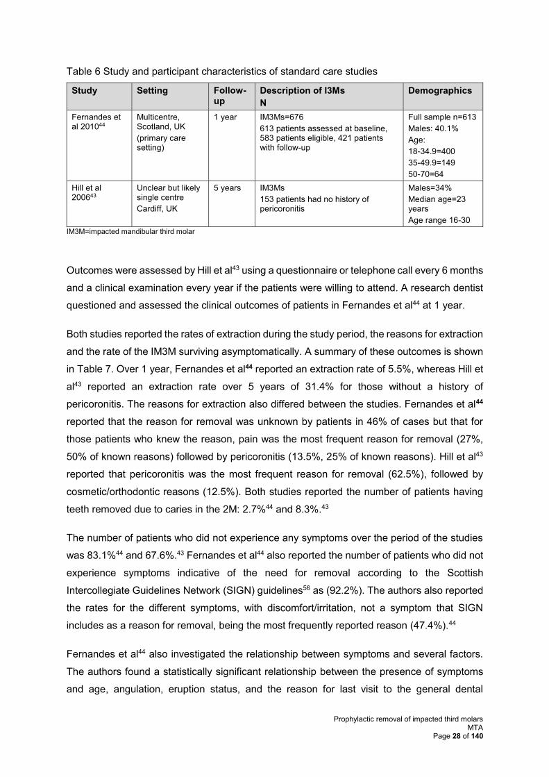
Table 6 Study and participant characteristics of standard care studies .................................... 28
Table 7 Outcomes of standard care studies.............................................................................. 29 Table 8 Systematic review characteristics ............................................................................... 33 Table 9 Systematic review results and conclusions ................................................................. 35
Table 10 Economic inclusion criteria (costs and outcomes) ................................................... 42 Table 11 Reasons for excluding papers from the cost-effectiveness review at Stage 2 .......... 44 Table 12 Three studies included in the Assessment Group’s economic evidence review ....... 45 Table 13 Characteristics of studies that were included in the economic evidence review ...... 46 Table 14 Probabilities of symptom, extraction complication and distal cervical caries of the
mandibular second molar used in the Assessment Group’s model base case.......................... 53 Table 15 Base case costs included in the model ...................................................................... 56 Table 16 Base case utility decrements and symptom duration ................................................ 57 Table 17 Parameter values in deterministic sensitivity analyses ............................................. 59 Table 18 Scenario analyses ...................................................................................................... 60
Table 19 Base case cost results for 1000 people with symptomatic impacted mandibular third
molars ....................................................................................................................................... 61
Table 20 Base case QALY results for 1000 people with symptomatic impacted mandibular
third molars .............................................................................................................................. 62 Table 21 Base case incremental cost-effectiveness ratios for 1000 people with symptomatic
impacted mandibular third molars ........................................................................................... 62
Table 22 Impact on cost-effectiveness results from assuming 100% and 0% of impacted
mandibular third molars extractions occur in primary care ..................................................... 65
Table 23 Impact on cost-effectiveness results of varying the start age ................................... 66 Table 24 Impact of varying the model time horizon on cost-effectiveness results ................. 66 Table 25 Impact on cost-effectiveness results from assuming no symptoms from impacted
mandibular third molars after the age of 63 years ................................................................... 67 Table 26 Impact on cost-effectiveness results from assuming no extraction from impacted
mandibular third molars after the age of 63 years ................................................................... 67
TABLE OF FIGURES Figure 1 PRISMA flow diagram: clinical evidence review ..................................................... 24
Figure 2 PRISMA flow diagram: economic evidence review ................................................. 43 Figure 3 Prophylactic removal of IM3M model pathway ....................................................... 50
Figure 4 Deterministic sensitivity analyses results .................................................................. 64
Prophylactic removal of impacted third molars MTA
Page 11 of 140
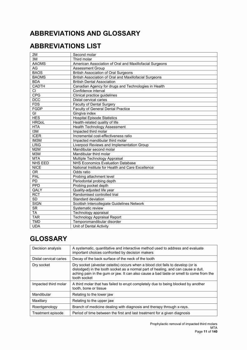
ABBREVIATIONS AND GLOSSARY
ABBREVIATIONS LIST 2M Second molar
3M Third molar
AAOMS American Association of Oral and Maxillofacial Surgeons
AG Assessment Group
BAOS British Association of Oral Surgeons
BAOMS British Association of Oral and Maxillofacial Surgeons
BDA British Dental Association
CADTH Canadian Agency for drugs and Technologies in Health
CI Confidence interval
CPG Clinical practice guidelines
DCC Distal cervical caries
FDS Faculty of Dental Surgery
FGDP Faculty of General Dental Practice
GI Gingiva index
HES Hospital Episode Statistics
HRQoL Health-related quality of life
HTA Health Technology Assessment
I3M Impacted third molar
ICER Incremental cost-effectiveness ratio
IM3M Impacted mandibular third molar
LRiG Liverpool Reviews and Implementation Group
M2M Mandibular second molar
M3M Mandibular third molar
MTA Multiple Technology Appraisal
NHS EED NHS Economics Evaluation Database
NICE National Institute for Health and Care Excellence
OR Odds ratio
PAL Probing attachment level
PD Periodontal probing depth
PPD Probing pocket depth
QALY Quality-adjusted life year
RCT Randomised controlled trial
SD Standard deviation
SIGN Scottish Intercollegiate Guidelines Network
SR Systematic review
TA Technology appraisal
TAR Technology Appraisal Report
TMD Temporomandibular disorder
UDA Unit of Dental Activity
GLOSSARY
Decision analysis A systematic, quantitative and interactive method used to address and evaluate important choices confronted by decision makers
Distal cervical caries Decay of the back surface of the neck of the tooth
Dry socket Dry socket (alveolar osteitis) occurs when a blood clot fails to develop (or is dislodged) in the tooth socket as a normal part of healing, and can cause a dull, aching pain in the gum or jaw. It can also cause a bad taste or smell to come from the tooth socket
Impacted third molar A third molar that has failed to erupt completely due to being blocked by another tooth, bone or tissue
Mandibular Relating to the lower jaw
Maxillary Relating to the upper jaw
Roentgenology Branch of medicine dealing with diagnosis and therapy through x-rays.
Treatment episode Period of time between the first and last treatment for a given diagnosis
Prophylactic removal of impacted third molars MTA
Page 12 of 140
1 BACKGROUND
1.1 Description of health problem
The four hindmost molars, known as third molars (3Ms) or wisdom teeth, are the last teeth to
erupt in the upper (maxillary) and lower (mandibular) jaws, and this usually happens during
young adulthood between the ages of 18 and 24. Third molars can be either impacted or non-
impacted, and an impacted 3M (I3M) can be classed as erupted, partially erupted or
unerupted. Impaction occurs when the eruption of the tooth is blocked either by soft tissue
(gum) or bone.
For some patients, 3Ms erupt fully, while for others 3Ms could remain unerupted and impacted
throughout the life of the tooth. Third molars can be potentially problematic to the individual by
causing pain and disease; however, many 3Ms are asymptomatic (trouble-free) or disease-
free/pathology-free. There has been significant debate over the past few decades surrounding
the management of 3Ms, and historically, the practice has been to surgically extract 3Ms
prophylactically to avoid potential problems in the future. However, 3M surgery is not without
risk to the patient. Despite the substantial amount of literature dedicated to the debate on
whether or not to prophylactically remove 3Ms, there is still disagreement and controversy
among dentists and oral surgeons as to what constitutes best practice.1 Current National
Institute for Health and Care Excellence (NICE) guidance2 advises against the routine
prophylactic removal of 3M teeth.
Kandasamy et al 20093 assert that “the literature pertaining to the extraction of third molars is
extensive. There is a large individual variation and a multitude of practitioners’ beliefs and
biases relating to the extraction of especially asymptomatic and pathology-free third molars.
With the current emphasis in dentistry being placed on clinicians to make evidence-based
decisions, the routine removal of third molars has been re-assessed and questioned.”(page
284)3
There is disagreement on the operational definition of what constitutes an asymptomatic or
pathology-/trouble-/disease-free 3M. In part, this is due to some inconsistent and misleading
use of vague terminology.1,4 In some studies “asymptomatic” denotes teeth that have no
associated pathology, in others it denotes an absence of symptoms.4 There is a significant
difference between disease-free and asymptomatic – asymptomatic does not equal disease-
free. It is argued that pathology always precedes symptoms, so it is therefore prudent for
decision makers to assume the development of pathology if teeth are symptomatic.1 The
terminology that is used in clinical research studies needs to convey the precise condition

Prophylactic removal of impacted third molars MTA
Page 13 of 140
being described (i.e. the presence or absence of pathology), otherwise inconsistent findings
will always be reported.4
To be clear, in this report, prophylactic removal of I3Ms is considered as relating to the removal
of pathology-free 3Ms to avoid potential problems in the future.
1.1.1 Aetiology, pathology and prognosis
Impacted third molars are classified on the basis of location (mandibular or maxillary), eruption
status, nature of impaction, angulation of impaction, and the depth of impaction relative to the
adjacent tooth. An impacted tooth can be visible in the mouth, can be explored with a
periodontal probe, or may only be observed through radiographic assessment.5 Eruption
status is described in Table 1.
Table 1 Eruption status
Erupted Partially erupted Unerupted
Crown is visible in mouth
• Functional position
• Non-functional position
• Unlikely to erupt into functional position
• Likely to develop into functional position
Part of crown is visible in mouth
• Partial bone impacted
• Soft tissue impacted
Crown not visible BUT
• May be soft tissue impacted and communicating with the mouth (probeable)
• Hard tissue impacted, i.e. under bone not communicating with the mouth
The nature of the impaction can be when the tooth is covered only by soft tissue and is referred
to as ‘soft tissue impaction’. The tooth can also be covered by bone, and this is known as
either ‘partial bony impaction’ when partially erupted or ‘complete bony impaction’ when
unerupted and not communicating with the mouth.
Angulation can be based on Winter’s classification6 and the 3M could be:
• Mesioangular (angled towards the second molar)
• Distoangular (away from the second molar)
• Horizontal
• Vertical
• Buccal (angled towards the cheek)
• Lingual (angled towards the tongue)
Based on Pell and Gregory’s classification7 relating to depth, the I3M can be class 1, 2, or 3
according to the amount of tooth covered by the mandibular ramus, or A, B, or C depending
on the depth of the impacted tooth compared with the second molar (2M).
Prophylactic removal of impacted third molars MTA
Page 14 of 140
Pathological changes
Impacted third molars may be associated with pathological changes such as infection
(pericoronitis), periodontal (gum) disease, dental caries, destruction of adjacent teeth, and
cysts and tumours. According to Worrall et al, the prevalence of pathological changes in I3Ms
is higher in impacted mandibular 3Ms (IM3Ms) compared with impacted maxillary 3Ms.8
Pericoronitis
This is an infection of the soft tissue surrounding the crown of the tooth and is caused by an
accumulation of bacteria and debris beneath the soft tissue. This can result in inflammation
and pain. Where 3Ms are impacted, this creates an area that is difficult to clean properly with
a toothbrush, making the molar in front of the 3M, as well as the 3M itself, vulnerable to plaque
accumulation, inflammation and infection. It is reported that 20-30% of partially erupted, and
10% of completely unerupted teeth are associated with pericoronitis. Partial soft bony
impaction and vertical or distal angulation are additional risk factors for pericoronitis.9
Gum/periodontal disease
Early stages of gum disease include red and swollen gums, and bleeding gums after tooth
brushing and is known as gingivitis. More advanced disease, known as periodontal disease
or periodontitis, can lead to bad breath, loose teeth, and gum abscesses. Periodontal
disease/gum disease is caused by bacteria in the mouth, which, when not removed by tooth
brushing, sets up chronic gum inflammation, which can affect the bone that supports the teeth
in the mouth.
Dental caries (decay)
Dental caries or decay is the demineralisation of tooth enamel or dentine caused by bacteria,
which metabolise sugar in the diet to form acids. A longitudinal study in the US followed
patients with at least one 3M below the occlusal plane at baseline, which had erupted during
the follow-up period (median 5.1 years). The study found that of the 49 patients who had no
3M caries at baseline, 36 (73%) had no caries experience at follow-up, and 13 (27%) had at
least one 3M with caries.10
Pathology in adjacent teeth
There is some evidence to suggest that horizontal or mesioangular I3Ms may increase the
risk of decay and cause possible damage to adjacent teeth.11 Longitudinal data from the U.S.
Department of Veterans Affairs Dental Longitudinal Study of 1,231 non-veteran volunteers,
revealed that the presence of a 3M that was soft tissue impacted increased the risk of incident
2M pathology 4.88-fold (95% confidence interval (CI): 2.62 to 9.08); however, the prevalence
of soft tissue impaction in the study population was only 3%. The relative risk for pathology in
Prophylactic removal of impacted third molars MTA
Page 15 of 140
the 2M was 39.6% for those with absent 3Ms, 52.8% for those with erupted 3Ms, and a similar
rate for those with bony impaction (56.6%).12 There appears to be a link, therefore, between
the presence of 3Ms and the development of 2M distal cervical caries (DCC), particularly with
mesioangular 3Ms.13
Cysts and tumours
Cysts and tumours may develop around I3Ms, though research has shown that the risk is low
and reduces with age.14 A study of surgically removed asymptomatic I3Ms found that
histological examination of the dental follicles showed the following pathological conditions:
14.1% were diagnosed as dentigerous cysts, 6.6% were calcifying odontogenic cysts, and
2.5% were odontogenic keratocysts.15
1.1.2 Natural history with no treatment
Little is known about the natural history of I3Ms left in situ. This is due in part to the historical
routine extraction of I3Ms, which means we have limited data on which to make reliable
estimates of the onset of pathology when the asymptomatic teeth are left in place.3 Collecting
the required data is also problematic in the UK, as clinical reporting systems are not sensitive
enough to capture information relating to 3M management.16 In addition, it would be costly to
conduct a non-interventional/observational study to gather data on untreated I3Ms, as it would
take decades because of the size of the study cohort needed to determine the occurrence of
pathological conditions.4
1.1.3 Epidemiology
The prevalence of I3Ms in the UK is unknown. Internationally, the prevalence of I3Ms is
reported to range from 18% to 68%.17 According to the results of a recent meta-analysis18 of
49 studies (83,484 individuals), the prevalence of 3M impaction worldwide in individuals aged
>17 years is 24.4% (95% CI: 18.97 to 30.80). The authors of the study also found that the risk
of having IM3Ms was higher than having impacted maxillary 3Ms – 57.6% (95% CI: 43.3 to
68.3; p<0.0001), and there was no difference in the incidence of impaction for men and women
(18.6%, 95% CI: –4.9% to 48.0%; p=0.12). The most common angulation of impaction was
found to be mesioangular (41.2%, 95% CI: 33.8 to 49.0).
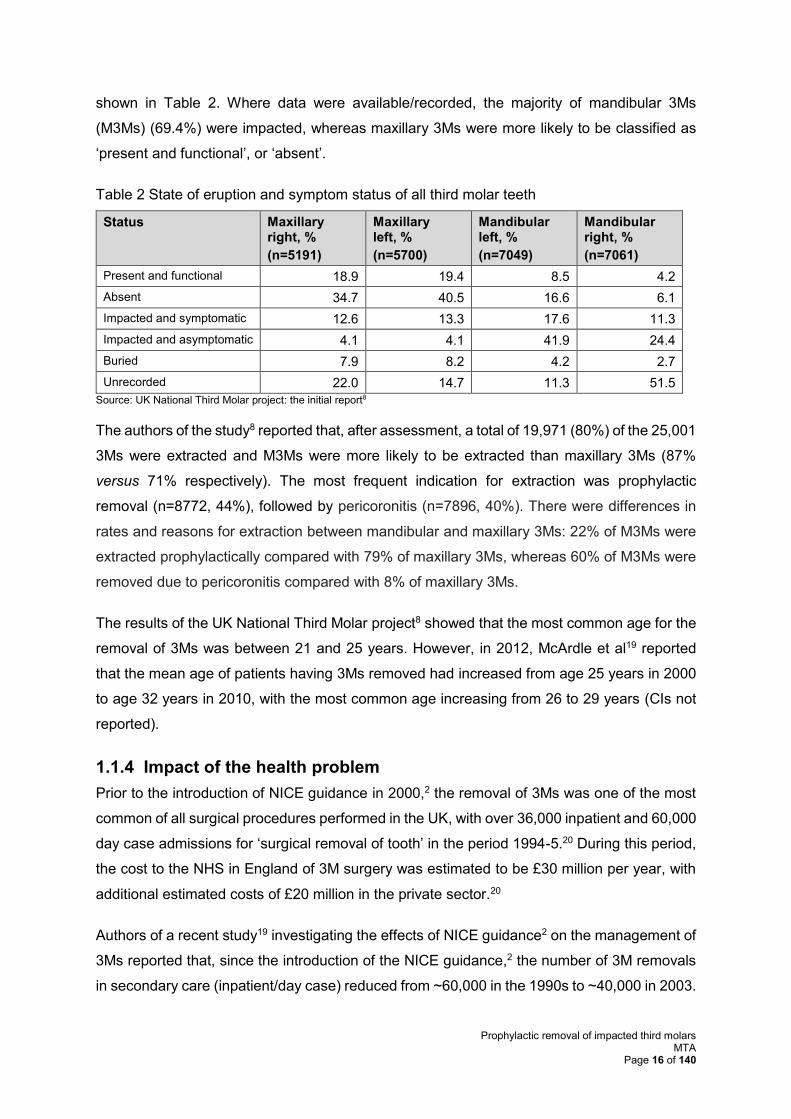
The UK National Third Molar project8 was a cross-sectional survey that was set up in 1997 to
assess the management of 3Ms in UK clinical practice. Clinical data were collected
prospectively from all of the patients referred for assessment of 3Ms to oral and maxillofacial
consultant surgeons during July 1995.8 Completed questionnaires were returned from 181
consultants and 8298 patients (with 25,001 3Ms) who were referred to hospital for
assessment. Details of eruption and symptom status of all 3Ms at the time of presentation are
Prophylactic removal of impacted third molars MTA
Page 16 of 140
shown in Table 2. Where data were available/recorded, the majority of mandibular 3Ms
(M3Ms) (69.4%) were impacted, whereas maxillary 3Ms were more likely to be classified as
‘present and functional’, or ‘absent’.
Table 2 State of eruption and symptom status of all third molar teeth
Status Maxillary right, %
(n=5191)
Maxillary left, %
(n=5700)
Mandibular left, %
(n=7049)
Mandibular right, %
(n=7061)
Present and functional 18.9 19.4 8.5 4.2
Absent 34.7 40.5 16.6 6.1
Impacted and symptomatic 12.6 13.3 17.6 11.3
Impacted and asymptomatic 4.1 4.1 41.9 24.4
Buried 7.9 8.2 4.2 2.7
Unrecorded 22.0 14.7 11.3 51.5
Source: UK National Third Molar project: the initial report8
The authors of the study8 reported that, after assessment, a total of 19,971 (80%) of the 25,001
3Ms were extracted and M3Ms were more likely to be extracted than maxillary 3Ms (87%
versus 71% respectively). The most frequent indication for extraction was prophylactic
removal (n=8772, 44%), followed by pericoronitis (n=7896, 40%). There were differences in
rates and reasons for extraction between mandibular and maxillary 3Ms: 22% of M3Ms were
extracted prophylactically compared with 79% of maxillary 3Ms, whereas 60% of M3Ms were
removed due to pericoronitis compared with 8% of maxillary 3Ms.
The results of the UK National Third Molar project8 showed that the most common age for the
removal of 3Ms was between 21 and 25 years. However, in 2012, McArdle et al19 reported
that the mean age of patients having 3Ms removed had increased from age 25 years in 2000
to age 32 years in 2010, with the most common age increasing from 26 to 29 years (CIs not
reported).
1.1.4 Impact of the health problem
Prior to the introduction of NICE guidance in 2000,2 the removal of 3Ms was one of the most
common of all surgical procedures performed in the UK, with over 36,000 inpatient and 60,000
day case admissions for ‘surgical removal of tooth’ in the period 1994-5.20 During this period,
the cost to the NHS in England of 3M surgery was estimated to be £30 million per year, with
additional estimated costs of £20 million in the private sector.20
Authors of a recent study19 investigating the effects of NICE guidance2 on the management of
3Ms reported that, since the introduction of the NICE guidance,2 the number of 3M removals
in secondary care (inpatient/day case) reduced from ~60,000 in the 1990s to ~40,000 in 2003.
Prophylactic removal of impacted third molars MTA
Page 17 of 140
However, since 2003, the number of removals appears to have increased to ~65,000 during
2009/10 (inpatient/day case only).
Information provided to NICE in the British Dental Association (BDA)21 and the Faculty of
General Dental Practice (FGDP)22 submissions, suggest that the prophylactic removal of 3Ms
prevents future harm to patients. They argue that the introduction of NICE guidance2 initially
resulted in a reduction in the number of 3Ms extracted. However, this figure has since
increased. It is argued that irrespective of the NICE guidance in 20002, the need for surgical
extraction was not negated, but postponed until a later date. It is further argued that patients
over the age of 25 years are at a higher risk of surgical morbidity relating to 3M extraction.22
Another possible explanation for the increase in 3M extractions could be that patients who
may have more than one I3M undergo multiple treatment episodes, as and when other 3Ms
become problematic (BDA).21
1.2 Current service provision
1.2.1 Management of disease
Treatment options for people with I3Ms include either surgical removal or standard care
without prophylactic removal of 3Ms.
Surgical removal
A report23 by the Royal College of Surgeons of England states that, “Third molar surgical
procedures are generally suitable for day case management, and it is recognised that
treatment under local anaesthesia with or without sedation is associated with reduced
complication rates.” (page 10)
Removal of I3Ms can be carried out by a dentist, or patients can be referred to an oral surgeon
in cases where the degree of impaction or position of the tooth indicates that a more complex
surgical procedure is required. In cases where general anaesthetic is required, the surgical
removal is conducted in hospital.
Generally, recovery from surgery for the removal of 3Ms is straightforward. The immediate
side effects of 3M surgery such as pain and swelling resolve within a few days, and jaw
stiffness usually subsides within 1-2 weeks.24 However, there may be potential additional
complications associated with the removal of I3Ms, including damage to surrounding teeth,
infection and dry socket (which can manifest as a throbbing pain in the gum or jaw and also
cause bad breath). Also, nerve damage may occur and is a serious complication that can
cause short- and long-term pain or a tingling sensation and numbness in the tongue, lower lip,
chin, teeth and gums.

Prophylactic removal of impacted third molars MTA
Page 18 of 140
Overall, the rate of complications following the surgical removal of 3Ms is reported to vary
between 2.6% and 30.9%.25 The removal of mandibular 3Ms (regardless of eruption status) is
much more likely to be associated with post-surgical complications than the removal of
maxillary 3Ms.26 The risk of infection following extraction of I3Ms is approximately 10% in
healthy patients; however, it may be up to 25% in patients with low immunity.27 Dry socket
occurs in 5-10% of patients who have undergone a 3M removal, and presents within 3-5 days
after the initial pain from surgery has subsided. Nerve damage occurs in up to 2% of patients
and is generally temporary, but in 0.5% (1 in 200) patients, the damage is permanent.24 The
risk of nerve injury is more common if the IM3M is located close to the inferior alveolar nerve,
with 20% of patients likely to then have temporary nerve damage and 2% to experience
permanent damage.24
Standard care without prophylactic removal
The alternative to surgical removal of an I3M is standard care without removal of the tooth.
Standard care is typically patient centred and comprises regular oral health reviews, oral
health advice, dental care plans and a decision on the length between recalls.28 Standard care
is carried out without the removal of the I3M. However, without the removal of the I3M, there
is a risk that pathological changes, as previously described, could lead to future surgical
removal of the impacted tooth.
Indications for removal or retention
The decision to remove or retain an I3M depends on whether it is asymptomatic (pathology-
or trouble-free). Where there are pathological changes, current NICE guidance2 states that
the I3M should be removed.
1.2.2 Variation in services and/or uncertainty about best practice
Internationally, there is a vast quantity of published literature relating to the management of
3Ms, and many published international guidelines with recommendations for best practice
relating to asymptomatic, or disease-free, 3Ms. However, there is still debate, and it remains
a contentious subject. According to the FGDP submission,22 there are differences of opinion
between professionals in the UK relating to best practice. However, the submission authors
assert that most UK dentists believe that erupted, non-functional, low-risk M3Ms should be
removed at a young age to prevent increased surgical morbidity in older age, and to prevent
future harm to the patient.22
There is significant geographical variation in current practice when international guidelines are
examined. The American29 guidelines recommend a more interventional approach to 3M
management. In the UK NHS setting, there is a ‘no intervention’ policy unless there are distinct
Prophylactic removal of impacted third molars MTA
Page 19 of 140
therapeutic indications, although there are differences of opinion between professionals. A
table summarising international guidelines is provided in Appendix 1.
There is variation in the services relating to the use of general anaesthesia or local
anaesthesia and sedation. There are published data30 which illustrate that only 3% of IM3M
cases in a London teaching hospital required general anaesthetic, with 40% of cases needing
intravenous sedation. However, our clinical advisor has pointed out that not all district general
hospitals offer sedation services for dental extractions, and therefore the proportion of patients
receiving higher-risk general anaesthetic is greater. In terms of service provision, many dental
practices in the UK do not provide intravenous sedation, which results in these patients being
referred to hospital to undergo surgical extraction under general anaesthetic. There is also
considerable variation in the perioperative care provided; for example, the provision of
informed consent, patient information, pre-operative mouth rinses, provision of analgesia, and
rates of antibiotic prescription.22
1.2.3 Relevant UK guidelines
The NICE TA120 was completed in 2000 and the resultant NICE guidance2 was that the
prophylactic removal of pathology-free I3Ms was not recommended (see Box 1).
Box 1 Current NICE guidance on the extraction of wisdom teeth
1.1 The practice of prophylactic removal of pathology-free impacted third molars should be discontinued in the NHS
1.2 The standard routine programme of dental care by dental practitioners and/or paraprofessional staff, need be no different, in general, for pathology-free impacted third molars (those requiring no additional investigations or procedures)
1.3 Surgical removal of impacted third molars should be limited to patients with evidence of pathology. Such pathology includes unrestorable caries, non-treatable pulpal and/or periapical pathology, cellulitis, abscess and osteomyelitis, internal/external resorption of the tooth or adjacent teeth, fracture of tooth, disease of follicle including cyst/tumour, tooth/teeth impeding surgery or reconstructive jaw surgery, and when a tooth is involved in or within the field of tumour resection
1.4 Specific attention is drawn to plaque formation and pericoronitis. Plaque formation is a risk factor but is not in itself an indication for surgery. The degree to which the severity or recurrence rate of pericoronitis should influence the decision for surgical removal of a third molar remains unclear. The evidence suggests that a first episode of pericoronitis, unless particularly severe, should not be considered an indication for surgery. Second or subsequent episodes should be considered the appropriate indication for surgery
Source: Guidance on the Extraction of Wisdom Teeth (TA1)2
A review of the existing NICE guidance2 via a ‘Review Proposal’ in 2014 concluded that no
new trial data on this topic were available. As a result, a decision was made that the NICE
guidance2 did not need to be revisited and the topic should remain on the static list. However,
as the recommendations set out in the NICE guidance2 were increasingly being perceived as
controversial by the dental profession, a NICE consultation with relevant stakeholders was
then undertaken. Consultation responses highlighted that additional pertinent trial data were
Prophylactic removal of impacted third molars MTA
Page 20 of 140
available and therefore should be assessed. In response, NICE instructed that the current
guidance2 should be partially updated (i.e. prophylactic indications only) via the Multiple
Technology Appraisal (MTA) process.
1.3 Description of technology under assessment
1.3.1 Summary of intervention
The surgical extraction of IM3Ms with evidence of pathology (see Box 1) can be undertaken
in primary care, secondary care, and specialist clinics. The NHS commissioning oral surgery
pathway31 clearly outlines social, medical and dental factors that dictate the optimal setting.22
Specialist radiographic equipment and assessment can be required for risk assessment of
IM3Ms, including panoral and cone beam computed tomography radiography which requires
the input of a radiologist. For patient requiring sedation (primary care), specialist nursing is
required. Intravenous sedation services require additional staff training, the correct facilities
and indemnity costs.
1.3.2 Identification of important subgroups
There is intrapatient variance in the presentation of I3Ms, i.e. a single patient can have multiple
3Ms (maxillary as well as mandibular and bilateral presence) with different types of impaction
(vertical, horizontal, distoangular, and mesioangular). These are the most common impaction
types considered as subgroups, though a smaller proportion of patients may have ectopic
impactions. The variability of 3M impactions results in different secondary disease distribution,
which is dependent on the nature of the impaction.21
Patients with high-risk M3Ms (the roots cross the inferior dental canal) could be 10 times more
likely to develop temporary or permanent inferior alveolar nerve injury.22
Prophylactic removal of impacted third molars MTA
Page 21 of 140
2 DEFINITION OF THE DECISION PROBLEM
The remit of this review was to appraise the clinical and cost-effectiveness of the prophylactic
removal of IM3Ms.
2.1 Decision problem
This MTA has been conducted in line with the decision problem issued by NICE in the final
scope.11 This is reproduced in Table 3.
Table 3 Decision problem issued by NICE
Interventions Prophylactic removal of third molars
Population People with pathology-free or trouble-free impacted mandibular third molars
Comparators Standard care without prophylactic removal of third molars
Outcomes The outcome measures to be considered include:
• Pathology associated with retention of third molars
• Post-operative complications following extraction (e.g. pain, dry socket, nerve injury)
• Adverse effects of treatment
• Health-related quality of life
Economic analysis
The reference case stipulates that the cost-effectiveness of treatments should be expressed in terms of incremental cost per quality-adjusted life year
The reference case stipulates that the time horizon for estimating clinical and cost-effectiveness should be sufficiently long to reflect any differences in costs or outcomes between the technologies being compared
Costs will be considered from an NHS and Personal Social Services perspective
Other considerations
If evidence allows, consideration may be given to the following subgroups:
• People with mesioangular or horizontally impacted third molars
Source NICE Final scope11
2.2 Overall aims of assessment
The aim of this assessment report is to synthesise the clinical and cost-effectiveness of the
prophylactic removal of IM3Ms compared with standard care without prophylactic removal.
2.2.1 What is not included in the assessment
It is beyond the remit of this assessment report to comment on or draw conclusions relating to
the wider topic of the management of 3Ms; this assessment report focusses primarily on
summarising the relevant evidence relating to the surgical extraction or retention of
asymptomatic IM3Ms.
It is worth noting that the aims of the original assessment report conducted by Song et al,20
which contributed to the NICE guidance2 issued in 2000, were not exactly the same as the
aims of this assessment, which is a partial update of TA1.20 Song et al20 aimed to “provide a
summary of existing evidence on prophylactic removal of impacted wisdom teeth, in terms of
the incidence of surgical complications associated with prophylactic removal, and the
morbidity associated with retention.”
Prophylactic removal of impacted third molars MTA
Page 22 of 140
3 ASSESSMENT OF CLINICAL EFFECTIVENESS
3.1 Methods for reviewing effectiveness
3.1.1 Identification of studies
Search strategy
The assessment group (AG) identified relevant clinical studies, systematic reviews (SRs) and
decision analyses by searching the following major medical databases: MEDLINE, EMBASE,
Cochrane Library, NHS Economics Evaluation Database (NHS EED) and EconLit, from 1999
onwards. The search strategies used are presented in Appendix 2.
In addition to the electronic databases, information on studies in progress were sought by
searching the Current Controlled Clinical Trials database.
Citation searching was conducted using all references in key articles and all identified SRs.
The sources referenced in the professional stakeholder submissions received as part of the
standard NICE process were cross checked to identify relevant references.
A database of the published literature was assembled from the aforementioned sources and
was held in the EndNote X7 software package.
3.1.2 Inclusion and exclusion criteria
Two of three reviewers (JH, GP, RD) independently screened all titles and abstracts identified
by the initial search using Covidence.32 Full-text copies of any titles/abstracts that may have
been eligible were obtained and assessed for inclusion by two reviewers (JH, GP), according
to the inclusion and exclusion criteria listed in Table 4. Discrepancies were resolved by
consultation with a third reviewer/clinical advisor. Studies that did not meet the inclusion
criteria were excluded and the reasons for exclusion summarised. For studies that were
identified as not meeting the criteria at the data abstraction stage, bibliographic details and
reasons for exclusion were summarised.
Prophylactic removal of impacted third molars MTA
Page 23 of 140
Table 4 Inclusion criteria (clinical effectiveness)
Inclusion Exclusion
Study design Clinical trials (randomised and non-randomised)
Observational studies
Systematic reviews
Decision analyses
Case studies
Non-systematic reviews
Patient population People with impacted mandibular third molars
Interventions Prophylactic removal of impacted mandibular third molars (as defined by study authors)
Comparators Standard care without prophylactic removal of impacted mandibular third molars
Outcomes The outcome measures to be considered include:
• Pathology associated with retention of third molars
• Post-operative complications following extraction
• Adverse effects of treatment
• Health-related quality of life
Setting/location Europe
North America
Australasia
Other considerations
If evidence allows, consideration may be given to the following subgroups:
• People with mesioangular or horizontally impacted third molars
Limits 1999 onwards
English language only
3.1.3 Data abstraction strategy
Data relating to study characteristics and outcomes were extracted by one of two reviewers
(JF or JH) and independently checked for accuracy by a second reviewer (JF or JH).
Disagreement was resolved through consensus, and where necessary a third reviewer was
consulted. Study data reported in multiple publications were extracted and reported as a single
study.
3.1.4 Critical appraisal strategy
The quality of the included studies was assessed by one reviewer (JH or JF), and
independently checked for agreement by a second reviewer (JH or JF). Disagreements were
resolved through consensus. The quality of the cohort studies were assessed using an
adapted version of the Newcastle–Ottawa quality assessment scale for cohort studies,33 and
SRs were assessed according to criteria outlined by the Centre for Reviews and
Dissemination.34
3.1.5 Methods of data synthesis
The results of the data extraction and quality assessment for each included study are
presented in structured tables and as a narrative summary.
Prophylactic removal of impacted third molars MTA
Page 24 of 140
3.2 Results
3.2.1 Quantity and quality of research available
The results of the electronic searches and the application of the inclusion criteria are shown
in Figure 1.
Figure 1 PRISMA flow diagram: clinical evidence review
In total, 22 citations20,35-55 reporting results of nine SRs20,36,38-42,46,52 and four cohort
studies43,44,54,55 were included in the review. No randomised controlled trials (RCTs) were
identified.
14,472
Searches
11373
Screened
10952
Excluded421 Full text
351 Excluded 70 Included
50 Excluded 22 Included
1
Prophylactic removal vs standard care
4
Standard care
(2 studies)
1
Prophylactic removal
(1 study)
16
SRs
(9 studies)
2 Citation searching
3099
Duplicates
Prophylactic removal of impacted third molars MTA
Page 25 of 140
The reasons for excluding papers at the full-text review stage are summarised in Appendix 3.
As shown in
Figure 1, 50 papers were initially included at full-text review but on further inspection no
relevant data for the specific population of interest for this review were available and these
papers were subsequently excluded. The bibliographic details with reasons for exclusion for
these 50 studies are also reported in Appendix 3.
One study54 reported on outcomes both for standard care with and without prophylactic
removal of IM3Ms. Four papers35,43,44,53 reported on two studies assessing the outcomes of
standard care without prophylactic removal of IM3Ms, and one paper55 reported on a study of
prophylactic removal of IM3Ms. A further 16 papers20,36-42,45-52 reported on nine SRs assessing
whether I3Ms should be removed prophylactically.
Quality assessment – Cohort studies
The quality of the four included cohort studies43,44,54,55 was assessed using an adapted version
of the Newcastle–Ottawa quality assessment scale for cohort studies33 and the results are
tabulated in Table 5.
Table 5 Quality assessment of cohort studies
Study Vares et al 201454
Fernandes et al
201044 Hill et al 200643 Petsos et al
201655
Representative of cohort
No description Truly representative Truly representative
Truly representative
Ascertainment of exposure
Clinical records Clinical records Clinical records Clinical records
Outcome present at start
Yes Yes Yes Yes
Assessment No description Record linkage Record linkage Record linkage
Length of follow-up Adequate Adequate Adequate Adequate
Attrition No description 31% 9% 14%
All but the Vares et al 2014 study54 had patients who were representative of the population of
interest. Clinical records were used in all four studies43,44,54,55 to ascertain whether patients
were ‘exposed’ to the intervention (either standard care or prophylactic removal), and all
studies demonstrated that the outcome of interest (e.g. pathology) was not present at the start
of the study and the assessment of outcome was through clinical assessment. None of the
studies used a blinded assessment as this was not possible. The length of follow-up was
adequate in all studies. The attrition rates differed between three studies; 9%,43 14%55 and
31%.44 No details of study attrition were reported by Vares et al 2014.54 To conclude, the AG
Prophylactic removal of impacted third molars MTA
Page 26 of 140
considered all but one54 of the studies to be generally good-quality cohort studies. However,
missing information in the Vares et al 201454 paper meant it was not possible to adequately
assess the quality of this study.
Quality assessment – Systematic reviews
The quality of the nine SRs20,36,38-42,46,52 was assessed according to criteria outlined by the
Centre for Reviews and Dissemination.34 The results of the quality assessment are shown in
Appendix 4.
Seven of the SRs had clear review questions defined in terms of population, interventions,
comparators and outcomes.20,36,38-40,42,46 However these details were missing in two SRs.41,52
Only three reviews had an adequate search strategy without language or date
restrictions.20,41,42 Two reviews were limited by language, one to English, Dutch, French, or
German36 and one to English40. One review was restricted by language (English, Danish,
Norwegian or Swedish) and date (1999-2003).39 Another review restricted by date only (1999-
2003).46 The search terms used were not reported in the Clinical Evidence publications52 nor
in the CADTH SR,38 which also restricted by date and language. Three SRs provided adequate
information to facilitate assessment of whether preventative steps had been taken to minimise
bias and errors in the selection process.20,36,42 Four SRs reported adequate methods for
assessing the quality of included studies.20,36,41,42 One SR reported using a recognised quality
assessment tool but did not provide details on how it was used.39 The remaining four
SRs38,40,46,52 did not report whether they had conducted a quality assessment of included
studies. Adequate details of the primary studies were presented in seven of the
SRs.20,36,39,40,42,46,52 In the CADTH publication, details were only presented narratively for each
primary study,38 and although Costa et al 201341 presented some details of the primary studies
no details of the outcomes of the primary studies were presented. Statistical data synthesis
was not appropriate for any of the SRs; instead, the authors of two SRs reported a narrative
synthesis,40,52 three summarised each study individually20,36,38 and one did not include any
studies.42 Costa et al 201341 only reported the results of the quality assessment and Suska et
al 201039 did not provide any synthesis. It was not possible to assess the SMM Report for this
item.46
3.2.2 Assessment of effectiveness from included studies
Prophylactic removal versus standard care
One study54 reported on outcomes both for surgical complications of the prophylactic removal
of asymptomatic IM3Ms and standard care without prophylactic removal of asymptomatic
IM3Ms. The study was an observational cohort study conducted in Ukraine between 2009 and
Prophylactic removal of impacted third molars MTA
Page 27 of 140
2013. It was designed to develop and assess a pre-operative assessment and create a
rationale for the prophylactic removal of asymptomatic IM3Ms. The assessment included
clinical and roentgenological parameters, and the 84 included patients were assigned to one
of three groups: requiring removal and subsequently had the tooth removed (n=52), requiring
removal but the patient refused (n=7), and those not requiring removal as determined by the
assessment (n=25). The first group (n=52) was then separated further into three age groups:
18-25 years (n=41), 25-45 years (n=10), and one patient of 68 years. Patients were followed
up annually for 5 years.
At the end of 5 years, the study authors reported that there were “No considerable intra- or
post-operative complications in the first subgroup” only “minor complications in the second
subgroup”, and “in the case of 68 year-old patient surgery, all complications were related to
considerable bone atrophy of the operated area”.
Of the seven patients who refused extraction, five required the tooth to be extracted within the
5 years. Of the 25 patients who were assessed at baseline as not requiring extraction, none
had the tooth extracted during the 5 years’ follow-up.
The study authors concluded that “The low-to-no percentage of intra- and post-operative
complications does not give any reason to leave a wisdom tooth with minor clinical
manifestations or an asymptomatic wisdom tooth with bad prognosis in place, since early
surgical procedures generate fewer complications, having shorter operative time and post-
operative period” (page 35).
Further details of the study characteristics and outcomes are reported in Appendix 5.
Standard care
We included two studies, Fernandes et al44 and Hill et al43 (reported in four
publications35,43,44,53), that reported relevant outcomes for the comparator of standard care
without prophylactic removal of IM3Ms. Both studies were single-cohort studies with follow-up
periods of 1 year44 and 5 years.43 Both studies were conducted in the UK and the number of
patients with trouble-free IM3Ms was 42144 and 153.43 The number of trouble-free IM3Ms
examined was only reported by one study (n=676).44 Participants were patients aged 16-30
years (median age 23 years)43 and 18-70 years (18-34.9 years, n=400; 35-49.9 years, n=149;
and 50-70 years, n=64).44 The percentage of males was 41%44 and 34%43 (Table 6). Further
study and participant characteristics are reported in Appendix 5.
Prophylactic removal of impacted third molars MTA
Page 28 of 140
Table 6 Study and participant characteristics of standard care studies
Study Setting Follow-up
Description of I3Ms
N
Demographics
Fernandes et al 201044
Multicentre, Scotland, UK
(primary care setting)
1 year IM3Ms=676
613 patients assessed at baseline, 583 patients eligible, 421 patients with follow-up
Full sample n=613
Males: 40.1%
Age:
18-34.9=400
35-49.9=149
50-70=64
Hill et al 200643
Unclear but likely single centre
Cardiff, UK
5 years IM3Ms
153 patients had no history of pericoronitis
Males=34%
Median age=23 years
Age range 16-30
IM3M=impacted mandibular third molar
Outcomes were assessed by Hill et al43 using a questionnaire or telephone call every 6 months
and a clinical examination every year if the patients were willing to attend. A research dentist
questioned and assessed the clinical outcomes of patients in Fernandes et al44 at 1 year.
Both studies reported the rates of extraction during the study period, the reasons for extraction
and the rate of the IM3M surviving asymptomatically. A summary of these outcomes is shown
in Table 7. Over 1 year, Fernandes et al44 reported an extraction rate of 5.5%, whereas Hill et
al43 reported an extraction rate over 5 years of 31.4% for those without a history of
pericoronitis. The reasons for extraction also differed between the studies. Fernandes et al44
reported that the reason for removal was unknown by patients in 46% of cases but that for
those patients who knew the reason, pain was the most frequent reason for removal (27%,
50% of known reasons) followed by pericoronitis (13.5%, 25% of known reasons). Hill et al43
reported that pericoronitis was the most frequent reason for removal (62.5%), followed by
cosmetic/orthodontic reasons (12.5%). Both studies reported the number of patients having
teeth removed due to caries in the 2M: 2.7%44 and 8.3%.43
The number of patients who did not experience any symptoms over the period of the studies
was 83.1%44 and 67.6%.43 Fernandes et al44 also reported the number of patients who did not
experience symptoms indicative of the need for removal according to the Scottish
Intercollegiate Guidelines Network (SIGN) guidelines56 as (92.2%). The authors also reported
the rates for the different symptoms, with discomfort/irritation, not a symptom that SIGN
includes as a reason for removal, being the most frequently reported reason (47.4%).44
Fernandes et al44 also investigated the relationship between symptoms and several factors.
The authors found a statistically significant relationship between the presence of symptoms
and age, angulation, eruption status, and the reason for last visit to the general dental
Prophylactic removal of impacted third molars MTA
Page 29 of 140
practitioner. They found no relationship between the presence of symptoms and sex, average
number of teeth, maximum basic periodontal examination score, average gingival bleeding
index, ‘average mean plaque’, education after minimum school-leaving age, employment
status, frequency of brushing teeth, occasional use of mouthwashes, occasional teeth
flossing, frequency of dental appointments, length of time since patient last visited the dentist,
smoking, drinking >14 units/week, and deprivation category.
Table 7 Outcomes of standard care studies
Study Outcomes assessed Rate n (%)
Extraction rate *Fernandes et al 201044 37/676 (5.5)
Hill et al 200643
Without a history of pericoronitis
With a history of pericoronitis
48/153 (31.4)
23/66 (34.8)
Reasons for extraction *Fernandes et al 201044
Pericoronitis 5/37 (13.5)
Pain 10/37 (27.0)
Caries in distal of adjacent molar 1/37 (2.7)
Caries in the third molar 2/37 (5.4)
Contralateral 2/37 (5.4)
Unknown 17/37 (46.0)
*Hill et al 200643(Without a history of pericoronitis)
Pericoronitis after start of study 30/48 (62.5)
Cosmetic/orthodontic 6/48 (12.5)
Food impacted/difficult to clean 4/48 (8.3)
Early caries in second molar 4/48 (8.3)
Painful when eating 2/48 (4.2)
Earache/TMJ pain 2/48 (4.2)
Survived asymptomatically Fernandes et al 201044
From any symptom 562/676 (83.1)
From SIGN symptoms only 623/676 (92.2)
Hill et al 200643 150/222+ (67.6)
Symptoms developed by tooth Fernandes et al 201044
Pericoronitis 15/114 (13.2)
Severe pain (SIGN) 16/114 (14.0)
Mild pain (SIGN) 22/114 (19.3)
Discomfort/irritation (non-SIGN) 54/114 (47.4)
Food stagnation (non-SIGN) 7/114 (6.1)
*per tooth; +Includes 66 patients with a history of pericoronitis TMJ=temporomandibular joint; SIGN=Scottish Intercollegiate Guidelines Network
Prophylactic removal
The final included study by Petsos et al55 assessed the effects of the prophylactic removal of
trouble-free IM3Ms. It was identified during forward citation searching as it was published after
Prophylactic removal of impacted third molars MTA
Page 30 of 140
the date of the review searches. Details of the study, patient characteristics and outcomes are
reported in Appendix 5.
The study55 was a prospective cohort study conducted in Germany that was self-funded and
recruited patients after extraction of asymptomatic IM3Ms over 5 months in 2014. The study55
was designed to assess changes in the periodontal health of adjacent 2Ms 6 months after the
removal of the asymptomatic IM3Ms. Results from 78 patients were included in the analyses.
Of these 78 patients, 58 had a submucosal IM3Ms removed and 20 of the 78 teeth were fully
impacted. The mean age of patients was 16 years and 37% were male. Only four patients
were smokers. At baseline, the plaque index, gingiva index (GI), probing pocket depth (PPD)
and probing attachment level (PAL) were measured. With measurements being obtained at
six sites around the M2 (i.e. mesiobuccal, buccal, distobuccal, distolingual, lingual,
mesiolingual).
To assess the change in periodontal health of the 2M at follow-up, the mean PPD and PAL
scores at the three sites located closest to the distovestibular incision (buccal, distobuccal,
distolingual) were used.
Whereas no significant change was reported in PIl and GI scores, the mean PPD score of the
three sites improved from 3.25±0.65 (range 2-5.7) to 2.57±0.5 (range 1.3-3.7). This was a
statistically significant reduction. Similarly, mean PAL score across the three sites significantly
improved, with a reduction from 2.96±0.53 (range 2.0-5.0) to 2.55 ±0.5 (range 1.3-3.7).
Surgical complications following the prophylactic removal of the IM3M were recorded. A total
of 20 patients (25.6%) reported complications. Intense pain for more than 1 day was the most
frequent complication, reported by 12 patients. A further five patients (6.4%) reported post-
operative infection (infiltrate or abscess) and the remaining three experienced wound
dehiscence. No incidences of secondary bleeding or nerve damage were reported.
The authors concluded that “Young patients may benefit from an early removal of mandibular
M3, especially in the presence of certain cofactors”. (page 453)
Systematic reviews
Nine SRs20,36,38-42,46,52 that were reported in 16 publications20,36-42,45-52 met the review inclusion
criteria and details are summarised in Table 8 and Table 9 with further details shown in
Appendix 5.
Two36,38 were rapid reviews that applied SR methodology. All but one40 attempted to assess
the evidence for the prophylactic removal of 3Ms compared with standard care without
Prophylactic removal of impacted third molars MTA
Page 31 of 140
prophylactic removal. Bouloux et al40 only assessed whether retention of asymptomatic 3Ms
led to future extraction.
No review restricted the population to trouble-free IM3Ms. Instead, four36,38,40,41 included all
trouble-free 3Ms regardless of impaction status or location, two42,52 included trouble-free I3Ms
regardless of location, one20 included all 3Ms regardless of symptoms or not, and one39
included I3Ms regardless of symptoms or location. A further review was published in
Norwegian and only had an English summary46 so the specific population was unclear.
Different types of study designs were included across the SRs: five36,38,39,41,52 included SRs
and five38-40,46,52 included non-RCTs (e.g. cohort studies and case series). One42 SR limited
inclusion to RCTs, and another40 limited to cohort studies only. One20 review also included
literature reviews. The dates of the searches ranged from 195042 to 2014.52
The different inclusion criteria adopted by the SRs meant that the studies identified and
included in the SRs differed. In total, 84 studies were identified across the nine SRs, with only
seven studies20,45,46,57-60 being identified by more than one review.
• Mettes et al45(SR) was included in five SRs36,38,39,41,52
• Harradine et al58 (RCT) was included in four SRs20,41,42,52
• NICE Guidance/Song et al publications2,20,61 were identified by three SRs36,38,52
• Senter for medisinsk metodevurdering (SMM)’s46 report was included in three
SRs38,39,52
• Lindqvist et al60 (RCT) was included in three SRs41,45,52
• Edwards et al57 (decision analysis) was included in two SRs20,46
• Kruger et al59 (cohort study) was included in two SRs40,46
Despite the differences in inclusion criteria across the SRs, the conclusions were similar.
Seven SRs20,36,38,39,41,42,52 stated that there was insufficient evidence to support or refute the
prophylactic removal of trouble-free 3Ms. Two38,52 SRs recommended that the decision to
remove an asymptomatic 3M should be based on careful consideration of the risks and
benefits, and that patient preferences should be taken into account. Two20,36 SRs
recommended that in light of insufficient evidence, retention/“first do no harm” may be
appropriate. Watchful monitoring was recommended in the Cochrane review by Mettes et al.42
The Norwegian SMM report46 recommended the prophylactic removal of 3Ms “when the
likelihood of 3Ms causing problems in the future is high and the incidence of post-operative
complications are low.” They restrict this to partially erupted 3Ms and state that this approach
Prophylactic removal of impacted third molars MTA
Page 32 of 140
is not recommended for people with fully retained (i.e. complete bony impacted) teeth. They
also state that patient preferences should be decisive.
The one40 SR that looked at the risk of future extraction following the retention of trouble-free
3Ms found that the mean incidence rate of future extraction was 3.0% annually (range 1-9%)
leading to a cumulative incidence rate of 5% at 1 year and 64% at 18 years. The reasons for
extraction were caries, periodontal disease, and other inflammatory conditions. The authors
concluded that: “The cumulative risk of M3 extraction for young adults with asymptomatic M3s
is sufficiently high to warrant its consideration when reviewing the risks and benefits of M3
retention as a management strategy”.40 (page 806)
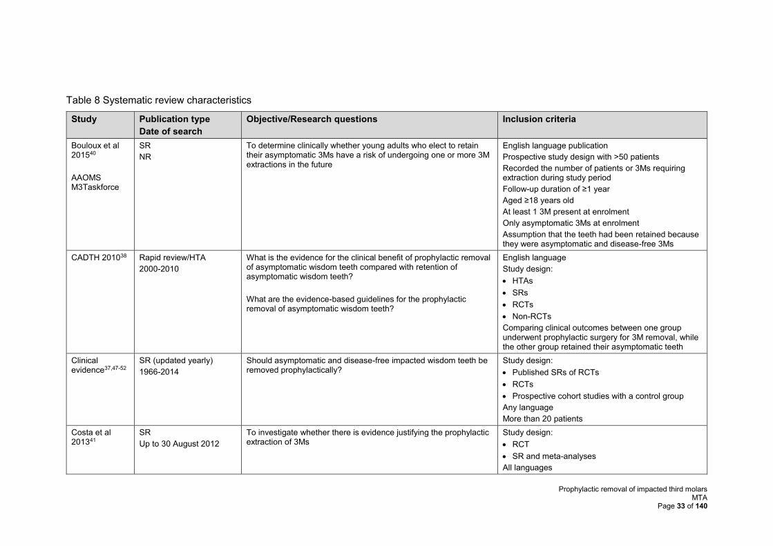
Prophylactic removal of impacted third molars MTA
Page 33 of 140
Table 8 Systematic review characteristics
Study Publication type
Date of search
Objective/Research questions Inclusion criteria
Bouloux et al 201540
AAOMS M3Taskforce
SR
NR
To determine clinically whether young adults who elect to retain their asymptomatic 3Ms have a risk of undergoing one or more 3M extractions in the future
English language publication
Prospective study design with >50 patients
Recorded the number of patients or 3Ms requiring extraction during study period
Follow-up duration of ≥1 year
Aged ≥18 years old
At least 1 3M present at enrolment
Only asymptomatic 3Ms at enrolment
Assumption that the teeth had been retained because they were asymptomatic and disease-free 3Ms
CADTH 201038 Rapid review/HTA
2000-2010
What is the evidence for the clinical benefit of prophylactic removal of asymptomatic wisdom teeth compared with retention of asymptomatic wisdom teeth?
What are the evidence-based guidelines for the prophylactic removal of asymptomatic wisdom teeth?
English language
Study design:
• HTAs
• SRs
• RCTs
• Non-RCTs
Comparing clinical outcomes between one group underwent prophylactic surgery for 3M removal, while the other group retained their asymptomatic teeth
Clinical evidence37,47-52
SR (updated yearly)
1966-2014
Should asymptomatic and disease-free impacted wisdom teeth be removed prophylactically?
Study design:
• Published SRs of RCTs
• RCTs
• Prospective cohort studies with a control group
Any language
More than 20 patients
Costa et al 201341
SR
Up to 30 August 2012
To investigate whether there is evidence justifying the prophylactic extraction of 3Ms
Study design:
• RCT
• SR and meta-analyses
All languages
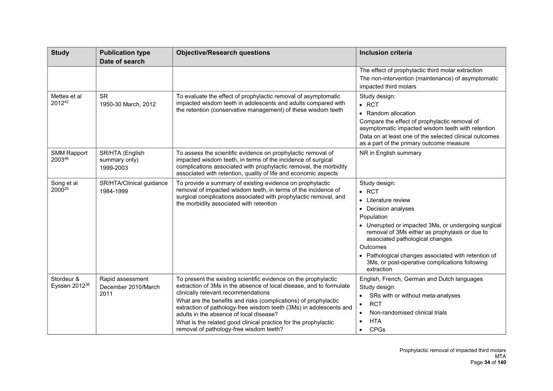
Prophylactic removal of impacted third molars MTA
Page 34 of 140
Study Publication type
Date of search
Objective/Research questions Inclusion criteria
The effect of prophylactic third molar extraction
The non-intervention (maintenance) of asymptomatic
impacted third molars
Mettes et al 201242
SR
1950-30 March, 2012
To evaluate the effect of prophylactic removal of asymptomatic impacted wisdom teeth in adolescents and adults compared with the retention (conservative management) of these wisdom teeth
Study design:
• RCT
• Random allocation
Compare the effect of prophylactic removal of asymptomatic impacted wisdom teeth with retention
Data on at least one of the selected clinical outcomes as a part of the primary outcome measure
SMM Rapport 200346
SR/HTA (English summary only)
1999-2003
To assess the scientific evidence on prophylactic removal of impacted wisdom teeth, in terms of the incidence of surgical complications associated with prophylactic removal, the morbidity associated with retention, quality of life and economic aspects
NR in English summary
Song et al 200020
SR/HTA/Clinical guidance
1984-1999
To provide a summary of existing evidence on prophylactic removal of impacted wisdom teeth, in terms of the incidence of surgical complications associated with prophylactic removal, and the morbidity associated with retention
Study design:
• RCT
• Literature review
• Decision analyses
Population
• Unerupted or impacted 3Ms, or undergoing surgical removal of 3Ms either as prophylaxis or due to associated pathological changes
Outcomes
• Pathological changes associated with retention of 3Ms, or post-operative complications following extraction
Stordeur & Eyssen 201236
Rapid assessment
December 2010/March 2011
To present the existing scientific evidence on the prophylactic extraction of 3Ms in the absence of local disease, and to formulate clinically relevant recommendations
What are the benefits and risks (complications) of prophylactic extraction of pathology-free wisdom teeth (3Ms) in adolescents and adults in the absence of local disease?
What is the related good clinical practice for the prophylactic removal of pathology-free wisdom teeth?
English, French, German and Dutch languages
Study design:
• SRs with or without meta-analyses
• RCT
• Non-randomised clinical trials
• HTA
• CPGs
Prophylactic removal of impacted third molars MTA
Page 35 of 140
Study Publication type
Date of search
Objective/Research questions Inclusion criteria
Comparing the effect of prophylactic removal of pathology-free wisdom teeth with no-treatment
Existing guidelines of high quality
Suska et al 201039
HTA/SR
May 2003 up to December 2009
Based on the Norwegian HTA so searches only conducted after 2003
Does removal of 3M teeth reduce the risk of infections and other local disease/pathological conditions in patients with asymptomatic or symptomatic impacted 3Ms compared with no intervention?
Study design:
• Studies with some kind of control group
• Case series etc. if ≥ 300 patients
Healthy individuals of all ages with totally or partially impacted wisdom teeth without symptoms, or healthy individuals of all ages with totally or partially impacted wisdom teeth with any kind of symptom or condition
Extraction of 3M tooth, or no extraction or any other treatment of 3M tooth
English, Danish, Norwegian and Swedish language only
3M=third molar; AAOMS=American Association of Oral and Maxillofacial Surgeons; CADTH=Canadian Agency for Drugs and Technologies in Health; HTA=health technology assessment; SR=systematic review; RCT=randomised controlled trial; CPG=clinical practice guidelines
Table 9 Systematic review results and conclusions
Study Number/type of studies included
Author conclusions as quoted in publications
Bouloux et al 201540
Cohort studies=7 The cumulative risk of 3M extraction for young adults with asymptomatic 3Ms is sufficiently high to warrant its consideration when reviewing the risks and benefits of 3M retention as a management strategy
CADTH 201038 SRs=4
Non-RCTs=1
Guidelines=2
Based on evidence and guidelines from the past ten years of evidence identified for inclusion in this review, there is currently insufficient evidence supporting or refuting the practice of prophylactic removal of asymptomatic third molars. Regarding clinical practice, the decision to remove asymptomatic wisdom teeth appears to be best based on careful consideration by practitioners of the potential risks and benefits for individual patients, as well as their attitude toward a potentially unnecessary surgical procedure
Clinical evidence37,47-52
Extraction of asymptomatic I3Ms: SR=5
Active surveillance of asymptomatic I3Ms: No studies
When managing asymptomatic, disease-free wisdom teeth, no RCT data are available to guide therapeutic choices. Consistent with the application of evidence-based medicine principles, after a thorough review of the risks and benefits of the treatment alternatives, patient preference should be the factor driving the clinical decision
Prophylactic removal of impacted third molars MTA
Page 36 of 140
Study Number/type of studies included
Author conclusions as quoted in publications
Costa et al 201341
SR=1
RCT=3
The results of the present review indicate a lack of scientific evidence to justify the indication of the prophylactic extraction of third molars
Mettes et al 201242
RCT=1 Insufficient evidence was found to support or refute routine prophylactic removal of asymptomatic impacted wisdom teeth in adults. A single trial comparing removal versus retention found no evidence of a difference on late lower incisor crowding at 5 years, however no other relevant outcomes were measured
Watchful monitoring of asymptomatic third molar teeth may be a more prudent strategy
SMM Rapport 200346
Patient series=13
Cohort studies=3
Case–control studies=2
Cross-sectional studies=6
Decision analysis=1
This report is based on evidence from studies that use small selected patient groups, and therefore it is difficult to conclude and give recommendations. Norwegian dentists recommend prophylactic removal of third molars when the likelihood of third molars causing problems in the future is high and the incidence of post-operative complications are low. This includes partially erupted wisdom teeth. Removal of asymptomatic fully retained wisdom teeth is not recommended. Since this report is based on studies that are not optimal the patient’s preferences need to be decisive
Song et al 200020
RCT=2
Decision analysis=4
Literature reviews=34
There is no reliable research evidence to support the prophylactic removal of disease-free impacted third molars. Available evidence suggests that retention may be more effective and cost-effective than prophylactic removal, at least in the short to medium term
Stordeur & Eyssen 201236
SRs=2
HTAs=2
CPG=1
There is mostly little debate on the fact that third molars associated with clinical and/or radiological pathology, such as unrestorable caries, should be removed. However, there is a lack of proven benefit from the systematic prophylactic removal of pathology-free third molars, impacted or not, in all adolescents or (young) adults, and the procedure is not free of risk. Preventive actions at the level of the population are only recommended if the benefits outweigh the disadvantages, and if this is not the case it is preferable not to intervene. If there is no scientific evidence that an intervention is beneficial, the largely accepted principle of medicine: “primum non nocere”, “first, do no harm”, should be respected
Suska et al 201039
HTA-report/SRs=2
Case series=16
(None reported on asymptomatic teeth)
A systematic literature search and review of published data has revealed that there is still no scientific documentation available to either support or refute routine prophylactic removal of asymptomatic impacted wisdom teeth in adults
3M=third molar; I3M=impacted third molar; AAOMS=American Association of Oral and Maxillofacial Surgeons; CADTH=Canadian Agency for Drugs and Technologies in Health; HTA=health technology assessment; SR=systematic review; RCT=randomised controlled trial; CPG=clinical practice guidelines
Prophylactic removal of impacted third molars MTA
Page 37 of 140
3.2.3 Additional evidence
References from included SRs
We reviewed all references included in the identified SRs for inclusion in this review. Of the
84 cited references, nine met our inclusion criteria and all had been identified through our
searches. However, the AG feels that another nine of these references warrant further
discussion as they are papers often cited in the debate on the management of 3Ms. Therefore,
study details and summaries of these nine studies are provided in Appendix 6.
Professional stakeholder’s submissions
As part of the NICE process three submissions from professional stakeholders were received.
One on behalf of the BDA, the second, a combined submission on behalf of the Faculty of
Dental Surgery (FDS), the FGDP, and the British Association of Oral Surgeons (BAOS), and
the third on behalf of the British Association of Oral and Maxillofacial Surgeons (BAOMS).
The submission forms provided by NICE to professional stakeholders enables healthcare
professionals to provide their perspectives on the technology in the context of clinical practice,
and includes questions within a predefined template to prompt and guide the process. The
submissions can also include references to additional sources of evidence that may not be
found by a technology-focussed SR. This could be information on recent and informal
unpublished evidence, registry and audit data. The information must include sufficient detail
to allow a judgement to be made as to the quality of the evidence, and to determine any
potential sources of bias.
The information from the submissions was reviewed to ascertain whether they included any
data that could inform this appraisal report.
Much of the content of the submissions were professional opinions and perspectives and the
full submissions are available for the committee to consider. Where references were provided,
no additional studies meeting our review inclusion criteria were identified and many were
excluded from this review as they did not meet all of our inclusion criteria. A summary of the
more pertinent papers is provided for information in Appendix 6
The key points from each submission are summarised below.
BDA
The BDA highlights that the treatment of 3Ms should be undertaken in a holistic manner, rather
than for each 3M in isolation. It is argued that NICE guidance,2 which does not recommend
the prophylactic removal of I3Ms, has led to an increase in the rate of 3M removal overall,
Prophylactic removal of impacted third molars MTA
Page 38 of 140
which causes a financial burden to the NHS and disadvantages patients. The submission
authors suggest that savings could be realised if repeat treatment episodes were reduced by
removing potentially problematic 3Ms at the same time as treating the symptomatic 3M.
FDS, FGDP and BAOS
The key points highlighted in the submission are that as a result of NICE guidance,2 patients
are retaining mandibular 3Ms, which results in problems for the surrounding teeth. There is
variation in surgical techniques used, in the sedation and anaesthetic used for patients, and
in the quality of follow-up care after the surgical removal of IM3Ms.
BAOMS
The key points highlighted in the submission are that as a result of NICE guidance,2 there is
little difference in clinical practice in the UK regarding the removal of 3Ms; however, there is a
difference in opinion between professionals in how these teeth should be managed. The
Finnish longitudinal study62 often cited to advocate the interventional removal of 3Ms to
prevent problems does not report the rationale for removal in the study and weakens the
rationale for interventional removal of 3Ms. Two subgroups with different prognoses are
described (i.e. those taking anti-resorptive or anti-angiogenic drugs, and those who are to
receive radiotherapy to the head and neck). The routine prophylactic removal of third molars
would put significant strain on NHS resources in both primary and secondary care.
3.3 Summary of clinical results
Searching of major electronic databases identified 14,472 citations; after screening and the
application of inclusion/exclusion criteria, 13 studies from 22 publications were included in the
SR (nine SRs20,36-42,45-52 and 4 cohort studies35,43,44,53-55).
Of the four cohort studies, one investigated the prophylactic removal of pathology or
asymptomatic IM3Ms in comparison with the standard care and retention of these pathology-
free or asymptomatic IM3Ms, two investigated the prophylactic removal of pathology or
asymptomatic IM3Ms without a comparison group, and one studied the retention and standard
care of pathology-free or asymptomatic IM3Ms. All studies described teeth as asymptomatic.
All four studies were European, and the two studies looking at the prophylactic removal of
pathology-free or asymptomatic IM3Ms, without a comparison group, were UK based. Follow-
up across the studies varied from 6 months to 5 years, with outcomes assessed through
clinical assessment for three of the studies. Of the two studies reporting on surgical
complications no serious complications were reported, though intense pain and post-operative
infection were reported by one study. The pathological changes due to retention of pathology-
free or asymptomatic IM3Ms were reported by three studies. The extraction rate for retained
Prophylactic removal of impacted third molars MTA
Page 39 of 140
teeth varied from 5.5% and 31.4% though this variation can be explained by the differing
follow-up periods (1 and 5 years).
3.4 Discussion of clinical effectiveness results
This SR aimed to identify and appraise the relevant evidence relating to the clinical
effectiveness of the prophylactic removal of IM3Ms in comparison with standard care without
the removal of IM3Ms. The rationale for the prophylactic removal of I3Ms is much debated in
the published literature in the UK and worldwide, with variation as to what is considered as the
best approach to the treatment of I3Ms. There are dental professionals who advocate for the
prophylactic removal of 3Ms, and those who argue for a more conservative approach. There
is a plethora of literature debating the controversies surrounding the prophylactic removal of
3Ms1,3,4,16,63-66 and there are a number of international clinical guidelines2,23,29,37-39,56,67-73 that
make recommendations on this topic. These clinical guidelines focus on the management of
3Ms in general and report indications for removal rather than reviewing the evidence for the
prophylactic removal of asymptomatic, pathology-free 3MS. The SR literature is consistent in
reporting a lack of evidence for or against the prophylactic removal of these teeth. The results
of this review have been limited by the decision problem set by NICE, focussing on people
with pathology-free or trouble-free, IM3Ms, which represents a more specific population than
the populations of all patients with 3Ms or I3Ms that were considered in much of the relevant
literature on the management of third molars.
Discussion of the results of the cohort studies are hampered by the different outcomes
reported by the studies; as different approaches to third molar management require different
outcome measures (e.g. the rate of infection of retained IM3Ms and the rate of surgical
complications following removal of IM3Ms). This means the different interventions cannot be
directly compared. However, from the included studies it appears that retention of
asymptomatic IM3Ms may lead to future symptoms and consequential extraction at a rate of
between 6% and 31% over a period of 1-5 years. For participants who had asymptomatic
IM3Ms removed, no major surgical complication rates were reported, though intense pain and
infection were reported at a rate of 15% and 6%, respectively.
None of the nine SRs reviews that were identified by this SR restricted their research question
to pathology-free or trouble-free IM3Ms; however, most were restrictive in the time periods
covered and/or languages included. The inclusion criteria for the SRs also differed especially
in relation to study design. This led to a disparate collection of studies being included, with 73
of the 84 studies only being included in one SR. This heterogeneity reflects the heterogeneity
in the literature in general and the lack of robust primary evidence. Despite these differences,
Prophylactic removal of impacted third molars MTA
Page 40 of 140
most reviews concluded that there was insufficient evidence to make a decision, regardless
of how inclusive an approach was used.
In conclusion, our findings are consistent with previous systematic reviews in that there is no
available RCT evidence to support or refute the practice of the prophylactic removal of
asymptomatic/pathology-free IM3Ms. However, the review did identify evidence from
longitudinal studies demonstrating what happens when asymptomatic IM3Ms are left in situ.
Prophylactic removal of impacted third molars MTA
Page 41 of 140
4 ASSESSMENT OF COST-EFFECTIVENESS
4.1 Systematic review of existing cost-effectiveness evidence
This section presents the methods and results of a systematic review of the published
literature comparing the cost-effectiveness of prophylactic removal of I3Ms versus no
prophylactic removal.
4.1.1 Search strategy
The search strategy developed for the clinical searches (see Appendix 2), with the addition of
an economics filter, was used to identify studies reporting the costs and benefits associated
with extracting/retaining I3Ms. As part of the search strategy, NHS EED, which is located
within the Cochrane Library, and EconLit (EBSCO) were also interrogated. All databases were
searched on 29th April 2016. The results were entered into an Endnote X7.4 library, de-
duplicated and exported into Covidence.
Informal searching activities were carried out to identify economic evaluations relevant to the
decision problem. These included contacting experts in the field and a search of Google
Scholar. The Google Scholar search was updated on 1st February 2017 and revealed no
relevant results.
The two clinical submissions from professional stakeholders that were submitted to NICE as
part of the MTA process were also checked for cost-effectiveness data.
4.1.2 Study selection and inclusion criteria
Studies were selected based on their relevance to the decision problem and the specific
economic criteria displayed in Table 10. Two reviewers (AB/SB) independently examined the
titles and abstracts of all studies identified by the search to find potentially eligible publications
(Stage 1). During the next stage (Stage 2), two reviewers (AB/SB) examined the full texts of
studies that were identified as being potentially relevant at Stage 1. During Stage 2, two
modifications were made to the inclusion criteria:
• due to only limited information about UK costs being available, studies that included
any costs were included in the review
• to align HRQoL outcomes with the outcomes reported in the clinical papers, papers
reporting short-term HRQoL outcomes were excluded from the review (i.e. only papers
with long-term HRQoL outcomes were included).
Prophylactic removal of impacted third molars MTA
Page 42 of 140
Disagreements about inclusion were resolved through discussion and, in all cases, a
consensus was reached; it was, therefore, not necessary to consult a third reviewer during the
screening and selection process.
Table 10 Economic inclusion criteria (costs and outcomes)
Criteria Inclusion
Patient population People with impacted third molars
Costs UK costs
Outcomes Any health outcomes, health-related quality of life
Study design All study designs
Date 2000 to present
Language English language only
4.1.3 Quantity of included evidence
From the main searches, the AG identified 493 potentially relevant papers for inclusion in the
review of economic evidence. Of these, 34 papers were considered for inclusion after Stage
1. As shown in Figure 2, eight studies17,20,74-79 were initially included at Stage 2. However, on
further inspection, five74-78 of the eight studies did not include information that was relevant to
the population of interest and these papers were, therefore, subsequently excluded from the
review. Bibliographic details and summary data from these five studies74-78 are available in
Appendix 3. So, of the 34 papers considered for inclusion after Stage 1, 31 papers were
excluded during Stage 2, leaving three papers17,20,79 to be included in the review. The reasons
for excluding the 31 studies are listed in Table 11.
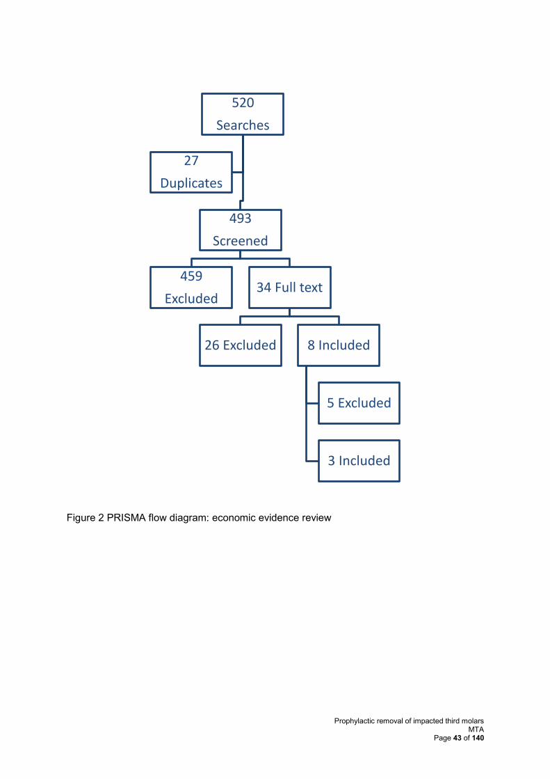
Prophylactic removal of impacted third molars MTA
Page 43 of 140
Figure 2 PRISMA flow diagram: economic evidence review
520
Searches
493
Screened
459
Excluded34 Full text
26 Excluded 8 Included
5 Excluded
3 Included
27
Duplicates
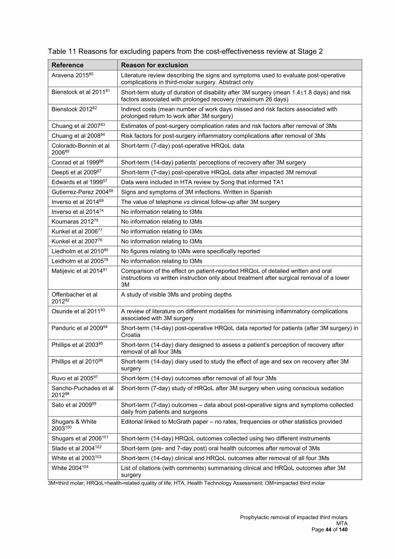
Prophylactic removal of impacted third molars MTA
Page 44 of 140
Table 11 Reasons for excluding papers from the cost-effectiveness review at Stage 2
Reference Reason for exclusion
Aravena 201580 Literature review describing the signs and symptoms used to evaluate post-operative complications in third-molar surgery. Abstract only
Bienstock et al 201181 Short-term study of duration of disability after 3M surgery (mean 1.41.8 days) and risk factors associated with prolonged recovery (maximum 26 days)
Bienstock 201282 Indirect costs (mean number of work days missed and risk factors associated with prolonged return to work after 3M surgery)
Chuang et al 200783 Estimates of post-surgery complication rates and risk factors after removal of 3Ms
Chuang et al 200884 Risk factors for post-surgery inflammatory complications after removal of 3Ms
Colorado-Bonnin et al 200685
Short-term (7-day) post-operative HRQoL data
Conrad et al 199986 Short-term (14-day) patients’ perceptions of recovery after 3M surgery
Deepti et al 200987 Short-term (7-day) post-operative HRQoL data after impacted 3M removal
Edwards et al 199957 Data were included in HTA review by Song that informed TA1
Gutierrez-Perez 200488 Signs and symptoms of 3M infections. Written in Spanish
Inverso et al 201489 The value of telephone vs clinical follow-up after 3M surgery
Inverso et al 201474 No information relating to I3Ms
Koumaras 201275 No information relating to I3Ms
Kunkel et al 200677 No information relating to I3Ms
Kunkel et al 200776 No information relating to I3Ms
Liedholm et al 201090 No figures relating to I3Ms were specifically reported
Leidholm et al 200578 No information relating to I3Ms
Matijevic et al 201491 Comparison of the effect on patient-reported HRQoL of detailed written and oral instructions vs written instruction only about treatment after surgical removal of a lower 3M
Offenbacher et al 201292
A study of visible 3Ms and probing depths
Osunde et al 201193 A review of literature on different modalities for minimising inflammatory complications associated with 3M surgery
Panduric et al 200994 Short-term (14-day) post-operative HRQoL data reported for patients (after 3M surgery) in Croatia
Phillips et al 200395 Short-term (14-day) diary designed to assess a patient’s perception of recovery after removal of all four 3Ms
Phillips et al 201096 Short-term (14-day) diary used to study the effect of age and sex on recovery after 3M surgery
Ruvo et al 200597 Short-term (14-day) outcomes after removal of all four 3Ms
Sancho-Puchades et al 201298
Short-term (7-day) study of HRQoL after 3M surgery when using conscious sedation
Sato et al 200999 Short-term (7-day) outcomes – data about post-operative signs and symptoms collected daily from patients and surgeons
Shugars & White 2003100
Editorial linked to McGrath paper – no rates, frequencies or other statistics provided
Shugars et al 2006101 Short-term (14-day) HRQoL outcomes collected using two different instruments
Slade et al 2004102 Short-term (pre- and 7-day post) oral health outcomes after removal of 3Ms
White et al 2003103 Short-term (14-day) clinical and HRQoL outcomes after removal of all four 3Ms
White 2004104 List of citations (with comments) summarising clinical and HRQoL outcomes after 3M surgery
3M=third molar; HRQoL=health-related quality of life; HTA, Health Technology Assessment; I3M=impacted third molar
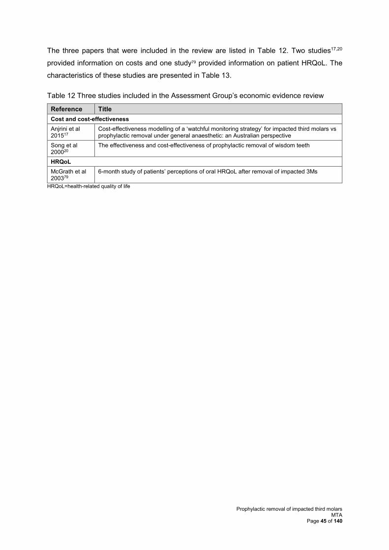
Prophylactic removal of impacted third molars MTA
Page 45 of 140
The three papers that were included in the review are listed in Table 12. Two studies17,20
provided information on costs and one study79 provided information on patient HRQoL. The
characteristics of these studies are presented in Table 13.
Table 12 Three studies included in the Assessment Group’s economic evidence review
Reference Title
Cost and cost-effectiveness
Anjrini et al 201517
Cost-effectiveness modelling of a ‘watchful monitoring strategy’ for impacted third molars vs prophylactic removal under general anaesthetic: an Australian perspective
Song et al 200020
The effectiveness and cost-effectiveness of prophylactic removal of wisdom teeth
HRQoL
McGrath et al 200379
6-month study of patients’ perceptions of oral HRQoL after removal of impacted 3Ms
HRQoL=health-related quality of life
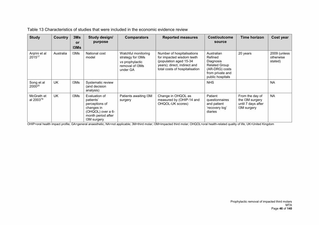
Prophylactic removal of impacted third molars MTA
Page 46 of 140
Table 13 Characteristics of studies that were included in the economic evidence review
Study Country 3Ms
or
I3Ms
Study design/ purpose
Comparators Reported measures Cost/outcome source
Time horizon Cost year
Anjrini et al 201517
Australia I3Ms National cost model
Watchful monitoring strategy for I3Ms
vs prophylactic removal of I3Ms under GA
Number of hospitalisations for impacted wisdom teeth (population aged 15-34 years); direct, indirect and total costs of hospitalisation
Australian Refined Diagnosis Related Group (AR-DRG) costs from private and public hospitals
20 years 2009 (unless otherwise stated)
Song et al 200020
UK I3Ms Systematic review (and decision analysis)
NHS NA
McGrath et al 200379
UK I3Ms Evaluation of patients’ perceptions of changes in (OHQOL) over a 6-month period after I3M surgery
Patients awaiting I3M surgery
Change in OHQOL as measured by (OHIP-14 and OHQOL-UK scores)
Patient questionnaires and patient ‘recovery log’ diaries
From the day of the I3M surgery until 7 days after I3M surgery
NA
OHIP=oral health impact profile; GA=general anaesthetic; NA=not applicable; 3M=third molar; I3M=impacted third molar; OHQOL=oral health-related quality of life; UK=United Kingdom
Prophylactic removal of impacted third molars MTA
Page 47 of 140
4.1.4 Quality of the included evidence
Contrary to the review protocol, the AG made the decision not to quality assess the papers
included in the review of cost-effectiveness evidence using a cost-effectiveness checklist. This
decision was made as only one paper directly considers the cost-effectiveness of prophylactic
removal of I3Ms in a UK setting (Song et al20), and a summary of this paper and its quality are
located in the clinical evidence section of this report (see section 3.2).
4.1.5 Economic review: overview of included papers
The AG concludes that relevant data on I3Ms are limited to three studies.17,20,79 Two of the
papers report details about the cost-effectiveness of the prophylactic removal of I3Ms. The
review by Song et al20 includes details about cost-effectiveness from a UK NHS perspective,
whilst the material presented in the study by Anjrini et al17 is of less direct relevance as
estimates are based on the Australian health care system and results are presented in
Australian dollars. The third paper79 reports findings relating to an assessment of oral HRQoL
after the removal of I3Ms.
4.1.6 Key results: cost-effectiveness of prophylactic removal of I3Ms
Cost-effectiveness: Song et al20
This publication is the AG report for TA1 (title: The effectiveness and cost-effectiveness of
prophylactic removal of wisdom teeth). It includes a summary of findings from a study reported
by Edwards et al,57 who estimated the cost-effectiveness of removal and retention of disease-
free 3Ms and concluded that the cost of prophylactic removal of I3Ms was about 33% higher
than the cost of retention. The Song et al20 report also includes findings from a paper by
Walters,105 who identified that the compensation awarded for permanent nerve damage after
3M surgery ranged from £4,000 to £14,000 per case, or higher (Walters).
Song et al20 conclude that in the short- to medium-term, based on available evidence, retention
of I3Ms may be more cost effective than prophylactic removal.
Cost-effectiveness: Anjrini et al17
The objective of the study reported by Anjrini et al17 was to develop a model to compare the
direct (and indirect) costs associated with a watchful monitoring strategy for I3Ms versus
prophylactic removal under general anaesthetic. Data were obtained from the Western
Australian Hospital Morbidity Data System. All episodes of discharge from all hospitals (private
and public) in Western Australia for the financial year 2008/2009 for the removal of impacted
or embedded teeth as the principal oral condition, as classified by the International
Classification of Diseases, Tenth Revision, Australian Modification (ICD-10-AM), were
Prophylactic removal of impacted third molars MTA
Page 48 of 140
included. The rate calculations for Western Australian Hospitalisation were measured using
population data obtained from the Australian Bureau of Statistics 2006 Census.
The annual direct cost to the state for I3M removal in hospital was estimated to be
AUS$259million, which equates to a direct cost of AUS$2,644 for each hospitalisation (i.e.
£1,536 using 22/08/16 conversion rate). The time frame for the analysis was 20 years. The
average watchful waiting strategy cost per participant was AUS$1,077. This cost included
clinical examinations (AU$60.30) and panoramic radiographs (AUS$47.40), both of which
were undertaken every 2 years. Thus, the estimated annual cost was AUS$53.80 per
individual, which is approximately 1% of the estimated cost of removal. The authors conclude:
‘With no evidence to support the prophylactic removal of asymptomatic
wisdom teeth, a proposed watchful monitoring strategy is a more cost
effective alternative in the Australian context. (page19)
Health-related quality of life: McGrath et al79
The study by McGrath et al79 in 2003 assessed oral HRQoL in patients after removal of I3Ms
over a period of 6 months using two specific oral HRQoL tools and a patient diary. Patients in
the study were a mix of people with asymptomatic (n=19) and symptomatic (n=69) 3Ms. Study
results demonstrated that people who had previously reported having pericoronitis symptoms
achieved greater oral HRQoL gains after I3M surgery than people who had not; the authors
considered the findings to be both statistically and clinically significant.
4.1.7 Cost-effectiveness review: conclusions
As there is very limited clinical effectiveness evidence comparing the prophylactic removal of
I3M versus a ‘watchful waiting’ strategy, it is unsurprising that economic evidence relating to
this comparison is also limited. There are only two published cost-effectiveness studies that
directly consider this comparison and, in both cases, the authors conclude that there is
currently no economic evidence to support the prophylactic removal of I3Ms. However, Song
et al20 restrict their conclusion to a short- to medium-term time frame.
4.2 Independent economic assessment
There are no existing cost–utility analyses that are relevant to the decision problem and
generalisable to the NHS in England. For these reasons, the AG constructed a de novo
economic model to determine the cost-effectiveness of the prophylactic removal of IM3Ms
compared with standard care where standard care refers to what is currently being done (i.e.
no prophylactic removal, referred to as ‘watchful waiting’) in a population with pathology-free
or trouble-free IM3Ms.
Prophylactic removal of impacted third molars MTA
Page 49 of 140
The model perspective is that of the UK NHS only, as Personal Social Services costs are not
relevant to the decision problem. Outcomes were measured in quality-adjusted life years
(QALYs), and both costs and QALYs were discounted at an annual rate of 3.5%, as
recommended by NICE in the Methods Guide to Technology Appraisal.106
In the AG model, in line with the age of the youngest patient recruited to the Fernandes et al
study,44 the AG has chosen to use a starting age of 20 years in the base case; sensitivity
analyses are used to explore the impact of using starting ages of 30, 40 and 50 years.
In the AG model, the base case time horizon is 80 years (i.e. up to the point when people
reach the age of 100 years, when less than 1% of patients are still alive). The time horizon is
varied in scenario analyses (10, 20, 30, 40, 50 years) to assess the impact that this change
has on model outputs.
4.2.1 Model pathways
The elements of both the intervention (prophylactic removal) and comparator (watchful
waiting) pathways were determined through consultation with clinical experts and examination
of clinical data identified via the AG’s systematic review of clinical effectiveness evidence.
In the NHS, the watchful waiting pathway (current standard of care) is complicated and, in
parts, unclear. There are many different points at which a tooth may, or should, be extracted.
For example, the decision to extract a tooth may be determined by a specific number of
instances of pericoronitis, the degree of severity of pericoronitis, a decayed adjacent tooth or
the amount of tooth pain. Following several searches of the literature and consultation with
experts, the AG concluded that data to populate the current watchful waiting pathway are
lacking. The AG has, therefore, chosen to design an economic model that is based on what
happens (i.e. use available data) rather than on what might happen. This approach minimises
the use of assumptions.
A visual representation of the intervention pathway (prophylactic removal) is shown in Figure
3. The pathway is modelled as a combination of a Markov process and decision trees. The
cycle length is one year. In each cycle, a person can develop IM3M symptoms but not have
the IM3M extracted, have the IM3M extracted (either with or without complications) or die from
any cause.
For every person with an IM3M in situ, there is a probability that, in each cycle, the person will
die (from any cause), their IM3M will develop symptoms and/or will be extracted. The
probability of extraction is independent of symptom development. The possible symptoms are
pericoronitis, mild pain and severe pain.
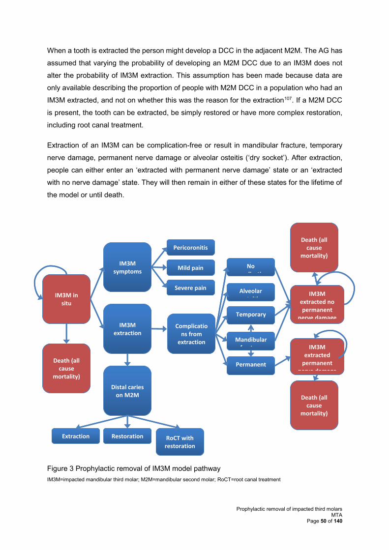
Prophylactic removal of impacted third molars MTA
Page 50 of 140
When a tooth is extracted the person might develop a DCC in the adjacent M2M. The AG has
assumed that varying the probability of developing an M2M DCC due to an IM3M does not
alter the probability of IM3M extraction. This assumption has been made because data are
only available describing the proportion of people with M2M DCC in a population who had an
IM3M extracted, and not on whether this was the reason for the extraction107. If a M2M DCC
is present, the tooth can be extracted, be simply restored or have more complex restoration,
including root canal treatment.
Extraction of an IM3M can be complication-free or result in mandibular fracture, temporary
nerve damage, permanent nerve damage or alveolar osteitis (‘dry socket’). After extraction,
people can either enter an ‘extracted with permanent nerve damage’ state or an ‘extracted
with no nerve damage’ state. They will then remain in either of these states for the lifetime of
the model or until death.
Figure 3 Prophylactic removal of IM3M model pathway
IM3M=impacted mandibular third molar; M2M=mandibular second molar; RoCT=root canal treatment
No complication
Alveolar osteitis
IM3M extraction
Pericoronitis
IM3M symptoms
Death (all cause
mortality)
IM3M in situ
Severe pain
Mild pain
Temporary nerve
Permanent nerve
Mandibular fracture
IM3M extracted no permanent
nerve damage
IM3M extracted
permanent nerve damage
Complications from
extraction
Death (all cause
mortality)
Death (all cause
mortality)
Distal caries on M2M
Extraction Restoration RoCT with restoration
Prophylactic removal of impacted third molars MTA
Page 51 of 140
4.2.2 Model transition probabilities
The transition probabilities used in the AG model have been drawn from published studies
identified in the clinical evidence review and evidence identified by clinical experts. Where
data are absent, assumptions based on clinical advice have been used.
Evidence presented by Fernandes et al44 has been used to represent the annual rate of tooth
extraction and the development of symptoms for people who have asymptomatic IM3Ms. In
this 1-year prospective cohort study, the authors collected data from patients with at least one
IM3M. All of the participants were registered with general dental practices in Scotland where
SIGN guidelines56 recommending a watchful waiting pathway have been in place since 2000.
The AG considered that these data were generalisable to an English NHS setting.
In the paper by Fernandes et al44 the annual rate of extraction (5.47%) was found to be
independent of age. The inclusion criteria in terms of age of the cohort studied by Fernandes
et al44 was 18-70, but only those aged 20 to 63 years were actually recruited into the study.
This determined the model start age of 20. It was assumed that annual rate of extraction
continued at 5.47% after the age of 63. A scenario analysis explores where no extractions
were undertaken after the age of 63.
An annual rate of extraction of 5.47% gives a five year extraction rate of 24.5% which is lower
than that reported by Hill et al43 at 31.4%. To check the impact of varying the annual rate of
extraction, the upper (7.39%pa equivalent to a five year extraction rate of 32.0%) and lower
(3.94%pa) bounds of the 95% CI quoted in the paper by Fernandes et al44 were used in the
AG’s sensitivity analysis.
For the prophylactic removal pathway, the AG’s base case assumption is that not all people
will accept the recommendation that the IM3M(s) should be extracted and, therefore, these
teeth will remain in situ. Fernandes et al44 reports that 45.9% of patients who had their IM3Ms
extracted did not know why the tooth had been extracted and so the extraction could have
been prophylactic (despite the SIGN guideline56 recommendation). Based on this finding, the
AG chose a base case value of 46.95% to represent the proportion of people who would
accept prophylactic removal of asymptomatic IM3Ms, if it were offered to them. The AG
recognises that it is likely that this figure overestimates the true rate of non-prophylactic
removal, as it is unlikely that all of the 46.95% of patients who could not recall why their IM3M
was extracted had the tooth extracted when the IM3M was asymptomatic. However, it could
also be an underestimate if future guidelines were to suggest that prophylactic removal were
to be recommended. Due to these uncertainties, the AG carried out sensitivity analyses to
Prophylactic removal of impacted third molars MTA
Page 52 of 140
explore the impact of different levels of acceptance of prophylactic removal (10%, 25%, 50%,
75% and 100%).
Findings from the Fernandes et al44 study suggest that symptom development is age
dependent (at least up the age of 63 years), declining as a patient ages. In the base case, the
AG assumes that the rate of symptom development follows a linear trend from ages 53 to 63
years and this trend is applied beyond the age of 63 years. A scenario analysis explores the
impact of symptom development no longer occurring past the age of 63 years. The
probabilities, by age, of experiencing symptoms can be found in Appendix 7.
The probability of a person who, on having an IM3M extracted, is found to have an associated
M2M DCC, and the likelihoods of the M2M being extracted, simply restored or undergoing
complex restoration, have been taken from the a study by McArdle et al.107 In this study, the
investigators undertook a retrospective review of 339 people in England across two cohorts
from 2006 and 2014 who had IM3Ms removed due to having an M2M DCC.
Probabilities of specific complications associated with tooth extraction were derived from the
Chuang et al study.83 Chuang et al83 also reported an odds ratio for all complications
(OR=1.46) for patients aged 25 years and over compared with those younger than 25 years.
The AG has adjusted the individual complication rates reported by Chuang et al83 by this odds
ratio to estimate the probability of specific complications for those younger than 25 years and
for those 25 years old and over.
One complication for which there is no evidence available in the study by Chuang et al83 is the
rate of permanent nerve damage. The AG has used a value reported by Valmaseda-Castellón
et al.108 This value has been adjusted for age (people younger than 25 years and 25 years old
or over) using the odds ratio reported by Chuang et al.83
Table 14 shows a full list of probabilities used in the AG model. Rates for death from any cause
have been taken from Office for National Statistics life tables.109
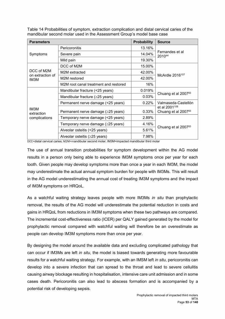
Prophylactic removal of impacted third molars MTA
Page 53 of 140
Table 14 Probabilities of symptom, extraction complication and distal cervical caries of the mandibular second molar used in the Assessment Group’s model base case
Parameters Probability Source
Symptoms
Pericoronitis 13.16% Fernandes et al 201044
Severe pain 14.04%
Mild pain 19.30%
DCC of M2M on extraction of IM3M
DCC of M2M 15.00%
McArdle 2016107 M2M extracted 42.00%
M2M restored 42.00%
M2M root canal treatment and restored 16%
IM3M extraction complications
Mandibular fracture (<25 years) 0.019% Chuang et al 200783
Mandibular fracture (25 years) 0.03%
Permanent nerve damage (<25 years) 0.22% Valmaseda-Castellón et al 2001108 Chuang et al 200783 Permanent nerve damage (25 years) 0.33%
Temporary nerve damage (<25 years) 2.89%
Chuang et al 200783 Temporary nerve damage (25 years) 4.16%
Alveolar osteitis (<25 years) 5.61%
Alveolar osteitis (25 years) 7.98%
DCC=distal cervical caries; M2M=mandibular second molar; IM3M=impacted mandibular third molar
The use of annual transition probabilities for symptom development within the AG model
results in a person only being able to experience IM3M symptoms once per year for each
tooth. Given people may develop symptoms more than once a year in each IM3M, the model
may underestimate the actual annual symptom burden for people with IM3Ms. This will result
in the AG model underestimating the annual cost of treating IM3M symptoms and the impact
of IM3M symptoms on HRQoL.
As a watchful waiting strategy leaves people with more IM3Ms in situ than prophylactic
removal, the results of the AG model will underestimate the potential reduction in costs and
gains in HRQoL from reductions in IM3M symptoms when these two pathways are compared.
The incremental cost-effectiveness ratio (ICER) per QALY gained generated by the model for
prophylactic removal compared with watchful waiting will therefore be an overestimate as
people can develop IM3M symptoms more than once per year.
By designing the model around the available data and excluding complicated pathology that
can occur if IM3Ms are left in situ, the model is biased towards generating more favourable
results for a watchful waiting strategy. For example, with an IMSM left in situ, pericoronitis can
develop into a severe infection that can spread to the throat and lead to severe cellulitis
causing airway blockage resulting in hospitalisation, intensive care unit admission and in some
cases death. Pericoronitis can also lead to abscess formation and is accompanied by a
potential risk of developing sepsis.
Prophylactic removal of impacted third molars MTA
Page 54 of 140
No published data could be found to estimate the risk of these serious events occurring.
However, one of the AG’s clinical experts reports treating one person per month who has been
admitted to the intensive care unit with life-threatening cellulitis due to having had an IM3M.
Although there is no way to generalise this experience into a probability that can be included
in the AG model, if such a complication were to be included it would result in a reduction in
QALYs and increase in costs for the watchful waiting strategy.
4.2.3 Resource use and unit cost estimation
The total number of patients with IM3M extractions each year in the NHS is unknown.
However, McArdle et al107 have estimated that, in 2014/15, there were 152,000 people with
IM3M extractions: 67,000 (44.1%) were carried out during inpatient admissions; 38,000
(25.0%) during outpatient attendances and 47,000 (30.9%) during a primary care
appointment. The AG has used these estimates as the basis for estimating the cost, to the
NHS, of IM3M extractions. Unit costs of extraction in an acute setting have been taken from
NHS Reference Costs (2015/16). The cost of an extraction in primary care is a Band 2
treatment and is charged to the NHS as three Units of Dental Activity (UDA).
The AG has assumed that 75% of M2Ms with DCC are extracted at the same time as the
IM3M, with no additional cost. For those M2Ms extracted independently, the cost of extraction
is assumed to be the same as the estimated cost of IM3M extraction. Sensitivity analyses
have been used to explore the impact of this assumption on model results. Values of
concurrent IM3M and M2M extraction of between 0% and 100% have been used in these
analyses.
To estimate the cost associated with pericoronitis and severe pain, in the absence of any
published information of clinical advice, the AG has assumed that 25% of people will self-
medicate and that symptoms will resolve without the need for dental or medical intervention.
For the 75% that require treatment, dental care will be required. Based upon clinical advice,
this dental care would comprise a Band 2 treatment (3 UDAs) plus an antibiotic prescription
for erythromycin (a further 0.75 UDAs for issuing a prescription). For the 75% of patients with
severe pain, the AG has assumed that an emergency dental appointment will be required (1.2
UDAs) and that people will be prescribed codeine (incurring a further 0.75 UDAs for issuing a
prescription). The AG has undertaken sensitivity analyses to explore the impact on model
results of varying the proportion of people self-treating from 0% to 100%.
The cost of a UDA varies across England. In the base case, the AG has used a figure of £25.
The BDA quotes this figure as being the mean UDA cost across England.110 The AG has
undertaken sensitivity analyses to explore the impact on model results of lower and higher
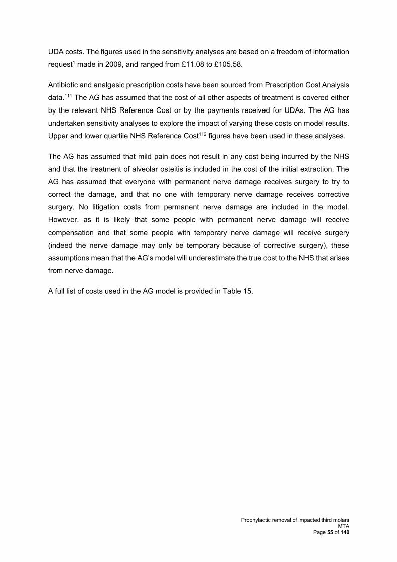
Prophylactic removal of impacted third molars MTA
Page 55 of 140
UDA costs. The figures used in the sensitivity analyses are based on a freedom of information
request1 made in 2009, and ranged from £11.08 to £105.58.
Antibiotic and analgesic prescription costs have been sourced from Prescription Cost Analysis
data.111 The AG has assumed that the cost of all other aspects of treatment is covered either
by the relevant NHS Reference Cost or by the payments received for UDAs. The AG has
undertaken sensitivity analyses to explore the impact of varying these costs on model results.
Upper and lower quartile NHS Reference Cost112 figures have been used in these analyses.
The AG has assumed that mild pain does not result in any cost being incurred by the NHS
and that the treatment of alveolar osteitis is included in the cost of the initial extraction. The
AG has assumed that everyone with permanent nerve damage receives surgery to try to
correct the damage, and that no one with temporary nerve damage receives corrective
surgery. No litigation costs from permanent nerve damage are included in the model.
However, as it is likely that some people with permanent nerve damage will receive
compensation and that some people with temporary nerve damage will receive surgery
(indeed the nerve damage may only be temporary because of corrective surgery), these
assumptions mean that the AG’s model will underestimate the true cost to the NHS that arises
from nerve damage.
A full list of costs used in the AG model is provided in Table 15.
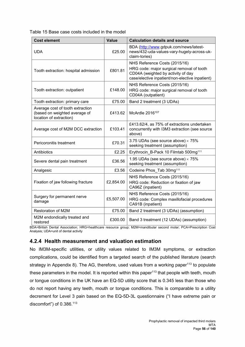
Prophylactic removal of impacted third molars MTA
Page 56 of 140
Table 15 Base case costs included in the model
Cost element Value Calculation details and source
UDA £25.00 BDA (http://www.gdpuk.com/news/latest-news/432-uda-values-vary-hugely-across-uk-claim-tories)
Tooth extraction: hospital admission £801.81
NHS Reference Costs (2015/16)
HRG code: major surgical removal of tooth CD04A (weighted by activity of day case/elective inpatient/non-elective inpatient)
Tooth extraction: outpatient £148.00
NHS Reference Costs (2015/16)
HRG code: major surgical removal of tooth CD04A (outpatient)
Tooth extraction: primary care £75.00 Band 2 treatment (3 UDAs)
Average cost of tooth extraction (based on weighted average of location of extraction)
£413.62 McArdle 2016107
Average cost of M2M DCC extraction £103.41 £413.62/4, as 75% of extractions undertaken concurrently with I3M3 extraction (see source above)
Pericoronitis treatment £70.31 3.75 UDAs (see source above) 75% seeking treatment (assumption)
Antibiotics £2.25 Erythrocin_B-Pack 10 Filmtab 500mg111
Severe dental pain treatment £36.56 1.95 UDAs (see source above) 75% seeking treatment (assumption)
Analgesic £3.56 Codeine Phos_Tab 30mg111
Fixation of jaw following fracture £2,854.00
NHS Reference Costs (2015/16)
HRG code: Reduction or fixation of jaw CA96Z (inpatient)
Surgery for permanent nerve damage
£5,507.00
NHS Reference Costs (2015/16)
HRG code: Complex maxillofacial procedures CA91B (inpatient)
Restoration of M2M £75.00 Band 2 treatment (3 UDAs) (assumption)
M2M endondically treated and restored
£300.00 Band 3 treatment (12 UDAs) (assumption)
BDA=British Dental Association; HRG=healthcare resource group; M2M=mandibular second molar; PCA=Prescription Cost Analysis; UDA=unit of dental activity
4.2.4 Health measurement and valuation estimation
No IM3M-specific utilities, or utility values related to IM3M symptoms, or extraction
complications, could be identified from a targeted search of the published literature (search
strategy in Appendix 8). The AG, therefore, used values from a working paper113 to populate
these parameters in the model. It is reported within this paper113 that people with teeth, mouth
or tongue conditions in the UK have an EQ-5D utility score that is 0.345 less than those who
do not report having any teeth, mouth or tongue conditions. This is comparable to a utility
decrement for Level 3 pain based on the EQ-5D-3L questionnaire (“I have extreme pain or
discomfort”) of 0.386.113
Prophylactic removal of impacted third molars MTA
Page 57 of 140
The AG model includes a 0.345 utility decrement to represent the experience of people with
any IM3M complication(s) or any complication(s) following extraction. For people experiencing
mild pain, the decrement was assumed to be 50% of 0.345 (i.e. 0.1725). Sensitivity analyses
were undertaken to explore the impact of varying these figures on model results. The
decrements were varied individually over the 95% CI (0.102 to 0.549) reported in the paper
by Ara and Brazier.113 The AG has assumed that M2M restoration is not associated with any
loss of utility and that extraction of the M2M would only result in a loss of utility if it occurred
independently of an IM3M extraction.
To generate a QALY loss when a person experiences a complication, the duration of
symptoms from the complication is required. The AG was not able to identify any evidence
describing the duration of symptoms associate with extraction complications. Thus, it was
necessary to make a number of assumptions about the durations of symptoms; the AG’s
clinical experts were contacted to verify these assumptions. The AG has undertaken sensitivity
analyses to assess the impact of varying these estimates of duration on model results
(duration varied by ±50%). To reflect declining HRQoL as people age in the model, utility
declines with age in line with age-related population norms described in the paper by Ara and
Brazier.113
Details of the values used in the model to represent utility decrement, duration of symptoms
assumed, and the resulting QALY loss associated with symptoms and complications are
shown in Table 16.
Table 16 Base case utility decrements and symptom duration
Cause of utility decrement Utility decrement
Duration of symptoms QALY loss
Symptoms
Pericoronitis 0.345 9 days 0.009
Severe pain 0.345 30 days 0.028
Mild pain 0.1725 30 days 0.014
Extraction IM3M or M2M 0.345 7 days 0.007
Complications following IM3M extraction
Mandibular fracture
0.345 42 days
0.040
Permanent nerve damage
0.345 Lifetime
0.345
Temporary nerve damage
0.345 30 days
0.028
Alveolar osteitis 0.345 9 days 0.009
IM3M=impacted mandibular third molar; M2M=mandibular second molar; QALY=quality-adjusted life year
Prophylactic removal of impacted third molars MTA
Page 58 of 140
4.2.5 Analysis of uncertainty
The AG explored the uncertainty surrounding model assumptions using deterministic
sensitivity analyses and scenario analyses. Probabilistic sensitivity analysis was not
undertaken as the only parameter for which a distribution could be drawn based solely on
published evidence was the annual rate of extraction. Creating distributions around the central
value of other parameters would not be meaningful either because the parameter values are
essentially fixed (e.g. in the case of costs) or because the central values are, at least partly,
based on assumptions due to lack of data (e.g. the actual utility values associated with
symptoms or the duration of symptoms). Furthermore, a probabilistic sensitivity analysis could
potentially lead to confounding results, as the uncertainty around most of the assumptions
used in the AG model is not known and, therefore, from a statistical perspective, cannot be
confidently modelled.
The parameter ranges explored in deterministic sensitivity analysis are summarised in Table
17, and the scenario analyses performed are summarised in Table 18.
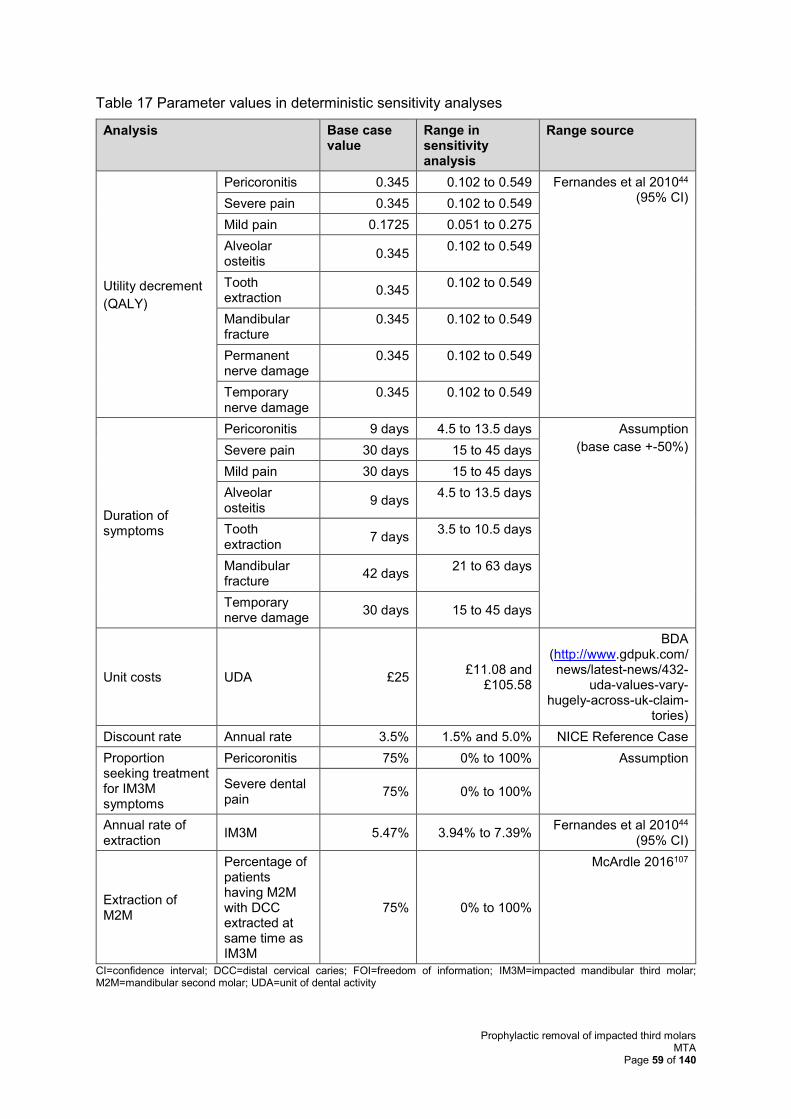
Prophylactic removal of impacted third molars MTA
Page 59 of 140
Table 17 Parameter values in deterministic sensitivity analyses
Analysis Base case value
Range in sensitivity analysis
Range source
Utility decrement
(QALY)
Pericoronitis 0.345 0.102 to 0.549 Fernandes et al 201044 (95% CI) Severe pain 0.345 0.102 to 0.549
Mild pain 0.1725 0.051 to 0.275
Alveolar osteitis
0.345 0.102 to 0.549
Tooth extraction
0.345 0.102 to 0.549
Mandibular fracture
0.345 0.102 to 0.549
Permanent nerve damage
0.345 0.102 to 0.549
Temporary nerve damage
0.345 0.102 to 0.549
Duration of symptoms
Pericoronitis 9 days 4.5 to 13.5 days Assumption
(base case +-50%) Severe pain 30 days 15 to 45 days
Mild pain 30 days 15 to 45 days
Alveolar osteitis
9 days 4.5 to 13.5 days
Tooth extraction
7 days 3.5 to 10.5 days
Mandibular fracture
42 days 21 to 63 days
Temporary nerve damage
30 days 15 to 45 days
Unit costs UDA £25 £11.08 and
£105.58
BDA (http://www.gdpuk.com/news/latest-news/432-
uda-values-vary-hugely-across-uk-claim-
tories)
Discount rate Annual rate 3.5% 1.5% and 5.0% NICE Reference Case
Proportion seeking treatment for IM3M symptoms
Pericoronitis 75% 0% to 100% Assumption
Severe dental pain
75% 0% to 100%
Annual rate of extraction
IM3M 5.47% 3.94% to 7.39% Fernandes et al 201044
(95% CI)
Extraction of M2M
Percentage of patients having M2M with DCC extracted at same time as IM3M
75% 0% to 100%
McArdle 2016107
CI=confidence interval; DCC=distal cervical caries; FOI=freedom of information; IM3M=impacted mandibular third molar; M2M=mandibular second molar; UDA=unit of dental activity
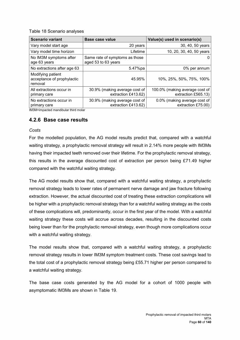
Prophylactic removal of impacted third molars MTA
Page 60 of 140
Table 18 Scenario analyses
Scenario variant Base case value Value(s) used in scenario(s)
Vary model start age 20 years 30, 40, 50 years
Vary model time horizon Lifetime 10, 20, 30, 40, 50 years
No IM3M symptoms after age 63 years
Same rate of symptoms as those aged 53 to 63 years
0
No extractions after age 63 5.47%pa 0% per annum
Modifying patient acceptance of prophylactic removal
45.95% 10%, 25%, 50%, 75%, 100%
All extractions occur in primary care
30.9% (making average cost of extraction £413.62)
100.0% (making average cost of extraction £565.13)
No extractions occur in primary care
30.9% (making average cost of extraction £413.62)
0.0% (making average cost of extraction £75.00)
IM3M=impacted mandibular third molar
4.2.6 Base case results
Costs
For the modelled population, the AG model results predict that, compared with a watchful
waiting strategy, a prophylactic removal strategy will result in 2.14% more people with IM3Ms
having their impacted teeth removed over their lifetime. For the prophylactic removal strategy,
this results in the average discounted cost of extraction per person being £71.49 higher
compared with the watchful waiting strategy.
The AG model results show that, compared with a watchful waiting strategy, a prophylactic
removal strategy leads to lower rates of permanent nerve damage and jaw fracture following
extraction. However, the actual discounted cost of treating these extraction complications will
be higher with a prophylactic removal strategy than for a watchful waiting strategy as the costs
of these complications will, predominantly, occur in the first year of the model. With a watchful
waiting strategy these costs will accrue across decades, resulting in the discounted costs
being lower than for the prophylactic removal strategy, even though more complications occur
with a watchful waiting strategy.
The model results show that, compared with a watchful waiting strategy, a prophylactic
removal strategy results in lower IM3M symptom treatment costs. These cost savings lead to
the total cost of a prophylactic removal strategy being £55.71 higher per person compared to
a watchful waiting strategy.
The base case costs generated by the AG model for a cohort of 1000 people with
asymptomatic IM3Ms are shown in Table 19.
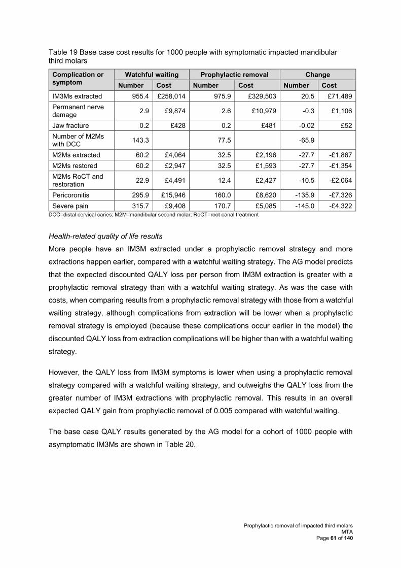
Prophylactic removal of impacted third molars MTA
Page 61 of 140
Table 19 Base case cost results for 1000 people with symptomatic impacted mandibular third molars
Complication or symptom
Watchful waiting Prophylactic removal Change
Number Cost Number Cost Number Cost
IM3Ms extracted 955.4 £258,014 975.9 £329,503 20.5 £71,489
Permanent nerve damage
2.9 £9,874 2.6 £10,979 -0.3 £1,106
Jaw fracture 0.2 £428 0.2 £481 -0.02 £52
Number of M2Ms with DCC
143.3 77.5 -65.9
M2Ms extracted 60.2 £4,064 32.5 £2,196 -27.7 -£1,867
M2Ms restored 60.2 £2,947 32.5 £1,593 -27.7 -£1,354
M2Ms RoCT and restoration
22.9 £4,491 12.4 £2,427 -10.5 -£2,064
Pericoronitis 295.9 £15,946 160.0 £8,620 -135.9 -£7,326
Severe pain 315.7 £9,408 170.7 £5,085 -145.0 -£4,322
DCC=distal cervical caries; M2M=mandibular second molar; RoCT=root canal treatment
Health-related quality of life results
More people have an IM3M extracted under a prophylactic removal strategy and more
extractions happen earlier, compared with a watchful waiting strategy. The AG model predicts
that the expected discounted QALY loss per person from IM3M extraction is greater with a
prophylactic removal strategy than with a watchful waiting strategy. As was the case with
costs, when comparing results from a prophylactic removal strategy with those from a watchful
waiting strategy, although complications from extraction will be lower when a prophylactic
removal strategy is employed (because these complications occur earlier in the model) the
discounted QALY loss from extraction complications will be higher than with a watchful waiting
strategy.
However, the QALY loss from IM3M symptoms is lower when using a prophylactic removal
strategy compared with a watchful waiting strategy, and outweighs the QALY loss from the
greater number of IM3M extractions with prophylactic removal. This results in an overall
expected QALY gain from prophylactic removal of 0.005 compared with watchful waiting.
The base case QALY results generated by the AG model for a cohort of 1000 people with
asymptomatic IM3Ms are shown in Table 20.
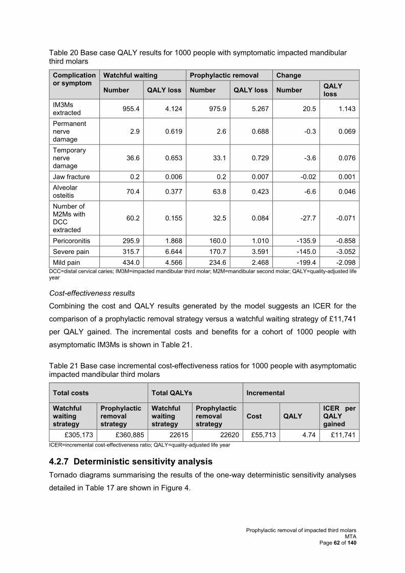
Prophylactic removal of impacted third molars MTA
Page 62 of 140
Table 20 Base case QALY results for 1000 people with symptomatic impacted mandibular third molars
Complication or symptom
Watchful waiting Prophylactic removal Change
Number QALY loss Number QALY loss Number QALY loss
IM3Ms extracted
955.4 4.124 975.9 5.267 20.5 1.143
Permanent nerve damage
2.9 0.619 2.6 0.688 -0.3 0.069
Temporary nerve damage
36.6 0.653 33.1 0.729 -3.6 0.076
Jaw fracture 0.2 0.006 0.2 0.007 -0.02 0.001
Alveolar osteitis
70.4 0.377 63.8 0.423 -6.6 0.046
Number of M2Ms with DCC extracted
60.2 0.155 32.5 0.084 -27.7 -0.071
Pericoronitis 295.9 1.868 160.0 1.010 -135.9 -0.858
Severe pain 315.7 6.644 170.7 3.591 -145.0 -3.052
Mild pain 434.0 4.566 234.6 2.468 -199.4 -2.098
DCC=distal cervical caries; IM3M=impacted mandibular third molar; M2M=mandibular second molar; QALY=quality-adjusted life year
Cost-effectiveness results
Combining the cost and QALY results generated by the model suggests an ICER for the
comparison of a prophylactic removal strategy versus a watchful waiting strategy of £11,741
per QALY gained. The incremental costs and benefits for a cohort of 1000 people with
asymptomatic IM3Ms is shown in Table 21.
Table 21 Base case incremental cost-effectiveness ratios for 1000 people with asymptomatic impacted mandibular third molars
Total costs Total QALYs Incremental
Watchful waiting strategy
Prophylactic removal strategy
Watchful waiting strategy
Prophylactic removal strategy
Cost QALY ICER per QALY gained
£305,173 £360,885 22615 22620 £55,713 4.74 £11,741
ICER=incremental cost-effectiveness ratio; QALY=quality-adjusted life year
4.2.7 Deterministic sensitivity analysis
Tornado diagrams summarising the results of the one-way deterministic sensitivity analyses
detailed in Table 17 are shown in Figure 4.
Prophylactic removal of impacted third molars MTA
Page 63 of 140
The tornado diagram shows that the most important parameters that affect model results are
the discount rate, the cost of a UDA, the annual extraction rate for IM3Ms, the utility
decrements applied to IM3M symptoms and the duration of the symptoms.
Although the results are most sensitive to changes in the discount rate – which is to be
expected given that the prophylactic strategy effectively ‘front loads’ extraction costs in the
model compared with the watchful waiting strategy, which spreads those cost over many years
– the ICER is still under £20,000 per QALY gained even when the discount rate is 5% per
annum. The AG considers that although the 3.5% per annum discount rate is the correct one
to apply, an argument could be made that prophylactic removal constitutes a public health
intervention similar to vaccination and so a 1.5% per annum discount rate should be applied
instead. At this lower rate, the model predicts an ICER of £3,377 per QALY gained for the
prophylactic removal strategy.
Given that there is no direct evidence on the QALY loss from symptoms of IM3Ms, it is
potentially concerning that the ICER per QALY gained is sensitive to values used in the AG
model to estimate the IM3M symptom QALY loss. However, over all of the parameter ranges
considered, only the lower bound for the utility decrement for severe pain (0.102 as opposed
to 0.345 in the base case) results in an ICER above £20,000 per QALY gained for prophylactic
removal (£21,469 per QALY gained).
As stated previously there is evidence from Hill et al43 that the annual rate of extraction of
IM3Ms is closer to the upper bound of 7.39%pa reported by Fernandes et al44 rather than the
5.47%pa used in the base case. If the annual rate of extraction was 7.39%pa then the ICER
would be £9,944 per QALY gained. However, even if the annual rate of extraction was at the
lower bound suggested by Fernandes et al44of 3.94%, the ICER would be £13,847 per QALY
gained which is still below the £20,000 per QALY gained threshold.
As stated previously, the cost of a UDA varies widely across the country. The value used in
the AG base case (£25) is at the lower end of the potential range used in England (£11.08 to
£105.58). At the lower UDA price (£11.08), the ICER for prophylactic removal is £12,925 per
QALY gained and thus remains below the £20,000 per QALY gained threshold. For
geographical areas where the UDA price is higher than the £25 base case value, the ICER
per QALY gained increasingly favours a prophylactic removal strategy.
The AG model results are insensitive to several parameters where no information was
available and assumptions had to be made. For example, the duration of symptoms for
complications from extraction, the percentage of patients seeking treatment for pericoronitis
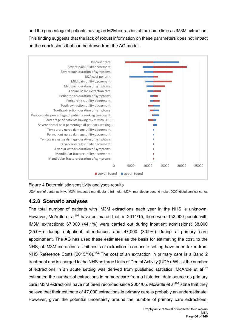
Prophylactic removal of impacted third molars MTA
Page 64 of 140
and the percentage of patients having an M2M extraction at the same time as IM3M extraction.
This finding suggests that the lack of robust information on these parameters does not impact
on the conclusions that can be drawn from the AG model.
Figure 4 Deterministic sensitivity analyses results
UDA=unit of dental activity; IM3M=impacted mandibular third molar; M2M=mandibular second molar; DCC=distal cervical caries
4.2.8 Scenario analyses
The total number of patients with IM3M extractions each year in the NHS is unknown.
However, McArdle et al107 have estimated that, in 2014/15, there were 152,000 people with
IM3M extractions: 67,000 (44.1%) were carried out during inpatient admissions; 38,000
(25.0%) during outpatient attendances and 47,000 (30.9%) during a primary care
appointment. The AG has used these estimates as the basis for estimating the cost, to the
NHS, of IM3M extractions. Unit costs of extraction in an acute setting have been taken from
NHS Reference Costs (2015/16).114 The cost of an extraction in primary care is a Band 2
treatment and is charged to the NHS as three Units of Dental Activity (UDA). Whilst the number
of extractions in an acute setting was derived from published statistics, McArdle et al107
estimated the number of extractions in primary care from a historical data source as primary
care IM3M extractions have not been recorded since 2004/05. McArdle et al107 state that they
believe that their estimate of 47,000 extractions in primary care is probably an underestimate.
However, given the potential uncertainty around the number of primary care extractions,
0 5000 10000 15000 20000 25000
Mandibular fracture duration of symptoms
Mandibular fracture utility decrement
Alveolar osteitis duration of symptoms
Alveolar osteitis utility decrement
Temporary nerve damage duration of symptoms
Permanent nerve damage utility decrement
Temporary nerve damage utility decrement
Severe dental pain percentage of patients seeking…
Percentage of patients having M2M with DCC…
Pericoronitis percentage of patients seeking treatment
Tooth extraction duration of symptoms
Tooth extraction utility decrement
Pericoronitis utility decrement
Pericoronitis duration of symptoms
Annual IM3M extraction rate
Mild pain duration of symptoms
Mild pain utility decrement
UDA cost per unit
Severe pain duration of symptoms
Severe pain utility decrement
Discount rate
Lower Bound upper Bound
Prophylactic removal of impacted third molars MTA
Page 65 of 140
scenario analyses were undertaken where (i) no extractions occurred in primary care and (ii)
all extractions occurred in primary care.
Proportion of extractions occurring in primary care
Increasing the proportion of people having extractions in primary care reduces the average
cost of extraction per person and vice versa.
If all extractions were to take place in primary care, the average cost per extraction decreases
to £75 and the ICER per QALY gained decreases such that prophylactic removal becomes a
dominant strategy compared to watchful waiting. If no extractions take place in primary care,
the average cost per extraction increases to £565.13 and the ICER increases to £17,116 per
QALY gained for prophylactic removal. The results of this scenario analysis are shown in Table
22.
Table 22 Impact on cost-effectiveness results from assuming 100% and 0% of impacted mandibular third molars extractions occur in primary care
IM3M symptoms
Total costs
Total QALYs Incremental
Watchful waiting strategy
Prophylactic removal strategy
Watchful waiting strategy
Prophylactic removal strategy
Cost QALY ICER per QALY gained
Base case £305,173 £360,885 22615 22620 £55,713 4.74 £11,741
100% of extractions in primary care
£90,616 £89,331 22615 22620 -£1,285 4.74 Dominates
0% of extractions in primary care
£401,173 £482,388 22615 22620 £81,215 4.74 £17,116
QALYs=quality adjusted life years; ICER=incremental cost effectiveness ratio
Model start age
The age at which people enter the model was varied between 30 and 50 years (base case=20
years). As the age at the start increases, so does the ICER per QALY gained for the
comparison of a prophylactic removal strategy with a watchful waiting strategy. However, even
when the starting age was set at 50 years, the ICER for this comparison remains below
£20,000 per QALY gained. The results of these sensitivity analyses are presented in Table
23.
Prophylactic removal of impacted third molars MTA
Page 66 of 140
Table 23 Impact on cost-effectiveness results of varying the start age
Model start age
Total costs Total QALYs Incremental
Watchful waiting strategy
Prophylactic removal strategy
Watchful waiting strategy
Prophylactic removal strategy
Cost QALY ICER per QALY gained
Base case
(20 years) £305,173 £360,885 22615 22620 £55,713 4.74 £11,741
30 years £303,175 £362,483 20616 20620 £59,308 4.36 £13,609
40 years £296,341 £358,764 18190 18194 £62,423 4.22 £14,787
50 years £283,219 £351,611 15335 15339 £68,392 3.94 £17,348
ICER=incremental cost-effectiveness ratio; QALY=quality-adjusted life year
Time horizon
The time horizon used in the AG model was varied in 10-year increments between 10 and 50
years. The results of these scenarios compared with the base case results are shown in Table
24.
As the prophylactic removal strategy ‘front loads’ the costs of extraction compared to a
watchful waiting strategy, the ICER per QALY gained is sensitive to the time horizon employed
in the model. The shorter the time horizon, there are fewer people in the watchful waiting
strategy who have an extraction and develop symptoms due their IM3Ms. By 21 years, the
ICER per QALY gained for the comparison of a prophylactic removal strategy with a watchful
waiting strategy has fallen below £20,000.
Table 24 Impact of varying the model time horizon on cost-effectiveness results
Total costs Total QALYs Incremental
Watchful waiting strategy
Prophylactic removal strategy
Watchful waiting strategy
Prophylactic removal strategy
Cost QALY
ICER per QALY gained
Base case (Lifetime)
£305,173 £360,885 22615 22620 £55,713 4.74 £11,741
10 years £186,095 £296,515 7364 7365 £110,419 1.57 £70,310
20 years £259,364 £336,122 13102 13106 £76,758 3.72 £20,620
30 years £288,021 £351,613 16987 16992 £63,593 4.42 £14,401
40 years £299,141 £357,625 19538 19543 £58,484 4.64 £12,598
50 years £303,325 £359,886 21184 21188 £56,562 4.72 £11,994
ICER=incremental cost-effectiveness ratio; QALY=quality-adjusted life year
No IM3M symptoms after the age of 63 years
The assumption that people continue to have IM3M symptoms after the age of 63 years makes
a minor difference to the size of the ICER per QALY gained. When comparing a prophylactic
removal strategy with a watchful waiting strategy, removing the assumption so that no people
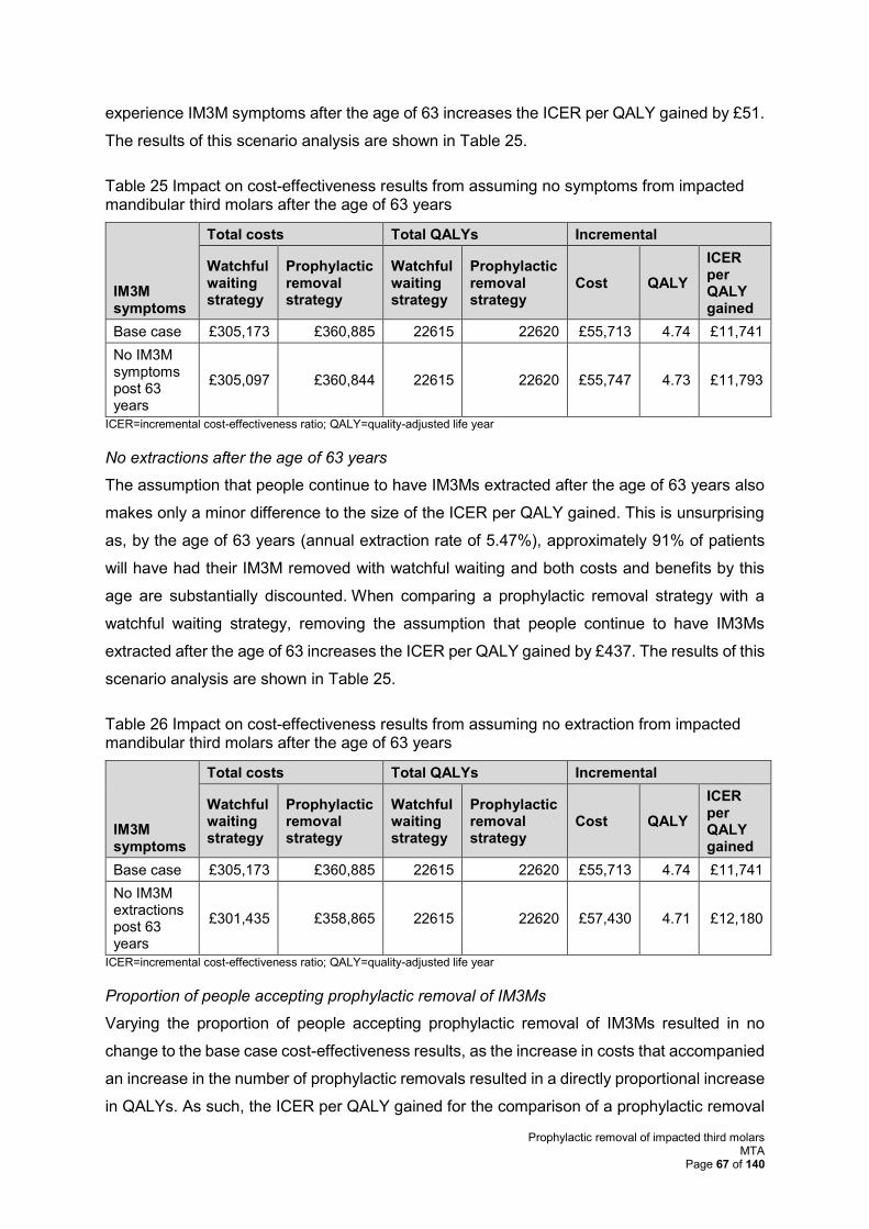
Prophylactic removal of impacted third molars MTA
Page 67 of 140
experience IM3M symptoms after the age of 63 increases the ICER per QALY gained by £51.
The results of this scenario analysis are shown in Table 25.
Table 25 Impact on cost-effectiveness results from assuming no symptoms from impacted mandibular third molars after the age of 63 years
IM3M symptoms
Total costs Total QALYs Incremental
Watchful waiting strategy
Prophylactic removal strategy
Watchful waiting strategy
Prophylactic removal strategy
Cost QALY
ICER per QALY gained
Base case £305,173 £360,885 22615 22620 £55,713 4.74 £11,741
No IM3M symptoms post 63 years
£305,097 £360,844 22615 22620 £55,747 4.73 £11,793
ICER=incremental cost-effectiveness ratio; QALY=quality-adjusted life year
No extractions after the age of 63 years
The assumption that people continue to have IM3Ms extracted after the age of 63 years also
makes only a minor difference to the size of the ICER per QALY gained. This is unsurprising
as, by the age of 63 years (annual extraction rate of 5.47%), approximately 91% of patients
will have had their IM3M removed with watchful waiting and both costs and benefits by this
age are substantially discounted. When comparing a prophylactic removal strategy with a
watchful waiting strategy, removing the assumption that people continue to have IM3Ms
extracted after the age of 63 increases the ICER per QALY gained by £437. The results of this
scenario analysis are shown in Table 25.
Table 26 Impact on cost-effectiveness results from assuming no extraction from impacted mandibular third molars after the age of 63 years
IM3M symptoms
Total costs Total QALYs Incremental
Watchful waiting strategy
Prophylactic removal strategy
Watchful waiting strategy
Prophylactic removal strategy
Cost QALY
ICER per QALY gained
Base case £305,173 £360,885 22615 22620 £55,713 4.74 £11,741
No IM3M extractions post 63 years
£301,435 £358,865 22615 22620 £57,430 4.71 £12,180
ICER=incremental cost-effectiveness ratio; QALY=quality-adjusted life year
Proportion of people accepting prophylactic removal of IM3Ms
Varying the proportion of people accepting prophylactic removal of IM3Ms resulted in no
change to the base case cost-effectiveness results, as the increase in costs that accompanied
an increase in the number of prophylactic removals resulted in a directly proportional increase
in QALYs. As such, the ICER per QALY gained for the comparison of a prophylactic removal
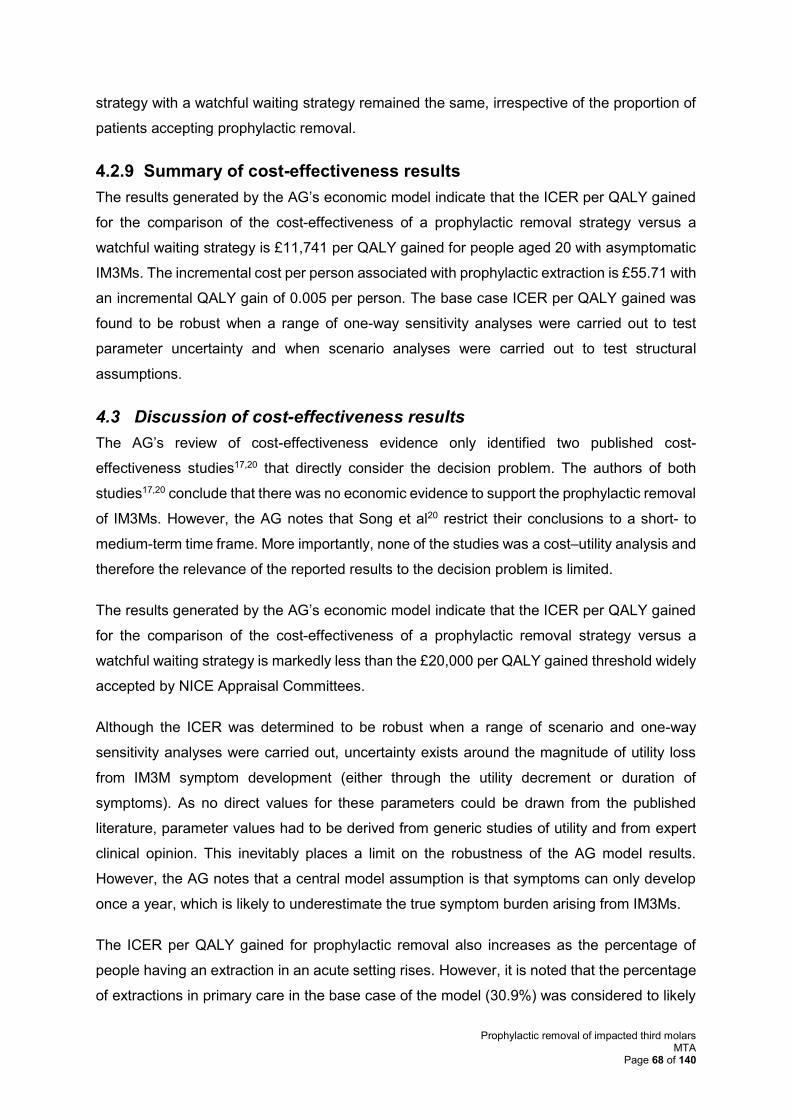
Prophylactic removal of impacted third molars MTA
Page 68 of 140
strategy with a watchful waiting strategy remained the same, irrespective of the proportion of
patients accepting prophylactic removal.
4.2.9 Summary of cost-effectiveness results
The results generated by the AG’s economic model indicate that the ICER per QALY gained
for the comparison of the cost-effectiveness of a prophylactic removal strategy versus a
watchful waiting strategy is £11,741 per QALY gained for people aged 20 with asymptomatic
IM3Ms. The incremental cost per person associated with prophylactic extraction is £55.71 with
an incremental QALY gain of 0.005 per person. The base case ICER per QALY gained was
found to be robust when a range of one-way sensitivity analyses were carried out to test
parameter uncertainty and when scenario analyses were carried out to test structural
assumptions.
4.3 Discussion of cost-effectiveness results
The AG’s review of cost-effectiveness evidence only identified two published cost-
effectiveness studies17,20 that directly consider the decision problem. The authors of both
studies17,20 conclude that there was no economic evidence to support the prophylactic removal
of IM3Ms. However, the AG notes that Song et al20 restrict their conclusions to a short- to
medium-term time frame. More importantly, none of the studies was a cost–utility analysis and
therefore the relevance of the reported results to the decision problem is limited.
The results generated by the AG’s economic model indicate that the ICER per QALY gained
for the comparison of the cost-effectiveness of a prophylactic removal strategy versus a
watchful waiting strategy is markedly less than the £20,000 per QALY gained threshold widely
accepted by NICE Appraisal Committees.
Although the ICER was determined to be robust when a range of scenario and one-way
sensitivity analyses were carried out, uncertainty exists around the magnitude of utility loss
from IM3M symptom development (either through the utility decrement or duration of
symptoms). As no direct values for these parameters could be drawn from the published
literature, parameter values had to be derived from generic studies of utility and from expert
clinical opinion. This inevitably places a limit on the robustness of the AG model results.
However, the AG notes that a central model assumption is that symptoms can only develop
once a year, which is likely to underestimate the true symptom burden arising from IM3Ms.
The ICER per QALY gained for prophylactic removal also increases as the percentage of
people having an extraction in an acute setting rises. However, it is noted that the percentage
of extractions in primary care in the base case of the model (30.9%) was considered to likely
Prophylactic removal of impacted third molars MTA
Page 69 of 140
be an underestimate by the authors of the study (McArdle et al105) from where the proportions
of location of extraction were derived. The greater the percentage of people that can have
their IM3Ms extracted in a primary care setting, the more cost effective prophylactic removal
becomes.
In addition to the limit of only developing symptoms once a year, eight other model
assumptions also suggest that the base case ICER per QALY gained generated by the model
may be conservative:
1. No serious complications arising from IM3M symptoms (e.g. from severe infection) are
included in the model.
2. No disutility from M2M decay and restorative treatment (including root canal treatment)
is included in the model.
3. Expert clinical advice to the AG is that tooth extraction becomes much more difficult as
people age, which could result in the extraction being more complex and, therefore,
more costly. The only way that this increase in difficulty is represented in the model is
through an increase in the complication rate.
4. No litigation costs from permanent nerve damage and/or fracture are included in the
model.
5. No surgical treatment costs for temporary nerve damage or ongoing costs of treating
permanent nerve damage are included in the model.
6. No costs of additional check-ups or x-rays are included for a watchful waiting strategy.
Routine dental care costs are assumed to be identical regardless of whether the IM3M
is in situ or not.
7. All people with IM3Ms are treated equally in the model regardless of impaction status
(i.e. whether partially erupted or a bony impaction). Expert clinical advice to the AG is
that it is only partially erupted teeth that tend to cause problems. If this is the case,
then both the extraction and complication rates reported by Fernandes et al44 (this
study included people with both bony and partially erupted IM3Ms) would be an
underestimate of the rates for people with partially erupted IM3Ms. The base case
ICERs generated by the model are, therefore, overestimates for those with partially
erupted IM3Ms and underestimates for those with bony impaction.
8. Patients who develop symptoms in the model but do not have their teeth removed have
the same risk of developing symptoms in future years as if they had stayed
asymptomatic. It may be the case that patients who develop symptoms are either
Prophylactic removal of impacted third molars MTA
Page 70 of 140
inherently more prone to symptom development, or prior symptoms and treatment
increase the likelihood of developing future symptoms.
The AG model results are ultimately driven by the finding that most individuals with
asymptomatic IM3Ms will eventually have their IM3Ms extracted. This finding arises from the
AG’s long-term extrapolation of the annual extraction data reported in the study by Fernandes
et al.44 In this sense, a watchful waiting strategy may be more accurately described as ‘putting
off the inevitable’. The findings reported by Fernandes et al44 on the rate of extraction of IM3Ms
are supported by the results of Hill et al.43 Importantly, even if the annual rates of extraction
are substantially lower then used in the model base case, the ICER per QALY gained remains
below £20,000.
Prophylactic removal of impacted third molars MTA
Page 71 of 140
5 DISCUSSION
5.1 Statement of principle findings
The SR of clinical evidence found no RCT data to support or refute the prophylactic removal
of pathology-free/trouble-free IM3Ms. The authors of the two included studies54,55 investigating
the rate of surgical complications concluded that no serious complications were reported, and
the three longitudinal studies43,44,54 assessing outcomes of retained IM3Ms reported varying
extraction rates due to the different lengths of follow-up. No studies reported the impact of
retention on the status of the second molars.
As there is very limited clinical effectiveness evidence comparing the prophylactic removal of
I3M versus a watchful waiting strategy, it is unsurprising that economic evidence relating to
this comparison is also limited. The two published cost-effectiveness studies17,20 that directly
consider this comparison conclude that there is currently no economic evidence to support the
prophylactic removal of I3Ms.
The results generated by the AG’s de novo economic model indicate that the ICER per QALY
gained for the comparison of the cost-effectiveness of a prophylactic removal strategy versus
a watchful waiting strategy is £11,741 per QALY gained for people aged 20 with asymptomatic
IM3Ms. The incremental cost per person associated with prophylactic extraction is £55.71 with
an incremental QALY gain of 0.005 per person. The base case ICER per QALY gained was
found to be robust when a range of one-way sensitivity analyses were carried out to test
parameter uncertainty and when scenario analyses were carried out to test structural
assumptions.
While the available published economic evidence is limited, the findings that prophylactic
removal is not cost-effective would seem to be contradicted by the findings from the results of
the AG’s de novo model. There are several reasons that may explain this apparent
contradiction. First, the model time horizon is important, as shown by the results of the
scenario analysis in Table 24. It is unlikely that economic models that only consider the short-
and medium-term would show that a prophylactic removal strategy was more cost effective
than a watchful waiting strategy. Second, there are data available43,44 on the annual rate of
extraction and symptom development in the UK under a watchful waiting strategy that were
not available at the time of the Song et al study.20 Additional sources of information107 are also
now available on, for example, the rate of M2M DCCs due to IM3Ms. Third, the costs of
extraction have now been robustly estimated and are significantly lower than those previously
estimated (see, for example, Anjrini et al17). Lower costs of extraction will make it more likely
that a prophylactic removal strategy will be more cost effective than a watchful waiting
Prophylactic removal of impacted third molars MTA
Page 72 of 140
strategy. Finally, the studies included in the review of cost-effectiveness evidence were not
cost–utility analyses and so the analysis of effectiveness in these studies is fundamentally
different to that considered here. The AG is, therefore, not surprised that the results generated
by the de novo model differ from those published previously.17,20
5.2 Strengths and limitations of the assessment
The main strength of this review is the breadth of literature that was considered. All clinical
study designs and SRs were included in an attempt to identify all relevant literature. However,
two limitations were the date of the search and the very specific population outlined in the
decision problem. As this review was an update of the current NICE guidance2 published in
2000, the conducted searches were from 2000 to 2016. The population outlined in the decision
problem was “People with pathology-free or trouble-free impacted mandibular third molars”;
much of the literature cited by the professional stakeholders and the identified SRs did not
provide data on the position of the tooth (maxillary or mandibular), whether teeth were
impacted or not, and several did not provide information on the state of the tooth (i.e.
pathology-free or trouble-free). This severely limited the number of studies relevant to this
review.
The findings of this review were in line with those from the other nine identified SRs,20,36,38-
42,46,52 suggesting that the limitations of this review did not overly affect the conclusions.
The strength of the de novo economic model is the use of existing evidence44 on the annual
rates of symptom development and on the annual rates of extraction of IM3Ms that are
currently pathology-free/trouble free. All of the assumptions employed in the model suggest
that the base case ICER per QALY gained for the comparison of prophylactic removal versus
watchful waiting may be conservative. An additional strength of the model is that it is robust to
variations across the range of parameter values that could be considered clinically plausible.
The economic model was limited by the lack of direct utility evidence around IM3M symptoms.
However, suitable proxies could be found and the cost-effectiveness findings were robust
across a range of potential values that could be chosen.
5.3 Uncertainties
The AG model results are driven by figures reported by Fernandes et al44 and McArdle et al.107
While the results reported by Fernandes et al44 on the rate of extraction of IM3Ms are
supported by results reported by Hill et al,43 the results reported by McArdle et al107 on the
different proportions of people having IM3Ms extracted in non-acute NHS settings have yet to
be confirmed by other studies. However, even if all extractions were carried out in the acute
Prophylactic removal of impacted third molars MTA
Page 73 of 140
setting, the size of the ICER for prophylactic removal versus watchful waiting would still remain
below £20,000 per QALY gained.
Model results show that the difference in costs between the prophylactic removal strategy and
the watchful waiting strategy is £55.71 per person. With such a small difference in the cost,
the level of confidence in the utility associated with the two strategies gains importance.
Although the model has been shown to be robust to variations in utility values, all of the utility
values used in the model are based on a figure published in 2010 study by Ara and Brazier;113
this figure has been adjusted for use in the AG model using assumptions verified by a clinical
expert.
6 CONCLUSIONS
6.1 Implications for service provision
The reintroduction of the prophylactic removal of pathology-free/trouble-free IM3Ms will have
resource implications both in primary and secondary care settings, with the rate of pathology-
free I3M3 extractions increasing. Expert clinical advice to the AG is that it can be argued that
the cost would be offset by the reduction in the number of complicated extractions being
performed when people are older. There is no published evidence that this is the case,
although there is evidence that complications associated with extraction increase with age.83
The results generated by the economic model show that most people with IM3Ms will have
their impacted teeth removed at some point and that, while prophylactic removal is probably
more costly than a watchful waiting strategy (although this may not be the case if, for example,
compensation pay-outs for permanent nerve damage are taken into consideration), the
improvements in HRQoL for people from a reduction in IM3M symptoms mean that
prophylactic removal is a cost effective strategy for the NHS.
6.2 Suggested research priorities
The AG was able to produce a robust economic model using the limited clinical evidence
relating to the prophylactic removal of pathology-free/trouble-free IM3Ms. However, there
remains a lack of head-to-head trial evidence comparing a prophylactic removal strategy with
a watchful waiting strategy. The practical difficulties (including, time, cost, and the need for
extended follow-up) associated with undertaking such studies means that it is unlikely that this
type of study will be conducted.
Future longitudinal studies on the pathology of retained IM3Ms could be designed to record
the impaction status and health of the retained IM3M with results being presented separately
for maxillary and mandibular teeth.
Prophylactic removal of impacted third molars MTA
Page 74 of 140
7 REFERENCES
1. Rafetto LK. Controversy, evidence, and third molar management. Atlas Oral
Maxillofac Surg Clin North Am. 2012; 20:253-61.
2. National Institute for Health and Care Excellence (NICE). Guidance on the Extraction
of Wisdom Teeth: NICE technology appraisal guidance [TA1]. 2000 [cited
13/01/2016]; Available from: http://www.nice.org.uk/guidance/ta1.
3. Kandasamy S, Rinchuse DJ, Rinchuse DJ. The wisdom behind third molar
extractions. Aust Dent J. 2009; 54:284-92.
4. Flick WG. The third molar controversy: framing the controversy as a public health
policy issue. J Oral Maxillofac Surg. 1999; 57:438-44.
5. Dodson TB. Management of Asymptomatic Wisdom Teeth: An Evidence-Based
Approach. In: Bagheri S, Bell B, Khan H, editors. Current Therapy in Oral and
Maxillofacial Surgery. Philadelphia: USA: Saunders; 2012. p. 122-26.
6. Winter GB. Impacted mandibular third molars. St Louis: American Medical Book Co
1926. p. 241–79.
7. Pell GJ, Gregory BT. Impacted mandibular third molars; classification and modified
techniques for removal. The Dental Digest. 1933; 39:330.
8. Worrall SF, Riden K, Haskell R, Corrigan AM. UK National Third Molar project: the
initial report. Br J Oral Maxillofac Surg. 1998; 36:14-8.
9. Kim SJ, Hwang CJ, Park JH, Kim HJ, Yu HS. Surgical removal of asymptomatic
impacted third molars: Considerations for orthodontists and oral surgeons. Seminars
in Orthodontics. 2016; 22:75-83.
10. Ahmad N, Gelesko S, Shugars D, White RP, Jr., Blakey G, Haug RH, et al. Caries
experience and periodontal pathology in erupting third molars. J Oral Maxillofac
Surg. 2008; 66:948-53.
11. National Institute for Health and Care Excellence (NICE). Prophylactic removal of
impacted third molars: Final scope. NICE; 2016 [cited 25/01/2017]; Available from:
https://www.nice.org.uk/guidance/indevelopment/gid-tag525/documents.
12. Nunn ME, Fish MD, Garcia RI, Kaye EK, Figueroa R, Gohel A, et al. Retained
asymptomatic third molars and risk for second molar pathology. J Dent Res. 2013;
92:1095-9.
13. McArdle LW, McDonald F, Jones J. Distal cervical caries in the mandibular second
molar: an indication for the prophylactic removal of third molar teeth? Br J Oral
Maxillofac Surg. 2014; 52:185-9.
14. Adeyemo WL. Do pathologies associated with impacted lower third molars justify
prophylactic removal? A critical review of the literature. Oral Surg Oral Med Oral
Pathol Oral Radiol Endod. 2006; 102:448-52.
15. Yildirim G, Ataoglu H, Mihmanli A, Kiziloglu D, Avunduk MC. Pathologic changes
in soft tissues associated with asymptomatic impacted third molars. Oral Surg Oral
Med Oral Pathol Oral Radiol Endod. 2008; 106:14-8.
16. Toedtling V, Yates JM. Revolution vs status quo? Non-intervention strategy of
asymptomatic third molars causes harm. Br Dent J. 2015; 219:11-2.
17. Anjrini A, Kruger E, Tennant M. Cost effectiveness modelling of a 'watchful
monitoring strategy' for impacted third molars vs prophylactic removal under GA: an
Australian perspective. Br Dent J. 2015; 219:19-23.
18. Carter K, Worthington S. Predictors of third molar impaction: A systematic review
and meta-analysis. J Dent Res. 2016; 95:267-76.
19. McArdle LW, Renton T. The effects of NICE guidelines on the management of third
molar teeth. Br Dent J. 2012; 213:E8.
Prophylactic removal of impacted third molars MTA
Page 75 of 140
20. Song F, O'Meara S, Wilson P, Golder S, Kleijnen J. The effectiveness and cost-
effectiveness of the prophylactic removal of wisdom teeth. Health Technol Assess.
2000; 4:55.
21. McArdle LW. Professional organisation submission to NICE, project ID898. 2016.
22. Renton T. Professional organisation submission to NICE, Project ID898. 2016.
23. The Royal College of Surgeons of England. The management of patients with third
molar (syn: wisdom) teeth: Current clinical practice and parameters of care. London:
Faculty of Dental Surgery Royal College of Surgeons; 1997 [cited 03/03/2016];
Available from: https://www.rcseng.ac.uk/-
/media/files/rcs/fds/publications/ncg97.pdf?la=en.
24. The Royal College of Surgeons of England. Helping you make a speedy recovery
after removal of wisdom teeth. [cited 09/03/2016]; Available from:
https://www.rcseng.ac.uk/patients/recovering-from-surgery/wisdom-teeth-
extraction/what-to-expect-after-the-operation.
25. Bui CH, Seldin EB, Dodson TB. Types, frequencies, and risk factors for
complications after third molar extraction J Oral Maxillofac Surg. 2003; 61:1379-89.
26. Brauer HU, Dent M, Green RA, Pynn BR. Complications During and After Surgical
Removal of Third Molars. 2013 [cited 10/03/2016]; Available from:
http://www.oralhealthgroup.com/features/complications-during-and-after-surgical-
removal-of-third-molars/.
27. Lodi G, Figini L, Sardella A, Carrassi A, Del Fabbro M, Furness S. Antibiotics to
prevent complications following tooth extractions. Cochrane Database of Systematic
Reviews; 2012 [cited 09/03/2016]; Available from:
http://onlinelibrary.wiley.com/doi/10.1002/14651858.CD003811.pub2/full.
28. National Institute for Health and Care Excellence (NICE). Oral and dental health
overview. NICE; 2016 [cited 09/03/2016]; Available from:
http://pathways.nice.org.uk/pathways/oral-and-dental-health.
29. American Association of Oral and Maxillofacial Surgeons. Management of Third
Molar Teeth: White Paper. American Association of Oral and Maxillofacial Surgeons;
2016 [cited 18/01/2016]; Available from:
http://www.aaoms.org/images/uploads/pdfs/management_third_molar_white_paper.p
df.
30. Devine M, Gerrard G, Renton T. Current practice in mandibular third molar surgery.
A national survey of British Association of Oral Surgeons membership. Oral Surgery.
2016; 10:11-9.
31. NHS England. Guide for Commissioning Oral Surgery and Oral Medicine. 2015
[cited 03/03/2016]; Available from: https://www.england.nhs.uk/commissioning/wp-
content/uploads/sites/12/2015/09/guid-comms-oral.pdf.
32. Veritas Health Innovation. Covidence systematic review software. Melbourne,
Australia. [cited 03/03/2016]; Available from: www.covidence.org.
33. Wells G, Shea B, O'Connell D, Peterson J, Welch V, Losos M, et al. The Newcastle-
Ottawa Scale (NOS) for assessing the quality of nonrandomised studies in meta-
analyses. 2014 [cited 15/08/2016]; Available from:
http://www.ohri.ca/programs/clinical_epidemiology/oxford.asp.
34. Centre for Reviews and Dissemination. Systematic Reviews: CRD's guidance for
undertaking reviews in healthcare. York: CRD; 2008 [cited 03/03/2016]; Available
from: http://www.york.ac.uk/inst/crd/pdf/Systematic_Reviews.pdf.
35. Fernandes MJ, Ogden GR, Pitts NB, Ogston SA, Ruta DA. Incidence of symptoms in
previously symptom-free impacted lower third molars assessed in general dental
practice. Br Dent J. 2009; 207:E10.
Prophylactic removal of impacted third molars MTA
Page 76 of 140
36. Stordeur S, Eyssen M. Prophylactic removal of pathology-free wisdom teeth: rapid
assessment. Brussels: Belgian Health Care Knowledge Centre (KCE); 2012 [cited
18/01/2016]; Available from: https://kce.fgov.be/publication/report/prophylactic-
removal-of-pathology-free-wisdom-teeth-rapid-assessment#.VuGgavmLS00.
37. Dodson TB, Susarla SM. Impacted wisdom teeth. Clin Evid (Online). 2010; 04:1302.
38. Canadian Agency for Drugs and Technologies in Health. Prophylactic removal of
wisdom teeth: A review of the clinical benefit and guidelines. Health Technology
Inquiry Service; 2010 [cited 03/03/2016]; Available from:
https://www.cadth.ca/prophylactic-removal-wisdom-teeth-review-clinical-benefit-
and-guidelines-0.
39. Suska F, Kjeller G, Molander A, Samuelsson O, Svanberg T, Liljegren A. Removal of
impacted wisdom teeth. Göteborg: The Regional Health Technology Assessment
Centre (HTA-centrum); 2010 [cited 03/03/2016]; Available from:
https://www2.sahlgrenska.se/upload/SU/HTA-centrum/HTA-rapporter/HTA-
rapport%20Wisdom%20teeth%202010-10-05%20till%20publicering.pdf.
40. Bouloux GF, Busaidy KF, Beirne OR, Chuang SK, Dodson TB. What is the risk of
future extraction of asymptomatic third molars? A systematic review. J Oral
Maxillofac Surg. 2015; 73:806-11.
41. Costa MG, Pazzini CA, Pantuzo MC, Jorge ML, Marques LS. Is there justification for
prophylactic extraction of third molars? A systematic review. Braz Oral Res. 2013;
27:183-8.
42. Mettes TD, Ghaeminia H, Nienhuijs ME, Perry J, van der Sanden WJ, Plasschaert A.
Surgical removal versus retention for the management of asymptomatic impacted
wisdom teeth. Cochrane Database Syst Rev. 2012; 6:CD003879.
43. Hill CM, Walker RV. Conservative, non-surgical management of patients presenting
with impacted lower third molars: a 5-year study. Br J Oral Maxillofac Surg. 2006;
44:347-50.
44. Fernandes MJ, Ogden GR, Pitts NB, Ogston SA, Ruta DA. Actuarial life-table
analysis of lower impacted wisdom teeth in general dental practice. Community
Dentistry & Oral Epidemiology. 2010; 38:58-67.
45. Mettes TG, Nienhuijs ME, van der Sanden WJ, Verdonschot EH, Plasschaert AJ.
Interventions for treating asymptomatic impacted wisdom teeth in adolescents and
adults. Cochrane Database Syst Rev. 2005:CD003879.
46. Norwegian Knowledge Centre for the Health Services. Prophylactic removal of
wisdom teeth. Oslo: John Wiley & sons. Ltd.; 2003 [updated 03/03/2016; cited
003/03/2016]; Available from: ftp://ftp.nokc.no/SMM/SMM-Rapport_03-
10_Profylaktisk_fjerning_av_visdomstenner.pdf.
47. Worrall S. Impacted wisdom teeth. Clin Evid (Online). 2002:1244-7.
48. Esposito M, Coulthard P. Impacted wisdom teeth. Clin Evid (Online). 2008.
49. Esposito M. Impacted wisdom teeth. Clin Evid (Online). 2006:1868-70.
50. Esposito M. Impacted wisdom teeth. Clin Evid (Online). 2005:1722-5.
51. Esposito M. Impacted wisdom teeth. Clin Evid (Online). 2004:1802-5.
52. Dodson TB, Susarla SM. Impacted wisdom teeth. Clin Evid (Online). 2014.
53. Hill CM, Walker RV. Conservative, non-surgical management of lower third molars
[Br J Or Maxillofac Surg 2006;44:347-350]. Br J Oral Maxillofac Surg. 2007;
45:341.
54. Vares Y, Kyyak S. Management of asymptomatic and mandibular impacted third
molars that do not present any considerable pathological changes. Dental and Medical
Problems. 2014; 51:35-42.
Prophylactic removal of impacted third molars MTA
Page 77 of 140
55. Petsos H, Korte J, Eickholz P, Hoffmann T, Borchard R. Surgical removal of third
molars and periodontal tissues of adjacent second molars. J Clin Periodontol. 2016;
43:453-60.
56. Scottish Intercollegiate Guidelines Network. Management of unerupted and impacted
third molar teeth - A national clincal guideline SIGN 43. Edinburgh: SIGN; 2000
[cited 03/03/2016]; Available from:
http://www.clinicalguidelines.scot.nhs.uk/National%20Guidelines/sign43.pdf.
57. Edwards MJ, Brickley MR, Goodey RD, Shepherd JP. The cost, effectiveness and
cost effectiveness of removal and retention of asymptomatic, disease free third
molars. Br Dent J. 1999; 187:380-4.
58. Harradine N, Pearson M, Toth B. The effect of extraction of third molars on late
lower incisor crowding: a randomized controlled trial. BR J Orthod. 1998; 25:117-22.
59. Kruger E, Thomson WM, Konthasinghe P. Third molar outcomes from age 18 to 26:
findings from a population-based New Zealand longitudinal study. Oral Surg Oral
Med Oral Pathol Oral Radiol Endod. 2001; 92:150-5.
60. Lindqvist B, Thilander B. Extraction of third molars in cases of anticipated crowding
in the lower jaw. Am J Orthod. 1982; 81:130-39.
61. Song F, Landes DP, Glenny AM, Sheldon TA. Prophylactic removal of impacted
third molars: an assessment of published reviews. Br Dent J. 1997; 182:339-46.
62. Venta I, Ylipaavalniemi P, Turtola L. Clinical outcome of third molars in adults
followed during 18 years. J Oral Maxillofac Surg. 2004; 62:182-5.
63. Bagheri S, Khan H. Extraction versus nonextraction management of third molars.
Oral Maxillofac. 2007; 19:15-21.
64. Friedman JW. The prophylactic extraction of third molars: A public health hazard.
Am J Public Health. 2007; 97:1554-59.
65. Hill CM. Removal of asymptomatic third molars: An opposing view. J Oral
Maxillofac Surg. 2006; 64:1816-20.
66. Task Force for Third Molar Summary. Summary of the third molar clinical trials:
report of the AAOMS Task Force for Third Molar Summary. J Oral Maxillofac Surg.
2012; 70:2238-48.
67. Agence Nationale d'Accréditation et d'Evaluation en Santé. Indications et non-
indications de l'avulsion des troisièmes molaires mandibulaires. Paris: ANAES; 1997
[cited 03/03/2016]; Available from:
http://maxillorisq.com/sites/maxillorisq.com/files/Recommandation_ANDEM_avulsi
on_1997.pdf.
68. Agency for Quality in Dentistry (ZZQ). Surgical removal of third molars. Kohn:
Institute of German Dentists; 2006 [cited 18/01/2016]; Available from:
http://www3.kzbv.de/zzqpubl.nsf/7549c7b9ec54d2dfc1257018002ad508/5f184e5d59
df47ebc125714d004890fa/$FILE/Leitlinie_Weish_EN.pdf.
69. American Academy of Pediatric Dentistry. Guideline on adolescent oral health care.
American Academy of Pediatric Dentistry; 2015 [cited 18/01/2016]; Available from:
http://www.aapd.org/media/policies_guidelines/g_adoleshealth.pdf.
70. American Academy of Pediatric Dentistry. Guideline on management considerations
for pediatric oral surgery and oral pathology. American Academy of Pediatric
Dentistry; 2015 [cited 18/01/2016]; Available from:
http://www.aapd.org/media/policies_guidelines/g_oralsurgery.pdf.
71. Ministry of Health Malysia (MoH). Management of unerupted and impacted third
molar teeth. Putrajaya: Ministry of Health Malaysia; [cited 05/03/2016]; Available
from: http://www.moh.gov.my/attachments/3931.pdf.
Prophylactic removal of impacted third molars MTA
Page 78 of 140
72. National Guideline Clearinghouse. HealthPartners Dental Group and Clinics third
molar guideline. Rockville MD: Agency for Healthcare Research and Quality
(AHRQ); [cited 03/03/2016]; Available from:
https://www.guideline.gov/content.aspx?id=47399.
73. The Finnish Medical Society Duodecim. Third molar current care guideline (English
translation). The Finnish Medical Society Duodecim; 2014 [cited 03/03/2016];
Available from:
http://www.kaypahoito.fi/web/english/guidelines/guideline?id=ccg00003.
74. Inverso G, Heald R, Padwa BL. The cost of third molar management. J Oral
Maxillofac Surg. 2014; 72:1038-9.
75. Koumaras GM. What costs are associated with the management of third molars? J
Oral Maxillofac Surg. 2012; 70:S8-10.
76. Kunkel M, Kleis W, Morbach T, Wagner W. Severe third molar complications
including death-lessons from 100 cases requiring hospitalization. J Oral Maxillofac
Surg. 2007; 65:1700-6.
77. Kunkel M, Morbach T, Kleis W, Wagner W. Third molar complications requiring
hospitalization. Oral Surg Oral Med Oral Pathol Oral Radiol Endod. 2006; 102:300-
6.
78. Liedholm R, Knutsson K, Lysell L, Rohlin M, Brickley M, Shepherd J. Third molar
treatment outcome: a comparison of patients' preferences in Sweden and Wales. Br
Dent J. 2005; 199.
79. McGrath C, Comfort MB, Edward CM, Luo Y. Can third molar surgery improve
quality of life? A 6-month cohort study? American Association of Oral and
Maxillofacial Surgeons. 2003; 61:759-63.
80. Aravena P, Cartes-Velasquez R. Signs and symptoms of postoperative complications
in third-molar surgery. Int J Oral Maxillofac Surg. 2011; 40 (10):1140.
81. Bienstock DA, Dodson TB, Perrott DH, Chuang SK. Prognostic factors affecting the
duration of disability after third molar removal. J Oral Maxillofac Surg. 2011;
69:1272-7.
82. Bienstock D. Work days missed following third molar surgery. J Oral Maxillofac
Surg. 2012; 2:e-9.
83. Chuang SK, Perrott DH, Susarla SM, Dodson TB. Age as a risk factor for third molar
surgery complications. J Oral Maxillofac Surg. 2007; 65:1685-92.
84. Chuang SK, Perrott DH, Susarla SM, Dodson TB. Risk factors for inflammatory
complications following third molar surgery in adults. J Oral Maxillofac Surg. 2008;
66:2213-8.
85. Colorado-Bonnin M, Valmaseda-Castellon E, Berini-Aytes L, Gay-Escoda C. Quality
of life following lower third molar removal. Int J Oral Maxillofac Surg. 2006;
35:343-7.
86. Conrad SM, Blakey GH, Shugars DA, Marciani RD, Phillips C, White RP, Jr.
Patients' perception of recovery after third molar surgery. J Oral Maxillofac Surg.
1999; 57:1288-94.
87. Deepti C, Rehan HS, Mehra P. Changes in quality of life after surgical removal of
impacted mandibular third molar teeth. J Max Oral Sur. 2009; 8:257-60.
88. Gutierrez-Perez JL. Third molar infections. Med Oral Patol Oral Cir Bucal. 2004; 9
Suppl:122-5; 0-2.
89. Inverso G, Desrochers HR, Padwa BL. The value of postoperative visits for third
molar removal. J Oral Maxillofac Surg. 2014; 72:30-4.
90. Liedholm R, Knutsson K, Norlund A. Economic aspects of mandibular third molar
surgery. Acta Odontol Scand. 2010; 68:43-8.
Prophylactic removal of impacted third molars MTA
Page 79 of 140
91. Matijevic M, Uzarevic Z, Ivanisevic Z, Gvozdic V, Leovic D, Popic B, et al.
Determining the quality of life after removing of impacted lower wisdom tooth using
the principal component analysis method. Coll Antropol. 2014; 38:691-9.
92. Offenbacher S, Beck JD, Moss KL, Barros S, Mendoza L, White RP, Jr. What are the
local and systemic implications of third molar retention? J Oral Maxillofac Surg.
2012; 70:S58-65.
93. Osunde OD, Adebola RA, Omeje UK. Management of inflammatory complications in
third molar surgery: a review of the literature. Afr Health Sci. 2011; 11:530-7.
94. Panduric DG, Brozovic J, Susic M, Katanec D, Bego K, Kobler P. Assessing health-
related quality of life outcomes after the surgical removal of a mandibular third molar.
Coll Antropol. 2009; 33:437-47.
95. Phillips C, White RP, Jr., Shugars DA, Zhou X. Risk factors associated with
prolonged recovery and delayed healing after third molar surgery. J Oral Maxillofac
Surg. 2003; 61:1436-48.
96. Phillips C, Gelesko S, Proffit WR, White RP, Jr. Recovery after third-molar surgery:
the effects of age and sex. Am J Orthod Dentofacial Orthop. 2010; 138:700.e1-8.
97. Ruvo AT, Shugars DA, White Jr RP, Phillips C. The impact of delayed clinical
healing after third molar surgery on health-related quality-of-life outcomes. J Oral
Maxillofac Surg. 2005; 63:929-35.
98. Sancho-Puchades M, Valmaseda-Castellon E, Berini-Aytes L, Gay-Escoda C. Quality
of life following third molar removal under conscious sedation. Med Oral Patol Oral
Cir Bucal. 2012; 17:e994-9.
99. Sato FR, Asprino L, de Araujo DE, de Moraes M. Short-term outcome of
postoperative patient recovery perception after surgical removal of third molars. J
Oral Maxillofac Surg. 2009; 67:1083-91.
100. Shugars D, White RP. Can third molar surgery improve quality of life? A 6-month
cohort study. J Oral Maxillofac Surg. 2003; 61:764-5.
101. Shugars DA, Gentile MA, Ahmad N, Stavropoulos MF, Slade GD, Phillips C, et al.
Assessment of oral health-related quality of life before and after third molar surgery. J
Oral Maxillofac Surg. 2006; 64:1721-30.
102. Slade GD, Foy SP, Shugars DA, Phillips C, White Jr RP. The impact of third molar
symptoms, pain, and swelling on oral health-related quality of life. J Oral Maxillofac
Surg. 2004; 62:1118-24.
103. White Jr RP, Shugars DA, Shafer DM, Laskin DM, Buckley MJ, Phillips C. Recovery
after third molar surgery: Clinical and health-related quality of life outcomes. J Oral
Maxillofac Surg. 2003; 61:535-44.
104. White RP. Recovery after third-molar surgery. Am J Orthod Dentofacial Orthop.
2004; 126:289.
105. Walters H. Reducing lingual nerve damage in third molar surgery: a clinical audit of
1350 cases. Br Dent J. 1995; 178:140-4.
106. National Institute for Health and Care Excellence (NICE). Guide to the methods of
technology appraisal. London: NICE; 2013 [cited 07/03/2010]; Available from:
http://publications.nice.org.uk/pmg9.
107. McArdle LW, N. P, J. J, F. M. The mesially impacted mandibular third molar: the
incidence and consequences of distal cervical caries in the mandibular second molar.
The Surgeon. 2016 [Epub ahead of print].
108. Valmaseda-Castellon E, Berini-Aytes L, Gay-Escoda C. Inferior alveolar nerve
damage after lower third molar surgical extraction: a prospective study of 1117
surgical extractions. Oral Surg Oral Med Oral Pathol Oral Radiol Endod. 2001;
92:377-83.
Prophylactic removal of impacted third molars MTA
Page 80 of 140
109. Office for National Statistics. National life tables, UK:2013-2015. 2013-15 [cited
05/12/2016]; Available from:
https://www.ons.gov.uk/peoplepopulationandcommunity/birthsdeathsandmarriages/lif
eexpectancies/bulletins/nationallifetablesunitedkingdom/20132015.
110. GDPUK. UDA values vry ugely across UK claim Tories. Available from:
http://www.gdpuk.com/news/latest-news/432-uda-values-vary-hugely-across-uk-
claim-tories.
111. Prescription Cost Analysis 2015. [cited 3 January 2017]; Available from:
http://content.digital.nhs.uk/catalogue/PUB20200.
112. Department of Health. NHS Reference Costs 2015/16. . [cited 3 January 2017];
Available from: https://www.gov.uk/government/publications/nhs-reference-costs-
2015-to-2016
113. Ara R, Brazier JE. Populating an Economic Model with Health State Utility Values:
Moving toward Better Practice. Value in Health. 2010; 13:509-18.
114. National Health Service. Reference costs 2015-20162016 November.
115. Al-Belasy FA, Tozoglu S, Ertas U. Mastication and late mandibular fracture after
surgery of impacted third molars associated with no gross pathology. J Oral
Maxillofac Surg. 2009; 67:856-61.
116. Allen RT, Witherow H, Collyer J, Roper-Hall R, Nazir MA, Mathew G. The
mesioangular third molar--to extract or not to extract? Analysis of 776 consecutive
third molars. Br Dent J. 2009; 206:E23.
117. Effectiveness matters. Prophylactic removal of impacted third molars: is it justified?
BR J Orthod. 1999; 26:149-51.
118. Baykul T, Saglam A, Aydin U, Basak K. Incidence of cystic changes in
radiographically normal impacted lower third molar follicles. Oral Surg Oral Med
Oral Pathol Oral Radiol Endod. 2005; 99:542-5.
119. Blakey GH, Gelesko S, Marciani RD, Haug RH, Offenbacher S, Phillips C, et al.
Third molars and periodontal pathology in American adolescents and young adults: a
prevalence study. J Oral Maxillofac Surg. 2010; 68:325-9.
120. Blakey GH, Golden BA, White RP, Jr., Offenbacher S, Phillips C, Haug RH. Changes
over time in the periodontal status of young adults with no third molar periodontal
pathology at enrollment. J Oral Maxillofac Surg. 2009; 67:2425-30.
121. Blakey GH, Hull DJ, Haug RH, Offenbacher S, Phillips C, White Jr RP. Changes in
third molar and nonthird molar periodontal pathology over time. J Oral Maxillofac
Surg. 2007; 65:1577-83.
122. Blakey GH, Jacks MT, Offenbacher S, Nance PE, Phillips C, Haug RH, et al.
Progression of periodontal disease in the second/third molar region in subjects with
asymptomatic third molars. J Oral Maxillofac Surg. 2006; 64:189-93.
123. Blakey GH, Marciani RD, Haug RH, Phillips C, Offenbacher S, Pabla T, et al.
Periodontal pathology associated with asymptomatic third molars. J Oral Maxillofac
Surg. 2002; 60:1227-33.
124. Blakey GH, Parker DW, Hull DJ, White RP, Jr., Offenbacher S, Phillips C, et al.
Impact of removal of asymptomatic third molars on periodontal pathology. J Oral
Maxillofac Surg. 2009; 67:245-50.
125. Divaris K, Fisher EL, Shugars DA, White RP, Jr. Risk factors for third molar occlusal
caries: a longitudinal clinical investigation. J Oral Maxillofac Surg. 2012; 70:1771-
80.
126. Phillips C, Norman J, Jaskolka M, Blakey GH, Haug RH, Offenbacher S, et al.
Changes over time in position and periodontal probing status of retained third molars.
J Oral Maxillofac Surg. 2007; 65:2011-7.
Prophylactic removal of impacted third molars MTA
Page 81 of 140
127. Shugars DA, Elter JR, Jacks MT, White RP, Phillips C, Haug RH, et al. Incidence of
occlusal dental caries in asymptomatic third molars. J Oral Maxillofac Surg. 2005;
63:341-6.
128. Shugars DA, Jacks MT, White RP, Jr., Phillips C, Haug RH, Blakey GH. Occlusal
caries experience in patients with asymptomatic third molars. J Oral Maxillofac Surg.
2004; 62:973-9.
129. Bloomer CR. Alveolar osteitis prevention by immediate placement of medicated
packing. Oral Surg Oral Med Oral Pathol Oral Radiol Endod. 2000; 90:282-4.
130. Brann CR, Brickley MR, Shepherd JP. Factors influencing nerve damage during
lower third molar surgery. Br Dent J. 1999; 186:514-6.
131. Chaparro-Avendano AV, Perez-Garcia S, Valmaseda-Castellon E, Berini-Aytes L,
Gay-Escoda C. Morbidity of third molar extraction in patients between 12 and 18
years of age. Med Oral Patol Oral Cir Bucal. 2005; 10:422-31.
132. Cunha-Cruz J, Rothen M, Spiekerman C, Drangsholt M, McClellan L, Huang GJ, et
al. Recommendations for third molar removal: a practice-based cohort study. Am J
Public Health. 2014; 104:735-43.
133. Huang GJ, Cunha-Cruz J, Rothen M, Spiekerman C, Drangsholt M, Anderson L, et al.
A prospective study of clinical outcomes related to third molar removal or retention.
Am J Public Health. 2014; 104:728-34.
134. Dicus C, Blakey GH, Faulk-Eggleston J, Hoverstad E, Offenbacher S, Phillips C, et
al. Second molar periodontal inflammatory disease after third molar removal in young
adults. J Oral Maxillofac Surg. 2010; 68:3000-6.
135. Faria AI, Gallas-Torreira M, Lopez-Raton M. Mandibular second molar periodontal
healing after impacted third molar extraction in young adults. J Oral Maxillofac Surg.
2012; 70:2732-41.
136. Inocencio Faria A, Gallas-Torreira M, Lopez-Raton M, Crespo-Vazquez E,
Rodriguez-Nunez I, Lopez-Castro G. Radiological infrabony defects after impacted
mandibular third molar extractions in young adults. J Oral Maxillofac Surg. 2013;
71:2020-8.
137. Figueiredo R, Valmaseda-Castellon E, Berini-Aytes L, Gay-Escoda C. Incidence and
clinical features of delayed-onset infections after extraction of lower third molars.
Oral Surg Oral Med Oral Pathol Oral Radiol Endod. 2005; 99:265-9.
138. Guven O, Keskin A, Akal UK. The incidence of cysts and tumors around impacted
third molars. Int J Oral Maxillofac Surg. 2000; 29:131-5.
139. Hanson BP, Cummings P, Rivara FP, John MT. The association of third molars with
mandibular angle fractures: a meta-analysis. J Can Dent Assoc. 2004; 70:39-43.
140. Juhl GI, Svensson P, Norholt SE, Jensen TS. Long-lasting mechanical sensitization
following third molar surgery. J Orofac Pain. 2006; 20:59-73.
141. Juhl GI, Jensen TS, Norholt SE, Svensson P. Central sensitization phenomena after
third molar surgery: a quantitative sensory testing study. Eur J Pain. 2008; 12:116-27.
142. Kucukkolbasi H, Esen A, Erinanc OH. Immunohistochemical analysis of Ki-67 in
dental follicle of asymptomatic impacted third molars. JOMFP. 2014; 18:189-93.
143. Monaco G, Tavernese L, Agostini R, Marchetti C. Evaluation of antibiotic
prophylaxis in reducing postoperative infection after mandibular third molar
extraction in young patients. J Oral Maxillofac Surg. 2009; 67:1467-72.
144. Montevecchi M, Incerti Parenti S, Checchi V, Palumbo B, Checchi L, Alessandri
Bonetti G. Periodontal healing after 'orthodontic extraction' of mandibular third
molars: a retrospective cohort study. Int J Oral Maxillofac Surg. 2014; 43:1137-41.
Prophylactic removal of impacted third molars MTA
Page 82 of 140
145. Naghipur S, Shah AA, Elgazzar RF. The effect of lower third molar presence and
position on fracture of the mandibular angle and condyle. J Oral Maxillofac Surg.
2013; 1):e95-e6.
146. Naghipur S, Shah A, Elgazzar RF. Does the presence or position of lower third molars
alter the risk of mandibular angle or condylar fractures? J Oral Maxillofac Surg.
2014; 72:1766-72.
147. Ozec I, Herguner Siso S, Tasdemir U, Ezirganli S, Goktolga G. Prevalence and
factors affecting the formation of second molar distal caries in a Turkish population.
Int J Oral Maxillofac Surg. 2009; 38:1279-82.
148. Pepper T, Konarzewski T, Grimshaw P, Combes J. Frequency of caries in the distal
surface of mandibular second molars with reference to third molar position. Br J Oral
Maxillofac Surg. 2012; 50:S16.
149. Phillips C, White RP, Jr. How predictable is the position of third molars over time? J
Oral Maxillofac Surg. 2012; 70:S11-4.
150. Poeschl PW, Eckel D, Poeschl E. Postoperative prophylactic antibiotic treatment in
third molar surgery--a necessity? J Oral Maxillofac Surg. 2004; 62:3-8.
151. Polat HB, Ozan F, Kara I, Ozdemir H, Ay S. Prevalence of commonly found pathoses
associated with mandibular impacted third molars based on panoramic radiographs in
Turkish population. Oral Surg Oral Med Oral Pathol Oral Radiol Endod. 2008;
105:e41-7.
152. Sarikov R, Juodzbalys G. Inferior alveolar nerve injury after mandibular third molar
extraction: a literature review. J Oral Maxillofac Res. 2014; 5:e1.
153. Simsek-Kaya G, Melih-Omezli M, Yapici G, Dayi E, Ertas U. Prevalence of impacted
premolars in a Turkish population and considerations for surgical treatment. Med Oral
Patol Oral Cir Bucal. 2011; 16:e781-6.
154. Venta I, Turtola L, Ylipaavalniemi P. Change in clinical status of third molars in
adults during 12 years of observation. J Oral Maxillofac Surg. 1999; 57:386-9.
155. Venta I, Turtola L, Ylipaavalniemi P. Radiographic follow-up of impacted third
molars from age 20 to 32 years. Int J Oral Maxillofac Surg. 2001; 30:54-7.
156. Venta I, Ylipaavalniemi P, Turtola L. Long-term evaluation of estimates of need for
third molar removal. J Oral Maxillofac Surg. 2000; 58:288-91.
157. Vondeling H, Maningky M, Bezemer D, Smeele LE, Baart JA, Waal I. Issues in the
design and preliminary results of a randomized controlled trial comparing the effects
and costs of preventive removal of third molars versus removal on indication
[abstract]. 1999 [cited 03/03/2016]; Available from:
http://onlinelibrary.wiley.com/o/cochrane/clcentral/articles/613/CN-
00476613/frame.html.
158. von Wowern N, Nielsen HO. The fate of impacted lower third molars after the age of
20. A four-year clinical follow-up. Int J Oral Maxillofac Surg. 1989; 18:277-80.
159. Garcia RI, Chauncey HH. The eruption of third molars in adults: A 10-year
longitudinal study. Oral Surgery, Oral Medicine, Oral Pathology. 1989; 68:9-13.
160. van der Sanden WJ, Mettes DG, Plasschaert AJ, Grol RP, Mulder J, Verdonschot EH.
Effectiveness of clinical practice guideline implementation on lower third molar
management in improving clinical decision-making: a randomized controlled trial.
Eur J Oral Sci. 2005; 113:349-54.
161. van de Waal I. Effects and costs of prophylactic removal of third molars versus
removal according to morbidity. Free University Amsterdam Department of Oral and
Maxillofacial Surgery 1999.
162. Berge T. Incidence of chronic neuropathic pain subsequent to surgical removal of
impacted third molars. Acta Odontol Scand. 2002; 60:108-12.
Prophylactic removal of impacted third molars MTA
Page 83 of 140
163. Gulicher D, Gerlach KL. Sensory impairment of the lingual and inferior alveolar
nerves following removal of impacted mandibular third molars. Int J Oral Maxillofac
Surg. 2001; 30:306-12.
164. Hill CM, Mostafa P, Thomas DW, Newcombe RG, Walker RV. Nerve morbidity
following wisdom tooth removal under local and general anaesthesia. Br J Oral
Maxillofac Surg. 2001; 39:419-22.
165. Rakprasitkul S. Pathologic changes in the pericoronal tissues of unerupted third
molars. Quintessence Int. 2001; 32:633-8.
166. Renton T, McGurk M. Evaluation of factors predictive of lingual nerve injury in third
molar surgery. Br J Oral Maxillofac Surg. 2001; 39:423-8.
167. Tay S. Third molar surgery under local anaesthesia--a clinical audit. Singapore Dent
J. 2000; 23:11-3.
168. Valmaseda-Castellon E, Berini-Aytes L, Gay-Escoda C. Lingual nerve damage after
third lower molar surgical extraction. Oral Surg Oral Med Oral Pathol Oral Radiol
Endod. 2000; 90:567-73.
169. White RP, Jr., Madianos PN, Offenbacher S, Phillips C, Blakey GH, Haug RH, et al.
Microbial complexes detected in the second/third molar region in patients with
asymptomatic third molars. J Oral Maxillofac Surg. 2002; 60:1234-40.
170. Yamaoka M, Furusawa K, Ikeda M, Hasegawa T. Root resorption of mandibular
second molar teeth associated with the presence of the third molars. Aust Dent J.
1999; 44:112-6.
171. Yoshii T, Hamamoto Y, Muraoka S, Kohjitani A, Teranobu O, Furudoi S, et al.
Incidence of deep fascial space infection after surgical removal of the mandibular
third molars. J Infect Chemother. 2001; 7:55-7.
172. Gungormus M. Pathologic status and changes in mandibular third molar position
during orthodontic treatment. J Contemp Dent Pract. 2002; 3:11-22.
173. Shafer DM, Frank ME, Gent JF, Fischer ME. Gustatory function after third molar
extraction. Oral Surg Oral Med Oral Pathol Oral Radiol Endod. 1999; 87:419-28.
174. Kan KW, Liu JK, Lo EC, Corbet EF, Leung WK. Residual periodontal defects distal
to the mandibular second molar 6-36 months after impacted third molar extraction. J
Clin Periodontol. 2002; 29:1004-11.
175. Libersa P, Roze D, Cachart T, Libersa JC. Immediate and late mandibular fractures
after third molar removal. J Oral Maxillofac Surg. 2002; 60:163-5; discussion 5-6.
176. Ma'aita J, Alwrikat A. Is the mandibular third molar a risk factor for mandibular angle
fracture? Oral Surg Oral Med Oral Pathol Oral Radiol Endod. 2000; 89:143-6.
177. Perry PA, Goldberg MH. Late mandibular fracture after third molar surgery: a survey
of Connecticut oral and maxillofacial surgeons. J Oral Maxillofac Surg. 2000;
58:858-61.
178. Punwutikorn J, Waikakul A, Ochareon P. Symptoms of unerupted mandibular third
molars. Oral Surg Oral Med Oral Pathol Oral Radiol Endod. 1999; 87:305-10.
179. Brickley M, Kay E, Shepherd JP, Armstrong RA. Decision analysis for lower-third-
molar surgery. Med Decis Making. 1995; 15:143-51.
180. Tulloch J, Antczak-Bouckoms A. Decision analysis in the evaluation of clinical
strategies for the management of mandibular third molars. Journal of Dental
Education. 1987; 51:652-60.
181. Tulloch JC, Antczak-Bouckoms AA, Ung N. Evaluation of the costs and relative
effectiveness of alternative strategies for the removal of mandibular third molars.
International journal of technology assessment in health care. 1990; 6:505-15.
182. Toth B. The appropriateness of prophylactic extraction of impacted third molars. A
review of the literature: Health Care Evaluation Unit, University of Bristol 1993.
Prophylactic removal of impacted third molars MTA
Page 84 of 140
183. Stephens RG, Kogon S, Reid J. The unerupted or impacted third molar--a critical
appraisal of its pathologic potential. J Can Dent Assoc. 1989; 55:201-07.
184. Mercier P, Precious D. Risks and benefits of removal of impacted third molars: a
critical review of the literature. Int J Oral Maxillofac Surg. 1992; 21:17-27.
185. Daley TD. Third molar prophylactic extraction: a review and analysis of the literature.
Gen Dent. 1996; 44:310-20; quiz 21-2.
186. Vasir N, Robinson R. The mandibular third molar and late crowding of the
mandibular incisors: a review. BR J Orthod. 1991; 18:59-66.
187. Anderson M. Removal of asymptomatic third molars: indications, contraindications,
risks and benefits. J Indiana Dent Assoc. 1998; 77:41.
188. Bertrand G. La dent de sagesse. Question mise en discussion. Orthod Fr. 1989;
60:T1.
189. Bishara SE. Third molars: a dilemma! Or is it? Am J Orthod Dentofacial Orthop.
1999; 115:628-33.
190. Bonetti GA, Prati C, Graziani E. The third molar: evaluation of its role and
indications for extraction. Dent Cadmos. 1988; 56:56-60, 3.
191. Bramante M. Controversies in orthodontics. Dent Clin North Am. 1990; 34:91-02.
192. Brokaw W. The third molar question: when and why should we recommend removal?
Va Dent J. 1991; 68:18.
193. Cade T. Paresthesia of the inferior alveolar nerve following the extraction of the
mandibular third molars: a literature review of its causes, treatment, and prognosis.
Mil Med. 1992; 157:389-92.
194. Chikhani L, Cartier S, Elamrani K, Guilbert F. Lésions du nerf lingual au cours de
l'extraction de la dent de sagesse mandibulaire. Conséquences. Etiopathogénie.
Prévention. Evolution. Aspects médicaux légaux. Rev Stomatol Chir Maxillofac.
1994; 95:369-73.
195. Denes J, Denes Z, Kaan M. Indications for preventive removal of impacted third
molars. Fogorv Sz. 1993; 86:227-30.
196. ECRI Institute. Removal of third molars: Health Technology Assessment Information
Service 1993.
197. Flick WG. The third molar controversy: framing the controversy as a public health
policy issue. J Oral Maxillofac Surg. 1999; 57:438-44.
198. Forssel H, Miettinen J. Indications for the extraction of mandibular third molars.
Suom Hammaslaakarilehti. 1988; 35:1078-81.
199. Garattini G, Grecchi MT, Vogel G. Role of lower third molars in the development of
dental malocclusion: review of the literature. Mondo Ortod. 1990; 15:145-50.
200. Goia F, Gilardino M, Cappella M, Bruno GB. Third molar: evaluation and indications
for extraction from an orthodontic point of view. Minerva Ortognatod. 1990; 8:135-8.
201. Pajarola GF. Extraction chirurgicale des dents de sagesse inferieures (II).
Schweizerische Monatsschrift für Zahnmedizin. 1994; 104:1531-48.
202. Kugelberg C. Third molar surgery. Curr Opin Cosmet Dent. 1992; 2:9.
203. Lechien P. Should we or should we not extract impacted teeth? Rev Belge Med Dent
1995; 50:29-39.
204. Mommaerts M, Jacobs W. Lingual nerve injury during extraction of lower wisdom
teeth. Rev Belge Med Dent (1984). 1991; 46:27-46.
205. Peterson LJ. Rationale for removing impacted teeth: when to extract or not to extract.
JADA. 1992; 123:198-04.
206. Robinson R, Vasir N. The great eights debate: do the mandibular third molars affect
incisor crowding? A review of the literature. Dent update. 1993; 20:242.
Prophylactic removal of impacted third molars MTA
Page 85 of 140
207. Robinson P. The impacted lower wisdom tooth: To remove or to leave alone? Dent
update. 1994; 21:245.
208. Sands T, Pynn B, Nenniger S. Third molar surgery: current concepts and
controversies. Part 2. Oral Health. 1993; 83:19-30.
209. Sands T, Pynn B, Nenniger S. Third molar surgery: current concepts and
controversies. Part 1. Oral Health. 1993; 83:11-4.
210. Southard TE. Third molars and incisor crowding: when removal is unwarranted.
JADA. 1992; 123:75-9.
211. Tate T. Impactions: observe or treat? J Calif Dent Assoc. 1994; 22:59-64.
212. Tealdi R, Domini R. Indications for extraction of the impacted 3d molar in an
orthodontic treatment program. A review of the literature. Minerva Stomatol. 1986;
35:461-3.
213. Torres J. Bénéfices et risques des avulsions de dents de sagesse. Rev Stomatol Chir
Maxillofac. 1997; 98:173-78.
214. van der Linden W, Lownie J, Cleaton-Jones P. Should impacted third molars be
removed? A review of the literature. Journal of the Dental Association of South
Africa. 1993; 48:235-40.
215. Waite PD, Reynolds RR. Surgical management of impactedthird molars. Seminars in
Orthodontics; 1998. Elsevier.
216. Weisenfeld M, Kondis S. Prophylactic removal of impacted third molars, revisited.
Gen Dent. 1991; 39:344.
217. Falci SG, de Castro CR, Santos RC, de Souza Lima LD, Ramos-Jorge ML, Botelho
AM, et al. Association between the presence of a partially erupted mandibular third
molar and the existence of caries in the distal of the second molars. Int J Oral
Maxillofac Surg. 2012; 41:1270-4.
218. McArdle LW, Renton TF. Distal cervical caries in the mandibular second molar: an
indication for the prophylactic removal of the third molar? Br J Oral Maxillofac Surg.
2006; 44:42-5.
219. Blondeau F, Daniel NG. Extraction of impacted mandibular third molars:
postoperative complications and their risk factors. J Can Dent Assoc. 2007; 73:325.
220. Haug RH, Perrott DH, Gonzalez ML, Talwar RM. The American Association of Oral
and Maxillofacial Surgeons Age-Related Third Molar Study. J Oral Maxillofac Surg.
2005; 63:1106-14.
221. Chang SW, Shin SY, Kum KY, Hong J. Correlation study between distal caries in the
mandibular second molar and the eruption status of the mandibular third molar in the
Korean population. Oral Surg Oral Med Oral Pathol Oral Radiol Endod. 2009;
108:838-43.
222. NHS Digital. Hospital Episode Statistics. Health and Social Care Information Centre;
[cited 03/03/2016]; Available from: http://www.hscic.gov.uk/hes.
223. Guy's and St Thomas' NHS Foundation Trust. Internal Audit GSTT Dept of Oral
Surgery (May 2016) Audit of patients attending for third molar surgery. 2016.
224. Kang F, Huang C, Sah MK, Jiang B. Effect of eruption status of the mandibular third
molar on distal caries in the adjacent second molar. J Oral Maxillofac Surg. 2016;
74:684-92.
225. McArdle L. NICE and the third molar debate. Fac Dental J. 2013; 4:166-71.
226. Oderinu OH, Adeyemo WL, Adeyemi MO, Nwathor O, Adeyemi MF. Distal cervical
caries in second molars associated with impacted mandibular third molars: a case-
control study. Oral Surg Oral Med Oral Pathol Oral Radiol Endod. 2012; Withdrawn.
227. Ozgun AN, Sherriff M, Renton T. An assessment of lower third molar treatment
needs. Unpublished.
Prophylactic removal of impacted third molars MTA
Page 86 of 140
228. World Health Organisation. International Statistical Classification of Diseases and
Related Health Problems 10th Revision (ICD-10) 2010.
Prophylactic removal of impacted third molars MTA
Page 87 of 140
8 APPENDICES
Appendix 1 International guidelines
International clinical practice guidelines
Guidelines Country Recommendation AAOMS 201629 US Position statement: Predicated on the best evidence-based data, third molar teeth that are associated with disease, or are at
high risk of developing disease, should be surgically managed. In the absence of disease or significant risk of disease, active clinical and radiographic surveillance is indicated
In the absence of evidence regarding current associated symptoms or disease to support surgical management, the surgeon should review the likelihood of pathology developing in the future, functionality, risks of removal, risks of retention, and protocol for active surveillance. Removal should be favoured when the third molar is currently or likely to be non-functional, there is an overlying removable prosthesis, orthodontic removal is justified (such as when the tooth is preventing the eruption of the second molar) and in the case of planned orthognathic surgery. Patients should also be informed of the greater difficulty and increased rate of complications associated with third molar removal as they age
AAPD 2005 Paediatric70 (Revised 2015)
US A systematic review of research literature from 1984 to 2013 concluded there is no evidence to support or refute the prophylactic removal of disease-free impacted third molars. Factors that increase the risk for surgical complications (e.g. coexisting systemic conditions, location of peripheral nerves, history of temporomandibular joint disease, presence of cysts or tumours) and position and inclination of the molar in question should be assessed. The age of the patient is only a secondary consideration. Referral to an oral and maxillofacial surgeon for consultation and subsequent treatment may be indicated. When a decision is made to retain impacted third molars, they should be monitored for change in position and/or development of pathology, which may necessitate later removal
AAPD 2010 Adolescents69 (Revised 2015)
US Evaluation of third molars, including radiographic diagnostic aids, should be an integral part of the dental examination of the adolescent. For diagnostic and extraction criteria, refer to AAPD’s Guideline on Pediatric Oral Surgery. Referral should be made if treatment is beyond the treating dentist’s scope of practice
ANAES 199767 France • The extraction of impacted or misaligned mandibular wisdom teeth, with signs of pericoronitis, is recommended for patients with a risk of chronic or acute infectious endocarditis
• The extraction of the four third molar buds is recommended for adolescents with a risk of infectious endocarditis and who present tooth-jaw disharmony
• For patients with a risk of infectious endocarditis, the operation should take place in the best conditions of asepsis and with respect to the protocol on prophylactic antibiotherapy concerning infectious endocarditis
• A course of antibiotics can follow if local infection persists
Faculty of Dental Surgery - RCS(eng) 199723 (reviewed in 2014 and currently under review)
England Indications for removal:
• Overt or previous history of infection including pericoronitis
• Unrestorable caries
• Non-treatable pulpal and/or periapical pathology
Prophylactic removal of impacted third molars MTA
Page 88 of 140
Guidelines Country Recommendation • Cellulitis, abscess and osteomyelitis
• Periodontal disease
• Orthodontic abnormalities
• Prophylactic removal in the presence of specific medical and surgical conditions
• Facilitation of restorative treatment including provision of prosthesis
• Internal/external resorption of tooth or adjacent teeth
• Pain directly related to a third molar
• Tooth in line of bony fracture or impeding trauma management
• Fracture of tooth
• Tooth/teeth impeding orthognathic surgery or reconstructive jaw surgery
• Tooth involved in/within field of tumour resection
• Satisfactory tooth for use as donor for transplantation
2.1 An impacted tooth which is totally covered by bone and which does not meet the above indications for surgery should not be removed; however, it is generally recognised that it should be monitored periodically by clinical and radiographic examination (usually dental panoramic tomograph) because of the potential for change in position and/or development of pathology. The relative risk of retaining/delaying removal of impacted third molars should be considered in all cases. However, surgical intervention in the absence of pathology is not usually indicated.
2.2 Consideration may be given to removal of an unerupted third molar by the third decade when a high probability of disease or pathology exists and when the risks associated with early removal are less than the anticipated risks of later removal (i.e. increased morbidity). Two situations in which a high probability of consequential local disease is present are: a) When a vertical or distoangular impacted tooth is at or close to the occlusal plane but the occlusal surface has been half or more covered for an extended period by soft tissue, pericoronitis is more likely; b) When a partly erupted impacted wisdom tooth in mesioangular or horizontal impaction has a contact point at or close to the amelocemental junction of the second molar the risk of caries of the latter is increased especially in the absence of a high standard of oral hygiene.
2.3 In a patient who has borderline indications for third molar excision and whose occupation will necessitate long periods away from civilisation (e.g. astronauts, nuclear submariners and explorers), consideration may be given to earlier rather than later third molar removal
2.4 Opposing and contralateral teeth: If there are indications for removal of one 3M it is in the patient’s best interests to determine whether the other three are present and if so whether their excision is required on the grounds of the clinical indications listed under items above. It is suggested that removal of other teeth should only be carried out when treatment under general anaesthetic is planned or selected by the patient and where there is no evidence of increased risk of post-operative complications such as sensory nerve impairment. It is important to recognise that medico-legal cases have arisen in relation to complications arising from removal of such opposing and/or contralateral teeth
MoH Malaysia 200571 Malaysia • Assessment of the unerupted and impacted third molar must involve history taking (including medical history), clinical examination and radiological investigations
• Asymptomatic and pathology-free impacted third molars need not be removed but would advise periodic review
• Impacted third molars should not be removed to prevent late anterior crowding
Prophylactic removal of impacted third molars MTA
Page 89 of 140
Guidelines Country Recommendation • The main indications for removal of impacted third molars are dental caries and third molar-associated infections
• Proper case assessment and careful surgical technique can prevent unwanted complications
• In third molar surgery, the buccal approach with minimal lingual soft tissue retraction minimises the likelihood of lingual nerve injury
• Excessive bone removal is not recommended
• The routine use of antibiotics in third molar surgery is not recommended
NGC-7156 200872 (Updated 2013)
US In summary, the committee have the following suggestions for treatment, referral, and monitoring asymptomatic impacted third molars:
• If the patient is over 30 years of age, third molars should be monitored. Suggested monitoring regimen is an annual radiograph and clinical examination
• If the patient is between 14 and 30 years of age and root formation is at least 1/2 to 2/3 complete, the examining dentist should review treatment options including risks and benefits. Referral to an oral and maxillofacial surgeon for consultation can be made as indicated
• If there are multiple third molars present, the treating general dentist or oral surgeon will consult on the advisability of removal of all third molars simultaneously
The decision to have asymptomatic teeth removed should be made by the well-informed patient in consultation with their care provider
SIGN 43 200056 (This guideline has now been removed at is >10 years old)
Scotland Removal of unerupted and impacted third molars is not advisable:
• In patients whose third molars would be judged to erupt successfully and have a functional role in the dentition
• In patients whose medical history renders the removal an unacceptable risk to the overall health of the patient or where the risk exceeds the benefit
• In patients with deeply impacted third molars with no history or evidence of pertinent local or systemic pathology
• In patients where the risk of surgical complications is judged to be unacceptably high, or where fracture of an atrophic mandible may occur
• Where the surgical removal of a single third molar tooth is planned under local anaesthesia the simultaneous extraction of asymptomatic contralateral teeth should not normally be undertaken
Removal of unerupted and impacted third molars is advisable:
• In patients who are experiencing or have experienced significant infection associated with unerupted or impacted third molar teeth
• In patients with predisposing risk factors whose occupation or lifestyle precludes ready access to dental care
• In patients with a medical condition when the risk of retention outweighs the potential complications associated with removal of third molars (e.g. prior to radiotherapy or cardiac surgery)
• In patients who have agreed to a tooth transplant procedure, orthognathic surgery, or other relevant local surgical procedure
• Where a general anaesthetic is to be administered for the removal of at least one third molar, consideration should be given to the simultaneous removal of the opposing or contralateral third molars when the risks of retention and a further general anaesthetic outweigh the risks associated with their removal
Prophylactic removal of impacted third molars MTA
Page 90 of 140
Guidelines Country Recommendation There are strong indications for removal when:
• There have been one or more episodes of infection such as pericoronitis, cellulitis, abscess formation; or untreatable pulpal/periapical pathology
• There is caries in the third molar and the tooth is unlikely to be usefully restored, or when there is caries in the adjacent second molar tooth which cannot satisfactorily be treated without the removal of the third molar
• There is periodontal disease due to the position of the third molar and its association with the second molar tooth
• In cases of dentigerous cyst formation or other related oral pathology
• In cases of external resorption of the third molar or of the second molar where this would appear to be caused by the third molar
Other indications for removal:
• For autogenous transplantation to a first molar socket
• In cases of fracture of the mandible in the third molar region or for a tooth involved in tumour resection
• An unerupted third molar in an atrophic mandible
• Prophylactic removal of a partially erupted third molar or a third molar that is likely to erupt may be appropriate in the presence of certain specific medical conditions
• Atypical pain from an unerupted third molar is a most unusual situation and it is essential to avoid any confusion with temporomandibular joint or muscle dysfunction before considering removal
• An acute exacerbation of symptoms occurring while the patient is on a waiting list for surgery may be managed by extraction of the opposing maxillary third molar
• A partially erupted or unerupted third molar, close to the alveolar surface, prior to denture construction or close to a planned implant
ZZQ 200668 Germany Removal is indicated in the following cases:
• Acute or chronic infection (acute pericoronitis)
• Exposed pulp due to caries
• Non-restorable caries-damaged teeth or untreatable pulpitis
• If it appears that the third molar is a significant source of pain
• Untreatable periapical changes
• Manifest pathological structures associated with dental follicles (e.g. cysts or a tumour) or suspicion of such changes
• Resorption of adjacent teeth
• In connection with the treatment of periodontal disease or limitation of its progression
• Teeth that impede orthodontic and reconstructive surgery
• Teeth in the fracture gap that impede fracture treatment
• Where the tooth is to be used for transplant purposes
• If the elongated or inclined third molar presents a manifest disturbance of dynamic occlusion
Prophylactic removal of impacted third molars MTA
Page 91 of 140
Guidelines Country Recommendation Indications for the removal of clinically and radiologically asymptomatic third molars having regard to the local risks of surgery
• Removal may be indicated in the following cases:
• Prophylactic removal for higher-level reasons associated with the patient’s life situation (e.g. non-availability of medical care)
• If other measures are being conducted under anaesthetic and further anaesthesia would be necessary for removal of a third molar
• Where prosthetic treatment is planned and secondary eruption due to further atrophy of the alveolar ridge or to pressure of the removable prosthesis is likely
• To facilitate orthodontic treatment such as tooth movement and/or retention
Indications for non-removal of clinically and radiologically asymptomatic third molars
• Removal is not indicated in the following cases:
• Where spontaneous regular positioning of the third molars in the dental arch is likely
• If the extraction of other teeth and/or orthodontic treatment with correct positioning of the tooth is appropriate
• Deeply impacted and malposed teeth without associated pathology, where a high risk of surgical complications exists
AAOMS=American Association of Oral and Maxillofacial Surgeons; AAPD=American Academy of Paediatric Dentistry; RCS(eng)=The Royal College of Surgeons of England; ZZQ=Agency for Quality in Dentistry; NGC=National Guideline, Clearinghouse; SIGN=Scottish Intercollegiate Guidelines Network; ANAES=Agence Nationale d'Accréditation et d'Evaluation en Santé; MoH=Ministry of Health; Source: Belgian Health Care Knowledge Centre (KCE). KCE Reports 182C36; ANAES 199767 is translated from the original French and AAOMS White paper29
Prophylactic removal of impacted third molars MTA
Page 92 of 140
Appendix 2 Literature search strategies
Cochrane library
Cochrane Database of systematic reviews/Central/ DARE/HTA
ID Search Hits
#1 MeSH descriptor: [Molar, Third] explode all trees 836
#2 ((third or three) near/1 molar*) 1756
#3 (wisdom near/1 (tooth or teeth)) 180
#4 (itm or itms) 69
#5 M3 and (tooth or teeth) 19
#6 MeSH descriptor: [Tooth, Impacted] explode all trees 506
#7 (impact* near/1 (tooth or teeth)) 598
#8 #1 or #2 or #3 or #4 or #5 or #6 or #7 Publication Year from 1999 1318
#9 age determin* 39875
#10 MeSH descriptor: [Age Determination by Teeth] explode all trees 5
#11 #9 or #10 39875
#12 #8 not #11 1206
N.B databases included Cochrane Database of systematic reviews (CDSR), Cochrane central register of controlled trials (Central), Database of abstracts of reviews of effects (DARE), Health Technology Assessments (HTA)
MEDLINE
: Strategy used Hits
1 Molar, Third/ 5258
2 ((third or three) adj1 molar*).tw. 6535
3 (wisdom adj1 (tooth or teeth)).tw. 937
4 Tooth, Impacted/ 5989
5 (impact* adj1 (tooth or teeth)).tw. 1066
6 (itm or itms).tw. 491
7 1 or 2 or 3 or 4 or 5 or 6 12952
8 M3.tw. 17929
9 (tooth or teeth).tw. 114879
10 8 and 9 147
11 7 or 10 13039
12 limit 11 to yr="1999 -Current" 7043
13 animal/ not human/ 4178280
14 12 not 13 6746
15 limit 14 to english language 6312
16 comment/ or editorial/ or letter/ or news/ 1532370
17 15 not 16 6033
18 Age Determination by Teeth/ 1410
19 "age determin*".tw. 898
20 18 or 19 2146
21 17 not 20 5895
Prophylactic removal of impacted third molars MTA
Page 93 of 140
EMBASE
Searches Results
1 Molar, Third/ 24449
2 ((third or three) adj1 molar*).tw. 7130
3 (wisdom adj1 (tooth or teeth)).tw. 1068
4 Tooth, Impacted/ 16122
5 (impact* adj1 (tooth or teeth)).tw. 1162
6 (itm or itms).tw. 825
7 1 or 2 or 3 or 4 or 5 or 6 42489
8 M3.tw. 34492
9 (tooth or teeth).tw. 123929
10 8 and 9 231
11 7 or 10 42616
12 limit 11 to yr="1999 -Current" 20887
13 animal/ not human/ 1297895
14 12 not 13 19854
15 limit 14 to english language 18383
16 comment/ or editorial/ or letter/ or news/ 1398219
17 15 not 16 17982
18 Age Determination by Teeth/ 5092
19 (age adj2 (determin* or estimat*)).tw. 15389
20 18 or 19 18641
21 17 not 20 17719
22 limit 21 to embase 6238
EconLit
Strategy used Hits
S10 (S6 NOT S9) 32
S9 (S7 OR S8) 6
S8 "age determin*" 6
S7 age determination on teeth 0
S6 S1 OR S2 OR S3 OR S4 OR S5 32
S5 M3 AND (teeth or tooth) 0
S4 (itm or itms) 32
S3 (impact* N1 (tooth or teeth)) 0
S2 (wisdom N1 (tooth or teeth)) 0
S1 ((third or three) N1 molar*) 0
NHS EED
04/04/2016 3
Prophylactic removal of impacted third molars MTA
Page 94 of 140
Appendix 3 Excluded studies
Reasons for exclusion of clinical studies excluded at full-text review
Reason for exclusion Number of studies
Wrong design 61
Non-systematic review 24
Non-English 8
Wrong setting 87
No relevant outcomes 20
Not mandibular third molar 24
Not impacted 13
Not pathology- or trouble-free 114
Total 351
Full bibliographic details of studies are available from the authors
Reasons for exclusion of clinical studies excluded at data abstraction
Study Reason for exclusion
Ahmad et al 200810 Not impacted
Al-Belasy et al 2009115 Not pathology-free or trouble-free
Allen et al 2009116 No data for impacted pathology-free/trouble-free
Anonymous 1999117 Reprint of an article based on Song 1997
Baykul et al 2005118 Not pathology-free or trouble-free
Blakey et al 2010119
Blakey et al 2009120
Blakey et al 2007121
Blakey et al 2006122
Blakey et al 2002123
Blakey et al 2009a124
Divaris et al 2012125
Phillips et al 2007126
Shugars et al 2005127
Shugars et al 2004128
Not all impacted, not all mandibular third molars, no relevant outcomes reported byIM3Ms
Bloomer 2000129 Not pathology-free or trouble-free
Brann et al 1999130 Not pathology-free or trouble-free
Chaparro-Avendano et al 2005131
No results for impacted pathology-free or trouble-free
Cunha-Cruz et al 2014132
Huang et al 2014133
Not all impacted pathology-free or trouble-free mandibular 3Ms
Dicus et al 2010134 Not results for impacted, mandibular pathology-free or trouble-free teeth
Faria et al 2012135
Faria et al 2013136
No relevant outcomes
Figueiredo et al 2005137 Not pathology-free or trouble-free
Guven et al 2000138 No results for impacted pathology-free or trouble-free
Hanson et al 2004139 Not impacted
Juhl et al 2006140 Not pathology-free or trouble-free
Juhl et al 2008141 Not pathology-free or trouble-free
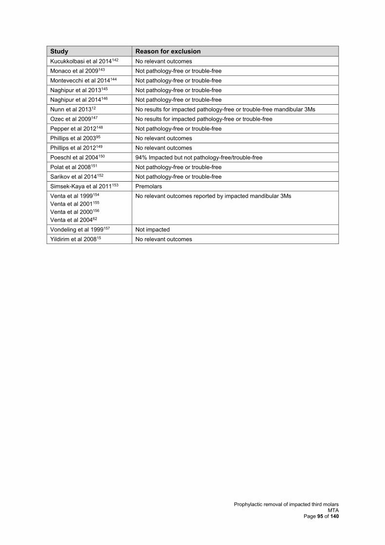
Prophylactic removal of impacted third molars MTA
Page 95 of 140
Study Reason for exclusion
Kucukkolbasi et al 2014142 No relevant outcomes
Monaco et al 2009143 Not pathology-free or trouble-free
Montevecchi et al 2014144 Not pathology-free or trouble-free
Naghipur et al 2013145 Not pathology-free or trouble-free
Naghipur et al 2014146 Not pathology-free or trouble-free
Nunn et al 201312 No results for impacted pathology-free or trouble-free mandibular 3Ms
Ozec et al 2009147 No results for impacted pathology-free or trouble-free
Pepper et al 2012148 Not pathology-free or trouble-free
Phillips et al 200395 No relevant outcomes
Phillips et al 2012149 No relevant outcomes
Poeschl et al 2004150 94% Impacted but not pathology-free/trouble-free
Polat et al 2008151 Not pathology-free or trouble-free
Sarikov et al 2014152 Not pathology-free or trouble-free
Simsek-Kaya et al 2011153 Premolars
Venta et al 1999154
Venta et al 2001155
Venta et al 2000156
Venta et al 200462
No relevant outcomes reported by impacted mandibular 3Ms
Vondeling et al 1999157 Not impacted
Yildirim et al 200815 No relevant outcomes
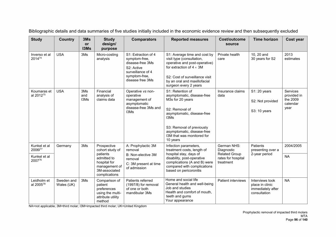
Prophylactic removal of impacted third molars MTA
Page 96 of 140
Bibliographic details and data summaries of five studies initially included in the economic evidence review and then subsequently excluded
Study Country 3Ms or
I3Ms
Study design/ purpose
Comparators Reported measures Cost/outcome source
Time horizon Cost year
Inverso et al 201474
USA 3Ms Micro-costing analysis
S1: Extraction of 4 symptom-free, disease-free 3Ms
S2: Active surveillance of 4 symptom-free, disease free 3Ms
S1: Average time and cost by visit type (consultation, operative and post-operative)
for extraction of 4 3M
S2: Cost of surveillance visit by an oral and maxillofacial surgeon every 2 years
Private health care
10, 20 and 30 years for S2
2013 estimates
Koumaras et al 201275
USA 3Ms and I3Ms
Financial analysis of claims data
Operative vs non-operative management of asymptomatic disease-free 3Ms and I3Ms
S1: Retention of asymptomatic, disease-free M3s for 20 years
S2: Removal of asymptomatic, disease-free I3Ms
S3: Removal of previously asymptomatic, disease-free I3M that was monitored for 10 years
Insurance claims data
S1: 20 years
S2: Not provided
S3: 10 years
Services provided in the 2009 calendar year
Kunkel et al 2006l77
Germany 3Ms Prospective cohort study of patients admitted to hospital for management of 3M-associated complications
A: Prophylactic 3M removal
B: Non-elective 3M removal
C: 3M present at time of admission
Infection parameters, treatment costs, length of hospital stay, days of disability, post-operative complications (A and B) were compared with complications based on pericoronitis
German NHS: Diagnostic Related Group rates for hospital treatment
Patients presenting over a 2-year period
2004/2005
Kunkel et al 200776
NA
Leidholm et al 200578
Sweden and Wales (UK)
3Ms Comparison of patient preferences using the multi-attribute utility method
Patients referred (1997/8) for removal of one or both mandibular 3Ms
Home and social life General health and well-being Job and studies Health and comfort of mouth, teeth and gums Your appearance
Patient interviews Interviews took place in clinic immediately after consultation
NA
NA=not applicable; 3M=third molar; I3M=impacted third molar; UK=United Kingdom
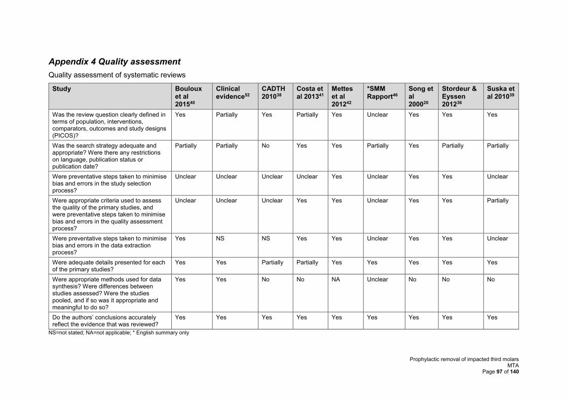
Prophylactic removal of impacted third molars MTA
Page 97 of 140
Appendix 4 Quality assessment
Quality assessment of systematic reviews
Study Bouloux et al 201540
Clinical evidence52
CADTH 201038
Costa et al 201341
Mettes et al 201242
*SMM Rapport46
Song et al 200020
Stordeur & Eyssen 201236
Suska et al 201039
Was the review question clearly defined in terms of population, interventions, comparators, outcomes and study designs (PICOS)?
Yes Partially Yes Partially Yes Unclear Yes Yes Yes
Was the search strategy adequate and appropriate? Were there any restrictions on language, publication status or publication date?
Partially Partially No Yes Yes Partially Yes Partially Partially
Were preventative steps taken to minimise bias and errors in the study selection process?
Unclear Unclear Unclear Unclear Yes Unclear Yes Yes Unclear
Were appropriate criteria used to assess the quality of the primary studies, and were preventative steps taken to minimise bias and errors in the quality assessment process?
Unclear Unclear Unclear Yes Yes Unclear Yes Yes Partially
Were preventative steps taken to minimise bias and errors in the data extraction process?
Yes NS NS Yes Yes Unclear Yes Yes Unclear
Were adequate details presented for each of the primary studies?
Yes Yes Partially Partially Yes Yes Yes Yes Yes
Were appropriate methods used for data synthesis? Were differences between studies assessed? Were the studies pooled, and if so was it appropriate and meaningful to do so?
Yes Yes No No NA Unclear No No No
Do the authors’ conclusions accurately reflect the evidence that was reviewed?
Yes Yes Yes Yes Yes Yes Yes Yes Yes
NS=not stated; NA=not applicable; * English summary only
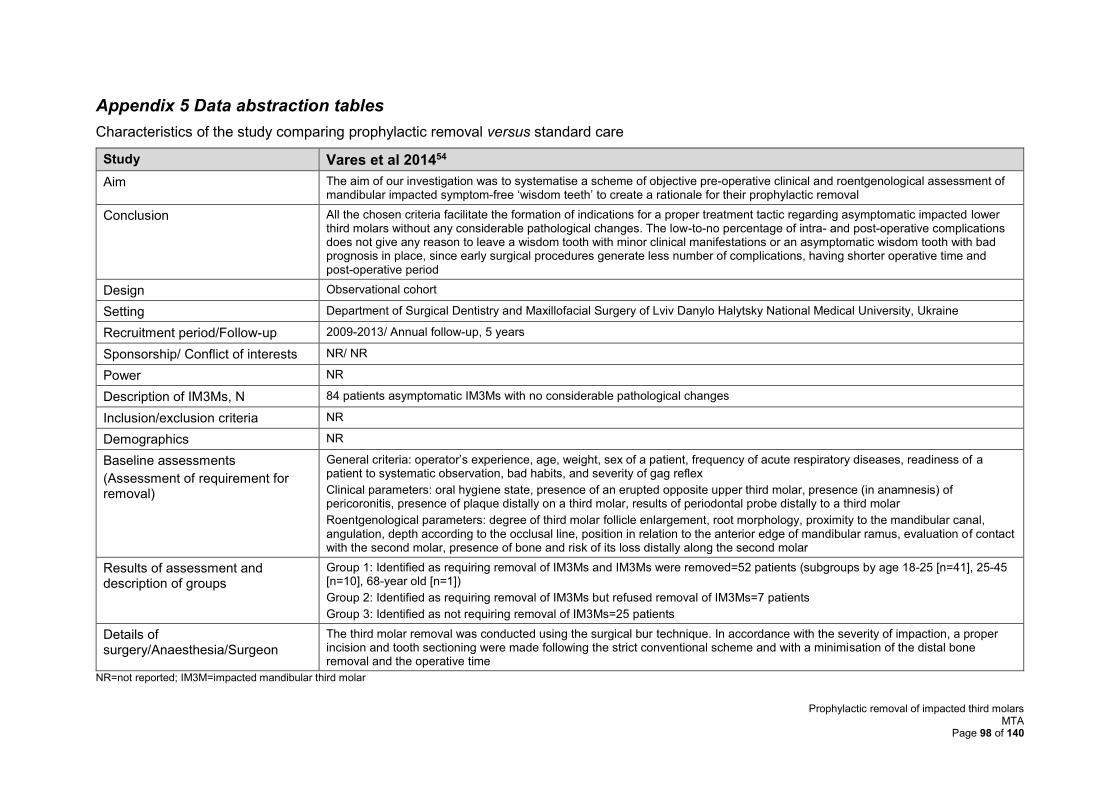
Prophylactic removal of impacted third molars MTA
Page 98 of 140
Appendix 5 Data abstraction tables
Characteristics of the study comparing prophylactic removal versus standard care
Study Vares et al 201454
Aim The aim of our investigation was to systematise a scheme of objective pre-operative clinical and roentgenological assessment of mandibular impacted symptom-free ‘wisdom teeth’ to create a rationale for their prophylactic removal
Conclusion All the chosen criteria facilitate the formation of indications for a proper treatment tactic regarding asymptomatic impacted lower third molars without any considerable pathological changes. The low-to-no percentage of intra- and post-operative complications does not give any reason to leave a wisdom tooth with minor clinical manifestations or an asymptomatic wisdom tooth with bad prognosis in place, since early surgical procedures generate less number of complications, having shorter operative time and post-operative period
Design Observational cohort
Setting Department of Surgical Dentistry and Maxillofacial Surgery of Lviv Danylo Halytsky National Medical University, Ukraine
Recruitment period/Follow-up 2009-2013/ Annual follow-up, 5 years
Sponsorship/ Conflict of interests NR/ NR
Power NR
Description of IM3Ms, N 84 patients asymptomatic IM3Ms with no considerable pathological changes
Inclusion/exclusion criteria NR
Demographics NR
Baseline assessments
(Assessment of requirement for removal)
General criteria: operator’s experience, age, weight, sex of a patient, frequency of acute respiratory diseases, readiness of a patient to systematic observation, bad habits, and severity of gag reflex
Clinical parameters: oral hygiene state, presence of an erupted opposite upper third molar, presence (in anamnesis) of pericoronitis, presence of plaque distally on a third molar, results of periodontal probe distally to a third molar
Roentgenological parameters: degree of third molar follicle enlargement, root morphology, proximity to the mandibular canal, angulation, depth according to the occlusal line, position in relation to the anterior edge of mandibular ramus, evaluation of contact with the second molar, presence of bone and risk of its loss distally along the second molar
Results of assessment and description of groups
Group 1: Identified as requiring removal of IM3Ms and IM3Ms were removed=52 patients (subgroups by age 18-25 [n=41], 25-45 [n=10], 68-year old [n=1])
Group 2: Identified as requiring removal of IM3Ms but refused removal of IM3Ms=7 patients
Group 3: Identified as not requiring removal of IM3Ms=25 patients
Details of surgery/Anaesthesia/Surgeon
The third molar removal was conducted using the surgical bur technique. In accordance with the severity of impaction, a proper incision and tooth sectioning were made following the strict conventional scheme and with a minimisation of the distal bone removal and the operative time
NR=not reported; IM3M=impacted mandibular third molar
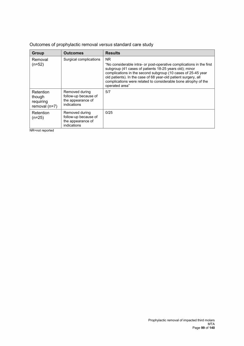
Prophylactic removal of impacted third molars MTA
Page 99 of 140
Outcomes of prophylactic removal versus standard care study
Group Outcomes Results
Removal (n=52)
Surgical complications NR
“No considerable intra- or post-operative complications in the first subgroup (41 cases of patients 18-25 years old); minor complications in the second subgroup (10 cases of 25-45 year old patients). In the case of 68 year-old patient surgery, all complications were related to considerable bone atrophy of the operated area”
Retention though requiring removal (n=7)
Removed during follow-up because of the appearance of indications
5/7
Retention (n=25)
Removed during follow-up because of the appearance of indications
0/25
NR=not reported
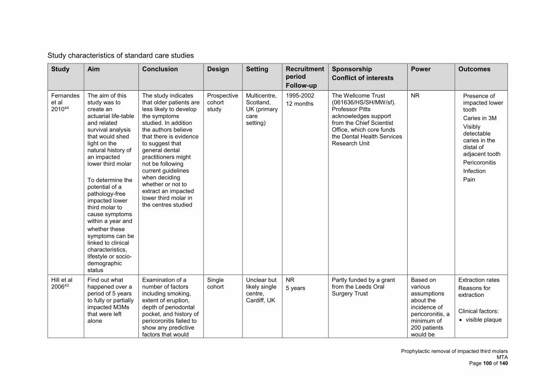
Prophylactic removal of impacted third molars MTA
Page 100 of 140
Study characteristics of standard care studies
Study Aim Conclusion Design Setting Recruitment period
Follow-up
Sponsorship
Conflict of interests
Power Outcomes
Fernandes et al 201044
The aim of this study was to create an actuarial life-table and related survival analysis that would shed light on the natural history of an impacted lower third molar
To determine the potential of a pathology-free impacted lower third molar to cause symptoms within a year and
whether these symptoms can be linked to clinical characteristics, lifestyle or socio-demographic status
The study indicates that older patients are less likely to develop the symptoms studied. In addition the authors believe that there is evidence to suggest that general dental practitioners might not be following current guidelines when deciding whether or not to extract an impacted lower third molar in the centres studied
Prospective cohort study
Multicentre, Scotland, UK (primary care setting)
1995-2002
12 months
The Wellcome Trust (061636/HS/SH/MW/sf). Professor Pitts acknowledges support from the Chief Scientist Office, which core funds the Dental Health Services Research Unit
NR • Presence of impacted lower tooth
• Caries in 3M
• Visibly detectable caries in the distal of adjacent tooth
• Pericoronitis
• Infection
• Pain
Hill et al 200643
Find out what happened over a period of 5 years to fully or partially impacted M3Ms that were left alone
Examination of a number of factors including smoking, extent of eruption, depth of periodontal pocket, and history of pericoronitis failed to show any predictive factors that would
Single cohort
Unclear but likely single centre, Cardiff, UK
NR
5 years
Partly funded by a grant from the Leeds Oral Surgery Trust
Based on various assumptions about the incidence of pericoronitis, a minimum of 200 patients would be
Extraction rates
Reasons for extraction
Clinical factors:
• visible plaque
Prophylactic removal of impacted third molars MTA
Page 101 of 140
Study Aim Conclusion Design Setting Recruitment period
Follow-up
Sponsorship
Conflict of interests
Power Outcomes
indicate which teeth would subsequently require removal. However, about one-third of the teeth in this series had to be removed within the 5-year period. Although this does not allow a ‘lifetime extrapolation’, it blurs the edges of our current thinking about asymptomatic wisdom teeth and certainly suggests that further (possibly longer-term) studies need to be completed. It does, however, provide little support for the reintroduction of prophylactic removal of wisdom teeth
needed to complete the study
• depth of pocket distal to the 2M
• bleeding on probing
• intra-bony defect
• evaluation of the position of the upper 3M
• any evidence of resorption
• radiographic measurement of the follicular space
2M=second molar; 3M=third molar; M3M=mandibular third molar; NR=not reported
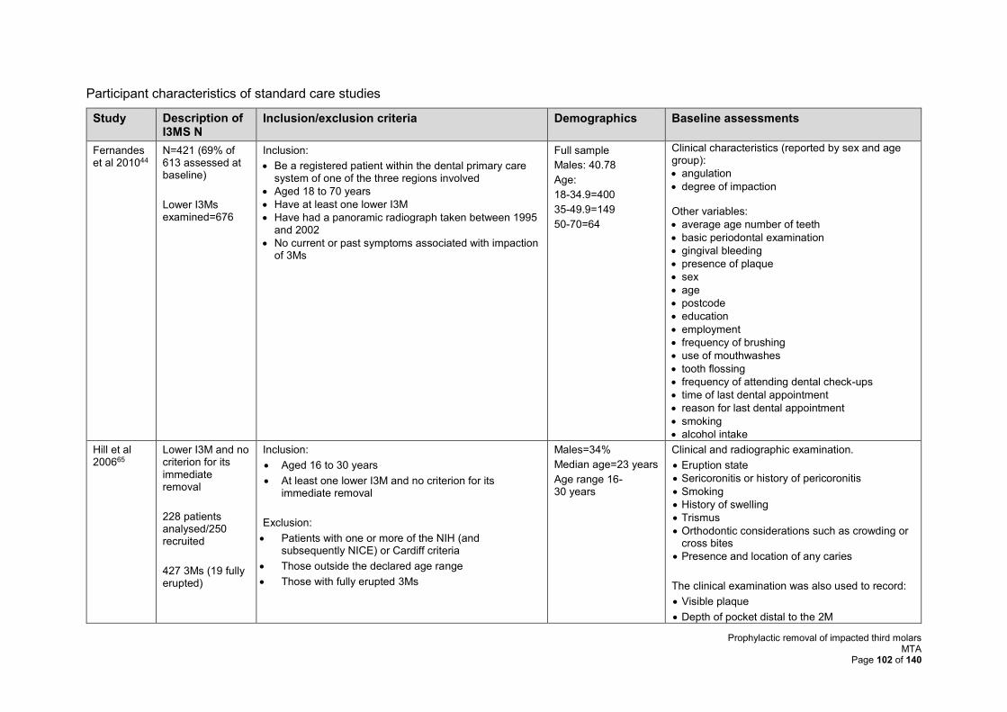
Prophylactic removal of impacted third molars MTA
Page 102 of 140
Participant characteristics of standard care studies
Study Description of I3MS N
Inclusion/exclusion criteria Demographics Baseline assessments
Fernandes et al 201044
N=421 (69% of 613 assessed at baseline)
Lower I3Ms examined=676
Inclusion:
• Be a registered patient within the dental primary care system of one of the three regions involved
• Aged 18 to 70 years
• Have at least one lower I3M
• Have had a panoramic radiograph taken between 1995 and 2002
• No current or past symptoms associated with impaction of 3Ms
Full sample
Males: 40.78
Age:
18-34.9=400
35-49.9=149
50-70=64
Clinical characteristics (reported by sex and age group):
• angulation
• degree of impaction Other variables:
• average age number of teeth
• basic periodontal examination
• gingival bleeding
• presence of plaque
• sex
• age
• postcode
• education
• employment
• frequency of brushing
• use of mouthwashes
• tooth flossing
• frequency of attending dental check-ups
• time of last dental appointment
• reason for last dental appointment
• smoking
• alcohol intake
Hill et al 200665
Lower I3M and no criterion for its immediate removal
228 patients analysed/250 recruited
427 3Ms (19 fully erupted)
Inclusion:
• Aged 16 to 30 years
• At least one lower I3M and no criterion for its immediate removal
Exclusion:
• Patients with one or more of the NIH (and subsequently NICE) or Cardiff criteria
• Those outside the declared age range
• Those with fully erupted 3Ms
Males=34%
Median age=23 years
Age range 16-30 years
Clinical and radiographic examination.
• Eruption state
• Sericoronitis or history of pericoronitis
• Smoking
• History of swelling
• Trismus
• Orthodontic considerations such as crowding or cross bites
• Presence and location of any caries
The clinical examination was also used to record:
• Visible plaque
• Depth of pocket distal to the 2M
Prophylactic removal of impacted third molars MTA
Page 103 of 140
Study Description of I3MS N
Inclusion/exclusion criteria Demographics Baseline assessments
153 patients had no history of pericoronitis
• Patients who were unwilling to be followed up for 5 years or who moved away from the geographical area were withdrawn
• Bleeding on probing
• Intra-bony defect
• Evaluation of the position of the upper 3M
• Any evidence of resorption
• Radiographic measurement of the follicular space
2M=second molar; 3M=third molar; I3M=impacted third molar; NIH=National Institute of Health; NICE=National Institute for Health and Care Excellence
Outcomes of standard care studies
Study Outcomes assessed Measured by/timing/Analysis Rate n (%) unless otherwise stated
Fernandes et al 201044
Teeth extracted Questioned and assessed by research dentist
1 year
NA
37 (5.47%)
Reasons for extraction Pericoronitis 5 (13.5%)
Pain 10 (27.0%)
Caries in distal of adjacent molar 1 (2.7%)
Caries in the 3M 2 (5.4%)
Contralateral 2 (5.4%)
Unknown 17 (46.0%)
Survived asymptomatically 562/676 (83.1%)
Symptoms developed by tooth
Pericoronitis (SIGN) 15 (13.2%)
Severe pain (SIGN) 16 (14.0%)
Mild pain (SIGN) 22 (19.3%)
Discomfort/irritation (non-SIGN) 54 (47.4%)
Food stagnation (non-SIGN) 7 (6.1%)
Distribution of lower I3Ms according to survival and the development of symptoms in 1 year
Survived symptom-free 552 (81.7%)
Survived with symptoms (SIGN) 31 (4.6%)
Survived with symptoms (non-SIGN)
55 (8.1%)
Extracted symptom-free 10 (1.5%)
Extracted with symptoms (SIGN) 23 (3.4%)
Prophylactic removal of impacted third molars MTA
Page 104 of 140
Study Outcomes assessed Measured by/timing/Analysis Rate n (%) unless otherwise stated
Extracted with symptoms (non-SIGN)
5 (0.7%)
Some form of symptoms 18-34.9 years of age Questioned and assessed by research dentist
1 year
Pearson chi square test
83 (22.6%) p=0.0028
35-49.9 years of age 28 (20.9%)
≥50 years of age 3 (5%)
Sex NR p>0.05
Vertical angulation 34 (22.7%) p≤0.001
Mesial angulation 43 (13.15%)
Distal angulation 31 (30.7%)
Horizontal angulation 6 (6.5%)
Unerupted 10.49% p≤0.001
Partially erupted 23.05%
Average number of teeth Questioned and assessed by research dentist
1 year
NR
NR p=>0.05
Maximum BPE scores NR p=>0.05
Average gingival bleeding index NR p=>0.05
Average mean plaque NR p=>0.05
Reason for last visit to the general dental practitioner
Questioned and assessed by research dentist
1 year
t-test
NR p=0.041
Education after minimum school-leaving age
NR p=0.191
Employment status NR p=0.560
Frequency of brushing teeth NR p=0.305
Occasional use of mouthwashes NR p=0.116
Occasional teeth flossing NR p=0.124
Frequency of dental appointments NR p=0.133
Length of time since patient last visited the dentist
NR p=0.335
Smoking NR p=0.291
Prophylactic removal of impacted third molars MTA
Page 105 of 140
Study Outcomes assessed Measured by/timing/Analysis Rate n (%) unless otherwise stated
Drinking >14 units/week NR p=0.447
Deprivation category NR p=0.058
Symptoms as SIGN symptoms only (infection, severe pain and caries)
Vertical angulation Questioned and assessed by research dentist
1 year
Pearson chi square test
10.29% p≤0.001
Mesial angulation 5.48%
Distal angulation 24.69%
Horizontal angulation 3.34%
Unerupted NR p=0.004
Partially erupted NR
Hill et al 200665
Extraction rates (per patient) Questionnaire/telephone (every 6 months)
Clinical examination (or telephone)
Every year
NA
48/153 (no history of pericoronitis)
23/66 (history of pericoronitis)
Reasons for extraction
Pericoronitis after start of study 30/48(62.5%)
Cosmetic/orthodontic 6/48 (12.5%)
Food impacted/difficult to clean 4/48(8.3%)
Early caries in 2M 4/48(8.35)
Painful when eating 2/48(4.2%)
Earache/TMJ pain 2/48(4.2%)
Clinical factors Clinical examination (or telephone)
Every year
NA
Visible plaque NR
Depth of pocket distal to the second molar NR
Bleeding on probing NR
Intra-bony defect NR
Evaluation of the position of the upper 3M NR
Any evidence of resorption 0/NR
Radiographic measurement of the follicular space NR
Remained symptomless 150/228
2M=second molar; 3M=third molar; I3M=impacted third molar; NA=not applicable; NR=not reported; BPE=basic periodontal examination; TMJ=temporomandibular joint; SIGN=Scottish Intercollegiate Guidelines Network *results for full sample but 91% were mandibular
Prophylactic removal of impacted third molars MTA
Page 106 of 140
Study and participant characteristics of the prophylactic removal study
Study Petsos et al 201655
Aim To investigate the effect of M3M removal on periodontal health of adjacent 2Ms. PPD and PAL have been described for primary outcome. As cofactors involved, sex, complications, two suture materials and two types of impaction were chosen as secondary outcomes
Conclusion Young patients may benefit from an early removal of M3M, especially in the presence of certain cofactors
Design Prospective cohort study
Setting Unclear on number of sites, Germany
Recruitment period/Follow-up 2nd June to 31th October 2014/ 6 months
Sponsorship/ Conflict of interests Self-funded/no conflicts
Power NR
Description of IM3Ms, N 78/91 recruited patients with a randomly selected 78/148 removed teeth selected for analysis
Submucosal=58, fully impacted=20
Inclusion/exclusion criteria Inclusion:
Completely impacted (entirely within the bone) or submucosal (completely below the mucous membrane), had a close positional relationship with the adjacent 2M (teeth 47 or 37, respectively), and no contact with the oral cavity. Completed root growth of the adjacent 2M was a prerequisite
Exclusion:
Presence of systemic disease (e.g. diabetes mellitus, cardiovascular, kidney, liver or lung disease), withdrawal of written consent, or failure to appear at the follow-up appointment
Demographics 37% male
Mean age=16.0±2.0 years
Non-smokers=74
Baseline assessments PlI=NR
GI=NR
PPD (measured from the gingival margin to the base of the pocket) mean=2.97±0.47 (range 2.0-4.2)
PAL (measured from cemento-enamel junction to the base of the pocket) mean=2.47±0.44 (range 1.5-3.3)
(Measurements were obtained at six sites around the 2M: mesiobuccal, buccal, distobuccal, distolingual, lingual, mesiolingual)
Details of surgery/Anaesthesia/Surgeon/other interventions
A mucoperiostal flap was reflected on the basis of a marginal incision on the 2M with a distovestibular releasing incision. A bone raspatorium was introduced on the lingual side to protect the lingual nerve. The wisdom tooth was horizontally dissected with rotary instruments using continuous sterile saline irrigation, and removed. The wound was closed with either pseudo-
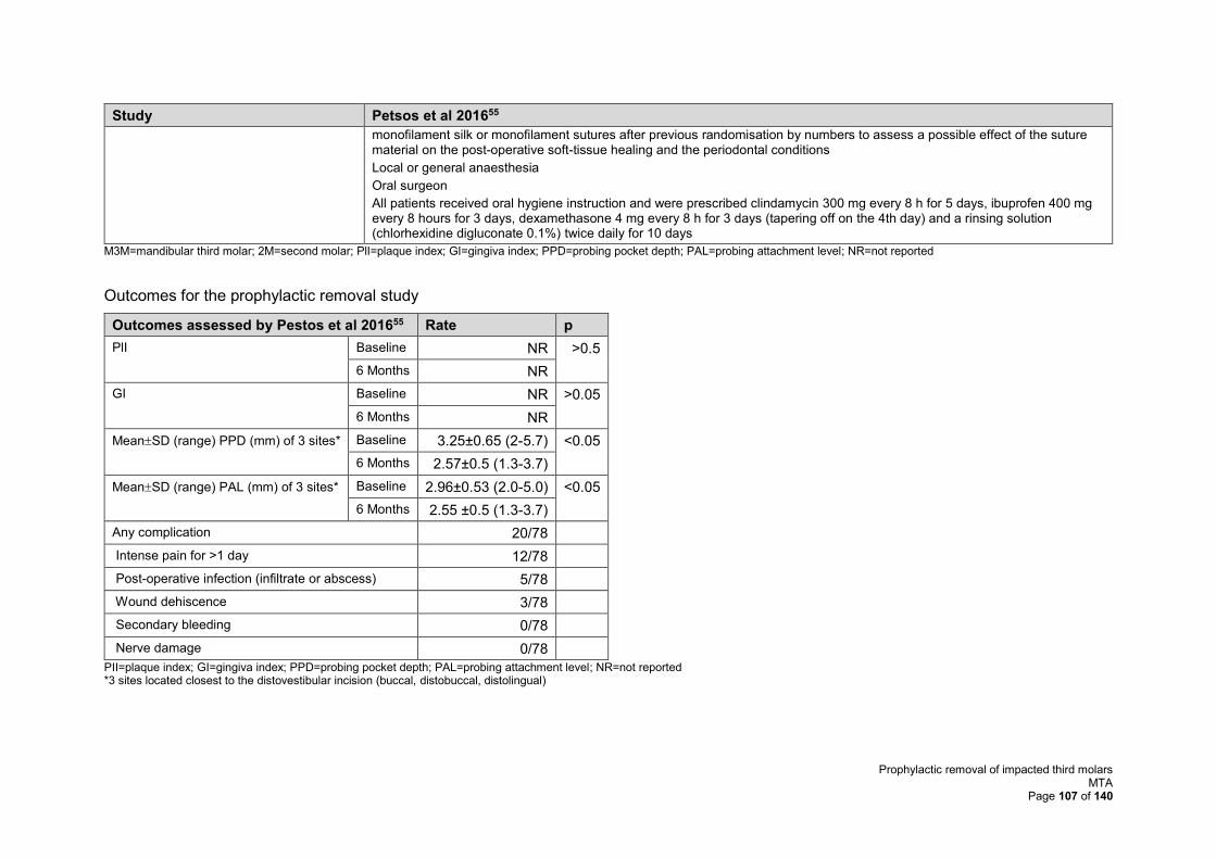
Prophylactic removal of impacted third molars MTA
Page 107 of 140
Study Petsos et al 201655
monofilament silk or monofilament sutures after previous randomisation by numbers to assess a possible effect of the suture material on the post-operative soft-tissue healing and the periodontal conditions
Local or general anaesthesia
Oral surgeon
All patients received oral hygiene instruction and were prescribed clindamycin 300 mg every 8 h for 5 days, ibuprofen 400 mg every 8 hours for 3 days, dexamethasone 4 mg every 8 h for 3 days (tapering off on the 4th day) and a rinsing solution (chlorhexidine digluconate 0.1%) twice daily for 10 days
M3M=mandibular third molar; 2M=second molar; PlI=plaque index; GI=gingiva index; PPD=probing pocket depth; PAL=probing attachment level; NR=not reported
Outcomes for the prophylactic removal study
Outcomes assessed by Pestos et al 201655 Rate p
PlI Baseline NR >0.5
6 Months NR
GI Baseline NR >0.05
6 Months NR
MeanSD (range) PPD (mm) of 3 sites* Baseline 3.25±0.65 (2-5.7) <0.05
6 Months 2.57±0.5 (1.3-3.7)
MeanSD (range) PAL (mm) of 3 sites* Baseline 2.96±0.53 (2.0-5.0) <0.05
6 Months 2.55 ±0.5 (1.3-3.7)
Any complication 20/78
Intense pain for >1 day 12/78
Post-operative infection (infiltrate or abscess) 5/78
Wound dehiscence 3/78
Secondary bleeding 0/78
Nerve damage 0/78
PII=plaque index; GI=gingiva index; PPD=probing pocket depth; PAL=probing attachment level; NR=not reported *3 sites located closest to the distovestibular incision (buccal, distobuccal, distolingual)
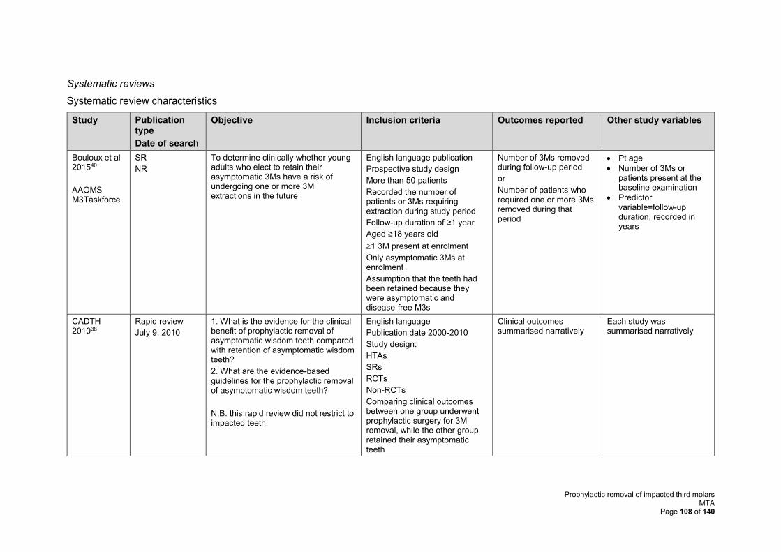
Prophylactic removal of impacted third molars MTA
Page 108 of 140
Systematic reviews
Systematic review characteristics
Study Publication type
Date of search
Objective Inclusion criteria Outcomes reported Other study variables
Bouloux et al 201540
AAOMS M3Taskforce
SR
NR
To determine clinically whether young adults who elect to retain their asymptomatic 3Ms have a risk of undergoing one or more 3M extractions in the future
English language publication
Prospective study design
More than 50 patients
Recorded the number of patients or 3Ms requiring extraction during study period
Follow-up duration of ≥1 year
Aged ≥18 years old
1 3M present at enrolment
Only asymptomatic 3Ms at enrolment
Assumption that the teeth had been retained because they were asymptomatic and disease-free M3s
Number of 3Ms removed during follow-up period
or
Number of patients who required one or more 3Ms removed during that period
• Pt age
• Number of 3Ms or patients present at the baseline examination
• Predictor variable=follow-up duration, recorded in years
CADTH 201038
Rapid review
July 9, 2010
1. What is the evidence for the clinical benefit of prophylactic removal of asymptomatic wisdom teeth compared with retention of asymptomatic wisdom teeth?
2. What are the evidence-based guidelines for the prophylactic removal of asymptomatic wisdom teeth?
N.B. this rapid review did not restrict to impacted teeth
English language
Publication date 2000-2010
Study design:
HTAs
SRs
RCTs
Non-RCTs
Comparing clinical outcomes between one group underwent prophylactic surgery for 3M removal, while the other group retained their asymptomatic teeth
Clinical outcomes summarised narratively
Each study was summarised narratively
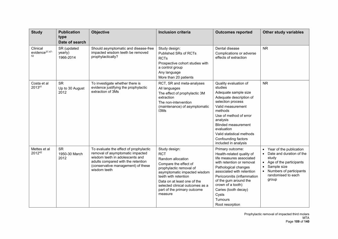
Prophylactic removal of impacted third molars MTA
Page 109 of 140
Study Publication type
Date of search
Objective Inclusion criteria Outcomes reported Other study variables
Clinical evidence37,47-
52
SR (updated yearly)
1966-2014
Should asymptomatic and disease-free impacted wisdom teeth be removed prophylactically?
Study design:
Published SRs of RCTs
RCTs
Prospective cohort studies with a control group
Any language
More than 20 patients
Dental disease
Complications or adverse effects of extraction
NR
Costa et al 201341
SR
Up to 30 August 2012
To investigate whether there is evidence justifying the prophylactic extraction of 3Ms
RCT, SR and meta-analyses
All languages
The effect of prophylactic 3M extraction
The non-intervention (maintenance) of asymptomatic I3Ms
Quality evaluation of studies
Adequate sample size
Adequate description of selection process
Valid measurement methods
Use of method of error analysis
Blinded measurement evaluation
Valid statistical methods
Confounding factors included in analysis
NR
Mettes et al 201242
SR
1950-30 March 2012
To evaluate the effect of prophylactic removal of asymptomatic impacted wisdom teeth in adolescents and adults compared with the retention (conservative management) of these wisdom teeth
Study design:
RCT
Random allocation
Compare the effect of prophylactic removal of asymptomatic impacted wisdom teeth with retention
Data on at least one of the selected clinical outcomes as a part of the primary outcome measure
Primary outcome:
Health-related quality of life measures associated with retention or removal
Pathological changes associated with retention
Pericoronitis (inflammation of the gum around the crown of a tooth)
Caries (tooth decay)
Cysts
Tumours
Root resorption
• Year of the publication
• Date and duration of the study
• Age of the participants
• Sample size
• Numbers of participants randomised to each group
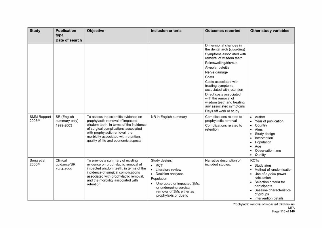
Prophylactic removal of impacted third molars MTA
Page 110 of 140
Study Publication type
Date of search
Objective Inclusion criteria Outcomes reported Other study variables
Dimensional changes in the dental arch (crowding)
Symptoms associated with removal of wisdom teeth
Pain/swelling/trismus
Alveolar osteitis
Nerve damage
Costs
Costs associated with treating symptoms associated with retention
Direct costs associated with the removal of wisdom teeth and treating any associated symptoms
Days off work or study
SMM Rapport 200346
SR (English summary only)
1999-2003
To assess the scientific evidence on prophylactic removal of impacted wisdom teeth, in terms of the incidence of surgical complications associated with prophylactic removal, the morbidity associated with retention, quality of life and economic aspects
NR in English summary Complications related to prophylactic removal
Complications related to retention
• Author
• Year of publication
• Country
• Aims
• Study design
• Intervention
• Population
• Age
• Observation time
• Quality
Song et al 200020
Clinical guidance/SR
1984-1999
To provide a summary of existing evidence on prophylactic removal of impacted wisdom teeth, in terms of the incidence of surgical complications associated with prophylactic removal, and the morbidity associated with retention
Study design:
• RCT
• Literature review
• Decision analyses
Population
• Unerupted or impacted 3Ms, or undergoing surgical removal of 3Ms either as prophylaxis or due to
Narrative description of included studies:
RCTs
• Study aims
• Method of randomisation
• Use of a priori power calculation
• Selection criteria for participants
• Baseline characteristics of groups
• Intervention details
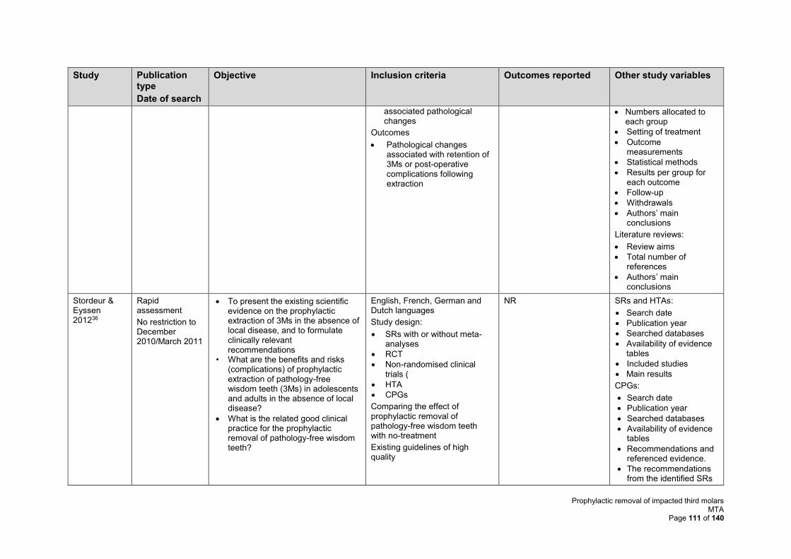
Prophylactic removal of impacted third molars MTA
Page 111 of 140
Study Publication type
Date of search
Objective Inclusion criteria Outcomes reported Other study variables
associated pathological changes
Outcomes
• Pathological changes associated with retention of 3Ms or post-operative complications following extraction
• Numbers allocated to each group
• Setting of treatment
• Outcome measurements
• Statistical methods
• Results per group for each outcome
• Follow-up
• Withdrawals
• Authors’ main conclusions
Literature reviews:
• Review aims
• Total number of references
• Authors’ main conclusions
Stordeur & Eyssen 201236
Rapid assessment
No restriction to December 2010/March 2011
• To present the existing scientific evidence on the prophylactic extraction of 3Ms in the absence of local disease, and to formulate clinically relevant recommendations
• What are the benefits and risks (complications) of prophylactic extraction of pathology-free wisdom teeth (3Ms) in adolescents and adults in the absence of local disease?
• What is the related good clinical practice for the prophylactic removal of pathology-free wisdom teeth?
English, French, German and Dutch languages
Study design:
• SRs with or without meta-analyses
• RCT
• Non-randomised clinical trials (
• HTA
• CPGs
Comparing the effect of prophylactic removal of pathology-free wisdom teeth with no-treatment
Existing guidelines of high quality
NR SRs and HTAs:
• Search date
• Publication year
• Searched databases
• Availability of evidence tables
• Included studies
• Main results
CPGs:
• Search date
• Publication year
• Searched databases
• Availability of evidence tables
• Recommendations and referenced evidence.
• The recommendations from the identified SRs
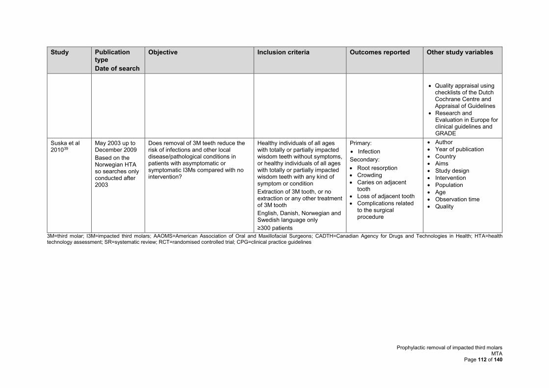
Prophylactic removal of impacted third molars MTA
Page 112 of 140
Study Publication type
Date of search
Objective Inclusion criteria Outcomes reported Other study variables
• Quality appraisal using checklists of the Dutch Cochrane Centre and Appraisal of Guidelines
• Research and Evaluation in Europe for clinical guidelines and GRADE
Suska et al 201039
May 2003 up to December 2009
Based on the Norwegian HTA so searches only conducted after 2003
Does removal of 3M teeth reduce the risk of infections and other local disease/pathological conditions in patients with asymptomatic or symptomatic I3Ms compared with no intervention?
Healthy individuals of all ages with totally or partially impacted wisdom teeth without symptoms, or healthy individuals of all ages with totally or partially impacted wisdom teeth with any kind of symptom or condition
Extraction of 3M tooth, or no extraction or any other treatment of 3M tooth
English, Danish, Norwegian and Swedish language only
≥300 patients
Primary:
• Infection
Secondary:
• Root resorption
• Crowding
• Caries on adjacent tooth
• Loss of adjacent tooth
• Complications related to the surgical procedure
• Author
• Year of publication
• Country
• Aims
• Study design
• Intervention
• Population
• Age
• Observation time
• Quality
3M=third molar; I3M=impacted third molars; AAOMS=American Association of Oral and Maxillofacial Surgeons; CADTH=Canadian Agency for Drugs and Technologies in Health; HTA=health technology assessment; SR=systematic review; RCT=randomised controlled trial; CPG=clinical practice guidelines
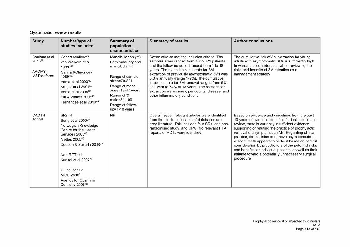
Prophylactic removal of impacted third molars MTA
Page 113 of 140
Systematic review results
Study Number/type of studies included
Summary of population characteristics
Summary of results Author conclusions
Bouloux et al 201540
AAOMS M3Taskforce
Cohort studies=7
von Wowern et al
1989158
Garcia &Chauncey 1989159
Venta et al 2000156
Kruger et al 200159
Venta et al 200462
Hill & Walker 200643
Fernandes et al 201044
Mandibular only=3
Both maxillary and mandibular=4
Range of sample sizes=70-821
Range of mean ages=18-47 years
Range of % male=31-100
Range of follow-up=1-18 years
Seven studies met the inclusion criteria. The samples sizes ranged from 70 to 821 patients, and the follow-up period ranged from 1 to 18 years. The mean incidence rate for 3M extraction of previously asymptomatic 3Ms was 3.0% annually (range 1-9%). The cumulative incidence rate for 3M removal ranged from 5% at 1 year to 64% at 18 years. The reasons for extraction were caries, periodontal disease, and other inflammatory conditions
The cumulative risk of 3M extraction for young adults with asymptomatic 3Ms is sufficiently high to warrant its consideration when reviewing the risks and benefits of 3M retention as a management strategy
CADTH 201038
SRs=4
Song et al 200020
Norwegian Knowledge Centre for the Health Services 200346
Mettes 200545
Dodson & Susarla 201037
Non-RCTs=1
Kunkel et al 200776
Guidelines=2
NICE 20002
Agency for Quality in Dentistry 200668
NR Overall, seven relevant articles were identified from the electronic search of databases and grey literature. This included four SRs, one non-randomised study, and CPG. No relevant HTA reports or RCTs were identified
Based on evidence and guidelines from the past 10 years of evidence identified for inclusion in this review, there is currently insufficient evidence supporting or refuting the practice of prophylactic removal of asymptomatic 3Ms. Regarding clinical practice, the decision to remove asymptomatic wisdom teeth appears to be best based on careful consideration by practitioners of the potential risks and benefits for individual patients, as well as their attitude toward a potentially unnecessary surgical procedure
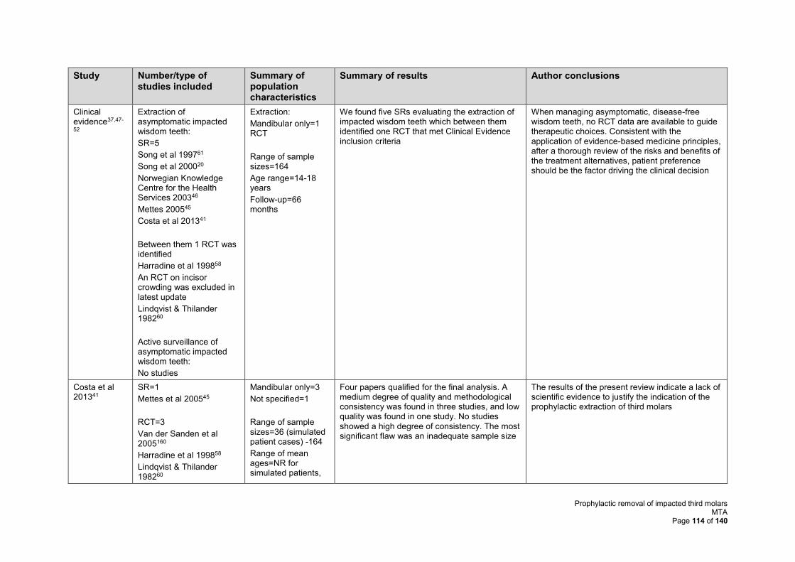
Prophylactic removal of impacted third molars MTA
Page 114 of 140
Study Number/type of studies included
Summary of population characteristics
Summary of results Author conclusions
Clinical evidence37,47-
52
Extraction of asymptomatic impacted wisdom teeth:
SR=5
Song et al 199761
Song et al 200020
Norwegian Knowledge Centre for the Health Services 200346
Mettes 200545
Costa et al 201341
Between them 1 RCT was identified
Harradine et al 199858
An RCT on incisor crowding was excluded in latest update
Lindqvist & Thilander 198260
Active surveillance of asymptomatic impacted wisdom teeth:
No studies
Extraction:
Mandibular only=1 RCT
Range of sample sizes=164
Age range=14-18 years
Follow-up=66 months
We found five SRs evaluating the extraction of impacted wisdom teeth which between them identified one RCT that met Clinical Evidence inclusion criteria
When managing asymptomatic, disease-free wisdom teeth, no RCT data are available to guide therapeutic choices. Consistent with the application of evidence-based medicine principles, after a thorough review of the risks and benefits of the treatment alternatives, patient preference should be the factor driving the clinical decision
Costa et al 201341
SR=1
Mettes et al 200545
RCT=3
Van der Sanden et al 2005160
Harradine et al 199858
Lindqvist & Thilander 198260
Mandibular only=3
Not specified=1
Range of sample sizes=36 (simulated patient cases) -164
Range of mean ages=NR for simulated patients,
Four papers qualified for the final analysis. A medium degree of quality and methodological consistency was found in three studies, and low quality was found in one study. No studies showed a high degree of consistency. The most significant flaw was an inadequate sample size
The results of the present review indicate a lack of scientific evidence to justify the indication of the prophylactic extraction of third molars
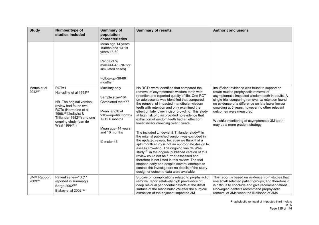
Prophylactic removal of impacted third molars MTA
Page 115 of 140
Study Number/type of studies included
Summary of population characteristics
Summary of results Author conclusions
Mean age 14 years 10mths and 13-19 years 13-60
Range of % male=44-45 (NR for simulated cases)
Follow-up=36-66 months
Mettes et al 201242
RCT=1
Harradine et al 199858
NB. The original version review had found two RCTs (Harradine et al 1998,58 Lindqvist & Thilander 198260) and one ongoing study (van de Waal 1999161)
Maxillary only
Sample size=164
Completed trial=77
Mean length of follow-up=66 months +/-12.6 months
Mean age=14 years and 10 months
% male=45
No RCTs were identified that compared the removal of asymptomatic wisdom teeth with retention and reported quality of life. One RCT on adolescents was identified that compared the removal of impacted mandibular wisdom teeth with retention and only examined the effect on late lower incisor crowding. This study at high risk of bias provided no evidence that extraction of wisdom teeth had an effect on lower incisor crowding over 5 years
The included Lindqvist & Thilander study60 in the original published version was excluded in the updated review, because we think that a split-mouth study is not an appropriate design to assess crowding. The ongoing van de Waal study161 in the original published version of this review could not be further assessed and therefore is not listed in this review. The trial stopped early and despite several attempts to contact the investigators no details of the study design or outcome data were available
Insufficient evidence was found to support or refute routine prophylactic removal of asymptomatic impacted wisdom teeth in adults. A single trial comparing removal vs retention found no evidence of a difference on late lower incisor crowding at 5 years, however no other relevant outcomes were measured
Watchful monitoring of asymptomatic 3M teeth may be a more prudent strategy
SMM Rapport 200346
Patient series=13 (11 reported in summary)
Berge 2002162
Blakey et al 2002123
Studies on complications related to prophylactic removal report relatively high prevalence of deep residual periodontal defects at the distal surface of the mandibular 2M after the surgical extraction of the adjacent impacted 3M.
This report is based on evidence from studies that use small selected patient groups, and therefore it is difficult to conclude and give recommendations. Norwegian dentists recommend prophylactic removal of 3Ms when the likelihood of 3Ms
Prophylactic removal of impacted third molars MTA
Page 116 of 140
Study Number/type of studies included
Summary of population characteristics
Summary of results Author conclusions
Conrad et al 199986
Gülicher & Gerlach 2001163
Hill et al 2001164
Rakprasitkul 2001165
Renton & McGurk 2001166
Tay 2000167
Valmaseda-Castellón et al 2000168
Valmaseda-Castellón 2001108
White et al 2002169
Yamaoka et al 1999170
Yoshii et al 2001171
Cohort studies=3 (5 reported in summary)
Kruger et al 200159
Ventä et al 1999154
Ventä et al 2001155
Case-controlled studies=2
Güngörmüs 2002172
Shafer et al 1999173
Cross-sectional studies=6
Güven et al 2000138
Kan et al 2002174
Libersa et al 2002175
Ma’aita & Alwrikat 2000176
Perry & Goldberg 2000177
Punwutikorn et al 1999178
Decision analysis=1
Edwards et al 199957
However, there was found low incidences of pain, permanent nerve damage (more than 6 months) on inferior alveolar and lingual nerve, fractures or serious infection
Studies on complications related to retention report a relatively high incidence of pericoronitis and caries, with higher incidence of pericoronitis related to partially erupted third molars compared to fully retained. Only low incidence of root resorption of 2M teeth, cysts and tumours was found
causing problems in the future is high and the incidence of post-operative complications are low. This includes partially erupted wisdom teeth. Removal of asymptomatic fully retained wisdom teeth is not recommended. Since this report is based on studies that are not optimal the patient’s preferences need to be decisive
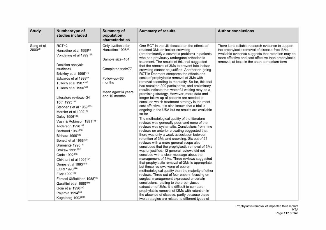
Prophylactic removal of impacted third molars MTA
Page 117 of 140
Study Number/type of studies included
Summary of population characteristics
Summary of results Author conclusions
Song et al 200020
RCT=2
Harradine et al 199858
Vondeling et al 1999157
Decision analysis studies=4
Brickley et al 1995179
Edwards et al 199957
Tulloch et al 1987180
Tulloch et al 1990181
Literature reviews=34
Toth 1993182
Stephens et al 1989183
Mercier et al 1992184
Daley 1996185
Vasir & Robinson 1991186
Anderson 1998187
Bertrand 1989188
Bishara 1999189
Bonetti et al 1988190
Bramante 1990191
Brokaw 1991192
Cade 1992193
Chikhani et al 1994194
Denes et al 1993195
ECRI 1993196
Flick 1999197
Forssel &Miettinen 1988198
Garattini et al 1990199
Goia et al 1990200
Pajarola 1994201
Kugelberg 1992202
Only available for Harradine 199858
Sample size=164
Completed trial=77
Follow-up=66 months
Mean age=14 years and 10 months
One RCT in the UK focused on the effects of retained 3Ms on incisor crowding (predominantly a cosmetic problem) in patients who had previously undergone orthodontic treatment. The results of this trial suggested that the removal of 3Ms to prevent late incisor crowding cannot be justified. Another on-going RCT in Denmark compares the effects and costs of prophylactic removal of 3Ms with removal according to morbidity. So far, this trial has recruited 200 participants, and preliminary results indicate that watchful waiting may be a promising strategy. However, more data and longer follow-up of patients are needed to conclude which treatment strategy is the most cost effective. It is also known that a trial is ongoing in the USA but no results are available so far
The methodological quality of the literature reviews was generally poor, and none of the reviews was systematic. Conclusions from nine reviews on anterior crowding suggested that there was only a weak association between retention of 3Ms and crowding. Six out of 21 reviews with a more general scope also concluded that the prophylactic removal of 3Ms was unjustified. 12 general reviews did not conclude with a clear message about the management of 3Ms. Three reviews suggested that prophylactic removal of 3Ms is appropriate, but these reviews were of poorer methodological quality than the majority of other reviews. Three out of four papers focusing on surgical management expressed uncertain conclusions relating to the prophylactic extraction of 3Ms. It is difficult to compare prophylactic removal of I3Ms with retention in the absence of disease, partly because these two strategies are related to different types of
There is no reliable research evidence to support the prophylactic removal of disease-free I3Ms. Available evidence suggests that retention may be more effective and cost effective than prophylactic removal, at least in the short to medium term
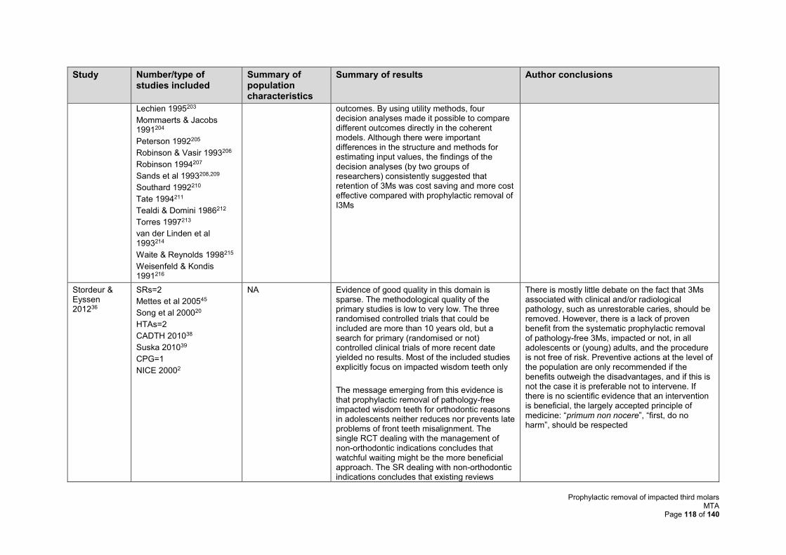
Prophylactic removal of impacted third molars MTA
Page 118 of 140
Study Number/type of studies included
Summary of population characteristics
Summary of results Author conclusions
Lechien 1995203
Mommaerts & Jacobs 1991204
Peterson 1992205
Robinson & Vasir 1993206
Robinson 1994207
Sands et al 1993208,209
Southard 1992210
Tate 1994211
Tealdi & Domini 1986212
Torres 1997213
van der Linden et al 1993214
Waite & Reynolds 1998215
Weisenfeld & Kondis 1991216
outcomes. By using utility methods, four decision analyses made it possible to compare different outcomes directly in the coherent models. Although there were important differences in the structure and methods for estimating input values, the findings of the decision analyses (by two groups of researchers) consistently suggested that retention of 3Ms was cost saving and more cost effective compared with prophylactic removal of I3Ms
Stordeur & Eyssen 201236
SRs=2
Mettes et al 200545
Song et al 200020
HTAs=2
CADTH 201038
Suska 201039
CPG=1
NICE 20002
NA Evidence of good quality in this domain is sparse. The methodological quality of the primary studies is low to very low. The three randomised controlled trials that could be included are more than 10 years old, but a search for primary (randomised or not) controlled clinical trials of more recent date yielded no results. Most of the included studies explicitly focus on impacted wisdom teeth only
The message emerging from this evidence is that prophylactic removal of pathology-free impacted wisdom teeth for orthodontic reasons in adolescents neither reduces nor prevents late problems of front teeth misalignment. The single RCT dealing with the management of non-orthodontic indications concludes that watchful waiting might be the more beneficial approach. The SR dealing with non-orthodontic indications concludes that existing reviews
There is mostly little debate on the fact that 3Ms associated with clinical and/or radiological pathology, such as unrestorable caries, should be removed. However, there is a lack of proven benefit from the systematic prophylactic removal of pathology-free 3Ms, impacted or not, in all adolescents or (young) adults, and the procedure is not free of risk. Preventive actions at the level of the population are only recommended if the benefits outweigh the disadvantages, and if this is not the case it is preferable not to intervene. If there is no scientific evidence that an intervention is beneficial, the largely accepted principle of medicine: “primum non nocere”, “first, do no harm”, should be respected
Prophylactic removal of impacted third molars MTA
Page 119 of 140
Study Number/type of studies included
Summary of population characteristics
Summary of results Author conclusions
favouring prophylactic removal, are generally of poorer methodological quality than those concluding that prophylactic removal is unjustified. Two HTA reports conclude that there is still no scientific documentation available to either support or reject routine prophylactic removal of pathology-free wisdom teeth
Decision analysis models compare prophylactic with symptomatic extraction for I3Ms, including frequencies and ratings of severity of complications in both cases. They consistently suggest that patients’ well-being is maximised if surgical removal is confined to wisdom teeth with pathological changes. Several of the included publications stress the importance of clear communication with patients about expected benefits and potential side effects and complications of the prophylactic removal of pathology-free 3Ms
Suska et al 201039
HTA-report/SRs=2
Mettes et al 200545
Norwegian Knowledge centre 200346
Case series=16
(None reported on asymptomatic teeth)
NR The literature search did not find any randomised or non-randomised, adequately controlled trial in which prophylactic removal of 3M teeth has been compared with no intervention
Both the SRs were of adequate quality according to the AMSTAR criteria. The Norwegian HTA-report stated “removal of asymptomatic fully retained wisdom teeth is not recommended”, whereas the Cochrane review concluded that “no evidence was found to support or refute routine prophylactic removal of asymptomatic impacted wisdom teeth in adults”
The level of evidence of prophylactic removal of asymptomatic 3M teeth as well as for removal of symptomatic 3M teeth is very low according to the GRADE system
Prophylactic removal of 3M teeth to prevent possible future complications is still frequently performed in Sweden. This intervention has been seriously questioned due to lack of supporting data of beneficial effects and the documented complications
A systematic literature search and review of published data has revealed that there is still no scientific documentation available to either support or refute routine prophylactic removal of asymptomatic impacted wisdom teeth in adults
Prophylactic removal of impacted third molars MTA
Page 120 of 140
Study Number/type of studies included
Summary of population characteristics
Summary of results Author conclusions
None of the case series reported the distribution of asymptomatic and symptomatic patients or the outcome of the extraction according to the presence or absence of symptoms at the time of the procedure
2M=second molar; 3M=third molar; I3M=impacted third molar; AAOMS=American Association of Oral and Maxillofacial Surgeons; CADTH=Canadian Agency for Drugs and Technologies in Health; HTA=health technology assessment; SR=systematic review; RCT=randomised controlled trial; CPG=clinical practice guidelines; NR=not reported
Prophylactic removal of impacted third molars MTA
Page 121 of 140
Appendix 6 Additional excluded studies
As mentioned earlier, many of the references included in previously published SRs and the
submissions from the BDA and the FDS, FGDP, and BAOS were excluded from this review.
However, due to the paucity of information available, the AG feels it would be pertinent to
discuss and summarise the results from some of these studies. The studies we have
summarised here are studies that nearly met our inclusion criteria but did not specifically report
results for the specific population of interest to this review (i.e. trouble-free IM3Ms).
References from included systematic reviews
Of the 84 references reported in the included SRs, only nine2,20,37,39,41,43-46 met the inclusion
criteria for this review, meaning that 75 references, previously included in SRs, were excluded.
For 41 of these references58,60,61,158,159,179-188,190-196,198-216 the date of publication was prior to
1999 and the studies were therefore published prior to the date limits of this SR. The remaining
34 references and the reasons for exclusion are shown in Table . Nine of these references did
not met our specific inclusion criteria but warrant further discussion as papers often cited in
the debate. The details of the study aims, the results and conclusions are summarised
narratively below and in Table .
Blakey et al 2002123 is one in a series of publications that report on a longitudinal clinical trial
including patients between 14 and 45 years of age, who had four asymptomatic 3Ms with
adjacent 2Ms. The Blakey et al 2002123 paper assessed the periodontal probing depth (PD) of
these molars at enrolment and found that 64 of the 329 patients had PD ≥5mm on any 3M, 35
of which were only on the M3M. However, two-thirds of all 3Ms in patients studied were
erupted to the occlusal plane (i.e not impacted or partially erupted/unerupted). Moreover,
these fully erupted teeth were found to be just as likely as teeth below the occlusal plane to
exhibit a change in probing depth. The White et al 2002169 paper is also part of this series of
papers, and reports on the detection and levels of pathogenic bacteria in subgingival plaque
of the same patients. The authors reported results in relation to position and angulation.
However, the results section did not include outcomes separately for mandibular and maxillary
3Ms.
The papers by Venta et al62,154-156 are based on a longitudinal study that followed 181 first-year
students at the University of Helsinki. Participant’s teeth were clinically examined and
panoramic radiographs were taken at baseline and 18 years later at the end of the study
(n=118). Some students were also examined at 6 years and 12 years (n=81). Not all teeth
were impacted and both mandibular and maxillary 3Ms were included. Not all were pathology-
free or trouble-free, though two of the published papers do report the results of a questionnaire
Prophylactic removal of impacted third molars MTA
Page 122 of 140
in which students were asked about symptoms. The authors of the studies conclude that 3Ms
“undergo continuous clinical change on a reduced scale at least up to the age of 38 years”
and that “considerable radiographic changes, without notable symptoms, may occur involving
inclination of the tooth and state of impaction in impacted third molars after the usual age of
eruption”. One paper156 reporting the results of the need for removal, reported that 33/54 3Ms
removed were asymptomatic and concluded that “Because need for surgical removal
decreases during early adulthood, routine prophylactic extraction of asymptomatic third molars
in young adults cannot be recommended. Well-defined indications for prophylactic removals
are needed”. (page 288)
The study by Edwards et al57 is a decision analysis that was identified by two SRs.20,46 The
study aimed to identify the “least costly, most effective and most cost-effective management
strategy for asymptomatic, disease free mandibular third molars”. Though the authors did
conduct a review to identify information to populate the model, no details on the results of the
clinical review are reported. The study did not restrict analyses to I3Ms. Further details on the
cost-effectiveness elements of the study are discussed in further details in the cost-
effectiveness section of this report (Section 4).
It was not possible to access any published data on either the Van de Waal et al161 or
Vondeling et al157 papers. However, both reported on discontinued trials, according to the SRs
citing them. The Van de Waal161 citation was a reference to an ongoing trial and was identified
by the original Cochrane review by Mettes et al 200545, but Van de Waal is also listed as an
author of the Vondeling et al abstract.157 As both have similar titles it is possible that the
citations are for the same study, which has been discontinued, and so no results have been
published. From the details reported in the citing SRs45,207 neither of the studies were restricted
to I3Ms.
Table 27 Studies included in previous systematic reviews and reasons for exclusion from this review
Study id Design and reason for exclusion
Agency for Quality in Dentistry (ZZQ) 200668
Guidelines German summary. Does not seem to be based on SR
‘Impaction’ refers to a tooth that has remained fully embedded in the bone; ‘retention’ denotes a position of a 3M in which the occlusal plane is not reached on completion of root growth; ‘malposed’ is if axis or position deviates
No mention of mandibular or not
Berge 2002162 Retrospective single cohort
IM3Ms no mention of prophylactic removal
Outcome is pain
Bishara 1999189 Literature review
Some of the pertinent studies related to the management of 3Ms in an orthodontic context
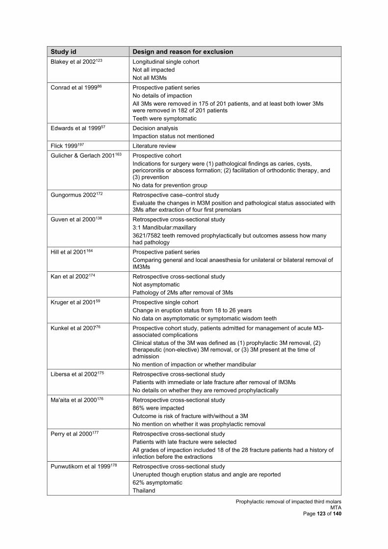
Prophylactic removal of impacted third molars MTA
Page 123 of 140
Study id Design and reason for exclusion
Blakey et al 2002123 Longitudinal single cohort
Not all impacted
Not all M3Ms
Conrad et al 199986 Prospective patient series
No details of impaction
All 3Ms were removed in 175 of 201 patients, and at least both lower 3Ms were removed in 182 of 201 patients
Teeth were symptomatic
Edwards et al 199957 Decision analysis
Impaction status not mentioned
Flick 1999197 Literature review
Gulicher & Gerlach 2001163 Prospective cohort
Indications for surgery were (1) pathological findings as caries, cysts, pericoronitis or abscess formation; (2) facilitation of orthodontic therapy, and (3) prevention
No data for prevention group
Gungormus 2002172 Retrospective case–control study
Evaluate the changes in M3M position and pathological status associated with 3Ms after extraction of four first premolars
Guven et al 2000138 Retrospective cross-sectional study
3:1 Mandibular:maxillary
3621/7582 teeth removed prophylactically but outcomes assess how many had pathology
Hill et al 2001164 Prospective patient series
Comparing general and local anaesthesia for unilateral or bilateral removal of IM3Ms
Kan et al 2002174 Retrospective cross-sectional study
Not asymptomatic
Pathology of 2Ms after removal of 3Ms
Kruger et al 200159 Prospective single cohort
Change in eruption status from 18 to 26 years
No data on asymptomatic or symptomatic wisdom teeth
Kunkel et al 200776 Prospective cohort study, patients admitted for management of acute M3-associated complications
Clinical status of the 3M was defined as (1) prophylactic 3M removal, (2) therapeutic (non-elective) 3M removal, or (3) 3M present at the time of admission
No mention of impaction or whether mandibular
Libersa et al 2002175 Retrospective cross-sectional study
Patients with immediate or late fracture after removal of IM3Ms
No details on whether they are removed prophylactically
Ma'aita et al 2000176 Retrospective cross-sectional study
86% were impacted
Outcome is risk of fracture with/without a 3M
No mention on whether it was prophylactic removal
Perry et al 2000177 Retrospective cross-sectional study
Patients with late fracture were selected
All grades of impaction included 18 of the 28 fracture patients had a history of infection before the extractions
Punwutikorn et al 1999178 Retrospective cross-sectional study
Unerupted though eruption status and angle are reported
62% asymptomatic
Thailand
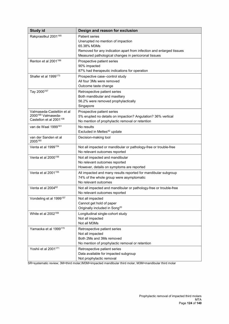
Prophylactic removal of impacted third molars MTA
Page 124 of 140
Study id Design and reason for exclusion
Rakprasitkul 2001165 Patient series
Unerupted no mention of impaction
65.38% M3Ms
Removed for any indication apart from infection and enlarged tissues
Measured pathological changes in pericoronal tissues
Renton et al 2001166 Prospective patient series
90% impacted
87% had therapeutic indications for operation
Shafer et al 1999173 Prospective case–control study
All four 3Ms were removed
Outcome taste change
Tay 2000167 Retrospective patient series
Both mandibular and maxillary
56.2% were removed prophylactically
Singapore
Valmaseda-Castellón et al 2000168 Valmaseda-Castellon et al 2001108
Prospective patient series
5% erupted no details on impaction? Angulation? 36% vertical
No mention of prophylactic removal or retention
van de Waal 1999161 No results
Excluded in Mettes42 update
van der Sanden et al 2005160
Decision-making tool
Venta et al 1999154 Not all impacted or mandibular or pathology-free or trouble-free
No relevant outcomes reported
Venta et al 2000156 Not all impacted and mandibular
No relevant outcomes reported
However, details on symptoms are reported
Venta et al 2001155 All impacted and many results reported for mandibular subgroup
74% of the whole group were asymptomatic
No relevant outcomes
Venta et al 200462 Not all impacted and mandibular or pathology-free or trouble-free
No relevant outcomes reported
Vondeling et al 1999157 Not all impacted
Cannot get hold of paper
Originally included in Song20
White et al 2002169 Longitudinal single-cohort study
Not all impacted
Not all M3Ms
Yamaoka et al 1999170 Retrospective patient series
Not all impacted
Both 2Ms and 3Ms removed
No mention of prophylactic removal or retention
Yoshii et al 2001171 Retrospective patient series
Data available for impacted subgroup
Not prophylactic removal
SR=systematic review; 3M=third molar;IM3M=impacted mandibular third molar; M3M=mandibular third molar
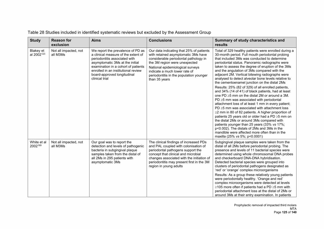
Prophylactic removal of impacted third molars MTA
Page 125 of 140
Table 28 Studies included in identified systematic reviews but excluded by the Assessment Group
Study Reason for exclusion
Aims Conclusions Summary of study characteristics and results
Blakey et al 2002123
Not all impacted, not all M3Ms
We report the prevalence of PD as a clinical measure of the extent of periodontitis associated with asymptomatic 3Ms at the initial examination in a cohort of patients enrolled in an institutional review board-approved longitudinal clinical trial
Our data indicating that 25% of patients with retained asymptomatic 3Ms have considerable periodontal pathology in the 3M region were unexpected
National epidemiological surveys indicate a much lower rate of periodontitis in the population younger than 35 years
Total of 329 healthy patients were enrolled during a 30-month period. Full mouth periodontal probing that included 3Ms was conducted to determine periodontal status. Panoramic radiographs were taken to assess the degree of eruption of the 3Ms and the angulation of 3Ms compared with the adjacent 2M. Vertical bitewing radiographs were analysed to detect alveolar bone levels relative to the cementoenamel junction on the distal 2Ms
Results: 25% (82 of 329) of all enrolled patients, and 34% (14 of 41) of black patients, had at least
one PD 5 mm on the distal 2M or around a 3M.
PD 5 mm was associated with periodontal attachment loss of at least 1 mm in every patient;
PD 5 mm was associated with attachment loss
2 mm in 80 of 82 patients. A higher proportion of
patients 25 years old or older had a PD 5 mm on the distal 2Ms or around 3Ms compared with patients younger than 25 years (33% vs 17%; p<0.002). The distals of 2Ms and 3Ms in the mandible were affected more often than in the maxilla (25% vs 5%; p<0.0001)
White et al 2002169
Not all impacted, not all M3Ms
Our goal was to report the detection and levels of pathogenic bacteria in subgingival plaque samples taken from the distal of all 2Ms in 295 patients with asymptomatic 3Ms
The clinical findings of increased PDs and PAL coupled with colonisation of periodontal pathogens support the concept that clinical and microbial changes associated with the initiation of periodontitis may present first in the 3M region in young adults
Subgingival plaque samples were taken from the distal of all 2Ms before periodontal probing. The presence and levels of 11 bacterial species were determined using whole chromosomal DNA probes and checkerboard DNA-DNA hybridisation. Detected bacterial species were grouped into clusters of periodontal pathogens designated as ‘red’ or ‘orange’ complex microorganisms
Results: As a group these relatively young patients were periodontally healthy. ‘Orange and red’ complex microorganisms were detected at levels
105 more often if patients had a PD 5 mm with periodontal attachment loss at the distal of 2Ms or around 3Ms at their entry examination. In patients
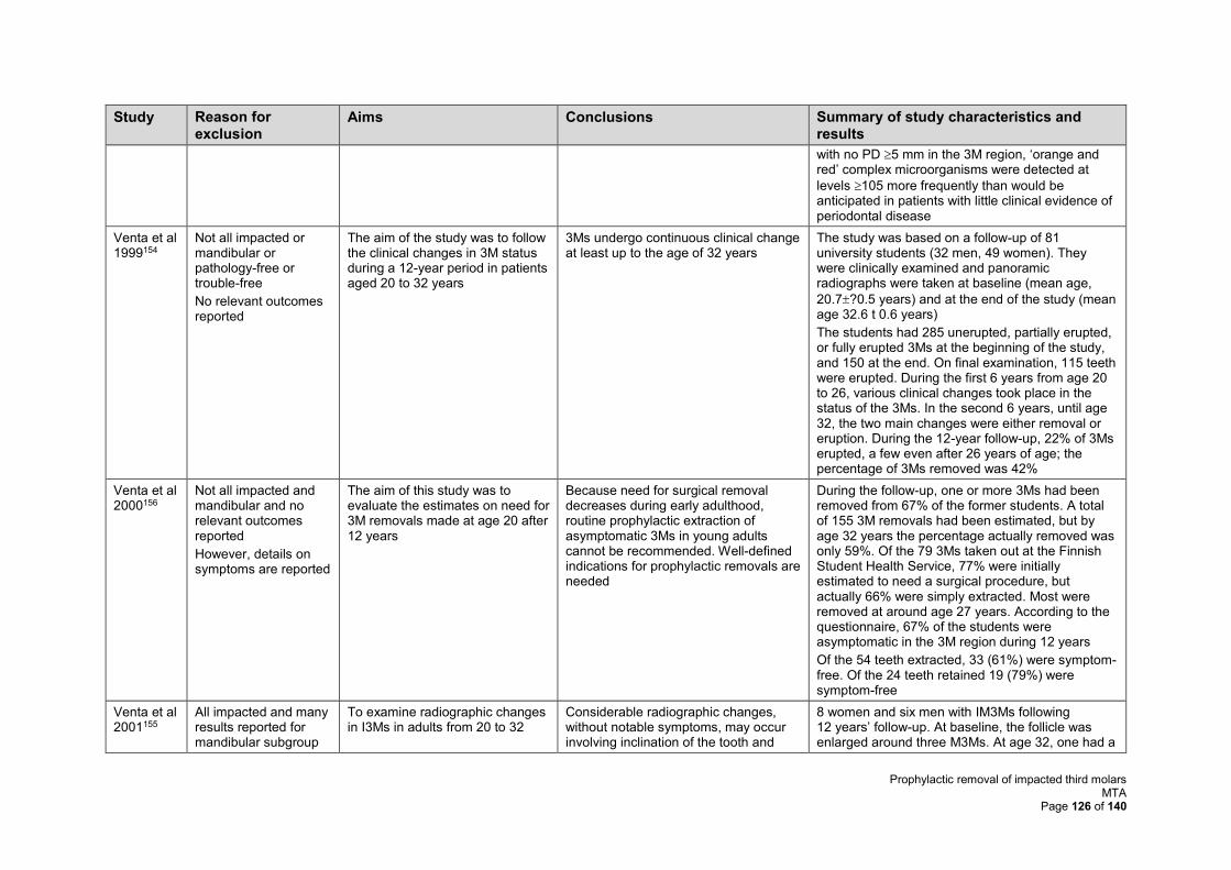
Prophylactic removal of impacted third molars MTA
Page 126 of 140
Study Reason for exclusion
Aims Conclusions Summary of study characteristics and results
with no PD 5 mm in the 3M region, ‘orange and red’ complex microorganisms were detected at
levels 105 more frequently than would be anticipated in patients with little clinical evidence of periodontal disease
Venta et al 1999154
Not all impacted or mandibular or pathology-free or trouble-free
No relevant outcomes reported
The aim of the study was to follow the clinical changes in 3M status during a 12-year period in patients aged 20 to 32 years
3Ms undergo continuous clinical change at least up to the age of 32 years
The study was based on a follow-up of 81 university students (32 men, 49 women). They were clinically examined and panoramic radiographs were taken at baseline (mean age,
20.7?0.5 years) and at the end of the study (mean age 32.6 t 0.6 years)
The students had 285 unerupted, partially erupted, or fully erupted 3Ms at the beginning of the study, and 150 at the end. On final examination, 115 teeth were erupted. During the first 6 years from age 20 to 26, various clinical changes took place in the status of the 3Ms. In the second 6 years, until age 32, the two main changes were either removal or eruption. During the 12-year follow-up, 22% of 3Ms erupted, a few even after 26 years of age; the percentage of 3Ms removed was 42%
Venta et al 2000156
Not all impacted and mandibular and no relevant outcomes reported
However, details on symptoms are reported
The aim of this study was to evaluate the estimates on need for 3M removals made at age 20 after 12 years
Because need for surgical removal decreases during early adulthood, routine prophylactic extraction of asymptomatic 3Ms in young adults cannot be recommended. Well-defined indications for prophylactic removals are needed
During the follow-up, one or more 3Ms had been removed from 67% of the former students. A total of 155 3M removals had been estimated, but by age 32 years the percentage actually removed was only 59%. Of the 79 3Ms taken out at the Finnish Student Health Service, 77% were initially estimated to need a surgical procedure, but actually 66% were simply extracted. Most were removed at around age 27 years. According to the questionnaire, 67% of the students were asymptomatic in the 3M region during 12 years
Of the 54 teeth extracted, 33 (61%) were symptom-free. Of the 24 teeth retained 19 (79%) were symptom-free
Venta et al 2001155
All impacted and many results reported for mandibular subgroup
To examine radiographic changes in I3Ms in adults from 20 to 32
Considerable radiographic changes, without notable symptoms, may occur involving inclination of the tooth and
8 women and six men with IM3Ms following 12 years’ follow-up. At baseline, the follicle was enlarged around three M3Ms. At age 32, one had a
Prophylactic removal of impacted third molars MTA
Page 127 of 140
Study Reason for exclusion
Aims Conclusions Summary of study characteristics and results
74% of the whole group were asymptomatic
No relevant outcomes
years, with special interest on sagittal changes in inclination
state of impaction in I3Ms after the usual age of eruption
follicle enlarged around a M3M different from the situation at baseline. Resorption of the crown of 3Ms or resorption of any 2M due to 3M impact was not seen in any of the teeth. In the mandible at baseline, development of the root was incomplete in 13 teeth. At the end of the study, all root ends were completed
Detailed longitudinal analysis showed that a change of more than 5 degrees in inclination had taken place for 76% of the teeth in the mandible
The M3Ms changed their angulation both in the distal and the mesial direction. The mean change in the mandible was 19 (SD 11.5) and in the maxilla 12 (SD 2.7), difference not statistically significant (t=1.03; df=17). In the mandible, all the teeth that moved were initially inclined in a mesial direction
Venta et al 200462
Not all impacted and mandibular or pathology-free or trouble-free
No relevant outcomes reported
The aim of the present study was to follow the clinical changes in 3M status during an 18-year period in patients aged 20 to 38 years
3Ms undergo continuous clinical change on a reduced scale at least up to the age of 38 years
The series consisted of 118 patients (37 men and 81 women). In the beginning of the study, the mean age was 20.2 years (SD 0.6), and at the end, it was 38.6 years (SD 6). Panoramic radiographs were taken at baseline and at age 38. All patients were clinically examined at baseline and at the end of the study. A portion of the patients (n=69) were also examined at age 32
Results: Most of the initially unerupted 3Ms were removed during the follow-up period (73%, maxilla and mandible together). More than half of the initially partially erupted 3Ms were removed during the follow-up period (64%, maxilla and mandible together). The percentage of erupted 3Ms found in the mouth at age 38 increased significantly depending on the initial status. Of the initially unerupted, partially erupted, or erupted 3Ms, 10%, 33%, and 50%, respectively, were erupted at age 38 (maxilla and mandible together). Changes in the status of 3Ms continued from age 32 to age 38, although to a lesser extent (8%). The three 3Ms with advanced eruption were all maxillary teeth in men
Prophylactic removal of impacted third molars MTA
Page 128 of 140
Study Reason for exclusion
Aims Conclusions Summary of study characteristics and results
Edwards et al 199957
Not impacted The study was undertaken to identify the least costly, most effective and most cost-effective management strategy for asymptomatic, disease-free M3Ms
M3M retention is less costly to the NHS, more effective for the patient and more cost-effective to both parties than removal. However, should the likelihood of developing pericoronitis, periodontal disease and caries increase substantially then removal becomes the more cost-effective strategy
Decision tree model
University of Wales Dental Hospital
“The various outcomes of lower third molar retention and removal, together with their incidences were obtained from a comprehensive computerised (Medline) and manual search of the medical literature (1966 to 1998).” No further details of the results of the search are reported
“The effectiveness of mandibular third molar management was rated as being greater for third molar retention (69.5) than for removal (63.3) giving a marginal effectiveness of -6.2”
van de Waal 1999161
An ongoing trial identified in Mettes review
It was stopped early in 2004 and no reports published so was subsequently excluded from Mettes update
NR NR NR
Vondeling et al 1999157
An ongoing trial identified by Song in 2000
No further results have been published and we have not been able to access the conference abstract identified by Song
NR NR NR
M3M=mandibular third molar; 3M=third molar; 2M=second molar; I3M=impacted third molar; IM3M=impacted mandibular third molar; PD=periodontal probing depth; PAL=periodontal attachment loss; DNA= deoxyribonucleic acid; SD=standard deviation; df=degrees of freedom; NR=not reported
Prophylactic removal of impacted third molars MTA
Page 129 of 140
References from submissions
The main reference lists included in the submissions (n=26) were checked by the AG to ensure
that a complete and thorough review of the available evidence could be conducted. Three
references had already been included by the AG. The remaining 23 references did not meet
the criteria for inclusion in this review; a list of these references, with comprehensive reasons
for exclusion is supplied in Table .
There were five references13,116,147,217,218 included in the submissions that partially met the
inclusion criteria for this SR and which warrant further discussion. In addition to the main
submission forms, the additional sources provided in the combined FDS, FGDP and BAOS
submission were also checked and a further nine references62,83,84,122,126,151,154,156,219 were
identified, which also fell into this category. The AG has therefore summarised these 14
studies13,62,83,84,116,122,126,147,151,154,156,217-219 in Table . Six studies13,116,147,151,217,218 were
retrospective in design, five publications62,122,126,154,156 reported the outcomes of longitudinal
studies, and three publications83,84,219 reported outcomes of prospective cohort studies. Six of
the studies13,116,147,151,217,218 reported outcomes relating to DCC in the 2M. The position of 3Ms,
and probing depth or clinical changes were reported in three publications.122,126,154 Clinical
complications of 3M surgery were reported by three,83,84,219 and one study156 reported the
estimation of the need for removal of 3Ms.
The publications by Blakey et al122 and Phillips et al126 report on different outcomes from the
same longitudinal study conducted in the US at the University of Kentucky and the University
of North Carolina. Two other linked publications123,169 from the same longitudinal study were
discussed in the previous section and are not repeated here. Blakey et al122 assessed the
changes in periodontal health over time, and it was concluded that for asymptomatic patients
with at least one PD ≥4 mm at enrolment, there were increased periodontal PDs ≥2 mm often
found in the 3M region. Phillips et al126 reported on the changes over time in the position of
3Ms relative to the occlusal plane, and concluded that the anatomic position of 3Ms was not
static over time; therefore, unerupted 3Ms should be monitored for changes in position and
periodontal pathology.
There were also three linked publications by Venta et al,62,154,156 which report on outcomes
from a longitudinal study conducted in Finland, at the University of Helsinki, which were also
discussed in detail in the previous section. Briefly, Venta et al154 followed the clinical changes
in 3M status in patients aged 20-32 years, and similarly to Phillips et al,126 found that 3Ms
undergo continuous clinical change. During the follow-up period, it was reported that 22% of
Prophylactic removal of impacted third molars MTA
Page 130 of 140
3Ms had erupted, and 42% of 3Ms were removed. In a subsequent publication,156 it was
reported that 67% of patients had one or more 3Ms removed during the follow-up period.
The publications by Chuang et al83,84 are linked to a series of publications,81,82,220 which report
the outcomes of the AAOMS Age-Related Third Molar Study, a prospective cohort study.
Chuang et al83 reports on the frequency of complications following 3M surgery, and it was
concluded from the results that increased age appears to be associated with a higher rate of
complications – patients aged 25 to 35 years were statistically significantly more likely to have
a complication compared with patients <25 years (OR=1.63; 95% CI: 1.12 to 2.37; p=0.01).
The level of impaction, evidence of periodontal condition, and pathology were also associated
with an increased risk of complications. Chuang et al84 reported the frequencies of
inflammatory complications after 3M surgery, and it was found that the level of impaction, pre-
existing infection and pathology were associated with inflammatory complications. Full bony
impacted teeth (OR=6.01; 95% CI: 4.7 to 7.7), followed by partially bony impacted teeth
(OR=4.7; 95% CI: 3.6 to 6.1), and soft tissue impacted teeth (OR=2.5; 95% CI: 1.7 to 3.7)
were more likely to have inflammatory complications compared with patients with erupted
teeth. Blondeau et al219 evaluated the incidence of post-surgical complications, and reported
that the overall complication rate differed significantly between men and women (2.2% and
10.2%, respectively; 2=13, p=0.0003).
Six studies13,116,147,151,217,218 reported outcomes relating to DCC in the 2M; all the studies
reported a relationship between I3Ms, in particular mesioangular I3Ms, and the presence of
2M DCC. Allen et al116 concluded that if 3Ms were left in situ, there is a need for close
monitoring and regular bitewing radiographs. McArdle et al13,218 and Ozec et al147 recommend
prophylactic removal of the 3M to prevent CDD of the 2M.
Table 29 Studies identified in the submissions and reasons for exclusion from this review
Study Design and reason for exclusion
AAOMS 201629 Wrong study design
Allen et al 2009116 Retrospective review of patient records
Not all patients pathology-free/trouble-free and no results for specific population of pathology-free/trouble-free, IM3M
Chang et al 2009221 Retrospective cohort
M3Ms
Not all impacted not all pathology-free or trouble-free
Wrong setting (Korea)
Devine et al 201630 Wrong study design
Falci et al 2012217 Retrospective review of patient records
All M3M
Unclear whether all patients pathology-free or trouble-free
Draft FDS RCS M3M Guidance
Wrong study design
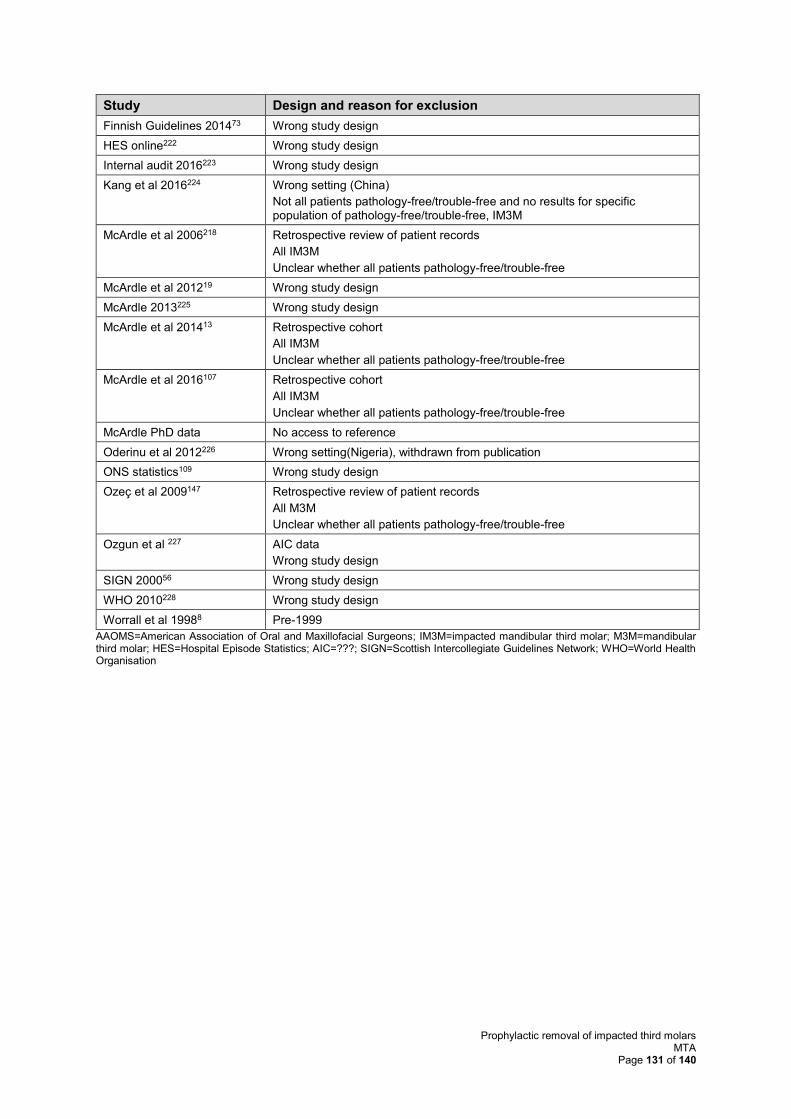
Prophylactic removal of impacted third molars MTA
Page 131 of 140
Study Design and reason for exclusion
Finnish Guidelines 201473 Wrong study design
HES online222 Wrong study design
Internal audit 2016223 Wrong study design
Kang et al 2016224 Wrong setting (China)
Not all patients pathology-free/trouble-free and no results for specific population of pathology-free/trouble-free, IM3M
McArdle et al 2006218 Retrospective review of patient records
All IM3M
Unclear whether all patients pathology-free/trouble-free
McArdle et al 201219 Wrong study design
McArdle 2013225 Wrong study design
McArdle et al 201413 Retrospective cohort
All IM3M
Unclear whether all patients pathology-free/trouble-free
McArdle et al 2016107 Retrospective cohort
All IM3M
Unclear whether all patients pathology-free/trouble-free
McArdle PhD data No access to reference
Oderinu et al 2012226 Wrong setting(Nigeria), withdrawn from publication
ONS statistics109 Wrong study design
Ozeç et al 2009147 Retrospective review of patient records
All M3M
Unclear whether all patients pathology-free/trouble-free
Ozgun et al 227 AIC data
Wrong study design
SIGN 200056 Wrong study design
WHO 2010228 Wrong study design
Worrall et al 19988 Pre-1999
AAOMS=American Association of Oral and Maxillofacial Surgeons; IM3M=impacted mandibular third molar; M3M=mandibular third molar; HES=Hospital Episode Statistics; AIC=???; SIGN=Scottish Intercollegiate Guidelines Network; WHO=World Health Organisation
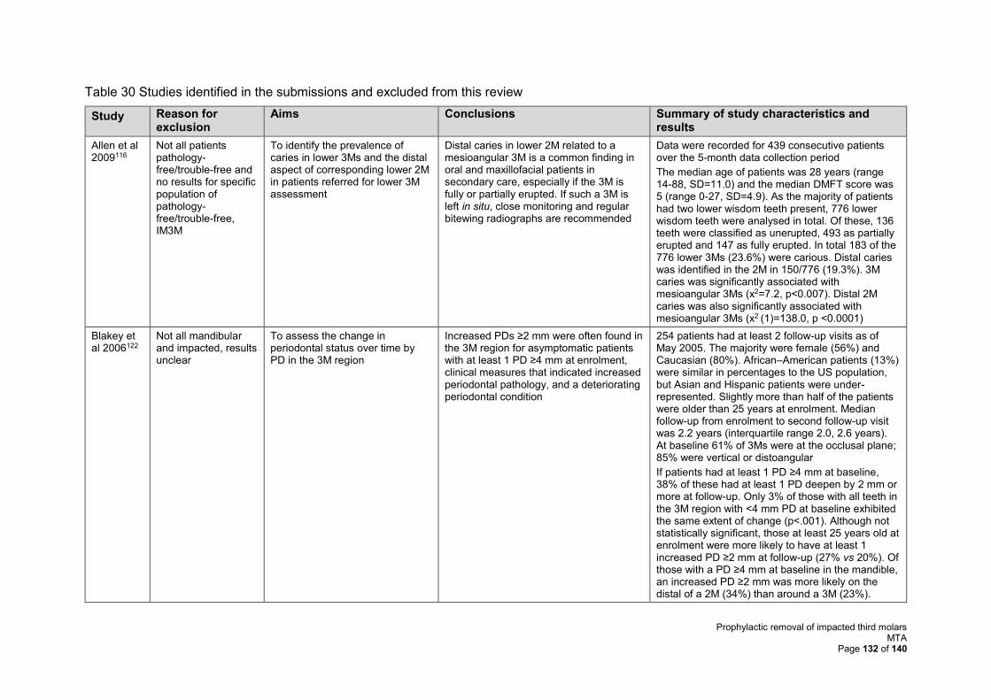
Prophylactic removal of impacted third molars MTA
Page 132 of 140
Table 30 Studies identified in the submissions and excluded from this review
Study Reason for exclusion
Aims Conclusions Summary of study characteristics and results
Allen et al 2009116
Not all patients pathology-free/trouble-free and no results for specific population of pathology-free/trouble-free, IM3M
To identify the prevalence of caries in lower 3Ms and the distal aspect of corresponding lower 2M in patients referred for lower 3M assessment
Distal caries in lower 2M related to a mesioangular 3M is a common finding in oral and maxillofacial patients in secondary care, especially if the 3M is fully or partially erupted. If such a 3M is left in situ, close monitoring and regular bitewing radiographs are recommended
Data were recorded for 439 consecutive patients over the 5-month data collection period
The median age of patients was 28 years (range 14-88, SD=11.0) and the median DMFT score was 5 (range 0-27, SD=4.9). As the majority of patients had two lower wisdom teeth present, 776 lower wisdom teeth were analysed in total. Of these, 136 teeth were classified as unerupted, 493 as partially erupted and 147 as fully erupted. In total 183 of the 776 lower 3Ms (23.6%) were carious. Distal caries was identified in the 2M in 150/776 (19.3%). 3M caries was significantly associated with mesioangular 3Ms (x2=7.2, p<0.007). Distal 2M caries was also significantly associated with mesioangular 3Ms (x2 (1)=138.0, p <0.0001)
Blakey et al 2006122
Not all mandibular and impacted, results unclear
To assess the change in periodontal status over time by PD in the 3M region
Increased PDs ≥2 mm were often found in the 3M region for asymptomatic patients with at least 1 PD ≥4 mm at enrolment, clinical measures that indicated increased periodontal pathology, and a deteriorating periodontal condition
254 patients had at least 2 follow-up visits as of May 2005. The majority were female (56%) and Caucasian (80%). African–American patients (13%) were similar in percentages to the US population, but Asian and Hispanic patients were under-represented. Slightly more than half of the patients were older than 25 years at enrolment. Median follow-up from enrolment to second follow-up visit was 2.2 years (interquartile range 2.0, 2.6 years). At baseline 61% of 3Ms were at the occlusal plane; 85% were vertical or distoangular
If patients had at least 1 PD ≥4 mm at baseline, 38% of these had at least 1 PD deepen by 2 mm or more at follow-up. Only 3% of those with all teeth in the 3M region with <4 mm PD at baseline exhibited the same extent of change (p<.001). Although not statistically significant, those at least 25 years old at enrolment were more likely to have at least 1 increased PD ≥2 mm at follow-up (27% vs 20%). Of those with a PD ≥4 mm at baseline in the mandible, an increased PD ≥2 mm was more likely on the distal of a 2M (34%) than around a 3M (23%).
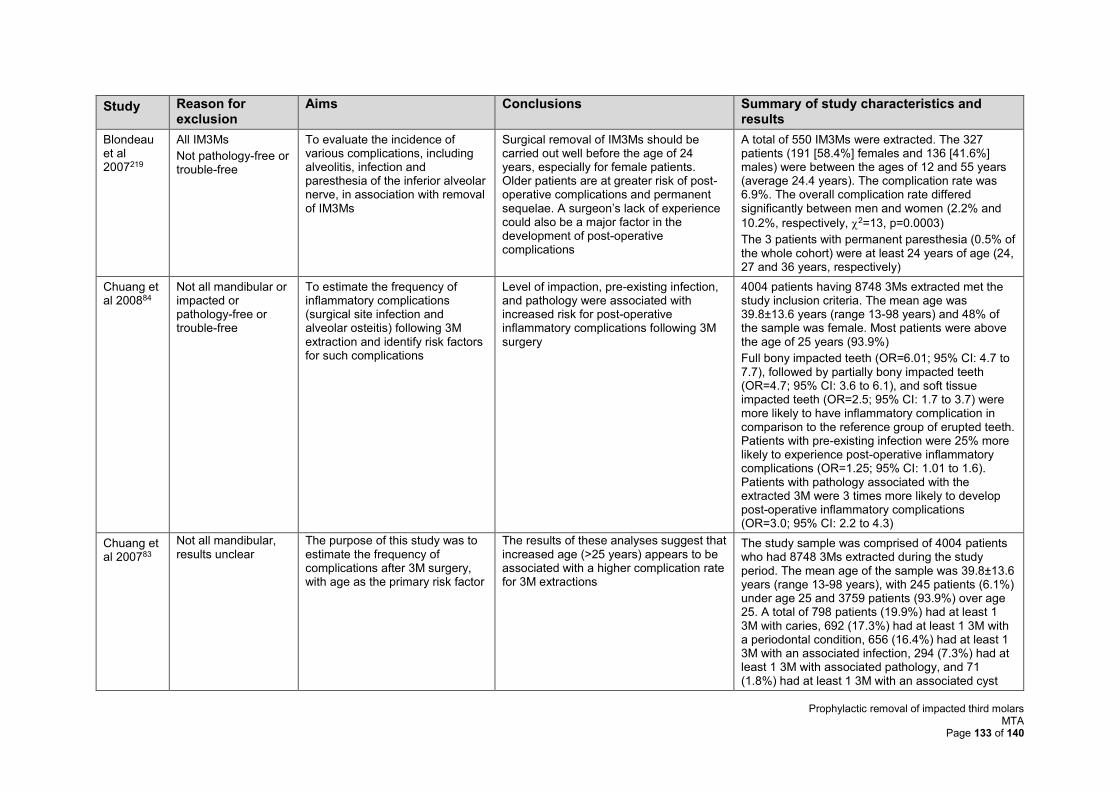
Prophylactic removal of impacted third molars MTA
Page 133 of 140
Study Reason for exclusion
Aims Conclusions Summary of study characteristics and results
Blondeau et al 2007219
All IM3Ms
Not pathology-free or trouble-free
To evaluate the incidence of various complications, including alveolitis, infection and paresthesia of the inferior alveolar nerve, in association with removal of IM3Ms
Surgical removal of IM3Ms should be carried out well before the age of 24 years, especially for female patients. Older patients are at greater risk of post-operative complications and permanent sequelae. A surgeon’s lack of experience could also be a major factor in the development of post-operative complications
A total of 550 IM3Ms were extracted. The 327 patients (191 [58.4%] females and 136 [41.6%] males) were between the ages of 12 and 55 years (average 24.4 years). The complication rate was 6.9%. The overall complication rate differed significantly between men and women (2.2% and
10.2%, respectively, 2=13, p=0.0003)
The 3 patients with permanent paresthesia (0.5% of the whole cohort) were at least 24 years of age (24, 27 and 36 years, respectively)
Chuang et al 200884
Not all mandibular or impacted or pathology-free or trouble-free
To estimate the frequency of inflammatory complications (surgical site infection and alveolar osteitis) following 3M extraction and identify risk factors for such complications
Level of impaction, pre-existing infection, and pathology were associated with increased risk for post-operative inflammatory complications following 3M surgery
4004 patients having 8748 3Ms extracted met the study inclusion criteria. The mean age was 39.8±13.6 years (range 13-98 years) and 48% of the sample was female. Most patients were above the age of 25 years (93.9%)
Full bony impacted teeth (OR=6.01; 95% CI: 4.7 to 7.7), followed by partially bony impacted teeth (OR=4.7; 95% CI: 3.6 to 6.1), and soft tissue impacted teeth (OR=2.5; 95% CI: 1.7 to 3.7) were more likely to have inflammatory complication in comparison to the reference group of erupted teeth. Patients with pre-existing infection were 25% more likely to experience post-operative inflammatory complications (OR=1.25; 95% CI: 1.01 to 1.6). Patients with pathology associated with the extracted 3M were 3 times more likely to develop post-operative inflammatory complications (OR=3.0; 95% CI: 2.2 to 4.3)
Chuang et al 200783
Not all mandibular, results unclear
The purpose of this study was to estimate the frequency of complications after 3M surgery, with age as the primary risk factor
The results of these analyses suggest that increased age (>25 years) appears to be associated with a higher complication rate for 3M extractions
The study sample was comprised of 4004 patients who had 8748 3Ms extracted during the study period. The mean age of the sample was 39.8±13.6 years (range 13-98 years), with 245 patients (6.1%) under age 25 and 3759 patients (93.9%) over age 25. A total of 798 patients (19.9%) had at least 1 3M with caries, 692 (17.3%) had at least 1 3M with a periodontal condition, 656 (16.4%) had at least 1 3M with an associated infection, 294 (7.3%) had at least 1 3M with associated pathology, and 71 (1.8%) had at least 1 3M with an associated cyst
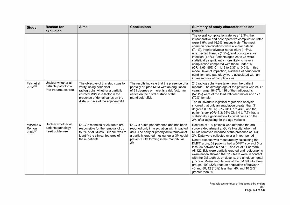
Prophylactic removal of impacted third molars MTA
Page 134 of 140
Study Reason for exclusion
Aims Conclusions Summary of study characteristics and results
The overall complication rate was 18.3%; the intraoperative and post-operative complication rates were 3.9% and 16.3%, respectively. The most common complications were alveolar osteitis (7.4%), inferior alveolar nerve injury (1.6%), unexpected trismus (1.2%), and post-operative infection (1.1%). Patients aged 25 to 35 were statistically significantly more likely to have a complication compared with those under 25 (OR=1.63; 95% CI: 1.12 to 2.37; p=0.01). In this model, level of impaction, evidence of periodontal condition, and pathology were associated with an increased risk of complications
Falci et al 2012217
Unclear whether all patients pathology-free free/trouble free
The objective of this study was to verify, using periapical radiographs, whether a partially erupted M3M is a factor in the presence of dental caries on the distal surface of the adjacent 2M
The results indicate that the presence of a partially erupted M3M with an angulation of 31 degrees or more, is a risk factor for caries on the distal surface of the mandibular 2Ms
246 radiographs were taken from the patient records. The average age of the patients was 24.17 years (range 16–57). 126 of the radiographs (52.1%) were of the third left-sided molar and 177 (72%) female
The multivariate logistical regression analysis showed that only an angulation greater than 31 degrees (OR=8.5; 95% CI: 1.7 to 43.8) and the patient’s sex (OR=3.3; 95% CI: 1.4 to 7.7), had a statistically significant link to distal caries on the 2M, after adjusting for the age variable
McArdle & Renton 2006218
Unclear whether all patients pathology-free/trouble-free
DCC in mandibular 2M teeth are responsible for the removal of up to 5% of all M3Ms. Our aim was to identify the clinical features of these patients
DCC is a late phenomenon and has been reported only in association with impacted 3Ms. The early or prophylactic removal of a partially erupted mesioangular 3M could prevent DCC forming in the mandibular 2M
Records of 100 patients who attended the oral surgery department at Guy’s Hospital who had M3Ms removed because of the presence of DCC 2M. Data were collected over a 1-year period
Dental disease was measured by calculating the DMFT score; 39 patients had a DMFT score of 5 or less; 36 between 6 and 10, and 24 of 11 or more. All 122 3Ms were partially erupted and radiographic examination showed that 119 teeth were in contact with the 2M tooth at, or close to, the amelocemental junction. Mesial angulations of the 3M fell into three groups; 100 (82%) had an angulation of between 40 and 80; 12 (10%) less than 40, and 10 (8%) greater than 80
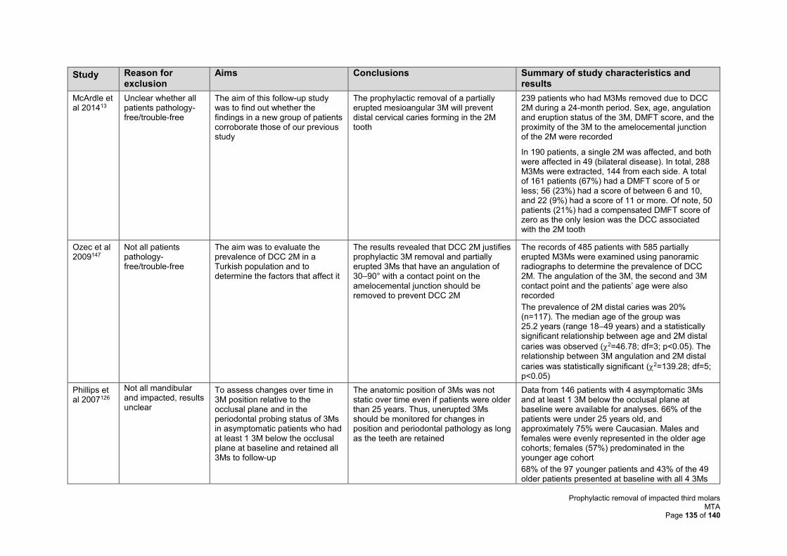
Prophylactic removal of impacted third molars MTA
Page 135 of 140
Study Reason for exclusion
Aims Conclusions Summary of study characteristics and results
McArdle et al 201413
Unclear whether all patients pathology-free/trouble-free
The aim of this follow-up study was to find out whether the findings in a new group of patients corroborate those of our previous study
The prophylactic removal of a partially erupted mesioangular 3M will prevent distal cervical caries forming in the 2M tooth
239 patients who had M3Ms removed due to DCC 2M during a 24-month period. Sex, age, angulation and eruption status of the 3M, DMFT score, and the proximity of the 3M to the amelocemental junction of the 2M were recorded
In 190 patients, a single 2M was affected, and both were affected in 49 (bilateral disease). In total, 288 M3Ms were extracted, 144 from each side. A total of 161 patients (67%) had a DMFT score of 5 or less; 56 (23%) had a score of between 6 and 10, and 22 (9%) had a score of 11 or more. Of note, 50 patients (21%) had a compensated DMFT score of zero as the only lesion was the DCC associated with the 2M tooth
Ozec et al 2009147
Not all patients pathology- free/trouble-free
The aim was to evaluate the prevalence of DCC 2M in a Turkish population and to determine the factors that affect it
The results revealed that DCC 2M justifies prophylactic 3M removal and partially erupted 3Ms that have an angulation of 30–90° with a contact point on the amelocemental junction should be removed to prevent DCC 2M
The records of 485 patients with 585 partially erupted M3Ms were examined using panoramic radiographs to determine the prevalence of DCC 2M. The angulation of the 3M, the second and 3M contact point and the patients’ age were also recorded
The prevalence of 2M distal caries was 20% (n=117). The median age of the group was 25.2 years (range 18–49 years) and a statistically significant relationship between age and 2M distal
caries was observed (2=46.78; df=3; p<0.05). The relationship between 3M angulation and 2M distal
caries was statistically significant (2=139.28; df=5; p<0.05)
Phillips et al 2007126
Not all mandibular and impacted, results unclear
To assess changes over time in 3M position relative to the occlusal plane and in the periodontal probing status of 3Ms in asymptomatic patients who had at least 1 3M below the occlusal plane at baseline and retained all 3Ms to follow-up
The anatomic position of 3Ms was not static over time even if patients were older than 25 years. Thus, unerupted 3Ms should be monitored for changes in position and periodontal pathology as long as the teeth are retained
Data from 146 patients with 4 asymptomatic 3Ms and at least 1 3M below the occlusal plane at baseline were available for analyses. 66% of the patients were under 25 years old, and approximately 75% were Caucasian. Males and females were evenly represented in the older age cohorts; females (57%) predominated in the younger age cohort
68% of the 97 younger patients and 43% of the 49 older patients presented at baseline with all 4 3Ms
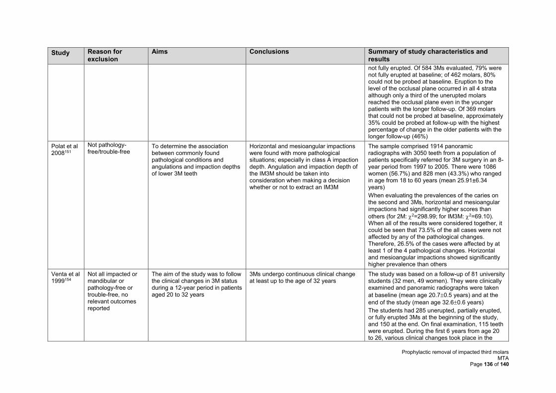
Prophylactic removal of impacted third molars MTA
Page 136 of 140
Study Reason for exclusion
Aims Conclusions Summary of study characteristics and results
not fully erupted. Of 584 3Ms evaluated, 79% were not fully erupted at baseline; of 462 molars, 80% could not be probed at baseline. Eruption to the level of the occlusal plane occurred in all 4 strata although only a third of the unerupted molars reached the occlusal plane even in the younger patients with the longer follow-up. Of 369 molars that could not be probed at baseline, approximately 35% could be probed at follow-up with the highest percentage of change in the older patients with the longer follow-up (46%)
Polat et al 2008151
Not pathology-free/trouble-free
To determine the association between commonly found pathological conditions and angulations and impaction depths of lower 3M teeth
Horizontal and mesioangular impactions were found with more pathological situations; especially in class A impaction depth. Angulation and impaction depth of the IM3M should be taken into consideration when making a decision whether or not to extract an IM3M
The sample comprised 1914 panoramic radiographs with 3050 teeth from a population of patients specifically referred for 3M surgery in an 8-year period from 1997 to 2005. There were 1086 women (56.7%) and 828 men (43.3%) who ranged in age from 18 to 60 years (mean 25.91±6.34 years)
When evaluating the prevalences of the caries on the second and 3Ms, horizontal and mesioangular impactions had significantly higher scores than
others (for 2M: 2=298.99; for IM3M: 2=69.10). When all of the results were considered together, it could be seen that 73.5% of the all cases were not affected by any of the pathological changes. Therefore, 26.5% of the cases were affected by at least 1 of the 4 pathological changes. Horizontal and mesioangular impactions showed significantly higher prevalence than others
Venta et al 1999154
Not all impacted or mandibular or pathology-free or trouble-free, no relevant outcomes reported
The aim of the study was to follow the clinical changes in 3M status during a 12-year period in patients aged 20 to 32 years
3Ms undergo continuous clinical change at least up to the age of 32 years
The study was based on a follow-up of 81 university students (32 men, 49 women). They were clinically examined and panoramic radiographs were taken
at baseline (mean age 20.70.5 years) and at the
end of the study (mean age 32.60.6 years)
The students had 285 unerupted, partially erupted, or fully erupted 3Ms at the beginning of the study, and 150 at the end. On final examination, 115 teeth were erupted. During the first 6 years from age 20 to 26, various clinical changes took place in the
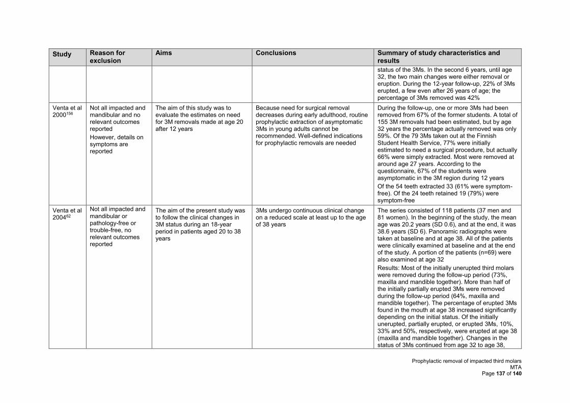
Prophylactic removal of impacted third molars MTA
Page 137 of 140
Study Reason for exclusion
Aims Conclusions Summary of study characteristics and results
status of the 3Ms. In the second 6 years, until age 32, the two main changes were either removal or eruption. During the 12-year follow-up, 22% of 3Ms erupted, a few even after 26 years of age; the percentage of 3Ms removed was 42%
Venta et al 2000156
Not all impacted and mandibular and no relevant outcomes reported
However, details on symptoms are reported
The aim of this study was to evaluate the estimates on need for 3M removals made at age 20 after 12 years
Because need for surgical removal decreases during early adulthood, routine prophylactic extraction of asymptomatic 3Ms in young adults cannot be recommended. Well-defined indications for prophylactic removals are needed
During the follow-up, one or more 3Ms had been removed from 67% of the former students. A total of 155 3M removals had been estimated, but by age 32 years the percentage actually removed was only 59%. Of the 79 3Ms taken out at the Finnish Student Health Service, 77% were initially estimated to need a surgical procedure, but actually 66% were simply extracted. Most were removed at around age 27 years. According to the questionnaire, 67% of the students were asymptomatic in the 3M region during 12 years
Of the 54 teeth extracted 33 (61% were symptom-free). Of the 24 teeth retained 19 (79%) were symptom-free
Venta et al 200462
Not all impacted and mandibular or pathology-free or trouble-free, no relevant outcomes reported
The aim of the present study was to follow the clinical changes in 3M status during an 18-year period in patients aged 20 to 38 years
3Ms undergo continuous clinical change on a reduced scale at least up to the age of 38 years
The series consisted of 118 patients (37 men and 81 women). In the beginning of the study, the mean age was 20.2 years (SD 0.6), and at the end, it was 38.6 years (SD 6). Panoramic radiographs were taken at baseline and at age 38. All of the patients were clinically examined at baseline and at the end of the study. A portion of the patients (n=69) were also examined at age 32
Results: Most of the initially unerupted third molars were removed during the follow-up period (73%, maxilla and mandible together). More than half of the initially partially erupted 3Ms were removed during the follow-up period (64%, maxilla and mandible together). The percentage of erupted 3Ms found in the mouth at age 38 increased significantly depending on the initial status. Of the initially unerupted, partially erupted, or erupted 3Ms, 10%, 33% and 50%, respectively, were erupted at age 38 (maxilla and mandible together). Changes in the status of 3Ms continued from age 32 to age 38,
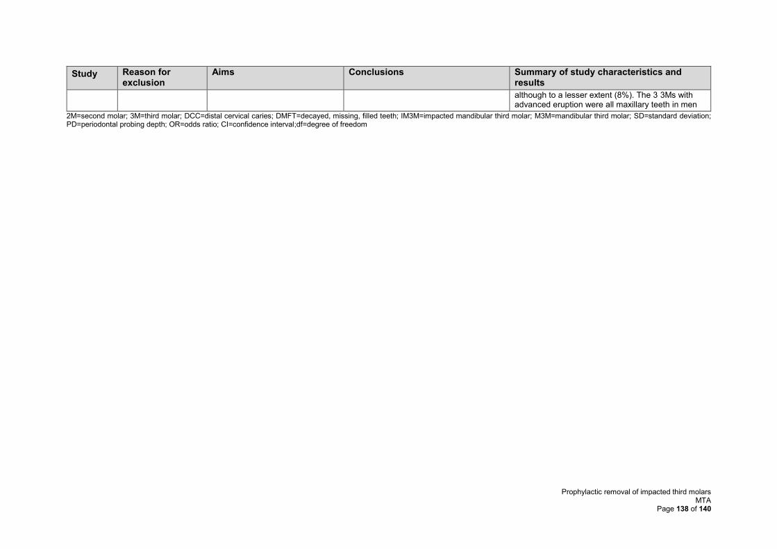
Prophylactic removal of impacted third molars MTA
Page 138 of 140
Study Reason for exclusion
Aims Conclusions Summary of study characteristics and results
although to a lesser extent (8%). The 3 3Ms with advanced eruption were all maxillary teeth in men
2M=second molar; 3M=third molar; DCC=distal cervical caries; DMFT=decayed, missing, filled teeth; IM3M=impacted mandibular third molar; M3M=mandibular third molar; SD=standard deviation; PD=periodontal probing depth; OR=odds ratio; CI=confidence interval;df=degree of freedom
Prophylactic removal of impacted third molars MTA
Page 139 of 140
Appendix 7 Transition probabilities used in the model
Age Mortality rate active
Probability of symptoms
Age Mortality rate active
Probability of symptoms
20 0.0003165 0.196694 61 0.007057382 0.045694
21 0.000328486 0.19048 62 0.007728454 0.043989
22 0.000324472 0.184418 63 0.008281076 0.042345
23 0.000372943 0.178507 64 0.009066209 0.040778235
24 0.000357425 0.172744 65 0.009661762 0.03926944
25 0.000375412 0.16713 66 0.010460394 0.037816471
26 0.000414361 0.161663 67 0.011719073 0.036417262
27 0.000424342 0.156341 68 0.012927631 0.035069823
28 0.00046136 0.151162 69 0.014311085 0.033772239
29 0.000475787 0.146125 70 0.015800852 0.032522667
30 0.000525243 0.141229 71 0.017563933 0.031319328
31 0.000569221 0.13647 72 0.02002717 0.030160513
32 0.000572207 0.131847 73 0.02188925 0.029044574
33 0.000626161 0.127357 74 0.024206902 0.027969925
34 0.000677156 0.122999 75 0.026799934 0.026935037
35 0.00074904 0.118769 76 0.029680244 0.025938441
36 0.000776518 0.114666 77 0.032715119 0.024978719
37 0.000864864 0.110687 78 0.03680341 0.024054506
38 0.00097521 0.106829 79 0.041100184 0.023164489
39 0.001062676 0.10309 80 0.046894523 0.022307403
40 0.00116042 0.099467 81 0.05270411 0.021482029
41 0.001236812 0.095958 82 0.059299519 0.020687194
42 0.0013093 0.092561 83 0.066956709 0.019921768
43 0.001457008 0.089271 84 0.075685636 0.019184663
44 0.001580834 0.086088 85 0.084829623 0.01847483
45 0.00175737 0.083007 86 0.095257973 0.017791261
46 0.001809657 0.080027 87 0.106315742 0.017132985
47 0.002005622 0.077145 88 0.118288053 0.016499064
48 0.002094736 0.074359 89 0.1331211 0.015888599
49 0.002342629 0.071665 90 0.146901404 0.015300721
50 0.002514115 0.069062 91 0.161803302 0.014734594
51 0.002791667 0.066546 92 0.184076189 0.014189414
52 0.003009129 0.064116 93 0.197660796 0.013664406
53 0.003313748 0.061769 94 0.210945629 0.013158823
54 0.003623468 0.059502 95 0.230126836 0.012671946
55 0.003987477 0.057313 96 0.260778283 0.012203084
56 0.004297595 0.0552 97 0.279505262 0.01175157
57 0.004825849 0.05316 98 0.301488605 0.011316762
58 0.005308646 0.051192 99 0.324712845 0.010898042
59 0.005923238 0.049293 100 0.335866615 0.010494814
60 0.006400301 0.047461
Prophylactic removal of impacted third molars MTA
Page 140 of 140
Appendix 8 Search strategy (I3M3 specific utilities)
1 Pericoronitis/
2 Toothache/
3 exp Dental Caries/
4 Dry Socket/
5 Tooth Extraction/
6 jaw fractures/ or mandibular fractures/ or maxillary fractures/
7 Facial Paralysis/
8 (Pericoronit* or toothache* or dry socket or tooth extract* or jaw fracture*).tw.
9 ((tooth or dental) adj1 (decay* or caries)).tw.
10 ((mandibular or maxillary) adj2 fracture*).tw.
11 ((Facial or face) adj2 nerve* adj2 damage*).tw.
12 ((Facial or face) adj2 paralysis*).tw.
13 or/1-12
14 (multiattribute$ or multi attribute$).ti,ab,kf.
15 utility.ab. /freq=2
16 utilities.ti,ab,kf.
17 disutili$.ti,ab,kf.
18 (standard gamble$ or sg).ti,ab,kf.
19 (time trade off$1 or time tradeoff$1 or tto or timetradeoff$1).ti,ab,kf.
20 (utility adj3 (score$1 or scoring or valu$ or measur$ or evaluat$ or scale$1 or
instrument$1 or weight or weights or weighting or information or data or unit or units or
health$ or life or estimat$ or elicit$ or disease$ or mean or cost$ or expenditure$1 or gain
or gains or loss or losses or lost or analysis or index$ or indices or overall or reported or
calculat$ or range$ or increment$ or state or states or status)).ti,ab,kf.
21 or/14-20
22 13 and 21


![Peri-operative Management of Impacted Third Molars [Autosaved]](https://img.pdfslide.net/doc/110x75/55cf8de3550346703b8c57cd/peri-operative-management-of-impacted-third-molars-autosaved.jpg)