Embed Size (px)
Citation preview

RUNX1 is essential for mesenchymal stem cellproliferation and myofibroblast differentiationWoosook Kima, David A. Barronb,1, Rebeca San Martinb, Keith S. Chana,b,c, Linda L. Trana, Feng Yangb, Steven J. Resslerb,2,and David R. Rowleya,b,3
aIntegrative Molecular and Biomedical Sciences Graduate Program, bDepartment of Molecular and Cellular Biology, and cScott Department of Urology, BaylorCollege of Medicine, Houston, TX 77030
Edited by Owen N. Witte, Howard Hughes Medical Institute, University of California, Los Angeles, CA, and approved September 22, 2014 (received for reviewApril 18, 2014)
Myofibroblasts are a key cell type in wound repair, cardiovasculardisease, and fibrosis and in the tumor-promoting microenviron-ment. The high accumulation of myofibroblasts in reactive stromais predictive of the rate of cancer progression in many differenttumors, yet the cell types of origin and the mechanisms thatregulate proliferation and differentiation are unknown. We reporthere, for the first time to our knowledge, the characterization ofnormal human prostate-derived mesenchymal stem cells (MSCs)and the TGF-β1–regulated pathways that modulate MSC prolifer-ation and myofibroblast differentiation. Human prostate MSCscombined with prostate cancer cells expressing TGF-β1 resultedin commitment to myofibroblasts. TGF-β1–regulated runt-relatedtranscription factor 1 (RUNX1) was required for cell cycle progres-sion and proliferation of progenitors. RUNX1 also inhibited, yetdid not block, differentiation. Knockdown of RUNX1 in prostateor bone marrow-derived MSCs resulted in cell cycle arrest, atten-uated proliferation, and constitutive differentiation to myofibro-blasts. These data show that RUNX1 is a key transcription factorfor MSC proliferation and cell fate commitment in myofibroblastdifferentiation. This work also shows that the normal human pros-tate gland contains tissue-derived MSCs that exhibit multilineagedifferentiation similar to bone marrow-derived MSCs. TargetingRUNX1 pathways may represent a therapeutic approach to affectmyofibroblast proliferation and biology in multiple disease states.
myofibroblast | MSC | RUNX1 | TGF-β1 | reactive stroma
Myofibroblasts are key mediators of homeostasis-associatedbiology in reactive stroma associated with tissue repair,
fibrosis, and the tumor microenvironment. However, the specificcell types of origin and the mechanisms that regulate their acti-vation, proliferation, and differentiation are not understood.Accordingly, identifying the niche and origins of myofibroblastsand pathways that regulate their proliferation and commitmentto myofibroblasts is important to developing antifibrotic andanticancer therapeutic approaches designed to target the evo-lution and proliferation of myofibroblasts.Adult mesenchymal stem cells (MSCs) are defined as multi-
potent stromal cells capable of self-renewal and differentiation tocartilage, bone, and adipose tissues (1). Studies have primarilyaddressed the biology of marrow-derived MSCs and far less isunderstood about putative tissue-resident MSCs and how theycontribute to the biology of their tissue of residence. Recent evi-dence from mouse models implicates tissue-resident MSCs in re-pair processes of disrupted local tissue homeostasis, suggesting thatthey are a critical component in wound repair, fibrosis, cardiovas-cular disease, and in the tumor microenvironment (2). Each ofthese is associated with evolution of reactive stroma with expres-sion of repair-centric genes. However, very little is known aboutadult human tissue-resident MSCs and the mechanisms that reg-ulate their proliferation and lineage commitment to local reactivestroma or fibrotic tissue.Reactive stroma is dynamic and responds rapidly to emerging
situations to restore disrupted homeostasis (3, 4). Reactive stroma
is found in most cancers and is typified by the coevolution ofmyofibroblasts (5–7). Importantly, the volume of reactive stromarelative to cancer is predictive of the rate of cancer progression inseveral tumor types (8–15). Furthermore, in vivo modeling hasshown that reactive stroma is tumor-promoting (16–19). Themyofibroblast is the principal cell type in reactive stroma, andtransforming growth factor beta 1 (TGF-β1) is a potent inducer ofmyofibroblast differentiation (3, 20). TGF-β1 is overexpressed inmost human cancers, including prostate cancer (2, 7). However, thecell types of origin and mechanisms that regulate their proliferationand differentiation to myofibroblasts remain unknown.To address tissue-resident MSCs in the prostate gland and their
potential induction to myofibroblasts, we have evaluated whethertissue-associated MSCs reside in normal adult human prostatetissue, whether they are induced to become myofibroblasts byTGF-β1, and the key mechanisms that regulate their proliferation.We report here for the first time, to our knowledge, the isolationof cells with prototypical MSC properties derived from normaladult human prostate gland. These cells exhibit multipotent dif-ferentiation similar to bone marrow-derived MSCs and formtypical reactive stroma myofibroblasts under TGF-β1 regulation.Importantly, we have identified runt-related transcription factor 1
Significance
Recruitment, proliferation, and differentiation of myofibroblastsare common in many disease states. Mechanisms that regulateproliferation and differentiation are poorly understood, al-though TGF-β is a key inducer of differentiation. Here, we report,for the first time to our knowledge, that runt-related transcrip-tion factor 1 (RUNX1) regulates mesenchymal stem cell (MSC)biology and progenitor cell commitment to myofibroblasts. Inthis work, we describe the first identification, to our knowledge,of tissue-resident MSCs from adult normal human prostategland and the role of these MSCs as myofibroblast precursors.We also pinpoint the role of RUNX1 in regulating proliferationand differentiation in both marrow-derived and tissue-residentMSCs. Perturbation of RUNX1 activity may provide insights fordeveloping antifibrotic and anticancer therapies via targetingthe reactive stroma microenvironment.
Author contributions: W.K., K.S.C., and D.R.R. designed research; W.K., D.A.B., R.S.M., L.L.T.,and F.Y. performed research; W.K., S.J.R., and D.R.R. analyzed data; and W.K. and D.R.R.wrote the paper.
The authors declare no conflict of interest.
This article is a PNAS Direct Submission.
Freely available online through the PNAS open access option.
See Commentary on page 16238.1Present address: Department of Radiation Oncology, Memorial Sloan Kettering CancerCenter, New York, NY 10065.
2Present address: Aptalis Pharmaceuticals, Bridgewater, NJ 08807.3To whom correspondence should be addressed. Email: [email protected].
This article contains supporting information online at www.pnas.org/lookup/suppl/doi:10.1073/pnas.1407097111/-/DCSupplemental.
www.pnas.org/cgi/doi/10.1073/pnas.1407097111 PNAS | November 18, 2014 | vol. 111 | no. 46 | 16389–16394
CELL
BIOLO
GY
SEECO
MMEN
TARY

(RUNX1) as a key transcription factor that is required for MSC/progenitor cell proliferation and that functions to moderatemyofibroblast differentiation.
ResultsProstate-Derived CD44+CD90+ Cells Are Functional MesenchymalStem Cells. Fresh tissue cores from the peripheral zone of nor-mal human prostate glands were obtained from 19-y-old (HPS-19Icells) and 33-y-old (HPS-33Q cells) cadaver donors with noprostate gland histopathology and explants cultured in a selectivemedium and protocol optimized for the culture of fetal rodenturogenital sinus mesenchyme (21). The resulting monolayers wereevaluated for phenotypic markers and biology. Both HPS-19I andHPS-33Q cells exhibited spindle-shaped fibroblast-like morphol-ogy (Fig. 1A) and were capable of long-term culture for more than25 passages, indicating a long-term growth potential. These cellsexhibited density-dependent inhibition of cell growth whenreaching more than 70% confluency, which restricted subsequentpassage efficiency and differentiation potential. Both HPS-19I andHPS-33Q cells exhibited a normal diploid karyotype and lackedchromosomal aberrations, including translocations as indicatedby spectral karyotyping (SI Appendix, Fig. S1). Flow cytometryshowed that both cell lines were positive for CD44 and CD90 (Fig.1B). To assess spatial distribution of CD44+CD90+ cells in pros-tate tissue, a tissue array with 32 normal human prostate tissuecores was evaluated via multispectral immunohistochemistry(IHC). Distinct clusters of CD44+CD90+ dual positive cells andindividual cells were localized in the stromal compartment (Fig.1C). In addition, dual positive immunoreactivity was observed insmall vessels of the microvasculature and nerves. Although manyfields exhibited a density of 1% or lower, the average density was2.83% of total cells (SI Appendix, Fig. S2).
Owing to the expression of MSC-associated markers, long-term passage potential, and diploid status, we addressed whetherthese cells exhibit multipotent MSC characteristics. To addressmultilineage differentiation potential, HPS-19I and HPS-33Q cellswere evaluated relative to human bone marrow-derived MSCs(hBM-MSCs). HPS-19I cells were induced to differentiate intoneurogenic, chondrogenic, and osteogenic lineages (Fig. 1D), butwere restricted from adipogenic differentiation. hBM-MSCs dif-ferentiated into all four lineages (SI Appendix, Fig. S3A). HPS-33Qcells could be induced to chondrocytes (SI Appendix, Fig. S3B),whereas osteogenic induction was less clear. Expression profiles ofMSC-specific markers and stem cell markers in HPS-19I cells werecongruent with profiles in hBM-MSCs (Fig. 1F). Moreover, flowcytometry revealed that the cell lines HPS-19I, HPS-33Q, andhBM-MSCs were each positive for CD44, CD90, CD13, CD29,CD73, and CD105, the most commonly reported human MSCmarkers, and were low for CD106 and STRO-1 (Fig. 1 E and G).Based on specific morphology, plastic adherence, multipotency,transcriptome, and cell-surface protein profiles of HPS-19I andHPS-33Q cells, we hereafter refer to these cells collectively ashuman prostate-derived mesenchymal stem cells (hP-MSCs).
Carcinoma hP-MSC Cell Interactions in 3D Organoid Cultures. A 3Dorganoid coculture system in defined medium that permitted thestudy of cell–cell interactions and differentiation potentials was de-veloped (Fig. 2A). A combination of LNCaP human prostate carci-noma cells with hP-MSCs resulted in free-floating self-organizingorganoids exhibiting a central core of hP-MSCs and an outer mantelof LNCaP (Fig. 2B). Organoids constructed with LNCaP and hBM-MSCs exhibited an identical phenotype (SI Appendix, Fig. S4A). IHCof 3D organoids showed that the core of hP-MSCs was positive forCD44 and CD90 and that the LNCaP mantel was positive for an-drogen receptor (AR) (Fig. 2C). LNCaP or hP-MSCs seeded alonealso self-organized as a free-floating 3D organoid (SI Appendix, Fig.S4A) and could be indirectly cocultured with other cells as mono-layers. Combination of LNCaP and hP-MSCs inoculated as s.c.xenografts in nude mice exhibited a similar self-organization in vivowith typical carcinoma foci and adjacent stroma (Fig. 2B; SI Ap-pendix, Fig. S4B).To investigate the function of TGF-β1 and hP-MSCs in the
genesis of reactive stroma, hP-MSCs were cocultured with LNCaPengineered to express constitutively active TGF-β1 (SI Appendix,Fig. S6A). Organoids constructed with TGF-β1–expressing LNCaPexhibited elevated immunoreactivity for α-smooth muscle actin(α-SMA) and tenascin-C in hP-MSCs relative to control (Fig. 2D).Both are prototypical markers of reactive stroma myofibroblasts(7). Tenascin-C was predominantly localized in stroma immedi-ately adjacent to epithelial cells, similar to patterns noted in humanprostate disease (7). In addition, hP-MSCs exhibited increasedimmunoreactivity for both vimentin and fibroblast activation pro-tein (FAP) under TGF-β1–stimulated conditions (SI Appendix, Fig.S5A). Furthermore, in vivo xenografts (day 11) generated with hP-MSCs combined with LNCaP resulted in a central core of α-SMA–positive myofibroblasts (SI Appendix, Fig. S4C). Labeling of thehP-MSCs via expression of RFP marker protein showed RFP andα-SMA dual positive cells in combined xenografts (day 10) (SIAppendix, Fig. S13).
TGF-β1–Induced Gene Expression Profiles of hP-MSCs Exhibit aReactive Stroma Myofibroblast Signature. To identify gene ex-pression profile alterations, we analyzed indirect cocultures of hP-MSC and engineered LNCaP organoids (Fig. 2A). ELISA analysisconfirmed that active TGF-β1 was observed only in coculturesmade with TGF-β1–expressing LNCaP (SI Appendix, Fig. S6B).hP-MSCs cocultured with LNCaP expressing TGF-β1 exhibitedan increased expression of reactive stroma genes such as TNC,ACTA2, FAP, and COL1A1 and showed distinct gene profiles in-cluding genes encoding cell-surface markers and growth factors
CD106 CD44CD29
CD105CD90
CD13CD73BMP2
OCT4
CD133
CD
44 ::
PE
CD90 :: FITC
HPS-19I HPS-33Q
CD44 CD90 CD29 CD73 CD13 CD106 CD105 STRO-1
HPS-19I (p9)
HPS-33Q (p7)
hBM-MSC (p4)
CD
44 ::
PE
CD90 :: FITC
CD
29 ::
PE
CD73 :: FITC
CD
13 ::
PE
CD106 :: FITC
CD
105
:: PE
STRO-1 :: FITC
HPS-19I hBM-MSC
D Neurofilament M
E
F G
OsteocalcinAggrecanHPS-19I HPS-33QA
B
C
Pseudo
CD44CD90 Pseudo
Log 10
HPS
-19I
2-∆
Ct
CD44CD90
Log10 hBM-MSC 2-∆Ct
Fig. 1. CD44+CD90+ stromal cells derived from normal human prostate aremesenchymal stem cells. (A) Cell morphology. (Scale bars, 100 μm.) (B) Flowcytometric analysis of HPS cell lines. (C) Immunostaining of normal humanprostate tissue for CD44 (blue), CD90 (red), and nuclei (green). Multispectraldeconvolution microscopy revealed localization of CD44+CD90+ cells (“Pseudo”yellow, arrows) in the stroma. (Scale bars, 100 μm.) (D) Multilineage differ-entiation potential of HPS-19I cells. Neurogenesis is shown by neurofilamentM staining (“Neurofilament M”). (Scale bars, 100 μm.) Chondrogenesis byaggrecan accumulation (“Aggrecan”). (Scale bars, 20 μm.) Osteogenesis byosteocalcin production (“Osteocalcin”). (Scale bars, 20 μm.) (E) Flow cytometricanalysis of cell lines HPS-19I, HPS-33Q, and hBM-MSCs. “p” denotes passage.The open and gray plots represent the isotype control and the specific anti-body indicated, respectively. (F) Similar gene expression profiles of HPS-19Icells compared with hBM-MSCs. (G) Flow cytometric analysis revealed thatHPS-19I cells and hBM-MSCs share expression of MSC surface markers.
16390 | www.pnas.org/cgi/doi/10.1073/pnas.1407097111 Kim et al.
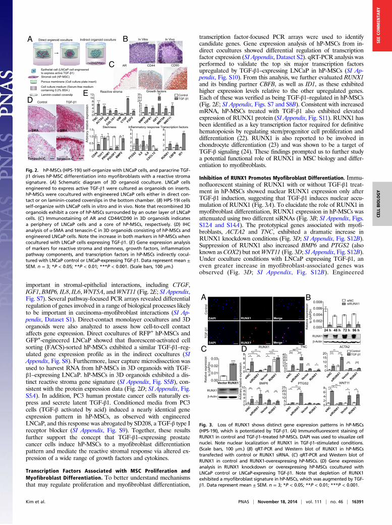
important in stromal-epithelial interactions, including CTGF,IGF1, BMP6, IL8, IL6,WNT5A, andWNT11 (Fig. 2E; SI Appendix,Fig. S7). Several pathway-focused PCR arrays revealed differentialregulation of genes involved in a range of biological processes likelyto be important in carcinoma–myofibroblast interactions (SI Ap-pendix, Dataset S1). Direct-contact monolayer cocultures and 3Dorganoids were also analyzed to assess how cell-to-cell contactaffects gene expression. Direct cocultures of RFP+ hP-MSCs andGFP+-engineered LNCaP showed that fluorescent-activated cellsorting (FACS)-sorted hP-MSCs exhibited a similar TGF-β1–reg-ulated gene expression profile as in the indirect cocultures (SIAppendix, Fig. S8). Furthermore, laser capture microdissection wasused to harvest RNA from hP-MSCs in 3D organoids with TGF-β1–expressing LNCaP. hP-MSCs in 3D organoids exhibited a dis-tinct reactive stroma gene signature (SI Appendix, Fig. S5B), con-sistent with the protein expression data (Fig. 2D; SI Appendix, Fig.S5A). In addition, PC3 human prostate cancer cells naturally ex-press and secrete latent TGF-β1. Conditioned media from PC3cells (TGF-β activated by acid) induced a nearly identical geneexpression pattern in hP-MSCs, as observed with engineeredLNCaP, and this response was abrogated by SD208, a TGF-β type Ireceptor blocker (SI Appendix, Fig. S9). Together, these resultsfurther support the concept that TGF-β1–expressing prostatecancer cells induce hP-MSCs to a myofibroblast differentiationpattern and mediate the reactive stromal response via altered ex-pression of a wide range of growth factors and cytokines.
Transcription Factors Associated with MSC Proliferation andMyofibroblast Differentiation. To better understand mechanismsthat may regulate proliferation and myofibroblast differentiation,
transcription factor-focused PCR arrays were used to identifycandidate genes. Gene expression analysis of hP-MSCs from in-direct cocultures showed differential regulation of transcriptionfactor expression (SI Appendix, Dataset S2). qRT-PCR analysis wasperformed to validate the top six major transcription factorsupregulated by TGF-β1–expressing LNCaP in hP-MSCs (SI Ap-pendix, Fig. S10). From this analysis, we further evaluated RUNX1and its binding partner CBFB, as well as ID1, as these exhibitedhigher expression levels relative to the other upregulated genes.Each of these was verified as being TGF-β1–regulated in hP-MSCs(Fig. 2E; SI Appendix, Figs. S7 and S8B). Consistent with increasedmRNA, hP-MSCs treated with TGF-β1 also exhibited elevatedexpression of RUNX1 protein (SI Appendix, Fig. S11). RUNX1 hasbeen identified as a key transcription factor required for definitivehematopoiesis by regulating stem/progenitor cell proliferation anddifferentiation (22). RUNX1 is also reported to be involved inchondrocyte differentiation (23) and was shown to be a target ofTGF-β signaling (24). These findings prompted us to further studya potential functional role of RUNX1 in MSC biology and differ-entiation to myofibroblasts.
Inhibition of RUNX1 Promotes Myofibroblast Differentiation. Immu-nofluorescent staining of RUNX1 with or without TGF-β1 treat-ment in hP-MSCs showed nuclear RUNX1 expression only afterTGF-β1 induction, suggesting that TGF-β1 induces nuclear accu-mulation of RUNX1 (Fig. 3A). To elucidate the role of RUNX1 inmyofibroblast differentiation, RUNX1 expression in hP-MSCs wasattenuated using two different siRNAs (Fig. 3B; SI Appendix, Figs.S12A and S14A). The prototypical genes associated with myofi-broblasts, ACTA2 and TNC, exhibited a dramatic increase inRUNX1 knockdown conditions (Fig. 3D; SI Appendix, Fig. S12B).Suppression of RUNX1 also increased BMP6 and PTGS2 (alsoknown as COX2) but notWNT11 (Fig. 3D; SI Appendix, Fig. S12B).Under coculture conditions with LNCaP expressing TGF-β1, aneven greater increase in myofibroblast-associated genes wasobserved (Fig. 3D; SI Appendix, Fig. S12B). Engineered
RUNX1CBFB ID
10
2
4
6
8
** **
**
PTGS2 IL8 IL6
CXCL12012345
*
***
CD44THY1
PROM1
POU5F1
BMP20.0
0.5
1.0
1.5
**** ***
BMP6
TGFB1IG
F1
WNT5A
WNT11012345
253035
** **
***
****
TNC
ACTA2
FAP
COL1A1
VIMCTGF
FGF202468
1012
******
****
***
*****
A Direct organoid coculture
Epithelial cell (LNCaP cell engineeredto express active TGF- 1)Stromal cell (hP-MSC)
Porous membrane (Cell culture plate insert)
Cell culture medium (Serum-free mediumcontaining 0.2% BSA )Laminin-coated coverslip
Indirect organoid coculture
C
In Vitro In Vivo
AR CD44 CD90
B
D Control TGF- 1
Stemness Transcription factors
Growth factors
Inflammatory response
Reactive stroma
TGF- 1Control
Arb
itrar
y re
lativ
e ex
pres
sion
E
-SMA-SMA
Tenascin-C Tenascin-C
Fig. 2. hP-MSCs (HPS-19I) self-organize with LNCaP cells, and paracrine TGF-β1 drives hP-MSC differentiation into myofibroblasts with a reactive stromasignature. (A) Schematic diagram of 3D organoid coculture. LNCaP cellsengineered to express active TGF-β1 were cultured as organoids on inserts.hP-MSCs were cocultured with engineered LNCaP cells either in direct con-tact or on laminin-coated coverslips in the bottom chamber. (B) HPS-19I cellsself-organize with LNCaP cells in vitro and in vivo. Note that recombined 3Dorganoids exhibit a core of hP-MSCs surrounded by an outer layer of LNCaPcells. (C) Immunostaining of AR and CD44/CD90 in 3D organoids indicatesa periphery of LNCaP cells and a core of hP-MSCs, respectively. (D) IHCanalysis of α-SMA and tenascin-C in 3D organoids consisting of hP-MSCs andengineered LNCaP cells. Note the increase in both markers in hP-MSCs whencocultured with LNCaP cells expressing TGF-β1. (E) Gene expression analysisof markers for reactive stroma and stemness, growth factors, inflammationpathway components, and transcription factors in hP-MSCs indirectly cocul-tured with LNCaP control or LNCaP-expressing TGF-β1. Data represent mean ±SEM. n = 3; *P < 0.05; **P < 0.01; ***P < 0.001. (Scale bars, 100 μm.)
siNC
siRUNX1
Vector
RUNX105
1015303540 *** ***
****
** *
siNC
siRUNX1
Vector
RUNX105
101520 *** ***
**
** ***
siNC
siRUNX1
Vector
RUNX102468 * * **
* *
siNC
siRUNX1
Vector
RUNX105
10152025 * **
**
siNC
siRUNX1
Vector
RUNX102468
10 ** **** **
siNC
siRUNX1
Vector
RUNX102468 * **
****
****
**
24 h 48 h 72 h 96 h0.000
0.002
0.004
0.006
0.008
* **** *
Vector RUNX10.00
0.01
0.02
0.03***
RUNX1
-Actin
Rel
ativ
e ex
pres
sion
to
GA
PD
H
RUNX1
-Actin
Rel
ativ
e ex
pres
sion
to
GA
PD
H
Arb
itrar
y re
lativ
e ex
pres
sion
RUNX1 TNC ACTA2
BMP6 PTGS2 WNT11
B
C
Con
trol
TGF-
1
DAPI RUNX1 Merge
DAPI Merge
A
RUNX1
D
siNCsiRUNX1
TGF- 1Control
Fig. 3. Loss of RUNX1 shows distinct gene expression patterns in hP-MSCs(HPS-19I), which is potentiated by TGF-β1. (A) Immunofluorescent staining ofRUNX1 in control and TGF-β1–treated hP-MSCs. DAPI was used to visualize cellnuclei. Note nuclear localization of RUNX1 in TGF-β1–stimulated conditions.(Scale bars, 100 μm.) (B) qRT-PCR and Western blot of RUNX1 in hP-MSCstransfected with control or RUNX1 siRNA. (C) qRT-PCR and Western blot ofRUNX1 in control and RUNX1-overexpressing hP-MSCs. (D) Gene expressionanalysis in RUNX1 knockdown or overexpressing hP-MSCs cocultured withLNCaP control or LNCaP-expressing TGF-β1. Note that depletion of RUNX1exhibited a myofibroblast signature in hP-MSCs, which was augmented by TGF-β1. Data represent mean ± SEM. n = 3; *P < 0.05; **P < 0.01; ***P < 0.001.
Kim et al. PNAS | November 18, 2014 | vol. 111 | no. 46 | 16391
CELL
BIOLO
GY
SEECO
MMEN
TARY

overexpression of RUNX1 (Fig. 3C) produced no significantincrease or decrease in the effect of TGF-β1 on hP-MSCs (Fig.3D). These results suggest that RUNX1 is permissive for myo-differentiation but may restrict complete differentiation, perhapsby maintenance of a proliferative (progenitor) status. Accord-ingly, we next evaluated the role of RUNX1 in mediating pro-liferation of MSCs.
RUNX1 Is Required for MSC Cell Cycle Progression and Proliferation.Knockdown of RUNX1 altered cell morphology. hP-MSCs withRUNX1 knockdown exhibited a more spread out, large, and flat-tened phenotype (Fig. 4A). Consistent with this phenotype, RUNX1knockdown resulted in decreased CD44, POU5F1 (also known asOCT4), and BMP2 expression, yet maintained THY1 (also knownas CD90) expression (Fig. 4B; SI Appendix, Fig. S14B). THY1 isa known marker of prostate cancer-associated fibroblasts (25). Ofmost interest, hP-MSCs with RUNX1 knockdown were restrictedfrom proliferation (Fig. 4C). Consistent with growth arrest, RUNX1knockdown led to decrease in expression of proliferation-associatedgenes (Fig. 4D; SI Appendix, Fig. S14B). Bromodeoxyuridine(BrdU) incorporation revealed that depletion of RUNX1 resultedin a decrease in the number of cells in S phase (Fig. 4E; SI Appendix,Fig. S14C). Analysis of cell cycle-related gene expression showeda significant decrease in cyclin A and B, required for both S-phaseprogression and G2/M-phase transition and for mitosis, respectively(26–28) (Fig. 4F; SI Appendix, Fig. S14B). No significant differenceswere observed in apoptotic and necrotic cell death between controland RUNX1 knockdown hP-MSCs, as analyzed by annexin Vconjugate and SYTOX Red dead cell stain (SI Appendix, Fig. S15).These data suggest that attenuated RUNX1 restricts proliferationby modulating cell cycle regulators without inducing cell death.RUNX1 knockdown in marrow-derived MSCs produced nearly
identical results. These cells also exhibited a more flattened phe-notype and were fully growth restricted under RUNX1 knockdown
conditions (Fig. 5A). In a similar manner, the percentage of cells inS phase was reduced (Fig. 5E). Expression of proliferation-associatedgenes and cell cycle control genes (Fig. 5 B and D), as well asdifferentiation/stemness-associated genes (Fig. 5C) exhibiteda pattern consistent with those observed in hP-MSCs.To assess how RUNX1 expression affects cell proliferative or
differentiation-committed states, we examined the localization ofRUNX1, Ki-67, and α-SMA after RUNX1 knockdown in both hP-MSCs and hBM-MSCs. Cell morphology changes and α-SMAexpression were correlated with the nuclear intensity of RUNX1staining (Fig. 6A). In control hP-MSCs, small and undifferentiatedcells showed higher RUNX1 nuclear staining and lower α-SMAcytoplasmic expression (arrowheads) than large and differentiatedcells (arrows). More interestingly, RUNX1-depleted hP-MSCs andhBM-MSCs revealed a myofibroblast phenotype characterized byactin polymerization, whereas controls expressed nonpolymerizedactin. A similar correlation to that seen between RUNX1 andα-SMA was observed between Ki-67 and α-SMA (Fig. 6B). Anincrease in α-SMA in MSCs corresponded to loss of Ki-67 ex-pression and morphology changes consistent with myofibroblastdifferentiation. Ki-67 negative cells in RUNX1 knockdown MSCsexhibited highly organized α-SMA (arrows). In addition, in vivoxenografts (day 3) constructed with LNCaP cells combined withRFP-expressing control or RUNX1 knockdown hP-MSCs alsoshowed reduced Ki-67 staining in RUNX1 knockdown conditions(SI Appendix, Fig. S16). Consistent with these data, xenograftsconstructed with RUNX1 knockdown hP-MSCs exhibited a greaterstaining intensity for α-SMA (SI Appendix, Fig. S13). Overall, theseresults suggest that RUNX1 in MSCs is required for cell pro-liferation and modulates terminal differentiation to myofibroblasts(Fig. 6C).
DiscussionWe report the first (to our knowledge) isolation and character-ization of tissue-resident MSCs derived from human normal adultprostate gland (21). The prostate-derived MSCs exhibited MSC-typical cell-surface antigen profiles and were multipotent and ca-pable of long-term culture. Previous reports of adult human tissue-resident MSCs are restricted to dental (29), adenoid (30), andadipose (31) tissues. Importantly, we report here a critical role ofRUNX1 in modulating MSC cell proliferation and committeddifferentiation to myofibroblasts. Myofibroblasts are stromal cellsthat are crucial for proper wound repair and the formation of re-active stroma associated with cancer, fibrosis, and vessel disease.Our data suggest that prostate-resident CD44+CD90+ MSCs areone potential source of reactive stroma myofibroblasts that co-evolve with foci of prostate cancer and in benign prostatic hyper-plasia. There are several reported sources of myofibroblasts andcarcinoma associated fibroblasts in tumor-associated reactivestroma, including circulating MSCs, normal fibroblasts, circulatingfibrocytes, and vessel-associated pericytes (1, 2, 32, 33). Our datado not exclude the involvement of these cell types. Our data fur-ther show that RUNX1 is an essential transcription factor for MSCproliferation and that RUNX1 modulates MSC differentiation tomyofibroblasts. TGF-β1, overexpressed by prostate carcinomacells, is a key inducer of myofibroblast differentiation and biology.We show here that RUNX1 expression is regulated by TGF-β1 inMSCs and that RUNX1 affects TGF-β1–stimulated gene expres-sion during differentiation to myofibroblasts.Myofibroblasts are important in normal wound closure and
contribute to the pathology of several diseases and disorders. Im-portantly, myofibroblasts provide a prowound repair environmentthat forms granulation tissue and contracts to affect wound closure.The transient appearance of myofibroblasts is beneficial in restoringand maintaining tissue homeostasis. However, excessive myofibro-blast proliferation and prolonged presence is detrimental for tissuefunction and thereby increases the risk of fibrotic diseases andcancers in many tissues including lung, liver, kidney, and prostate (4,
1 2 3 40
2
4
6
8
***
Days
Cel
l Num
ber (
x105 )
CD44THY1
POU5F1
BMP20.0
0.5
1.0
1.5
** ***
TNC
ACTA2
02468
*****
A siNC
siRUNX1
B C D
Stemness
Myofibroblast signature
F
DNA content :: 7-AAD
Brd
U ::
APC
E
Arb
itrar
y re
lativ
e ex
pres
sion
siNC siRUNX1
S18.3%
S4.94%
siNCsiRUNX1
PCNAMKI67
cMYC
BIRC5
0.0
0.5
1.0
1.5
*
**
Arb
itrar
y re
lativ
e ex
pres
sionsiNC
siRUNX1
CCNA2
CCNB1
CCNB2
CCND1
CCNE10.0
0.5
1.0
1.5
2.0
***
**
**
CDKN1A
CDKN1B
CDKN2A
CDKN2B0
1
2
3
***
siNCsiRUNX1
Arb
itrar
y re
lativ
e ex
pres
sion
Fig. 4. Suppressing RUNX1 induces hP-MSC (HPS-19I) growth and cell cyclearrest. (A) Effect of RUNX1 knockdown on hP-MSC morphology. (Scale bars,50 μm.) (B) qRT-PCR analysis of myofibroblast and stemness genes in controland RUNX1 knockdown hP-MSCs. (C) Growth curves of hP-MSCs followingtransfection with control or RUNX1 siRNA. (D) qRT-PCR analysis of pro-liferation-related genes in control and RUNX1 knockdown hP-MSCs. (E)Representative cell cycle distribution of hP-MSCs transfected with control orRUNX1 siRNA. Cells were analyzed by flow cytometry after BrdU incor-poration and 7-AAD staining at 72 h posttransfection. (F) qRT-PCR analysisof multiple cyclins and cyclin-dependent kinase inhibitors in hP-MSCsfollowing RUNX1 knockdown. For B, D, and F, data represent mean ± SEM. ForC, data represent mean ± SD; two-way ANOVA. n = 3; *P < 0.05; **P < 0.01;***P < 0.001.
16392 | www.pnas.org/cgi/doi/10.1073/pnas.1407097111 Kim et al.

34, 35). Thus, reactive stroma is associated with elevated accumu-lation of myofibroblasts, a hallmark of fibrosis and a predictor ofcancer progression. Accordingly, identifying mechanisms that affectthe genesis of myofibroblasts from precursor cells may providepotential therapeutic targets in the treatment of fibrotic diseasesand cancers.RUNX1 is a key transcription factor that regulates hematopoietic
stem cells and hematopoiesis (36); however, its role in MSC andmyofibroblast biology has not been reported. Our data show thatRUNX1 expression is required for extended self-renewal and pro-liferation of MSCs and that RUNX1 expression is stimulated byTGF-β1. Attenuation of RUNX1 induced complete myofibroblastdifferentiation and quiescence of hP-MSCs, and this was potenti-ated by TGF-β1. Interestingly, these data show that TGF-β1 inducesexpression of genes associated with differentiation of myofibro-blasts, yet also stimulates expression of RUNX1 that inhibits dif-ferentiation and maintains proliferation. The genesis of reactivestroma myofibroblasts depends on the proliferation of MSCs andtheir transit-amplifying progeny. Hence, one possible explanationfor our data is that TGF-β1 induces MSC commitment to myofi-broblast progenitor cells while maintaining proliferative status viaelevated or sustained expression of RUNX1. This concept is con-sistent with previous reports that show that TGF-β1 stimulatesproliferation of mesenchymal stem cells (37–39), progenitor cells,and several types of fibroblasts (40–43) and also induces myofi-broblast differentiation and the fibrosis phenotype (44). TGF-β hasbeen shown to stimulate fibroblast proliferation and subsequentlyinduce their quiescence and differentiation to myofibroblasts ina coordinate manner (45). Additional factors, including CXCL12(SDF-1), have also been shown to regulate the myofibroblast phe-notype (46). TGF-β and CXCL12 pathways interact and regulateaccumulation of myofibroblasts from resident fibroblasts in breastcancer (47). Hence, TGF-β1 acts in a coordinate and sequentialmanner to both stimulate proliferation of progenitors and inducetheir differentiation.Another advance reported here was the development of a cancer
cell–MSC recombined 3D in vitro organoid coculture model. Thismodel requires no additional growth factors, serum substitutes, or
extracellular matrix components. Organoid coculture of MSCs andtumor cells consistently formed a self-organizing organoid witha core of MSC-derived stromal cells surrounded by tumor cells. The3D indirect coculture model permits a more detailed understandingof the complex mechanisms underlying carcinoma cell–stromalcell crosstalk.Myofibroblasts are currently a potent target for developing
antifibrotic therapies (48). Our findings provide insights to helpidentify potentially specific pathways. The overall key mecha-nisms and contributing cell types in the formation of reactivestroma have yet been fully identified. Perturbation of pathwaysthat produce a biologically effective mass of myofibroblasts mayoffer potential for the development of antifibrotic and anticancertherapies via targeting the reactive stroma microenvironment.
Materials and MethodsIsolation of Stromal Cells from Normal Human Prostate Gland. A fresh tissuecore from the peripheral zone of normal prostate was obtained from 19- and33-y-old cadaver donors following a Baylor College of Medicine InstitutionalReview Board approved protocol and consents. The core was cut into discsand placed in a 96-well tissue plate containing Bfs medium [DMEM; highglucose (GIBCO) supplemented with 5% (vol/vol) FBS (HyClone), 5% (vol/vol)NuSerum (Collaborative Research), 0.5 μg/mL testosterone, 5 μg/mL insulin,100 units/mL penicillin, and 100 μg/mL streptomycin (Sigma)]. The explantswere incubated at 37 °C with 5% (vol/vol) CO2, and medium was changedevery 48 h or as necessary. Stromal cells migrated out of the explant andattached to the tissue culture dish. After the cells reached confluence, theexplant was moved and the cells were passaged. Cultures at passages 7–15were used for all experiments.
CCNA2
CCNB1
CCNB2
CCND1
CCNE10.0
0.5
1.0
1.5
** ****
**
CDKN1A
CDKN1B
CDKN2A
CDKN2B0
1
2
3
4
*****
**
1 2 3 40
2
4
6
8
***
Days
Cel
l Num
ber (
x105 )
CD44THY1
POU5F1
BMP20.00.51.01.52.0
***
**
***
TNC
ACTA2
01234
***
DNA content :: 7-AAD
A
Brd
U ::
APC
B C
D
E siNC siRUNX1
Stemness
Myofibroblast signature
S26.4%
S3.73%
PCNAMKI67
cMYC
BIRC5
0.0
0.5
1.0
1.5
******
***
Arb
itrar
y re
lativ
e ex
pres
sion
siNCsiRUNX1
siNCsiRUNX1
Arb
itrar
y re
lativ
e ex
pres
sionsiNC
siRUNX1
siNCsiRUNX1
Arb
itrar
y re
lativ
e ex
pres
sion
Fig. 5. Depletion of RUNX1 inhibits growth and cell cycle progression inhBM-MSCs. (A) Growth curve of hBM-MSCs in control and RUNX1 knock-down conditions. (B–D) qRT-PCR analysis in hBM-MSCs transfected withcontrol or RUNX1 siRNA at 48 h posttransfection. Gene expression profiles ofproliferation-associated genes (B), myofibroblast and stemness genes (C),and cell cycle control genes (D). (E) Representative cell cycle distribution ofhBM-MSCs pulse-labeled with BrdU following transfection with control orRUNX1 siRNA for 72 h. Note that loss of RUNX1 in hBM-MSCs exhibits con-sistent phenotype and gene expression patterns as those shown in hP-MSCs.For A, data represent mean ± SD; two-way ANOVA. For B–D, data representmean ± SEM. n = 3; *P < 0.05; **P < 0.01; ***P < 0.001.
A
BRUNX1
-SMA
siRUNX1siNC
hP-MSCs
hBM-MSCs
hP-MSCs
C
RUNX1
TGF- 1
RUNX1RUNX
CD44+CD90+
hP-MSC hBM-MSC
Cell proliferation
Blockedcell proliferation
RUNX1 RUNX1RUNX1
RUNX1RUNX1
Transit-amplifyingcell population
RUNX1RUNX
TGF- 1
Differentiation
TGF- 1
Differentiation
Differentiaff
-SMA, tenascin-CMyofibroblast
Myofibroblast enriched stroma
(reactive)
Myofibroblast depleted stroma
RUNX1-SMA
Ki-67-SMA
Ki-67-SMA
Ki-67-SMA
Ki-67-SMA
Fig. 6. The expression level of RUNX1 regulates cell proliferative or differen-tiation-committed state in hP-MSCs (HPS-19I) and hBM-MSCs. (A) Immunofluo-rescent staining for RUNX1 (red) and α-SMA (green) in hP-MSCs transfected withcontrol or RUNX1 siRNA for 72 h. (Scale bars, 100 μm.) (B) Immunofluorescentstaining for Ki-67 (red) and α-SMA (green) in hP-MSCs and hBM-MSCs withsiRNA-mediated knockdown RUNX1 or control siRNA at 72 h posttransfection.Note that silencing RUNX1 increased polymerized α-SMA that corresponded toloss of Ki-67 expression and morphology changes consistent with myofibroblastdifferentiation (arrows). (Scale bars, 100 μm.) (C) Schematic diagram depictinga working hypothesis of how RUNX1 and TGF-β1 may regulate the transit am-plification of MSCs to a critical mass of myofibroblasts typical of wound repairstroma and reactive stroma associated with cancer and fibrosis.
Kim et al. PNAS | November 18, 2014 | vol. 111 | no. 46 | 16393
CELL
BIOLO
GY
SEECO
MMEN
TARY

Coculture of hP-MSCs and Prostate Cancer Cells. For 3D organoid coculture,a mixture of LNCaP cells and/or either hBM-MSCs (Lonza) or hP-MSCs at differentratios was seeded onto a 12-mm Millicell-CM culture plate insert (Millipore) ina 24-well cultureplate (Falcon) and incubated in LNCaP cell growthmedium.After24 h of incubation, the medium was switched to M20 medium [DMEM; highglucose (GIBCO) supplemented with 0.2% BSA, 5 μg/mL insulin, 5 μg/mL trans-ferrin, and 5 ng/mL sodium selenite (ITS; insulin, trasnsferrin, selenium supple-ment, Sigma)]. After 72 h of coculture, organoids were either fixed in 4% (vol/vol)paraformaldehyde for 15 min at room temperature for paraffin embeddingor immediately processed for cryo-embedding. For indirect coculture, hP-MSCs(5 × 10⁴) were seeded on a 12-mm round coverslip coated with poly-D-lysine/laminin (BD BioCoat) on a 24-well culture plate (Falcon) in Bfs medium. LNCaP orPC3 cells (4 × 10⁵) were seeded onto a 12-mm Millicell-CM culture plate insert(Millipore) in a 24-well culture plate (Falcon) in growth medium. After 24 h ofincubation, the insert containing LNCaP cells was transferred to the well con-taining hP-MSCs, and these cells were cocultured for 72 h in M20 medium. Fordirect coculture in monolayer, RFP-expressing hP-MSCs (1 × 10⁶) were seededonto a 150-mm dish (Falcon) in Bfs medium. After 24 h of incubation, an equalnumber of GFP-expressing engineered LNCaP cells were directly loaded onto hP-MSCs and cultured for 24 h in LNCaP cell growth medium. The next day, themedium was switched to M20 medium, and cells were cocultured for 72 h. hP-MSCs (RFP+) and engineered LNCaP (GFP+) cells from coculture experiments were
suspended in Ca2+/Mg2+-free Dulbecco’s PBS (GIBCO). RFP- and GFP-labeled cellsorting was performed with a BD FACSAria II cell sorter.
Differential Reactive Stroma Xenograft Model. Mice were injected sub-cutaneously with LNCaP cells and hP-MSCs with or without Matrigel as de-scribed previously (16). Xenografts were harvested after 3, 10, and 11 d.
Statistical Analysis. Student t test or two-way ANOVA was used to de-termine significance between groups. For all statistical tests, P < 0.05 wasconsidered significant.
ACKNOWLEDGMENTS. We thank Drs. Owen Witte and Li Xin for theFUCRW lentiviral vector and packaging plasmids; Truong D. Dang fortechnical assistance; and Joel M. Sederstrom and Michael Ittmann, MD,PhD, for expert assistance. This work was supported by grants from theNational Institutes of Health (NIH) (R01 CA58093 and R01 DK083293) andthe Department of Defense (W81XWH-12-1-0197). This project was sup-ported by the Cytometry and Cell Sorting Core at Baylor College of Med-icine with funding from the NIH (AI036211, CA125123 and RR024574)and by the Pathology and Histology Core at Baylor College of Medicinewith funding from the NIH (National Cancer Institute Grant P30-CA125123).
1. Uccelli A, Moretta L, Pistoia V (2008) Mesenchymal stem cells in health and disease.Nat Rev Immunol 8(9):726–736.
2. Barron DA, Rowley DR (2012) The reactive stroma microenvironment and prostatecancer progression. Endocr Relat Cancer 19(6):R187–R204.
3. Desmoulière A, Darby IA, Gabbiani G (2003) Normal and pathologic soft tissue re-modeling: Role of the myofibroblast, with special emphasis on liver and kidney fi-brosis. Lab Invest 83(12):1689–1707.
4. Gabbiani G (2003) The myofibroblast in wound healing and fibrocontractive diseases.J Pathol 200(4):500–503.
5. Rowley DR (1998-1999) What might a stromal response mean to prostate cancerprogression? Cancer Metastasis Rev 17(4):411–419.
6. Tuxhorn JA, Ayala GE, Rowley DR (2001) Reactive stroma in prostate cancer pro-gression. J Urol 166(6):2472–2483.
7. Tuxhorn JA, et al. (2002) Reactive stroma in human prostate cancer: Induction ofmyofibroblast phenotype and extracellular matrix remodeling. Clin Cancer Res 8(9):2912–2923.
8. Peddareddigari VG, Wang D, Dubois RN (2010) The tumor microenvironment in co-lorectal carcinogenesis. Cancer Microenviron 3(1):149–166.
9. Gao Q, et al. (2011) Tumor stroma reaction-related gene signature predicts clinicaloutcome in human hepatocellular carcinoma. Cancer Sci 102(8):1522–1531.
10. Bremnes RM, et al. (2011) The role of tumor stroma in cancer progression andprognosis: Emphasis on carcinoma-associated fibroblasts and non-small cell lungcancer. J Thorac Oncol 6(1):209–217.
11. Dekker TJ, et al. (2013) Prognostic significance of the tumor-stroma ratio: Validation studyin node-negative premenopausal breast cancer patients from the EORTC perioperativechemotherapy (POP) trial (10854). Breast Cancer Res Treat 139(2):371–379.
12. Freeman MR, Li Q, Chung LW (2013) Can stroma reaction predict cancer lethality? ClinCancer Res 19(18):4905–4907.
13. Ayala G, et al. (2003) Reactive stroma as a predictor of biochemical-free recurrence inprostate cancer. Clin Cancer Res 9(13):4792–4801.
14. Ayala GE, et al. (2011) Determining prostate cancer-specific death through quantificationof stromogenic carcinoma area in prostatectomy specimens. Am J Pathol 178(1):79–87.
15. Yanagisawa N, et al. (2007) Stromogenic prostatic carcinoma pattern (carcinomaswith reactive stromal grade 3) in needle biopsies predicts biochemical recurrence-freesurvival in patients after radical prostatectomy. Hum Pathol 38(11):1611–1620.
16. Tuxhorn JA, McAlhany SJ, Dang TD, Ayala GE, Rowley DR (2002) Stromal cells pro-mote angiogenesis and growth of human prostate tumors in a differential reactivestroma (DRS) xenograft model. Cancer Res 62(11):3298–3307.
17. Tuxhorn JA, McAlhany SJ, Yang F, Dang TD, Rowley DR (2002) Inhibition of trans-forming growth factor-beta activity decreases angiogenesis in a human prostatecancer-reactive stroma xenograft model. Cancer Res 62(21):6021–6025.
18. Yang F, Strand DW, Rowley DR (2008) Fibroblast growth factor-2 mediates trans-forming growth factor-beta action in prostate cancer reactive stroma. Oncogene27(4):450–459.
19. Yang F, et al. (2005) Stromal expression of connective tissue growth factor promotesangiogenesis and prostate cancer tumorigenesis. Cancer Res 65(19):8887–8895.
20. Hinz B (2007) Formation and function of the myofibroblast during tissue repair.J Invest Dermatol 127(3):526–537.
21. Rowley DR, Tindall DJ (1987) Responses of NBT-II bladder carcinoma cells to condi-tioned medium from normal fetal urogenital sinus. Cancer Res 47(11):2955–2960.
22. Nimmo R, Woollard A (2008) Worming out the biology of Runx. Dev Biol 313(2):492–500.
23. Wang Y, et al. (2005) Runx1/AML1/Cbfa2 mediates onset of mesenchymal cell dif-ferentiation toward chondrogenesis. J Bone Miner Res 20(9):1624–1636.
24. Ito Y, Miyazono K (2003) RUNX transcription factors as key targets of TGF-beta su-perfamily signaling. Curr Opin Genet Dev 13(1):43–47.
25. Zhao H, Peehl DM (2009) Tumor-promoting phenotype of CD90hi prostate cancer-associated fibroblasts. Prostate 69(9):991–1000.
26. Fung TK, Ma HT, Poon RY (2007) Specialized roles of the two mitotic cyclins in somaticcells: Cyclin A as an activator of M phase-promoting factor. Mol Biol Cell 18(5):1861–1873.
27. Gong D, Ferrell JE, Jr (2010) The roles of cyclin A2, B1, and B2 in early and late mitoticevents. Mol Biol Cell 21(18):3149–3161.
28. Pagano M, Pepperkok R, Verde F, Ansorge W, Draetta G (1992) Cyclin A is required attwo points in the human cell cycle. EMBO J 11(3):961–971.
29. Jo YY, et al. (2007) Isolation and characterization of postnatal stem cells from humandental tissues. Tissue Eng 13(4):767–773.
30. Lee YS, et al. (2013) Isolation of mesenchymal stromal cells (MSCs) from human ad-enoid tissue. Cell Physiol Biochem 31(4-5):513–524.
31. Araña M, Mazo M, Aranda P, Pelacho B, Prosper F (2013) Adipose tissue-derivedmesenchymal stem cells: Isolation, expansion, and characterization.Methods Mol Biol1036:47–61.
32. Gabbiani G (1996) The cellular derivation and the life span of the myofibroblast.Pathol Res Pract 192(7):708–711.
33. Brennen WN, Chen S, Denmeade SR, Isaacs JT (2013) Quantification of Mesenchymalstem cells (MSCs) at sites of human prostate cancer. Oncotarget 4(1):106–117.
34. Hinz B, et al. (2012) Recent developments in myofibroblast biology: Paradigms forconnective tissue remodeling. Am J Pathol 180(4):1340–1355.
35. Desmoulière A, Chaponnier C, Gabbiani G (2005) Tissue repair, contraction, and themyofibroblast. Wound Repair Regen 13(1):7–12.
36. Chen MJ, Yokomizo T, Zeigler BM, Dzierzak E, Speck NA (2009) Runx1 is required for theendothelial to haematopoietic cell transition but not thereafter. Nature 457(7231):887–891.
37. Jian H, et al. (2006) Smad3-dependent nuclear translocation of beta-catenin is re-quired for TGF-beta1-induced proliferation of bone marrow-derived adult humanmesenchymal stem cells. Genes Dev 20(6):666–674.
38. Rodrigues M, Griffith LG, Wells A (2010) Growth factor regulation of proliferationand survival of multipotential stromal cells. Stem Cell Res Ther 1(4):32.
39. Walenda G, et al. (2013) TGF-beta1 does not induce senescence of multipotentmesenchymal stromal cells and has similar effects in early and late passages. PLoS ONE8(10):e77656.
40. Chen G, Deng C, Li YP (2012) TGF-β and BMP signaling in osteoblast differentiationand bone formation. Int J Biol Sci 8(2):272–288.
41. Cheng L, Zhang C, Li D, Zou J, Wang J (2014) Transforming growth factor-β1 (TGF-β1)induces mouse precartilaginous stem cell proliferation through TGF-β receptor II(TGFRII)-Akt-β-catenin signaling. Int J Mol Sci 15(7):12665–12676.
42. Mendias CL, Gumucio JP, Lynch EB (2012) Mechanical loading and TGF-β change theexpression of multiple miRNAs in tendon fibroblasts. J Appl Physiol (1985) 113(1):56–62.
43. Stouffer GA, Owens GK (1994) TGF-beta promotes proliferation of cultured SMC viaboth PDGF-AA-dependent and PDGF-AA-independent mechanisms. J Clin Invest 93(5):2048–2055.
44. Desmoulière A, Geinoz A, Gabbiani F, Gabbiani G (1993) Transforming growth factor-beta 1 induces alpha-smooth muscle actin expression in granulation tissue myofi-broblasts and in quiescent and growing cultured fibroblasts. J Cell Biol 122(1):103–111.
45. Grotendorst GR, Rahmanie H, Duncan MR (2004) Combinatorial signaling pathwaysdetermine fibroblast proliferation and myofibroblast differentiation. FASEB J 18(3):469–479.
46. Gharaee-Kermani M, et al. (2012) CXC-type chemokines promote myofibroblastphenoconversion and prostatic fibrosis. PLoS ONE 7(11):e49278.
47. Kojima Y, et al. (2010) Autocrine TGF-beta and stromal cell-derived factor-1 (SDF-1)signaling drives the evolution of tumor-promoting mammary stromal myofibroblasts.Proc Natl Acad Sci USA 107(46):20009–20014.
48. Kramann R, DiRocco DP, Humphreys BD (2013) Understanding the origin, activationand regulation of matrix-producing myofibroblasts for treatment of fibrotic disease.J Pathol 231(3):273–289.
16394 | www.pnas.org/cgi/doi/10.1073/pnas.1407097111 Kim et al.