Embed Size (px)
Citation preview
541Case Report
Ruptured Aneurysm of the Accessory Middle Cerebral ArteryAssociated with Moyamoya Disease – A Case Report
Cheng-Chi Lee, MD; Zhuo-Hao Liu, MD; Shih-Ming Jung1, MD; Tao-Chieh Yang, MD
The accessory middle cerebral artery can provide collateral blood supply in moyamoyadisease. We report a case of unilateral moyamoya disease which demonstrates the anatomyof the right accessory middle cerebral artery and a ruptured peripheral aneurysm on theartery. Our patient was a 56-year-old woman who initially suffered from headache andlethargy. Right caudate nucleus hemorrhage with intraventricular extension and spontaneoussubarachnoid hemorrhage were found on brain computed tomography. A ruptured peripheralaccessory middle cerebral artery aneurysm associated with unilateral moyamoya disease wasdiagnosed on cerebral angiography. Surgical intervention to excise the peripheral accessorymiddle cerebral artery aneurysm assisted by frameless navigation guidance to reduce the riskof damage to collateral vessels was done successfully. Histopathology of excised tissueshowed this anomaly was a pseudoaneurysm. The management of an aneurysm inmoyamoya disease should be modified based on its location and collateral vessels.Prevention of aneurysm bleeding and preservation of collateral vessels during craniotomyare the critical when managing hemorrhagic moyamoya disease. This case suggests that sur-gical intervention for ruptured intracranial aneurysms is safe with the use of frameless navi-gation guidance to minimize collateral vessel injuries. (Chang Gung Med J 2011;34:541-7)
Key words: accessory middle cerebral artery, moyamoya disease, ruptured cerebral aneurysm
From the Department of Neurosurgery; 1Department of Pathology, Chang Gung Memorial Hospital at Linkou, Chang GungUniversity College of Medicine, Taoyuan, Taiwan.Received: May 12, 2009; Accepted: Apr. 13, 2010Correspondence to: Dr. Tao-Chieh Yang, Department of Neurosurgery, Chang Gung Memorial Hospital at Linkou. 5, Fusing St.,Gueishan Township, Taoyuan County 333, Taiwan (R.O.C.) Tel: 886-3-3281200 ext. 2119; Fax: 886-3-3285818; E-mail: [email protected]
Moyamoya disease is a chronic occlusive cere-brovascular disorder of unknown etiology. It is
characterized by progressive steno-occlusivechanges in the terminal portions of the intracranialinternal carotid arteries and the circle of Willis, alongwith concomitant development of fine networks ofcollateral vessels at the skull base which collateralizevessels distal to the occlusions and appear to serve asa source of supplemental blood flow to ischemicregions of the brain.(1,2) Unilateral involvement withdevelopment of moyamoya vessels is called ‘unilat-eral’ moyamoya disease. Moyamoya disease is com-monly accompanied by intracranial aneurysms.(3) Theaccessory middle cerebral artery (MCA) is a varia-
tion of middle cerebral artery branching, and its inci-dence has been reported to be 0.3-4.0% in angio-graphic studies.(1) The accessory MCA can providecollateral blood supply in moyamoya disease.(4) Tothe best of our knowledge, this type of accessorymiddle cerebral artery aneurysm associated withmoyamoya disease is extremely rare. We report arare case of a ruptured peripheral right accessoryMCA aneurysm with unilateral moyamoya disease.
CASE REPORT
A 56-year-old Taiwanese woman without a his-tory of systemic medical disease was sent to our
Chang Gung Med J Vol. 34 No. 5September-October 2011
Cheng-Chi Lee, et alRuptured aneurysm in moyamoya disease
542
emergency room after the sudden onset of headache,dizziness, nausea, and lethargy. The patient had ahead injury with a right occipital contusion hemor-rhage about 20 years before. Neurological examina-tion on admission revealed an unsteady gait withoutfocal limb weakness. A brain computed tomography(CT) scan demonstrated a diffuse, thin subarachnoidhemorrhage in the basal cistern, hemorrhage in theright caudate nucleus with intraventricular extension,an enlarged ventricle suggestive of acute hydro-cephalus and one small, old stroke in the right anteri-or frontal area. (Fig. 1A, B). Bilateral frontal externalventricular drains were placed. The patient’s symp-toms and discomfort were relieved after intracranialpressure was controlled by cerebrospinal fluiddrainage. Cerebral digital subtraction angiography
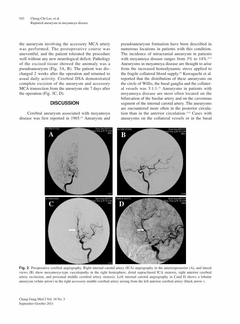
(DSA) was performed 7 days after the intracerebralhemorrhage, revealing moyamoya-type vasculopathyin the right hemisphere, including distal supraclinoidinternal carotid artery stenosis, right anterior cerebralartery occlusion, proximal MCA stenosis (Fig. 2A,B) and a lobular aneurysm in the right accessoryMCA arising from the left anterior cerebral artery.(Fig. 2C, D). Surgical intervention was undertaken toprevent aneurysm rebleeding. We performed a rightfrontal craniotomy using transfrontal-transventricularapproach with the aid of frameless CT-guided stereo-tactic navigation (Stryker system, Kalamazoo, MI,U.S.A.) (Fig. 1C). After identifying the peripheralright accessory MCA aneurysm and parent artery(accessory MCA artery), excision of the lobularaneurysm with transection of the distal segment of
Fig. 1 Serial brain CT studies. (A) Brain CT shows a right caudate nucleus hemorrhage with interventricular hemorrhage and onesmall stroke in the right anterior frontal area. (B) Brain CT shows a diffuse thin subarachnoid hemorrhage in the basal cistern. (C)Frameless stereotatic navigator system for aneurysm localization. (D) Right frontal lobe encephalomalacia is seen 3 months postop-eratively via a transfrontal transventricular approach. Abbreviation used: CT: computed tomography.
A B
C D
Chang Gung Med J Vol. 34 No. 5September-October 2011
Cheng-Chi Lee, et alRuptured aneurysm in moyamoya disease
543
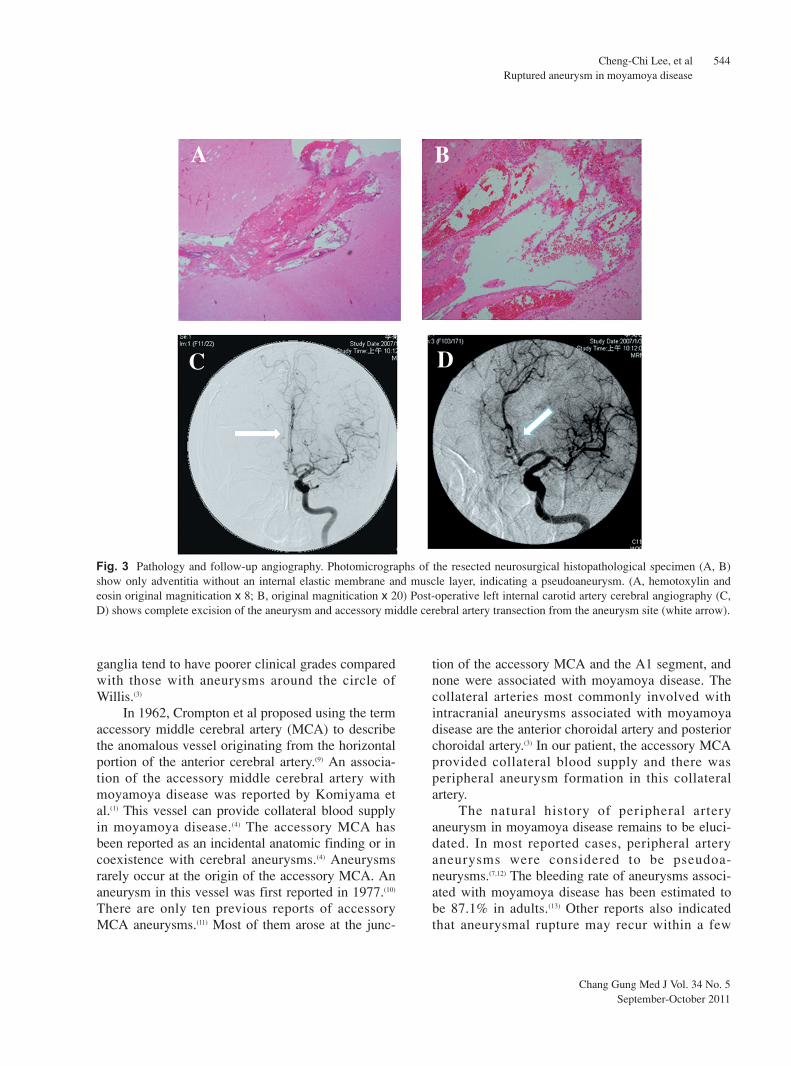
the aneurysm involving the accessory MCA arterywas performed. The postoperative course wasuneventful, and the patient tolerated the procedurewell without any new neurological deficit. Pathologyof the excised tissue showed the anomaly was apseudoaneurysm (Fig. 3A, B). The patient was dis-charged 2 weeks after the operation and returned tousual daily activity. Cerebral DSA demonstratedcomplete excision of the aneurysm and accessoryMCA transection from the aneurysm site 7 days afterthe operation (Fig. 3C, D).
DISCUSSION
Cerebral aneurysm associated with moyamoyadisease was first reported in 1965.(5) Aneurysm and
pseudoaneurysm formation have been described innumerous locations in patients with this condition.The incidence of intracranial aneurysm in patientswith moyamoya disease ranges from 3% to 14%.(6,7)
Aneurysms in moyamoya disease are thought to arisefrom the increased hemodynamic stress applied tothe fragile collateral blood supply.(8) Kawaguchi et al.reported that the distribution of these aneurysms onthe circle of Willis, the basal ganglia and the collater-al vessels was 3:1:1.(3) Aneurysms in patients withmoyamoya disease are most often located on thebifurcation of the basilar artery and on the cavernoussegment of the internal carotid artery. The aneurysmsare encountered more often in the posterior circula-tion than in the anterior circulation.(3,6) Cases withaneurysms on the collateral vessels or in the basal
Fig. 2 Preoperative cerebral angiography. Right internal carotid artery (ICA) angiography in the anteriorposterior (A), and lateralviews (B) show moyamoya-type vasculopathy in the right hemisphere, distal supraclinoid ICA stenosis, right anterior cerebralartery occlusion, and proximal middle cerebral artery stenosis. Left internal carotid angiography in Cand D shows a lobularaneurysm (white arrow) in the right accessory middle cerebral artery arising from the left anterior cerebral artery (black arrow ).
A B
C D
Chang Gung Med J Vol. 34 No. 5September-October 2011
Cheng-Chi Lee, et alRuptured aneurysm in moyamoya disease
544
ganglia tend to have poorer clinical grades comparedwith those with aneurysms around the circle ofWillis.(3)
In 1962, Crompton et al proposed using the termaccessory middle cerebral artery (MCA) to describethe anomalous vessel originating from the horizontalportion of the anterior cerebral artery.(9) An associa-tion of the accessory middle cerebral artery withmoyamoya disease was reported by Komiyama etal.(1) This vessel can provide collateral blood supplyin moyamoya disease.(4) The accessory MCA hasbeen reported as an incidental anatomic finding or incoexistence with cerebral aneurysms.(4) Aneurysmsrarely occur at the origin of the accessory MCA. Ananeurysm in this vessel was first reported in 1977.(10)
There are only ten previous reports of accessoryMCA aneurysms.(11) Most of them arose at the junc-
tion of the accessory MCA and the A1 segment, andnone were associated with moyamoya disease. Thecollateral arteries most commonly involved withintracranial aneurysms associated with moyamoyadisease are the anterior choroidal artery and posteriorchoroidal artery.(3) In our patient, the accessory MCAprovided collateral blood supply and there wasperipheral aneurysm formation in this collateralartery.
The natural history of peripheral arteryaneurysm in moyamoya disease remains to be eluci-dated. In most reported cases, peripheral arteryaneurysms were considered to be pseudoa-neurysms.(7,12) The bleeding rate of aneurysms associ-ated with moyamoya disease has been estimated tobe 87.1% in adults.(13) Other reports also indicatedthat aneurysmal rupture may recur within a few
Fig. 3 Pathology and follow-up angiography. Photomicrographs of the resected neurosurgical histopathological specimen (A, B)show only adventitia without an internal elastic membrane and muscle layer, indicating a pseudoaneurysm. (A, hemotoxylin andeosin original magnitication x 8; B, original magnitication x 20) Post-operative left internal carotid artery cerebral angiography (C,D) shows complete excision of the aneurysm and accessory middle cerebral artery transection from the aneurysm site (white arrow).
A B
C D
Chang Gung Med J Vol. 34 No. 5September-October 2011
Cheng-Chi Lee, et alRuptured aneurysm in moyamoya disease
545
months after the initial bleeding episode.(14,15)
Intracranial hemorrhage is the major catastrophicevent in the natural course of moyamoya disease,and the outcome of patients with rebleeding is verypoor.(16) The rate of recurrent hemorrhage in untreatedaneurysm in moyamoya disease is approximately30%, and therefore definitive treatment is necessaryto avoid aneurysm rebleeding to reduce morbidityand mortality in these already compromisedpatients.(8)
Definitive treatment of aneurysms associatedwith moyamoya disease can involve both surgicaland endovascular methods. Surgical approaches aredifficult because of interference by abundant fragilecollateral vessels which easily bleed. The collateralpathways play an important role in moyamoya dis-ease, and cannot be severely sacrificed.(17) Theseaneurysms are thought to be pseudoaneurysms,which cannot be safely clipped. Sacrificing the distalparent artery is necessary to prevent intra-operativerupture and post-operative rebleeding. It is alsoimportant to minimize brain retraction, because thetolerance of brain tissue to ischemia and the hemody-namic reserve capacity are poor in patients withmoyamoya disease.(3) Excessive brain retraction maycause cerebral flow disturbance, resulting in postop-erative complications such as cerebral infarction andintracerebral hemorrhage. The use of stereotacticguidance with either CT angiography, magnetic reso-nance imaging, or three-dimensional angiographycan aid in the localization of such small, deeplesions.(1,14) Once the lesion is localized, a directapproach is used to identify the aneurysm with mini-mal brain manipulation. Surgical intervention withthe use of stereotactic navigation can result in lesscollateral supply damage and minimal brain manipu-lation during surgery.
An endovascular approach to aneurysms associ-ated with moyamoya disease has also been consid-ered.(18-20) The greatest advantage of endovasculartreatment is the avoidance of direct invasion of thebrain, such as occurs with retraction, and theaneurysm can be approached without affecting themoyamoya vessel. To the best of our knowledge,reported endovascular treatments for cerebralaneurysm associated with moyamoya disease allinvolved the main trunk of the posterior circula-tion.(18-20) We found no reports of attempts at treat-ment of distal peripheral aneurysms associated with
moyamoya disease with an endovascular approach inour review of the literature. Endovascular interven-tion for distal peripheral aneurysms associated withmoyamoya disease is often difficult because of thesmall caliber of the involved parent vessels and oftenextremely tortuous endovascular vessel route. Inaddition, the use of an endovascular approach toobliterate this peripheral type of aneurysm by coilsor glues often requires sacrifice of a longer segmentof the parent vessel than does surgical intervention.
ConclusionPeripheral accessory MCA aneurysms associat-
ed with moyamoya disease are extremely rare. Theoptimal management of hemorrhagic moyamoya dis-ease associated with this type of aneurysm remainsto be determined because of the condition’s rarity.Rapid diagnosis and early intervention are importantto avoid rebleeding with subsequent morbidity andmortality.
REFERENCES
1. Komiyama M, Yasui T. Accessory middle cerebral arteryand moyamoya disease. J Neurol Neurosurg Psychiatry2001;71:129-30.
2. Ueki K, Meyer FB, Mellinger JF. Moyamoya disease: thedisorder and surgical treatment. Mayo Clin Proc1994;69:749-57.
3. Kawaguchi S, Sakaki T, Morimoto T, Kakizaki T,Kamada K. Characteristics of intracranial aneurysmsassociated with moyamoya disease. A review of 111cases. Acta Neurochir 1996;138:1287-94.
4. Komiyama M, Nishikawa M, Yasui T. The accessory mid-dle cerebral artery as a collateral blood supply. AJNR AmJ Neuroradiol 1997;18:587-90.
5. Maki Y, Nakata Y. Autopsy of hemangiomatous malfor-mation of the internal carotid artery at the base of brain.No To Shinkei 1965;17:764-6.
6. Borota L, Marinkovic S, Bajic R, Kovacevic M.Intracranial aneurysms associated with moyamoya dis-ease. Neurol Med Chir 1996;36:860-4.
7. Kodama N, Suzuki J. Moyamoya disease associated withaneurysm. J Neurosurg 1978;48:565-9.
8. Yoshida Y, Yoshimoto T, Shirane R, Sakurai Y. Clinicalcourse, surgical management, and long-term outcome ofmoyamoya patients with rebleeding after an episode ofintracerebral hemorrhage: An extensive follow-Up study.Stroke 1999;30:2272-6.
9. Cromptom MR. The pathology of ruptured middle-cere-bral aneurysms with special reference to the differencesbetween sexes. Lancet 1962;2:421-5.
Chang Gung Med J Vol. 34 No. 5September-October 2011
Cheng-Chi Lee, et alRuptured aneurysm in moyamoya disease
546
10. Waga S, Kojima T, Morooka Y, Sakakura M. Aneurysmof the accessory middle cerebral artery. Surg Neurol1977;8:359-60.
11. Fujiwara K, Saito K, Ebina T. Saccular aneurysm of theaccessory middle cerebral artery--case report. Neurol MedChir 2003;43:31-4.
12. Konishi Y, Kadowaki C, Hara M, Takeuchi K. Aneurysmsassociated with moyamoya disease. Neurosurgery1985;16:484-91.
13. Kwak R, Ito S, Yamamoto N, Kadoya S. Significance ofintracranial aneurysms associated with moyamoya dis-ease. Differences between intracranial aneurysms associ-ated with moyamoya disease and usual saccularaneurysms--review of the literature. Neurol Med Chir1984;24:97-103.
14. Ali MJ, Bendok BR, Getch CC, Gottardi-Littell NR,Mindea S, Batjer HH. Surgical management of a rupturedposterior choroidal intraventricular aneurysm associatedwith moyamoya disease using frameless stereotaxy: casereport and review of the literature. Neurosurgery2004;54:1019-24.
15. Hamada J, Hashimoto N, Tsukahara T. Moyamoya dis-
ease with repeated intraventricular hemorrhage due toaneurysm rupture. Report of two cases. J Neurosurg1994;80:328-31.
16. Iwama T, Morimoto M, Hashimoto N, Goto Y, Todaka T,Sawada M. Mechanism of intracranial rebleeding inmoyamoya disease. Clin Neurol Neurosurg 1997;99 Suppl2:S187-90.
17. Iwama T, Todaka T, Hashimoto N. Direct surgery formajor artery aneurysm associated with moyamoya dis-ease. Clin Neurol Neurosurg 1997;99 Suppl 2:S191-3.
18. Nishio A, Hara M, Otsuka Y, Tsuruno T, Murata T.Endovascular treatment of posterior cerebral aneurysmassociated with moyamoya disease. J Neuroradiol2004;31:60-2.
19. Massoud TF, Guglielmi G, Viñuela F, Duckwiler GR.Saccular aneurysms in moyamoya disease: endovasculartreatment using electrically detachable coils. Surg Neurol1994;41:462-7.
20. Kagawa K, Ezura M, Shirane R, Takahashi A, YoshimotoT. Intraaneurysmal embolization of an unruptured basilartip aneurysm associated with moyamoya disease. J ClinNeurosci 2001;8:462-4.
547
1
( )
56
( 2011;34:541-7)
1
98 5 12 99 4 13333 5
Tel: (03)3281200 2119; Fax: (03)3285818; E-mail:[email protected]