Embed Size (px)
Citation preview
www.appliedradiology.com APPLIED RADIOLOGY©
n May–June 2020
SA-CME
DescriptionMany acute infectious conditions of the face and neck re-
sulting from common sources, such as pharyngitis, dental infection, and penetrating trauma, are evaluated in the emer-gency department. The clinical features of these conditions overlap, and clinical evaluation is often insufficient to localize or determine the extent of infection within the deep spaces of the neck.
This article showcases the classic imaging features and lo-cations of a variety of acute face and neck infections encoun-tered in the emergency department. In addition, the imaging findings of potentially life-threatening complications, such as mediastinitis resulting from retropharyngeal abscess and sep-tic pulmonary emboli resulting from Lemierre syndrome, are described.
In the emergency setting, the radiologist’s ability to cor-rectly identify and categorize acute face and neck infections and their complications is paramount to direct appropriate sur-gical and medical management.
Learning ObjectivesAfter completing this activity, the participant will be able to:• Describe pathophysiology, clinical features, and treat-
ments for a variety of acute face and neck infections con-fronted in the emergency department;
• Discuss the classic imaging findings and locations of fre-quently encountered acute face and neck infections; and
• Recognize potential complicating features of acute faceand neck infections which may be detected on imaging.
Accreditation/Designation StatementThe Institute for Advanced Medical Education is accredited
by the Accreditation Council for Continuing Medical Educa-tion (ACCME) to provide continuing medical education for physicians.
The Institute for Advanced Medical Education designates this journal-based CME activity for a maximum of 1 AMA PRA Cat-egory 1 Credit™. Physicians should only claim credit commen-surate with the extent of their participation in the activity.
These credits qualify as SA-CME credits.
SA–CME Information
AuthorsBlair A. Winegar, MD, is an Assistant Professor in the De-
partments of Medical Imaging, Ophthalmology and Vision Science, and Neurosurgery; Ethan A. Neufeld, MD, is an As-sistant Professor in the Department of Medical Imaging; and Dr. Kubal is a Professor in the Departments of Medical Imag-ing and Neurosurgery, all at the University of Arizona College of Medicine, Tucson, AZ.
Target Audience• Radiologists• Related Imaging Professionals
System RequirementsIn order to complete this program, you must have a com-
puter with a recently updated browser and a printer. For as-sistance accessing this course online or printing a certificate, email [email protected].
InstructionsThis activity is designed to be completed within the des-
ignated time period. To successfully earn credit, participants must complete the activity during the valid credit period. To receive SA–CME credit, you must:
1. Review this article in its entirety.2. Visit www.appliedradiology.org/SAM2.3. Login to your account or (new users) create
an account.4. Complete the posttest and review the discussion
and references.5. Complete the evaluation.6. Print your certificate.
Estimated time for completion: 1 hourDate of release and review: May 1, 2020Expiration date: April 30, 2022
DisclosuresNo authors, faculty, or any individuals at IAME or Applied
Radiology who had control over the content of this program have any relationships with commercial supporters.
Imaging Acute Face and Neck Infections
28 n APPLIED RADIOLOGY©
www.appliedradiology.com May–June 2020
SA-CME
Avariety of acute infectious dis-orders of the face and neck, resulting from such common
sources as pharyngitis, dental infection, and penetrating trauma, may present to the emergency department. Clinical lo-calization of such infections, especially those involving the deep neck, is lim-ited. In one series of patients with deep neck involvement, clinical examination localized the space involved in only 42.9% of cases.1 Accurate characteri-zation of infections can determine the most appropriate therapy; ie, aspiration/drainage versus antibiotic therapy.
Nevertheless, despite widespread ac-cess to antibiotics, face and neck infec-tions still present significant morbidity and mortality. Potentially life-threat-ening complications, including airway obstruction, mediastinitis, and septic emboli, are reported in 10-20% of deep neck infections.2,3 Emergency radiolo-gists play a critical role in diagnosing, localizing, characterizing, and identi-fying the complications resulting from acute face and neck infections.
Dental AbscessDental caries, broken teeth, or peri-
odontal disease can predispose teeth and adjacent soft tissues to bacterial infections. Dental abscesses, focal col-lections of pus associated with bacterial infection, occur most commonly adja-cent to the tooth root tip, and are termed
periapical abscesses. Infection can also arise from the tissues along the mar-gins of the tooth roots, and is known as a periodontal abscess. Such abscesses can result in focal cortical destruction of the alveolar ridge and extend into the adjacent subperiosteal and extraosseous spaces, resulting in facial cellulitis.
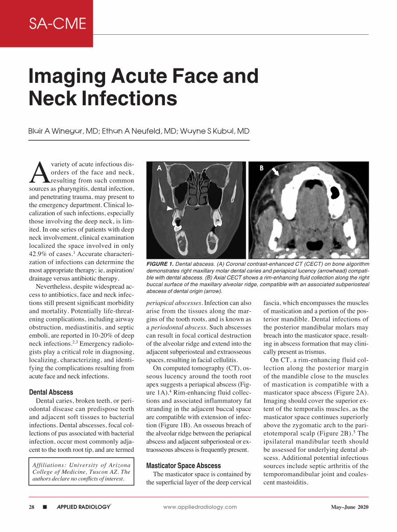
On computed tomography (CT), os-seous lucency around the tooth root apex suggests a periapical abscess (Fig-ure 1A).4 Rim-enhancing fluid collec-tions and associated inflammatory fat stranding in the adjacent buccal space are compatible with extension of infec-tion (Figure 1B). An osseous breach of the alveolar ridge between the periapical abscess and adjacent subperiosteal or ex-traosseous abscess is frequently present.
Masticator Space AbscessThe masticator space is contained by
the superficial layer of the deep cervical
fascia, which encompasses the muscles of mastication and a portion of the pos-terior mandible. Dental infections of the posterior mandibular molars may breach into the masticator space, result-ing in abscess formation that may clini-cally present as trismus.
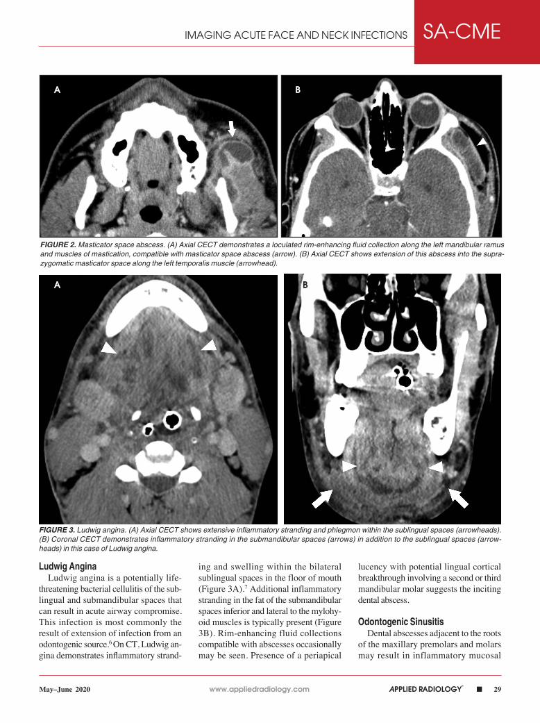
On CT, a rim-enhancing fluid col-lection along the posterior margin of the mandible close to the muscles of mastication is compatible with a masticator space abscess (Figure 2A). Imaging should cover the superior ex-tent of the temporalis muscles, as the masticator space continues superiorly above the zygomatic arch to the pari-etotemporal scalp (Figure 2B).5 The ipsilateral mandibular teeth should be assessed for underlying dental ab-scess. Additional potential infectious sources include septic arthritis of the temporomandibular joint and coales-cent mastoiditis.
Imaging Acute Face and Neck InfectionsBlair A Winegar, MD; Ethan A Neufeld, MD; Wayne S Kubal, MD
Affiliations: University of Arizona College of Medicine, Tuscon AZ. The authors declare no conflicts of interest.
FIGURE 1. Dental abscess. (A) Coronal contrast-enhanced CT (CECT) on bone algorithm demonstrates right maxillary molar dental caries and periapical lucency (arrowhead) compati-ble with dental abscess. (B) Axial CECT shows a rim-enhancing fluid collection along the right buccal surface of the maxillary alveolar ridge, compatible with an associated subperiosteal abscess of dental origin (arrow).
A B
www.appliedradiology.com APPLIED RADIOLOGY©
n 29May–June 2020
IMAGING ACUTE FACE AND NECK INFECTIONS SA-CME
Ludwig AnginaLudwig angina is a potentially life-
threatening bacterial cellulitis of the sub-lingual and submandibular spaces that can result in acute airway compromise. This infection is most commonly the result of extension of infection from an odontogenic source.6 On CT, Ludwig an-gina demonstrates inflammatory strand-
ing and swelling within the bilateral sublingual spaces in the floor of mouth (Figure 3A).7 Additional inflammatory stranding in the fat of the submandibular spaces inferior and lateral to the mylohy-oid muscles is typically present (Figure 3B). Rim-enhancing fluid collections compatible with abscesses occasionally may be seen. Presence of a periapical
lucency with potential lingual cortical breakthrough involving a second or third mandibular molar suggests the inciting dental abscess.
Odontogenic SinusitisDental abscesses adjacent to the roots
of the maxillary premolars and molars may result in inflammatory mucosal
FIGURE 2. Masticator space abscess. (A) Axial CECT demonstrates a loculated rim-enhancing fluid collection along the left mandibular ramus and muscles of mastication, compatible with masticator space abscess (arrow). (B) Axial CECT shows extension of this abscess into the supra-zygomatic masticator space along the left temporalis muscle (arrowhead).
FIGURE 3. Ludwig angina. (A) Axial CECT shows extensive inflammatory stranding and phlegmon within the sublingual spaces (arrowheads). (B) Coronal CECT demonstrates inflammatory stranding in the submandibular spaces (arrows) in addition to the sublingual spaces (arrow-heads) in this case of Ludwig angina.
A B
A B
30 n APPLIED RADIOLOGY©
www.appliedradiology.com May–June 2020
IMAGING ACUTE FACE AND NECK INFECTIONSSA-CME
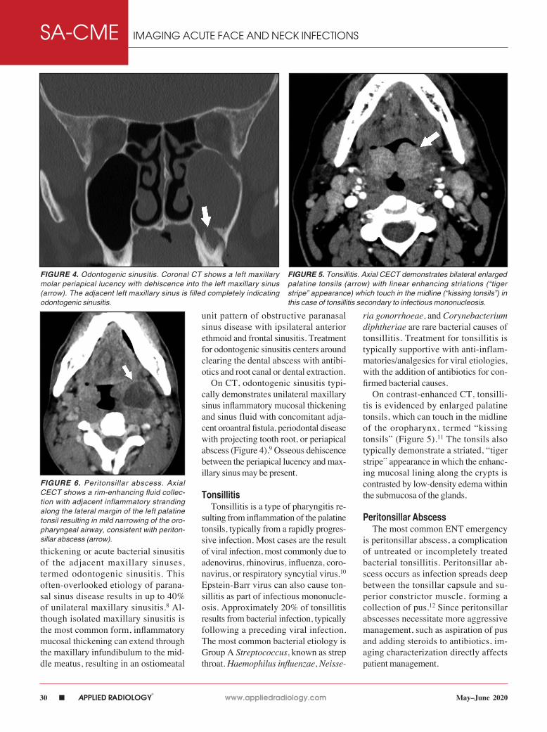
thickening or acute bacterial sinusitis of the adjacent maxillary sinuses, termed odontogenic sinusitis. This often-overlooked etiology of parana-sal sinus disease results in up to 40% of unilateral maxillary sinusitis.8 Al-though isolated maxillary sinusitis is the most common form, inflammatory mucosal thickening can extend through the maxillary infundibulum to the mid-dle meatus, resulting in an ostiomeatal
unit pattern of obstructive paranasal sinus disease with ipsilateral anterior ethmoid and frontal sinusitis. Treatment for odontogenic sinusitis centers around clearing the dental abscess with antibi-otics and root canal or dental extraction.
On CT, odontogenic sinusitis typi-cally demonstrates unilateral maxillary sinus inflammatory mucosal thickening and sinus fluid with concomitant adja-cent oroantral fistula, periodontal disease with projecting tooth root, or periapical abscess (Figure 4).9 Osseous dehiscence between the periapical lucency and max-illary sinus may be present.
TonsillitisTonsillitis is a type of pharyngitis re-
sulting from inflammation of the palatine tonsils, typically from a rapidly progres-sive infection. Most cases are the result of viral infection, most commonly due to adenovirus, rhinovirus, influenza, coro-navirus, or respiratory syncytial virus.10 Epstein-Barr virus can also cause ton-sillitis as part of infectious mononucle-osis. Approximately 20% of tonsillitis results from bacterial infection, typically following a preceding viral infection. The most common bacterial etiology is Group A Streptococcus, known as strep throat. Haemophilus influenzae, Neisse-
ria gonorrhoeae, and Corynebacterium diphtheriae are rare bacterial causes of tonsillitis. Treatment for tonsillitis is typically supportive with anti-inflam-matories/analgesics for viral etiologies, with the addition of antibiotics for con-firmed bacterial causes.
On contrast-enhanced CT, tonsilli-tis is evidenced by enlarged palatine tonsils, which can touch in the midline of the oropharynx, termed “kissing tonsils” (Figure 5).11 The tonsils also typically demonstrate a striated, “tiger stripe” appearance in which the enhanc-ing mucosal lining along the crypts is contrasted by low-density edema within the submucosa of the glands.
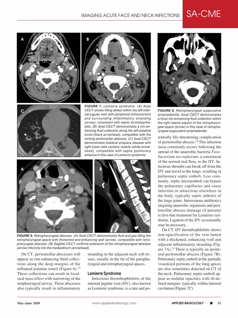
Peritonsillar AbscessThe most common ENT emergency
is peritonsillar abscess, a complication of untreated or incompletely treated bacterial tonsillitis. Peritonsillar ab-scess occurs as infection spreads deep between the tonsillar capsule and su-perior constrictor muscle, forming a collection of pus.12 Since peritonsillar abscesses necessitate more aggressive management, such as aspiration of pus and adding steroids to antibiotics, im-aging characterization directly affects patient management.
FIGURE 6. Peritonsillar abscess. Axial CECT shows a rim-enhancing fluid collec-tion with adjacent inflammatory stranding along the lateral margin of the left palatine tonsil resulting in mild narrowing of the oro-pharyngeal airway, consistent with periton-sillar abscess (arrow).
FIGURE 4. Odontogenic sinusitis. Coronal CT shows a left maxillary molar periapical lucency with dehiscence into the left maxillary sinus (arrow). The adjacent left maxillary sinus is filled completely indicating odontogenic sinusitis.
FIGURE 5. Tonsillitis. Axial CECT demonstrates bilateral enlarged palatine tonsils (arrow) with linear enhancing striations (“tiger stripe” appearance) which touch in the midline (“kissing tonsils”) in this case of tonsillitis secondary to infectious mononucleosis.
www.appliedradiology.com APPLIED RADIOLOGY©
n 31May–June 2020
IMAGING ACUTE FACE AND NECK INFECTIONS SA-CME
On CT, peritonsillar abscesses will appear as rim-enhancing fluid collec-tions along the deep margins of the inflamed palatine tonsil (Figure 6).13 These collections can result in local-ized mass effect with narrowing of the oropharyngeal airway. These abscesses also typically result in inflammatory
stranding in the adjacent neck soft tis-sues, usually in the fat of the parapha-ryngeal and retropharyngeal spaces.
Lemierre SyndromeInfectious thrombophlebitis of the
internal jugular vein (IJV), also known as Lemierre syndrome, is a rare and po-
tentially life-threatening complication of peritonsillar abscess.14 This infection most commonly occurs following the spread of the anaerobic bacteria Fuso-bacterium necrophorum, a constituent of the normal oral flora, to the IJV. In-fectious thrombi can break off from the IJV and travel to the lungs, resulting in pulmonary septic emboli. Less com-monly, septic microemboli can bypass the pulmonary capillaries and cause infection or infarctions elsewhere in the body, typically septic arthritis of the large joints. Intravenous antibiotics targeting anaerobic organisms and peri-tonsillar abscess drainage (if present) is first-line treatment for Lemierre syn-drome. Ligation of the IJV occasionally may be necessary.
On CT, IJV thrombophlebitis shows non-opacification of the vein lumen with a thickened, enhancing wall and adjacent inflammatory stranding (Fig-ure 7A).15 There is typically an ipsilat-eral peritonsillar abscess (Figure 7B). Pulmonary septic emboli in the partially visualized portions of the lung apices are also sometimes detected on CT of the neck. Pulmonary septic emboli ap-pear as nodular opacities with ill-de-fined margins, typically within internal cavitation (Figure 7C).
FIGURE 7. Lemierre syndrome. (A) Axial CECT shows filling defect within the left inter-nal jugular vein with peripheral enhancement and surrounding inflammatory stranding (arrow), consistent with septic thrombophle-bitis. (B) Axial CECT demonstrates a rim-en-hancing fluid collection along the left palatine tonsil (black arrowhead), compatible with the inciting peritonsillar abscess. (C) Axial CECT demonstrates bilateral airspace disease with right lower lobe cavitary nodule (white arrow-head), compatible with septic pulmonary embolus in this case of Lemierre syndrome.
FIGURE 8. Retropharyngeal suppurative lymphadenitis. Axial CECT demonstrates a focal rim-enhancing fluid collection within the right lateral aspect of the retropharyn-geal space (arrow) in this case of retropha-ryngeal suppurative lymphadenitis.
FIGURE 9. Retropharyngeal abscess. (A) Axial CECT demonstrates fluid and gas filling the retropharyngeal space with thickened and enhancing wall (arrow), compatible with retro-pharyngeal abscess. (B) Sagittal CECT confirms extension of the retropharyngeal abscess (arrow) inferiorly into the mediastinum (arrowhead).
A
C
B
A B
32 n APPLIED RADIOLOGY©
www.appliedradiology.com May–June 2020
IMAGING ACUTE FACE AND NECK INFECTIONSSA-CME
Retropharyngeal Suppurative Lymphadenitis
Retropharyngeal suppura t ive lymphadenitis denotes lateral retro-pharyngeal lymph node inflammation resulting from a bacterial infection that has progressed to internal lique-faction. This condition is most com-monly encountered in young children (2-6 years of age) exhibiting fever, neck pain, dysphagia, and/or torti-collis. The most commonly involved bacteria are Streptococcus sp. and Staphylococcus aureus.16 Intravenous antibiotics are the first-line treatment for this infection, with aspiration or surgical drainage reserved for refrac-tory cases or large fluid collections.
On CT, a rim-enhancing fluid col-lection confined to the lateral margin of the retropharyngeal space with adjacent inflammatory stranding is compatible with retropharyngeal suppurative lymph-adenitis (Figure 8).17 There is typically associated retropharyngeal edema or ef-fusion, but rim-enhancing fluid should not cross the midline or fill the retropha-ryngeal space, which would suggest de-velopment of a retropharyngeal abscess.
Retropharyngeal AbscessInfection from pus-forming bacteria
in the retropharyngeal space results in retropharyngeal abscess. In adults, this
condition most frequently is the result of a penetrating trauma with inoculation of normal oral flora into the retropha-ryngeal space. Retropharyngeal spread of infection from adjacent cervical dis-citis-osteomyelitis or ruptured retropha-ryngeal suppurative lymphadenitis in children are other potential etiologies. Retropharyngeal abscesses may spread to the danger space, a potential space sit-uated between the true retropharyngeal space and prevertebral musculature. This permits free communication to the medi-astinum, resulting in the dreaded compli-cation of mediastinitis.18 Treatment with IV antibiotics and surgical drainage is standard of care.
On CT, retropharyngeal abscesses are rim-enhancing fluid collections that fill the retropharyngeal space (Figure 9A).19 Larger fluid collections typically demonstrate bowing of the walls of the retropharyngeal space. In cases of ret-ropharyngeal abscess, it is essential to image the chest to search for inflamma-tory stranding or rim-enhancing fluid collections in the mediastinum, indicat-ing the complication of mediastinitis (Figures 9B & C).
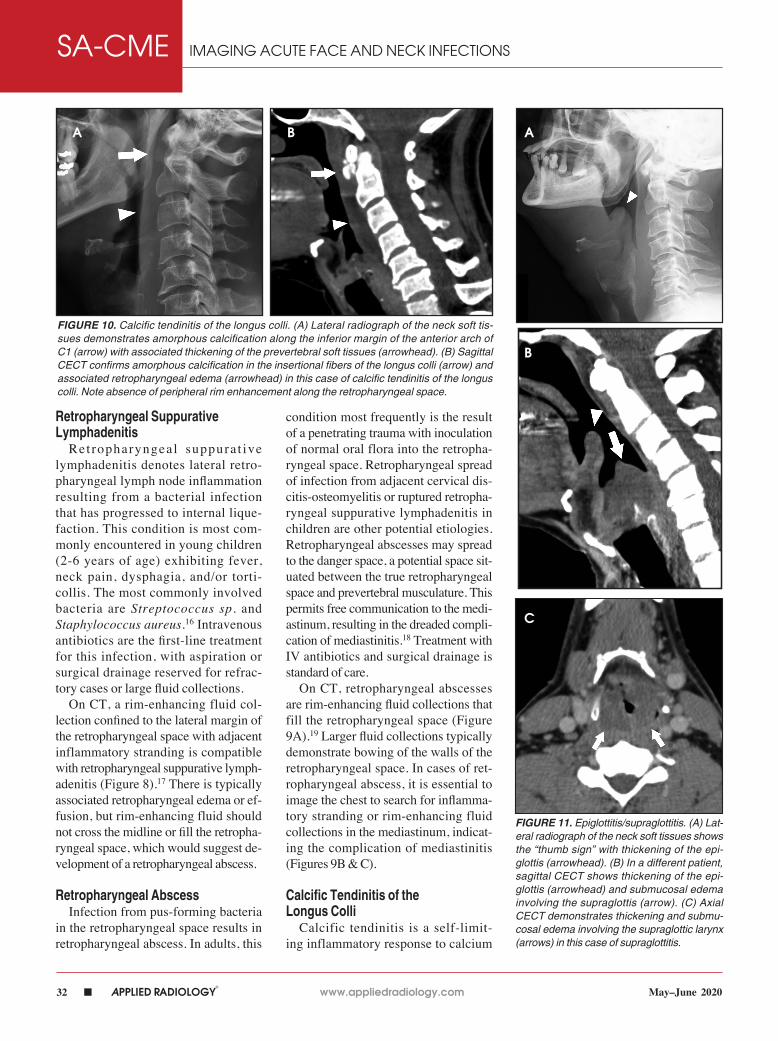
Calcific Tendinitis of the Longus Colli
Calcific tendinitis is a self-limit-ing inflammatory response to calcium
FIGURE 10. Calcific tendinitis of the longus colli. (A) Lateral radiograph of the neck soft tis-sues demonstrates amorphous calcification along the inferior margin of the anterior arch of C1 (arrow) with associated thickening of the prevertebral soft tissues (arrowhead). (B) Sagittal CECT confirms amorphous calcification in the insertional fibers of the longus colli (arrow) and associated retropharyngeal edema (arrowhead) in this case of calcific tendinitis of the longus colli. Note absence of peripheral rim enhancement along the retropharyngeal space.
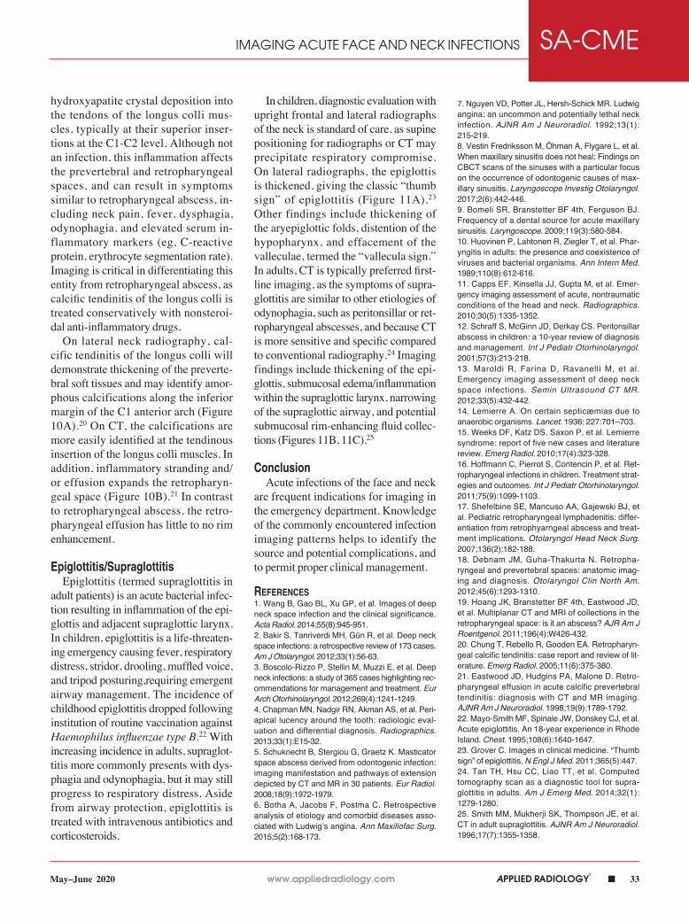
FIGURE 11. Epiglottitis/supraglottitis. (A) Lat-eral radiograph of the neck soft tissues shows the “thumb sign” with thickening of the epi-glottis (arrowhead). (B) In a different patient, sagittal CECT shows thickening of the epi-glottis (arrowhead) and submucosal edema involving the supraglottis (arrow). (C) Axial CECT demonstrates thickening and submu-cosal edema involving the supraglottic larynx (arrows) in this case of supraglottitis.
A B A
B
C
www.appliedradiology.com APPLIED RADIOLOGY©
n 33May–June 2020
IMAGING ACUTE FACE AND NECK INFECTIONS SA-CME
hydroxyapatite crystal deposition into the tendons of the longus colli mus-cles, typically at their superior inser-tions at the C1-C2 level. Although not an infection, this inflammation affects the prevertebral and retropharyngeal spaces, and can result in symptoms similar to retropharyngeal abscess, in-cluding neck pain, fever, dysphagia, odynophagia, and elevated serum in-flammatory markers (eg, C-reactive protein, erythrocyte segmentation rate). Imaging is critical in differentiating this entity from retropharyngeal abscess, as calcific tendinitis of the longus colli is treated conservatively with nonsteroi-dal anti-inflammatory drugs.
On lateral neck radiography, cal-cific tendinitis of the longus colli will demonstrate thickening of the preverte-bral soft tissues and may identify amor-phous calcifications along the inferior margin of the C1 anterior arch (Figure 10A).20 On CT, the calcifications are more easily identified at the tendinous insertion of the longus colli muscles. In addition, inflammatory stranding and/or effusion expands the retropharyn-geal space (Figure 10B).21 In contrast to retropharyngeal abscess, the retro-pharyngeal effusion has little to no rim enhancement.
Epiglottitis/SupraglottitisEpiglottitis (termed supraglottitis in
adult patients) is an acute bacterial infec-tion resulting in inflammation of the epi-glottis and adjacent supraglottic larynx. In children, epiglottitis is a life-threaten-ing emergency causing fever, respiratory distress, stridor, drooling, muffled voice, and tripod posturing,requiring emergent airway management. The incidence of childhood epiglottitis dropped following institution of routine vaccination against Haemophilus influenzae type B.22 With increasing incidence in adults, supraglot-titis more commonly presents with dys-phagia and odynophagia, but it may still progress to respiratory distress. Aside from airway protection, epiglottitis is treated with intravenous antibiotics and corticosteroids.
In children, diagnostic evaluation with upright frontal and lateral radiographs of the neck is standard of care, as supine positioning for radiographs or CT may precipitate respiratory compromise. On lateral radiographs, the epiglottis is thickened, giving the classic “thumb sign” of epiglottitis (Figure 11A).23 Other findings include thickening of the aryepiglottic folds, distention of the hypopharynx, and effacement of the valleculae, termed the “vallecula sign.” In adults, CT is typically preferred first-line imaging, as the symptoms of supra-glottitis are similar to other etiologies of odynophagia, such as peritonsillar or ret-ropharyngeal abscesses, and because CT is more sensitive and specific compared to conventional radiography.24 Imaging findings include thickening of the epi-glottis, submucosal edema/inflammation within the supraglottic larynx, narrowing of the supraglottic airway, and potential submucosal rim-enhancing fluid collec-tions (Figures 11B, 11C).25
ConclusionAcute infections of the face and neck
are frequent indications for imaging in the emergency department. Knowledge of the commonly encountered infection imaging patterns helps to identify the source and potential complications, and to permit proper clinical management.
RefeRences1. Wang B, Gao BL, Xu GP, et al. Images of deepneck space infection and the clinical significance.Acta Radiol. 2014;55(8):945-951.2. Bakir S, Tanriverdi MH, Gün R, et al. Deep neck space infections: a retrospective review of 173 cases.Am J Otolaryngol. 2012;33(1):56-63.3. Boscolo-Rizzo P, Stellin M, Muzzi E, et al. Deep neck infections: a study of 365 cases highlighting rec-ommendations for management and treatment. Eur Arch Otorhinolaryngol. 2012;269(4):1241-1249.4. Chapman MN, Nadgir RN, Akman AS, et al. Peri-apical lucency around the tooth: radiologic eval-uation and differential diagnosis. Radiographics. 2013;33(1):E15-32.5. Schuknecht B, Stergiou G, Graetz K. Masticator space abscess derived from odontogenic infection: imaging manifestation and pathways of extensiondepicted by CT and MR in 30 patients. Eur Radiol. 2008;18(9):1972-1979.6. Botha A, Jacobs F, Postma C. Retrospectiveanalysis of etiology and comorbid diseases asso-ciated with Ludwig’s angina. Ann Maxillofac Surg. 2015;5(2):168-173.
7. Nguyen VD, Potter JL, Hersh-Schick MR. Ludwig angina: an uncommon and potentially lethal neckinfection. AJNR Am J Neuroradiol. 1992;13(1): 215-219.8. Vestin Fredriksson M, Öhman A, Flygare L, et al. When maxillary sinusitis does not heal: Findings on CBCT scans of the sinuses with a particular focus on the occurrence of odontogenic causes of max-illary sinusitis. Laryngoscope Investig Otolaryngol. 2017;2(6):442-446.9. Bomeli SR, Branstetter BF 4th, Ferguson BJ.Frequency of a dental source for acute maxillarysinusitis. Laryngoscope. 2009;119(3):580-584.10. Huovinen P, Lahtonen R, Ziegler T, et al. Phar-yngitis in adults: the presence and coexistence of viruses and bacterial organisms. Ann Intern Med. 1989;110(8):612-616.11. Capps EF, Kinsella JJ, Gupta M, et al. Emer-gency imaging assessment of acute, nontraumatic conditions of the head and neck. Radiographics. 2010;30(5):1335-1352.12. Schraff S, McGinn JD, Derkay CS. Peritonsillar abscess in children: a 10-year review of diagnosis and management. Int J Pediatr Otorhinolaryngol. 2001;57(3):213-218.13. Maroldi R, Farina D, Ravanelli M, et al.Emergency imaging assessment of deep neckspace infections. Semin Ultrasound CT MR. 2012;33(5):432-442.14. Lemierre A. On certain septicæmias due toanaerobic organisms. Lancet. 1936; 227:701–703.15. Weeks DF, Katz DS, Saxon P, et al. Lemierresyndrome: report of five new cases and literaturereview. Emerg Radiol. 2010;17(4):323-328.16. Hoffmann C, Pierrot S, Contencin P, et al. Ret-ropharyngeal infections in children. Treatment strat-egies and outcomes. Int J Pediatr Otorhinolaryngol. 2011;75(9):1099-1103.17. Shefelbine SE, Mancuso AA, Gajewski BJ, et al. Pediatric retropharyngeal lymphadenitis: differ-entiation from retrophyarngeal abscess and treat-ment implications. Otolaryngol Head Neck Surg. 2007;136(2):182-188.18. Debnam JM, Guha-Thakurta N. Retropha-ryngeal and prevertebral spaces: anatomic imag-ing and diagnosis. Otolaryngol Clin North Am. 2012;45(6):1293-1310.19. Hoang JK, Branstetter BF 4th, Eastwood JD,et al. Multiplanar CT and MRI of collections in the retropharyngeal space: is it an abscess? AJR Am JRoentgenol. 2011;196(4):W426-432.20. Chung T, Rebello R, Gooden EA. Retropharyn-geal calcific tendinitis: case report and review of lit-erature. Emerg Radiol. 2005;11(6):375-380.21. Eastwood JD, Hudgins PA, Malone D. Retro-pharyngeal effusion in acute calcific prevertebraltendinitis: diagnosis with CT and MR imaging.AJNR Am J Neuroradiol. 1998;19(9):1789-1792.22. Mayo-Smith MF, Spinale JW, Donskey CJ, et al. Acute epiglottitis. An 18-year experience in RhodeIsland. Chest. 1995;108(6):1640-1647.23. Grover C. Images in clinical medicine. “Thumbsign” of epiglottitis. N Engl J Med. 2011;365(5):447.24. Tan TH, Hsu CC, Liao TT, et al. Computedtomography scan as a diagnostic tool for supra-glottitis in adults. Am J Emerg Med. 2014;32(1): 1279-1280.25. Smith MM, Mukherji SK, Thompson JE, et al. CT in adult supraglottitis. AJNR Am J Neuroradiol.1996;17(7):1355-1358.