Embed Size (px)
Citation preview

DOI: 10.1542/pir.32-3-1092011;32;109Pediatrics in Review
Holly A. Zywicke and Curtis J. RozzelleSacral Dimples
http://pedsinreview.aappublications.org/content/32/3/109located on the World Wide Web at:
The online version of this article, along with updated information and services, is
dfhttp://pedsinreview.aappublications.org/http://pedsinreview.aappublications.org/content/32/5/207.full.p
dfhttp://pedsinreview.aappublications.org/http://pedsinreview.aappublications.org/content/32/4/151.full.p
An erratum has been published regarding this article. Please see the attached page for:
http://pedsinreview.aappublications.org/content/suppl/2011/03/04/32.3.109.DC1.htmlData Supplement at:
Pediatrics. All rights reserved. Print ISSN: 0191-9601. Boulevard, Elk Grove Village, Illinois, 60007. Copyright © 2011 by the American Academy of published, and trademarked by the American Academy of Pediatrics, 141 Northwest Pointpublication, it has been published continuously since 1979. Pediatrics in Review is owned, Pediatrics in Review is the official journal of the American Academy of Pediatrics. A monthly
at UNIV OF CHICAGO on May 23, 2013http://pedsinreview.aappublications.org/Downloaded from

Sacral DimplesHolly A. Zywicke, MD,*
Curtis J. Rozzelle, MD†
Author Disclosure
Drs Zywicke and
Rozzelle have
disclosed no financial
relationships relevant
to this article. This
commentary does not
contain a discussion
of an unapproved/
investigative use of a
commercial
product/device.
Objectives After completing this article, readers should be able to:
1. Explain the difference between open and closed neural tube defects.2. Describe the characteristics of spinal skin dimples that warrant further evaluation.3. Describe the characteristics of spinal skin dimples that do not warrant further
evaluation.4. Discuss the evaluation of spinal skin dimples and name the findings that suggest
occult spinal dysraphism.5. Discuss the neurosurgical treatment of occult spinal dysraphism.6. Explain the natural history and clinical manifestations of occult spinal dysraphism.
DefinitionsNeural tube defects are among the most common forms of birth defect, affecting 1 in every1,000 pregnancies. (1)(2) These defects, which result from abnormal fusion of the neuraltube during embryonic development, are placed into two broad categories: open andclosed. Open neural tube defects are lesions in which brain, spinal cord, or spinal nerves areexposed through obvious defects of the meninges and skull or vertebral column. Examplesare anencephaly, myelomeningocele, and meningocele. Closed neural tube defects areskin-covered lesions under which the nervous system structures have not formed normally.These include split cord malformation, dermal sinus tract, tethered spinal cord, andintraspinal lipoma (Table).
Spina bifida is an imprecise term often used to describe a variety of congenital spinalanomalies that range in consequence from insignificant to severe. Spina bifida occulta(SBO) is a radiographic finding that describes incomplete osseous fusion of the posteriorelements. It may occur in conjunction with a cutaneous abnormality but is clinically benignand is considered a normal variant. (3) Occult spinal dysraphisms (OSDs) are much lesscommon than SBO and encompass a variety of skin-covered neural tube defects. Becausethe neural structures are affected, however, neurologic impairment is common. Mostforms of OSD have an associated overlying cutaneous abnormality.
Most open neural tube defects are diagnosed prenatally with ultrasonography andserum marker concentrations. Those defects not identified before delivery are apparent atbirth. An OSD, on the other hand, is less obvious and may not be diagnosed until later inlife, despite its presence at birth. The occult nature can be problematic because the clinicalimpairments associated with closed neural tube defects, which include paresis, spasticity,sensory disturbance, orthopedic deformity or contracture, and bowel and bladder dysfunc-tion, often progress insidiously over time.
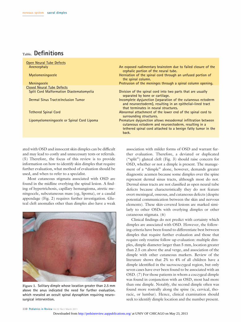
DiagnosisMore than 50% of OSDs are diagnosed when a dimple (Fig. 1) is noted in the lowerspine/sacral region. Although the natural history of OSD is not fully understood, earlydiagnosis and intervention are believed to improve outcome in almost all cases. (4) Hence,the recognition of a suspicious skin dimple and identification of underlying anomalies withprompt radiographic evaluation and neurosurgical referral is crucial. However, not alldimples are associated with an OSD. Distinguishing between cutaneous stigmata associ-
*Division of Pediatric Neurosurgery, Department of Surgery, University of Alabama at Birmingham and Children’s Hospital ofAlabama, Birmingham, AL.†Assistant Professor, Department of Surgery, Division of Pediatric Neurosurgery, University of Alabama at Birmingham andChildren’s Hospital of Alabama, Birmingham, AL.
Article nervous system
Pediatrics in Review Vol.32 No.3 March 2011 109
at UNIV OF CHICAGO on May 23, 2013http://pedsinreview.aappublications.org/Downloaded from
ated with OSD and innocent skin dimples can be difficultand may lead to costly and unnecessary tests or referrals.(5) Therefore, the focus of this review is to provideinformation on how to identify skin dimples that requirefurther evaluation, what method of evaluation should beused, and when to refer to a specialist.
Most cutaneous stigmata associated with OSD arefound in the midline overlying the spinal lesion. A find-ing of hypertrichosis, capillary hemangioma, atretic me-ningocele, subcutaneous mass (eg, lipoma), or a caudalappendage (Fig. 2) requires further investigation. Glu-teal cleft anomalies other than dimples also have a weak
association with milder forms of OSD and warrant fur-ther evaluation. Therefore, a deviated or duplicated(“split”) gluteal cleft (Fig. 3) should raise concern forOSD, whether or not a dimple is present. The manage-ment of a “dimple” alone, however, demands greaterdiagnostic acumen because some dimples over the spinerepresent dermal sinus tracts, although most do not.Dermal sinus tracts are not classified as open neural tubedefects because characteristically they do not featureovert meningeal, osseous, and cutaneous defects (despitepotential communication between the skin and nervouselements). These skin-covered lesions are marked simi-larly to other OSDs with overlying dimples or othercutaneous stigmata. (6)
Clinical findings do not predict with certainty whichdimples are associated with OSD. However, the follow-ing criteria have been found to differentiate best betweendimples that require further evaluation and those thatrequire only routine follow-up evaluation: multiple dim-ples, dimple diameter larger than 5 mm, location greaterthan 2.5 cm above the anal verge, and association of thedimple with other cutaneous markers. Review of theliterature shows that 2% to 4% of all children have adimple identified in the sacrococcygeal region, but onlyseven cases have ever been found to be associated with anOSD. (7) For those patients in whom a coccygeal dimplewas found in conjunction with an OSD, most had morethan one dimple. Notably, the second dimple often wasfound more rostrally along the spine (ie, cervical, tho-racic, or lumbar). Hence, clinical examination shouldseek to identify dimple location and the number present.
Table. DefinitionsOpen Neural Tube Defects
Anencephaly An exposed rudimentary brainstem due to failed closure of thecephalic portion of the neural tube.
Myelomeningocele Herniation of the spinal cord through an unfused portion ofthe spinal column.
Meningocele Protrusion of the meninges through a spinal column opening.Closed Neural Tube Defects
Split Cord Malformation Diastematomyelia Division of the spinal cord into two parts that are usuallyseparated by bone or cartilage.
Dermal Sinus Tract�Inclusion Tumor Incomplete dysjunction (separation of the cutaneous ectodermand neuroectoderm), resulting in an epithelial-lined tractthat terminates in neural structures.
Tethered Spinal Cord Abnormal attachment of the lower end of the spinal cord tosurrounding structures.
Lipomyelomeningocele or Spinal Cord Lipoma Premature dysjunction allows mesodermal infiltration betweencutaneous ectoderm and neuroectoderm, resulting in atethered spinal cord attached to a benign fatty tumor in theback.
Figure 1. Solitary dimple whose location greater than 2.5 mmabove the anus indicated the need for further evaluation,which revealed an occult spinal dysraphism requiring neuro-surgical intervention.
nervous system sacral dimples
110 Pediatrics in Review Vol.32 No.3 March 2011
at UNIV OF CHICAGO on May 23, 2013http://pedsinreview.aappublications.org/Downloaded from

Solitary sacrococcygeal pits located entirely within thegluteal cleft (Fig. 4) have no clinical significance andshould be considered anatomic variations of normal.Typically, the coccyx is palpable beneath the dimple andintact skin can be seen at the base (Fig. 5). If there isdifficulty discerning whether the lesion is covered com-pletely by skin, otoscopic examination of the dimpleoften can determine if there is a bottom to the pit.Although most lesions occur in the midline, eccentriclesions (Fig. 6) are not in themselves abnormal unlessoccurring in conjunction with other lesions or outsidethe sacral spine. No radiographic evaluation or neuro-surgical consultation is required; parental reassurance isthe only intervention necessary.
In addition to a thorough inspection of the skin, the
pediatrician must perform a careful physical examination,with particular attention to the neurologic and ortho-pedic aspects. Associated orthopedic findings can includeclubfeet, arthrogryposis (contracture of multiple jointsleading to fixation of the joints in extension or flexion) ofthe lower extremities, and hip dislocation. Abnormal cur-vature of the spine, including kyphosis or scoliosis, alsomay be present. Abnormal neurologic or orthopedicexamination findings indicate the need for further eval-uation.
ManagementWhen detailed history and physical examination raise theclinical suspicion for OSD, radiographic imaging shouldbe obtained (Fig. 7). Either ultrasonography or magneticresonance imaging (MRI) can be employed to evaluate
Figure 2. Cutaneous lesions. Skin dimples are often the cu-taneous marking found with occult spinal dysraphism. How-ever, multiple other markings can signify underlying spinalelement malformation, including caudal appendage (A) andhypertrichosis (B).
Figure 3. A duplicated gluteal cleft associated with occultspinal dysraphism.
Figure 4. A prototypical benign sacral dimple that is locatedwithin the gluteal cleft (less than 2.5 cm above the anus) andsolitary.
nervous system sacral dimples
Pediatrics in Review Vol.32 No.3 March 2011 111
at UNIV OF CHICAGO on May 23, 2013http://pedsinreview.aappublications.org/Downloaded from

OSD. Ultrasonography of the lumbosacral spine gener-ally is useful only in children younger than 3 months ofage because ossification of the vertebral arches has notyet occurred. (8) However, the decision to use ultra-sonography versus MRI (for children of any age) as first-line imaging appears somewhat institution-dependent. Inone study of a pediatric population who had sacrococcy-geal cutaneous lesions, a discordance rate of 17% be-tween ultrasonography and MRI studies was found inwhich ultrasonography suggested an OSD while MRIyielded normal results. (9) Pediatricians, therefore,should be aware of the possible discrepancy in findingswith these imaging modalities and know which study ismost appropriate at their respective institutions.
Spinal ultrasonography can assess the level of theconus medullaris, the diameter and echogenicity of thefilum terminale, and the position and movement patternof the spinal cord and nerve roots. Abnormal findings caninclude a low-lying conus, in which the tip is below thelevel of the second lumbar vertebral body; a filum termi-nale diameter greater than 2 mm; and a posteriorlypositioned or nonmobile cord, which can indicate teth-ering. If ultrasonographic findings are abnormal, MRI ofthe spine is indicated. Findings on MRI vary, based onthe type of OSD present. In general, MRI is more reliableand exact in diagnosing OSD.
Neurosurgical referral is appropriate if radiographicevaluation reveals any spinal abnormality. Considerationfor early referral (before imaging) is appropriate for dim-ples superior to the gluteal cleft, especially if any dis-charge is observed or reported. Such dimples are the
hallmark of dermal sinus tracts that predispose the pa-tient to bacterial meningitis or intraspinal abscess. (10)Surgical intervention is aimed at untethering the spinalcord and removing abnormal tissue, when present.
PrognosisAlmost all neurosurgical referrals for suspected OSD inchildren younger than 1 year of age are for evaluationof a dimple. Although the natural history of OSD issomewhat unpredictable, the overall risk of neurologiccompromise increases with time. Neurologic deficits canbe difficult to identify in young children because theonset of dysfunction is generally insidious and occursabout the same time as expected neurologic functiondevelopment (eg, crawling, walking, standing). Accord-ingly, OSD deficits may be mistaken for delayed accrue-ment of normal function, and irreversible damage mayoccur before symptomatic manifestation. The reasons forneurosurgical referral for children older than 1 year ofage suspected of having OSD include chronic urinarytract infections, lower limb deformity (eg, foot drop,
Figure 6. A right eccentric dimple that occurs outside of themidline but does not carry a high degree of suspicion for anoccult spinal dysraphism because it is an isolated sacrococcy-geal lesion.
Figure 5. Solitary sacrococcygeal dimple that demonstratescomplete covering with skin over the entire dimpled area whenthe skin is stretched laterally and, therefore, is not an occultspinal dysraphism-associated lesion.
nervous system sacral dimples
112 Pediatrics in Review Vol.32 No.3 March 2011
at UNIV OF CHICAGO on May 23, 2013http://pedsinreview.aappublications.org/Downloaded from

weakness or atrophy in a lower extremity, talipes equino-varus, or dragging one foot), bowel/bladder dysfunc-tion, pain, and lower extremity spasticity or paresis.However, careful inspection of this population oftenreveals subtle cutaneous stigmata. Therefore, it is impor-tant for the pediatrician to be vigilant in searching formidline skin anomalies. Even as common a condition asprimary nocturnal enuresis warrants careful examinationfor midline skin anomalies.
ConclusionEarly diagnosis of OSD often comes from identificationof spinal skin dimples. Recognition of suspicious lesionsis important to reduce the risk of neurologic, urologic,and orthopedic dysfunction. During examination, thepediatrician should not only look for dimples along thespine but also for other markings such as abnormalhair growth, asymmetric gluteal creases, dermal sinuses/dimples/pits, hyper- or hypopigmentation, capillaryhemangiomas, skin tags, and subcutaneous fatty massesthat are associated with OSDs. Any lesion along the spineoutside of the sacrococcygeal region or identification ofmore than one skin marking anywhere along the spinewarrants further evaluation, including radiographic im-
aging and neurosurgical referral. (11) Optimal outcomeis most likely with early diagnosis and surgical interven-tion.
References1. Wiswell TE, Tuttle DJ, Northam RS, Simonds GR. Majorcongenital neurologic malformations. A 17-year survey. Am J DisChild. 1990;144:61–672. Williams LJ, Rasmussen SA, Flores A, Kirby RS, Edmonds LD.Decline in the prevalence of spina bifida and anencephaly by race/ethnicity: 1995–2002. Pediatrics. 2005;116:580–5863. Boone D, Parsons D, Lachmann SM, Sherwood T. Spina bifidaocculta: lesion or anomaly? Clin Radiol. 1985;36:159–1614. Soonawala N, Overweg-Plandsoen WCG, Brouwer OF. Earlyclinical signs and symptoms in occult spinal dysraphism: a retrospec-tive case study of 47 patients. Clin Neurol Neurosurg. 1999;101:11–145. Medina LS, Crone K, Kuntz KM. Newborns with suspectedoccult spinal dysraphism: a cost-effectiveness analysis of diagnosticstrategies. Pediatrics. 2001;108:e101–e1086. Ackerman LL, Menezes AH. Spinal congenital dermal sinuses: a30-year experience. Pediatrics. 2003;112:641–6477. Weprin BE, Oakes WJ. Coccygeal pits. Pediatrics. 2000;105:e69–e738. Szyszko TA, Watson M. The value of ultrasonographic exami-nation of the lumbar spine in infants with specific reference tocutaneous markers of occult spinal dysraphism. Clin Radiol. 2005;60:9359. Sasani M, Asghari B, Asghari Y, Afsharian R, Ozer AF. Correla-tion of cutaneous lesions with clinical radiological and urodynamicfindings in the prognosis of underlying spinal dysraphism disorders.Pediatr Neurosrg. 2008;44:360–37010. Bhatia S, Gullu MS, Date NB, Muzumdar D, Muranjan MN,Lahiri KR. Anterior sacral pyocele with meningitis: a rare presenta-tion of occult spinal dysraphism with congenital dermal sinus.J Child Neurol. 2010;25:1393–139711. Hall DE, Udvarhelyi GB, Altman J. Lumbosacral skin lesionsas markers of occult spinal dysraphism. JAMA. 1981;246:2606
Figure 7. An algorithm for evaluation of dimples overlyingthe neural axis. MRI�magnetic resonance imaging
Summary• Spinal skin dimples and other cutaneous markings
located outside of the sacrococcygeal region areassociated most often with closed neural tubedefects or OSD.
• The presence of more than one skin dimpleanywhere along the neural axis is an indicator of thelikely presence of OSD.
• The neurologic deficits associated with OSD areprogressive and frequently not detected untilpermanent dysfunction has been sustained whendiagnosed later in life.
• Early neurosurgical intervention is believed toprevent or halt progression of neurologic deficitsdue to spinal cord tethering.
nervous system sacral dimples
Pediatrics in Review Vol.32 No.3 March 2011 113
at UNIV OF CHICAGO on May 23, 2013http://pedsinreview.aappublications.org/Downloaded from

PIR QuizQuiz also available online at http://pedsinreview.aappublications.org.
11. Which of the following is the best example of an open neural tube defect?
A. Anencephaly.B. Dermal sinus tract.C. Diastematomyelia.D. Spinal cord lipoma.E. Tethered spinal cord.
12. Among the following, the child most likely to benefit from early referral to a neurosurgeon is:
A. 1-month-old who has an eccentric sacral dimple.B. 1-week-old who has a solitary sacrococcygeal pit.C. 2-month-old who has a sacrococcygeal dimple.D. 3-month-old who has a dimple superior to the gluteal cleft with discharge.E. 3-week-old who has a palpable coccyx beneath the dimple.
13. An 8-month-old girl presents to your clinic with multiple dimples superior to the gluteal cleft, and yoususpect OSD. Among the following, the most appropriate next step in her evaluation is:
A. Computed tomography scan of the spine.B. Lumbar puncture.C. Magnetic resonance imaging of the spine.D. Ultrasonography of the spine.E. Radiographs of the spinal column.
14. A father brings his 6-year-old son to you for evaluation of nocturnal enuresis and occasional daytimewetting. On physical examination, you note a sacral dimple. Among the following, the feature mostconcerning for OSD is:
A. Eccentric sacral location of the dimple.B. History of one urinary tract infection.C. Hypertrophy of one foot.D. Spasticity of the lower extremities.E. Truncal hypotonia.
15. Of the following, the feature that best distinguishes a dimple associated with OSD is:
A. Cutaneous marker associated with the dimple.B. Greater than 3 mm maximal dimension.C. Location greater than 1 cm above the anus.D. Sacrococcygeal location.E. Single dimple.
nervous system sacral dimples
114 Pediatrics in Review Vol.32 No.3 March 2011
at UNIV OF CHICAGO on May 23, 2013http://pedsinreview.aappublications.org/Downloaded from

DOI: 10.1542/pir.32-3-1092011;32;109Pediatrics in Review
Holly A. Zywicke and Curtis J. RozzelleSacral Dimples
ServicesUpdated Information &
http://pedsinreview.aappublications.org/content/32/3/109including high resolution figures, can be found at:
References
http://pedsinreview.aappublications.org/content/32/3/109#BIBLat: This article cites 11 articles, 5 of which you can access for free
Subspecialty Collections
c_disorders_subhttp://pedsinreview.aappublications.org/cgi/collection/neurologiNeurologic Disorders_subhttp://pedsinreview.aappublications.org/cgi/collection/neurologyNeurologyborn_infant_subhttp://pedsinreview.aappublications.org/cgi/collection/fetus:newFetus/Newborn Infantfollowing collection(s): This article, along with others on similar topics, appears in the
Permissions & Licensing
/site/misc/Permissions.xhtmltables) or in its entirety can be found online at: Information about reproducing this article in parts (figures,
Reprints/site/misc/reprints.xhtmlInformation about ordering reprints can be found online:
at UNIV OF CHICAGO on May 23, 2013http://pedsinreview.aappublications.org/Downloaded from

CorrectionsThe caption for Figure 2 in the article entitled “Focus on Diagnosis: Urine Electrolytes” inthe February issue of the journal (Pediatr Rev. 2011;32:65–68) is incorrect. The correctcaption should read, “A graphic illustration of a positive urine anion gap, with the numberof unmeasured anions exceeding the number of unmeasured cations. When this situationoccurs in the context of metabolic acidosis, it is consistent with renal tubular acidosis,indicating an impaired ability to excrete protons in the urine as ammonium.” We regret theerror.
The caption for Figure 1 in the article entitled “Sacral Dimples” in the March issue ofthe journal (Pediatr Rev. 2011;32:109–114) is incorrect. The correct caption should read,“Solitary dimple whose location is greater than 2.5 cm above the anus indicated the needfor further evaluation. . . .” We regret the error.
5. A 7-year-old girl presents with a 3-day history of bruising and an episode of epistaxis lasting 30 minutes.On physical examination, the only abnormalities are scleral icterus, widespread bruising, and cutaneous aswell as mucosal petechiae. Laboratory results include a platelet count of 3�103/�L (3�109/L), hemoglobinof 7.8 g/dL (78 g/L), white blood cell count of 12.9�103/�L (12.9�109/L), absolute neutrophil count of8.8�103/�L (8.8�109/L), and mean corpuscular volume of 86 fL. Urinalysis is negative for red blood cells.The most appropriate next study is:
A. Antiplatelet antibodies.B. Bone marrow aspirate.C. Direct antiglobulin (Coombs) test.D. Flow cytometry on peripheral blood.E. Serum blood urea nitrogen and creatinine assessment.
hematology thrombocytopenia
Pediatrics in Review Vol.32 No.4 April 2011 151 at UNIV OF CHICAGO on May 23, 2013http://pedsinreview.aappublications.org/Downloaded from
rection, but not far enough to con-sider broader community concerns.
3. Prestige and ego. Some neona-tologists (including the acclaimed fa-ther of neonatology) admit that thisfactor is a motivation in some cases ofovertreatment. (9)
4. Indirect and direct applicationof the law. Many wrongly believe thatBaby Doe regulations demand morethan what is actually required. (10)
5. Profitability. The market andCMS rates help to contribute to aculture of disproportionate spend-ing. Many hospitals are buildingNICUs because of this profitability.Lantos and Meadow (11) make thisargument in some detail. For exam-ple, they cite a study that showed thatfrom 1980 to 1995 the number ofhospitals grew by 99%, the numberof NICU beds by 138%, and thenumber of neonatologists by 268%.By contrast, the growth in neededNICU bed days was only 84%.
If clinicians accept the central ar-gument of this article and its applica-bility to the NICU, attacking thepreviously cited problems is a goodstarting point.
References1. American Academy of Pediatrics Com-mittee on Child Health Care Financing.Principles of child health care financing.Pediatrics. 1988;102:994–9952. Camosy C. Too Expensive to Treat?—Finitude, Tragedy, and the Neonatal ICU.Grand Rapids, MI: Wm. B. Eerdmans Press;20103. Camosy C. Common ground on surgicalabortion?—Engaging Peter Singer on themoral status of potential persons. J MedPhilos. 2009;33:577–5934. American Academy of Pediatrics Com-mittee on Pediatric Workforce. Nondis-crimination in pediatric health care. Pediat-rics. 2007;120:9225. Pellegrino E. The goals and ends of med-icine: how are they to be defined? In: Han-son MJ, Callahan D, eds. The Goals of Med-icine: The Forgotten Issue in Health Care
Reform. Washington, DC: GeorgetownUniversity Press; 1999:55–686. Nuffield Council on Bioethics. CriticalCare Issues in Fetal and Neonatal Medicine:Ethical Issues. London, United Kingdom:Nuffield Council on Bioethics; 2006. Ac-cessed February 2011 at: http://www.nuffieldbioethics.org/sites/default/files/CCD%20web%20version%2022%20June%2007%20(updated).pdf7. Lantos J, Meadow W. Changes in mor-tality for extremely low birth weight infantsin the 1990s: implications for treatment de-cisions and resource use. Pediatrics. 2004;113:12268. Guillemin JH, Holmstrom LL. Thesanctity of newborn life: aggressive inter-vention. In: Mixed Blessings: Intensive Carefor Newborns. New York, NY: Oxford Uni-versity Press; 1986: 114–1159. Silverman WA. Overtreatment of neo-nates? A personal retrospective. Pediatrics.1992;90:97110. Stanley JM. The Appleton Consensus:Suggested International Guidelines for Deci-sions to Forego Medical Treatment. Vol. 15.London, United Kingdom: British MedicalAssociation; 1989:12911. Lantos J, Meadow W. Neonatal Bioeth-ics. Baltimore, MD: Johns Hopkins Univer-sity Press; 2006: 31, 131
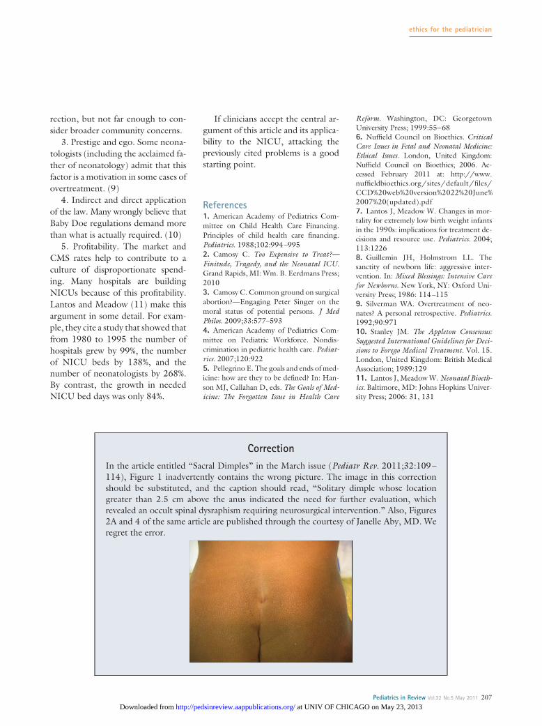
CorrectionIn the article entitled “Sacral Dimples” in the March issue (Pediatr Rev. 2011;32:109–114), Figure 1 inadvertently contains the wrong picture. The image in this correctionshould be substituted, and the caption should read, “Solitary dimple whose locationgreater than 2.5 cm above the anus indicated the need for further evaluation, whichrevealed an occult spinal dysraphism requiring neurosurgical intervention.” Also, Figures2A and 4 of the same article are published through the courtesy of Janelle Aby, MD. Weregret the error.
ethics for the pediatrician
Pediatrics in Review Vol.32 No.5 May 2011 207 at UNIV OF CHICAGO on May 23, 2013http://pedsinreview.aappublications.org/Downloaded from