Embed Size (px)
Citation preview

www.eurosurveillance.org
Vol. 18 | Weekly issue 46 | 14 November 2013
E u r o p e ’ s j o u r n a l o n i n f e c t i o u s d i s e a s e e p i d e m i o l o g y, p r e v e n t i o n a n d c o n t r o l
Rapid communications
Salmonella enterica serotype Paratyphi A carrying CTX-M-15 type extended-spectrum beta-lactamase isolated from a Japanese traveller returning from India, Japan, July 2013 2by M Mawatari, Y Kato, K Hayakawa, M Morita, K Yamada, K Mezaki, T Kobayashi, Y Fujiya, S Kutsuna, N Takeshita, S Kanagawa, M Ohnishi, H Izumiya, N Ohmagari
Isolation of NDM-1-producing Pseudomonas aeruginosa sequence type ST235 from a stem cell transplant patient in Italy, May 2013 5by A Carattoli, D Fortini, R Galetti, A Garcia-Fernandez, G Nardi, D Orazi, A Capone, I Majolino, A Proia, B Mariani, G Parisi, A Morrone, N Petrosillo
Research articles
Tick-borne encephalitis in Bulgaria, 2009 to 2012 8by E Mohareb, I Christova, A Soliman, R Younan, T Kantardjiev
Evidence for the transmission of Salmonella from reptiles to children in Germany, July 2010 to October 2011 12by M Pees, W Rabsch, B Plenz, A Fruth, R Prager, S Simon, V Schmidt, S Münch, PG Braun
News
ECDC launches the Geoportal for the European Environment and Epidemiology (E3) Network 22by B Sudre

2 www.eurosurveillance.org
Rapid communications
Salmonella enterica serotype Paratyphi A carrying CTX-M-15 type extended-spectrum beta-lactamase isolated from a Japanese traveller returning from India, Japan, July 2013
M Mawatari ([email protected])1, Y Kato1, K Hayakawa1, M Morita2, K Yamada3, K Mezaki3, T Kobayashi1, Y Fujiya1, S Kutsuna1, N Takeshita1, S Kanagawa1, M Ohnishi2, H Izumiya2, N Ohmagari1
1. Disease Control and Prevention Center, National Center for Global Health and Medicine, Tokyo, Japan 2. Bacteriology I, National Institute of Infectious Diseases, Tokyo, Japan3. Clinical Laboratory, National Center for Global Health and Medicine, Tokyo, Japan
Citation style for this article: Mawatari M, Kato Y, Hayakawa K, Morita M, Yamada K, Mezaki K, Kobayashi T, Fujiya Y, Kutsuna S, Takeshita N, Kanagawa S, Ohnishi M, Izumiya H, Ohmagari N. Salmonella enterica serotype Paratyphi A carrying CTX-M-15 type extended-spectrum beta-lactamase isolated from a Japanese traveller returning from India, Japan, July 2013. Euro Surveill. 2013;18(46):pii=20632. Available online: http://www.eurosurveillance.org/ViewArticle.aspx?ArticleId=20632
Article submitted on 30 October 2013 / published on 14 November 2013
Emerging drug resistance in Salmonella Typhi and S. Paratyphi is a substantial public health concern. We report what appears to be the first case and isolation of multidrug resistant S. Paratyphi A carrying CTX-M-15-type extended-spectrum beta-lactamase from a Japanese traveller returning from India.
Here, we report the isolation of multidrug resistant S. Paratyphi A producing CTX-M-15-type extended-spec-trum beta-lactamase (ESBL) from a traveller returning from India. To our knowledge, this is the first report of S. Paratyphi A with CTX-M-15-type ESBL isolated from a human. Enteric fever, including typhoid fever caused by S. enterica serotype Typhi (S. Typhi) and paraty-phoid fever caused by S. enterica serotype Paratyphi A (S. Paratyphi A), is one of the most important febrile illnesses in tropical and subtropical countries, with high rates of morbidity and mortality [1]. In industri-alised countries, enteric fever is a common cause of fever in returned travellers [2]. The emergence of drug-resistance in S. Typhi and S. Paratyphi A is an emerg-ing public health problem. Owing to the recent increase of fluoroquinolone resistance, third-generation cepha-losporins, such as ceftriaxone or cefotaxime, have become the primary drugs for treatment of enteric fever [3].
Case reportIn July 2013, a Japanese woman in her mid-20s was admitted to a hospital in Manali, India for fever over 38.5 ºC, diarrhoea, and anorexia, where she was diag-nosed with typhoid fever based on clinical symptoms and a non-specified rapid serological test. She had been in India for five weeks prior to the admission. Before her visit to India, she had been on a seven-week trip to China, Myanmar, Thailand, and Nepal. She was treated with parenteral cephalosporin for three days.
No further details on the treatment regimen in India were available. The patient returned to Japan on day 4 of illness owing to sustained fever, diarrhoea, and ano-rexia. During her travel from India to Japan (days 5–7), the patient took oral combined ofloxacin-cefixime. On day 7, she was admitted to our hospital with diarrhoea and anorexia but without fever. On examination, deep palpation of the lower abdominal area caused mild dis-comfort, otherwise, physical examination was normal. She did not have any underlying illness, and was not on any regular medication. Eight months prior to the admission at the hospital in India she had been vac-cinated against Salmonella Typhi (Vi polysaccharide).
Laboratory analysesOn admission, stool and blood samples were taken for culture. Antimicrobial susceptibility test was per-formed by broth microdilution in accordance with the Clinical and Laboratory Standards Institutes [4].
The patient’s stool was screened on admission for met-icillin-resistant Staphylococcus aureus, vancomycin-resistant Enterococci, and Gram-negative rods resistant to one or multiple agents in the extended-spectrum cephalosporin class and/or that demonstrated elevated minimum inhibitory concentrations (MICs) (>1 mg/L) to imipenem and/or meropenem. Screening culture of stool was positive only for drug-sensitive Escherichia coli and Enterobacteriaceae spp.
Results from blood cultures were positive for S. Paratyphi A two days after admission and S. Paratyphi A was found to be resistant to cefotaxime (MIC = 64 mg/L) and ceftazidime (MIC > 16 mg/L). Addition of cla-vulanic acid to each cephalosporin lowered the MIC to 0.25 mg/L and 1 mg/L, respectively. This isolate was also resistant to nalidixic acid, and categorised as

3www.eurosurveillance.org
intermediate to ciprofloxacin (Table). Due to the atypi-cal susceptibility pattern and the requirement for the verification susceptibility test process, it took longer than one week for the final susceptibility result to be available.
The presence of the gene conferring the ESBL pheno-type was verified by polymerase chain reaction (PCR) as described elsewhere [5]. Sequencing of the PCR products revealed that S. Paratyphi A was positive for blaCTX-M-15. The presence of the plasmid-mediated quinolone resistance genes including qnrA, qnrB, qnrC, qnrS, and aac(6’)-Ib was investigated using PCR described elsewhere [6-9]. However plasmid-mediated quinolone resistance genes were not detected in this isolate.
Patient treatment and outcomeUpon admission, we treated the patient with ceftriax-one (2 g per day for 8 days), and the patient’s symp-toms resolved after eight days of admission. After the final susceptibility results were available, the ini-tial antibiotic treatment was changed on day 9 from ceftriaxone to azithromycin (orally, 500 mg per day),
which was administered for seven days. A stool culture obtained on admission was negative for S. Paratyphi A. Stool cultures repeated before changing antibiotic treatment as well as eight, nine, and 10 days after com-pleting azithromycin treatment also remained negative for S. Paratyphi A. The patient was discharged on 10 days after admission.
Discussion and conclusionAlthough ESBL-producing organisms are emerging among Enterobacteriaceae, ESBL-producing S. Typhi or S. Paratyphi A have been reported rarely in Nepal and the Middle East [10,11]; CTX-M-15 type ESBL-producing S. Typhi were previously reported in patients in Middle Eastern countries [11,12].
blaCTX-M-15 genes have been identified worldwide in Enterobacteriaceae; further, isolation of these genes from Escherichia coli has been reported to be increas-ing [13]. The acquisition of blaCTX-M-15 as a transferable plasmid from another enteric bacterium has previously been reported in S. Typhi [5].
Of particular concern are reports of increasing isola-tion rates of S. Paratyphi A from India and other parts of Asia [14], which might be attributed to use of pro-tective vaccines (Vi polysaccharide and live oral Ty21a) effective against S. Typhi, but not S. Paratyphi A [14].
The number of reports on ESBL in S. Typhi and S. Paratyphi A is still limited, and thus, extended-spectrum cephalosporins would still be a reasonable empiric treatment for a suspected case of enteric fever in the current situation. The patient improved on day 8 before antibiotics were changed. The oral treatment by a fluoroquinolone (ofloxacin) prior to the admission might have contributed to her clinical improvement. Other possible explanations include clinical recovery by natural history of S. Paratyphi A, or infection of two different antimicrobial patterns of S. Paratyphi A, i.e. ESBL-S. Paratyphi A and non-ESBL-S. Paratyphi A, with non-ESBL mainly contributing to clinical symptoms.
Potential increase in the plasmid-mediated spread of ESBL in S. Paratyphi A in the future would pose a threat to public health. Judicious use of antibiotics to avoid unnecessary selective pressure on intestinal bacterial flora, and careful microbiological analysis of patients with typhoid fever, especially those returning from the Middle East or south Asia are approaches critical for prevention of the potential spread of multidrug resist-ant S. Paratyphi A.
Acknowledgements This work was partly supported by funding from the Research on Emerging and Reemerging Infectious Diseases by the Ministry of Health, Labour, and Welfare, Japan (H24-shinkou-ippan-013).
TableMinimum inhibitory concentrations of various antibiotics on Salmonella enterica serotype Paratyphi A isolated from a Japanese traveller returning from India, Japan, July 2013
Antibiotics MIC (mg/L), interpretation result
Ampicillin >16, R
Amoxicillin/clavulanic acid =16, I
Cefotaxime =64, R
Cefotaxime/clavulanic acid =0.25, S
Ceftazidime >16, R
Ceftazidime/clavulanic acid =1, S
Ceftriaxone >16, R
Nalidixic acid >16, R
Ciprofloxacin =0.5, I
Levofloxacin =1, I
Azithromycin =16a
Aztreonam >16, R
Trimethoprim/sulfamethoxazole >2, R
Chloramphenicol =4, S
Tetracycline =2, S
MIC: Minimum inhibitory concentration; R: resistant; S: susceptible; I: intermediate.
a The MIC breakpoint for azithromycin is not provided by the Clinical and Laboratory Standards Institutes. We thus refer to the breakpoints table by the European Committee on Antimicrobial Susceptibility Testing, which mentions that azithromycin has been used in the treatment of infections with Salmonella Typhi (MIC <16mg/L for wild type isolates) [15].

4 www.eurosurveillance.org
Conflict of interestNone declared.
Authors’ contributionsM. Mawatari collected the data and drafted the manuscript; YK and KH participated in the coordination and concept of the manuscript and edited the manuscript and helped with the draft of the manuscript; M. Morita, MO and HI performed and analysed the molecular tests; KY and KM performed and analysed the microbiological tests; TK, YF, S. Kutsuna and NT collected the data and participated in the concept of the manuscript; S. Kanagawa and NO revised the article for intellectual content. All authors read and critically revised the first as well as the subsequent and final drafts of this manuscript.
References1. Crump JA, Luby SP, Mintz ED. The global burden of typhoid
fever. Bull World Health Org. 2004;82(5):346–53. PMid:15298225 PMCid:PMC2622843
2. Leder K, Torresi J, Libman MD, Cramer JP, Castelli F, Schlagenhauf P, et al. GeoSentinel surveillance of illness in returned travelers, 2007-2011. Ann Intern Med. 2013;158(6):456–68. http://dx.doi.org/10.7326/0003-4819-158-6-201303190-00005 PMid:23552375
3. Humphries RM, Fang FC, Aarestrup FM, Hindler JA. In vitro susceptibility testing of fluoroquinolone activity against Salmonella: recent changes to CLSI standards. Clin Infect Dis. 2012;55(8):1107–13. http://dx.doi.org/10.1093/cid/cis600 PMid:22752519
4. Clinical and Laboratory Standards Institute (CLSI). Performance standards for antimicrobial susceptibility testing; twenty-second informational supplement. M100-S22. Wayne, PA: CLSI; 2012.
5. Morita M, Takai N, Terajima J, Watanabe H, Kurokawa M, Sagara H, et al. Plasmid-mediated resistance to cephalosporins in Salmonella enterica serovar Typhi. Antimicrob Agents Chemother. 2010;54(9):3991–2. http://dx.doi.org/10.1128/AAC.00225-10 PMid:20585124 PMCid:PMC2935013
6. Kim HB, Park CH, Kim CJ, Kim EC, Jacoby GA, Hooper DC. Prevalence of plasmid-mediated quinolone resistance determinants over a 9-year period. Antimicrob Agents Chemother. 2009;53(2):639–45. http://dx.doi.org/10.1128/AAC.01051-08 PMid:19064896 PMCid:PMC2630623
7. Cattoir V, Poirel L, Rotimi V, Soussy CJ, Nordmann P. Multiplex PCR for detection of plasmid-mediated quinolone resistance qnr genes in ESBL-producing enterobacterial isolates. J Antimicrob Chemother. 2007;60(2):394-7. http://dx.doi.org/10.1093/jac/dkm204 PMid:17561500
8. Park CH, Robicsek A, Jacoby GA, Sahm D, Hooper DC. Prevalence in the United States of aac(6’)-Ib-cr encoding a ciprofloxacin-modifying enzyme. Antimicrob Agents Chemother. 2006;50(11):3953-5. http://dx.doi.org/10.1128/AAC.00915-06 PMid:16954321 PMCid:PMC1635235
9. Robicsek A, Strahilevitz J, Sahm DF, Jacoby GA, Hooper DC. qnr prevalence in ceftazidime-resistant Enterobacteriaceae isolates from the United States. Antimicrob Agents Chemother. 2006;50(8):2872-4. http://dx.doi.org/10.1128/AAC.01647-05 PMid:16870791 PMCid:PMC1538681
10. Pokharel BM, Koirala J, Dahal RK, Mishra SK, Khadga PK, Tuladhar NR. Multidrug-resistant and extended-spectrum beta-lactamase (ESBL)-producing Salmonella enterica (serotypes Typhi and Paratyphi A) from blood isolates in Nepal: surveillance of resistance and a search for newer alternatives. Int J Infect Dis. 2006;10(6):434–8. http://dx.doi.org/10.1016/j.ijid.2006.07.001 PMid:16978898
11. Rotimi VO, Jamal W, Pal T, Sovenned A, Albert MJ. Emergence of CTX-M-15 type extended-spectrum beta-lactamase-producing Salmonella spp. in Kuwait and the United Arab Emirates. J Med Microbiol. 2008;57(Pt 7):881–6.
http://dx.doi.org/10.1099/jmm.0.47509-0 PMid:18566147
12. Pfeifer Y, Matten J, Rabsch W. Salmonella enterica serovar Typhi with CTX-M beta-lactamase, Germany. Emerg Infect Dis. 2009;15(9):1533–5. http://dx.doi.org/10.3201/eid1509.090567 PMid:19788837 PMCid:PMC2819882
13. Pitout JD, Laupland KB. Extended-spectrum beta-lactamase-producing Enterobacteriaceae: an emerging public-health concern. Lancet Infect Dis. 2008;8(3):159–66. http://dx.doi.org/10.1016/S1473-3099(08)70041-0
14. Ochiai RL, Wang X, von Seidlein L, Yang J, Bhutta ZA, Bhattacharya SK, et al. Salmonella paratyphi A rates, Asia. Emerg Infect Dis. 2005;11(11):1764–6. http://dx.doi.org/10.3201/eid1111.050168 PMid:16318734 PMCid:PMC3367370
15. The European Committee on Antimicrobial Susceptibility Testing (EUCAST). Breakpoint tables for interpretation of MICs and zone diameters Version 3.1, valid from 2013-02-11. Växjö: EUCAST; 2013. Available from: http://www.eucast.org/fileadmin/src/media/PDFs/EUCAST_files/Breakpoint_tables/Breakpoint_table_v_3.1.pdf

5www.eurosurveillance.org
Rapid communications
Isolation of NDM-1-producing Pseudomonas aeruginosa sequence type ST235 from a stem cell transplant patient in Italy, May 2013
A Carattoli1, D Fortini1, R Galetti1, A Garcia-Fernandez1, G Nardi2, D Orazi2, A Capone3, I Majolino2, A Proia2, B Mariani2, G Parisi2, A Morrone2, N Petrosillo ([email protected])3
1. Istituto Superiore di Sanità, Rome Italy2. Azienda Ospedaliera S.Camillo-Forlanini Hospital, Rome Italy3. National Institute of Infectious Disease “L. Spallanzani”, Rome Italy
Citation style for this article: Carattoli A, Fortini D, Galetti R, Garcia-Fernandez A, Nardi G, Orazi D, Capone A, Majolino I, Proia A, Mariani B, Parisi G, Morrone A, Petrosillo N. Isolation of NDM-1-producing Pseudomonas aeruginosa sequence type ST235 from a stem cell transplant patient in Italy, May 2013. Euro Surveill. 2013;18(46):pii=20633. Available online: http://www.eurosurveillance.org/ViewArticle.aspx?ArticleId=20633
Article submitted on 25 October 2013 / published on 14 November 2013
We describe the first isolation of an NDM-1-producing Pseudomonas aeruginosa in Italy. In May 2013, a patient with acute lymphoblastic leukaemia and history of prior hospitalisation in Belgrad, Serbia, underwent stem cell transplantation at a tertiary care hospital in Rome, Italy. After transplantion, sepsis by NDM-1-producing P. aeruginosa occurred, leading to septic shock and fatal outcome.
In May 2013, a Pseudomonas aeruginosa strain pro-ducing New Delhi metallo-beta-lactamase-1 (NDM-1) was isolated in Italy from a patient previously hospi-talised in Serbia and found to be resistant to all anti-biotics tested except colistin. Multidrug-resistant and extensively drug-resistant P. aeruginosa represent an increasing therapeutic challenge worldwide. In partic-ular, the ST235 clone is the founder of the successful epidemic clonal complex 235 (CC235), associated with various carbapenemase genes, but very rarely with NDM-1 [1,2].
Case descriptionIn late May 2013, a man in his early 40s with a diag-nosis of acute lymphoblastic leukaemia (ALL) in first remission was admitted to the haematology unit of a hospital in Rome, Italy, in order to undergo stem cell transplantation. He had been hospitalised in December 2012 at a general hospital in Belgrad, Serbia. In that hospital, the patient had undergone four courses of chemotherapy (HYPER-C-VAD) from December 2012 to February 2013 with a complete remission of disease.
At hospital admission in Rome, the patient was febrile, in good general condition. According to the haematol-ogy ward’s routine procedure adopted for transplanta-tion candidates, nasal, pharyngeal, rectal and groin swabs, as well as blood, urine and stool cultures were screened and found negative for multidrug-resist-ant bacteria. In brief, all samples were screened for
methicillin-resistant Staphylococcus aureus (MRSA), extended-spectrum beta-lactamase (ESBL)-producing Enterobacteriaceae, Acinetobacter baumannii, P. aer-uginosa, carbapenem-resistant Enterobacteriaceae, vancomycin-resistant Enterococcus faecium (VRE), using MacConkey and chromogenic agar plates (Becton Dickinson, Heidelberg, Germany). Antimicrobial drug susceptibility testing and minimal inhibitory concen-trations (MICs) were obtained by the Phoenix System (Becton Dickinson, Sparks, MD) and interpreted according to the recommendations from the European Committee on Antimicrobial Susceptibility Testing (EUCAST) [3].
Lacking an available HLA-compatible donor, the patient received on 28 May a stem cell transplant from a hap-loidentical donor, with a high-dose conditioning regi-men containing thiotepa, busulfan, and also containing fludarabine-unmanipulated bone marrow as the stem cell source. Prophylaxis of graft-versus-host disease was post-transplant cyclophosphamide, mycopheno-late and cyclosporine, as in the Baltimore programme [4].Two days later he developed severe neutropenia (absolute neutrophil count (ANC) =0.1x10⁹/L) and treat-ment was started with piperacillin/tazobactam, amika-cin and vancomycin, with an immediate defervescence. The patient remained neutropenic (ANC <0.1x10⁹/L), and fever reappeared some days later. Meropenem was added in substitution of piperacillin/tazobactam.
A pharyngeal swab taken 10 days after transplanta-tion was positive for Klebsiella pneumoniae. Species identification and antimicrobial susceptibilities were determined by the Phoenix System (Becton Dickinson, Sparks, MD) and the tigecycline MIC was obtained by Etest (Becton-Dickinson, United States). The K. pneu-moniae isolate was an ESBL producer with resist-ance to cefotaxime (MIC >4 mg/L) ceftazidime (MIC >8 mg/ml), ertapenem (MIC >1 mg/L) and tigecycline

6 www.eurosurveillance.org
(MIC 3 mg/L), but remained susceptible to imipenem (MIC ≤1 mg/L), meropenem (MIC ≤1 mg/L) and colistin (MIC ≤1 mg/L), according to interpretive criteria from the EUCAST [3]. The strain was assigned to ST101 by multilocus sequence typing [5] and resulted negative for carbapenemase genes in PCR using previously described primers and protocols [6,7]. Colistin (given every 12 hours for a total daily dose of 9,000,000 IU after a loading dose of 9,000,000 IU) was added to the ongoing antimicrobial treatment.
At Day 15 after transplantation, the patient was still febrile and neutropenic. Another rectal swab was again positive for the ertapenem-resistant K. pneumoniae. A perianal abscess appeared on Day 16 and draining yielded P. aeruginosa and VRE. This isolate was resist-ant to all antibiotics tested (Table) except colistin.
The blood cultures of the patient, taken on Day 15 yielded a P. aeruginosa with an identical phenotype. Vancomycin treatment was halted, and tigecycline (200 mg/day administered every 12 hours, after a load-ing dose of 200 mg), rifampicin (600 mg given every 24 hours) and daptomycin (700 mg every 24 hours), were added to the previous treatment.
On Day 17 after transplantation, the patient rapidly deteriorated and was transferred to the intensive care unit, where he died few hours later from septic shock still in profound aplasia.
Molecular characterisation of the P. aeruginosa isolateThe P. aeruginosa strain isolated from the blood cul-tures was identified as sequence type (ST) 235 by MLST [8]. The ST235 strain resulted positive for the blaNDM-1 gene. Plasmid and total DNA was tested by Southern blot hybridisation with the blaNDM-1 gene as probe. Results suggested a chromosomal location of the blaNDM-1 gene (data not shown). Total DNA was restricted with HindIII and a genome library was con-structed in the HindIII-pZErO-2 kanamycin-resistant vector (Invitrogen, Milan Italy). Transformants were obtained selecting on plates containing 30 mg/L ampi-cillin and 30 mg/L kanamycin. One transformant carried a HindIII insert of approximately 3.3 KB, which resulted positive in a PCR for the blaNDM-1 gene. The insert was fully sequenced using universal primers and the primer walking approach. Comparative DNA sequence analy-sis showed that the HindIII insert in our construct had 100% of DNA identity to the HindIII-fragment of the ST235 P. aeruginosa strain HIABP11(EMBL ACC. No. KC170992) carrying blaNDM-1 that was isolated in France in March 2012 [9]. Based on the DNA sequence of the HIABP11 strain (EMBL ACC No. KC170992), 12,762 kb containing the entire blaNDM-1 gene environment and flanking regions were analysed by PCR and sequenc-ing. The presence of ISPa7 was identified upstream of a complex class 1 integron, carrying the aacA7, aadA6, orfD gene cassettes, suggesting a chromosomal loca-tion of this genetic determinant. The blaNDM-1 gene was embedded in the same genetic environment previously described for the HIABP11 ST235 P. aeruginosa isolate from France [9].
Control measures
The ST235 strain could represent a serious risk for potential spreading in the hospital environment. However, during the hospitalisation and after the iden-tification of the strain, strict contact isolation pre-cautions (CIP), and screening of the personnel were implemented on the haematology ward. CIP included wearing a gown and gloves when entering the patient’s room, promptly removing gloves after care and hand decontamination, as well as using disposable single-use or patient dedicated non-critical equipment such as blood pressure cuff and stethoscope. Moreover, since the hands of healthcare workers are the most common vehicle for the transmission of microorgan-isms, including P. aeruginosa and K. pneumoniae, from patient to patient and within the healthcare environ-ment [10], hand hygiene practices were strengthened.
All contacts, room-mates, relatives, and personnel involved in the care of the patient were screened for carriage of this organism. In addition, environmen-tal screening for Pseudomonas and Klebsiella was performed at several points of the ward, including the rooms where the patient stayed. Neither NDM-1-producing P. aeruginosa nor Klebsiella were obtained
TableSusceptibility pattern of NDM-1-positive Pseudomonas aeruginosa isolated from blood cultures and perianal abscess, Rome, May 2013
Antibiotics MIC (µg/mL), interpretation resulta
Amikacin >16, R
Aztreonam =16, R
Cefepime >8, R
Ceftazidime >8, R
Ciprofloxacin >1, R
Colistin ≤1, S
Gentamicin >4, R
Imipenem >8, R
Levofloxacin >2, R
Meropenem >8, R
Piperacillin >16, R
Piperacillin/tazobactam >16/4, R
Tobramycin >4, R
MIC: minimum inhibitory concentration R: resistant; S: susceptiblea Based on EUCAST interpretive criteria [1].

7www.eurosurveillance.org
from these cultures. Moreover, after a one month of strict surveillance, neither any infection nor any coloni-sation by NDM-1-producing P. aeruginosa or Klebsiella were observed on the wards where the patient stayed.
DiscussionIt has been recently demonstrated that the emergence of multidrug-resistant, metallo-beta lactamase (MBL)-positive P. aeruginosa in Russia was largely due to the spread of one dominant clone, CC235 producing VIM-2 [1]. The association of CC235 with MBL genes has been reported outside Russia in several European countries [1]. In particular, VIM-1 was the more common MBL identified in CC235 P. aeruginosa from Italy, VIM-4 in Greece, Sweden, Hungary, and Belgium, VIM-13 in Spain, and IMP-29 in France [1]. To the best of our knowl-edge, only few NDM-1-producing P. aeruginosa strains were reported in Europe before this Italian case: two strains were described in Serbia in 2010 and one strain was identified in France in 2012 [2,9]. Both the Italian and French NDM-1 producing P. aeruginosa were identi-fied in patients previously hospitalised in Serbian hos-pitals, where a reservoir of NDM-1-producing CC235 P. aeruginosa can be suspected. Moreover, six of 55 travel-associated cases of Enterobacteriaceae produc-ing NDM-1 reported in European countries had a link to the Balkan region, presumably to Serbia, Kosovo*, Montenegro, and Bosnia and Herzegovina [11].
* This designation is without prejudice to positions on status, and is in line with United Nations Security Council Resolution 1244/99 and the International Court of Justice Opinion on the Kosovo declaration of independence.
AcknowledgementsThis work was supported by The Italian FLAGSHIP “InterOmics” project (PB.P05) funded by MIUR and coordi-nated by the CNR. Renata Galetti’s fellowship was supported by the grant 2012/24864-1, Sao Paulo Research Foundation (FAPESP)
Conflict of interestNone declared.
Authors’ contributionsAll authors of this research paper have directly participated in the planning, execution and analysis of this study
References1. Edelstein MV, Skleenova EN, Shevchenko OV, D’souza JW,
Tapalski DV, Azizov IS, et al. Spread of extensively resistant VIM-2-positive ST235 Pseudomonas aeruginosa in Belarus, Kazakhstan, and Russia: a longitudinal epidemiological and clinical study. Lancet Infect Dis. 2013;13(10):867-76. http://dx.doi.org/10.1016/S1473-3099(13)70168-3
2. Jovcic B, Lepsanovic Z, Suljagic V, Rackov G, Begovic J, Topisirovic L, et al. Emergence of NDM-1 Metallo-beta-Lactamase in Pseudomonas aeruginosa Clinical Isolates from Serbia. Antimicrob. Agents Chemother. 2011;55(8):3929-31. http://dx.doi.org/10.1128/AAC.00226-11 PMid:21646490 PMCid:PMC3147624
3. European Committee on Antimicrobial Susceptibility Testing (EUCAST). Breakpoint tables for interpretation of MICs and zone diameters. Version 3.1 EUCAST; 2013.Available from: http://www.eucast.org/clinical-breakpoints/
4. Fuchs EJ. Human leukocyte antigen-haploidentical stem cell transplantation using T-cell-replete bone marrow grafts. Curr Opin Hematol. 2012;19(6):440-7. http://dx.doi.org/10.1097/MOH.0b013e32835822dc PMid:22954723
5. Diancourt L, Passet V, Verhoef J, Grimont PA, Brisse S. Multilocus sequence typing of Klebsiella pneumoniae nosocomial isolates. J Clin Microbiol. 2005;43(8):4178-82. http://dx.doi.org/10.1128/JCM.43.8.4178-4182.2005 PMid:16081970 PMCid:PMC1233940
6. Yong D, Toleman MA, Giske CG, Cho HS, Sundman K, Lee K, et al. Multiplex PCR for rapid detection of genes encoding acquired metallo-beta-lactamases. J Antimicrob Chemother. 2007;59(2):321-2.
7. Poirel L, Walsh TR, Cuvillier V, Nordmann P. Multiplex PCR for detection of acquired carbapenemase genes. Diagn Microbiol Infect Dis.2011;70(1):119-23. http://dx.doi.org/10.1016/j.diagmicrobio.2010.12.002 PMid:21398074
8. Curran B, Jonas D, Grundmann H, Pitt T, Dowson CG. Development of a multilocus sequence typing scheme for the opportunistic pathogen Pseudomonas aeruginosa. J. Clin. Microbiol. 2004;42(12):5644-9. http://dx.doi.org/10.1128/JCM.42.12.5644-5649.2004 PMid:15583294 PMCid:PMC535286
9. Janvier F, Jeannot K, Tessé S, Robert-Nicoud M, Delacour H, Rapp C, et al. Molecular Characterization of blaNDM-1 in a Sequence Type 235 Pseudomonas aeruginosa Isolate from France. Antimicrob. Agents Chemother. 2013; 57(7):3408-11. http://dx.doi.org/10.1128/AAC.02334-12 PMid:23612200
10. Monistrol O, López ML, Riera M, Font R, Nicolás C, Escobar MA, et al. Hand contamination during routine care in medical wards: the role of hand hygiene compliance. J Med Microbiol. 2013;62(Pt 4):623-9. http://dx.doi.org/10.1099/jmm.0.050328-0 PMid:23329322
11. Struelens MJ, Monnet DL, Magiorakos AP, Santos O’Connor F, Giesecke J. New Delhi metallo-beta-lactamase 1-producing Enterobacteriaceae: emergence and response in Europe. Euro Surveill. 2010;15(46):pii=19716. Available from: http://www.eurosurveillance.org/ViewArticle.aspx?ArticleId=19716

8 www.eurosurveillance.org
Research articles
Tick-borne encephalitis in Bulgaria, 2009 to 2012E Mohareb1, I Christova ([email protected])2, A Soliman2, R Younan1, T Kantardjiev2
1. United States Naval Medical Research Unit-3, Cairo, Egypt2. National Center of Infectious and Parasitic Diseases, Sofia, Bulgaria
Citation style for this article: Mohareb E, Christova I, Soliman A, Younan R, Kantardjiev T. Tick-borne encephalitis in Bulgaria, 2009 to 2012. Euro Surveill. 2013;18(46):pii=20635. Available online: http://www.eurosurveillance.org/ViewArticle.aspx?ArticleId=20635
Article submitted on 16 December 2012 / published on 14 November 2013
For the last 60 years, only a few cases of tick-borne encephalitis (TBE) have been detected in Bulgaria. Considering the remarkable increase in TBE morbidity in Europe over the past two decades, we conducted a study of TBE among patients with acute viral menin-gitis who were hospitalised in Bulgaria during 2009 to 2012. A total of 86 patients with viral meningitis of unknown aetiology during this period were tested. Acute TBE was confirmed in three of these patients. The last TBE case was detected in October 2012; the other two were diagnosed in 2009. To the best of our knowledge, these three patients are the first con-firmed TBE cases reported in Bulgaria. The risk of TBE is underestimated in Bulgaria due to the low aware-ness of medical doctors.
IntroductionTick-borne encephalitis (TBE) occurs in north and central Europe, Russia, Far East, Asia and Japan [1-3]. The aetiological agent, tick-borne encephalitis virus (TBEV), is a member of the genus Flavivirus of the fam-ily Flaviviridae, which also includes aetiological agents of yellow fever, dengue, West Nile fever and Japanese encephalitis. Three subtypes of the virus are known: European, Siberian and Far Eastern [4]. The severity of the disease and its outcome depends largely on the causative subtype [4].
TBEV is transmitted to humans through bites of infected Ixodes ricinus ticks or by consumption of unpasteurised milk from infected animals [5], usually goats, but also sheep and cows [6]. The incubation period is between 2 and 28 days, most commonly 7–14 days. A shorter incubation period is connected to milk-borne TBE [5].
Between 70% and 98% of TBEV infections are subclini-cal [5]. In clinically manifested cases, about two thirds of the patients only develop a non-specific febrile syndrome during the first phase of the infection [5]. Neurological disorders, usually meningitis or menin-goencephalitis, appear during a second febrile phase. Biphasic febrile illness is typical for infection with the Western subtype of the virus, while patients infected with the Eastern subtype develop only a monophasic course [5].
In Bulgaria, reporting of TBE has been mandatory since 1953, but is rarely reported. Over the past 60 years, only a few cases of TBE have been detected [7,8] which, according to the current European Union case defini-tion criteria [9] would not be considered as confirmed cases. Before our study presented here, the last TBE cases were reported in 2006 [10]: laboratory diagno-sis of these and previous cases was based on detec-tion of TBE-specific antibodies by complement fixation assay. Most of these cases were due to consumption of unpasteurised goats’ milk [10]. Surprisingly, the tick vector, Ixodes ricinus, is widely distributed in Bulgaria and Lyme borreliosis, caused by borreliae transmitted by the same tick species, is endemic in the country, with about 1,000 cases reported annually [11].
Considering the remarkable increase in TBE morbidity in Europe over the past three decades [12], we conducted a study among patients with acute viral meningitis who were hospitalised in Bulgaria during 2009 to 2012, to determine whether some of the acute viral meningitis caes could be due to infection with TBEV.
Methods
Patients and serum samplesSerum samples from hospitalised patients with acute viral meningitis of unknown aetiology (with or without history of a tick bite, according to the anamnesis) were collected between 2009 and 2012. The samples were drawn by physicians at the infectious diseases units of regional hospitals in the largest districts of Bulgaria (Sofia, Pazardzhik, Plovdiv, Burgas). Serum samples from 86 patients were drawn during the acute phase and 49 sera were collected at the convalescence phase, 7–30 days after the first sample.
Case definitionIn this study, we used the 2012 European Union case definition for TBE [9]. A case is classified as probable when a patient met the clinical criteria (symptoms of inflammation of the central nervous system (CNS)) and laboratory criteria (detection of TBE-specific IgM anti-bodies in a unique serum sample). For a confirmed

9www.eurosurveillance.org
TBE case, in addition to meeting the clinical criteria, at least one of the following laboratory criteria was met: (i) TBE-specific IgM and IgG antibodies in blood; (ii) seroconversion or fourfold increase of TBE-specific antibodies in paired serum samples; (iii) detection of TBE viral nucleic acid in blood or CSF.
Enzyme-linked immunosorbent assay All 135 serum samples from the 86 patients were tested for TBEV-specific IgM antibodies; those that were posi-tive were also tested for IgG antibodies against TBEV using commercially available enzyme-linked immuno-sorbent assay (ELISA) (Euroimmun, Germany). The test uses highly purified TBEV proteins. Serum samples were diluted 1:101. Peroxidase-labelled anti-human IgM or IgG antibodies and 3,3’,5,5’ -tetramethylben-zidine (TMB)/peroxide substrate were used to detect specific interactions. Calculated values below 0.8 were interpreted as negative; those between 0.8 and 1.1 were accepted as borderline and those above 1.1 were considered positive.
Polymerase chain reactionTBEV RNA was detected by reverse transcription (RT) polymerase chain reaction (PCR) based on quantita-tive real-time technology (TaqMan), as described else-where [13]. The system detected a fragment of the 3’ non-coding region of the TBEV genome.
Testing for other pathogensSerum samples of patients that were found positive for TBEV-specific antibodies were further tested for IgM antibodies against Borrelia burgdorferi by ELISA (anti-Borrelia ELISA IgM, Euroimmun, Germany), West Nile virus by ELISA (West Nile virus IgM capture – DxSelect, Focus Diagnostics, United States) and immunofluores-cence assay (IFA) (Euroimmun, Germany) and against yellow fever virus by IFA (Euroimmun, Germany).
ResultsSamples from 86 patients with viral meningitis of unknown aetiology during 2009 to 2012 were tested to detect acute TBE: three confirmed TBE cases were
FigurePlace of residence of all reported cases of tick-borne encephalitis, Bulgaria, 1953–2012 (n=15)
Romania
Serbia
Former Yugoslav Republic of Macedonia
Greece
TurkeyBlagoevgrad
Burgas
Dobrich
Gabrovo
Haskovo
Kardzhali
Kyustendil
Lovech
Montana
Pazardzhik
Pernik
Pleven
Plovdiv
Razgrad Ruse
Shumen
Silistra
Sliven
Smolyan
Sofia Sofia Province
Stara Zagora
Targovishte Varna
Veliko Tarnovo
Vidin
Vratsa
Yambol
50 km
Confirmed icase, i2009–2012 i(n=3)
Not-confirmed i case, i1972–2008 i(n=7)
Not-confirmedicase, i1953–1971i(n=5)
Source of data from 1953 to 2008: [7,8,10,14]
Map adapted from: http://d-maps.com/carte.php?num_car=5668&lang=en

10 www.eurosurveillance.org
found. The last TBE case was detected in October 2012; the other two were diagnosed in 2009. The place of residence of the three confirmed cases, as well as the previous not-confirmed cases reported since 1953, is shown (Figure).
Case 1A teenager residing in Velingrad (Pazardzhik district, south Bulgaria) was admitted to the regional hospi-tal in early 2009 with high fever (40 °C) and malaise. The patient’s temperature returned to normal (four days after admission) and then about a week later, the patient’s condition again deteriorated, with fever, headache, stiff neck, sore throat, nausea, vomiting and a depressed mood. The patient had a history of possible tick exposure in the forest surrounding the village. Cerebrospinal fluid (CSF) collected four days after admission showed a high number of leucocytes (160/µL; norm: 0–5/µL) with 75% granulocytes (norm: 60–70% lymphocytes, up to 30% monocytes), high protein content (125 mg/dL; norm: 15–45 mg/dL) and normal glucose level (0.31 mmol/L; norm: 0.22–0.44 mmol/L). TBEV was detected by real-time RT-PCR in a serum sample taken the same day. The patient was transferred to a hospital in Sofia and a second CSF sample was obtained on 22 April 2009. CSF pressure was increased, the leucocytes count was increased (400/µL) with 65% lymphocytes, the protein content was decreased slightly but was still high (100 mg/dL) and the glucose level was still normal (0.30 mmol/L). Mycobacterium tuberculosis was isolated from this CSF sample. A serum sample drawn the same day showed high titres of TBEV-specific IgM antibodies by ELISA. IgG antibodies against TBEV were not found.
Case 2In early September 2009, a person aged in their early 20s was admitted to the regional Plovdiv hospital (south Bulgaria) with fever (38.5 °C), headache, fatigue, nausea and vomiting. Physical examination revealed stiff neck, muscle soreness, conjunctivitis, stupor and abnormal reflexes with pain in joints. The symp-toms started 5–6 days before hospital admission. The patient lived in a village with a high risk for exposure to tick bites (many village residents had had tick bites). CSF analysis showed an increased count of leuco-cytes 301/µL, with 82% lymphocytes, slightly elevated protein (56 mg/dL), and normal glucose level (0.38 mmol/L). After initial improvement within a week, the patient’s condition worsened again with fever, severe headache and prominent dizziness. CSF analysis of the sample collected at that time reflected the worsened condition of the patient: the leucocytes count reached 442/µL, with 90% lymphocytes and the protein level was remarkably elevated (134 mg/dL); the glucose level was normal (0.28 mmol/L). The patient gradually recovered within a month after symptom onset. Paired serum samples from this patient – one upon admission and a second during the convalescence – were tested by ELISA: the first sample was positive and the sec-ond borderline for TBEV-specific IgM antibodies. IgG
antibodies were detected by ELISA in the second serum sample, but not in the first.
Case 3 A resident of the Burgas district (east Bulgaria) in their late 20s was admitted to the regional hospital in late September 2012 with fever (37.5–38 °C), con-siderable numbness in the muscles and weakness. Physical examination revealed mild neck stiffness, mild left hemiplegia and hypaesthesia of the limbs. The patient’s symptoms started two days before admission. Upon admission, a tick was found on their body and was removed. Four days later, the patient’s condition improved, after a further four days, the fever, weakness and numbness in muscles was exacerbated. CSF analysis showed slightly elevated leucocyte count (60/µL) and protein level (74 mg/dL); the glucose level was normal (0.38 mmol/L). A serum sample taken at that time and a later sample taken 9 days later, were tested by ELISA: in both samples, IgM and IgG antibod-ies specific to TBEV were detected. The patient was discharged in an improved condition three weeks after the admission.
The serum samples of the three patients tested neg-ative by ELISA and IFA for West Nile virus and IFA for yellow fever virus, and were also negative for IgM anti-bodies to Borrelia burgdorferi by ELISA. Bacterial cul-ture from the CSF samples of the three patients was negative.
DiscussionEarlier investigations, carried out between 1974 and 2002, confirmed that TBEV was present in ticks in Bulgaria: 6,849 ticks were investigated and eight TBEV strains were isolated [14]. TBE cases in humans have been occasionally reported in Bulgaria (Figure); how-ever, the fact that cases do occur – even though only diagnosed and reported sporadically – and are associ-ated with tick bites or consumption of unpasteurised milk, shows that TBEV circulates in the country. Given that patients who develop neurological symptoms rep-resent a small proportion of those infected, it can be predicted that the number of TBEV-infected people in Bulgaria is many times higher.
There has been significant increase in the number of registered cases of TBE in Europe, Russia and the Far East since 1990 [12]. Since then, about 10,000 to 12,000 clinical cases are reported per year in 30 TBE-endemic countries in Europe and Russia [12]. The epi-demiology of TBE after 1990 is characterised not only by a global increase in the number of cases but also by an expansion of risk areas. For example, a significant increase in TBE was recorded in Sweden in the last dec-ade (2000–2011), especially in 2011, when there was a record annual number of TBE cases in Sweden [15]. New endemic areas in Switzerland were identified by detec-tion of TBEV RNA in field-collected ticks in 2007–2010 [16]. Since September 2012, given the importance and

11www.eurosurveillance.org
spread of TBE in the European Union, the European Commission included TBE in the list of communicable diseases covered by epidemiological surveillance in the Member States [9].
In all three patients described here, the typical bipha-sic course of the disease was noted. These patients are, to the best of our knowledge, the first confirmed cases in Bulgaria, having been laboratory confirmed by PCR and IgG ELISA.
Usually, IgM and IgG antibodies to TBEV are present by the time CNS involvement manifests itself in the sec-ond phase of TBE [12]. However, TBEV RNA is very rarely detected by PCR during the viraemic second phase of the disease [13]. Surprisingly, we detected TBEV infec-tion by RT-PCR in the first case. This patient proved to have a mixed infection with M. tuberculosis, which could have promoted the primary progressive course of the meningo-encephalitis, as previously reported [17].
The detection of three cases among the 86 patients tested shows that TBE is probably not uncommon in Bulgaria. The risk of TBE is underestimated in Bulgaria because of the low awareness of medical doctors. TBE should be considered for patients with various mani-festations of CNS infection in Bulgaria.
Acknowledgements This work was supported by the Global Emerging Infections Surveillance Response System (GIES) funds DOD # NAMRU3.2008.0003.
References1. Pavlidou V, Geroy S, Diza E, Antoniadis A, Papa A.
Epidemiological study of tick-borne encephalitis virus in northern Greece. Vector Borne Zoonotic Dis. 2007;7(4):611-5. http://dx.doi.org/10.1089/vbz.2007.0107 PMid:18171108
2. Hukić M, Numanović F, Sisirak M, Moro A, Dervović E, Jakovec S, et al. Surveillance of wildlife zoonotic diseases in the Balkans Region. Med Glas (Zenica). 2010;7(2):96-105.
3. Schultze D, Dollenmaier G, Rohner A, Guidi T, Cassinotti P. Benefit of detecting tick-borne encephalitis viremia in the first phase of illness. J Clin Virol. 2007;38(2):172-5. http://dx.doi.org/10.1016/j.jcv.2006.11.008 PMid:17204453
4. Ecker M, Allison SL, Meixner T, Heinz FX. Sequence analysis and genetic classification of tick-borne encephalitis viruses from Europe and Asia. J Gen Virol. 1999;80(Pt 1):179-85. PMid:9934700
5. Dumpis U, Crook D, Oksi J. Tick-borne encephalitis. Clin Infect Dis. 1999;28(4):882-90. http://dx.doi.org/10.1086/515195 PMid:10825054
6. Caini S, Szomor K, Ferenczi E, Székelyné Gáspár Á, Csohán Á, Krisztalovics K, et al. Tick-borne encephalitis transmitted by unpasteurised cow milk in western Hungary, September to October 2011. Euro Surveill. 2012;17(12):pii=20128. Available from: http://www.eurosurveillance.org/ViewArticle.aspx?ArticleId=20128 PMid:22490310
7. Vaptzarov I, Turpomanov A, Spasov Z, Nikov D, Dragiev M. [Recurrent viral meningitis in South Bulgaria]. Suvr Med (Sofiia). 1954;5(2):86-103. Bulgarian.
8. Georgiev B, Rosický B, Pavlov P, Daniel M, Arnaudov D. The ticks of the natural focus of tick-borne encephalitis of sheep and man in the Rhodope Mountains (Bulgaria). Folia Parasitol (Praha). 1971;18(3):267-73.
9. European Commission. Commission implementing decision of 8 August 2012 amending Decision 2002/253/EC laying down case definitions for reporting communicable diseases to the Community network under Decision No 2119/98/EC of the European Parliament and of the Council. Official Journal of the European Union. Luxembourg: Publications Office of the European Union. 27.9.2012:L 262. Available from: http://eur-lex.europa.eu/LexUriServ/LexUriServ.do?uri=OJ:L:2012:262:0001:0057:EN:PDF
10. Kaneva Z. [Infectious diseases]. Sofia: Znanie; 2006, 261-2. Bulgarian.
11. Kojuharova M, Parmakova K, Kurachatova A, Vladimirova N, Marinova, Georgieva T et al. [Annual report on infectious diseases in Bulgaria]. Informacionen Journal. 2010;3-4:5-47. Bulgarian.
12. Süss J. Tick-borne encephalitis in Europe and beyond--the epidemiological situation as of 2007. Euro Surveill. 2008;13(26):pii=18916. Available from: http://www.eurosurveillance.org/ViewArticle.aspx?ArticleId=18916 PMid:18761916
13. Schwaiger M, Cassinotti P. Development of a quantitative real-time RT-PCR assay with internal control for the laboratory detection of tick borne encephalitis virus (TBEV) RNA. J Clin Virol. 2003;27(2):136-45. http://dx.doi.org/10.1016/S1386-6532(02)00168-3
14. Dikov I, Gacheva N, Kamarinchev B. [Tick-borne encephalitis]. In: Serbezov V, Kalvatchev Z, editors. [Arboviral infections]. Sofia: Iztok-Zapad Ltd; 2005. p. 92-106. Bulgarian.
15. Lundkvist A, Wallensten A, Vene S, Hjertqvist M. Tick-borne encephalitis increasing in Sweden, 2011. Euro Surveill. 2011;16(39):pii=19981. Available from: http://www.eurosurveillance.org/ViewArticle.aspx?ArticleId=19981
16. Lommano E, Burri C, Maeder G, Guerne M, Bastic V, Patalas E, et al. Prevalence and genotyping of tick-borne encephalitis virus in questing Ixodes ricinus ticks in a new endemic area in western Switzerland. J Med Entomol. 2012;49(1):156-64. http://dx.doi.org/10.1603/ME11044 PMid:22308784
17. Meirova RA. [Tick-borne encephalitis associated with other infections]. Klin.Med. (Mosk). 1991;69(5):71-3. Russian.

12 www.eurosurveillance.org
Research articles
Evidence for the transmission of Salmonella from reptiles to children in Germany, July 2010 to October 2011
M Pees ([email protected])1, W Rabsch2, B Plenz1, A Fruth2, R Prager2, S Simon2, V Schmidt1, S Münch2, P G Braun3
1. Clinic for Birds and Reptiles, University of Leipzig, An den Tierkliniken, Leipzig, Germany2. National Reference Centre for Salmonella and other bacterial Enterics, Robert Koch Institute, Wernigerode, Germany3. Institute of Food Hygiene, An den Tierkliniken, Leipzig, Germany
Citation style for this article: Pees M, Rabsch W, Plenz B, Fruth A, Prager R, Simon S, Schmidt V, Münch S, Braun PG. Evidence for the transmission of Salmonella from reptiles to children in Germany, July 2010 to October 2011. Euro Surveill. 2013;18(46):pii=20634. Available online: http://www.eurosurveillance.org/ViewArticle.aspx?ArticleId=20634
Article submitted on 21 December 2012 / published on 14 November 2013
This study examines the Salmonella status in reptiles kept in households with children suffering from gas-troenteritis due to an exotic Salmonella serovar, to obtain information on possible transmission paths. A number of affected households (n=79) were contacted, and almost half (34/79) comprised at least one reptile in the home. Of the households, 19 were further stud-ied, whereby a total of 36 reptiles were investigated. Samples were taken from the reptiles including the oral cavity, the cloaca, the skin and, in the case of liz-ards, the stomach, and isolation of Salmonella strains was performed using repeated enrichment and typing. Where the Salmonella serovars of the infected child and the reptile were identical, typing was followed by pulsed-field gel electrophoresis (PFGE). Bearded dragons (Pogona vitticeps) constituted 19 of 36 exam-ined reptiles. Altogether 319 Salmonella isolates were investigated and 24 different serovars identified in the reptiles. In 15 of 19 households, an identical serovar to the human case was confirmed in at least one rep-tile (including 16 of all 19 bearded dragons examined). The results demonstrate that reptiles and especially bearded dragons shed various Salmonella serovars including those isolated from infected children in the respective households. Hygiene protocols and parents’ education are therefore highly necessary to reduce the risk of transmission. From a terminological point of view, we propose to call such infections ‘Reptile-Exotic-Pet-Associated-Salmonellosis’ (REPAS).
IntroductionAccording to Thomas et al. [1] the potential of captive and wild animals to transmit salmonellae to humans should not be underestimated, and epidemiological studies on sources of human salmonellosis should simultaneously investigate both the human cases and the wild and domestic animals in contact with them. A study in 1997 from Canada also estimated that three to five per cent of human Salmonella cases were associ-ated with exposure to exotic pets (including reptiles,
sugar gliders, and hedgehogs) and involved a great variety of Salmonella serovars with for example S. Stanley, S. Poona, S. Jangwani, S. Pomona, S. subsp. IV 48:g,z51:- (former S. Marina) [2].
Numerous reports exist on the prevalence of Salmonella enterica in captive reptiles, with recent publications demonstrating a higher prevalence in lizards (up to 76%) compared to tortoises and turtles [3,4]. Geue and Löschner [5] showed that reptile collections with pur-chased animals had a significantly higher prevalence of Salmonella than collections from pure breeders. Furthermore, animals from pet shops were more fre-quently affected (89%) than wild caught animals (59%).
Beside the reptiles per se, the reptile feed, in the form of rodents, can also cause infections in humans. In England, a S. Typhimurium definite phage-type was identified as a source of salmonellosis in humans with a strong association to those keeping reptiles, and was also confirmed in frozen feeding-mice originating from a specific rodent breeding facility [6]. In a further out-break of S. Typhimurium related to snakes in the United States (US), pulsed-field gel electrophoresis (PFGE)-patterns identical to the human isolates were con-firmed for isolates from mice used to feed the snakes as well as the snakes and the environment [7].
Among reptiles, turtles have been reported to be the most common source of Salmonella in the 1970s [8]. However, later studies and surveys indicate that other reptile species, especially lizards, may play a more important role [9-13]. Measures targeted at the preven-tion of turtle-associated salmonellosis in the US and Canada in the 1970s temporary helped to reduce its occurrence, however reptile-associated salmonellosis is suspected to be a resurgent problem and estimated to cause three to 11% of all human salmonellosis cases in these countries [2,14,15].

13www.eurosurveillance.org
Most reports of reptile-associated salmonellosis con-cern babies (less than one year of age) and young children (up to six years-old). However, adults can be affected, especially immunocompromised hosts, and patients with impaired gastric acid production [16]. Fatal outcomes following reptile-associated salmonel-losis (RAS) in babies have been observed [17,18]. In Germany, a recent study [19] reported an increasing number of salmonellosis related to reptiles, with most patients being less than one year-old. The aim of this study was therefore to obtain data on possible links between captive reptiles and salmonellosis in children by examining both the children with salmonellosis and all reptiles kept in the respective households.
MethodsThe study was conducted from July 2010 to October 2011. Within this period, the National Reference Centre (NRC) for Salmonella and other Bacterial Enterics at the Robert Koch Institute (Wernigerode, Germany) examined samples from 206 households with salmo-nellosis in children not older than three years. Of the Salmonella isolates, 65% (134/206) did not belong to S. Typhimurium and S. Enteritidis and were therefore of interest for this study. The responsible federal health institutes were asked to contact these households. A total of 79 parents, corresponding to 79 households, were successfully contacted by mail and asked about the presence of reptiles in the respective households. Thirty-five of 79 parents confirmed having reptiles and were contacted by phone to attend this study. As inclu-sion criteria for participation, parents had to agree that all reptiles in the respective household could be sam-pled and their health status assessed, and the time period between detection of clinical salmonellosis in the child and the sampling of the reptiles was not more than three weeks.
Species and health status of the reptiles were assessed and sampling was conducted following established guidelines using sterile cotton swabs (Heinz Herenz, Hamburg, Germany). Swabs were taken from the oral cavity, the cloaca, and the skin on the ventral region of the reptile. For lizards, an additional swab sample was taken from the stomach. Bacterial isolation and identification were conducted with repeated enrich-ment and examination of several colonies in each sample, in order to find as many different Salmonella serovars as possible: All samples were immediately placed into a tube containing Rappaport-Vassiliadis (RV) medium (Oxoid, Wesel, Germany). This medium was cultured aerobically for 24h at 39°C. A sample was then plated onto sheep blood agar as well as XLT-4-Agar (Oxoid, Wesel, Germany) and cultured aerobically for further 24h at 39°C. From the RV bouillon culture, 1 ml was transferred to a new tube containing RV, and enrichment as well as culture was repeated. This pro-cedure was repeated again, so that in total isolation of Salmonella spp. was attempted from three enrichment cycles. From Salmonella-suspicious colonies, at least five different colonies were subcultured on Brilliant
Green agar (Sifin, Berlin, Germany) for confirmatory testing with biochemical methods. Confirmed colonies were agglutinated against Salmonella surface antigens (sera from Sifin, Berlin, Germany) and all strains were typed at the NRC according to the White-Kauffmann-Le Minor scheme [20].
The isolates of identical serovars confirmed in the infected child and the reptile were compared using PFGE. PFGE was carried out according to the standard-ised protocol for subtyping Salmonella [21]. 40 strains (human and animal) were investigated by PFGE. In case of minor pattern differences between the reptile and the human isolate of the respective household, the PFGE was repeated. Interpretation of the PFGE results followed recent recommendations from the literature [22-24]: Identical PFGE patterns were considered to rep-resent the same epidemiological type. Depending on the time the outbreak has been going on and if person to person spread is the prominent feature, two to three [23] or up to four differences [22] in PFGE restriction fragment pattern are considered to be the result of a single genetic event, and isolates can be designated as ’subpatterns’ or related patterns. Given the maximum time period (several weeks between initial infection of the child and final sampling of the reptile) for this study, more than two differences were considered to represent an epidemiologically-significant difference.
ResultsNineteen households met the inclusion criteria for participation in the study and reptiles were thus investigated. Altogether, 36 reptiles were kept in the households and included in this study (per household between 1 and 7, on average 1.9). Details are listed in Table 1. In these 19 households, Salmonella serovars isolated from the children belonged mainly to subspe-cies I (12 households), but also subspecies II (1 house-hold), subspecies IIIa (1 household), subspecies IIIb (2 households) and subspecies IV (3 households) (Table 1). One S. Newport strain (6,8:e,h:1,2:z67, d-tartrate-, malonate+) was investigated in Paris (Institut Pasteur) because of its unusual biochemical properties. The strain exhibited two atypical characteristics: D-tartrate negative and malonate positive. The ‘e,h’ and the ‘1,2’ flagellar phases have been confirmed by fliC and fljB sequencing. The multilocus sequence typing (MLST) profile (ST118) was identical to those of serovar Newport lineage II [25].
Investigated households were spread well over Germany, including nine (of 16 possible) federal states (Bavaria, Baden-Württemberg, Berlin, North Rhine-Westphalia, Rhineland-Palatinate, Saxony, Saxony-Anhalt, Schleswig-Holstein, Thuringia). From the investigated households, all children except one were less than 15 months of age, most (11/19) were less than six months. All children showed clinical symptoms of gastroenteritis including fever, with some being critically ill. Except for three serovars (S. Eastbourne 9,12:e,h:1,5; S. subspec. IV 44:z4,z23:-; S. Monschaui

14 www.eurosurveillance.org
Tabl
e 1a
Com
pari
son
of S
alm
onel
la se
rova
rs in
rept
iles a
nd sa
lmon
ello
sis-a
ffect
ed c
hild
ren
livin
g in
the
sam
e ho
useh
olds
, with
resu
lts o
f pul
sed-
field
gel
ele
ctro
phor
esis
whe
n th
e se
rova
rs
wer
e id
entic
al, G
erm
any,
July
201
0–O
ctob
er 2
011
Hous
e-ho
ld
Child
Rept
ile s
peci
es (t
ype
of
rept
ilea )
Sero
vars
iden
tifie
d in
the
rept
ile
Loca
lisat
ion
on
rept
ileb o
f ser
ovar
id
entic
al to
chi
ld
PGFE
resu
ltc (n
umbe
r of
pat
tern
di
ffer
ence
s)
Age
(mon
ths)
Salm
onel
la s
erov
arN
Non
-iden
tical
isol
ates
to th
e ch
ild (l
ocal
isat
ion
on th
e re
ptile
b )
111
S. E
astb
ourn
e 9,
12:e
,h:1
,5Po
gona
vitt
icep
s (l
)3
S. s
ubsp
ec. I
ser
ol. r
ough
(sc,
ch)
; S. s
ubsp
ec. I
IIb 5
0:r:
z (c
l)Ch
, cl,
sks
(2)
Pogo
na v
ittic
eps
(l)
1–
Cl
23
S. E
alin
g 35
:g,m
,s:-
Pogo
na v
ittic
eps
(l)
1S.
Eas
tbou
rne
9,12
:e,h
:1,5
(cl)
––
38
S. C
otha
m 2
8:i:1
,5Po
gona
vitt
icep
s (l
)3
S. T
enne
ssee
6,7
:z29
:- (c
l); S
. sub
spec
. I s
erol
. rou
gh (c
l)Sk
s (2
)
42
S. s
ubsp
ec. I
V 44
:z4,
z23:
-Po
gona
vitt
icep
s (l
)3
S. E
astb
ourn
e 9,
12:e
,h:1
,5 (c
l); S
. Ten
ness
ee 6
,7:z
29:-
(cl)
Sti
56
S. T
enne
ssee
6,7
:z29
:-Po
gona
vitt
icep
s (l
)3
S. E
nter
itidi
s 9,
12:g
,m:-,
pha
ge ty
pe 8
/7 (c
l); S
. Kis
araw
e 11
:k:e
,n,x
(cl)
Sk
i
635
S. M
onsc
haui
35:
m,t:
-Po
gona
vitt
icep
s (l
)1
–Cl
s (1
)Po
gona
vitt
icep
s (l
)2
S. P
omon
a 28
:y:1
,7 (c
l)Cl
72
S. s
ubsp
ec. I
IIa 4
1:z4
,z23
:-
Pant
hero
phis
gut
tatu
s (s
)4
S. s
ubsp
ec. I
IIb 4
2:k:
z35
(cl);
S. P
arat
yphi
B V
ar.d
T+ Ja
va 4
,5,1
2:b:
1,2,
ph
age
type
Wor
ksop
(cl);
S. F
lorid
a 1,
6,14
,25:
d:1,
7 (c
l)Cl
iPa
nthe
roph
is g
utta
tus
(s)
1S.
sub
spec
. IIIb
42:
k:z3
5 (c
l)–
Cham
eleo
cal
yptr
atus
(l)
1S.
Pom
ona
28:y
:1,7
(cl)
–
83
S. s
ubsp
ec. I
IIb 1
4,24
:z10
:zPa
nthe
roph
is g
utta
tus
(s)
1S.
sub
spec
. IIIb
42:
k:z3
5 (c
l)–
–
97
S. N
ewpo
rt 6
,8:e
,h:1
,2:z
67
(d-t
artr
ate-
,mal
onat
e+)
Pant
hero
phis
gut
tatu
s (s
)2
S. s
ubsp
ec. I
IIb 1
4,24
:z10
:z (c
l)Cl
s (2
)Pa
nthe
roph
is g
utta
tus
(s)
1–
Cl
1013
S. K
andl
a 17
:z29
:-Po
gona
vitt
icep
s (l
)2
S. K
isar
awe
11:k
:e,n
,x (c
h, c
l)Cl
iPo
gona
vitt
icep
s (l
)2
S. K
isar
awe
11:k
:e,n
,x (c
l)Cl
115
S. M
onsc
haui
35:
m,t:
-Po
gona
vitt
icep
s (l
)2
S. T
enne
ssee
6,7
:z29
:- (s
k); S
. Car
mel
17:
l,v:e
,n,x
(ch,
sk,
st)
––
Pogo
na v
ittic
eps
(l)
2S.
Ten
ness
ee 6
,7:z
29:-
(ch,
cl);
S. C
arm
el 1
7:l,v
:e,n
,x (s
k)–
125
S. s
ubsp
ec. I
IIb 4
8:r:
z
Cham
eleo
cal
yptr
atus
(l)
1S.
Ent
eriti
dis
9,12
:g,m
:-, p
hage
type
8/7
(cl)
–
–
Cham
eleo
cal
yptr
atus
(l)
1S.
Ent
eriti
dis
9,12
:g,m
:-, p
hage
type
8/7
(cl)
–
Pyth
on s
p. (s
)0
––
Pyth
on s
p. (s
)1
S. s
ubsp
ec. I
IIb s
erol
. rou
gh (c
h)–
Pyth
on s
p. (s
)2
S. s
ubsp
ec. I
IIb s
erol
. rou
gh (c
l), S
. sub
spec
. IIIb
50r
:z (c
l)–
Pyth
on s
p. (s
)1
S. s
ubsp
ec. I
IIb s
erol
. rou
gh (c
l)–
Pyth
on s
p. (s
)1
S. s
ubsp
ec. I
IIb s
erol
. rou
gh (c
h)–
PFGE
: pul
sed-
field
gel
ele
ctro
phor
esis
; ser
ol.:
sero
logi
cal s
peci
ficit
y; s
p.:s
peci
es; s
ubsp
ec.:
subs
peci
es;
a l:
lizar
d; s
: sna
ke; t
: tor
tois
e/tu
rtle
.b
ch:
choa
na; c
l: cl
oaca
; sk:
ski
n; s
t: st
omac
h.c
Whe
re th
e se
rova
rs fr
om th
e ch
ild a
nd th
e re
ptile
wer
e id
entic
al, P
FGE
was
per
form
ed; i
: ide
ntic
al p
atte
rn; s
: sim
ilar p
atte
rn.

15www.eurosurveillance.org
Tabl
e 1B
Com
pari
son
of S
alm
onel
la se
rova
rs in
rept
iles a
nd sa
lmon
ello
sis-a
ffect
ed c
hild
ren
livin
g in
the
sam
e ho
useh
olds
, with
resu
lts o
f pul
sed-
field
gel
ele
ctro
phor
esis
whe
n th
e se
rova
rs
wer
e id
entic
al, G
erm
any,
July
201
0–O
ctob
er 2
011
Hous
e-ho
ld
Child
Rept
ile s
peci
es (t
ype
of
rept
ilea )
Sero
vars
iden
tifie
d in
the
rept
ileLo
calis
atio
n on
rept
ileb
of s
erov
ar
iden
tical
to
child
PGFE
resu
ltc (n
umbe
r of
pat
tern
di
ffer
ence
s)
Age
(mon
ths)
Salm
onel
la s
erov
arN
Non
-iden
tical
isol
ates
to th
e ch
ild (l
ocal
isat
ion
on th
e re
ptile
b )
133
S. s
ubsp
ec. I
I 42:
r:-Po
gona
vitt
icep
s (l
)2
S. K
isar
awe
11:k
:e,n
,x (c
h, c
l)Ch
, st,
cl, s
ki
1411
S.
Pot
sdam
6,7
:l,v:
e,n,
z15
Test
udo
herm
anni
(t)
1–
Cls
(1)
157
S. Ja
ngw
ani 1
7:a:
1,5
Pogo
na v
ittic
eps
(l)
2S.
Kum
asi 3
0:z1
0:e,
n,z1
5 (c
h, c
l)Cl
iPo
gona
vitt
icep
s (l
)1
–Ch
, cl
165
S. K
umas
i 30:
z10:
e,n,
z15
Phys
igna
thus
coc
inci
nus
(l)
1–
Chi
Phys
igna
thus
coc
inci
nus
(l)
1–
Cl
175
S. s
ubsp
ec. I
V 48
:g,z
51:-
Pogo
na v
ittic
eps
(l)
2S.
Eas
tbou
rne
9,12
:e,h
:1,5
(cl,
sk)
Chs
(2)
Pogo
na v
ittic
eps
(l)
2S.
Kisa
raw
e 11
:k:e
,n,x
(ch,
cl,
st)
Sk
183
S. s
ubsp
ec. I
V 44
:z4,
z23:
-Po
gona
vitt
icep
s (l
)1
–Cl
, sk
iM
aure
mys
reev
esii
(t)
1–
Cl, s
k
193
S. E
astb
ourn
e 9,
12:e
,h:1
,5Po
gona
vitt
icep
s (l
)1
–Ch
, cl
i
PFGE
: pul
sed-
field
gel
ele
ctro
phor
esis
; ser
ol.:
sero
logi
cal s
peci
ficit
y; s
p.:s
peci
es; s
ubsp
ec.:
subs
peci
es;
a l:
lizar
d; s
: sna
ke; t
: tor
tois
e/tu
rtle
.b
ch:
choa
na; c
l: cl
oaca
; sk:
ski
n; s
t: st
omac
h.c
Whe
re th
e se
rova
rs fr
om th
e ch
ild a
nd th
e re
ptile
wer
e id
entic
al, P
FGE
was
per
form
ed; i
: ide
ntic
al p
atte
rn; s
: sim
ilar p
atte
rn.
16 www.eurosurveillance.org
35:m,t:-) found to have caused infections of two chil-dren each, the serovars isolated from the children dif-fered from each other.
A total of 319 Salmonella isolates were investigated and 24 different serovars were identified in the rep-tiles. The number of serovars within individual reptiles varied between one and four. Only from one snake was no Salmonella isolated. In ten reptiles with Salmonella serovar identical to the salmonellosis-affected child, no other Salmonella serovar but that identical to the child was isolated (Table 1).
In 15 of the 19 examined households, an identical serovar to that of the salmonellosis-affected child was found in at least one reptile. In six reptiles, the respec-tive identical Salmonella serovar was isolated from the oral cavity and in two lizards from the stomach. In 17 reptiles with the same serovar as the child, Salmonella was detected in the cloaca, and in seven the skin swab sample was positive. The identical serovars found in the infected children as well as in the reptiles in the respective households belonged to the following sub-species: I (10), II (1), IIIa (1), IV(3) (details for individual reptiles and also non-identical serovars are presented in Table 1).
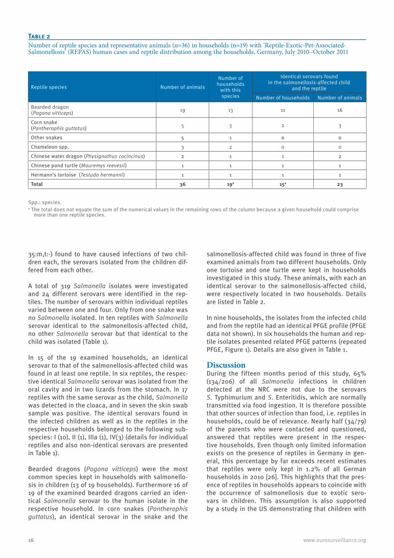
Bearded dragons (Pogona vitticeps) were the most common species kept in households with salmonello-sis in children (13 of 19 households). Furthermore 16 of 19 of the examined bearded dragons carried an iden-tical Salmonella serovar to the human isolate in the respective household. In corn snakes (Pantherophis guttatus), an identical serovar in the snake and the
salmonellosis-affected child was found in three of five examined animals from two different households. Only one tortoise and one turtle were kept in households investigated in this study. These animals, with each an identical serovar to the salmonellosis-affected child, were respectively located in two households. Details are listed in Table 2.
In nine households, the isolates from the infected child and from the reptile had an identical PFGE profile (PFGE data not shown). In six households the human and rep-tile isolates presented related PFGE patterns (repeated PFGE, Figure 1). Details are also given in Table 1.
DiscussionDuring the fifteen months period of this study, 65% (134/206) of all Salmonella infections in children detected at the NRC were not due to the serovars S. Typhimurium and S. Enteritidis, which are normally transmitted via food ingestion. It is therefore possible that other sources of infection than food, i.e. reptiles in households, could be of relevance. Nearly half (34/79) of the parents who were contacted and questioned, answered that reptiles were present in the respec-tive households. Even though only limited information exists on the presence of reptiles in Germany in gen-eral, this percentage by far exceeds recent estimates that reptiles were only kept in 1.2% of all German households in 2010 [26]. This highlights that the pres-ence of reptiles in households appears to coincide with the occurrence of salmonellosis due to exotic sero-vars in children. This assumption is also supported by a study in the US demonstrating that children with
Table 2Number of reptile species and representative animals (n=36) in households (n=19) with ‘Reptile-Exotic-Pet-Associated-Salmonellosis’ (REPAS) human cases and reptile distribution among the households, Germany, July 2010–October 2011
Reptile species Number of animals
Number of households
with this species
Identical serovars found in the salmonellosis-affected child
and the reptile
Number of households Number of animals
Bearded dragon (Pogona vitticeps) 19 13 11 16
Corn snake (Pantherophis guttatus) 5 3 2 3
Other snakes 5 1 0 0
Chameleon spp. 3 2 0 0
Chinese water dragon (Physignathus cocincinus) 2 1 1 2
Chinese pond turtle (Mauremys reevesii) 1 1 1 1
Hermann’s tortoise (Testudo hermanni) 1 1 1 1
Total 36 19a 15a 23
Spp.: species.a The total does not equate the sum of the numerical values in the remaining rows of the column because a given household could comprise
more than one reptile species.

17www.eurosurveillance.org
confirmed Salmonella infections had more contact to reptiles and cats in comparison to a control group [27].
In most published case reports on RAS, only reptile fae-cal samples or faecal swabs were used for Salmonella detection. In contrast, this study was designed to obtain as much information as possible on Salmonella serovars shed by the reptiles. Sampling of the oral cavity (and stomach) as well as the cloaca should pro-vide information on shedding via both orifices and if Salmonella are present within the whole digestive sys-tem, whereas sampling from the skin should demon-strate whether Salmonella may also be transmitted via direct contact with the animal. Repeated enrichment and culture was necessary to provide reliable informa-tion on the Salmonella status in the reptiles examined. Combining the molecular typing using PFGE with other available data, such as serological typing as in this study, is highly recommended for accurate analysis and comparison of samples [24].
The extensive sampling and testing protocol used in this study is a possible explanation for the high preva-lence of Salmonella (in 35 of 36 reptiles) found among
the reptiles investigated. Intermittent shedding of Salmonella in reptiles and the wide array of collection and sampling techniques have been proposed to be the main reason for the variability in detection rates [28]. None of the reptiles examined showed clinical signs indicative for salmonellosis. In consequence, a high prevalence of Salmonella in reptiles should generally be assumed, and reptiles should be considered posi-tive for Salmonella until the contrary has been proven, as reported previously [29].
Up to four different serovars were found within one rep-tile. Reptiles were frequently colonised with the same serovars within a given household. This indicates that Salmonella as a part of the normal flora can spread amongst individuals within captive reptile collections and therefore probably shed over long periods of time. These results are in accordance with observations that if one reptile carries Salmonella, nearly all other rep-tiles of the respective owner are also affected [5].
Most Salmonella isolates were found in cloacal swabs. However, since in some reptiles the identical serovar to the salmonellosis-affected child was only found on
Figure 1Patterns obtained by XbaI restriction and pulsed-field gel electrophoresis (PFGE) for isolates from children with ‘Reptile-Exotic-Pet-Associated-Salmonellosis’ (REPAS) (n=6) and reptiles (n=6) living in the same respective households
H: human isolate; R: reptile isolate.Minor PFGE pattern differences between human and reptile isolates from the same households are shown and indicated by arrows.
200
500
1,00
0 1,
500
2,00
0 80
0 60
0 35
0 30
0 25
0 15
0
100
60
40
20
kbp Salmonella serovar
Eastbourne
Eastbourne
Cotham
Cotham
Monschaui
Monschaui
Newport
Newport
IV 48:g,z51:-
IV 48:g,z51:-
Potsdam
Potsdam
Isolate source (household number)
H (1)
R (1)
H (3)
R (3)
H (6)
R (6)
H (9)
R (9)
H (14)
R (14)
R (17)
H (17)

18 www.eurosurveillance.org
the skin or the stomach, it can be assumed that shed-ding via the cloaca is intermittent. A negative cloacal or faecal sample will therefore not prove that the animal does not harbour Salmonella. Given the anatomy and behaviour of most reptiles, the presence of Salmonella on skin samples was not surprising but underlines the general transmission risk if handling the reptiles is not followed by appropriate hand washing. In conclusion, considering the various shedding sites, several sam-pling points should be used to increase the detection rate of Salmonella in reptiles.
In the majority (15/19) of the examined households, an identical serovar was found in the infected child and at least one reptile from the respective household. The epidemiological association between these isolates was first confirmed using biochemical typing. PFGE confirmed all reptiles, with either completely identical patterns (nine households) or only minor differences in up to two fragments indicating single genetic vari-ations (six households, Figure 1), as described in dis-ease outbreaks [24]. Most serovars found belonged to subspecies I and some have already been described as part of the reptile flora or even as potential human pathogens [2,8,28]. One serovar, S. Tennessee was iso-lated from four reptiles in three households. However, so far there seems to be no specific serovar that is of special zoonotic importance. In consequence, at the moment all Salmonella strains found in reptiles need to be considered to be potentially infectious for children.
An important finding of this study was that the reptile species involved could play an important role in trans-mission of Salmonella to children. Little data exists on the proportion of reptile species kept as pets in Germany. In a post-mortem survey, about 17% were water turtles, 34% tortoises, 22% snakes and 27% lizards (including 1.8% bearded dragons) [30]. In one of the authors’ clinic specialising in reptile medicine, about 12% of more than 1,000 reptiles presented in the period of the running study were bearded dragons. However, in this study, the majority (13/19) of all house-holds studied kept bearded dragons (Pogona vitticeps), and identical Salmonella strains to the salmonellosis-affected child were confirmed in 16 of 19 of these rep-tiles. In contrast, only one turtle and one tortoise were kept in the affected households studied. This result is in strong contrast to early reports of possible RAS in humans, where mainly turtles were suspected to be a source of infection. Interestingly, more recent case reports also indicate that bearded dragons present a risk for transmission of salmonellosis to young chil-dren [31,32].
Weiss et al. [12] reported a marked increase in infec-tions in infants caused by S. Tennessee in Germany in 2008 and indicated the possibility of bearded drag-ons being a natural reservoir for this serovar, as this species was kept in seven of 16 households affected and S. Tennessee was confirmed in three households. Furthermore, probable transmission from bearded
dragons has also recently been described in adult humans [33]. One explanation for the increasing num-ber of reports on bearded dragons as a source of Salmonella infection in humans in recent years might be that this species is becoming more popular as a pet, and that bearded dragons are more often handled and petted due to their peaceful nature compared to several other reptile species. Since nearly all children included in this study were too young to handle reptiles them-selves, it is likely that vectors, such as the parents or the environment may have played a role. Furthermore it is conceivable that bearded dragons naturally harbour a variety of potentially zoonotic Salmonella serovars. The fact that more than one serovar of Salmonella was isolated in 13 of a total of 19 bearded dragons supports this assumption.
Husbandry conditions and the households’ hygiene status were not assessed according to a protocol, as this was not agreed upon with the owners. However, the overall impression was that most of the house-holds visited demonstrated at least some deficits that needed corrections and advice, e.g. the reptile terrar-ium placed in the kitchen or in the same room as the child’s bed. Further investigations in the environment of the reptile and possible interactions between the reptile and the persons in the households could give important information on possible transmission paths. None of the owners was aware of any risk of infection due to reptiles before, and all were therefore either willing to improve the situation or decided not to keep reptiles any longer.
All publications cited above indicate that although infections attributed to exposure to reptiles and other exotic pets represent only a small proportion of all human salmonellosis cases, it is likely an underesti-mated and growing problem in Europe [34] and in the US [35] that deserves closer attention. In 2007 more than 500,000 reptiles were imported to Germany only via the Frankfurt/Main airport [36]. According to the European Surveillance System (TESSy) database, the number of infections affecting children younger than three years with serovars associated to reptiles also increased in the last years (Figure 2). Beside the focus on exotic serovars as in this study, it should be kept in mind that there is also a potential risk for the transmis-sion of S. Typhimurium and S. Enteriditis due to reptiles or as already described, via the reptile feed [6,7]. Also these common serotypes can be shedded by reptiles and therefore have a potential to cause salmonellosis in humans. For example, in this study S. Paratyphi B variant java was isolated from a corn snake. Although the identical serovar has not been found in the child from the respective household, this isolate belongs to phage type Worksop and is therefore of relevance for human infections.
From a terminological point of view, in addition to an earlier suggestion (RAS [37]), we propose that Salmonella infections related to reptiles as observed

19www.eurosurveillance.org
in this study be called ‘Reptile-Exotic-Pet-Associated-Salmonellosis’ (REPAS) to give an indication of a pos-sible source of exposure. The main argument for this proposal is that over the last years, the way of trading reptiles has changed considerably and this will prob-ably continue in the future. The main risk of Salmonella transmission from reptiles to humans is not due to European wild species, but to ‘exotic’ (non-native) rep-tile species, as the results of this study also demon-strate. Furthermore, following recent examinations, Salmonella shedding is higher in reptiles kept in captiv-ity in comparison to wild reptiles [5,28] and ‘pet’ rep-tiles are obviously in closer contact to humans. These arguments justify the inclusion of ‘exotic pet’ into the term describing the problem. The risk to human health connected to the reptile pet market has been high-lighted recently [38] and the accurate description of the problem using REPAS might be important to convey the problem in education and assist risk managers in giving recommendations to harmonise animal welfare and public health.
Concluding, emphasis on educational measures will be key to reduce the risk of Salmonella transmission from reptiles to children. Professionals dealing with reptiles (pet shop owners, veterinarians, breeders) should be aware of such a risk and, together with those dealing
with human health, should be responsible to inform more about this problem. Given the fact that most own-ers in this study were unaware of the risk of Salmonella transmission from reptiles, we recommend that, in pet shops, any new exotic pet owner be provided with appropriate information sheets on Salmonella prevention. In the authors’ opinion the risk of REPAS can easily be minimised, using a reasonable manage-ment protocol without the need to remove reptiles from households with young children.
Acknowledgements The authors thank François-Xavier Weill, Centre for Reference and Research on Salmonella, Enteric Bacterial Pathogens Unit, Institut Pasteur, Paris, for the confirmation of the unu-sual S. Newport. We thank the Member States in the FWD network for submitting detailed serovar data, and Johanna Takkinen, European Center for Disease Prevention and Control (ECDC) for the European data. Furthermore, we thank Marita Wahnfried, Susanne Kulbe, Dagmar Busse for skillful technical assistance, and Rachel Marschang, University of Hohenheim, for linguistic support.
Figure 2Occurrence of ‘Reptile-Exotic-Pet-Associated-Salmonellosis’ (REPAS) serovars in children less than three years-old in the European Union, 2007–2010
Salmonella Arizona includes subspecies IIIa and subspecies IIIb. Arrows indicate where REPAS cases have increased in 2010 compared to previous years.
Data from the European Surveillance System (TESSy) (29 May 2012).
0
10
20
30
40
50
60
70
80
90
100
Apap
a
Carm
el
Ealin
g
East
bour
ne
Mon
scha
ui
Ora
nien
burg
Pom
ona
Poon
a
Tenn
esse
e
Ariz
ona
Subs
p. II
Ia
Subs
p. II
Ib
Subs
p. IV
Num
ber o
f cas
es
Salmonella serovar
2007 2008 2009 2010

20 www.eurosurveillance.org
References1. Thomas AD, Forbes-Faulkner JC, Speare R, Murray C.
Salmonellosis in wildlife from Queensland. J Wildl Dis. 2001;37(2):229–38. http://dx.doi.org/10.7589/0090-3558-37.2.229 PMid:11310872
2. Woodward DL, Khakhria R, Johnson WM. Human salmonellosis associated with exotic pets. J. Clin Microbiol. 1997;35(11):2786-90. PMid:9350734 PMCid:PMC230062
3. Pasmans F, Martel A, Boyen F, Vandekerchove D, Wybo I, Van Immerseel F, et al. Characterization of Salmonella isolates from captive lizards. Vet Microbiol. 2005;110(3-4):285–91. http://dx.doi.org/10.1016/j.vetmic.2005.07.008 PMid:16153787
4. Kikillus KH, Gartrell BD, Motion E. Prevalence of Salmonella spp., and serovars isolated from captive exotic reptiles in New Zealand. N Z Vet. J. 2011;59(4):174-8. http://dx.doi.org/10.1080/00480169.2011.579246 PMid:21660846
5. Geue L, Löschner U. Salmonella enterica in reptiles of German and Austrian origin. Vet Microbiol. 2002;84 (1-2):79-91. http://dx.doi.org/10.1016/S0378-1135(01)00437-0
6. Harker KS, Lane C, De Pinna E, Adak GK. An outbreak of Salmonella Typhimurium DT191a associated with reptile feeder mice. Epidemiol Infect. 2011;139(8):1254-61. http://dx.doi.org/10.1017/S0950268810002281 PMid:20943001
7. Fuller CC, Jawahir SL, Leano FT, Bidol SA, Signs K, Davis C, et al. A multi-state Salmonella Typhimurium outbreak associated with frozen vacuum-packed rodents used to feed snakes. Zoonoses Public Health. 2008;55(8-10):481-7. PMid:18833597
8. Chiodini RJ, Sundberg JP. Salmonellosis in reptiles: a review. Am J Epidemiol. 1981;113(5): 494–9. PMid:7013476
9. Willis C, Wilson T, Greenwood M, Ward L. Pet reptiles associated with a case of salmonellosis in an infant were carrying multiple strains of Salmonella. J Clin Microbiol. 2002;40(12):4802–3. http://dx.doi.org/10.1128/JCM.40.12.4802-4803.2002 PMid:12454202 PMCid:PMC154656
10. Center for Disease Control and Prevention (CDC). Reptile-associated salmonellosis – selected states, 1998-2002. MMWR Morb Mort Wkly Rep. 2003;52(49):1206-9. PMid:14668712
11. Kaibu H, Iida K, Ueki S, Ehara H, Simasaki Y, Anzai H, et al. Salmonellosis of infants presumably originating from an infected turtle in Nagasaki, Japan. Jpn J Infect Dis. 2006;59(4):281. PMid:16936357
12. Weiss B, Rabsch W, Prager R, Tietze E, Koch J, Mutschmann F, et al. Babies and bearded dragons: sudden increase in reptile-associated Salmonella enterica serovar Tennessee infections, Germany 2008. Vector Borne Zoonotic Dis. 2011;11(9):1299–301. http://dx.doi.org/10.1089/vbz.2010.0239 PMid:21612527
13. Hernández E, Rodriguez JL, Herrera-León S, García I, de Castro V, Muniozguren N. Salmonella Paratyphi B var Java infections associated with exposure to turtles in Bizkaia, Spain, September 2010 to October 2011. Euro Surveill. 2012;17(25):pii= 20201. Available from: http://www.eurosurveillance.org/ViewArticle.aspx?ArticleId=20201 PMid:22748004
14. Warwick C, Lambiris AJ, Westwood D, Steedman C. Reptile-related salmonellosis. J R Soc Med. 2001;94(3):124-6. PMid:11285792 PMCid:PMC1297927
15. Mermin J, Hutwagner L, Vugia D, Shallow S, Daily P, Bender J, et al. Reptiles, amphibians, and human Salmonella infection: a population-based, case-control study. Clin Infect Dis. 2004;38 Suppl 3:S253-61. http://dx.doi.org/10.1086/381594 PMid:15095197
16. Stam F, Römkens TE, Hekker TAM, Smulders YM. Turtle-associated human salmonellosis. Clin Infect Dis. 2003;37(11):e167-9. http://dx.doi.org/10.1086/379612 PMid:14614690
17. Baby dies of Salmonella poona infection linked to pet reptile. Commun Dis Rep CDR Wkly. 2000;10(18):161.
18. Wisconsin Division of Public Health (DPH). Disease fact sheet series: Reptile associated salmonellosis. Madison: Wisconsin Department of Health Services; 2004. P-42082.
19. Böhme H, Fruth A, Rabsch W. Reptilien-assoziierte Salmonelleninfektionen bei Säuglingen und Kleinkindern in Deutschland. [Reptile-associated Salmonellosis in Infants in Germany]. Klin Padiatr. 2009;221(2):60-4. German. http://dx.doi.org/10.1055/s-0028-1112156 PMid:19263324
20. Grimont PA, Weill FX. Antigenic formulae of the Salmonella serovars. Paris: WHO Collaborating Centre for Reference and Research on Salmonella, Institut Pasteur; 2007.
21. Ribot EM, Fair MA, Gautom R, Cameron DN, Hunter SB, Swaminathan B, et al. Standardization of pulsed-field gel electrophoresis protocols for the subtyping of Escherichia coli O157:H7, Salmonella, and Shigella for PulseNet. Foodborne Pathog Dis. 2006;3(1):59-67. http://dx.doi.org/10.1089/fpd.2006.3.59 PMid:16602980
22. Van Belkum A, Tassios PT, Dijkshoorn L, Haeggman S, Cookson B, Fry NK, et al. Guidelines for the validation and application of typing methods for use in bacterial epidemiology. Clin Microbiol Infect. 2007;13 Supp 3:1–46. http://dx.doi.org/10.1111/j.1469-0691.2007.01786.x PMid:17716294
23. Goering RV. Pulsed field gel electrophoresis: A review of application and interpretation in the molecular epidemiology of infectious disease. Infect Genet Evol. 2010;10(7):866–75. http://dx.doi.org/10.1016/j.meegid.2010.07.023 PMid:20692376
24. Barrett TJ, Gerner-Smidt P, Swaminathan B. Interpretation of pulsed-field gel electrophoresis patterns in foodborne disease investigations and surveillance. Foodborne Pathog Dis. 2006;3(1):20-31. http://dx.doi.org/10.1089/fpd.2006.3.20 PMid:16602976
25. Sangal V, Harbottle H, Mazzoni CJ, Helmuth R, Guerra B, Didelot X, et al. Evolution and population structure of Salmonella enterica serovar Newport. J Bacteriol. 2010;92(24):6465-76. http://dx.doi.org/10.1128/JB.00969-10 PMid:20935094 PMCid:PMC3008538
26. Statistica. Anteil der Haushalte in Deutschland mit Haustieren nach Tierarten in den Jahren 2008 bis 2012. Statistica. [Accessed 19 Sep 2012]. German. Available from: http://de.statista.com/statistik/daten/studie/30153/umfrage/haustiere-heimtierhaltung-in-deutschen-haushalten-seit-2008/
27. Younus M, Wilkins MJ, Davies HD, Rahbar MH, Funk J, Nguyen C, et al. The role of exposures to animals and other risk factors in sporadic, non-typhoidal Salmonella infections in Michigan children. Zoonoses Public Health. 2010;57(7-8):e170-6. http://dx.doi.org/10.1111/j.1863-2378.2010.01324.x PMid:20202185
28. Scheelings TF, Lightfoot D, Holz P. Prevalence of Salmonella in Australian reptiles. J Wildlife Dis. 2011;47(1):1–11. http://dx.doi.org/10.7589/0090-3558-47.1.1 PMid:21269991
29. Pasmans F, Blahak S, Martel A, Pantchev N. Introducing reptiles into a captive collection: The role of the veterinarian. Vet J. 2008;175(1):53–68. http://dx.doi.org/10.1016/j.tvjl.2006.12.009 PMid:17346998
30. Sinn AD. Pathologie der Reptilien - eine retrospektive Studie [PhD thesis]. Munich: Ludwig-Maximilian-University; 2004. German.
31. Tabarani CM, Bennett NJ, Kiska DL, Riddell SW, Botash AS, Domachowske JB. Empyema of preexisting subdural hemorrhage caused by a rare Salmonella species after exposure to bearded dragons in a foster home. J Pediatr. 2010;156(2):322–3. http://dx.doi.org/10.1016/j.jpeds.2009.07.050 PMid:20105642
32. Haase R, Beier T, Bernstadt M, Merkel N, Bartnicki J. Neugeborenensepsis durch Salmonella Apapa nach Reptilienkontakt im Haushalt. [Neonatal infection with Salmonella apapa after contact with a reptile in the home]. Z. Geburtshilfe Neonatol. 2011; 215(2):86–8. German. http://dx.doi.org/10.1055/s-0031-1275308 PMid:21541908
33. Lowther SA, Medus C, Scheftel J, Leano F, Jawahir S, Smith K. Foodborne outbreak of Salmonella subspecies IV infections associated with contamination from bearded dragons. Zoonoses Public Health. 2011;58(8):560–6. http://dx.doi.org/10.1111/j.1863-2378.2011.01403.x PMid:21824356
34. Editorial team, Bertrand S, Rimhanen-Finne R, Weill FX, Rabsch W, Thornton L, et al. Salmonella infections associated with reptiles: The current situation in Europe. Euro Surveill. 2008;13(24):pii=18902. Available from: http://www.eurosurveillance.org/ViewArticle.aspx?ArticleId=18902

21www.eurosurveillance.org
35. Center for Disease Control and Prevention (CDC). Notes from the field: outbreak of salmonellosis associated with pet turtle exposures - United States, 2011. MMWR Morb.Mort Wkly Rep. 2012;61(4):79. PMid:22298304
36. Hatt JM, Fruth A, Rabsch W. Reptilienassoziierte Salmonellosen - aktuelle Informationen für Tierärztinnen und Tierärzte. [Reptile-associated salmonellosis –information update for veterinarians]. Tierärztliche Praxis. 2009;37(3):188-93. German.
37. De Jong B, Andersson Y, Ekdahl K. Effect of Regulation and Education on Reptile-associated Salmonellosis. Emerg Infect Dis. 2005;11(3):398–403. http://dx.doi.org/10.3201/eid1103.040694 PMid:15757554 PMCid:PMC3298264
38. Arena PC, Steedman C, Warwick C. Amphibian and reptile pet markets in the EU: An investigation and Assessment. Düsseldorf: Animal public e.V.; 2012. [Accessed 15 Dec 2012]. Available from: http://animal-public.de/wp-content/uploads/2012/04/ARPM2012_v131.pdf

22 www.eurosurveillance.org
News
ECDC launches the Geoportal for the European Environment and Epidemiology (E3) Network
B Sudre ([email protected])1
1. European Centre for Disease Prevention and Control (ECDC), Stockholm, Sweden
Citation style for this article: Sudre B. ECDC launches the Geoportal for the European Environment and Epidemiology (E3) Network. Euro Surveill. 2013;18(46):pii=20631. Available online: http://www.eurosurveillance.org/ViewArticle.aspx?ArticleId=20631
Article published on 14 November 2013
Infectious diseases are (re-) emerging in Europe. Tropical infectious diseases such as chikungunya and dengue fever have recently been transmitted in Europe in part due to global air traffic and climate change [1,2]. This calls for a proactive public health response to (re-) emerging infectious disease threats both in Europe and globally as well as careful monitoring of infectious dis-ease drivers. Such drivers are globalisation and envi-ronmental change, social and demographic change and public health system factors [3]. In order to address this need, the European Centre for Disease Prevention and Control (ECDC) has developed the European Environment and Epidemiology (E3) Network [4].
The long term goal of the E3 Network is to gain a bet-ter understanding of drivers of disease and the envi-ronmental and socio-economic determinants of public health threats. Such an understanding will strengthen the capacity for formulating public health intervention strategies, and increase the effectiveness of preven-tion and control measures and general policies within the European Union [5].
The E3 Network provides access to climatic/environ-mental geospatial data for epidemiologic analysis that
are currently collected and analysed by a variety of European agencies, public health institutes, as well as research organisations. The E3 Network will serve as a platform for collaborative dataset compilation, advanced analyses, data processing, and reporting and monitoring of data for risk assessments. It can also be used to enhance the rapid detection of emerg-ing public health threats driven by environmental fac-tors through prediction tools and forecasting models. To facilitate this objective, it will promote the exchange and sharing of the outputs of this collaborate effort through the E3 Geoportal service (https://e3geoportal.ecdc.europa.eu/).
References1. Semenza JC, Domanović D. Blood supply under threat. Nat Clim
Chang. 2013; 3(5): 432-5. 2. Semenza JC, Menne B. Climate change and infectious diseases
in Europe. Lancet Infect Dis. 2009; 9(6): 365-75. 3. Suk JE, Semenza JC. Future infectious disease threats to
Europe. Am J Public Health. 2011; 101(11): 2068-79. 4. Semenza JC, Sudre B, Oni T, Suk JE, Giesecke J. Linking
environmental drivers to infectious diseases: the European environment and epidemiology network. PLoS negl. trop. dis. 2013; 7(7): e2323.
5. Lindgren E, Andersson Y, Suk JE, Sudre B, Semenza JC. Public health. Monitoring EU emerging infectious disease risk due to climate change. Science. 2012; 336(6080): 418-9.


![Salmonella Typhimurium e variante monofasica 4,[5],12:i:- circolanti · Salmonella enterica serotype 4,5,12:i:-, an emerging serotype that represents multiple distinct clones. J Clin](https://img.pdfslide.net/doc/110x75/5fd6fde1df433a52d9650c6a/salmonella-typhimurium-e-variante-monofasica-4512i-salmonella-enterica-serotype.jpg)