Embed Size (px)
Citation preview
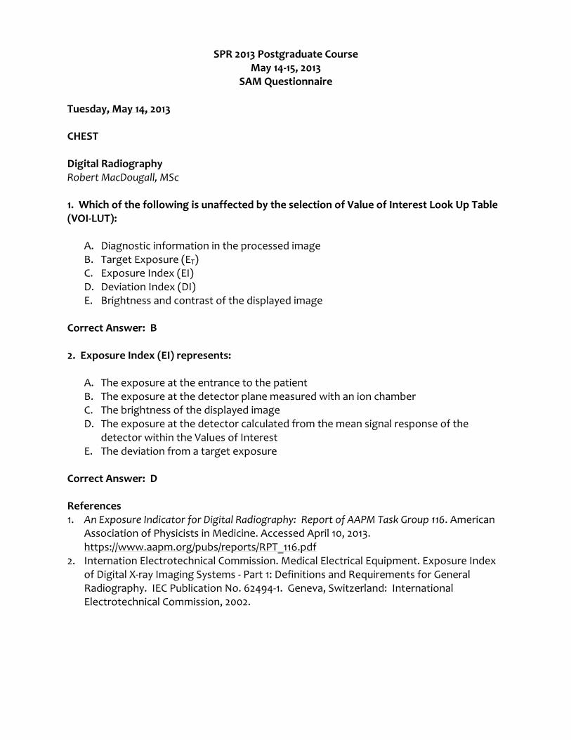
SPR 2013 Postgraduate Course May 14-15, 2013
SAM Questionnaire Tuesday, May 14, 2013 CHEST
Digital Radiography Robert MacDougall, MSc 1. Which of the following is unaffected by the selection of Value of Interest Look Up Table (VOI-LUT):
A. Diagnostic information in the processed image B. Target Exposure (ET) C. Exposure Index (EI) D. Deviation Index (DI) E. Brightness and contrast of the displayed image
Correct Answer: B 2. Exposure Index (EI) represents:
A. The exposure at the entrance to the patient B. The exposure at the detector plane measured with an ion chamber C. The brightness of the displayed image D. The exposure at the detector calculated from the mean signal response of the
detector within the Values of Interest E. The deviation from a target exposure
Correct Answer: D
References 1. An Exposure Indicator for Digital Radiography: Report of AAPM Task Group 116. American
Association of Physicists in Medicine. Accessed April 10, 2013. https://www.aapm.org/pubs/reports/RPT_116.pdf
2. Internation Electrotechnical Commission. Medical Electrical Equipment. Exposure Index of Digital X-ray Imaging Systems - Part 1: Definitions and Requirements for General Radiography. IEC Publication No. 62494-1. Geneva, Switzerland: International Electrotechnical Commission, 2002.
Functional Chest MR Imaging Hyun Woo Goo, MD, PhD
3. Which one of the followings is the LEAST likely limitation of thoracic MR imaging?
A. Low signal-to-noise ratio due to the low proton density of the lung B. Potential hazards from ionizing radiation C. Motion artifacts from respiratory motion and cardiac pulsation D. Relatively long examination time E. Susceptibility artifacts from multiple air-tissue interfaces
Correct Answer: B References 1. Puderbach M, Kauczor HU. Assessment of lung function in children by cross-sectional
imaging: techniques and clinical applications. Pediatr Radiol 2006;36:192-204. 2. Lee EY. Advancing CT and MR imaging of the lungs and airways in children: imaging into
practice. Pediatr Radiol 2008;38 Suppl 2:S208-S212. 3. Goo HW. Advanced functional thoracic imaging in children: from basic concepts to clinical
applications. Pediatr Radiol 2013;43:262-268. 4. Which one of the followings is the MR pulse sequence most commonly used for real-time respiratory dynamic imaging?
A. Balanced steady-state free precession (SSFP) B. T1-weighted In-phase gradient echo (GE) C. Short tau inversion-recovery (STIR) D. Ultra-short echo time (UTE) E. Velocity encoded cine (VEC)
Correct Answer: A References 1. Lee CH, Goo JM, Kim YT, et al. The clinical feasibility of using non-breath-hold real-time
MR-echo imaging for the evaluation of mediastinal and chest wall tumor invasion. Korean J Radiol 2010;11:37-45.
2. Goo HW. Regional and whole-body imaging in pediatric oncology. Pediatr Radiol 2011;41 Suppl1:S186-S194.
3. Goo HW. Advanced functional thoracic imaging in children: from basic concepts to clinical applications. Pediatr Radiol 2013;43:262-268.

PE Evaluation in Children: What is New? Edward Y. Lee, MD, MPH
5. What is the current imaging modality of choice for evaluating pediatric patients with clinically suspected pulmonary embolism?
A. Chest radiograph B. Computed Tomography Pulmonary Angiography (CTPA) C. Ventilation / Perfusion scan D. Conventional catheter based pulmonary angiography
Correct Answer: B 6. Which one of following risk factors has a statistically significant association with the presence of unsuspected pulmonary emboli on routine thoracic MDCT examinations of pediatric oncology patients?
A. Surgery within 1 month of MDCT B. Prior history of transplant C. Radiation therapy within 1 month of MDCT D. Underlying coagulation disorder
Correct Answer: D References 1. Kritsaneepaiboon S, Lee EY, Zurakowski D, Strauss KJ, Boiselle PM. MDCT pulmonary
angiography evaluation of pulmonary embolism in children. AJR Am J Roentgenol. 2009; 192(5): 1246 - 1252.
2. Lee EY, Kritsaneepaiboon S, Arellano CM, Grace FR, Zurakowski D, Boiselle PM. Unsuspected pulmonary emboli in pediatric oncology patients: detection with MDCT. AJR Am J Roentgenol. 2010; 194(5); 1216 - 1222.
Imaging of Acquired Thoracic Cardiovascular Diseases Beverley Newman, MD, FACR
7. The accompanying image is a frontal chest radiograph in an 11yo girl with prolonged fever of unknown origin and an elevated erythrocyte sedimentation rate (ESR). Based on the most likely diagnosis, what do you consider the most appropriate next imaging study?
A. No further imaging, the chest radiograph is normal B.MR angiogram to evaluate for congenital aortic coarctation C. MRI and MR angiogram to evaluate for vasculitis D. Functional MRI to evaluate for cardiomyopathy E. Gated CT angiogram to evaluate for coronary abnormality
Correct Answer: C References 1. Nastri MV, Baptista LPS, Baroni RH, et al: Gadolinium-enhanced three-dimensional MR
angiography of Takayasu arteritis. Radiographics 2004;24:773-786 2. Weyand CM, Goronzy JJ: Medium- and large-vessel vasculitis. N Engl J Med 2003;349:160-
169 3. Mason JC (2010) Takayasu arteritis--advances in diagnosis and management. Nat Rev
Rheumatol 6 (7):406-415. 4. Tann OR, Tulloh RM, Hamilton MC (2008) Takayasu's disease: a review. Cardiol Young 18
(3):250-259. 5. Aluquin VP, Albano SA, Chan F, et al. (2002) Magnetic resonance imaging in the diagnosis
and follow up of Takayasu's arteritis in children. Ann Rheum Dis 61 (6):526-529. 6. Towbin A, Newman B, In Slovis T ( ed) Caffeys Pediatric Diagnostic Imaging. Cardiac
Involvement by Systemic Diseases 11th edition, Mosby; 2007. Pg1684 8. Current advantages of MR over CT cardiovascular imaging include all of the following EXCEPT:
A. No exposure to ionizing radiation B. Better spatial resolution C. Superior for cardiac functional and viability assessment D. Vascular flow quantification E. Contrast safety and utility of non contrast imaging
Correct Answer: B References 1. Chan FP. MR and CT imaging of the pediatric patient with structural heart disease. Semin
Thorac Cardiovasc Surg Pediatr Card Surg Annu 2009; 12(1): 99–105 2. Puranik R, Muthurangu V, Celermajer DS et al. Congenital heart disease and multi-
modality imaging. Heart Lung Circ 2010; 19(3): 133–144 3. Taylor AM. Cardiac imaging: MR or CT? Which to use when. Pediatr Radiol 2008; 38(Suppl
3): S433–S438 4. Walker MF, Souza SP, Dumoulin CL. Quantitative flow measurement in phase contrast
MR angiography. J Comput Assist Tomogr 1988; 12:304–313 5. Newman B, Vasanawala SS. Point/counterpoint: dose related issues in cardiac CT
imaging. Pediatr Radiol 2011 Sep; 41(Suppl 2): 528-33

Interesting Cases R. Paul Guillerman, MD 9. The likelihood that a lung cyst represents a pleuropulmonary blastoma rather than a congenital pulmonary airway malformation is increased by the presence of which of the following conditions?
A. Spontaneous pneumothorax B. Multifocal lung cysts C. Cystic nephroma D. All of the above E. None of the above
Correct Answer: D References 1. Priest JR, Williams GM, Hill DA, et al (2009) Pulmonary cysts in early childhood and the
risk of malignancy. Pediatr Pulmonol 44:14-30 2. Slade I, Bacchelli C, Davies H, et al (2011) DICER1 syndrome – clarifying the diagnosis,
clinical features and management implications of a pleiotropic tumor predisposition syndrome. J Med Genet 48:273-278
10. Which of the following conditions is not known to be associated with pulmonary alveolar growth abnormalities?
A. Oligohydramnios B. Congenital heart disease C. Cystic fibrosis D. Prematurity E. Pulmonary interstitial glycogenosis
Correct Answer: C References 1. Deutsch GH, Young LR, Deterding RR, et al (2007) Diffuse lung disease in young children.
Application of a novel classification scheme. Am J Respir Crit Care Med 176:1120-1128 2. Guillerman RP, Brody AS (2011) Contemporary perspectives on pediatric diffuse lung
disease. Radiol Clin N Am 49:847-868 11. Which of the following statements about pulmonary veno-occlusive disease is true?
A. Distinction from primary pulmonary arterial hypertension is usually possible on the basis of the clinical presentation
B. Centrilobular ground-glass opacities, septal thickening and lymphadenopathy are the most suggestive imaging features
C. Pulmonary vasodilators are the safest and most effective treatment D. Prognosis is favorable with medical therapy E. All of the above

Correct Answer: B References 1. Montani D, Price LC, Dorfmuller P, et al (2009) Pulmonary veno-occlusive disease. Eur
Respir J 33:189-200 2. Resten A, Maitre S, Humber M, et al (2004) Pulmonary hypertension: CT of the chest in
pulmonary venoocclusive disease. AJR Am J Roentgenol 183:65-70
NEURORADIOLOGY Optimizing Head US Imaging Lisa H. Lowe, MD, FAAP
12. A 5-week-old term infant undergoes head sonogram. Which sagittal image(s) of the peripheral cortex is/are abnormal?
A. Right B. Left C. Both D. Neither
Correct Answer: A References 1. Lowe LH, Bailey Z. State-of-the-Art Cranial Sonography: Part 1, Modern Techniques and
Image Interpretation. AJR Am J Roentgenol 2011; 196:1028-33; PMID 21512067 2. Lowe LH, Bailey Z. State-of-the-Art Cranial Sonography: Part 2, Pitfalls and Variants. AJR
Am J Roentgenol 2011; 196:1034-9; PMID 21512068
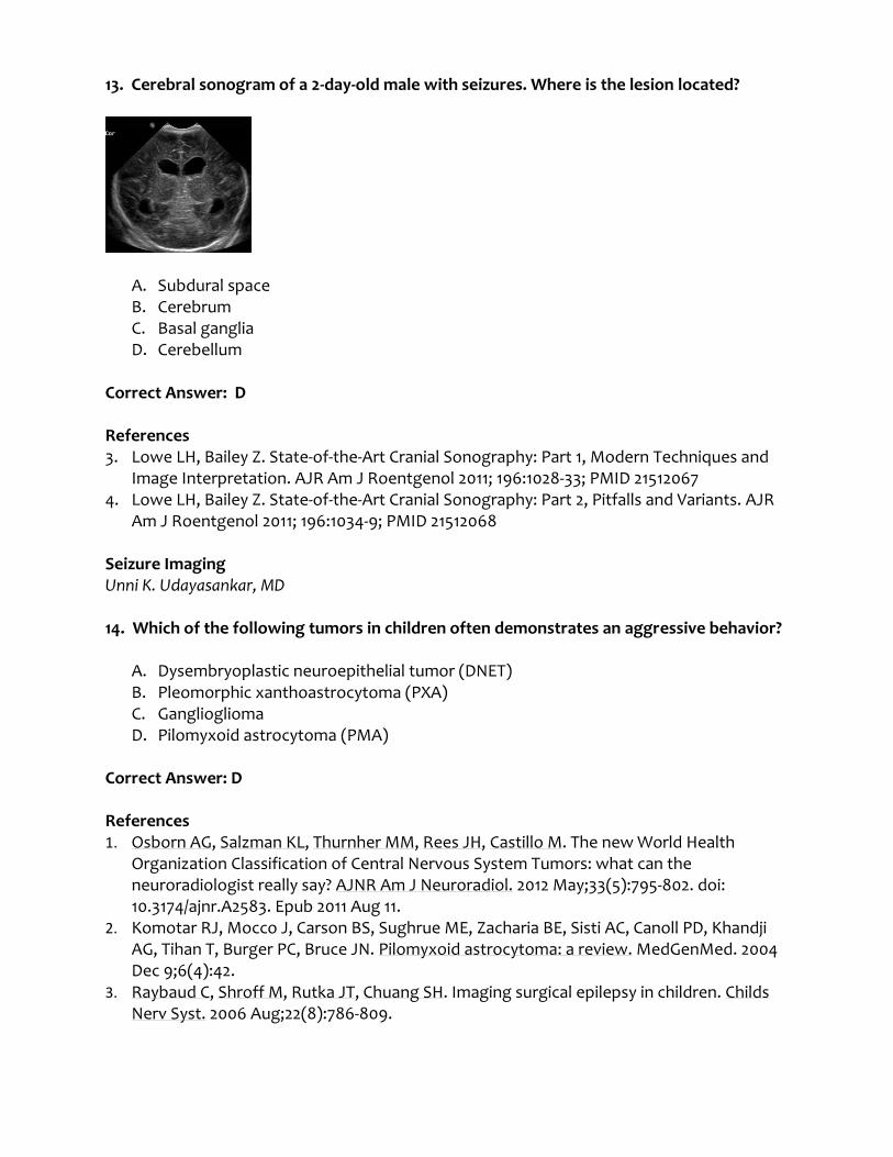
13. Cerebral sonogram of a 2-day-old male with seizures. Where is the lesion located?
A. Subdural space B. Cerebrum C. Basal ganglia D. Cerebellum
Correct Answer: D References 3. Lowe LH, Bailey Z. State-of-the-Art Cranial Sonography: Part 1, Modern Techniques and
Image Interpretation. AJR Am J Roentgenol 2011; 196:1028-33; PMID 21512067 4. Lowe LH, Bailey Z. State-of-the-Art Cranial Sonography: Part 2, Pitfalls and Variants. AJR
Am J Roentgenol 2011; 196:1034-9; PMID 21512068 Seizure Imaging Unni K. Udayasankar, MD 14. Which of the following tumors in children often demonstrates an aggressive behavior?
A. Dysembryoplastic neuroepithelial tumor (DNET) B. Pleomorphic xanthoastrocytoma (PXA) C. Ganglioglioma D. Pilomyxoid astrocytoma (PMA)
Correct Answer: D References 1. Osborn AG, Salzman KL, Thurnher MM, Rees JH, Castillo M. The new World Health
Organization Classification of Central Nervous System Tumors: what can the neuroradiologist really say? AJNR Am J Neuroradiol. 2012 May;33(5):795-802. doi: 10.3174/ajnr.A2583. Epub 2011 Aug 11.
2. Komotar RJ, Mocco J, Carson BS, Sughrue ME, Zacharia BE, Sisti AC, Canoll PD, Khandji AG, Tihan T, Burger PC, Bruce JN. Pilomyxoid astrocytoma: a review. MedGenMed. 2004 Dec 9;6(4):42.
3. Raybaud C, Shroff M, Rutka JT, Chuang SH. Imaging surgical epilepsy in children. Childs Nerv Syst. 2006 Aug;22(8):786-809.
15. Which of the following statements about focal cortical dysplasia (FCD) is true?
A. Most children with FCD do not benefit from epilepsy surgery B. Non-Taylor types are associated with dysmorphic neurons and balloon cells on
pathological examination C. Tuberous sclerosis and hemimegalencephaly are considered variants of FCD D. Contrast enhancement and focal mass effect are often present on MRI.
Correct Answer: C References 1. Barkovich AJ, Raybaud CA (ed) Pediatric Neuroimaging. Congenital malformations of the
Brain and Skull 5th edition, LWW; 2012. Pg 403-407. 2. Raybaud C, Shroff M, Rutka JT, Chuang SH. Imaging surgical epilepsy in children. Childs
Nerv Syst. 2006 Aug;22(8):786-809. 3. Lortie A, Plouin P, Chiron C, Delalande O, Dulac O. Characteristics of epilepsy in focal
cortical dysplasia in infancy. Epilepsy Res. 2002 Sep;51(1-2):133-45. 4. Hauptman JS, Mathern GW. Surgical treatment of epilepsy associated with cortical
dysplasia: 2012 update. Epilepsia. 2012 Sep;53 Suppl 4:98-104. Advances in Brain Tumor Imaging Ashok Panigrahy, MD 16. Which best characterizes hypercellular brain tumor:
A. hyperdensity on CT, hypointensity on T2 and decreased ADC B. hypodensity on CT, hypointensity on T2 and decreased ADC C. hyperdensity on CT, hyperintensity on T2 and decreased ADC D. hyperdensity on CT, hypointensity on T2 and increased ADC
Correct Answer: A References 1. Panigrahy A, Blüml S. Neuroimaging of pediatric brain tumors: from basic to advanced
magnetic resonance imaging (MRI). J Child Neurol. 2009 Nov;24(11):1343-65 17. In the modern era of targeted molecular therapies and immunotherapies which can be used to characterize pseudoprogression?
A. intra-tumoral hemorrhage B. tumor recurrence C. heterogeneous tumor response D. leptomeningeal metastases E. biopsies
Correct Answer: C References

1. Hygino da Cruz LC Jr, Rodriguez I, Domingues RC, et al Pseudoprogression and pseudoresponse: imaging challenges in the assessment of posttreatment glioma AJNR Am J Neuroradiol 2011 Dec; 32 (11): 1978-85.
2. Okada H, Pollack IF. Do we need novel radiologic response criteria for brain tumor immunotherapy? Expert Rev Neurother. 2011 May; 11(5):619-22.
Interesting Cases Yutaka Sato, MD, PhD 18. Which one of the following structures is more often affected in the middle interhemispheric variant (MIV) when compared to classic holoprosencephaly (HPE)?
A. Body of the corpus callosum B. Hypothalamus C. Anterior cerebral fissure D. The 3rd ventricle E. Ventral frontal cortex
Correct Answer: A References 1. Simon E, Hevner R, Pinter J, et al; The middle interhemispheric variant of
holoprosencephaly. AJNR 2002; 23:151-156. 2. Lewis A, Simon E, Barkovich A, et al; Middle interhemispheric variant of
holoprosencephaly: a distinct cliniconeuroradiologic subtype. Neurology 2002; 59:1860-1865.
3. Pulitzer S, Simon E, Crombleholme T, Golden J; Prenatal MR findings of the middle interhemispheric variant of holoprosencephaly. AJNR 2004: 25:1034-1036.
19. Which of the following statements regarding neonatal alloimmune thrombocytopenia (NAIT) is incorrect?
A. NAIT is caused by paternal-fetal antigen incompatibility. B. NAIT accounts for approximately 9% of neonatal thrombocytopenia. C. Intracranial hemorrhage occurs in 10 – 30% of NAIT, half of which occur in utero. D. Parenchymal hemorrhage, extraaxial hemorrhage, intraventricular hemorrhage and
porencephalic cysts are all seen in NAIT. E. There is no cost-effective screening program of primiparous women and neonates
for this disease. Correct Answer: A References 1. Dale S, Coleman L; Neonatal alloimmune thrombocytopenia: antenatal and postnatal
imaging findings in the pediatric brain. AJNR 2002; 23:1457-1465. 2. Pacheco L, Berkowitz R, Moise K, et al; Fetal and neonatal alloimmune thrombocytopenia.
Obstet Gynecol 2011; 118:1157-1163.


20. All of the below statements regarding infections of the deep neck are correct except:
A. The danger space is dorsal to the retropharyngeal space and ventral to the prevertebral space.
B. The danger space is separated from the retropharyngeal space by the alar fascia and from the prevertebral space by the prevertebral fascia.
C. The danger space extends from the skull base to the coccyx. D. Retropharyngeal space abscess most commonly originates from lymphadenitis and is
seen predominantly in infants. E. Parapharyngeal and peritonsillar abscesses are more common among adults and
older children. Correct Answer: C References 1. Hedge A, Mohan S, Lim WEH; Infections of the deep neck spaces. Singapore Med J 2012;
53:305-311. GASTROINTESTINAL Abdominal Applications of DWI Rutger A.J. Nievelstein, MD
21. The most important advantage of both a respiratory triggered and a free-breathing DWI sequence over a breath-hold DWI sequence in the abdomen is:
A. the possibility to obtain only thick slices B. the possibility to apply a higher number of b-values C. the lower susceptibility to motion artifacts and volume averaging D. the shorter scan time
Correct Answer: B References 1. Bittencourt LK, Matos C, Coutinho AC. Diffusion-weighted magnetic resonance imaging
in the upper abdomen: technical issues and clinical applications. Magn Reson Imaging Clin N Am 2011;19:111-131.
2. Kwee TC, Takahara T, Koh DM, et al. Comparison and reproducibility of ADC measurements in breathhold, respiratory triggered, and free-breathing diffusion-weighted MR imaging of the liver. J Magn reson Imaging 2008;28(5): 1141-1148.
22. When DWI of the abdomen is primarily used for visual assessment of pathology typically high b-values are used. However, in the liver the use of low b-value DWI is preferred for lesion detection, because:
A. low b-value DWI is less prone to cardiac motion-induced signal loss B. in low b-value DWI the background is better suppressed than in high b-value DWI C. low b-value DWI allows using shorter echo times

D. low b-value DWI is more effective in suppressing the vasculature than high b-value DWI
Correct Answer: A References 1. Coenegrachts K, Delanote J, Ter Beek L, et al. Improved focal liver lesion detection:
comparison of single-shot diffusion weighted echo planar and single-shot T2 weighted turbo spin echo techniques. Br J Radiol 2007;80:524–531.
2. Goshima S, Kanematsu M, Kondo H, et al. Diffusion-weighted imaging of the liver: optimizing b value for the detection and characterization of benign and malignant hepatic lesions. J Magn Reson Imaging 2008;28:691–697.
3. Takahara T, Kwee TC. Low b-value diffusion-weighted imaging: Emerging applications in the body. J Magn Reson Imaging 2012;35:1266–1273.
Update on MR Contrast Agents and Applications Shreyas S. Vasanawala, MD, PhD 23. Which of the following agents is a conventional extracellular agent?
A. Gadofosveset B. Gadoxetate C. Gadobutrol D. Ferumoxytol E. None of the above
Correct Answer: C 24. Which agent reversibly binds to albumin?
A. Gadofosveset B. Gadoxetate C. Gadobutrol D. Ferumoxytol E. None of the above
Correct Answer: A References 1. MS-325: albumin-targeted contrast agent for MR angiography. 2. Lauffer RB, Parmelee DJ, Dunham SU, Ouellet HS, Dolan RP, Witte S, McMurry TJ,
Walovitch RC. Radiology. 1998 May;207(2):529-38 3. Blood pool contrast agents for cardiovascular MR imaging. Kroft LJ, de Roos A. J Magn
Reson Imaging. 1999 Sep;10(3):395-403. 4. Targeted MRI contrast agents for pediatric hepatobiliary disease. Courtier JL, Perito ER,
Rhee S, Tsai P, Heyman MB, MacKenzie JD. J Pediatr Gastroenterol Nutr. 2012 Apr;54(4):454-62
Rex Shunt Interventions James S. Donaldson, MD 25. The Rex Shunt is appropriate for which of the following:
A. Abernethy malformation Type I B. A complication of umbilical venous catheterization C. Veno-occlusive disease D. Budd Chiari E. Alagilles syndrome
Correct Answer: B
26. The following represent advantages of the Rex shunt over a Warren shunt EXCEPT:
A. Reduced bleeding from esophageal and gastric varices B. Reduced incidence of hepatopulmonary syndrome C. Improved portal venous blood flow D. Improved cognition E. Improved BMI
Correct Answer: A
27. Indications for IR investigation of a Rex shunt include all EXCEPT:
A. Thrombocytopenia B. A suspected stenosis by US, CT, or MR imaging C. Splenomegaly D. Recurrent UGI bleeding E. Lower extremity edema
Correct Answer: E
References 1. J. de Ville de Goyet, et al. Direct bypassing of extrahepatic portal vein obstruction in
children: A new technique for combined hepatic revascularization and managing portal hypertension. J Pediatr Surg 33 (1998), pp. 597-601
2. Dasgupta R, Roberts E, Superina RA, Kim PC Effectiveness of Rex shunt in treatment of portal hypertension. J Pediatr Surg 41 (2006), pp. 108-112
3. Lautz T, Kim ST, Donaldson JS, Superina R. Outcomes of percutaneous interventions for managing stenosis of meso-Rex bypass in patients with extrahepatic portal vein obstruction. JVIR 2012; 23:377-383

Interesting Cases Marta Hernanz-Schulman, MD, FACR
28. Regarding chronic or intermittent volvulus, which of the following presentations is NOT characteristic?
A. Failure to thrive B. Malabsorption C. Intermittent abdominal pain D. Distal bowel obstruction
Correct Answer: D References 1. Spigland N, Brandt ML, Yazbeck S. Malrotation presenting beyond the neonatal period.
J Pediatr Surg 1990;25(11):1139-1142 2. Applegate KE Anderson JM, Klatte EC. Intestinal malrotation in children: a problem-
solving approach to the Upper GI series. Radiographics 2006;26:1485-1500 3. Strouse PJ. Disorders of intestinal rotation. Pediatr Radiol 2004;34:837-851 4. Berdon WE. The diagnosis of malrotation and volvulus in the older child and adult: a trap
for radiologists. Pediatr Radiol 1995;25:101-103 29. Typical characteristics of transient small bowel intussusception include all of the following, EXCEPT:
A. Segment length < 4 cm B. Diameter < 18 mm C. Small lead point D. Preserved wall motion
Correct Answer: C References 1. Kim JH US features of transient small bowel intussusception in pediatric patients.
Korean J Radiolo 2004;5:178-184 2. Strouse PJ, DiPietro MA, Saez F. Transient small bowel intussusception in children on CT.
Pediatr Radiol 2003;33:316-320 30. Tumors occurring in patients with dysgenetic gonads include the following EXCEPT:
A. Gonadoblastoma B. Juvenile granulosa cell tumor C. Wilms tumor D. Ovarian dermoid
Correct Answer: D

References 1. Young RH, Lawrence WD, Scully RE. Juvenile granulosa cell tumor- another neoplasm
associated with abnormal chromosomes and ambiguous genitalia The American Journal of Surgical Pathology 1985; 9(10):737-743
2. Vhsbhsn GB, Parra DA, Oudjhane, K et al. Imaging of ambiguous genitalia: classification and diagnostic approach. Radiographics 2008;28:1891-1904
GENITOURINARY Update on Contrast Material Use in Children Jonathan R. Dillman, MD
31. A 15 year-old boy undergoes a CT examination with intravenous iodinated contrast material. The patient becomes unresponsive two minutes after the scan and is noted to have diffuse skin erythema. Initial vital signs confirm a blood pressure of 68/42 and a heart rate of 125. After calling for help, what is the next most appropriate step in the medical management of this patient?
A. Administer IV diphenhydramine (Benadryl) and IV corticosteroid B. Administer inhaled B-agonist medication (e.g., albuterol) C. Administer IV atropine D. Administer IM epinephrine
Correct Answer: D Reference 1. ACR Manual on contrast media, volume 8.
32. Regarding nephrogenic systemic fibrosis (NSF) in the pediatric population, which of the following is TRUE?
A. NSF has been documented mostly in children with normal renal function. B. Patients with an estimated glomerular filtration rate (eGFR) between 45 and 60
ml/min are considered to be at-risk for NSF. C. Macrocyclic gadolinium chelates are less likely to be associated with the
development of NSF than linear gadolinium chelates. D. Macrocyclic gadolinium chelates absolutely must be avoided in children determined
to be at-risk for NSF, even if the benefits of imaging outweigh the risk of NSF.
Correct Answer: C Reference 1. ACR Manual on contrast media, volume 8.

Update on UTI Imaging Assessment: ACR and AAP Guidelines Boaz Karmazyn, MD 33. What is the most common acquired end stage renal disease in children in North America?
A. Renal scarring from pyelonephritis B. Focal segmental glomerulosclerosis C. Obstructive uropathy D. Renal aplasia/hypoplasia/dysplasia
Correct Answer: B References 1. Mitsnefes M, Ho PL, McEnery PT. Hypertension and progression of chronic renal
insufficiency in children: a report of the North American Pediatric Renal Transplant Cooperative Study (NAPRTCS). J Am Soc Nephrol. 2003 Oct;14(10):2618-2622.
2. Novak TE, Mathews R, Martz K, Neu A. NAPRTCS 2010 Annual Transplant Report https://web.emmes.com/study/ped/annlrept/2010_Report.pdf
3. Reuss A, Wladimiroff JW, Niermeijer MF. Antenatal diagnosis of renal tract anomalies by ultrasound. Pediatr Nephrol 1987;1:546-552.
4. Montini G, Tullus K, Hewitt I. Febrile urinary tract infections in children. N Engl J Med. 2011 Jul 21;365(3):239-250
34. What is correct about acute phase (within 2 weeks) DMSA in febrile UTI?
A. Best study to detect renal scaring B. Accurate study in detecting dilated vescicoureteral reflux C. Positive in 50%-60% of patients D. Pyelonephritis most often will result in a scar
Correct Answer: C References 1. Shaikh N, Ewing AL, Bhatnagar S, Hoberman A. Risk of renal scarring in children with a
first urinary tract infection: a systematic review. Pediatrics. 2010;126(6):1084-91 2. Mantadakis E, Vouloumanou EK, Georgantzi GG, Tsalkidis A, Chatzimichael A, Falagas ME.
Acute Tc-99m DMSA scan for identifying dilating vesicoureteral reflux in children: a meta-analysis. Pediatrics. 2011;128(1):e169-79.

Advances in Pediatric Urosonography Kassa Darge, MD, PhD 35. The “twinkling artifact” is a color Doppler artifact that can be used to better depict stones in the urinary tract. The most conspicuous and clear visualization of the “twinkling artifact” can be best achieved by one of the following measures:
A. Switching on harmonic imaging B. Increasing the pulse repetition frequency (PRF) to the maximum C. Making the color Doppler window as small as possible D. Placing the focus at the level of the echogenic structure to be evaluated E. None of the above
Correct Answer: B References 1. Darge K, Heidemeier A US diagnosis of urinary tract calculi in children using the twinkling
sign. Ultrasound 2006; 14: 167-173 2. Mitterberger M, Aigner F, Pallwein L, Pinggera GM, Neururer R, Rehder P, Frauscher F.
Sonographic detection of renal and ureteral stones. Value of the twinkling sign. Int Braz J Urol 2009 Sep-Oct;35(5):532-9
3. Ripollés T, Martínez-Pérez MJ, Vizuete J, Miralles S, Delgado F, Pastor-Navarro T. Sonographic diagnosis of symptomatic ureteral calculi: usefulness of the twinkling artifact. Abdom Imaging 2012 Aug 7. [online]
36. In contrast enhanced voiding urosonography, US contrast agent is administered into the bladder for detection of vesicoureteric reflux. The advantages over fluoroscopic voiding cystourethrography include the following except:
A. Elimination of radiation exposure B. Higher detection rate of vesicoureteric reflux C. Better detection of small amount of US contrast agent in a dilated ureter/pelvis D. No need for use of bladder catheter E. Improved patient comfort as it is conducted in an US room setting
Correct Answer: D
References 1. Darge K (2008) Voiding urosonography with ultrasound contrast agents for the diagnosis
of vesicoureteric reflux in children. I. Procedure. Pediatr Radiol 38:40-53 2. Darge K (2008) Voiding urosonography with US contrast agents for the diagnosis of
vesicoureteric reflux in children. II. Comparison with radiological examinations. Pediatr Radiol 38:54-63
3. Riccabona M, Avni FE, Damasio MB, Ording-Müller LS, Blickman JG, Darge K, Lobo ML, Papadopoulou F, Vivier PH, Willi U (2012) ESPR Uroradiology Task Force and ESUR Paediatric Working Group Imaging recommendations in paediatric uroradiology, Part V: childhood cystic kidney disease, childhood renal transplantation and contrast-enhanced ultrasonography in children. Pediatr Radiol 42:1275-1283.

Imaging of Mullerian Anomalies S. Pinar Karakas, MD
A B
C D 37. Above MRI images (T2 coronal (a), T2 axial (b), T1 fat sat axial before (c) and after contrast (d)) demonstrate a non-communicating cavitated unicornuate uterus. Which of the following is TRUE regarding a unicornuate uterus?
A. The majority of the unicornuate uterus cases are isolated without a rudimentary horn.
B. Associated renal anomalies are uncommon in unicornuate uterus. C. A unicornuate uterus is not compatible with a viable offspring. D. Elongation failure of one Müllerian duct which cannot reach to the urogenital sinus
results in unicornuate uterus. E. A cavitary communicating rudimentary horn does not require surgical correction.
Correct Answer: D References 1. Behr SC, Courtier JL, Qayyum A. Imaging of müllerian duct anomalies. Radiographics.
2012 Oct;32(6):E233-50. doi: 10.1148/rg.326125515. PubMed PMID: 23065173. 2. Dykes TM, Siegel C, Dodson W. Imaging of congenital uterine anomalies: review and self-
assessment module. AJR Am J Roentgenol. 2007 Sep;189(3 Suppl):S1-10. PubMed PMID: 19642254.
3. Junqueira BL, Allen LM, Spitzer RF, Lucco KL, Babyn PS, Doria AS. Müllerian duct anomalies and mimics in children and adolescents: correlative intraoperative assessment
with clinical imaging. Radiographics. 2009 Jul-Aug;29(4):1085-103. PubMed PMID: 19605658.
4. Troiano RN, McCarthy SM. Mullerian duct anomalies: imaging and clinical issues. Radiology. 2004 Oct;233(1):19-34. Epub 2004 Aug 18. Review. PubMed PMID: 15317956.
38. Which of the following statements is true?
A. Septate uterus & uterus didelphys 0ccur due to failed lateral fusion of the Mullerian ducts.
B. Shape of the uterine fundus is the most important determinant for correct classification of a uterus with duplicated cavity.
C. Septate uterus is the least common form of Mullerian malformation. D. Incomplete degeneration of embryologic septum results in bicornuate uterus. E. Any uterine duplication with double cervix is in keeping with uterus didelphys.
Correct Answer: D References 1. Behr SC, Courtier JL, Qayyum A. Imaging of müllerian duct anomalies. Radiographics.
2012 Oct;32(6):E233-50. doi: 10.1148/rg.326125515. PubMed PMID: 23065173. 2. Junqueira BL, Allen LM, Spitzer RF, Lucco KL, Babyn PS, Doria AS. Müllerian duct
anomalies and mimics in children and adolescents: correlative intraoperative assessment with clinical imaging. Radiographics. 2009 Jul-Aug;29(4):1085-103. PubMed PMID: 19605658.
3. Troiano RN, McCarthy SM. Mullerian duct anomalies: imaging and clinical issues. Radiology. 2004 Oct;233(1):19-34. Epub 2004 Aug 18. Review. PubMed PMID: 15317956.
4. Marcal L, Nothaft MA, Coelho F, Volpato R, Iyer R. Mullerian duct anomalies: MR imaging. Abdom Imaging. 2011 Dec;36(6):756-64.
Interesting Cases D. Gregory Bates, MD
39. The ureteral bud arises from which segment of the distal Wolffian duct?
A. MB (metanephric blastema) B. PVP (proximal vas precursor) C. UGS (urogenital sinus) D. CMD (common mesonephric duct) E. UMD (upper mesonephric duct)
Correct Answer: D References 1. Mackie GG, Stephens FD; Duplex kidneys: a correlation of renal dysplasia with position of
the ureteral orifice. J Urol. 1975 Aug;114(2):274-80. 2. Gibbons MD, Cromie WJ, Duckett JW Jr; Ectopic vas deferens. J Urol. 1978
Nov;120(5):597-604.
3. Berrocal T, López-Pereira P, Arjonilla A et al (2002) Anomalies of the distal ureter, bladder, and urethra in children: embryologic, radiologic, and pathologic features. Radiographics 22:1139–1164
4. Kriss VM, Miller SD, McRoberts WJ; Ectopia of the vas deferens. Pediatr Radiol. 1995;25(5):381-2.
40. Which of the following are associated with ureteral triplication?
A. Occurs as an isolated anomaly. B. Occurs in association with other urologic anomalies. C. As part of the VACTERAL association. D. Inherited as an autosomal dominant condition with amastia. E. All of the above.
Correct Answer: E References 1. Kokabi N, Price N, Smith GH, Gibbons PJ, Holland AJ; Ureteral triplication: a rare anomaly
with a variety of presentations. J Pediatr Urol. 2011 Aug;7(4):484-7. 2. Kudela G, Koszutski T, Mikosinski M, Utrata W; Ureteral triplication--report of four cases.
Eur J Pediatr Surg. 2006 Aug;16(4):279-81. 3. Rich MA, Heimler A, Waber L, Brock WA; Autosomal dominant transmission of ureteral
triplication and bilateral amastia. J Urol. 1987 Jan;137(1):102-5. 41. Which of the following statements regarding renal lymphangiomatosis is incorrect?
A. Histopathologic lining of the cysts is endothelium, characterizing its vascular origin. B. Can appear suddenly, grow rapidly, cease growth, or regress spontaneously. C. Developmental malformation of lymphatic drainage of the renal parenchyma, renal
capsule and perinephric tissues. D. Aspiration of the cysts reveals chylous fluid. E. Result in renin-dependent hypertension secondary to mechanical compression and
compromised perfusion. Correct Answer: D References 1. Vasquez E, Kallen RJ, Shore RM, Lindgren BW; Pediatric renal lymphangiectasia:
importance of recognition and accurate renal imaging. Urology. 2012 Aug;80(2):434-6. 2. Upreti L, Dev A, Kumar Puri S; Imaging in renal lymphangiectasia: report of two cases and
review of literature. Clin Radiol. 2008 Sep;63(9):1057-62 3. Schwarz A, Lenz T, Klaen R, Offermann G, Fiedler U, Nussberger J; Hygroma renale:
pararenal lymphatic cysts associated with renin-dependent hypertension (Page kidney). Case report on bilateral cysts and successful therapy by marsupialization. J Urol. 1993 Sep;150(3):953-7.

Wednesday, May 15, 2013 MUSCULOSKELETAL Advanced Imaging of Arthritis Andrea S. Doria, MD
42. Which are NOT subtypes of Juvenile Idiopathic Arthritis (JIA)?
A. Enthesitis-related arthritis B. Psoriasis C. C.Oligoarthritis D. Systemic arthritis E. CRMO
Correct Answer: E Reference 1. Petty RE, Southwood TR, Baum J, Bhettay E, Glass DN, Manners P, Maldonado-Cocco J,
Suarez-Almazor M, Orozco-Alcala J, Prieur AM. Revision of the proposed classification criteria for juvenile idiopathic arthritis: Durban, 1997. J Rheumatol 1998; 25:1991.
43. Which is NOT an advantage of tridimensional ultrasound (3DUS) over conventional bi-dimensional (2DUS) imaging for assessment of arthritis?
A. Improved reliability and reproducibility in serial measurements B. Electronically controlled transducers: less motion artifact with the use of mechanical
arms C. Possibility of compression of images during scanning D. D. Images in different viewing planes can be reconstructed using data acquired,
including the coronal plane which is not always possible in 2DUS imaging E. E. Superior visualization of the synovial space (compared to 2DUS) including subtle
changes in the microvasculature and morphology of the synovial membrane and articular cartilage
Correct Answer: C References 1. Watanabe T, Takemura M, Sato M, Sekine A, Fukuoka D, Seishima M, Shimizu K,
Matsuoka T. Quantitative analysis of vascularization in the finger joints in patients with rheumatoid arthritis using three-dimensional volumetric ultrasonography with power Doppler. Clin Rheumatol 2012; 31:299-307.
2. Iagnocco A, Perella C, D'Agostino MA, Sabatini E, Valesini G, Conaghan PG. Magnetic resonance and ultrasonography real-time fusion imaging of the hand and wrist in osteoarthritis and rheumatoid arthritis. Rheumatology (Oxford) 2011; 50:1409-1413.
44. Which is an MR imaging technique that provides estimates of the physiologic properties of the synovial microvessels in juvenile idiopathic arthritis (JIA), including blood/plasma volume, and transendothelial permeability of the contrast agent?
A. Nuclear MR (NMR) Spectroscopy B. Dynamic MRI (positive contrast agent: gadolinium) C. Blood oxygen level dependent (BOLD) D. MRI (negative contrast agent: ultrasmall paramagnetic iron oxide [USPIO]) E. Diffusion-weighted imaging (DWI)
Correct Answer: B References 1. Holmes E, Antti H. Chemometric contributions to the evolution of metabonomics:
mathematical solutions to characterising and interpreting complex biological NMR spectra. Analyst 2002;127:1549-1557.
2. Damyanovich AZ, Staples JR, Marshall KW. 1H NMR investigation of changes in the metabolic profile of synovial fluid in bilateral canine osteoarthritis with unilateral joint denervation. Osteoarthritis Cartilage 1999;7:165-172.
3. Doria AS, Crawley A, Gahunia H, Moineddin R, Rayner T, Tassos V, Zhong A, Pritzker K, Mendes M, Jong R, Salter RB. Correlative BOLD MR imaging of stages of synovitis in a rabbit model of antigen-induced arthritis. Pediatr Radiol 2012;42: 63-75.
4. Nasui C, Nathanael G, Miller E, Belik J, Crawley A, Weiss R, Detzler G; Zhong A; Moineddin R, Doria AS. Responsiveness of BOLD MRI to Short-Term Temperature Changes in Rabbit Knees with Inflammatory Arthritis. Rheumatology (Current Research) 2012 ISSN: 2161-1149 (Suppl 2):1-9.
5. Lutz AM, Göpfert K, Jochum W, Nanz D, Fröhlich JM, Weishaupt D. USPIO-enhanced MR imaging for visualization of synovial hyperperfusion and detection of synovial macrophages: preliminary results in an experimental model of antigen-induced arthritis. J Magn Reson Imaging 2006;24:657-666.
6. MacKenzie JD, Gonzalez L, Hernandez A, Ruppert K, Jaramillo D. Diffusion-weighted and diffusion tensor imaging for pediatric musculoskeletal disorders. Pediatr Radiol 2007;37:781-788.
Imaging of Osteochondral Lesions J. Herman Kan, MD
45. Which of the following findings is consistently seen with normal femoral condylar irregularities and not seen with an OCD by MRI?
1. Absence of marrow edema 2. Jigsaw fragmentation of the subchondral epiphysis 3. Spherical growth plate widening 4. Trochlear cartilage location
Correct Answer: A

Reference 1. Jans LBO et al. Radiology (2011); 258(3):880-8
46. Which of the following is/are features of an unstable OCD?
A. Fluid insinuation between osteochondral lesion and parent bone B. Overlying cartilage thinning C. Loose bodies D. All of the above
Correct Answer: D
Reference 1. De Smet AA. AJR (1990) 155:549-553
Update on Child Abuse Imaging Jeannette Perez-Rossello, MD
47. Which of the following statements regarding spinal fractures in infants and young children is FALSE?
A. The spinal fractures seen in abuse are usually compression fractures in the thoraco-lumbar spine.
B. Spinal fractures are usually clinically occult and can be missed if appropriate imaging is not performed.
C. Spinal fractures in abusive trauma are associated with other skeletal and intracranial injuries.
D. Spine and pelvic fractures are extremely rare in child abuse. E. Bone scintigraphy and MRI can increase the detection of spinal fractures.
Correct Answer: D References 1. Kemp A, Joshi AD, Mann M, et al. What are the clinical and radiological characteristics of
spinal injuries from physical abuse: a systematic review. Arch Dis Child 2010;95(5):355-360. 2. Karmazyn B, Lewis ME, Jennings SG, Hibbard RA, Hicks RA. The prevalence of uncommon
fractures on skeletal surveys performed to evaluate for suspected abuse in 930 children: should practice guidelines change? AJR Am J Roentgenol 2011; 197:W159-163.
3. Kleinman PK, Morris NB, Makris J, Moles RL, Kleinman PL. Yield of radiographic skeletal surveys for detection of hand, foot, and spine fractures in suspected child abuse. AJR Am J Roentgenol 2013;200(3):641-4.
48. Which of the following statements regarding skeletal surveys for suspected abuse is FALSE?
A. The SPR-ACR guidelines include oblique views of the chest to increase detection of rib fractures.
B. F-18 NaF PET has similar radiation dose to 99mTC MDP but has higher resolution and multiplanar imaging.
C. Fractures can be missed if the guidelines are not followed or if the images are not of high quality.
D. The follow-up skeletal survey adds additional information in 15-60% of cases. E. The initial and follow-up skeletal survey may be limited by not including images of the
hands, feet, pelvis and spine. Correct Answer: E References 1. American College of Radiology. ACR-SPR Practice Guidelines for Skeletal Surveys in
Children. 2011; http://www.acr.org/~/media/ACR/Documents/PGTS/guidelines/Skeletal_Surveys.pdf.
2. Drubach LA, Johnston PR, Newton AW, Perez-Rossello JM, Grant FD, Kleinman PK. Skeletal Trauma in Child Abuse: Detection with 18F-NaF PET1. Radiology. 2010;255(1):173-181.
3. Kleinman PK. Diagnostic imaging of child abuse. 2nd ed. St. Louis: Mosby; 1998. 4. Harlan SR, Nixon GW, Campbell KA, et al. Follow-up skeletal surveys for nonaccidental
trauma: can a more limited survey be performed? Pediatr. Radiol. 2009;39(9):962-968.
Imaging of Myopathies Tal Laor, MD
49. MRI of the pelvis in boys with Duchenne muscular dystrophy is characterized by:
A. Generalized unilateral muscle involvement. B. Hypointense T1-weighted signal in muscles from fatty replacement. C. Hyperintense T2-weighted fat suppressed signal in muscles, reflecting edema and/or
inflammation. D. Relative sparing of the gluteus maximus muscles.
Correct Answer: C References 1. Finanger EL, Russman B, Forbes SC., et al. Use of skeletal muscle MRI in diagnosis and
monitoring disease progression in Duchenne muscular dystrophy. Phys Med Rehabil Clin N Am. 2010;23 1-10.
2. Kim HK, Laor T, Horn PS, Racadio JM, Wong B, Dardzinski BJ. T2 relaxation time mapping in Duchenne muscular dystrophy (DMD): distribution of disease activity and correlation with clinical assessments. Radiology. 2010;255(3): 899-908.
3. Lovitt S, Marden FA, Gundogdu B, OStrowski ML. MRI in myopathy. Neurol Clin. 2004; 22:509-538.
50. Fat signal in muscle on MRI is characteristic of:
A. Acute rhabdomyolysis B. Chronic denervation

C. Delayed onset muscle soreness (DOMS) D. Pyomyositis
Correct Answer: B References 1. Kamath, S, Venkatanarasimha N, Walsh MA, Hughes PM. MRI appearance of muscle
denervation. Skel Radiol. 2008; 37:397-404. 2. May DA, DIsler DG, Jones EA, Balkissoon AA, Manaster BJ. Abnormal signal intensity in
skeletal muscle at MR imaging: Patterns, pearls, and pitfalls. Radiographics 2000; 20: S295-S315.
Interesting Cases Mary B. Wyers, MD
51. What is the most likely etiology of this finger lesion?
A. Ganglion cyst B. Hemangioma C. Bursitis D. Giant cell tumor E. Rhabdomyosarcoma
Correct Answer: D References 1. Teh J and Whitely G, MRI of Soft Tissue Masses of the Hand and Wrist. BJR Jan 1, 2007
vol. 80 no. 949, pp 47-63. 2. Wu J and Hochman M, Soft Tissue Tumors and Tumorlike Lesions: A Systematic
Approach. Radiology: Volume 253: No 2—Nov 2009, pp 297-316. 52. Which of these may be associated with this anomaly?

A. Congenital vertical talus B. tibial hypoplasia C. ddh D. polydactyly
Correct Answer: B References 1. Gilsanz v, teitelbaum g, condon v, clubfoot deformity and tibiofibular diastasis. Ajr vol
140, april 1983, pp 759-761. 2. Bajuifer s, letts m, congenital diastasis of the inferior tibiofibular joint: case report and
treatment analysis. Can j surg, vol 47(2), april 2004, pp 138-141. 53. What is the etiology of these findings?
A. Infection/osteomyelitis B. Pathologic physeal bar C. Stress D. Something strange and rheumatologic
Correct Answer: C References 1. Zbojniewicz a, laor t. Focal periphyseal edema (fope) zone on mri of the adolescent knee:
a potentially painful manifestation of physiologic physeal fusion? Ajr 2011: vol 197, pp 998-1004.
ONCOLOGY IMAGING Late Effects of Cancer Treatment Kirsten K. Ness, PhD
54. A 35 year old survivor of childhood onset Hodgkin Lymphoma who was treated with 30 Gy mantle field radiation to the chest as part of her cancer therapy when she was 12 years of age and whose ovaries were not in the radiation field is:
A. Not at an elevated risk for breast cancer because her ovaries were not treated with
radiation B. Does not have an elevated risk of breast cancer compared to other 40 year old
females C. Not at risk for thyroid cancer because the dose of radiation was too high D. At a higher risk for thyroid than for breast cancer E. At risk for both thyroid and breast cancer
Correct Answer: E References 1. Friedman DL, Whitton J, Leisenring W, Mertens AC, Hammond S, Stovall M, Donaldson
SS, Meadows AT, Robison LL, Neglia JP. Subsequent neoplasms in 5-year survivors of childhood cancer: the Childhood Cancer Survivor Study. J Natl Cancer Inst. 2010 Jul 21;102(14):1083-95. doi: 10.1093/jnci/djq238. Epub 2010 Jul 15. PubMed PMID: 20634481; PubMed Central PMCID: PMC2907408.
2. Inskip PD, Robison LL, Stovall M, Smith SA, Hammond S, Mertens AC, Whitton JA, Diller L, Kenney L, Donaldson SS, Meadows AT, Neglia JP. Radiation dose and breast cancer risk in the childhood cancer survivor study. J Clin Oncol. 2009 Aug 20;27(24):3901-7. doi: 10.1200/JCO.2008.20.7738. Epub 2009 Jul 20. PubMed PMID: 19620485; PubMed Central PMCID: PMC2734395.
3. Kenney LB, Yasui Y, Inskip PD, Hammond S, Neglia JP, Mertens AC, Meadows AT, Friedman D, Robison LL, Diller L. Breast cancer after childhood cancer: a report from the Childhood Cancer Survivor Study. Ann Intern Med. 2004 Oct 19;141(8):590-7. PubMed PMID: 15492338.
4. Sigurdson AJ, Ronckers CM, Mertens AC, Stovall M, Smith SA, Liu Y, Berkow RL, Hammond S, Neglia JP, Meadows AT, Sklar CA, Robison LL, Inskip PD. Primary thyroid cancer after a first tumour in childhood (the Childhood Cancer Survivor Study): a nested case-control study. Lancet. 2005 Jun 11-17;365(9476):2014-23. PubMed PMID: 15950715.
55. A 45 year old survivor of childhood onset Hodgkin Lymphoma who was treated with 30 Gy mantle field radiation to the chest as part of his cancer therapy when he was 14 years of age is:
A. At an elevated risk of heart valve disease when compared to other cancer survivors
who did not receive radiation

B. Does not have an elevated risk of cognitive impairment when compared to population normative values
C. Not at risk for thyroid cancer because the dose of radiation was too high D. Not at risk for lung problems E. Not at risk for skin cancer
Correct Answer: A References 1. Krull KR, Sabin ND, Reddick WE, Zhu L, Armstrong GT, Green DM, Arevalo AR, Krasin MJ,
Srivastava DK, Robison LL, Hudson MM. Neurocognitive function and CNS integrity in adult survivors of childhood hodgkin lymphoma. J Clin Oncol. 2012Oct 10;30(29):3618-24. PMID: 22949149
2. Mulrooney DA, Yeazel MW, Kawashima T, Mertens AC, Mitby P, Stovall M, Donaldson SS, Green DM, Sklar CA, Robison LL, Leisenring WM. Cardiac outcomes in a cohort of adult survivors of childhood and adolescent cancer: retrospective analysis of the Childhood Cancer Survivor Study cohort. BMJ. 2009 Dec 8;339: PMID: 19996459.
3. Mertens AC, Yasui Y, Liu Y, Stovall M, Hutchinson R, Ginsberg J, Sklar C, Robison LL; Childhood Cancer Survivor Study. Pulmonary complications in survivors of childhood and adolescent cancer. A report from the Childhood Cancer Survivor Study. Cancer. 2002 Dec 1;95(11):2431-41. PubMed: 12436452.
4. Sigurdson AJ, Ronckers CM, Mertens AC, Stovall M, Smith SA, Liu Y, Berkow RL, Hammond S, Neglia JP, Meadows AT, Sklar CA, Robison LL, Inskip PD. Primary thyroid cancer after a first tumour in childhood (the Childhood Cancer Survivor Study): a nested case-control study. Lancet. 2005 Jun 11-17;365(9476):2014-23. PMID: 15950715.
5. Watt TC, Inskip PD, Stratton K, Smith SA, Kry SF, Sigurdson AJ, Stovall M, Leisenring W, Robison LL, Mertens AC. Radiation-related risk of basal cell carcinoma: a report from the Childhood Cancer Survivor Study. J Natl Cancer Inst. 2012 Aug 22;104(16):1240-50. PMID: 22835387.
Pre and Post-operative Imaging of Limb Salvage Therapy Laura M. Fayad, MD
56. Which of the following statements regarding limb salvage surgery are correct:
A. Amputation is an extreme form of limb salvage surgery. B. Autografts are ideally used for a large resection C. Allografts, although readily available, are prone to complications of fracture,
infection and nonunion. D. Intercalary segmental allografts are used for resection and reconstruction of the end
of bone (epiphysis). E. Endoprostheses provide tendinous attachments for reconstructions of the shoulder,
knee and hip, and are therefore, less prone to instability than other reconstruction options.
Correct Answer: C References
1. Fritz J, Fishman EK, Corl F, Carrino JA, Weber KL, Fayad LM. Imaging of Limb Salvage Surgery. AJR 2012; 198:647-660.
2. Veth R, van Hoesel R, Prusczczynski M, Hoogenhout J, Schreuder B, Wobbest T. Limb salvage in musculoskeletal oncology. Lancet Oncol 2003; 4:343-350.
3. Muscolo DL DL, Ayerza MA, Aponte-Tinao LA. Massive allograft use in orthopedic oncology. Orthop Clin North Am 2006; 37:65-74.
57. CT imaging of the patient who has undergone limb salvage surgery offers the following:
A. Metal reduction techniques with CT are usually effective at reducing streak artifact associated with metal hardware.
B. CT offers superior contrast resolution to MRI for the assessment of the post-operative patient.
C. 3D CT is ineffective at displaying implanted metal. D. CT, although advantageous for the assessment of the osseous structures, is very
insensitive to recurrent disease. Correct Answer: A References 1. Fayad LM, Johnson P, Fishman EK . Multidetector CT of musculoskeletal disease in the
pediatric patient: principles, techniques, and clinical applications. RadioGraphics 2005; 25:603–618
2. Fayad LM, Patra A, Fishman EK. Value of 3D CT in defining skeletal complications of orthopedic hardware in the postoperative patient. AJR Am J Roentgenol. 2009;193(4):1155-63.
Detection of CNS Metastases Noah Sabin, MD, JD
58. Which of the following statements is incorrect concerning methods of detecting metastatic lesions in the brain and spine of a child?
A. CISS imaging and the “fill in the gap” technique can help distinguish small spinal leptomeningeal metastases from blood vessels.
B. Diffusion-weighted imaging is especially useful for detection of nonenhancing hypercellular metastases in or along the brain.
C. The technique of subtracting precontrast T1-weighted images from postcontrast T1-weighted images may help detect small enhancing metastatic lesions in the brain as well as perineural tumor spread and infiltrating tumor.
D. Post gadolinium FLAIR images may help visualize intracranial leptomeningeal metastases by highlighting the T2 shortening properties of gadolinium.
E. Motion can cause artifactual hyperintensity that can be confused for enhancement on subtraction images.
Correct Answer: D
References 1. Ercan N, Gultekin S, Celik H, et al. Diagnostic value of contrast-enhanced fluid-attenuated
inversion recovery MR imaging of intracranial metastases. AJNR Am J Neuroradiol 2004;25:761-5.
2. Fukuoka H, Hirai T, Okuda T, et al. Comparison of the added value of contrast-enhanced 3D fluid-attenuated inversion recovery and magnetization-prepared rapid acquisition of gradient echo sequences in relation to conventional postcontrast T1-weighted images for the evaluation of leptomeningeal diseases at 3T. AJNR Am J Neuroradiol 2010;31:868-73.
3. Griffiths PD, Coley SC, Romanowski CAJ, et al. Contrast-enhanced fluid-attenuated inversion recovery imaging for leptomeningeal disease in children. AJNR Am J Neuroradiol 2003;24:719-23.
4. Neville KA, Blaney SM. Leptomeningeal cancer in the pediatric patient. Cancer Treat Res 2005;125:87-106.
5. Porto L, Kieslich M, Bartels M, et al. Leptomeningeal metastases in pediatrics: magnetic resonance image manifestations and correlation with cerebral spinal fluid cytology. Pediatr Int 2010;52:541-6.
6. Ramli N, Cooper A, Jaspan T. High resolution CISS imaging of the spine. Br J Radiol Sep 2001;74:862-873. (This article does not discuss the use of CISS for detection of metastases but does provide a good overview of the use of the imaging sequence.)
59. Which of the following statements is correct concerning leptomeningeal metastases in children?
A. Tumor may only reach the CSF by erosion through pia or dura mater or by seeding of the CSF during a surgical procedure.
B. On post gadolinium MR imaging, leptomeningeal metastases may appear as diffuse linear enhancement or enhancing nodules but never as a combination of linear and nodular enhancement.
C. MRI techniques are now so sensitive for the detection of leptomeningeal metastases that CSF cytology is no longer routinely needed.
D. If no abnormal intracranial enhancement is detected on an MRI examination of the brain or spine, leptomeningeal metastases can be confidently ruled out.
E. The differential diagnosis for MRI findings compatible with leptomeningeal metastases includes meningitis, viral encephalitis and recent intracranial surgery.
Correct Answer: E References 1. Engelhard HH, Corsten LA. Leptomeningeal metastasis of primary central nervous
system (CNS) neoplasms. Cancer Treat Res 2005;125:71-85. 2. Lee EK, Lee EJ, Kim MS, Park H-J, Park NH, Park SI, Lee YS. Intracranial metastases:
spectrum of MR imaging findings. Acta Radiologica 2012;53:1173-85. 3. Maroldi R, Ambrosi C, Farina D. Metastatic disease of the brain: extra-axial metastases
(skull, dura, leptomeningeal) and tumour spread. Eur Radiol 2005;15:617-26. 4. Neville KA, Blaney SM. Leptomeningeal cancer in the pediatric patient. Cancer Treat Res
2005;125:87-106.
PET-MR: Radiation Reduction and More Geetika Khanna, MD
60. Regarding MR based attenuation correction of PET data, which of the following is TRUE?
A. MR based attenuation correction is required for both sequential and simultaneous PET-MR scanners
B. MR provides improved attenuation correction of bone compared to PET-CT C. MR provides improved attenuation correction of metal compared to PET-CT D. Current evidence suggests good correlation between PET- MR and PET-CT estimated
SUVs
Correct Answer: D 61. Which of the following statements regarding hybrid PET-MR imaging is TRUE?
A. MR acquisition is truly simultaneous with PET acquisition. B. PET-MR is as sensitive as CT in detecting <1cm pulmonary metastasis C. Radiation exposure from PET-MR is not significantly different as compared to PET-CT D. Hybrid systems use low strength magnets which can only be used for anatomic
localization Correct Answer: A References 1. Hirsch FW, Sattler B, Sorge I, et al. PET/MR in children. Initial clinical experience in
paediatric oncology using an integrated PET/MR scanner. Pediatr Radiol. 2013 Jan 11. [Epub ahead of print]
2. Schwenzer NF, Schraml C, Müller M, et al. Pulmonary lesion assessment: comparison of whole-body hybrid MR/PET and PET/CT imaging--pilot study. Radiology. 2012 Aug; 264(2):551-8.
3. Antoch G, Bockisch A. Combined PET/MRI: a new dimension in whole-body oncology imaging? Eur J Nucl Med Mol Imaging. 2009 Mar;36 Suppl 1:S113-20.