Embed Size (px)
Citation preview
8/8/2019 Sampathkumar Proteins-StructFunctBioinform 2010a
http://slidepdf.com/reader/full/sampathkumar-proteins-structfunctbioinform-2010a 1/7
proteinsSTRUCTURE O FUNCTION O BIOINFORMATICS
STRUCTURE NOTE
Structure of a putative BenF-like porin fromPseudomonas fluorescens Pf-5 at 2.6 A resolution
Parthasarathy Sampathkumar,1* Frances Lu,1 Xun Zhao,1 Zhenzhen Li,1 Jeremiah Gilmore,1
Kevin Bain,1 Marc E. Rutter,1 Tarun Gheyi,1 Kenneth D. Schwinn,1 Jeffrey B. Bonanno,2
Ursula Pieper,3,4,5 J. Eduardo Fajardo,2,6 Andras Fiser,2,6 Steven C. Almo,2
Subramanyam Swaminathan,7
Mark R. Chance,8
David Baker,9
Shane Atwell,1
Devon A. Thompson,1 J. Spencer Emtage,1 Stephen R. Wasserman,10 Andrej Sali,3,4,5
J. Michael Sauder,1 and Stephen K. Burley 1
1 New York SGX Research Center for Structural Genomics (NYSGXRC). Eli Lilly and Company, Lilly Biotechnology Center, San Diego, California 92121
2 Department of Biochemistry, Albert Einstein College of Medicine, Bronx, New York 10461
3 Department of Bioengineering and Therapeutic Sciences, University of California, San Francisco, California 94158
4 Department of Pharmaceutical Chemistry, University of California, San Francisco, California 94158
5 California Institute for Quantitative Biosciences, University of California, San Francisco 94158
6 Department of Systems and Computational Biology, Albert Einstein College of Medicine, Bronx, NY 10461
7 Department of Biology, Brookhaven National Laboratory, Upton, New York 11973
8 Department of Physiology and Biophysics, Center for Proteomics and Bioinformatics, Case Western Reserve University, Cleveland, Ohio 44106
9 Department of Biochemistry, University of Washington, Seattle, Washington 98195
10 LRL-CAT, Eli Lilly and Company, Advanced Photon Source, Argonne National Laboratory, Argonne, Illinois 60439
Key words: BenF-like; substrate-specific porin; OprD superfamily; OprD subfamily; OpdK subfamily; benzoate; Pseudomonas ;
integral membrane protein.
INTRODUCTION
Gram-negative bacteria typically overcome poor perme-ability of outer membranes through general porins likeOmpF and OmpC, which form water-filled transmembrane
pores permitting diffusion of hydrophilic molecules with no
particular selectivity.1 Many bacteria lacking such generalporins use substrate-specific porins to overcome growth-lim-iting conditions and facilitate selective transport of metabo-
lites. Exclusive reliance on substrate-specific porins yieldslower membrane permeability to small molecules (<600 Da)
versus that seen for Escherichia coli . In Pseudomonads , transitof most small molecules across the cell membrane is thought
to be mediated by substrate-specific channels of the OprDsuperfamily.2 This property explains, at least in part, the high
incidence of Pseudomonas aeruginosa antibiotic resistance.High-throughput DNA sequencing of the P. aeruginosa chro-
mosome revealed the presence of 19 genes encoding structur-
ally related, substrate-specific porins (with 30–45% pairwise
amino acid sequence identity) that mediate transmembranepassage of small, water-soluble compounds. The OprD super-family encompasses the eponymous OprD subfamily, which
includes 9 P. aeruginosa proteins that convey basic amino
acids and carbapenem antibiotics,3 and the OpdK subfamily,which includes 11 P. aeruginosa proteins that convey aromatic
acids and other small aromatic compounds.4 Genomesequencing of other gram-negative bacteria has revealed addi-
Additional Supporting Information may be found in the online version of this article.
Grant sponsor: NIH; Grant number: U54 GM074945, NIH R01 GM54762; Grant
sponsor: U.S. Department of Energy, Office of Basic Energy Sciences*Correspondence to: Parthasarathy Sampathkumar, Eli Lilly and Company, 10300
Campus Point Drive, Suite 200, San Diego, CA 92121.E-mail: [email protected]
Received 9 May 2010; Accepted 26 June 2010Published online 23 July 2010 in Wiley Online Library (wileyonlinelibrary.com).
DOI: 10.1002/prot.22829
3056PROTEINS VVC
2010 WILEY-
LISS,
INC.

8/8/2019 Sampathkumar Proteins-StructFunctBioinform 2010a
http://slidepdf.com/reader/full/sampathkumar-proteins-structfunctbioinform-2010a 2/7
tional members of the OprD and OpdK subfamilies in vari-
ous organisms, including other pseudomonads. Among the
many bacteria in which OprD superfamily members have
been identified are P. putida , P. fluorescens Pf-5 , P. syringae ,
and Azotobacter vinelandii , all of which share closely related
genes that encode the so-called BenF-like porins. In P. putida ,
benF is part of an operon involved in benzoate catabolism
regulated by benR .5 Within this operon, benK , benE , andbenF genes have been suggested to contribute toward either
influx or efflux of benzoate.5,6 BLAST7 analysis of the amino
acid sequence of P. fluorescens Pf-5 gene PFL1329 (Uniprot8
id: http://www.uniprot.org/uniprot/Q4KH25)9 against P.
putida KT2440 strain10 identified 20 related porins. The top
six hits include P. putida KT2440 genes PP1383 (annotated as
BenF-like), PP2517 (annotated as BenF-like), and PP3168
(annotated as BenF), which share sequence identities of 76%,
66%, and 44% with PFL1329, respectively. The precise func-
tions of these genes are not yet known. Therefore, we refer to
the protein product of gene PFL1329 as PflBenF-like, which
reflects its current annotation in the Uniprot database.Crystal structures of OprD11 and OpdK (vanillate spe-
cific porin),12 both from P. aeruginosa (designated below
as PaOprD and PaOpdK, respectively) have been deter-
mined. Herein, we report the crystal structure of a puta-
tive BenF-like porin from P. fluorescens Pf-5 (PflBenF-
like). For the sake of brevity, all subsequent references to
the PflBenF-like porin will be made using PflBenF. X-ray
crystallography revealed a canonical 18-stranded b-barrel
fold that forms a central pore with a diameter of 4.6 E.
We describe detailed comparisons of the PflBenF struc-
ture with those of PaOprD and PaOpdK.
METHODS
Cloning and expression of PflBenF
The gene encoding putative P. fluorescens BenF-likeporin was cloned from genomic DNA of the strain Pf-5(American Type Culture Collection, USA). The desiredtruncation (encoding residues 30–420) was PCR amplifiedusing GAGTCGGGCTTTCTCGAAGATGC and CCAG-CAGGCTGAGGGGATAGC as forward and reverse pri-mers, respectively. The purified PCR product was subse-
quently TOPO
1
(Invitrogen, USA) cloned into pSGX3, aderivative of pET26b(1), giving rise to a fusion proteinwith a noncleavable C-terminal hexa-histidine tag. Plas-mids were transfected into BL21(DE3)-Condon1RIL(Invitrogen) cells for overexpression. Expression was car-ried out at 228C in 4 L of Terrific Broth13 supplementedwith kanamycin (50 lg/mL) and chloramphenicol (35 lg/mL). Protein expression was induced by addition of 0.4mM IPTG at an OD600 of 1.0. Cells were harvested after 21
h by centrifugation at 48C. Virtually, all PflBenF proteinwas found in inclusion bodies.
Purification and refolding of PflBenF
The E. coli cell pellet was resuspended in 30 mL of coldbuffer (20 mM Tris–HCl pH 8.0, 500 mM NaCl, 25 mM
imidazole, and 0.1% Tween 20), and cells were lysed viasonication. Inclusion bodies were pelleted by centrifuga-tion at 48C and solubilized in 20 mM Tris–HCl pH 8.0,1 mM b-mercaptoethanol, and 6M guanidinium chloride.Solubilized PflBenF was purified under denaturing condi-tions via immobilized metal ion affinity chromatography with NiNTA resin by washing the column with solubiliza-tion buffer plus 50 mM imidazole. Following elution withthe solubilization buffer plus 400 mM imidazole, PflBenF
was diluted to a concentration of 1 mg/mL and refoldedby drop wise dilution into a buffer containing 20 mM
Tris–HCl pH 8.0, 150 mM NaCl, 0.5 mM TCEP, and 0.5%(w/v) lauryldimethylamineoxide (LDAO) to a final proteinconcentration of 250 lg/mL. The refolded protein wasconcentrated by immobilization on NiNTA resin followedby elution with 20 mM Tris–HCl pH 8.0, 200 mM NaCl,
10% (v/v) glycerol, 0.5 mM TCEP, 5 mM LDAO, and400 mM imidazole. Refolded PflBenF was further purified
by size exclusion chromatography using a 120 mL Super-dex S200 column equilibrated with 20 mM Tris–HCl pH8.0, 200 mM NaCl, 10% (v/v) glycerol, 0.5 mM TCEP,and 5 mM LDAO. SDS–PAGE analysis showed greater than95% purity and protein fractions corresponding to the sym-metric portion of the size exclusion chromatography profilewere pooled and concentrated using spin filters. Size exclu-sion chromatography with multiangle laser light scattering(SEC-MALLS) revealed PflBenF to be a monomer associ-ated with one LDAO micelle (data not shown).
Crystallization, data collection, andstructure determination
PflBenF (protein concentration 9.0 mg/mL; 0.3-lL
protein-containing solution 10.3-lL reservoir solution)was subjected to crystallization screening with the Clas-sics, Classics II, ComPAS, and PEG kits (Qiagen, USA)using a Phoenix Liquid Handling System (Art RobbinsInstruments, USA) via sitting drop vapor diffusion at 4and 218C. Several conditions yielded thin needles andmicrocrystals. Subsequent optimization was performedwith additive and detergent screening. A single crystaldiffracting to 2.6 A resolution was obtained with 21%
PEG 3350, 100 mM citrate, and 10% (w/v) ANAPOE-X-114 (from the detergent screen) at 218C. Diffractiondata were recorded under standard cryogenic conditionsusing the LRL-CAT 31-ID beamline at the advancedphoton source and processed with MOSFLM14 andSCALA (Collaborative Computing Project Number 4,1994).15 A polyalanine model of PaOpdK was used formolecular replacement with PHASER 16 as implementedin CCP4. The atomic model of PflBenF was built
manually by visual inspection with COOT17 and refinedto convergence using REFMAC518 (Table I). Structural
Structure of a Putative Benzoate Specific Porin
PROTEINS 3057

8/8/2019 Sampathkumar Proteins-StructFunctBioinform 2010a
http://slidepdf.com/reader/full/sampathkumar-proteins-structfunctbioinform-2010a 3/7
analyses were carried out using COOT and CCP4,and illustrations were prepared using PyMol (http://
pymol.sourceforge.net).
RESULTS AND DISCUSSION
Overall structure of PflBenF-like porin
An N-terminal truncation of the putative BenF-likeporin from P. fluorescens Pf-5 (PflBenF, residues 30–420) was overexpressed in E. coli , purified under dena-turing conditions from inclusion bodies by immobi-lized metal ion affinity chromatography, refolded by dilution into a buffer containing LDAO, and furtherpurified by size exclusion chromatography. PflBenF
crystallized in the monoclinic space group P 21 withfour molecules in the asymmetric unit (Table I). ThePflBenF polypeptide chain could be continuously traced for one protomer (denoted chain A and usedfor all illustrations and analyses herein). In each of chains B, C, and D, residues 112–115 were disorderedin the experimental electron density map and couldnot be modeled. Otherwise, the structures of the fourprotomers are very similar [pairwise root mean squaredeviations (r.m.s.d.s) range from 0.31 to 0.39 A forcommon a-carbon atomic pairs].
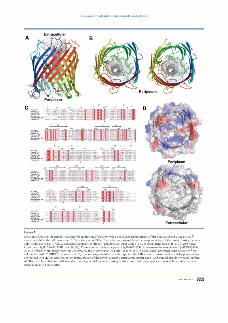
PflBenF adopts the canonical b-barrel porin foldconsisting of 18 antiparallel b-strands arranged ina cylinder [Fig. 1(A,B)]. Pairwise comparisons of thestructure of PflBenF with those of PaOpdK andPaOprD using SSM24 revealed highly similar polypep-
tide chain folds (PflBenF vs. PaOpdK: r.m.s.d. 5 0.9A, sequence identity 5 53% for 369 a-carbon pairs;
PflBenF vs. PaOprD: r.m.s.d. 5 1.3 A, sequence iden-tity 5 40% f or 342 a-carbon pairs; PaOpdK vs.PaOprD: r.m.s.d. 5 1.3 A, sequence identity 5 39%for 382 a-carbon pairs). The b-strands of PflBenF areconnected by shorter turns T1–T8 and longer loopsL1–L7 on the periplasmic and extracellular faces of thebarrel, respectively. Loops L3 and L7 fold into the
interior of the b-barrel, where they help define the ge-ometry of the pore [Fig. 1(B)]. As for PaOpdKand PaOprD, PflBenF also possesses short S5 and S6
b-strands, which are characteristic of porins that formtrimers in the outer membrane.25 Notwithstandingthis similarity, PflBenF protomers do not make exten-
sive intermolecular interactions within the crystal lat-tice. Recombinant forms of both PaOpdK and PaOprDalso appear monomeric in the crystal lattice. However,
recombinant PaOpdK inserted into artificial lipidbilayer oligomerizes and behaves as a trimeric ion-conductor.11 Given the short lengths of the S5 and S6
b-strands in the PflBenF-like porin and the PaOpdKprecedent, there is no compelling reason to believethat PflBenF functions as a monomer.
Structure of the PflBenF benzoate-specificchannel
The structure of the PflBenF b-barrel revealed aquasi-circular pore [Fig. 1(B)] with a diameter of 4.6
A (as estimated with HOLE21). Sequence alignment of PflBenF, P. putida BenF, PaOpdK, PaOprD, and threeother porins with various annotations (pairwise sequenceidentities with PflBenF ranging from 34 to 76%) revealedsignificant conservation for b-strand residues [Fig. 1(C)]and those within the loops L2, L3, and L7. The electrostaticproperties of the periplasmic and extracellular faces of PflBenF are illustrated in Figure 1(D). On the periplasmicface, conserved basic and nonpolar surface features are visi-
ble in the immediate vicinity of channel constriction. Thereis no corresponding concentration of polar surface features
on the extracellular side of the channel immediately beyondthe constriction. In fact, the extracellular face of the proteinis largely devoid of polar features [Fig. 1(D), lower panel].The entire periplasmic face of PflBenF is also considerably more polar than the extracellular face of the protein.
Evidence for an aromatic acid selectivityfilter shared by PflBenF and P. aeruginosa OpdK
The pore constriction of PflBenF is lined by residuesAsp151 and Arg154 from loop L3; Ser310, Asp317,
Table ICrystallographic Data Collection and Refinement Statistics for PflBenF
Data collectionPDB code 3JTYSpace group P 21Unit-cell dimensions (, 8) a 5 62.1, b 5 210.6,
c 5 84.1, b 5 97.8Resolution () 53.68–2.58 (2.72–2.58)a
Number of unique reflections 67,053 (9774)Completeness (%) 100.0 (100.0)R symm (%) 14.0 (52.7)Multiplicity 3.9 (3.8)<I /r(I )> 6.1 (2.7)
RefinementResolution () 35.11–2.58R -factor (%) 22.0R free (%) 27.5
Number of nonhydrogen atomsProtein 11,859Ligands 32Water molecules 25
Average B-factors (2)
Protein 41.9Ligands 75.4Water molecules 35.2
RMS deviations from ideal valuesBond length () 0.018Bond angles (8) 1.60Ramachandran plot19
MolProbity20 residues in favored region (%) 95.6Allowed region (%) 99.9
aValues in parenthesis correspond to highest-resolution shell.
P. Sampathkumar et al.
3058 PROTEINS
8/8/2019 Sampathkumar Proteins-StructFunctBioinform 2010a
http://slidepdf.com/reader/full/sampathkumar-proteins-structfunctbioinform-2010a 4/7
Figure 1
Structure of PflBenF. A: Rainbow-colored ribbon drawing of PflBenF with a dot surface representation of the pore calculated using HOLE 21
viewed parallel to the cell membrane. B: Stereodrawing of PflBenF with the pore viewed from the periplasmic face of the protein [using the samecolor coding as in Fig. 1(A)]. C: Sequence alignment of PlfBenF (gi|123657525; PDB Code 3JTY), P. putida BenF (gi|81441797), P. aeruginosa
OpdK porin (gi|81539679; PDB Code 2QTK), P. putida outer membrane protein (gi|167033173), Acinetobacter baumannii VanP (gi|193076825),A. sp. RUH2654 OprD family porin (gi|260549927), and P. aeruginosa D2 porin (gi|417518; PDB Code 2ODJ) generated using ClustalW22 andcolor coded with ESPRIPT23; numbers after ‘‘_’’ denote sequence identity with respect to the PflBenF and aromatic acid selectivity pore residuesare marked with ~. D: Semitransparent representation of the solvent accessible periplasmic (upper panel) and extracellular (lower panel) surfacesof PlfBenF, color coded for qualitative electrostatic potential (generated using PyMol) shown with polypeptide chain as ribbon, using the sameorientation as in Figure 1(B).
Structure of a Putative Benzoate Specific Porin
PROTEINS 3059
8/8/2019 Sampathkumar Proteins-StructFunctBioinform 2010a
http://slidepdf.com/reader/full/sampathkumar-proteins-structfunctbioinform-2010a 5/7
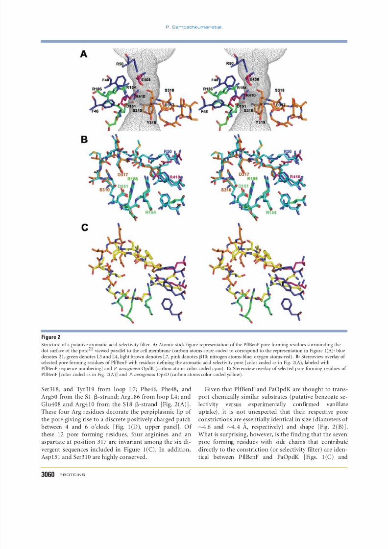
Ser318, and Tyr319 from loop L7; Phe46, Phe48, andArg50 from the S1 b-strand; Arg186 from loop L4; andGlu408 and Arg410 from the S18 b-strand [Fig. 2(A)].These four Arg residues decorate the perpiplasmic lip of the pore giving rise to a discrete positively charged patchbetween 4 and 6 o’clock [Fig. 1(D), upper panel]. Of these 12 pore forming residues, four arginines and anaspartate at position 317 are invariant among the six di-
vergent sequences included in Figure 1(C). In addition,Asp151 and Ser310 are highly conserved.
Given that PlfBenF and PaOpdK are thought to trans-port chemically similar substrates (putative benzoate se-lectivity versus experimentally confirmed vanillateuptake), it is not unexpected that their respective poreconstrictions are essentially identical in size (diameters of
4.6 and 4.4 A, respectively) and shape [Fig. 2(B)].What is surprising, however, is the finding that the sevenpore forming residues with side chains that contribute
directly to the constriction (or selectivity filter) are iden-tical between PflBenF and PaOpdK [Figs. 1(C) and
Figure 2
Structure of a putative aromatic acid selectivity filter. A: Atomic stick figure representation of the PflBenF pore forming residues surrounding thedot surface of the pore21 viewed parallel to the cell membrane (carbon atoms color coded to correspond to the representation in Figure 1(A): bluedenotes b1, green denotes L3 and L4, light brown denotes L7, pink denotes b10; nitrogen atoms-blue; oxygen atoms-red). B: Stereoview overlay of selected pore forming residues of PlfBenF with residues defining the aromatic acid selectivity pore [color coded as in Fig. 2(A), labeled withPflBenF sequence numbering] and P. aeruginosa OpdK (carbon atoms color coded cyan). C: Stereoview overlay of selected pore forming residues of PlfBenF [color coded as in Fig. 2(A)] and P. aeruginosa OprD (carbon atoms color-coded yellow).
P. Sampathkumar et al.
3060 PROTEINS

8/8/2019 Sampathkumar Proteins-StructFunctBioinform 2010a
http://slidepdf.com/reader/full/sampathkumar-proteins-structfunctbioinform-2010a 6/7
2(B)]. Specifically, Arg50 (Arg22 , italics denotes PaOpdKresidues), Asp151 (Asp123), Arg154 (Arg126 ), Arg186(Arg158 ), Ser310 (Ser282 ), Asp317 (Asp289 ), and Arg410(Arg381) are invariant between these two porins [Figs.1(C) and 2(B)]. The only notable difference is the side-
chain conformation of Arg410, which brings its guanidi-nium group closer to the constriction in the case of
PflBenF. In contrast, the PaOprD pore constriction issmaller than that of PlfBenF and PaOpdK (3.7 A vs.4.5 A), differs in shape, and is defined by a differentconfiguration of selectivity filter residues [Fig. 2(C)].Thus, the selectivity determining residues of the vanillateand putative benzoate pores appear to be similar. Furthercharacterization of the function of PflBenF and other
porins sharing this sequence motif will be required to es-tablish whether or not it is in fact a signature of aromaticacid channels.
One of the metrics guiding target selection in struc-tural genomics is modeling leverage at the level of greaterthan 30% sequence identity (see Supporting Information
Methods).26,27 Searching of the UniProtKB database of all known protein sequences using the sequences of PlfBenF, PaOpdK, and PaOprD identified 221 uniqueprotein sequences with sequence identity greater than30% to at least one of these three proteins. The inauguralstructure of PaOprD, enabled modeling of 165 related
proteins (Supporting Information Fig. S1). Subsequentdetermination of the structure of PaOpdK enabledhomology modeling of an additional three proteinsequences. In contrast, determination of the PflBenFstructure enabled homology modeling of 53 additionalprotein sequences (Supporting Information Fig. S1).Thus, the structure of PflBenF, reported herein, expandssignificantly the number of homology models of OprDand OpdK family of substrate-specific porins. Experimen-tal structures of additional OprD/OpdK subfamily mem-
bers should provide useful guides for planning experi-ments aimed at defining the mechanisms governing poreselectivity.
ACKNOWLEDGMENTS
Access to the LRL-CAT beam-line facilities at Sector 31of the advanced photon source was provided by Eli Lilly
and Company, which operates the facility. Use of theAdvanced Photon Source was supported by the U.S.Department of Energy, Office of Science, Office of BasicEnergy Sciences, under Contract No. DE-AC02-06CH11357.Atomic coordinates and structure factors of PflBenF weredeposited to the PDB (www.rcsb.org) on 14 September 2009with PDB accession code 3JTY. The NYSGXRC targetidentifier for PflBenF in TargetDB (http://targetdb.pdb.org) is ‘‘NYSGXRC-10383p.’’ Expression clone sequences
and selected interim experimental results are available inPepcDB (http://pepcdb.pdb.org/).
REFERENCES
1. Nikaido H. Molecular basis of bacterial outer membrane permeabil-
ity revisited. Microbiol Molbiol Rev 2003;67:593–656.
2. Hancock RE, Brinkman FS. Function of pseudomonas porins in
uptake and efflux. Annu Rev Microbiol 2002;56:17–38.
3. Trias J, Nikaido H. Protein D2 channel of the Pseudomonas aerugi-
nosa outer membrane has a binding site for basic amino acids and
peptides. J Biol Chem 2006;265:15680–15684.4. Tamber S, Ochs MM, Hancock RE. Role of the novel OprD family
of porins in nutrient uptake in Pseudomonas aeruginosa . J Bacteriol
2006;188:45–54.
5. Cowles CE, Nichols NN, Harwood CS. BenR, a XylS homologue,
regulates three different pathways of aromatic acid degradation in
Pseudomonas putida . J Bacterol 2000;182:6339–6346.
6. Nishikawa Y, Yasumi Y, Noguchi S, Sakamoto H, Nikawa J. Func-
tional analyses of Pseudomonas putida benzoate transporters
expressed in the yeast Saccharomyces cerevisiae . Biosci Biotechnol
Biochem 2008;72:2034–2038.
7. Altschul SF, Madden TL, Schaffer AA, Zhang J, Zhang Z, Miller W,
Lipman DJ. Gapped BLAST and PSI-BLAST: a new generation of
protein database search programs. Nucleic Acids Res 1997;25:3389–
3402.
8. The UniProt Consortium. The universal protein resource (UniProt)in 2010. Nucleic Acids Res 2010;38(Database issue):D142–D148.
9. Paulsen IT, Press CM, Ravel J, Kobayashi DY, Myers GS, Mavrodi
DV, De Boy RT, Seshadri R, Ren Q, Madupu R, Dodson RJ, Durkin
AS, Brinkac LM, Daugherty SC, Sullivan SA, Rosovitz MJ, Gwinn
ML, Zhou L, Schneider DJ, Cartinhour SW, Nelson WC, Weidman
J, Watkins K, Tran K, Khouri H, Pierson EA, Pierson LS, III, Tho-
mashow LS, Loper JE. Complete genome sequence of the plant
commensal Pseudomonas fluorescens Pf-5 . Nat Biotechnol
2005;23:873–878.
10. Nelson KE, Weinel C, Paulsen IT, Dodson RJ, Hilbert H, Martins
dos Santos VA, Fouts DE, Gill SR, Pop M, Holmes M, Brinkac L,
Beanan M, DeBoy RT, Daugherty S, Kolonay J, Madupu R, Nelson
W, White O, Peterson J, Khouri H, Hance I, Chris Lee P, Holtzap-
ple E, Scanlan D, Tran K, Moazzez A, Utterback T, Rizzo M, Lee K,
Kosack D, Moestl D, Wedler H, Lauber J, Stjepandic D, Hoheisel J,
Straetz M, Heim S, Kiewitz C, Eisen JA, Timmis KN, Dusterhoft A,
Tummler B, Fraser CM. Complete genome sequence and compara-
tive analysis of the metabolically versatile Pseudomonas putida
KT2440. Environ Microbiol 2002;4:799–808.
11. Biswas S, Mohammad MM, Patel DR, Movileanu L, van den Berg
B. Structural insight into OprD substrate specificity. Nat Struct Mol
Biol 2007;14:1108–1109.
12. Biswas S, Mohammad MM, Movileanu L, van den Berg B. Crystal
structure of the outer membrane protein OpdK from Pseudomonas
aeruginosa . Structure 2008;16:1027–1035.
13. Sambrook J, Fritsch EF, Maniatis T. Molecular cloning: a laboratory
manual, 2nd ed. Cold Spring Harbour, NY: Cold Spring Harbour
Laboratory; 1989.
14. Leslie AGW, Brick P, Wonacott AJ. An improved program package
for the measurement of oscillation photographs. CCP4 News Lett
1986;18:33–39.
15. Collaborative Computing Project Number 4. The CCP4 suite: pro-
grams for protein crystallography. Acta Crystallog Sect D Biol Crys-
tallogr 1994;50:760–763.
16. McCoy AJ, Grosse-Kunstleve RW, Adams PD, Winn MD, Storoni L,
Read RJ. Phaser crystallographic software. J Appl Cryst 2007;
40:658–674.
17. Emsley P, Cowtan K. COOT: model-building tools for molecular
graphics. Acta Crystallogr Sect D Biol Crystallogr 2004;60:2126–2132.
18. Murshudov GN, Vagin AA, Dodson EJ. Refinement of macromolec-
ular structures by the Maximum-Likelihood Method. Acta Crystal-
logr D Biol Crystallogr 1997;53:240–255.
Structure of a Putative Benzoate Specific Porin
PROTEINS 3061

8/8/2019 Sampathkumar Proteins-StructFunctBioinform 2010a
http://slidepdf.com/reader/full/sampathkumar-proteins-structfunctbioinform-2010a 7/7
19. Ramakrishnan C, Ramachandran GN. Stereochemical criteria for
polypeptide and protein chain conformations. II. Allowed confor-
mations for a pair of peptide units. Biophys J 1965;5:909–933.
20. Davis IW, Leaver-Fay A, Chen VB, Block JN, Kapral GJ, Wang X,
Murray LW, Arendall WB, III, Snoeyink J, Richardson JS, Richard-
son DC. Mol Probity: all-atom contacts and structure validation
for proteins and nucleic acids. Nucl Acids Res 2007;35:W375–
W383.
21. Smart OS, Neduvelil JG, Wang X, Wallace BA, Sansom MSP.
HOLE: a program for the analysis of the pore dimensions of ion
channel structural models. J Mol Graph 1996;14:354–360.
22. Larkin MA, Blackshields G, Brown NP, Chenna R, McGettigan PA,
McWilliam H, Valentin F, Wallace IM, Wilm A, Lopez R, Thomp-
son JD, Gibson TJ, Higgins DG. ClustalW and ClustalX version 2.
Bioinformatics 2007;23:2947–2948.
23. Gouet P, Courcelle E, Stuart DI, Metoz F. ESPript: multiple
sequence alignments in PostScript. Bioinformatics 1999;15:305–308.
24. Krissinel E, Henrick K. Secondary-structure matching (SSM), a new
tool for fast protein structure alignment in three dimensions. Acta
Crystallogr D Biol Crystallogr 2004;60:2256–2268.
25. Schulz GE. The structure of bacterial outer membrane proteins.
Biochim Biophys Acta 2002;1565:308–317.
26. Eswar N, John B, Mirkovic N, Fiser A, Ilyin VA, Pieper U, Stuart
AC, Marti-Renom MA, Madhusudhan MS, Yerkovich B, Sali A.
Tools for comparative protein structure modeling and analysis.
Nucleic Acids Res 2003;31:3375–3380.
27. Pieper U, Eswar N, Webb BM, Eramian D, Kelly L, Barkan DT, Carter
H, Mankoo P, Karchin R, Marti-Renom MA, Davis FP, Sali A.
MODBASE, a database of annotated comparative protein structure
models and associated resources. Nucleic Acids Res 2009;37:D347–354.
P. Sampathkumar et al.
3062 PROTEINS