Embed Size (px)
Citation preview

Scientia Chromatographica 2018; 10(3):174-194Instituto Internacional de Cromatografiahttp://dx.doi.org/10.5935/sc.2018.011ISSN 1984-4433
SAMPLE PREPARATION
174 Scientia Chromatographica 2018; 10(3)
AbstractIt is essential to determine drugs and their metabolites or biomarmarkers in biological samples (e.g., serum, plasma, breast milk, cerebrospinal fluid, and urine) quantitatively for clinical studies including therapeutic drug monitoring, drug metabolism investigation, and diagnostic or prognostic purposes. Because biological samples are extremely complex matrices, direct injection of complex samples into conventional chromatographic systems is not convenient. A sample preparation step is usually necessary to eliminate the majority of endogenous compounds and to concentrate the analytes that often exist at traces levels. In this context, this review presents an overview of how our research group has contributed to the development of innovative microextraction methods (solid-phase microextraction, in-tube solid-phase microextraction, stir bar sorptive extraction, microextraction by packed sorbent, and disposable pipette extraction) for bioanalysis. The theoretical fundamentals, extraction configurations, and current technology used for the synthesis of selective sorbents for microextractions, such as (i) polypyrrole, (ii) restricted-access materials, (iii) immunosorbents, (iv) molecular imprinting polymers, (v) monolithic polymers, (vi) ionic liquids, and (vii) bi-functional materials are also discussed, as well as future trends in sample preparation techniques.
Keywords: microextraction techniques, biological samples, selective sorbents for microextractions, online coupling techniques, and LC-MS/MS.
Maria Eugênia Costa Queiroz* Israel Donizeti de Souza
Departamento de Química, Facul-dade de Filosofia Ciências e Letras de Ribeirão Preto, Universidade de São Paulo, Ribeirão Preto, SP, Brazil
* Maria Eugênia C. Queiroz, Av. Bandeirantes, 3900 - CEP 14040-901, Ribeirão Preto - SP – [email protected] Tel.: + 55 16 33159172
Sample preparation techniques for biological samples
1. Introduction
Biological fluids, such as plasma, serum, cerebrospinal fluid, and urine, are complex samples that contain proteins, phospholipids, inorganic salts, and organic compounds. Thus, chromatographic instruments cannot handle these matrixes directly.
Chromatographic analysis of biological samples includes sampling, sample preparation, chromatographic separation, detection, and data handling. Among all
these steps, sample preparation is both the most error-
prone part of the process and the most tedious procedure
and typically takes 60-80% of the total analysis time.
During the sample preparation step, most endogenous
compounds are excluded from the biological matrixes
(to decrease the matrix effects), and the target analytes
(which are present in trace amounts in the matrix) are
isolated and concentrated (enrichment) to suitable
concentration levels for detection. Therefore, the sample
preparation step has been regarded as a bottleneck in

Queiroz M. E. C., Souza I. D. Sample preparation techniques for biological samples
Scientia Chromatographica 2018; 10(3):174-194 175
the development of sensitive, selective, and precise analytical methods.
Conventional biological sample preparation techniques, including protein precipitation (PP), 1–3 liquid-liquid extraction (LLE), 4,5 and solid-phase extraction (SPE), 6,7 involve well-established and well-optimized techniques. PP and LLE are simple, fast sample treatments that do not require special equipment. However, PP is poorly selective, which might compromise the sensitivity of the method and cause serious matrix effects. Conventional LLE demands a large volume of biological sample and organic solvents. Consequently, the organic solvent needs to be evaporated and the extracted analyte(s) must be reconstituted in an adequate solvent after the process, to provide a pre-concentrated sample that is compatible for injection into the chromatographic system. In turn, manual SPE is a selective technique that requires smaller volume of organic solvents as compared to conventional LLE. Nevertheless, it is quite a time-consuming, multistep procedure. In some cases, when higher levels of analytes are present in the sample and the complexity of the matrix does not affect the results, classic sample preparation techniques are the first choice for sample treatment. Table 1 lists some successful applications of these classic sample preparation techniques developed in our research laboratory for bioanalysis of various compounds.
Over the last decades, works regarding sample preparation techniques have focused on analytical system miniaturization, online coupling techniques, environmentally friendly approaches, and development of selective stationary phases.
To achieve these goals, our research group has been developing new miniaturized or online sample preparation methods for chromatographic bioanalysis since 2003. We have applied most of these methods to determine drugs, their metabolites, and biomarkers in biological samples for studies about neuropsychiatric disorders. This review describes theoretical fundamentals,
extraction configurations, experimental parameters, new selective capillary coatings, applications, and current and future challenges of these innovative sample preparation techniques.
2. Solid-phase microextraction (SPME)SPME is based on the sorption equilibrium of
analytes between the sample matrix and the stationary phase. It is a non-exhaustive process; that is, it extracts only a small fraction of the initial amount of analyte. This technique combines extraction and concentration of the analyte in a single step, thereby reducing the time required for sample preparation.
A SPME device (Figure 1) consists of a fiber assembly containing a built-in extraction fiber inside a needle and an assembly holder. The SPME fiber itself is a 1- or 2-cm-long thin fused-silica optical fiber coated with a thin polymer film or solid sorbent (stationary phase).
SPME was introduced by Arthur and Pawliszyn in 1990. 8 It initially gained wide acceptance for the analysis of volatile and semi-volatile organic compounds in environmental samples by gas chromatography (GC) because it easily combined solvent-free SPME headspace extraction with thermal desorption in the GC injection port. The first challenge faced by our research group was to evaluate the SPME technique to extract drugs (weakly volatile compounds) by directly immersing the SPME fiber into human plasma (complex sample) and then conducting liquid desorption and high-performance liquid chromatography (HPLC) analysis.
Since then, our research group has successfully developed and validated different SPME/HPLC-UV methods to determine anticonvulsants (phenylethylmalonamide, phenobarbital, primidone, carbamazepine, phenytoin, and lamotrigine) 9 and antidepressants (selective serotonin reuptake inhibitors, or SSRIs) 9–12 in human plasma for therapeutic drug monitoring (TDM), Table 2.

Queiroz M. E. C., Souza I. D. Sample preparation techniques for biological samples
176 Scientia Chromatographica 2018; 10(3):174-194
Tabl
e 1.
Cla
ssic
sam
ple
prep
arat
ion
tech
niqu
es fo
r bio
logi
cal s
ampl
es
DAD:
dio
de a
rray
dete
ctor
, FD:
fluo
resc
ence
det
ecto
r, M
IP: m
olec
ular
ly im
prin
ted
poly
mer
, NAC
E: n
on-a
queo
us c
apill
ary
elec
troph
ores
is
Anal
ytes
Mat
rix (s
ampl
e vo
lum
e, µ
L)Sa
mpl
e pr
epar
atio
n pr
oced
ure
Chro
mat
ogra
phic
sys
tem
Chro
mat
ogra
phic
col
umn
Line
ar ra
nge
(ng
mL-1
)Re
fere
nce
Prot
ein
prec
ipta
tion
Fluo
xetin
e an
d no
rfluo
xetin
ePl
asm
a (1
00)
Sam
ple/
acet
onitr
ile 1
:3 (v
/v)
HPLC
-FD
Chira
lcel
OD
-R(2
50 ×
4.6
mm
, 10
µm)
30-1
000
1
Amin
o ac
ids a
ndNe
urot
rans
mitt
ers
Plas
ma
(50)
Sam
ple/
acet
onitr
ile 1
:2 (v
/v) f
ollo
wed
by
ultra
-filtr
atio
n w
ith A
mic
on U
ltra-
4 (M
illip
ore,
Be
dfor
d, M
A, U
SA) d
evic
eHP
LC-M
S/M
SAs
cent
is Ex
pres
s HIL
IC c
olum
n(4
.6 ×
100
mm
, 2.7
µm
)4.
5-65
600
2
5 an
tipsy
chot
ics,
7 an
tidep
ress
ants
, 2
antic
onvu
lsant
s, an
d 2
anxi
olyt
ics
Plas
ma
(200
)Sa
mpl
e/ac
eton
itrile
1:2
(v/v
)HP
LC-M
S/M
SXS
elec
t CSH
C18
XP
colu
mn
(2.1
× 1
00 m
m, 2
.5 μ
m)
0.2-
40.5
3
Liqu
id-li
quid
ext
ract
ion
Antid
epre
ssan
tsPl
asm
a (1
000)
Extra
ctio
n w
ith h
exan
e/iso
amyl
alc
ohol
99:
1 (v
/v) m
ixtu
reNA
CE-D
ADFu
sed
silic
a ca
pilla
ry(4
00 ×
0.7
5 m
m)
15-3
04
Anan
dam
ide
and
2-AG
Cere
bros
pina
l flu
id
(200
)Pr
otei
n pr
ecip
itatio
n w
ith a
ceto
ne fo
llow
ed
by e
xtra
ctio
n w
ith to
luen
eUH
LC-M
S/M
SKi
nete
x C1
8(1
00 ×
2.1
mm
, 1.7
µm
)0.
5-50
5
Solid
-pha
se e
xtra
ctio
n
Venl
afax
ine,
o-d
esm
ethy
lven
lafa
x-in
e, a
nd n
-des
met
hylv
enla
faxi
nePl
asm
a(2
00)
Extra
ctio
n (M
IP):
pre-
treat
ed sa
mpl
e (3
00
µL).
Clea
n-up
: ace
tic a
cid
0.1%
follo
wed
by
met
hano
l sol
utio
n (1
0%).
Deso
rptio
n: M
eOH.
UHLC
-MS/
MS
Kine
tex
C18
(100
× 2
.1 m
m, 1
.7 µ
m)
3-70
06
Para
bens
Plas
ma
(200
)Ex
tract
ion
(MIP
): pr
e-tre
ated
sam
ple
(300
µL)
. Cl
ean-
up: w
ater
. Des
orpt
ion:
met
hano
lUH
LC-M
S/M
SKi
nete
x C1
8(1
00 ×
2.1
mm
, 1.7
µm
)1-
507

Queiroz M. E. C., Souza I. D. Sample preparation techniques for biological samples
Scientia Chromatographica 2018; 10(3):174-194 177
Figure 1. Commercial SPME device
Table 2. SPME/HPLC methods to determine drugs in plasma samples for therapeutic drug monitoring
Chromatographic column: RP18 LichroCART® (125 × 4 mm, 5 µm)
AnalytesMatrix,
(volume, µL)Stationary
phaseExtraction procedure
Linear range
(ng mL-1)Reference
Phenylethylmalonamide, phenobarbital, primidone,
carbamazepine, and carbamazepine-epoxide
Plasma(1000) Carbowax
Extraction: 30 min at 30 °C.Desorption: 10 min in phosphate buffer solution/acetoni-trile/ methanol 65:18:17 (v/v/v) mixture.
500-12000 9
Imipramine, amitriptyline, desipramine, and nortrip-
tyline
Plasma(1000) PDMS/DVB
Extraction: 40 min at 65 °C.Desorption: 10 min in sodium acetate buffer solution/ace-tonitrile 50:50 (v/v) mixture.
75-500 9
Mirtazapine, citalopram, paroxetine, duloxetine,
fluoxetine, and sertraline
Plasma(250) Poly(pyrrole)
Extraction: 40 min at 25 °C.Desorption: 15 min at 25 °C in phosphate buffer solution/acetonitrile 57:43 (v/v) mixture.
16-1200 10
Fluoxetine, sertraline, par-oxetine, and citalopram
Plasma(250) Polythiophene
Extraction: 40 min at 25 °C.Desorption: 15 min at 25 °C in phosphate buffer solution/ methanol 40:60 (v/v) mixture.
200-4000 11
Mirtazapine, citalopram, paroxetine, fluoxetine, and
sertraline
Plasma(1000)
PDMS/DVBExtraction: 45 min at 60 °C.Desorption: 15 min at 60 °C in acetonitrile.
25-500 12

Queiroz M. E. C., Souza I. D. Sample preparation techniques for biological samples
178 Scientia Chromatographica 2018; 10(3):174-194
Our group has prepared PPY and PTh phases on a stainless-steel wire for SPME by electrochemical deposition (cyclic voltammetry) and has evaluated how the electrolyte solution (lithium perchlorate or tetrabutylammonium perchlorate) and the number of cycles (50, 100, or 200) applied during the polymerization process affect the SPME performance. 10,11 Figure 2 shows that 100 cycles provides the best results for both electrolyte solutions. Meanwhile, lithium perchlorate leads to better efficiency for most of the antidepressants. Film thickness increases with increasing number of polymerization coating cycles, which allows a higher amount of analytes to be extracted. This happens because, in the case of thicker porous coatings like PPY, increasing coating thickness increases not only the total coating volume, but also the total surface area. 14 Some articles by our group have discussed important factors that impact SPME efficiency, such as fiber coating, extraction time, sample pH, sample ionic strength, influence of plasma proteins, and liquid desorption conditions. 9–12,15
Our group has observed that the SPME extraction efficiency depends on the relative affinity of the analyte for the fiber stationary phase (coating) and its thickness. A thicker coating increases the sorption capacity of the fiber, but it also demands a longer equilibration time.
Our group has evaluated commercial fibers, including polydimethylsiloxane/divinylbenzene (PDMS/DVB, 65-μm film thickness), polyacrylate (PA, 85-μm film thickness), and the carbowax-templated resin (CW/TPR-100, 50-μm film thickness), as well as lab-made fibers like polyurethane, octadecylsilane (5-μm dp), polypyrrole (PPY), and polythiophene (PTh) for drug analysis. 9–12
Conductive polymers, such as polyaniline (PPY) and PTh, are promising extraction phases for analysis of ionizable drugs. These polymers display excellent permeability (porous structure) and multifunctional properties, which result in ion exchange and intermolecular interactions, including acid-base, TT-TT, dipole–dipole, hydrophobic, and hydrogen bonding, between the polymer and the analytes. 13
Figure 2. Effect of the number of cycles during PPY electrochemical polymerization (SPME fiber) at 50 mVs−1 on SPME performance. Electrolyte solution: (A) Lithium perchlorate solution; (B) tetrabutylammonium perchlorate solution (reimprinted from reference 10 with permission).
Queiroz M. E. C., Souza I. D. Sample preparation techniques for biological samples
Scientia Chromatographica 2018; 10(3):174-194 179
3. In-tube solid-phase microextraction (in-tube SPME)
In 1997, Eisert and Pawliszyn introduced the
in-tube SPME technique 16 to overcome the limitations
of SPME fibers (first generation) related to (a) low
extraction efficiency for weakly volatile or thermally
labile compounds not amenable to GC or GC-MS, and
(b) stability against solvents used in HPLC. 17
Our group has further developed in-tube SPME
by coupling the SPME technique directly with HPLC
systems. In-tube SPME uses an open tubular fused-silica
capillary column as the SPME device instead of SPME fiber. Hence, it can be considered an online automated sample preparation technique. 18 Column switching in-tube SPME can be carried out in two configurations: the draw/eject and the flow-through extraction systems.
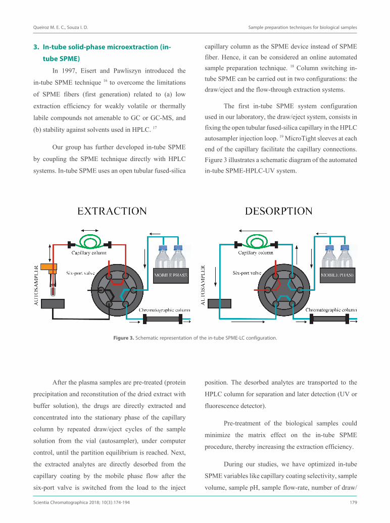
The first in-tube SPME system configuration used in our laboratory, the draw/eject system, consists in fixing the open tubular fused-silica capillary in the HPLC autosampler injection loop. 19 MicroTight sleeves at each end of the capillary facilitate the capillary connections. Figure 3 illustrates a schematic diagram of the automated in-tube SPME-HPLC-UV system.
Figure 3. Schematic representation of the in-tube SPME-LC configuration.
After the plasma samples are pre-treated (protein
precipitation and reconstitution of the dried extract with
buffer solution), the drugs are directly extracted and
concentrated into the stationary phase of the capillary
column by repeated draw/eject cycles of the sample
solution from the vial (autosampler), under computer
control, until the partition equilibrium is reached. Next,
the extracted analytes are directly desorbed from the
capillary coating by the mobile phase flow after the
six-port valve is switched from the load to the inject
position. The desorbed analytes are transported to the
HPLC column for separation and later detection (UV or
fluorescence detector).
Pre-treatment of the biological samples could
minimize the matrix effect on the in-tube SPME
procedure, thereby increasing the extraction efficiency.
During our studies, we have optimized in-tube
SPME variables like capillary coating selectivity, sample
volume, sample pH, sample flow-rate, number of draw/

Queiroz M. E. C., Souza I. D. Sample preparation techniques for biological samples
180 Scientia Chromatographica 2018; 10(3):174-194
eject cycles (only draw/eject mode), and desorption conditions. This optimization not only improved method selectivity and sensitivity, but also reduced analysis time. 17,18,20,21
In-tube SPME capillary coating
Initially, our research group used this classic in-tube SPME-HPLC system with photometric detectors to evaluate open tubular fused-silica capillary columns (a short piece of commercial GC column) such as OV-1701 (14% cyanopropylphenyl methylpolysiloxane, 100 cm × 250 μm i.d., 0.05 μm) and polyethylene glycol (60 cm × 0.32 mm i.d., 0.05μm) for analysis of antidepressants (OV-1701) 19, lidocaine and its metabolites (OV-1701) 22, and rifampicin (polyethylene glycol) 23 in plasma samples.
To improve in-tube SPME efficiency and selectivity, we synthesized alternative stationary phases, including:
(a) an open tubular fused-silica capillary with PPY coating (oxidative polymerization - positively charged) for enantioselective analysis of fluoxetine and norfluoxetine in plasma samples. 24
(b) open tubular fused-silica capillaries with immunosorbent (molecular recognition, covalently
bonded polyclonal antibodies) for analysis of fluoxetine 25 and monoclonal antibodies for interferon alpha 2a analysis 26 in plasma samples.
(c) a packed capillary (polyether ether ketone, PEEK tube) with molecularly imprinted polymers (MIP, nanocavities with specific shape and defined arrangement of the functional group synthesized by two-step sol–gel procedure) for analysis of interferon alpha 2a in plasma samples. 27
(d) a packed capillary (PEEK tube) with restricted-access materials (RAM, C18-BSA) for analysis of interferon alpha 2a in plasma samples and RAM (SCX particles with the outer surface covered with BSA) for analysis of sulfonamides in milk samples. 28 RAM phases exclude macromolecules by means of a chemical diffusion barrier created by a protein network (e.g., BSA covering the outer silica surface).
Figure 4 depicts the in-tube PPY-coated SPME/HPLC chromatograms for enantioselective analysis of fluoxetine and norfluoxetine in plasma samples for therapeutic drug monitoring. 24
Directly packing adsorbent particles into the PEEK tube improves the extraction capacity, but the high column pressure resulting from tightly packed micrometric particles limits the sample loading speed.
Figure 4. In-tube PPY-coated SPME-HPLC chromatograms. (A) Blank plasma sample. (B) Blank plasma sample spiked with FLX and NFLX resulting in 300 ng/mL (retention times: S-NFLX 15.252 min, R-NFLX 16.735 min, S-FLX 18.834 min, R-FLX 20.618 min). (C) Plasma sample from elderly depressed patient receveing therapeutic dosage. (reimprinted from reference 24 with permission).

Queiroz M. E. C., Souza I. D. Sample preparation techniques for biological samples
Scientia Chromatographica 2018; 10(3):174-194 181
After the bidimensional UHPLC coupled to tandem mass spectrometry (UHPLC-MS/MS) was purchased, our research group evaluated the flow-through in-tube SPME-LC/MS/MS system with two pumps and one six-port valve. In this system, the quaternary pump (QSM) is connected to the capillary column (first dimension – for enrichment with trace analytes), and the binary pump (BSM) is connected to the analytical column (second dimension – for chromatographic separation) (Figure 5). The use of two binary pumps allows application of different flow rates to extract and to separate the analytes. During chromatographic separation (second dimension), the capillary column (first dimension) is re-equilibrated for the next injection (parallel column regeneration).
By using this flow-through in-tube SPME-LC/MS/MS system with innovative capillary coating (first dimension), our group has developed methods for bioanalysis, including:
(a) an organic-inorganic hybrid monolithic capillary column functionalized with cyano groups to determine sixteen drugs (antidepressants, anticonvulsants, anxiolytics, and antipsychotics) in plasma samples from schizophrenic patients for TDM. 29 The properties of the monolithic phases – low backpressure, high mass transfer rate, and high permeability – make the innovative hybrid monolith more attractive than conventional particulate-
packed phases. Incorporation of the cyano groups into the monolith increases selectivity for the target drugs.
(b) a wall-coated open tubular capillary column with polymeric ionic liquids (PILs) to determine anandamide (AEA) and 2-arachidonoyl glycerol (2AG) in plasma samples from patients with Parkinson’s disease. Selective PILs were synthetized from the [VC6IM][Cl], [VC16IM][Br], and [(VIM)
2C10]2[Br] ionic liquids
by in-situ thermally initiated polymerization in a fused silica capillary column. 30
(c) an organic poly(butyl methacrylate-co-ethylene glycol dimethacrylate) monolithic capillary to determine antipsychotics (chlopromazine, clozapine, quetiapine, and olanzapine) and their metabolites (desmethyl chlorpromazine, 7-hydroxy-chlorpromazine, N-desmethyl clozapine, N-desmethyl olanzapine, and norquetiapine) in plasma samples from schizophrenic patient. 31
By means of offline in-tube SPME followed by UHPLC-MS/MS analysis, our group has determined parabens in breast milk samples. 32 For this new approach, a MIP (for selective interaction) modified with restricted access material (a non-adsorptive network hydrophilic external layer – for exclusion of endogenous compounds) (MIP-RAM) was synthesized by in situ polymerization in an open fused silica capillary, Fig 7b.
Figure 5. (a) Graphic diagrams of in-tube SPME configurations: flow-through mode with two pumps and (b) labmade device for offline in-tube SPME (adaptated from reference 17 and 32).

Queiroz M. E. C., Souza I. D. Sample preparation techniques for biological samples
182 Scientia Chromatographica 2018; 10(3):174-194
In our research group, a column switching technique that uses in the first dimension a “capillary column” as extraction device is called “in-tube SPME-LC system”, but a column switching technique that uses a “commercial HPLC cartridge” is called a “column switching LC system”.
Our group has used a column switching UHPLC-MS/MS system with a commercial RAM column (RP-8 ADS) in the first dimension (for enrichment with trace analytes while endogenous interference is excluded) and a reverse phase (RP18) in the second dimension (for chromatographic separation) to determine (a) antipsychotics, antidepressants, anticonvulsants, and anxiolytics in plasma samples from
schizophrenic patients, 33 and (b) anandamide (AEA) and
2-arachidonoylglycerol (2-AG) in plasma samples 34 and
in cerebrospinal fluid 5 from patients with Alzheimer’s
disease. 34
Our group has evaluated how the recently
introduced RP18 superficially porous columns and
RP18 fully porous columns with different particle sizes
perform during chromatographic separation of drugs in
plasma samples by MS/MS detection. 35 On the basis of
this study, we have selected which analytical column
(best performance) to use in the second dimension of the
column switching system for drug analysis. 33

Queiroz M. E. C., Souza I. D. Sample preparation techniques for biological samples
Scientia Chromatographica 2018; 10(3):174-194 183
Anal
ytes
Mat
rix,
(vol
ume,
µL)
Stat
iona
ry p
hase
Extr
actio
n pr
oced
ure
Det
ectio
n sy
stem
[Lin
ear r
ange
(ng
mL-1
)]*
Chro
mat
ogra
phic
co
lum
nRe
fere
nce
in-tu
be S
PME-
LC
Nont
ricyc
lic a
n-tid
epre
ssan
tsPl
asm
a(5
00)
OV-
1701
(8
00 ×
0.2
5 m
m, 0
.05-
µm fi
lm th
ickn
ess)
Extra
ctio
n: d
raw
-eje
ct m
ode
(15
× 10
0 µL
). De
sorp
tion:
pho
spha
te b
uffe
r sol
utio
n/ac
eton
itrile
57
:43
(v/v
) mix
ture
UV[2
0-50
]
RP18
Lic
hroC
ART®
(2
50 ×
4 m
m, 5
µm
)19
Lido
cain
ePl
asm
a(5
00)
OV-
1701
(1
000
× 0.
25 m
m, 0
.05-
µm fi
lm th
ickn
ess)
Extra
ctio
n: d
raw
-eje
ct m
ode
(5 ×
200
µL)
. De
sorp
tion:
(pho
spha
te b
uffe
r sol
utio
n w
ith
0.16
% o
f trie
thyl
amin
e)/a
ceto
nitri
le 6
0:40
(v/v
).
UV[5
0-50
00]
RP18
Lic
hroC
ART®
(1
25 ×
4 m
m, 5
µm
)22
Rifa
mpi
cin
Plas
ma
(500
)
PEG
(800
× 0
.25
mm
, 0.0
5-µm
film
thic
knes
s)Ex
tract
ion:
dra
w-e
ject
mod
e (1
0 ×
200
µL).
Deso
rptio
n: p
hosp
hate
buf
fer s
olut
ion/
ace
toni
-tri
le 6
0:40
(v/v
).
FD[1
00-1
0000
0]RP
18 L
ichr
oCAR
T®
(125
× 4
mm
, 5 µ
m)
23
Fluo
xetin
e an
d no
rfluo
xetin
e en
antio
mer
s
Plas
ma
(200
)
Poly
pyrro
le
(600
× 0
.25
mm
, 0.2
-µm
film
thic
knes
s)Ex
tract
ion:
dra
w-e
ject
mod
e (2
0 ×
100
µL).
Deso
rptio
n: 1
× 6
0 µL
of m
etha
nol
FD[1
0-70
0]
Chira
lcel
OD
-R
(250
× 4
.6m
m, 1
0 µm
) 2
4
Fluo
xetin
eSe
rum
(160
)An
tibod
y(fu
sed
silic
a ca
pilla
ry: 7
00 ×
0.2
5 m
m)
Extra
ctio
n: d
raw
-eje
ct m
ode
(20
× 34
µL)
. De
sorp
tion:
am
mon
ium
ace
tate
solu
tion
with
0.
1% fo
rmic
aci
d/ac
eton
itrile
with
1%
of a
cetic
ac
id) 4
0:60
(v/v
).
MS
[5-5
0]
C18
(150
× 3
.9 m
m, 5
µm
)25
Inte
rfero
n-al
pha
Plas
ma
(250
)
Antib
ody
(fuse
d sil
ica
capi
llary
: 600
× 0
.25
mm
)Ex
tract
ion:
dra
w-e
ject
mod
e (2
0 ×
250
µL).
Deso
rptio
n: tr
ifluo
roac
etic
aci
d so
lutio
n, 0
.1%
pH
2.5)
ace
toni
trile
86:
14 (v
/v).
FD[0
.006
-3.0
MIU
/m
L]
RP18
Lic
hroC
ART®
(1
25 ×
4 m
m, 5
µm
)26
Inte
rfero
n-al
pha
2aPl
asm
a(5
0)M
IP
(PEE
K tu
be: 5
0 ×
0.50
mm
)
Extra
ctio
n: d
raw
-eje
ct m
ode
(20
× 50
µL)
. De
sorp
tion:
0.1
% tr
ifluo
roac
etic
aci
d so
lutio
n/ac
e-to
nitri
le 8
6:14
(v/v
).
FD[8
-300
]RP
18 L
ichr
oCAR
T®
(125
× 4
mm
, 5 µ
m)
27
Tabl
e 3.
Aut
omat
ed c
olum
n sw
itchi
ng L
C-M
S//M
S an
d in
-tube
SPM
E-LC
met
hods
for b
ioan
alys
is

Queiroz M. E. C., Souza I. D. Sample preparation techniques for biological samples
184 Scientia Chromatographica 2018; 10(3):174-194
Tabl
e 3.
(con
tinua
tion)
Sulfo
nam
ides
Milk
(500
)
SAX-
BSA
(PEE
K tu
be: 5
0 ×
0.50
mm
, 45-
µm p
artic
le si
ze)
Extra
ctio
n: d
raw
-eje
ct m
ode
(20
× 50
µL)
. De
sorp
tion:
(ace
toni
trile
/met
hano
l 60:
40, v
/v)/
(wat
er 1
0:90
(v/v
).
UV[3
0-15
0]C1
8 Ch
rom
Sep
(250
x 3
.0 m
m, 5
µm
)28
Antid
epre
s-sa
nts,
anti-
conv
ulsa
nts,
anxi
olyt
ics,
and
antip
sych
otic
s dr
ugs
Plas
ma
(200
)
Hybr
id si
lica-
cyan
opro
pyl m
onol
ith
(45
× 0.
53 m
m)
Extra
ctio
n: fl
ow-th
roug
h m
ode
(1 ×
5 µ
L).
Clea
n-up
: wat
er.
Deso
rptio
n: w
ater
with
0.1
% fo
rmic
aci
d/ac
eton
i-tri
le 7
0:30
(v/v
).
MS/
MS
[0.0
6-10
500]
X Se
lect
CSH
C18
XP
(2.1
× 1
00m
m, 2
.5
µm)
29
AEA
and
2-AG
Plas
ma
(200
)
Poly
mer
ic io
nic
liqui
d (fu
sed
silic
a ca
pilla
ry: 1
10 ×
0.5
3 m
m, 1
.7-µ
m fi
lm th
ickn
ess)
Extra
ctio
n: fl
ow-th
roug
h m
ode
(8 ×
10
µL).
Clea
n-up
: wat
er.
Deso
rptio
n: w
ater
with
0.5
% fo
rmic
aci
d/ a
ceto
ni-
trile
with
0.5
% fo
rmic
aci
d 30
:70
(v/v
).
MS/
MS
[0.0
5-10
0]
Kine
tex
C18
(100
× 2
.1 m
m, 1
.7
µm)
30
Chlo
prom
azin
e,
cloz
apin
e,
quet
iapi
ne,
olan
zapi
ne, a
nd
thei
r met
abo-
lites
Plas
ma
(300
)po
ly(B
MA-
co-E
GDM
A)
(100
0 ×
0.25
mm
)
Extra
ctio
n: fl
ow-th
roug
h m
ode
(1 ×
10
µL).
Clea
n-up
: wat
er.
Deso
rptio
n: a
mm
oniu
m a
ceta
te so
lutio
n w
ith
0.1%
form
ic a
cid/
acet
onitr
ile 8
0:20
(v/v
).
MS/
MS
[10-
700]
Acqu
ity C
SH C
18
(100
× 2
.1 m
m, 1
.7
µm)
31
Para
bens
Brea
st m
ilk(2
00)
MIP
-RAM
(0
.53
mm
ID, 5
.0 c
m, 0
.12-
µm fi
lm th
ickn
ess)
Extra
ctio
n: d
raw
-eje
ct m
ode
(3 ×
200
µL)
. Cle
an-
up: 1
× 1
00 µ
L of
wat
er.
Deso
rptio
n: 1
× 1
00 µ
L of
met
hano
l/eth
anol
solu
-tio
n 1:
1 (v
/v).
MS/
MS
[10-
400]
Kine
tex
C18
(100
× 2
.1 m
m, 1
.7
µm)
32
Inte
rfero
n-al
pha
Plas
ma
(250
)C1
8-BS
A (p
eek
tube
: 50
× 0.
50 m
m, 4
5-µm
par
ticle
size
)
Extra
ctio
n: fl
ow-th
roug
h m
ode
(1 ×
500
). Cl
ean-
up: w
ater
. De
sorp
tion:
0.1
% tr
ifluo
roac
etic
aci
d so
lutio
n/ac
e-to
nitri
le 8
6:14
(v/v
).
FD[0
.06-
3.0
MIU
/mL]
RP18
Lic
hroC
ART®
(1
25 ×
4 m
m, 5
µm
)36

Queiroz M. E. C., Souza I. D. Sample preparation techniques for biological samples
Scientia Chromatographica 2018; 10(3):174-194 185
Tabl
e 3.
(con
tinua
tion)
colu
mn
switc
hing
LC-
MS/
MS
AEA
and
2-AG
Cere
bros
pina
l flu
id(2
00)
C8-A
DS
(25
× 4
mm
, 25-
μm
par
ticle
size
)fir
st d
imen
sion
Extra
ctio
n: fl
ow-th
roug
h m
ode
(10
× 10
µL)
. Cl
ean-
up: w
ater
/ace
toni
trile
60:
40, v
/v.
Deso
rptio
n: w
ater
with
0.5
% fo
rmic
aci
d/ a
ceto
ni-
trile
30:
70 (v
/v).
MS/
MS
[0.5
-50]
Kine
tex
C18
(100
× 2
.1 m
m, 1
.7
µm)
5
Antid
epre
s-sa
nts,
anti-
conv
ulsa
nts,
anxi
olyt
ics,
and
antip
sych
otic
s dr
ugs
Plas
ma
(200
)C8
-ADS
(25
× 4
mm
, 25-
μm
par
ticle
size
)fir
st d
imen
sion
Extra
ctio
n: fl
ow-th
roug
h m
ode
(1 ×
5 µ
L).
Clea
n-up
: wat
er.
Deso
rptio
n: a
mm
oniu
m a
ceta
te w
ith fo
rmic
aci
d 0.
1%/ a
ceto
nitri
le 6
3:37
(v/v
).
MS/
MS
[0.0
25-5
0]
Kine
tex
C18
(100
× 2
.1 m
m, 1
.7
µm)
33
AEA
and
2-AG
Plas
ma
(250
)
C8-A
DS
(25
× 4
mm
, 25-
μm
par
ticle
size
)fir
st d
imen
sion
Extra
ctio
n: fl
ow-th
roug
h m
ode
(10
× 10
µL)
. Cl
ean-
up: w
ater
/ace
toni
trile
60:
40, v
/v.
Deso
rptio
n: w
ater
with
0.5
% fo
rmic
aci
d/ a
ceto
ni-
trile
30:
70 (v
/v).
MS/
MS
[0.0
4-0.
12]
Kine
tex
C18
(100
× 2
.1 m
m, 1
.7
µm)
34
* Li
near
rang
e of
the
met
hod;
Abb
revi
atio
ns: A
EA: a
nand
amid
e, 2
-AG:
2-a
rach
idon
oylg
lyce
rol,
BSA:
bov
ine
seru
m a
lbum
in, F
D: F
luor
esce
nce
dete
ctio
n, L
LE: l
iqui
d-liq
uid
extra
ctio
n, M
IP: m
olec
ular
ly
impr
inte
d po
lym
er, M
P: m
obile
pha
se, p
oly(
BMA-
co-E
GDM
A), P
oly(
Buty
l Met
hacr
ylat
e-Co
-Eth
ylen
e Gl
ycol
Dim
etha
cryl
ate)
, NAC
E: n
onaq
ueou
s cap
illar
y el
ectro
phor
esis,
PDM
S: p
olyd
imet
hylsi
loxa
ne,
PEG:
pol
yeth
ylen
e gl
ycol
, RA
M: r
estri
cted
acc
ess m
edia
, TDM
: the
rape
utic
dru
g m
onito
ring.

Queiroz M. E. C., Souza I. D. Sample preparation techniques for biological samples
186 Scientia Chromatographica 2018; 10(3):174-194
4. Stir bar sorptive extraction Stir bar sorptive extraction (SBSE) was introduced
by Baltussen et al. 37 in 1999. SBSE is also based on partitioning of a solute between the aqueous sample and the stationary phase. The analytes are dissolved in the coating and diffuse into the bulk of said coating during the extraction process, which is conducted under agitation. Stir bars (10-20 mm) coated with polydimethylsiloxane (PDMS, 25-125 μL) have higher surface area, and consequently higher sorption capacity, than SPME fibers (0.5 μL for 100-μm film thickness). 38–40 A magnetic rod is usually encapsulated in a glass jacket on which a PDMS coating is placed (Figure 6). Thermal (GC special thermal desorption unit) or liquid (GC or LC analysis) desorption can be used after the extraction. 38–40
Figure 6. Stir bar sorptive extraction device.
Our research group has developed and validated SBSE methods with liquid desorption (polar solvents) followed by LC–UV analysis to determine antidepressants [selective serotonin reuptake inhibitors (SSRI) and an antagonist of central 2-adrenergic autoreceptors], 41 antiepileptics, 42 and rifampicin, 43 in plasma samples, as well as parabens in different cosmetic products. 44 We have also analyzed SSRI drugs by SBSE followed by non-aqueous capillary electrophoresis (NACE) with spectrophotometric detection. 45 In the case of plasma samples, we have been able to reuse the stir bars twenty times after liquid desorption.
Table 4 details the development of the SBSE methods. We have optimized the SBSE parameters, including pH, extraction time, and desorption conditions (solvents, magnetic or ultrasonic stirring, stirring time, and number of steps). 38–46 The extraction time (typically 30–240 min), which is kinetically controlled, is directly related to the sample volume, stirring rate, and stir bar dimensions.
The sensitivity of the SBSE/HPLC-UV method can be improved by diluting the biological samples with buffer solution, where the drugs are partially in the non-ionic form. This enables the drugs to be extracted by the PDMS phase. Sample dilution also favors the stirring SBSE process.
Our group has also developed a new polymeric coating consisting of a dual-phase, polydimethylsiloxane (PDMS) and polypyrrole (PPY), to conduct SBSE of antidepressants from plasma samples, followed by LC analysis (SBSE/LC-UV). Extractions are based on both adsorption (PPY) and sorption (PDMS) mechanisms. 46
5. Microextraction by packed sorbentMicroextraction by packed sorbent (MEPS) is
based on miniaturization of the conventional solid phase extraction (SPE) device. In MEPS, the sorbent material (1 or 2 mg) is inserted between the syringe barrel and the injection needle as a cartridge. The primary factor of this technique is that the solvent volume employed to elute the analytes during the extraction process must be of a suitable order of magnitude to allow the analytes to be directly injected into an LC or GC system. 47,48
Commercial sorbents include silica-based sorbents, mixed mode (C8 with SCX with sulfonic acid bonded silica), polystyrene-divinylbenzene (PS-DVB), and porous graphitic carbon. 48
The MEPS procedure steps involve conditioning, loading, washing, and analyte elution/introduction into

Queiroz M. E. C., Souza I. D. Sample preparation techniques for biological samples
Scientia Chromatographica 2018; 10(3):174-194 187
multi-residue analysis of 22 pesticides in honey samples, 53 and (d) MEPS/UHPLC–MS/MS to determine parabens in urine samples from 30 postpartum volunteers, to assess human exposure to these compounds. 54
Our group has used an innovative hybrid silica monolith functionalized with cyano group (sol-
chromatographic methods and several sample matrixes: (a) MEPS (C8/SCX)/LC–UV to determine new-generation antidepressants in human plasma samples for TDM, 50 (b) MEPS (C8)/LC-DAD to determine sulfonamides in egg samples51 and in poultry litter wastewater samples, 52 (c) MEPS (C8/SCX)/GC-MS for
Our research group has successfully applied the MEPS technique in combination with different
the analytical instrument. Figure 7 displays the schematic diagram for the MEPS procedure.
Figure 7. Schematic diagram for the MEPS procedure (translated from reference 49).
Queiroz M. E. C., Souza I. D. Sample preparation techniques for biological samples
188 Scientia Chromatographica 2018; 10(3):174-194
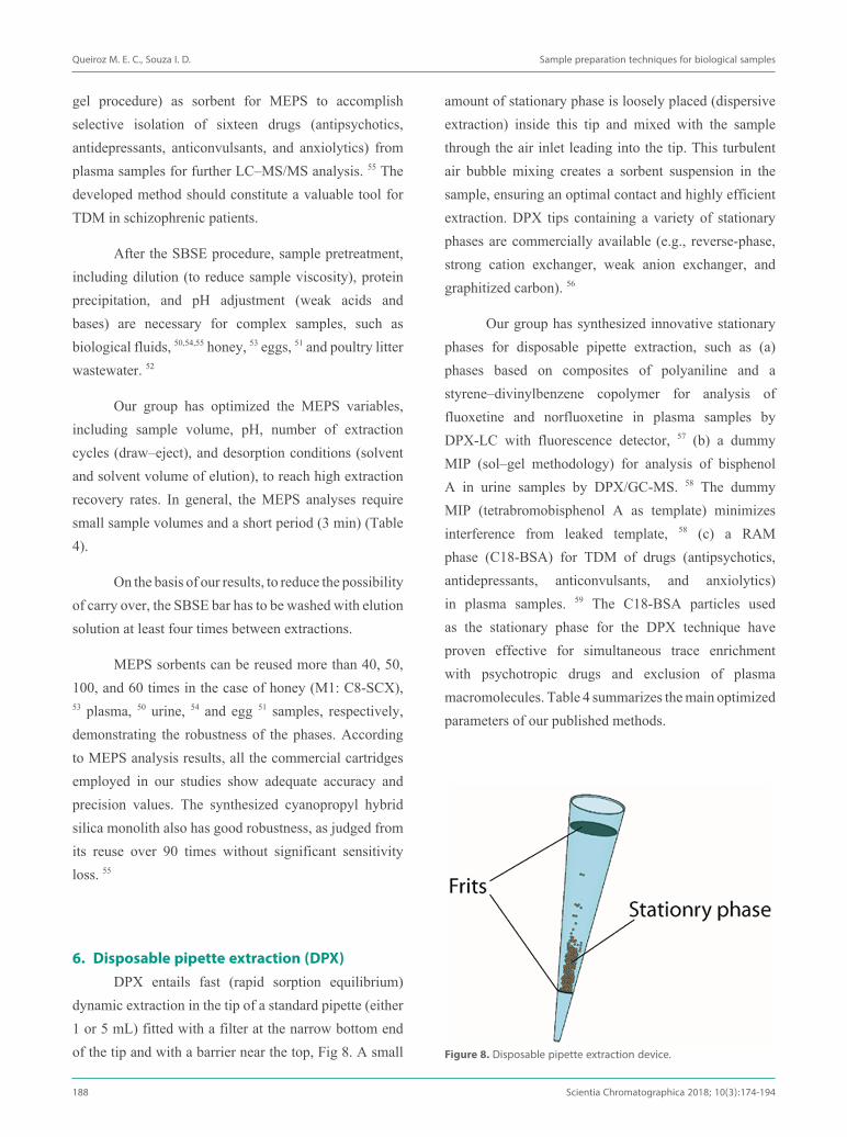
amount of stationary phase is loosely placed (dispersive extraction) inside this tip and mixed with the sample through the air inlet leading into the tip. This turbulent air bubble mixing creates a sorbent suspension in the sample, ensuring an optimal contact and highly efficient extraction. DPX tips containing a variety of stationary phases are commercially available (e.g., reverse-phase, strong cation exchanger, weak anion exchanger, and graphitized carbon). 56
Our group has synthesized innovative stationary phases for disposable pipette extraction, such as (a) phases based on composites of polyaniline and a styrene–divinylbenzene copolymer for analysis of fluoxetine and norfluoxetine in plasma samples by DPX-LC with fluorescence detector, 57 (b) a dummy MIP (sol–gel methodology) for analysis of bisphenol A in urine samples by DPX/GC-MS. 58 The dummy MIP (tetrabromobisphenol A as template) minimizes interference from leaked template, 58 (c) a RAM phase (C18-BSA) for TDM of drugs (antipsychotics, antidepressants, anticonvulsants, and anxiolytics) in plasma samples. 59 The C18-BSA particles used as the stationary phase for the DPX technique have proven effective for simultaneous trace enrichment with psychotropic drugs and exclusion of plasma macromolecules. Table 4 summarizes the main optimized parameters of our published methods.
gel procedure) as sorbent for MEPS to accomplish selective isolation of sixteen drugs (antipsychotics, antidepressants, anticonvulsants, and anxiolytics) from plasma samples for further LC–MS/MS analysis. 55 The developed method should constitute a valuable tool for TDM in schizophrenic patients.
After the SBSE procedure, sample pretreatment, including dilution (to reduce sample viscosity), protein precipitation, and pH adjustment (weak acids and bases) are necessary for complex samples, such as biological fluids, 50,54,55 honey, 53 eggs, 51 and poultry litter wastewater. 52
Our group has optimized the MEPS variables, including sample volume, pH, number of extraction cycles (draw–eject), and desorption conditions (solvent and solvent volume of elution), to reach high extraction recovery rates. In general, the MEPS analyses require small sample volumes and a short period (3 min) (Table 4).
On the basis of our results, to reduce the possibility of carry over, the SBSE bar has to be washed with elution solution at least four times between extractions.
MEPS sorbents can be reused more than 40, 50, 100, and 60 times in the case of honey (M1: C8-SCX), 53 plasma, 50 urine, 54 and egg 51 samples, respectively, demonstrating the robustness of the phases. According to MEPS analysis results, all the commercial cartridges employed in our studies show adequate accuracy and precision values. The synthesized cyanopropyl hybrid silica monolith also has good robustness, as judged from its reuse over 90 times without significant sensitivity loss. 55
6. Disposable pipette extraction (DPX)DPX entails fast (rapid sorption equilibrium)
dynamic extraction in the tip of a standard pipette (either 1 or 5 mL) fitted with a filter at the narrow bottom end of the tip and with a barrier near the top, Fig 8. A small Figure 8. Disposable pipette extraction device.

Queiroz M. E. C., Souza I. D. Sample preparation techniques for biological samples
Scientia Chromatographica 2018; 10(3):174-194 189
Tabl
e 4.
Offl
ine
mic
roex
tract
ion
met
hods
for d
iffer
ent c
lass
es o
f com
poun
ds
Anal
ytes
Mat
rix,
(vol
ume,
µL)
Stat
iona
ry p
hase
Extr
actio
n pr
oced
ure
Chro
mat
orap
h-ic
sys
tem
[Lin
ear r
ange
(n
g m
L-1)]*
Ch
rom
atog
raph
ic c
olum
nRe
fere
nce
SBSE
Sert
ralin
e, m
irtaz
apin
e,
fluox
etin
e, c
italo
pram
, pa
roxe
tine,
imip
ram
ine,
no
rtrip
tylin
e, a
mitr
ipty
ne,
and
desip
ram
ine
Plas
ma
(100
0)PD
MS
Extra
ctio
n fo
r 45
min
at 5
0 °C
.De
sorp
tion
for 5
0 m
in a
t 50
°C in
ace
toni
trile
.HP
LC-U
V[1
0-10
000]
RP18
Lic
hroC
ART®
(125
× 4
mm
, 5
µm)
41
Carb
amaz
epin
e, c
arba
maz
e-pi
ne-1
0,11
-epo
xide
, phe
-ny
toin
, and
phe
noba
rbita
lPl
asm
a(1
000)
PDM
S Ex
tract
ion
for 5
0 m
in a
t 50
°C.
Deso
rptio
n fo
r 50
min
at 5
0 °C
in a
ceto
nitri
le.
HPLC
-UV
[80-
4000
0]RP
18 L
ichr
oCAR
T® (1
25 ×
4 m
m,
5 µm
)42
Rifa
mpi
cin
Plas
ma
(200
)Bi
olog
ical
ana
lysis
PDM
S Ex
tract
ion
for 5
0 m
in a
t 38
°C.
Deso
rptio
n fo
r 20
min
in a
ceto
nitri
le.
HPLC
-UV
[125
-500
00]
RP18
Lic
hroC
ART®
(125
× 4
mm
, 5
µm)
43
Para
bens
Cosm
etic
form
ulat
ions
(30
mg)
PDM
S Ex
tract
ion
for 4
0 m
in a
t 30
°C.
Deso
rptio
n fo
r 20
min
at 2
5 °C
in w
ater
/met
hano
l 70
:30
(v/v
) mix
ture
.
HPLC
-UV
[30-
2000
ng/
mg]
RP18
Lic
hroC
ART®
(125
× 4
mm
, 5
µm)
44
Fluo
xetin
e, se
rtra
line,
cita
lo-
pram
, and
par
oxet
ine
Plas
ma
(800
)PD
MS
Extra
ctio
n fo
r 45
min
at 5
0 °C
. De
sorp
tion
for 1
5 m
in a
t 50
°C in
ace
toni
trile
.NA
CE-D
AD[1
0-50
0]Fu
sed
silic
a ca
pilla
ry (4
00 ×
0.7
5 m
m)
45
Mirt
azap
ine,
cita
lopr
am,
paro
xetin
e, d
ulox
etin
e,
fluox
etin
e, a
nd se
rtra
line
Plas
ma
(100
0)PD
MS/
pyrro
leEx
tract
ion
for 4
0 m
in a
t 40
°C.
Deso
rptio
n fo
r 15
min
at 2
5 °C
in p
hosp
hate
buf
fer
solu
tion/
acet
onitr
ile 5
3:47
(v/v
).
HPLC
-UV
[20-
500]
RP18
Lic
hroC
ART®
(125
× 4
mm
, 5
µm)
46
MEP
S
Sert
ralin
e, m
irtaz
apin
e,
fluox
etin
e, c
italo
pram
, and
pa
roxe
tine
Plas
ma
(400
)C8
-SCX
Extra
ctio
n: 3
× 2
50 μ
L).
Clea
n-up
: 1 ×
50 μL
of w
ater
/met
hano
l, 95
:5 (v
/v).
Deso
rptio
n: 1
× 1
50 µ
L of
pho
spha
te b
uffe
r sol
utio
n/m
etha
nol 5
5:45
(v/v
) mix
ture
.
HPLC
-UV
[10-
1000
]RP
18 L
ichr
oCAR
T® (1
25 ×
4 m
m,
5 µm
)50

Queiroz M. E. C., Souza I. D. Sample preparation techniques for biological samples
190 Scientia Chromatographica 2018; 10(3):174-194
* Li
near
rang
e of
the
met
hod;
Abb
revi
atio
ns: A
EA: a
nand
amid
e, 2
-AG:
2-a
rach
idon
oylg
lyce
rol,
BSA:
bov
ine
seru
m a
lbum
in, F
D: F
luor
esce
nce
dete
ctio
n, L
LE: l
iqui
d-liq
uid
extra
ctio
n, M
IP: m
olec
ular
ly
impr
inte
d po
lym
er, M
P: m
obile
pha
se, N
ACE:
non
aque
ous c
apill
ary
elec
troph
ores
is, P
DMS:
pol
ydim
ethy
lsilo
xane
, RAM
: res
trict
ed a
cces
s med
ia, S
CX: s
trong
cat
ion
exch
ange
, TDM
: the
rape
utic
dru
g m
onito
ring.
Tabl
e 4.
(con
tinua
tion)
sulfa
ceta
mid
e, su
lfadi
azin
e,
sulfa
thia
zole
, sul
fam
etha
-zin
e, su
lfam
etho
xypi
rida-
zine,
and
sulfa
met
hoxa
zole
Eggs
(0.5
g)
C8-S
CX
Extra
ctio
n: 4
× 2
50 μ
L.
Clea
n-up
: -.
Deso
rptio
n: 1
× 1
00 μ
L of
90:
10 (v
/v) w
ater
/(ace
toni
-tri
le/m
etha
nol 6
0:40
, v/v
) mix
ture
.
HPLC
-DAD
[30-
300]
C18
Chro
mSe
p HP
LC (2
50 x
3.0
m
m, 5
µm
)51
sulfa
ceta
mid
e, su
lfadi
azin
e,
sulfa
thia
zole
, sul
fam
etha
-zin
e, su
lfam
etho
xypy
rida-
zine,
and
sulfa
met
hoxa
zole
Poul
try
litte
r was
te
wat
er(5
000)
C8
Extra
ctio
n: 4
× 2
50 μ
L. C
lean
-up:
-.
Deso
rptio
n: 1
× 1
00 μ
L of
90:
10 (v
/v) w
ater
/(ace
toni
-tri
le/m
etha
nol 6
0:40
, v/v
) mix
ture
.
HPLC
-DAD
[5-2
00]
C18
Chro
mSe
p HP
LC (2
50 x
3.0
m
m, 5
µm
)52
22 p
estic
ide
Hone
y(3
g)
C8-S
CXEx
tract
ion:
4 ×
250
μL.
Cl
ean-
up: 1
× 1
00 μ
L of
met
hano
l. De
sorp
tion:
1 ×
20
mL
of e
thyl
ace
tate
GC-M
S[2
-75]
5% p
heny
l dim
ethy
lpol
ysilo
x-an
e an
alyt
ical
col
umn
(30
m ×
0.
25 m
m,0
.25
mM
)53
Para
bens
Urin
e(2
00)
C18
Extra
ctio
n: 4
× 1
00 μ
L.
Clea
n-up
: 1 ×
250
µL
of 0
.1%
ace
tic a
cid
aque
ous s
olu-
tion.
De
sorp
tion:
1 ×
50
µL o
f met
hano
l/wat
er so
lutio
n,
80:2
0 (v
/v) m
ixtu
re.
UHLC
-MS/
MS
[0.5
-50]
Kine
tex
C18
(100
× 2
.1 m
m,
1.7
µm)
54
Antid
epre
ssan
ts, a
ntic
on-
vulsa
nts,
anxi
olyt
ics,
and
antip
sych
otic
s
Plas
ma
(200
)Hy
brid
silic
a-cy
anop
ro-
pyl m
onol
ith
Extra
ctio
n: 4
× 1
00 μ
L.
Clea
n-up
: 1 ×
150
μL
of w
ater
. De
sorp
tion:
1 ×
100
μL
of m
etha
nol/a
ceto
nitri
le 5
0:50
(v
/v) m
ixtu
re.
HPLC
-MS/
MS
[0.0
5-40
.5]
X Se
lect
CSH
C18
XP
(2.1
× 1
00
mm
, 2.5
µm
)55
DPX
Fluo
xetin
e an
d no
rfluo
x-et
ine
Plas
ma
(200
)Po
lyan
iline
Ex
tract
ion:
3 ×
200
µL.
Cl
ean-
up: -
. De
sorp
tion:
1 ×
200
µL
of a
ceto
nitri
le.
HPLC
-FD
[10-
1000
]RP
18 L
ichr
oCAR
T® (1
25 ×
4 m
m,
5 µm
)57
Bisp
heno
l AUr
ine
(500
)M
IP (1
50 m
g)
Extra
ctio
n: 1
× 5
00 µ
L (1
min
). Cl
ean-
up: 1
× 2
50 µ
L of
ace
tic a
cid
0.1%
follo
wed
by
1 ×
250
µL o
f 5%
) met
hano
l sol
utio
n.
Deso
rptio
n: 1
× 5
00 µ
L of
met
hano
l.
GC-M
S[5
0-50
0]HP
5MS
colu
mn
(30
m ×
0.2
5 m
m; 0
.25-μm
film
thic
knes
s)58
Antid
epre
ssan
ts, a
ntic
on-
vulsa
nts,
anxi
olyt
ics,
and
antip
sych
otic
s
Plas
ma
(200
)C1
8-BS
A
Extra
ctio
n: 1
× 2
00 µ
L (4
min
). Cl
ean-
up: 1
× 2
50 µ
L of
ace
tic a
cid
0.1%
follo
wed
by
wat
er.
Deso
rptio
n: 1
× 5
00 µ
L of
met
hano
l.
HPLC
-MS/
MS
[0.5
-32.
5]X
Sele
ct C
SH C
18 X
P (2
.1 ×
100
m
m, 2
.5 µ
m)
59

Queiroz M. E. C., Souza I. D. Sample preparation techniques for biological samples
Scientia Chromatographica 2018; 10(3):174-194 191
neurotransmitters (alanine, serine, isoleucine, leucine, aspartic acid, glutamic acid, lysine, methionine, tyrosine, and tryptophan) in plasma samples from schizophrenic patients. This method offers several beneficial characteristics, including high extraction efficiency and high analytical frequency (Figure 9).
7. Direct online coupling of sample preparation technique with MS/MS system
Recently, the in-tube SPME system with an organic-silica hybrid monolithic capillary containing bifunctional groups (amino and cyano) has been directly coupled to MS/MS online to determine amino acids and
Conclusion and future challengesThe main advantages of the miniaturized
techniques are related not only to the small volumes of organic solvent and biological samples that these techniques require, but also to the high degree of sample cleanup and selective analyte pre-concentration. Moreover, online coupling of the microextraction techniques to chromatographic systems, the so-called automated system, enhances accuracy, precision, and high-throughput analysis.
The main disadvantage of the automated column switching systems, such as in-tube SPME technique, is the need to pre-treat the samples, which prevents coupling of the capillary column and flow lines. However, if monolithic and RAMIP capillaries are used as coatings that serve as a physical barrier to exclude large molecules, the need for biological sample pre-treatment (protein precipitation) is dismissed.
Finally, we would like to emphasize the diversity of the innovative stationary phases (polypyrrole, polythiophene, polydimethylsiloxane, C8/strong cation
Figure 9. Schematic diagram direct online coupling of sample preparation technique with MS/MS system

Queiroz M. E. C., Souza I. D. Sample preparation techniques for biological samples
192 Scientia Chromatographica 2018; 10(3):174-194
received a PhD in Analytical Chemistry (IQSC-USP). In 2007, she worked several months at the University of Waterloo (Canada) during her international PhD project. Her research focuses on development of selective stationary phases (polypyrrole, restricted-access materials, immunosorbents, molecular imprinting polymers, monolithic polymers, ionic liquids, and bi-functional materials), innovative microextraction techniques and on-line chromatographic techniques (in-tube SPME-LC-MS/MS, column-switching LC-MS/MS) in combination with tandem mass spectrometry to determine drugs and biomarkers in biological samples for neurological studies.
Israel D Souza received his BS and MS degree at University of Sao Paulo (Brazil) in 2013 and 2015, respectively. He is currently a PhD student at University of Sao Paulo under the supervision of Dr Maria Eugênia Costa Queiroz. In 2018, he worked at Iowa State University (EUA) during his international Post-graduation course. His research direction has always been developing new sorbents phases and applied them in miniaturazed sample preparation techniques, specially capillaries ones. He has worked with monolithic phases, MIP, RAM, and polymeric ionic liquids phases.
exchange, C8-ADS, C18-BSA, and hybrid silica monolith) evaluated by our group and their capacity to improve the performance of microextraction techniques, especially in terms of selectivity and sorption capacity.
Future trends in sample preparation should be oriented toward directly coupling of the microextraction techniques with miniaturized chromatographic systems (CapLC and NanoLC) or direct coupling of these coatings to MS/MS or NanoESI-MS systems. In tube SPME-NanoLC/NanoESI-MS could be a very promising system to enhance analytical sensitivity and to minimize suppression of co-eluting analyte ionization.
AcknowledgmentsThe authors would like to acknowledge FAPESP
(Fundação de Amparo à Pesquisa do Estado de São Paulo, process 2017/02147-0 and 2016/01082-9) and INCT-TM (465458/2014-9) (Instituto Nacional de Ciência e Tecnologia Translacional em Medicina).
Prof. Dr. Maria Eugênia Costa Queiroz is Associate Professor at the Departamento de Química da Faculdade de Filosofia Ciências e Letras de Ribeirão Preto da Universidade de São Paulo (USP), Brasil. In 2000, she

Queiroz M. E. C., Souza I. D. Sample preparation techniques for biological samples
Scientia Chromatographica 2018; 10(3):174-194 193
References[1] Bueno, J. S.; Silva, B. J. G.; Queiroz, M. E. C. ;J. Braz. Chem. Soc. 2011, 22, 1221.
[2] Domingues, D. S.; Crevelin, E. J.; De Moraes, L. A. B.; Hallak, J. E. C.; De Souza Crippa, J. A.; Queiroz, M. E. C. ;J. Sep. Sci. 2015, 38, 780.
[3] Domingues, D. S.; Pinto, M. A. L.; De Souza, I. D.; Hallak, J. E. C.; Crippa, J. A. de S.; Queiroz, M. E. C. ;J. Anal. Toxicol. 2016, 40, 28.
[4] Catai, A. P. F.; Carrilho, E.; Lanças, F. M.; Queiroz, M. E. C. ;J. Chromatogr. A 2009, 1216, 5779.
[5] Marchioni, C.; de Souza, I. D.; Acquaro, V. R.; de Souza Crippa, J. A.; Tumas, V.; Queiroz, M. E. C. ;Anal. Chim. Acta 2018, 1044, 12.
[6] Miranda, L. F. C.; Domingues, D. S.; Queiroz, M. E. C. ;J. Chromatogr. A 2016, 1458, 46.
[7] Roldão, M. V.; Melo, L. P.; Miranda, L. F. C.; Resende, M. G.; Queiroz, M. E. C. ;J. Braz. Chem. Soc. 2017, 28, 257.
[8] Arthur, C. L.; Pawliszyn, J. ;Anal. Chem. 1990, 62, 2145.
[9] Cantú, M. D.; Toso, D. R.; Lacerda, C. A.; Lanças, F. M.; Carrilho, E.; Queiroz, M. E. C. ;Anal. Bioanal. Chem. 2006, 386, 256.
[10] Chaves, A. R.; Chiericato Júnior, G.; Queiroz, M. E. C. ;J. Chromatogr. B Anal. Technol. Biomed. Life Sci. 2009, 877, 587.
[11] Caris, J. A.; Chaves, A. R.; Queiroz, M. E. C. ;J. Braz. Chem. Soc. 2012, 23, 57.
[12] Goncalves Silva, B. J.; Costa Queiroz, R. H.; Costa Queiroz, M. E. ;J. Anal. Toxicol. 2007, 31, 313.
[13] Olszowy, P.; Szultka, M.; Fuchs, P.; Kegler, R.; Mundkowski, R.; Miekisch, W.; Schubert, J.; Buszewski, B. ;J. Pharm. Biomed. Anal. 2010, 53, 1022.
[14] Wu, J.; Xie, W.; Pawliszyn, J. ;Analyst 2000, 125, 2216.
[15] Queiroz, M. E. C.; Lanças, F. M. ;LCGC North Americ 2004, 22, 970.
[16] Eisert, R.; Pawliszyn, J. ;Anal. Chem. 1997, 69, 3140.
[17] Costa Queiroz, M. E.; Donizeti de Souza, I.; Marchioni, C. ;TrAC Trends Anal. Chem. 2019, 111, 261.
[18] Queiroz, M. E. C.; Lanças, F. M. Análise de fármacos em material biológico: acoplamento microextração em fase sólida “no tubo” e cromatografia líquida de alta eficiência . Química Nov. 2005, 28, 880–886.
[19] Silva, B. J. G.; Lancas, F. M.; Queiroz, M. E. C. ;J. Chromatogr. B, Anal. Technol. Biomed. life Sci. 2008, 862, 181.
[20] Queiroz, M. E. C.; Melo, L. P. ;Anal. Chim. Acta 2014, 826, 1.
[21] Queiroz, M. E. C.; Acquaro Junior, V. R. ;Sci. Chromatogr. 2017, 9, 73.
[22] Caris, J. A.; Silva, B. J. G.; Moisés, E. C. D.; Lanchote, V. L.; Queiroz, M. E. C. ;J. Sep. Sci. 2012, 35, 734.
[23] Melo, L. P.; Queiroz, R. H. C.; Queiroz, M. E. C. ;J. Chromatogr. B Anal. Technol. Biomed. Life Sci. 2011, 879, 2454.
[24] Silva, B. J. G.; Lanças, F. M.; Queiroz, M. E. C. ;J. Chromatogr. A 2009, 1216, 8590.
[25] Queiroz, M. E. C.; Oliveira, E. B.; Breton, F.; Pawliszyn, J. ;J. Chromatogr. A 2007, 1174, 72.
[26] Chaves, A. R.; Queiroz, M. E. C. ;J. Chromatogr. B Anal. Technol. Biomed. Life Sci. 2013, 928, 37.
[27] Chaves, A. R.; Costa Queiroz, M. E. ;J. Chromatogr. A 2013, 1318, 43.
[28] Jardim, V. C.; Salami, F. H. ;Chromatographia 2015, 6, 269.
[29] Domingues, D. S.; Souza, I. D. de; Queiroz, M. E. C. ;J. Chromatogr. B 2015, 993–994, 26.
[30] Souza, I. D.; Hantao, L. W.; Queiroz, M. E. C. ;Anal. Chim. Acta 2019, 1045, 108.
[31] Beloti, L. G. M.; Miranda, L. F. C.; Queiroz, M. E. C. ;Molecules 2019, 24.
[32] Souza, I. D.; Melo, L. P.; Jardim, I. C. S. F.; Monteiro, J. C. S.; Nakano, A. M. S.; Queiroz, M. E. C. ;Anal. Chim. Acta 2016, 932, 49.
[33] Acquaro, V. R.; Domingues, D. S.; Costa Queiroz, M. E. ;Bioanalysis 2017, 9, 555.
[34] Marchioni, C.; de Souza, I. D.; Grecco, C. F.; Crippa, J. A.; Tumas, V.; Queiroz, M. E. C. ;Anal. Bioanal. Chem. 2017, 409, 3587.
[35] Acquaro, V. R.; Lanças, F. M.; Queiroz, M. E. C. ;J. Chromatogr. B Anal. Technol. Biomed. Life Sci. 2017, 1048, 1.

Queiroz M. E. C., Souza I. D. Sample preparation techniques for biological samples
194 Scientia Chromatographica 2018; 10(3):174-194
[36] Chaves, A. R.; Silva, B. J. G.; Lanças, F. M.; Queiroz, M. E. C. ;J. Chromatogr. A 2011, 1218, 3376.
[37] Baltussen, E.; Sandra, P.; David, F.; Cramers, C. ;J. Microcolumn Sep. 1999, 11, 737.
[38] Lancas, F. M.; Queiroz, M. E. C.; Grossi, P.; Olivares, I. R. B. ;J. Sep. Sci. 2009, 32, 813.
[39] Queiroz, M. E. C. ;Sci. Chromatogr. 2009, 1, 21.
[40] Chaves, A. R.; Queiroz, M. E. C. ;Quim. Nova 2008, 31, 1814.
[41] Chaves, A. R.; Silva, S. M.; Queiroz, R. H. C.; Lanças, F. M.; Queiroz, M. E. C. ;J. Chromatogr. B Anal. Technol. Biomed. Life Sci. 2007, 850, 295.
[42] Queiroz, R. H. C.; Bertucci, C.; Malfará, W. R.; Dreossi, S. A. C.; Chaves, A. R.; Valério, D. A. R.; Queiroz, M. E. C. ;J. Pharm. Biomed. Anal. 2008, 48, 428.
[43] Balbão, M. S.; Bertucci, C.; Bergamaschi, M. M.; Queiroz, R. H. C.; Malfará, W. R.; Dreossi, S. A. C.; de Paula Mello, L.; Queiroz, M. E. C. ;J. Pharm. Biomed. Anal. 2010, 51, 1078.
[44] Melo, L. P.; Queiroz, M. E. C. ;J. Sep. Sci. 2010, 33, 1849.
[45] Catai, A. P. F.; Picheli, F. P.; Carrilho, E.; Queiroz, M. E. C. ;J. Braz. Chem. Soc. 2013, 24, 1635.
[46] Melo, L. P.; Nogueira, A. M.; Lanças, F. M.; Queiroz, M. E. C. ;Anal. Chim. Acta 2009, 633, 57.
[47] Queiroz, M. E. C. In Preparo de amostras para análise de compostos orgânicos; LTC: Rio de Janeiro, 2015.
[48] Queiroz, M. E. C. ;Sci. Chromatogr. 2011, 3, 223.
[49] Souza, I. D. de Synthesis of hybrid silica monoliths for microextraction by packed sorbent (MEPS) to determine drugs from plasma samples by liquid chromatography-tandem mass (LC-MS/MS), Faculdade de Filosofia Ciências e Letras de Ribeirão Preto/USP, 2015.
[50] Chaves, A. R.; Leandro, F. Z.; Carris, J. A.; Queiroz, M. E. C. ;J. Chromatogr. B Anal. Technol. Biomed. Life Sci. 2010, 878, 2123.
[51] Salami, F. H.; Queiroz, M. E. C. ;J. Braz. Chem. Soc. 2011, 22, 1656.
[52] Salami, F. H.; Queiroz, M. E. C. ;J. Liq. Chromatogr. Relat. Technol. 2014, 37, 2377.
[53] Salami, F. H.; Queiroz, M. E. C. ;J. Chromatogr. Sci. 2013, 51, 899.
[54] Cristina Jardim, V.; de Paula Melo, L.; Soares Domingues, D.; Queiroz, M. E. ;J. Chromatogr. B Anal. Technol. Biomed. Life Sci. 2015, 974, 35.
[55] De Souza, I. D.; Domingues, D. S.; Queiroz, M. E. C. ;Talanta 2015, 140, 166.
[56] Pinto, M. A. L.; Queiroz, M. E. C. ;Sci. Chromatogr. 2015, 7, 101.
[57] Chaves, A. R.; Moura, B. H. F.; Caris, J. A.; Rabelo, D.; Queiroz, M. E. C. ;J. Chromatogr. A 2015, 1399, 1.
[58] Brigante, T. A. V.; Miranda, L. F. C.; de Souza, I. D.; Acquaro Junior, V. R.; Queiroz, M. E. C. ;J. Chromatogr. B Anal. Technol. Biomed. Life Sci. 2017, 1067, 25.
[59] Pinto, M. A. L.; de Souza, I. D.; Queiroz, M. E. C. ;J. Pharm. Biomed. Anal. 2017, 139, 116.