Embed Size (px)
Citation preview
AnalyticalMethods
CRITICAL REVIEW
Ope
n A
cces
s A
rtic
le. P
ublis
hed
on 2
4 O
ctob
er 2
016.
Dow
nloa
ded
on 2
/18/
2022
6:5
7:53
AM
. T
his
artic
le is
lice
nsed
und
er a
Cre
ativ
e C
omm
ons
Attr
ibut
ion
3.0
Unp
orte
d L
icen
ce.
View Article OnlineView Journal | View Issue
Sampling, isolati
aDepartment of Animal Ecology I, University
Bayreuth, Germany. E-mail: amy.lusher7@gbFaculty of Science, Technology, Engineeri
Keynes, MK7 6AA, UKcMARE – Marine and Environmental Scie
Tecnologia, Universidade NOVA de Lisb
Caparica, PortugaldCollege of Life and Environmental Sciences:
Pope Building, Stocker Road, Exeter, EX4 4Q
† Electronic supplementary informa10.1039/c6ay02415g
Cite this: Anal. Methods, 2017, 9, 1346
Received 27th August 2016Accepted 22nd October 2016
DOI: 10.1039/c6ay02415g
www.rsc.org/methods
1346 | Anal. Methods, 2017, 9, 1346–13
ng and identifying microplasticsingested by fish and invertebrates†
A. L. Lusher,*a N. A. Welden,b P. Sobralc and M. Coled
Microplastic debris (<5mm) is a prolific environmental pollutant, found worldwide in marine, freshwater and
terrestrial ecosystems. Interactions between biota and microplastics are prevalent, and there is growing
evidence that microplastics can incite significant health effects in exposed organisms. To date, the
methods used to quantify such interactions have varied greatly between studies. Here, we critically
review methods for sampling, isolating and identifying microplastics ingested by environmentally and
laboratory exposed fish and invertebrates. We aim to draw attention to the strengths and weaknesses of
the suite of published microplastic extraction and enumeration techniques. Firstly, we highlight the risk
of microplastic losses and accumulation during biotic sampling and storage, and suggest protocols for
mitigating contamination in the field and laboratory. We evaluate a suite of methods for extracting
microplastics ingested by biota, including dissection, depuration, digestion and density separation. Lastly,
we consider the applicability of visual identification and chemical analyses in categorising microplastics.
We discuss the urgent need for the standardisation of protocols to promote consistency in data
collection and analysis. Harmonized methods will allow for more accurate assessment of the impacts
and risks microplastics pose to biota and increase comparability between studies.
1 Introduction
Over the past century there has been an exponential increase inplastic demand and production.1 Concurrently, improperdisposal, accidental loss, and fragmentation of plastic mate-rials, have led to an increase in tiny plastic particles and bres(microplastic, <5 mm) polluting the environment.2,3 Micro-plastics have been observed in marine,4 freshwater5,6 andterrestrial7 ecosystems across the globe, and biotic interactionsare widely evidenced (Fig. 1). Microplastics can be consumed bya diverse array of marine organisms, across trophic levels,including protists,8 zooplankton,9–17 annelids,18–26 echino-derms,27–31 cnidaria,32 amphipods,19,26,33 decapods,34–41
isopods,42 bivalves,43–60 cephalopods,61 barnacles,62 sh,58,66–94
turtles,95 birds96 and cetaceans.97,98 Over 220 different specieshave been found to consume microplastic debris in natura. Ofthese, ingestion is reported in over 80% of the sampled
of Bayreuth, Universitaetsstr. 30, 95440
mail.com
ng and Maths, Open University, Milton
nces Centre, Faculdade de Ciencias e
oa, Campus da Caparica. 2829-516
Biosciences, University of Exeter, Geoffrey
D, UK
tion (ESI) available. See DOI:
60
populations of some invertebrate species.34,38,41 Interactionsbetween microplastics and freshwater invertebrates, sh andbirds are increasingly reported99–107 although some researchersare focussing on model species such as Daphnia magna.108–111
The consumption of microplastics by terrestrial organisms ispoorly documented, however, laboratory studies indicateearthworms (Lumbricus terrestris) can consume plastic particlespresent in soil.112
There are a number of exposure pathways by which organ-isms may interact with microplastic debris. Direct consumptionof microplastic is prevalent in suspension feeders, including
Fig. 1 Publication trend of studies investigating biota interactions withmicroplastics until 30th June 2016.
This journal is © The Royal Society of Chemistry 2017

Fig. 2 Studies of biota interactions with microplastic in the laboratoryand field.
Critical Review Analytical Methods
Ope
n A
cces
s A
rtic
le. P
ublis
hed
on 2
4 O
ctob
er 2
016.
Dow
nloa
ded
on 2
/18/
2022
6:5
7:53
AM
. T
his
artic
le is
lice
nsed
und
er a
Cre
ativ
e C
omm
ons
Attr
ibut
ion
3.0
Unp
orte
d L
icen
ce.
View Article Online
zooplankton,10 oysters59 and mussels,43–58,60–63 and depositfeeders, such as sea cucumbers,28 crabs35–37,39,40 and Neph-rops,34,41 owing to their inability to differentiate betweenmicroplastics and prey. Predators and detritivores may indi-rectly ingest plastic while consuming prey (i.e. trophic transfer)or scavenging detrital matter (e.g. marine snows, faecal pellets,carcasses) containing microplastic.13,34,35,41,113–115 Micro- andnanoplastics can adhere to external appendages, including thegills of the shore crab (Carcinus maenas)37 and mussels (Mytilusedulis),62 and setae of copepod swimming legs and antennules.10
Other studies have identied that microplastics can bind tomicroalgae116–118 or macroalgae.119 Microplastic exposure hasbeen associated with a suite of negative health effects, includingincreased immune response,49 decreased food consump-tion,20,22 weight loss,20 decreased growth rate,112 decreasedfecundity,59 energy depletion22 and negative impacts on subse-quent generations59,104. Microplastics have also been shown toreadily accumulate waterborne persistent organic pollutantsincluding pesticides, solvents and pharmaceuticals, which maypose further health effects such as endocrine disruption andmorbidity.106,120,121
The United Nations Environment Programme (UNEP) hasidentied plastic pollution as a critical problem, the scale anddegree of this environmental issue is comparable to that ofclimate change.3 There is currently much public and politicaldebate surrounding the issue of microplastics as additives tohousehold and industrial products, and the methods by whichimpacts of said microplastics on the environment are to bemeasured. Determining the degree to which biota consumemicroplastics is essential to determine and monitor ‘goodenvironmental status’ for plastic pollution (e.g., EU MarineStrategy Framework Directive, 2008/56/EC; UNEA, US EPA).Equally, the development of robust environmental legislation isreliant on toxicological studies with ecological relevance,requiring an accurate measure of microplastic loads innatura.122 As such, it is imperative that researchers are able toaccurately isolate, identify and enumerate microplastic debrisconsumed by or entangled with biota. Here we systematicallyand critically review methods employed in the extraction,identication and quantication of microplastic particlesingested by biota. We consider the effectiveness and limitationsof a range of eld sampling, laboratory exposure, extraction,and analytical techniques, and consider steps for mitigatingcontamination. Our review primarily focuses on peer-reviewedpublications that have investigated the interactions betweeninvertebrates and sh from the wild, and following controlledlaboratory exposure. A review on extraction of microplasticsfrom larger marine organisms has been conducted by Pro-vencher et al. (this issue).
2 Methodological review
For this literature review, we examined original peer-reviewedresearch articles, grey literature and conference proceedingsfrom the 1970s to July 2016. We identied literature referring tothe extraction of microplastics from marine, freshwater andterrestrial biota using Web of Knowledge, Science Direct,
This journal is © The Royal Society of Chemistry 2017
Scopus and Google Scholar. We also mined the journals MarinePollution Bulletin, Environmental Pollution and EnvironmentalScience and Technology owing to the regularity with which theypublish relevant material. Analysis of microplastic specicstudies was expanded to include historical literature that didnot necessarily have microplastics as the central theme of theresearch, such as studies which used uorescent latex beads asa tracer for feeding and retention experiments. Of the 120papers included in our meta analysis, 58.3% of studies wereconducted in the laboratory, 38.3% focused on organismscollected from the wild and 3.4% involved both laboratoryexposure and eld collection (Fig. 2). There were 96 studieswholly focused on marine organisms, 21 on freshwater, twostudies on both marine and freshwater organisms and onepublished study on a terrestrial species.
2.1. Sampling
2.1.1. Field collected organisms. Observations of micro-plastic uptake by environmentally exposed organisms have nowbeen reported in a range of habitats, including the sea-surface,water column, benthos, estuaries, beaches and aquaculture.4
The diversity of the organisms studied and the habitats fromwhich they are sampled require a range of collection tech-niques:123 the sampling method employed is determined by theresearch question, available resources, habitat and targetorganism. Benthic invertebrate species such as Nephrops nor-vegicusmay be collected in grabs, traps, and creels, or by bottomtrawling,34,41 and planktonic and nektonic invertebrates by wayof manta and bongo nets.10,12,14,16 Fish species are generallyrecovered in surface, midwater and benthic trawls, dependingon their habitats.69–92 Gill nets have been used in riverinesystems.102 Some species are collected from the eld by hand;this is common practice for bivalves, crustaceans and anne-lids.21,35,37,42,56 Another method is direct collection from shellshor sh farms15,55,56 or from commercial sh markets, where thecapture method is oen unknown.58,103 Avoiding contaminationand biases during sampling and sample analysis is paramount,and mitigation protocols are described below.
Anal. Methods, 2017, 9, 1346–1360 | 1347

Analytical Methods Critical Review
Ope
n A
cces
s A
rtic
le. P
ublis
hed
on 2
4 O
ctob
er 2
016.
Dow
nloa
ded
on 2
/18/
2022
6:5
7:53
AM
. T
his
artic
le is
lice
nsed
und
er a
Cre
ativ
e C
omm
ons
Attr
ibut
ion
3.0
Unp
orte
d L
icen
ce.
View Article Online
2.1.1.1. Microplastic losses during eld sampling. Handlingstress, physical movement, and the physiological and behav-ioural specicities of the sampled organism, may result in theloss of microplastics prior to animal preservation. Gut evacua-tion times for animals are varied, ranging from as little as30 minutes for decapod crustaceans (N. Welden, personalobservations), <2 hours for calanoid copepods,10 10 to 52 hoursfor sh68,124 to over 150 hours in larger lobsters.125 Therefore,some animals might egest microplastic debris prior to anal-ysis.41 In such cases, the time between sample collection and thepreservation of the animal must be as short as possible.
Care must also be taken to minimise handling stress orphysical damage. This will reduce the potential for microplasticregurgitation; the frequency with which animals expelconsumed plastics during sampling is unknown. The copepodEurytemora affinis126 and some sh species have been observedregurgitating their stomach contents.127 The main cause ofregurgitation in sh is thought to be related to the expansion ofgas in the swim bladder: this causes the compression of thestomach and may, in extreme cases, result in total stomachinversion.128 Compression of a catch in the cod end mightinduce regurgitation in sh.129 The likelihood of regurgitationincreases with depth of capture, and gadoids are more prone toregurgitation than atsh. Piscivorous predators are prone toregurgitation due to their large distensive oesophagus andstomach.128,130 As such, regurgitation may bias the stomachcontent estimation, affecting consumption estimates and thepresence of plastic debris.
2.1.1.2. Microplastic accumulation during eld sampling.Laboratory studies have identied that nano- and micro-plastics can adhere to external appendages of marine cope-pods.10 Cataloguing such interactions in natura is complicatedas determining whether the resulting accumulation hasoccurred naturally, or as a by-product of the samplingregimen, is prohibitive. While most studies focus on theconsumption of plastic, any research considering externaladherence of microplastics should be aware that observedentanglement may have occurred during sampling and may beunrepresentative of microplastic–biota interactions at large. Asimilar interaction may occur with organisms feeding onmicroplastics during capture in nets, this is particularlya concern when the mesh size of the net is capable of col-lecting microplastics, for example, in manta nets (commonmesh size 0.33 mm.69 Control of microplastic contamination isdiscussed in Section 2.4.
2.1.1.3. Sample storage. Consideration should be given tothe storage of biotic samples. Choice of preservation tech-nique will largely depend on the research question beingconsidered; for example, will the xative affect the structure,microbial surface communities, chemical composition, colouror analytical properties of any microplastics within thesample? 4% formaldehyde and 70% ethanol are commonlyused xatives, however, consultation of resistance tablessuggests these preservatives, albeit at higher concentrations,can damage some polymers; for example, polyamide is onlypartially resistant to 10% formaldehyde solution, while
1348 | Anal. Methods, 2017, 9, 1346–1360
polystyrene can be damaged by 100% alcohol (ESI, Table S1†).Alternative methods for storage of organisms include desic-cation12 and freezing.41,77,83,89
2.1.2. Laboratory exposed organisms. Laboratory studieshave been implemented to better understand the interactionsbetween microplastics and biota. Controlled laboratory expo-sures facilitate monitoring of the uptake, movement anddistribution of synthetic particles in whole organisms andexcised tissues (e.g. gills, intestinal tract, liver). Fluorescentlylabelled plastics, either purchased or dyed in the lab17 allowvisualization of microplastics in organisms with transparentcarapaces,10,15,30 circulatory uids,47,49 or histological sections.105
Where dissection is prohibitive (e.g. mussels) uorescentmicroplastics can be quantied by physically homogenisingtissues followed by microscopic analysis of sub-sampledhomogenate.35 Coherent anti-Stokes Raman scattering (CARS)has also been used to visualise non-uorescent nano- andmicroplastics in intestinal tracts and those adhered to externalappendages of copepods and gill lamellae of crabs.10,35 Bio-imaging techniques, however, are not feasible with eld-sampled biota as environmental plastics do not uoresce, andmay be obscured by tissues or algal uorescence.
2.2. Isolating microplastics
In recent years an increasing number of techniques have beendeveloped to detect microplastics consumed by biota. Methodsfor extracting microplastics from biotic material includedissection, depuration, homogenisation and digestion oftissues with chemicals or enzymes. Here we consolidate a rangeof optimised methods, and evaluate their benets, biases andareas of concern:
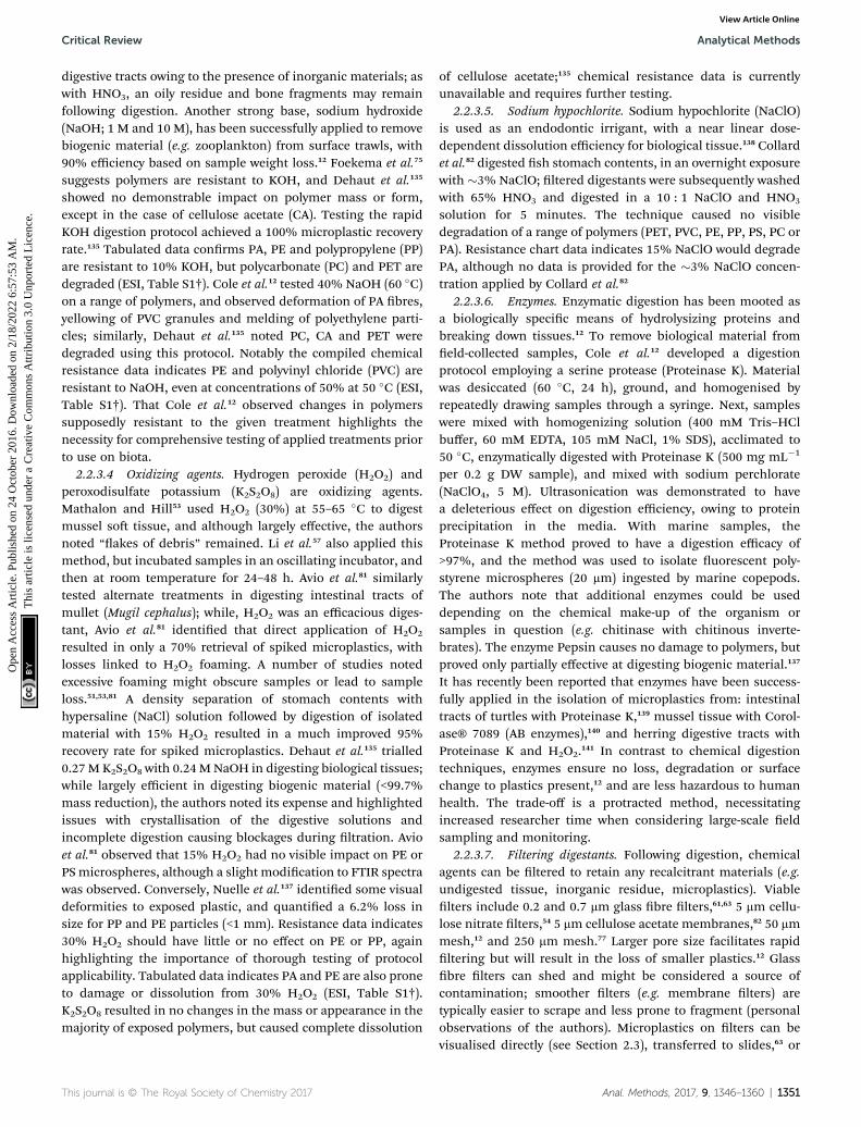
2.2.1. Dissection. In a large proportion of studiesresearchers target specic tissues, primarily the digestive tract(including the stomach and intestine). In larger animals,including squid,64 whales,97,98 turtles95 and seabirds,96 dissec-tion of the gastrointestinal tract and subsequent quanticationof synthetic particles from the gut is the predominant methodfor assessing plastic consumption. In laboratory studies, it ismore common for the whole organism (42% of studies) or thedigestive tract (26% of studies) to be digested or analysed(Fig. 3A). In comparison, 69% of eld studies targeted thedigestive tract, and 27% looked at the whole organism (Fig. 3B).Excision of the intestinal tract can also be used to ascertainconsumption of microplastics by invertebrates and vertebratesincluding pelagic and demersal sh.19,34,41,65–67,69–80,83–93 Investi-gation of stomachs and intestines is relevant for microplastic>0.5 mm in size. Microplastics larger than this do not readilypass through the gut wall without pre-existing damage, andthe likelihood of translocation into tissues is too low towarrant regular investigation.131,132 Localisation of microplastics<0.5 mm can be determined by excising organs, such as the liveror gills,62,81,105 or, where the research question relates to risks ofhuman consumption: edible tissues, for example, tail musclesof shrimp.38 Microplastics present in dissected tissues can beisolated using saline washes, density otation, visual inspec-tion, or digestion (see below).
This journal is © The Royal Society of Chemistry 2017
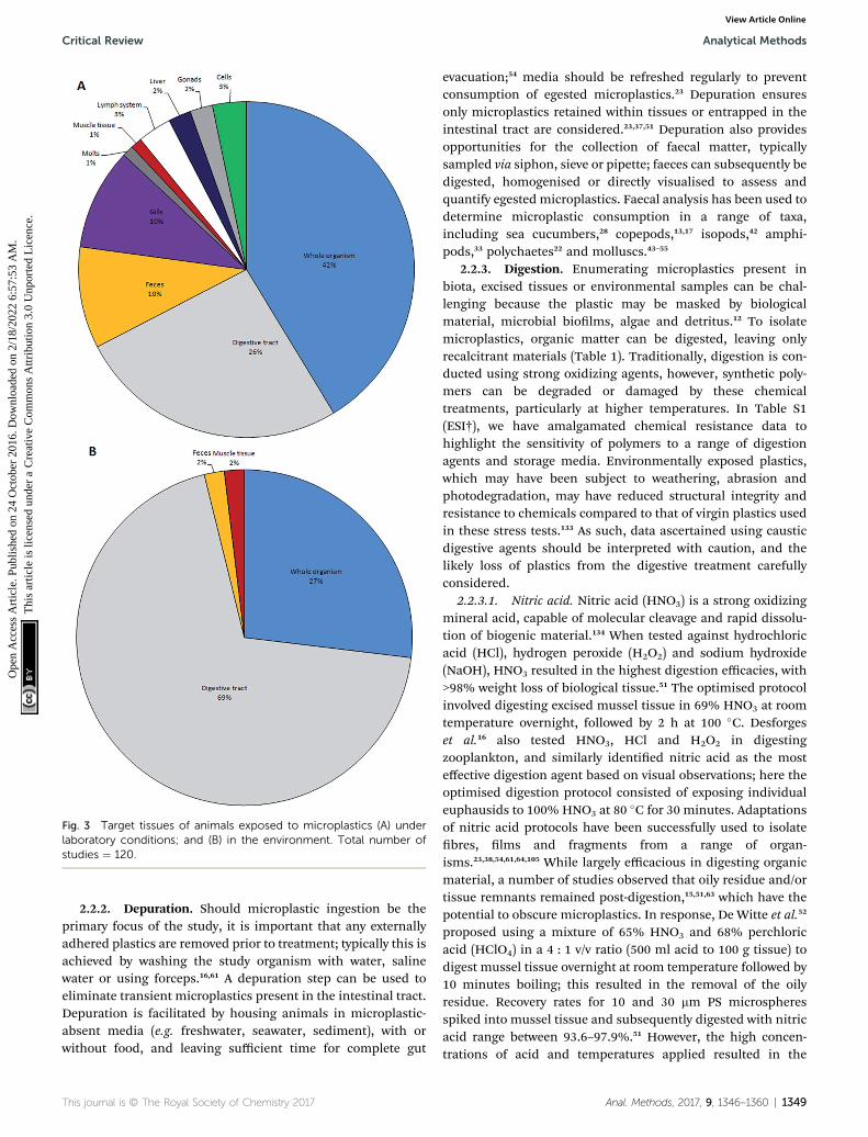
Fig. 3 Target tissues of animals exposed to microplastics (A) underlaboratory conditions; and (B) in the environment. Total number ofstudies ¼ 120.
Critical Review Analytical Methods
Ope
n A
cces
s A
rtic
le. P
ublis
hed
on 2
4 O
ctob
er 2
016.
Dow
nloa
ded
on 2
/18/
2022
6:5
7:53
AM
. T
his
artic
le is
lice
nsed
und
er a
Cre
ativ
e C
omm
ons
Attr
ibut
ion
3.0
Unp
orte
d L
icen
ce.
View Article Online
2.2.2. Depuration. Should microplastic ingestion be theprimary focus of the study, it is important that any externallyadhered plastics are removed prior to treatment; typically this isachieved by washing the study organism with water, salinewater or using forceps.16,61 A depuration step can be used toeliminate transient microplastics present in the intestinal tract.Depuration is facilitated by housing animals in microplastic-absent media (e.g. freshwater, seawater, sediment), with orwithout food, and leaving sufficient time for complete gut
This journal is © The Royal Society of Chemistry 2017
evacuation;54 media should be refreshed regularly to preventconsumption of egested microplastics.23 Depuration ensuresonly microplastics retained within tissues or entrapped in theintestinal tract are considered.23,37,51 Depuration also providesopportunities for the collection of faecal matter, typicallysampled via siphon, sieve or pipette; faeces can subsequently bedigested, homogenised or directly visualised to assess andquantify egested microplastics. Faecal analysis has been used todetermine microplastic consumption in a range of taxa,including sea cucumbers,28 copepods,13,17 isopods,42 amphi-pods,33 polychaetes22 and molluscs.43–55
2.2.3. Digestion. Enumerating microplastics present inbiota, excised tissues or environmental samples can be chal-lenging because the plastic may be masked by biologicalmaterial, microbial biolms, algae and detritus.12 To isolatemicroplastics, organic matter can be digested, leaving onlyrecalcitrant materials (Table 1). Traditionally, digestion is con-ducted using strong oxidizing agents, however, synthetic poly-mers can be degraded or damaged by these chemicaltreatments, particularly at higher temperatures. In Table S1(ESI†), we have amalgamated chemical resistance data tohighlight the sensitivity of polymers to a range of digestionagents and storage media. Environmentally exposed plastics,which may have been subject to weathering, abrasion andphotodegradation, may have reduced structural integrity andresistance to chemicals compared to that of virgin plastics usedin these stress tests.133 As such, data ascertained using causticdigestive agents should be interpreted with caution, and thelikely loss of plastics from the digestive treatment carefullyconsidered.
2.2.3.1. Nitric acid. Nitric acid (HNO3) is a strong oxidizingmineral acid, capable of molecular cleavage and rapid dissolu-tion of biogenic material.134 When tested against hydrochloricacid (HCl), hydrogen peroxide (H2O2) and sodium hydroxide(NaOH), HNO3 resulted in the highest digestion efficacies, with>98% weight loss of biological tissue.51 The optimised protocolinvolved digesting excised mussel tissue in 69% HNO3 at roomtemperature overnight, followed by 2 h at 100 �C. Desforgeset al.16 also tested HNO3, HCl and H2O2 in digestingzooplankton, and similarly identied nitric acid as the mosteffective digestion agent based on visual observations; here theoptimised digestion protocol consisted of exposing individualeuphausids to 100% HNO3 at 80 �C for 30 minutes. Adaptationsof nitric acid protocols have been successfully used to isolatebres, lms and fragments from a range of organ-isms.23,38,54,61,64,105 While largely efficacious in digesting organicmaterial, a number of studies observed that oily residue and/ortissue remnants remained post-digestion,15,51,63 which have thepotential to obscure microplastics. In response, De Witte et al.52
proposed using a mixture of 65% HNO3 and 68% perchloricacid (HClO4) in a 4 : 1 v/v ratio (500 ml acid to 100 g tissue) todigest mussel tissue overnight at room temperature followed by10 minutes boiling; this resulted in the removal of the oilyresidue. Recovery rates for 10 and 30 mm PS microspheresspiked into mussel tissue and subsequently digested with nitricacid range between 93.6–97.9%.51 However, the high concen-trations of acid and temperatures applied resulted in the
Anal. Methods, 2017, 9, 1346–1360 | 1349

Table 1 Optimised protocols for digesting biota or biogenic material to isolate microplastics. Assumptions: ‘overnight’ given as 12 h; ‘roomtemperature’ given as 20 �C
Treatment Exposure Organism Author
HNO3 (22.5 M) 20 �C (12 h) + 100 �C (2 h) Blue mussels Claessens et al. (2013)51
HNO3 (22.5 M) 20 �C (12 h) + 100 �C (2 h) Blue mussels oysters Van Cauwenberghe & Jansen (2014)54
HNO3 (22.5 M) 20 �C (12 h) + 100 �C (2 h) Blue mussels lugworms Van Cauwenberghe et al. (2013)23
HNO3 (100%) 20 �C (30 min) Euphausids copepods Desforges et al. (2015)16
HNO3 (69–71%) 90 �C (4 h) Manilla clams Davidson & Dudas (2016)61
HNO3 (70%) 2 h Zebrash Lu et al. (2016)105
HNO3 (22.5 M) 20 �C (12 h) + 100 �C (15 min) Brown mussels Santana et al. (2016)63
HNO3 (65%) 20 �C (12 h) + 100 �C (10 min) Blue mussels De Witte et al. (2014)52
HClO4 (68%) (4 : 1)
HNO3 (65%) 20 �C (12 h) + 100 �C (10 min) Brown shrimp Devriese et al. (2015)38
HClO4 (68%) (4 : 1)
CH2O2 (3%) 72 h Corals Hall et al. (2015)32
KOH (10%) 2–3 weeks Fish Foekema et al. (2013)75
KOH (10%) 60 �C (12 h) Fish Rochman et al. (2015)58
KOH (10%) 2–3 weeks Fish Lusher et al. (2016)89
H2O2 (30%) 60 �C Blue mussels Mathalon & Hill (2014)53
H2O2 (30%) 20 �C (7 d) Biogenic matter Nuelle et al. (2015)137
H2O2 (15%) 55 �C (3 d) Fish Avio et al. (2015)81
H2O2 (30%) 65 �C (24 h) + 20 �C (<48 h) Bivalves Li et al. (2015)57
NaClO (3%) 20 �C (12 h) Fish Collard et al. (2015)82
NaClO3 (10 : 1) 20 �C (5 min)
Proteinase K 50 �C (2 h) Zooplankton copepods Cole et al. (2014)12
Analytical Methods Critical Review
Ope
n A
cces
s A
rtic
le. P
ublis
hed
on 2
4 O
ctob
er 2
016.
Dow
nloa
ded
on 2
/18/
2022
6:5
7:53
AM
. T
his
artic
le is
lice
nsed
und
er a
Cre
ativ
e C
omm
ons
Attr
ibut
ion
3.0
Unp
orte
d L
icen
ce.
View Article Online
destruction of 30 � 200 mm Nylon bres and melding of 10 mmpolystyrene microbeads following direct exposure. Researchershave found that polymeric particles, including polyethylene(PE) and polystyrene (PS), dissolved following overnight expo-sure and 30 minutes boiling with 22.5 M HNO3.81,135 Polyamide(PA, Nylon), polyester (PET) and polycarbonate have low resis-tance to acids, even at low concentrations; furthermore, highconcentrations of nitric, hydrouoric, perchloric and sulphuricacid are likely to destroy or severely damage the majority ofpolymers tested, particularly at higher temperatures (ESI, TableS1†). The absence of synthetic bres in biota digested usingHNO3 is likely a reection of the destructive power of the acid.57
2.2.3.2. Other acids. Formic and hydrochloric acid (HCl)have also been suggested as digestive agents. With scleractiniancorals (Dipsastrea pallida), formic acid (3%, 72 h) has been usedto decalcify polyps to assist in the visualisation of ingested blue
1350 | Anal. Methods, 2017, 9, 1346–1360
polypropylene shavings.32 HCl has also be trialled as a digestantof microplastics from pelagic and sediment samples; howeverthis non-oxidizing acid proved inconsistent and inefficient indigesting organic material.12
2.2.3.3. Alkalis. Strong bases can be used to remove bio-logical material by hydrolysing chemical bonds and denaturingproteins.136 Excised sh tissues, including the oesophagus,stomach and intestines, have been successfully digested usingpotassium hydroxide (KOH, 10%) following a 2–3 week incu-bation.75,89 The protocol has been adapted for the dissolution ofgastrointestinal tracts of sh and mussel, crab and oystertissues, either directly or following baking (450 �C, 6 h), byincubating tissues in 10% KOH at 60 �C overnight.58,135 Thislatter method has proven largely efficacious in removingbiogenic material, being well suited to the dissolution ofinvertebrates and sh llets, but proving less applicable for sh
This journal is © The Royal Society of Chemistry 2017
Critical Review Analytical Methods
Ope
n A
cces
s A
rtic
le. P
ublis
hed
on 2
4 O
ctob
er 2
016.
Dow
nloa
ded
on 2
/18/
2022
6:5
7:53
AM
. T
his
artic
le is
lice
nsed
und
er a
Cre
ativ
e C
omm
ons
Attr
ibut
ion
3.0
Unp
orte
d L
icen
ce.
View Article Online
digestive tracts owing to the presence of inorganic materials; aswith HNO3, an oily residue and bone fragments may remainfollowing digestion. Another strong base, sodium hydroxide(NaOH; 1 M and 10 M), has been successfully applied to removebiogenic material (e.g. zooplankton) from surface trawls, with90% efficiency based on sample weight loss.12 Foekema et al.75
suggests polymers are resistant to KOH, and Dehaut et al.135
showed no demonstrable impact on polymer mass or form,except in the case of cellulose acetate (CA). Testing the rapidKOH digestion protocol achieved a 100% microplastic recoveryrate.135 Tabulated data conrms PA, PE and polypropylene (PP)are resistant to 10% KOH, but polycarbonate (PC) and PET aredegraded (ESI, Table S1†). Cole et al.12 tested 40% NaOH (60 �C)on a range of polymers, and observed deformation of PA bres,yellowing of PVC granules and melding of polyethylene parti-cles; similarly, Dehaut et al.135 noted PC, CA and PET weredegraded using this protocol. Notably the compiled chemicalresistance data indicates PE and polyvinyl chloride (PVC) areresistant to NaOH, even at concentrations of 50% at 50 �C (ESI,Table S1†). That Cole et al.12 observed changes in polymerssupposedly resistant to the given treatment highlights thenecessity for comprehensive testing of applied treatments priorto use on biota.
2.2.3.4 Oxidizing agents. Hydrogen peroxide (H2O2) andperoxodisulfate potassium (K2S2O8) are oxidizing agents.Mathalon and Hill53 used H2O2 (30%) at 55–65 �C to digestmussel so tissue, and although largely effective, the authorsnoted “akes of debris” remained. Li et al.57 also applied thismethod, but incubated samples in an oscillating incubator, andthen at room temperature for 24–48 h. Avio et al.81 similarlytested alternate treatments in digesting intestinal tracts ofmullet (Mugil cephalus); while, H2O2 was an efficacious diges-tant, Avio et al.81 identied that direct application of H2O2
resulted in only a 70% retrieval of spiked microplastics, withlosses linked to H2O2 foaming. A number of studies notedexcessive foaming might obscure samples or lead to sampleloss.51,53,81 A density separation of stomach contents withhypersaline (NaCl) solution followed by digestion of isolatedmaterial with 15% H2O2 resulted in a much improved 95%recovery rate for spiked microplastics. Dehaut et al.135 trialled0.27 M K2S2O8 with 0.24 M NaOH in digesting biological tissues;while largely efficient in digesting biogenic material (<99.7%mass reduction), the authors noted its expense and highlightedissues with crystallisation of the digestive solutions andincomplete digestion causing blockages during ltration. Avioet al.81 observed that 15% H2O2 had no visible impact on PE orPS microspheres, although a slight modication to FTIR spectrawas observed. Conversely, Nuelle et al.137 identied some visualdeformities to exposed plastic, and quantied a 6.2% loss insize for PP and PE particles (<1 mm). Resistance data indicates30% H2O2 should have little or no effect on PE or PP, againhighlighting the importance of thorough testing of protocolapplicability. Tabulated data indicates PA and PE are also proneto damage or dissolution from 30% H2O2 (ESI, Table S1†).K2S2O8 resulted in no changes in the mass or appearance in themajority of exposed polymers, but caused complete dissolution
This journal is © The Royal Society of Chemistry 2017
of cellulose acetate;135 chemical resistance data is currentlyunavailable and requires further testing.
2.2.3.5. Sodium hypochlorite. Sodium hypochlorite (NaClO)is used as an endodontic irrigant, with a near linear dose-dependent dissolution efficiency for biological tissue.138 Collardet al.82 digested sh stomach contents, in an overnight exposurewith �3% NaClO; ltered digestants were subsequently washedwith 65% HNO3 and digested in a 10 : 1 NaClO and HNO3
solution for 5 minutes. The technique caused no visibledegradation of a range of polymers (PET, PVC, PE, PP, PS, PC orPA). Resistance chart data indicates 15% NaClO would degradePA, although no data is provided for the �3% NaClO concen-tration applied by Collard et al.82
2.2.3.6. Enzymes. Enzymatic digestion has been mooted asa biologically specic means of hydrolysizing proteins andbreaking down tissues.12 To remove biological material fromeld-collected samples, Cole et al.12 developed a digestionprotocol employing a serine protease (Proteinase K). Materialwas desiccated (60 �C, 24 h), ground, and homogenised byrepeatedly drawing samples through a syringe. Next, sampleswere mixed with homogenizing solution (400 mM Tris–HClbuffer, 60 mM EDTA, 105 mM NaCl, 1% SDS), acclimated to50 �C, enzymatically digested with Proteinase K (500 mg mL�1
per 0.2 g DW sample), and mixed with sodium perchlorate(NaClO4, 5 M). Ultrasonication was demonstrated to havea deleterious effect on digestion efficiency, owing to proteinprecipitation in the media. With marine samples, theProteinase K method proved to have a digestion efficacy of>97%, and the method was used to isolate uorescent poly-styrene microspheres (20 mm) ingested by marine copepods.The authors note that additional enzymes could be useddepending on the chemical make-up of the organism orsamples in question (e.g. chitinase with chitinous inverte-brates). The enzyme Pepsin causes no damage to polymers, butproved only partially effective at digesting biogenic material.137
It has recently been reported that enzymes have been success-fully applied in the isolation of microplastics from: intestinaltracts of turtles with Proteinase K,139 mussel tissue with Corol-ase® 7089 (AB enzymes),140 and herring digestive tracts withProteinase K and H2O2.141 In contrast to chemical digestiontechniques, enzymes ensure no loss, degradation or surfacechange to plastics present,12 and are less hazardous to humanhealth. The trade-off is a protracted method, necessitatingincreased researcher time when considering large-scale eldsampling and monitoring.
2.2.3.7. Filtering digestants. Following digestion, chemicalagents can be ltered to retain any recalcitrant materials (e.g.undigested tissue, inorganic residue, microplastics). Viablelters include 0.2 and 0.7 mm glass bre lters,61,63 5 mm cellu-lose nitrate lters,54 5 mm cellulose acetate membranes,82 50 mmmesh,12 and 250 mm mesh.77 Larger pore size facilitates rapidltering but will result in the loss of smaller plastics.12 Glassbre lters can shed and might be considered a source ofcontamination; smoother lters (e.g. membrane lters) aretypically easier to scrape and less prone to fragment (personalobservations of the authors). Microplastics on lters can bevisualised directly (see Section 2.3), transferred to slides,63 or
Anal. Methods, 2017, 9, 1346–1360 | 1351
Analytical Methods Critical Review
Ope
n A
cces
s A
rtic
le. P
ublis
hed
on 2
4 O
ctob
er 2
016.
Dow
nloa
ded
on 2
/18/
2022
6:5
7:53
AM
. T
his
artic
le is
lice
nsed
und
er a
Cre
ativ
e C
omm
ons
Attr
ibut
ion
3.0
Unp
orte
d L
icen
ce.
View Article Online
extracted. Collard et al.82 suggests placing lters in methanolsolution, ultrasonicating (50 Hz), centrifuging (5000 rpm,5 min, 20 �C) and then removing pelleted plastic by pipette;while this method was suitable for a range of polymers, themethanol caused a 25% weight reduction in tested PVCparticles.
2.2.4. Density separation. Although most commonly uti-lised in studies of water and sediment samples, density sepa-ration has been used in four biotic studies. Three studies usedNaCl to separate less dense particles53,57,81 while Collard et al.82
used a centrifuge. Following settlement of denser materials, thesupernatant is ltered and the resulting material examinedunder microscope. Density separation can be useful in studiesfollowing digestion. Saturated salt solutions, such as NaCl (aq)allow the separation of less dense particles where there is a largeamounts of inorganic matter (e.g. sand, chitin, bone) that hasnot been dissolved (A. Lusher, personal observations). Densityseparation has been recommended by the MSFD (EU) forEurope. NaCl is recommended because it is inexpensive andnon-hazardous; however, the use of NaCl could lead to anunderestimation of more dense particles (>1.2 g cm�3). NaI andZnCl2 solutions have been considered as viable alternatives toNaCl (aq).142 Their high density makes them capable of oatinghigh-density plastics including polyvinylchloride (PVC).
2.3. Microplastic identication
Following the preparation of target tissues, the quantity andtypes of microplastics should be ascertained. Of the methodscurrently employed, visual identication is most widely utilized;oen in combination with one or more follow-up analyticaltechniques. Researchers can use characteristics, includingmorphology and density, to identify the presence of micro-plastics. Visual identication is based on the morphologicaland physical characteristics of particles whereas chemicalcharacteristics are determined by more advanced analyticaltechniques.
2.3.1. Visual identication. Early reports quantifying envi-ronmental plastics primarily relied upon visual identication;this method remains an essential step in classifying micro-plastics, and is perfectly acceptable when supported by subse-quent polymer analysis of sub-samples. Visual identicationcan be conducted using light, polarised or electron microscopy.Semi-automated methods, including ZooScan,143 ow cytom-etry,144 cell sorters and coulter counters,14 allow for a largenumber of samples to be analysed rapidly; however, theserequire technical expertise, specialised equipment, and timemust still be given to sample preparation and data analysis.Scanning Electron Microscopes (SEM) produce high resolutionimages and have been implemented in several studies either toimage recovered plastics23,41 or as a way of identifying microbialcolonisation.145
Visual identication is rapid, relatively cheap and can beconducted without the need for additional technical staff andconsumables; however, accurately differentiating microplastics,particularly in the smaller size ranges, requires training andexperience. Criteria for visually identifying microplastics
1352 | Anal. Methods, 2017, 9, 1346–1360
include: the absence of cellular or organic structures;a homogenous thickness across the particles; and, homogenouscolors and gloss.77,123 Manually sorting plastics under a micro-scope is most effective for particles >500 micrometers; the effortand accuracy required for sorting increases with decreasingparticle size. Owing to the difficulties in handling and differ-entiating microplastics from organic and inorganic matter,146
error rates could be as high as 70%, increasing with decreasingparticle size,61 with incorrect identication most prevalent withmicrobres.123,147 To gauge the accuracy of visual discrimina-tion, sub-samples of potential plastics should be chemicallyanalysed.77,123,147–150 It has been observed that training andexperience can signicantly lower the error rates and misiden-tication stemming from visual identication.83
Plastics are largely classied by their morphological char-acteristics: size, shape, and colour. Size is typically based on thelongest dimension of a particle; size categories can be usedwhere appropriate. When reporting microplastic shape,researchers tend to use ve main categories, although thenomenclature used varies between research groups (Table 2).Finally, colours are oen reported across a wide spectrum;colour differentiation is subjective, and visual identication ofmicroplastics cannot be based on colour alone. Caution shouldbe given to categorising microplastics suffering embrittlement,fragmentation or bleaching, or encrusted with biota, as this mayskew results.
2.3.2. Polymer verication. Due to the challenges in visu-ally identifying microplastics, secondary analyses should beused to conrm the identity of suspected polymeric material.The method employed is oen dictated by the equipmentavailable and whilst any chemical characterisation of the poly-mers recovered is useful, some techniques are more robust thanothers. The European Commission suggests that a subsample(5–10%) of particles with a size between 100 mm and 5 mm andall particles between 20 and 100 mm should be subjected tofurther verication techniques. Post-visual analyses have shownmisidentication of microplastics in wild caught animals of upto 70%.82,147,150 It should be noted here, that errors in identi-cation oen include un-matched spectra that could not beassigned with condence to a known polymer type, condencethresholds for spectra matches are usually set at 70–75%.77,83,89
Conrming the identity of suspected plastics may be carriedout in a number of ways depending on the funds and equip-ment available to the researcher. Perhaps the simplest tech-nique is the use of a hot needle to observe meltingpoints.38,52,60,86 While both cheap and fast, this method does notallow for the accurate identication of the polymer; however,the temperature range at which melting occurs does providea specic range of potential plastics. A converse method is toexclude non-plastics rather than identifying the plasticspresent, oven and freeze drying removes water from organicmaterial causing it to wither. This increases the likelihood ofnon-plastic material being identied and removed from mixedsamples.151,152 Combining these two techniques providesa cheap, if laborious method of plastic identication.
Another low cost technique involves the examination ofmicroplastics under a polarised light microscope to observe the
This journal is © The Royal Society of Chemistry 2017

Table 2 Categorises used when classifying microplastic by shape
Shape classication Other terms used
Fragments Irregular shaped particles, crystals, uff,powder, granules, shavings
Fibres Filaments, microbres, strands, threadsBeads Grains, spherical microbeads, microspheresFoams Polystyrene, EPSPellets Resin pellets, nurdles,
pre-production pellets, nibs
Critical Review Analytical Methods
Ope
n A
cces
s A
rtic
le. P
ublis
hed
on 2
4 O
ctob
er 2
016.
Dow
nloa
ded
on 2
/18/
2022
6:5
7:53
AM
. T
his
artic
le is
lice
nsed
und
er a
Cre
ativ
e C
omm
ons
Attr
ibut
ion
3.0
Unp
orte
d L
icen
ce.
View Article Online
birefringent properties of the suspected polymer. The birefrin-gence of a polymer is the result of its chemical structure andmanufacturing methods which results in unique anisotropicproperties; by passing polarised light through a sample, uniquespectra are created, from which it is possible to conrm theidentity of plastic materials.153 As with the hot needle technique,this method require plastics to be viewed individually; whilstinitial costs are low, the time taken makes it prohibitive forlarge samples. More complex – and costly –methods can also beused to infer resin constituents, plastic additives and dyes.Oen, these techniques require the purication of potentialmicroplastic prior to analysis. The removal of biolms, organicand inorganic matter adhered to the surface will avoidimpeding polymer identication and the removal of non-plasticparticles.146 Following purication, suspected plastics aresubmitted to analytical techniques including: Fourier Trans-formed Infra-Red spectrometry (FT-IR) in transmittance orreectance; Attenuated Total Reectance (ATR); Raman spec-trometry for colour pigment spectra; and, Pyrolysis-Gas Chro-matography combined with Mass Spectroscopy (Pyr-GC-MS),which analyzes particles using their thermal degradationproperties and can be used to analyse polymer type and organicplastic additives simultaneously.154 Alternate analyticalmethods include: high temperature gel-permeation chroma-tography (HT-GPC) with IR detection; SEM-EDS and thermoex-traction; and, desorption coupled with GC/MS.150,155,156
If coupled with microscopy, FTIR and Raman can be used toidentify microplastics with a size >20 mm.123,149 Raman spec-troscopy combined with microscopy has a higher resolution(approx. 1–2 mm)100,149 and can be used to locate particles withinbiological tissues.10 FT-IR and Raman have been recommendedfor determining resin constituents.123,149 There is minimalsample preparation, other than clean up, required for FT-IR.However, FT-IR and PYr-GS-MS are both destructive. Raman isnon-destructive as it does not require the sample to be attenedor manipulated. The disadvantage of PYr-GS-MS is the manualplacement of the particle in the instrument, which can incursize limitations and only one particle can be run per sample.However, qualitative and quantitative analysis are being devel-oped.141,157 A drawback of chemical analysis is that the isolationof small, highly degraded samples increases the chances ofmisidentication and producing noisy spectra in which the vitalngerprint areas are obscured, although this can be improvedby the use of microscope aided instrumentation (micro-FTIRand micro-Raman), which is designed to target and readresponses from samples of a smaller size.
2.4. Contamination
At all stages care must be taken to prevent the contamination orcross-contamination of samples. Airborne contamination ofsamples with synthetic bres stemming from clothing75,158 oratmospheric fallout159 is a recurrent issue within the litera-ture.52,53,58,61,63 Sources of contamination should be eliminatedwhere possible, and otherwise quantied using environmentallters or procedural blanks. Here we highlight sources of
This journal is © The Royal Society of Chemistry 2017
contamination during sampling and sample processing, andconsider protocols for contamination mitigation.
2.4.1. Contamination during eld sampling. With marinespecies, animals are oen sampled by way of polymer rope, netsor traps.123 In these situations, animals should only be exposedfor minimal periods and a reference sample of the gear shouldbe retained to exclude contamination during the identicationphase.77 Avoiding airborne contamination of samples in theeld is understandably more complex than in the laboratory,but remains an important consideration nevertheless. Steps formitigating contamination include: thorough cleaning of allequipment prior to sampling, which will also mitigate cross-contamination; covering samples and equipment between use;wearing polymer-free clothing or cotton coveralls, and gloves;and the use of procedural blanks.
2.4.2. Contamination during sample processing and anal-ysis. In the laboratory, forensic techniques, good laboratorypractice and common sense should be applied to mitigatecontamination.160 Wherever feasible, researchers shouldprocess samples in a laminar ow hood (e.g. cell or algal cultureunit);12,54,55,82 alternatively a fume-hood63 or ‘clean room’ (i.e.non-ventilated or negative ow) with low foot-traffic or embar-goed to non-essential personnel can be used. Glassware ispreferential to plastic consumables; Cole et al.12 observedphysical homogenisation of specimens in polypropylene Falcontubes resulted in the introduction of plastic shavings to thesample. Filtering media or liquids used in sample preparationhas been recommended by some researchers.54,55 Glassware,benches and equipment should be rinsed with deionisedwater,53–55,58,63,77,89 ethanol12,82 or acetone52 prior to use. Collardet al.82 further suggests drying equipment with cellulose-ligninbased cloths from which reference samples can be taken. Aswith eld sampling, all materials should be covered betweenuse, and cotton coveralls or laboratory coats are widely recom-mended. Environmental lters (e.g. glass bre lters) can beplaced near equipment to quantify external contamination.77,89
Lastly, procedural blanks (i.e. controls) are highly recom-mended for quantifying contamination and for identifyingaspects of the experimental design where contamination canoccur. Analysis of procedural blanks can reveal substantialcontamination of synthetic bres, ranging 5.8 � 2.261 to 33–39bres137 per replicate. Where contaminating plastics are easilyidentiable, for example being brightly coloured,137 >1.5 mm38
or <36 mm,82 or resembling laboratory coat bres,58 thesemicroplastics can be removed from subsequent analysis.
Anal. Methods, 2017, 9, 1346–1360 | 1353

Analytical Methods Critical Review
Ope
n A
cces
s A
rtic
le. P
ublis
hed
on 2
4 O
ctob
er 2
016.
Dow
nloa
ded
on 2
/18/
2022
6:5
7:53
AM
. T
his
artic
le is
lice
nsed
und
er a
Cre
ativ
e C
omm
ons
Attr
ibut
ion
3.0
Unp
orte
d L
icen
ce.
View Article Online
2.5. Data analysis
The varied methods by which microplastic uptake by biota ismeasured understandably results in differing levels ofrecording. At the highest level, researchers record the numberof items, oen in relation to organism size. This may berecorded simply, as the percentage of individuals seen to ingestmicroplastic,36,66,71,76,77,79,84,89 the number of microplastics perindividual,16,53,54,57,58,65,74,78,80,85,88–91,102 or as the number ofmicroplastic items by length or weight.38,54,60,61,73,77,89 Many typesof plastic, for example microbre boluses, do not lend them-selves to the enumeration of individual plastic items. In addi-tion, mastication and peristaltic action may break down plasticitems within the gut; as a result, the number of items in the gutmay exceed that originally ingested. In such cases, researchershave reported the weight of plastic aggregations,41 descriptionsof the aggregation of microplastic observed,34 or a combinationof the two. Such issues in enumeration are more oen observedin studies of wild caught organisms, where the initial level ofmicroplastic exposure is not known and the type of microplasticrecovered being susceptible to tangling. A similar issue may beobserved in the study of microplastic uptake in laboratoryexperiments; here concentrations of introduced microplasticmay be recorded solely by number or mass per individual,19,161
or as a value in relation to mass of food33,34,92,99,161–163 or volumeof water.10,26,164
The use of multiple methods to quantify the level of micro-plastic uptake by sh and invertebrates is also an issue in thereporting of environmental plastic levels. Inconsistency in theuse of units can mask or inate the apparent impact of micro-plastic on a species or location. This increases the likelihood oferrors arising when comparing multiple studies carried out byunrelated researchers. The manner in which plastic abundanceand concentration is recorded inuences the range of statisticalanalyses available; for example, grouping aggregations intospecic classications reduces the power of the available tests.In eld experiments, a range of techniques have been used todetermine the relationship between microplastic uptake andboth biological and environmental factors. Many of thesemethods combine continuous and categorical variables inlinear models of varying complexity, and require careful struc-turing in statistical soware such as R statistical soware.165
3 Discussion
Techniques used to isolate and enumerate microplastics in shand invertebrates have largely been adapted from studiesfocussed on large vertebrates (e.g. marine mammals, seabirds;Provencher, this issue), or have derived from traditional biologymethods; as these elds advance, it is vital that we continue toobserve their progress and incorporate relevant methods.Owing to the challenges in sampling, isolating and identifyingmicroplastics, and the diverse physiology of taxonomic groupsunder investigation, a degree of exibility, innovation andingenuity on the part of the researcher is clearly required.
Of the numerous studies investigating microplastic uptakein sh and invertebrates, it is analysis of wild-caught animals
1354 | Anal. Methods, 2017, 9, 1346–1360
that presents the most issues. In these studies, potential sour-ces of error are numerous, including microplastic losses orcontamination during sampling; furthermore, there aresubstantial challenges in drawing links between exposure andeffect. These studies, however, are essential for establishingecologically relevant data, which ultimately providesresearchers with a clear view of the quantity and types of plastic(and associated contaminants) experienced by biota in thenatural environment. In this section, we address the need forstandardising protocols for microplastic quantication,166 out-lining preferred methods, best practice, and steps for miti-gating contamination. It is anticipated that compilingstandardized methodologies will provide researchers witha grounding in developing future experimental design.
3.1. Controlling sampling bias
Throughout the sampling process, utmost caremust be taken toprevent the articial ination or loss of microplastics. In Table 3we outline the commonly used methods for sampling andisolating microplastics across a range of taxa. In all cases, theleast damaging sampling gear is preferable, and samplingperiods should be kept as short as practically realistic. Organ-isms that spend longer in nets are subject to additional stressthat increases the likelihood of regurgitation or stomachinversion and articially increases contact time betweenmicroplastics and biota; this could facilitate microplasticingestion and adherence to external appendages. Individualsshould be rinsed following capture to remove adhered particles,and samples of shing gear should be taken to exclude materialingested as a result of capture. We recommend researchersover-sample where practical, so that individuals with recentlyemptied stomachs, or otherwise damaged during sampling,should be omitted from the dataset; this will help reduce thebias caused by regurgitation, and enable more robust compar-isons between animals sampled from different sites or collectedusing alternate sampling methods. Specimens should berapidly transported to the laboratory or preserved promptly toavoid microplastic egestion in transit. We note that collectionfrom commercial sh markets or artisanal shers is not ideal,as the researcher will have less, if any, control on the method ofcapture and the handling conditions on transport. Whereapplicable, we suggest researchers work closely with shers toensure animals are sampled appropriately and sufficientinformation on the capture procedure is collected.
3.2. Effective plastic isolation
Researchers are presented with a range of techniques forisolating microplastics from biota, including: dissection, dep-uration, digestion and density separation. Determining theappropriate method will largely depend on the research ques-tion (e.g. risks of human consumption, total body burden,localised accumulation). Digestion of whole organisms orexcised tissues is widely used (Table 2), however caution mustbe given in selecting an appropriate digestive agent due to thepotential destruction of contaminants. In Table S1 (ESI†) wehighlight the damage that a range of digestive agents can cause
This journal is © The Royal Society of Chemistry 2017

Table 3 Standard sampling and plastic isolation strategies employed across a variety of subphyla
Ecosystem Life strategy Subphylum Size range Sampling methodInitial plasticseparation
Aquatic Benthic Annelida — Grabs DigestionAquatic Benthic Crustacea >50 mm Trawls/creels DissectionAquatic Benthic Crustacea <50 mm Otter-/beam-trawls DigestionAquatic Benthic Echindodermata — Grab/trawls DissectionAquatic Benthic Mollusca >30 mm Grabs DissectionAquatic Benthic Mollusca <30 mm Grabs DigestionAquatic Benthic Flatsh — Otter-/beam-trawls DissectionAquatic Nektonic Crustacea (juv.) <50 mm Mid-water trawls DigestionAquatic Nektonic Gadids Otter-/mid-water trawls DissectionAquatic Nektonic Echindodermata (juv.) <2 cm Trawls DigestionAquatic Nektonic Mollusca (juv.) <2 cm Trawls DigestionAquatic Nektonic Fish <10 cm Mid-water trawls DissectionAquatic Planktonic Annelida <2 cm Trawls DigestionAquatic Planktonic Cnidaria <10 cm Trawls DigestionAquatic Planktonic Crustacea <2 cm Trawls DissectionTerrestrial — Annelida — Sediment collection DigestionTerrestrial — Arachnida — Trapping/hand gathering DigestionTerrestrial — Crustacea — Trapping/hand gathering DigestionTerrestrial — Insecta — Trapping/hand gathering DigestionTerrestrial — Mollusca — Trapping/hand gathering Digestion
Critical Review Analytical Methods
Ope
n A
cces
s A
rtic
le. P
ublis
hed
on 2
4 O
ctob
er 2
016.
Dow
nloa
ded
on 2
/18/
2022
6:5
7:53
AM
. T
his
artic
le is
lice
nsed
und
er a
Cre
ativ
e C
omm
ons
Attr
ibut
ion
3.0
Unp
orte
d L
icen
ce.
View Article Online
to polymers. For example, >50% formic acid, >35% HClO4, >40%hydrouoric acid, >80% H2O2, >50% HNO3, >70% HClO4, >50%KOH and >95% sulphuric acid can be particularly damaging tospecic polymers. Some digestive agents, including H2O andHClO4, are simply ineffective in breaking down tissues. Ouranalysis found that some recent studies used high percentages ofacids to which several polymers are not resistant. In particular,high percentages of and HClO4 have been utilised in severalstudies.16,23,38,51,52,54,60,61,105 For example, only PET and PVC areresistant to 50%HNO3, whereas, PE and PP are partially resistantand PA, PC and PS are not resistant. Typically a balance will needto be struck between nding a cost-effective digestive agent withthe capacity to effectively break down tissue, without losingmicroplastics. Based on our analyses, the rapid 10% KOH (60 �Covernight)58,135 and enzymatic digestion protocols12,140 appear, onbalance, to be among the most widely tested and effectivedigestive treatments currently available; in all cases, the costs,strengths and weaknesses, and applicability of each method tothe study organism in question should be carefully considered.As with sampling, steps for mitigating and accounting forexternal contamination are paramount.
3.3. Polymer verication
Methods for verifying isolated microplastics vary in complexityand expense. The method used is dependent not only onresources available to the research group, but also the degree ofinformation required by the study. Studies examining the totalbody burden or the rate of uptake may only require the mostcursory identication to conrm that the particles recovered areindeed plastic; whereas research examining the potentialorigins of plastics or the presence of adsorbed contaminantsand additives require more rigorous testing. We concur with theEuropean standard of polymeric identication of a 5–10%
This journal is © The Royal Society of Chemistry 2017
subsample of isolated microplastics. Whilst it has been sug-gested that this level is insufficient to accurately determine theability of the researcher to accurately identify plastics, this canbe improved when selecting subsamples for verication.Researchers must ensure a representative sample encompass-ing all categories of recovered microplastics, and particularconsideration should be given to commonly misidentiedforms, such as bres and small size fractions. Where possible,researchers should include the weight and number of plasticitems, and themass of sampled organism should be recorded incases where animals are grouped prior to analysis. Suchstandardisation will improve comparability between studies.
3.4. Mitigating contamination
Contamination, cross-contamination and loss of plastics area major challenge for microplastics research. It is recom-mended that all laboratory processing should include steps forpreventing or limiting airborne contamination, and proceduralblanks used to account for this.124 Additional processing may beutilised during microplastic isolation to improve the detect-ability or identication of microplastic, however each addi-tional step increases the opportunity for contamination. Wherepossible plastic consumables should be avoided.167 All samplesshould be preserved by freezing, desiccation, or in lteredethanol or formalin, although the latter may result in loss ofsome plastics. On research vessels, glassware may not befeasible, so plastic may be used following sufficient cleaningwith ltered water.
3.5. Data analysis
Researchers should be aware of biases in sampling environ-mentally exposed individuals. Firstly, the condition of
Anal. Methods, 2017, 9, 1346–1360 | 1355

Analytical Methods Critical Review
Ope
n A
cces
s A
rtic
le. P
ublis
hed
on 2
4 O
ctob
er 2
016.
Dow
nloa
ded
on 2
/18/
2022
6:5
7:53
AM
. T
his
artic
le is
lice
nsed
und
er a
Cre
ativ
e C
omm
ons
Attr
ibut
ion
3.0
Unp
orte
d L
icen
ce.
View Article Online
organisms prior to capture is unknown, and linking micro-plastic (and co-contaminant) burden with condition is prohib-itive. Secondly, sampling may lead to an underestimation of themicroplastic burden in a population because highly contami-nated individuals are dead or dying, or remain in shelters andburrows owing to reduced functionality. Thirdly, it is vital thatsampling is spatially and/or temporally broad to ensure thatobserved levels of contamination are representative of the widerpopulation. For example, Welden et al.41 observed signicantspatial variation in bre contamination in three Scottish pop-ulations of Nephrops norvegicus.
3.6. Recommendations for future work
In reviewing the relevant literature, it is apparent that researchis currently skewed towards vertebrates (Fig. 4). The range ofecological functions carried out by invertebrates and the diverseniches which they occupy all suggest that the impacts ofmicroplastics on these groups may have a marked effect on theenvironment. We recommend further assessment of the uptakeand impact of microplastics in these groups as this is essentialif we are to predict the extent of the effects on biodiversity,ecosystems and ecological processes. A comparison of therelative uptake and retention of the different categories andshapes of microplastics is also required to determine which arethe most harmful. Many laboratory studies of microplasticingestion rely solely on pre-produced plastics which are easilypurchased from suppliers (e.g. polystyrene microbe-ads10–15,17,26,47,105,117); however, these are not representative of thediverse forms currently present in the environment. Under-standing which plastics are readily retained is necessary todetermine the threat posed by the relative levels of environ-mental microplastics and to link these to evidence of negativeeffects in physiology and behaviour which may impair functionat the ecosystem level.
In fact, a number of studies have considered the observableimpacts of microplastic on organisms. In 11/120 studiesreviewed here, researchers examined the relationship between
Fig. 4 Laboratory and field studies investigating microplastic inter-actions with biota separated by Phyla. The value in parentheses aboveeach bar indicates the number of species studied within the taxa. Totalnumber of studies included in this review ¼ 120.
1356 | Anal. Methods, 2017, 9, 1346–1360
microplastic uptake and changes in physiology and bodychemistry. Endpoints have included: organism behaviour,104,168
lysosomal response,56 lipid content,169,170 protein content,170
population tness,161 cellular population growth171 and indi-vidual growth.15,170 They also utilised ecotoxicological assays tomonitor embryonic development31,172 and the uptake ofmetals94,173 and chemicals.162,163,174 Laboratory studies must useenvironmentally realistic concentrations of micro particles, toallow evaluation of potential harm to the individual as a resultof eld exposure. In wild populations, where the presence ofconfounding factors make it difficult to attribute biologicalresponses and condition directly to plastic exposure, directobservation of plastic type and abundance remains the mostreliable method of determining microplastic impacts.
Microplastics may be selectively or non-selectively ingestedor acquired through trophic transfer. Again, the articiallyinated levels of non representative plastics used in a numberof currently available studies greatly increases the potential forchance transfer of plastics. The result of such studies, whilstuseful in indicating the potential for transfer, display transfer ina way not feasible at normal contamination levels. In studiesnot focusing on the ecotoxicological impacts, we recommendinated levels of microplastic should only be used in the pres-ence of an ecologically valid control determined by reference torigorous environmental sampling.
Lastly, few studies have addressed the movement of micro-plastic within ecosystems. Many rely on seeding plastic intonon-natural food sources, in mesocosm experiments with noalternate food sources, and whilst these experiments haveshown the transfer of plastic between food and organisms,there is a clear need for more robust information on the validityof these results in natura.175
The need for rapid, accurate assessment of the levels ofmicroplastic in wild populations is essential for determiningbaseline levels of contamination, and assessing the risk ofmicroplastic to organisms and ecosystems. The diverse physi-ology of the organisms covered in this review has necessitatedthe analysis of a range of protocols for microplastic extractionand enumeration which could limit comparability betweenstudies. As such, we recommend the development of a standardmethodology per subphylum or class of organism which willcombine efficiency in digestion and recovery of microplasticwith the use of the least toxic chemicals to preserve plasticpolymers.
Particular attention should be given to harmonising the wayin which data is recorded (e.g. mass and number of isolatedmicroplastics per mass of organism) to promote comparability.Prevention of overestimation of plastic contamination must becontrolled by conrming the identity of a proportion of therecovered materials. This may be carried out either by chemicalanalysis, density separation, birefringent microscopy or otherphysical examination.
4 Concluding remarks
In this review various methods for sampling, isolating andidentifying microplastics ingested by sh and invertebrates
This journal is © The Royal Society of Chemistry 2017
Critical Review Analytical Methods
Ope
n A
cces
s A
rtic
le. P
ublis
hed
on 2
4 O
ctob
er 2
016.
Dow
nloa
ded
on 2
/18/
2022
6:5
7:53
AM
. T
his
artic
le is
lice
nsed
und
er a
Cre
ativ
e C
omm
ons
Attr
ibut
ion
3.0
Unp
orte
d L
icen
ce.
View Article Online
were examined. As research progresses the need for methodstandardisation becomes clear, so that a ner picture of thethreat of microplastic to organisms emerges and ecological andenvironmental risk can be assessed. Such standardised meth-odologies must take into account the numerous potentialsources of error and contamination, as outlined above, and alsothe general need for monitoring which demands that a greatnumber of samples be processed hastily.
Many of the studies covered in this review have focused onthe issue of whether microplastic uptake occurs, and onlyaddresses the level of plastic contamination in a single speciesor group and do not allow the assess of risk and disturbance atthe ecosystem level. These studies have resulted in numerousfurther questions regarding the uptake and transfer of micro-plastics within ecosystems: is plastic uptake selective, orpassive? To what extent does trophic transfer occur? And, arethe negative effects of plastic ingestion observed in laboratoryexperiments valid in the environment? There is a strong need todesign studies in such a way that their results contribute toclarify these issues, for example contrasting microplastic loadswith environmental contamination or diet, to give a moreholistic approach to the study of microplastic pollution.
Acknowledgements
MC is funded by the Natural Environment Research Council(NERC UK: NE/L007010). Thanks to S. Nelms for her usefulcomments on an earlier version of the manuscript. Thanks areextended to C. Rochman and R. Thompson for inviting us tocontribute to this special issue.
References
1 Plastics – the Facts 2014/2015, http://issuu.com/plasticseuropeebook/docs/nal_plastics_the_facts_2014_19122, accessed August2015.
2 M. Cole, P. Lindeque, C. Halsband and T. S. Galloway, Mar.Pollut. Bull., 2011, 62(12), 2588–2597.
3 UNEP, Marine Plastic Debris and Microplastic TechnicalReport, United Nations Environmental Programme,Nairobi, 2016.
4 A. Lusher, in Marine Anthropogenic Litter, ed. M. Bergmann,L. Gutow and M. Klages, Springer, Berlin, 2015, ch. 10, pp.245–308.
5 M. Wagner, C. Scherer, D. Alvarez-Munoz, N. Brennholt,X. Bourrain, S. Buchinger, E. Fries, C. Grosbois,J. Klasmeier, T. Marti, S. Rodriguez-Mozaz, R. Urbatazka,D. Vertaak, M. Winther-Nielsen and G. Reifferscheid,Environ. Sci. Eur., 2014, 26(12), 1–9.
6 D. Eerkes-Medrano, R. C. Thompson and D. C. Aldridge,Water Res., 2015, 75, 63–82.
7 K. Duis and A. Coors, Environ. Sci. Eur., 2016, 28(1), 1–25.8 U. Christaki, J. R. Dolan, S. Pelegri and F. Rassoulozadegan,Limnol. Oceanogr., 1998, 43(3), 458–464.
9 D. S. Wilson, Ecology, 1973, 54(4), 909–914.
This journal is © The Royal Society of Chemistry 2017
10 M. Cole, P. Lindeque, E. Fileman, C. Halsband,R. Goodhead, J. Moger and T. S. Galloway, Environ. Sci.Technol., 2013, 47(12), 6646–6655.
11 K.-W. Lee, W. J. Shim, O. Y. Kwon and J.-H. Kang, Environ.Sci. Technol., 2013, 47(19), 11278–11283.
12 M. Cole, H. Webb, P. K. Lindeque, E. S. Fileman,C. Halsband and T. S. Galloway, Sci. Rep., 2014, 4, 4528,DOI: 10.1038/srep04528.
13 O. Setala, V. Flemming-Lehtinen and M. Lehtiniemi,Environ. Pollut., 2014, 185, 77–83.
14 M. Cole, H. Lindeque, E. S. Fileman, C. Halsband andT. S. Galloway, Environ. Sci. Technol., 2015, 49(2), 1130–1137.
15 M. Cole and T. S. Galloway, Environ. Sci. Technol., 2015,49(24), 14625–14632.
16 J. P. W. Desforges, M. Galbraith and P. S. Ross, Arch.Environ. Contam. Toxicol., 2015, 69(3), 320–330.
17 M. Cole, P. Lindeque, E. S. Fileman, C. Halsband andT. S. Galloway, Environ. Sci. Technol., 2016, 50(6), 3239–3246.
18 T. F. Bolton and J. N. Havenhand, J. Plankton Res., 1998,20(11), 2153–2164.
19 R. C. Thompson, Y. Olsen, R. P. Mitchell, A. Davis,S. J. Rowland, A. W. John, D. McGonigle and A. E. Russell,Science, 2004, 304(5672), 838.
20 E. Besseling, A. Wegner, E. M. Foekema, M. J. Van DenHeuvel-Greve and A. A. Koelmans, Environ. Sci. Technol.,2013, 47(1), 593–600.
21 M. A. Browne, S. J. Niven, T. S. Galloway, S. J. Rowland andR. C. Thompson, Curr. Biol., 2013, 23(23), 2388–2392.
22 S. L. Wright, D. Rowe, R. C. Thompson and T. S. Galloway,Curr. Biol., 2013, 23(23), R1031–R1033.
23 L. Van Cauwenberghe, M. Claessens, M. B. Vandegehuchteand C. R. Janssen, Environ. Pollut., 2015, 199, 10–17.
24 D. S. Green, B. Boots, J. Sigwart, S. Jiang and C. Rocha,Environ. Pollut., 2016, 208, 426–434.
25 F. Gusmao, M. Di Domenico, A. C. Z. Amaral, A. Martinez,B. C. Gonzalez, K. Worsaae, J. A. I. do Sul and P. daCunha Lana, Environ. Pollut., 2016, 216, 584–590.
26 O. Setala, J. Norkko and M. Lehtiniemi, Mar. Pollut. Bull.,2016, 102(1), 95–101.
27 M. W. Hart, Biol. Bull., 1991, 180(1), 12–27.28 E. R. Graham and J. T. Thompson, J. Exp. Mar. Biol. Ecol.,
2009, 368(1), 22–29.29 C. Della Torre, E. Beergami, A. Salvati, C. Faleri, P. Cirino,
K. A. Dawson and I. Corsi, Environ. Sci. Technol., 2014,48(20), 12302–12311.
30 K. L. Kaposi, B. Mos, B. P. Kelaher and S. A. Dworjanyn,Environ. Sci. Technol., 2014, 48(3), 1638–1645.
31 C. R. Nobre, M. F. M. Santana, A. Maluf, F. S. Cortez,A. Cesar, C. D. S. Pereira and A. Turra, Mar. Pollut. Bull.,2015, 92(1–2), 99–104.
32 N. M. Hall, K. L. E. Berry, L. Rintoul andM. O. Hoogenboom, Mar. Biol., 2015, 162(3), 725–732.
33 A. Ugolini, G. Ungherese, M. Cioni, A. Lapucci andM. Camaiti, Estuarine, Coastal Shelf Sci., 2013, 129, 19–22.
Anal. Methods, 2017, 9, 1346–1360 | 1357

Analytical Methods Critical Review
Ope
n A
cces
s A
rtic
le. P
ublis
hed
on 2
4 O
ctob
er 2
016.
Dow
nloa
ded
on 2
/18/
2022
6:5
7:53
AM
. T
his
artic
le is
lice
nsed
und
er a
Cre
ativ
e C
omm
ons
Attr
ibut
ion
3.0
Unp
orte
d L
icen
ce.
View Article Online
34 F. Murray and P. R. Cowie, Mar. Pollut. Bull., 2011, 62(6),1207–1217.
35 A. J. Watts, C. Lewis, R. M. Goodhead, D. J. Beckett,J. Moger, C. Tyler and T. S. Galloway, Environ. Sci.Technol., 2014, 48(15), 8823–8830.
36 D. Brennecke, E. C. Ferreira, T. M. M. Costa, D. Appel,B. A. P. da Gama and M. Lenz, Mar. Pollut. Bull., 2015, 96,491–495.
37 A. J. Watts, M. A. Urbina, S. Corr, C. Lewis andT. S. Galloway, Environ. Sci. Technol., 2015, 49(24), 14597–14604.
38 L. I. Devriese, M. D. van der Meulen, T. Maes, K. Bekaert,I. Paul-Pont, L. Frere, J. Robbens and A. D. Vethaak, Mar.Pollut. Bull., 2015, 98(1–2), 179–187.
39 G. Stasolla, G. Innocenti and B. S. Galil, Isr. J. Ecol. Evol.,2016, 61(3–4), 130–134.
40 A. J. Watts, M. A. Urbina, R. Goodhead, J. Moger, C. Lewisand T. S. Galloway, Environ. Sci. Technol., 2016, 50(10),5364–5369.
41 N. A. Welden and P. R. Cowie, Environ. Pollut., 2016, 214,859–865.
42 J. Hamer, L. Gutow, A. Kohler and R. Saborowski, Environ.Sci. Technol., 2012, 48(22), 13451–13458.
43 J. E. Ward and N. M. Targett, Mar. Biol., 1989, 101(3), 313–321.
44 M. G. S. Brilliant and B. A. MacDonald, J. Exp. Mar. Biol.Ecol., 2000, 253(2), 211–227.
45 M. Brillant and B. MacDonald,Mar. Biol., 2002, 141(3), 457–465.
46 J. E. Ward, J. S. Levinton and S. E. Shumway, J. Exp. Mar.Biol. Ecol., 2003, 293(2), 129–149.
47 M. A. Browne, A. Dissanayake, T. S. Galloway, D. M. Loweand R. C. Thompson, Environ. Sci. Technol., 2008, 42(13),5026–5031.
48 J. E. Ward and D. J. Kach, Mar. Environ. Res., 2009, 68(3),137–142.
49 N. Von Moos, P. Burkhardt-Holm and A. Kohler, Environ.Sci. Technol., 2012, 46(20), 11327–11335.
50 A. Wegner, E. Besseling, E. M. Foekema, P. Kamermans andA. A. Koelmans, Environ. Toxicol. Chem., 2012, 31, 2490–2497.
51 M. Claessens, L. Van Cauwenberghe, M. B. Vandegehuchteand C. R. Janssen,Mar. Pollut. Bull., 2013, 70(1–2), 227–233.
52 B. De Witte, L. Devriese, K. Bekaert, S. Hoffman,G. Vandermeersch, K. Cooreman and J. Robbens, Mar.Pollut. Bull., 2014, 85(1), 146–155.
53 A. Mathalon and P. Hill, Mar. Pollut. Bull., 2014, 81(1), 69–79.
54 L. Van Cauwenberghe and C. R. Janssen, Environ. Pollut.,2014, 193, 65–70.
55 C. G. Avio, S. Gorbi, M. Milan, M. Benedetti, D. Fattorini,G. d'Errico, M. Pauletto, L. Bargelloni and F. Regoli,Environ. Pollut., 2015, 198, 211–222.
56 L. Canesi, C. Ciacci, E. Bergami, M. P. Monopoli,K. A. Dawson, S. Papa, B. Canonico and I. Corsi, Mar.Environ. Res., 2015, 111, 34–40.
1358 | Anal. Methods, 2017, 9, 1346–1360
57 J. Li, D. Yang, L. Li, K. Jabeen and H. Shi, Environ. Pollut.,2015, 207, 190–195.
58 C. M. Rochman, A. Tahir, S. L. Williams, D. V. Baxa, R. Lam,J. T. Miller, F. C. Teh, S. Werorilangi and S. J. Teh, Sci. Rep.,2015, 5, 14340, DOI: 10.1038/srep14340.
59 R. Sussarellu, M. Suquet, Y. Thomas, C. Lambert,C. Fabioux, M. E. J. Pernet, N. Le Goıc, V. Quillien,C. Mingant, Y. Epelboin and C. Corporeau, Proc. Natl.Acad. Sci. U. S. A., 2016, 113(9), 2430–2435.
60 G. Vandermeersch, L. Van Cauwenberghe, C. R. Janssen,A. Marques, K. Granby, G. Fait, M. J. Kotterman,J. Diogene, K. Bekaert, J. Robbens and L. Devriese,Environ. Res., 2015, 143, 46–55.
61 K. Davidson and S. E. Dudas, Arch. Environ. Contam.Toxicol., 2016, 71(2), 147–156.
62 I. Paul-Pont, C. Lacroix, C. Gonzalez Fernandez, H. Hegaret,C. Lambert, N. Le Goic, L. Frere, A.-L. Cassone,R. Sussarellu, C. Fabioux, J. Guyomarch, M. Albentosa,A. Huvet and P. Soudant, Environ. Pollut., 2016, 216, 724–737.
63 M. F. M. Santana, L. G. Ascer, M. R. Custodio, F. T. Moreiraand A. Turra, Mar. Pollut. Bull., 2016, 106(1–2), 183–189.
64 H. E. Braid, J. Deeds, S. L. DeGreasse, J. J. Wilson,J. Osborne and R. H. Hanner, Mar. Biol., 2012, 159(1), 25–31.
65 M. C. Goldstein and D. S. Goodwin, PeerJ, 2014, 1, e841.66 E. J. Carpenter, S. J. Anderson, G. R. Harvey, H. P. Miklas
and B. B. Peck, Science, 1972, 178, 749–750.67 S. Kartar, R. A. Milne and M. Sainsbury, Science, 1976, 79(3),
52.68 J. Dos Santos and M. Jobling, ICES J. Mar. Sci., 1992, 49(2),
145–154.69 C. M. Boerger, G. L. Lattin, S. L. Moore and C. J. Moore,Mar.
Pollut. Bull., 2010, 60(12), 2275–2278.70 P. Davison and R. G. Asch, Mar. Ecol.: Prog. Ser., 2011, 432,
173–180.71 F. E. Possatto, M. Barletta, M. F. Costa, J. A. I. do Sul and
D. A. Dantas, Mar. Pollut. Bull., 2011, 62(5), 1098–1102.72 D. V. Dantas, M. Barletta and M. F. Costa, Environ. Sci.
Pollut. Res., 2012, 19(2), 600–606.73 J. A. A. Ramos, M. Barletta and M. F. Costa, Aquat. Biol.,
2012, 17, 29–34.74 C. A. Choy and J. C. Drazen,Mar. Ecol.: Prog. Ser., 2013, 485,
155–163.75 E. M. Foekema, C. De Gruijter, M. T. Mergia, J. A. van
Franeker, A. J. Murk and A. A. Koelmans, Environ. Sci.Technol., 2013, 47(15), 8818–8824.
76 M. Gassel, S. Harwani, J.-S. Park and A. Jahn, Mar. Pollut.Bull., 2013, 73, 231–242.
77 A. L. Lusher, M. McHugh and R. C. Thompson, Mar. Pollut.Bull., 2013, 67(1–2), 94–99.
78 K. K. Saji Kumar, N. Ragesh, R. Remya and K. S. Mohamed,Marine Fisheries Information Service; Technical and ExtensionSeries, 2013, vol. 217, p. 13.
79 V. Kripa, P. G. Nair, A. M. Dhanya, V. P. Pravita,K. S. Abhilash, A. A. Mohamed, D. Vijayan, P. G. Vishnu,G. Morhan, P. S. Anilkkumar and L. R. Khambadkar,
This journal is © The Royal Society of Chemistry 2017

Critical Review Analytical Methods
Ope
n A
cces
s A
rtic
le. P
ublis
hed
on 2
4 O
ctob
er 2
016.
Dow
nloa
ded
on 2
/18/
2022
6:5
7:53
AM
. T
his
artic
le is
lice
nsed
und
er a
Cre
ativ
e C
omm
ons
Attr
ibut
ion
3.0
Unp
orte
d L
icen
ce.
View Article Online
Marine Fisheries Information Service; Technical and ExtensionSeries, 2014, vol. 219, pp. 27–28.
80 B. Sulochanan, G. S. Bhat, S. Lavanya, A. P. Dineshbabu andP. Kaladharan, Indian J. Geo-Mar. Sci., 2013, 43(9), 1–6.
81 C. G. Avio, S. Gorbi and F. Regoli, Mar. Environ. Res., 2015,111, 18–26.
82 F. Collard, B. Gilbert, G. Eppe, E. Parmentier and K. Das,Arch. Environ. Contam. Toxicol., 2015, 69(3), 331–339.
83 D. Neves, P. Sobral, J. L. Ferreira and T. Pereira,Mar. Pollut.Bull., 2015, 101, 119–126.
84 M. B. Phillips and T. H. Bonner,Mar. Pollut. Bull., 2015, 100,264–269.
85 T. Romeo, B. Pietro, C. Peda, P. Consoli, F. Andaloro andM. C. Fossi, Mar. Pollut. Bull., 2015, 95, 358–361.
86 J. Bellas, J. Martınez-Armental, A. Martınez-Camara,V. Besada and C. Martınez- Gomez, Mar. Pollut. Bull.,2016, 109(1), 55–60.
87 S. M. E. Cannon, J. L. Lavers and B. Figueiredo, Mar. Pollut.Bull., 2016, 107, 286–291.
88 D. D. A. Miranda and G. F. de Carvalho-Souza, Mar. Pollut.Bull., 2016, 103, 109–114.
89 A. L. Lusher, C. O'Donnell, R. Officer and I. O'Connor, ICESJ. Mar. Sci., 2016, 73(4), 1214–1225.
90 M. A. Nadal, C. Alomar and S. Deudero, Environ. Pollut.,2016, 214, 517–523.
91 T. Naidoo, A. J. Smit and D. Glassom, Afr. J. Mar. Sci., 2016,38(1), 145–149.
92 C. Peda, L. Caccamo, M. C. Fossi, F. Gai, F. Andaloro,L. Genovese, A. Perdichizzi, T. Romeo and G. Maricchiolo,Environ. Pollut., 2016, 212, 251–256.
93 C. D. Rummel, M. G. Loder, N. F. Fricke, T. Lang,E. M. Griebeler, M. Janke and G. Gerdts, Mar. Pollut. Bull.,2016, 102, 134–141.
94 P. Ferreira, E. Fonte, M. E. Soares, F. Carvalho andL. Guilhermino, Aquat. Toxicol., 2016, 170, 89–103.
95 P. S. Tourinho, J. A. I. do Sul and G. Fillmann, Mar. Pollut.Bull., 2010, 60(3), 396–401.
96 J. A. Van Franeker, C. Blaize, J. Danielsen, K. Fairclough,J. Gollan, N. Guse, P. L. Hansen, M. Heubeck,J. K. Jensen, G. Le Guillou and B. Olsen, Environ. Pollut.,2011, 159(10), 2609–2615.
97 E. Besseling, E. M. Foekema, J. A. van Franeker,M. F. Leopold, S. Kuhn, E. B. Rebolledo, E. Heße,L. Mielke, J. IJzer, P. Kamminga and A. A. Koelmans, Mar.Pollut. Bull., 2015, 95(1), 248–252.
98 A. L. Lusher, G. Hernandez-Milian, J. O'Brien, S. Berrow,I. O'Connor and R. Officer, Environ. Pollut., 2015, 199,185–191.
99 P. Rosenkranz, Q. Chaudhry, V. Stone and T. F. Fernandes,Environ. Toxicol. Chem., 2009, 28(10), 2142–2149.
100 H. K. Imhof, J. Schmid, R. Niessner, N. P. Ivleva andC. Laforsch, Limnol. Oceanogr.: Methods, 2012, 10(7), 524–537.
101 W. Sanchez, C. Bender and J. M. Porcher, Environ. Res.,2014, 128, 98–100.
102 F. Faure, C. Demars, O. Wieser, M. Kunz and L. F. DeAlencastro, Environ. Chem., 2015, 12(5), 582–591.
This journal is © The Royal Society of Chemistry 2017
103 F. J. Biginagwa, B. S. Mayoma, Y. Shashoua, K. Syberg andF. R. Khan, J. Great Lakes Res., 2016, 42(1), 146–149.
104 O. M. Lonnstedt and P. Eklov, Science, 2016, 352(6290),1213–1216.
105 Y. Lu, Y. Zhang, Y. Deng, W. Jiang, Y. Zhao, J. Geng, L. Dingand H. Ren, Environ. Sci. Technol., 2016, 50(7), 4054–4060.
106 P. Wardrop, J. Shimeta, D. Nugegoda, P. D. Morrison,A. Miranda, M. Tang and B. O. Clarke, Environ. Sci.Technol., 2016, 50(7), 4037–4044.
107 C. A. Peters and S. P. Bratton, Environ. Pollut., 2016, 210,380–387.
108 E. Besseling, B. Wang, M. Lurling and A. A. Koelmans,Environ. Sci. Technol., 2014, 48(20), 12336–12343.
109 M. Ogonowski, C. Schur, A. Jarsen and E. Gorokhova, PLoSOne, 2016, 11(5), e0155063.
110 F. Nasser and I. Lynch, J. Proteomics, 2016, 137, 45–51.111 S. Rehse, W. Kloas and C. Zar, Chemosphere, 2016, 153,
91–99.112 E. Huerta Lwanga, H. Gertsen, H. Gooren, P. Peters,
T. Salanki, M. van der Ploeg, E. Besseling, A. A. Koelmansand V. Geissen, Environ. Sci. Technol., 2016, 50(5), 2685–2691.
113 T. Cedervall, L. A. Hansson, M. Lard, B. Frohm and S. Linse,PLoS One, 2012, 7(2), e32254.
114 P. Farrell and K. Nelson, Environ. Pollut., 2013, 177, 1–3.115 A. Batel, F. Linti, M. Scherer, L. Erdinger and T. Braunbeck,
Environ. Toxicol. Chem., 2016, 35(7), 1656–1666.116 P. Bhattacharya, J. P. Turner and P.-C. Ke, J. Phys. Chem. C,
2010, 114(39), 16556–16561.117 M. Long, B. Moriceau, M. Gallinari, C. Lambert, A. Huvet,
J. Raffray and P. Soudant, Mar. Chem., 2015, 175, 39–46.118 F. Lagarde, O. Olivier, M. Zanella, P. Daniel, S. Hiard and
A. Caruso, Environ. Pollut., 2016, 215, 331–339.119 L. Gutow, A. Eckerlebe, L. Gimenez and R. Saborowski,
Environ. Sci. Technol., 2016, 50, 915–923.120 A. Bakir, S. J. Rowland and R. C. Thompson, Environ.
Pollut., 2014, 185, 16–23.121 C. M. Rochman, in Marine Anthropogenic Litter, ed. M.
Bergmann, L. Gutow and M. Klages, Springer, Berlin,2015, ch. 5, pp. 117–140.
122 C. M. Rochman, Science, 2016, 352(6290), 1172.123 V. Hidalgo-Ruz, L. Gutow, R. C. Thompson and M. Thiel,
Environ.Sci. Technol., 2012, 46(6), 3060–3075.124 B. S. Sæther and M. Jobling, Aquat. Living Resour., 1997,
10(6), 359–364.125 I. J. McGaw and D. L. Curtis, J. Comp. Physiol., B, 2013,
183(4), 443–465.126 M. D. Powell and A. J. Berry, Estuarine, Coastal Shelf Sci.,
1990, 31(6), 763–773.127 P. J. Bromley, Rev. Fish Biol. Fish., 1994, 4(1), 36–66.128 R. E. Bowman, Environ. Biol. Fishes, 1986, 16(1), 171–181.129 S. H. Bowen, in Fisheries Techniques, ed. B. R. Murphy and
D. W. Willis, American Fisheries Society, Bethesda, MD,2nd edn, 1996, pp. 513–532.
130 N. Daan, Neth. J. Sea Res., 1973, 6(4), 479–517.131 D. Barltrop and F. Meek, Arch. Environ. Health, 1979, 34(4),
280–385.
Anal. Methods, 2017, 9, 1346–1360 | 1359

Analytical Methods Critical Review
Ope
n A
cces
s A
rtic
le. P
ublis
hed
on 2
4 O
ctob
er 2
016.
Dow
nloa
ded
on 2
/18/
2022
6:5
7:53
AM
. T
his
artic
le is
lice
nsed
und
er a
Cre
ativ
e C
omm
ons
Attr
ibut
ion
3.0
Unp
orte
d L
icen
ce.
View Article Online
132 J. J. Reineke, D. Y. Cho, Y.-T. Dingle, A. P. Morello, J. Jacob,C. G. Thanos and E. Mathiowitz, Proc. Natl. Acad. Sci. U. S.A., 2013, 110(34), 13803–13808.
133 A. L. Andrady, in Marine Anthropogenic Litter, ed. M.Bergmann, L. Gutow and M. Klages, Springer, Berlin,2015, ch. 3, pp. 57–72.
134 S. Nakashima, R. E. Sturgeon, S. N. Willie and S. S. Berman,Analyst, 1988, 113, 159–163.
135 A. Dehaut, A.-L. Cassone, L. Frere, L. Hermabessiere,C. Himber, E. Rinnert, G. Riviere, C. Lambert, P. Soudant,A. Huvet, G. Duos and I. Paul-Pont, Environ. Pollut.,2016, 215, 223–233.
136 Y. Jin, H. Li, R. B. Mahar, Z. Wang and Y. Nie, J. Environ.Sci., 2009, 21, 279–284.
137 M. T. Nuelle, J. H. Dekiff, D. Remy and E. Fries, Environ.Pollut., 2014, 184, 161–169.
138 S. Stojicic, S. Zivkovic, W. Qian, H. Zhang andM. Haapasalo, J. Endod., 2010, 36(9), 1558–1562.
139 E. Duncan, presented in part at Micro 2016, Lanzarote,Canary Islands, Spain, May, 2016.
140 A. I. Catarino, R. Thompson, W. Sanderson and T. B. Henry,Environ. Toxicol. Chem., 2016, DOI: 10.1002/etc.3608,accepted author manuscript.
141 B. Scholz-Bottcher, presented in part at Micro 2016,Lanzarote, Canary Islands, Spain, May, 2016.
142 A. A. Horton, C. Svendsen, R. J. Williams, D. J. Spurgeonand E. Lahive, Mar. Pollut. Bull., 2016, DOI: 10.1016/j.marpolbul.2016.09.004.
143 L. R. Gilllan, M. D. Ohman, M. J. Doyle and W. Watson,California Cooperative Oceanic and Fisheries Investigations,2009, vol. 50, pp. 123–133.
144 L. Sgier, R. Freimann, A. Zupanic and A. Kroll, Nat.Commun., 2016, 7, 11587, DOI: 10.1038/ncomms11587.
145 J. Reisser, J. Shaw, G. Hallegraeff, M. Proietti, D. A. Barnes,M. Thums, C. Wilcox, B. D. Hardesty and C. Pattiaratchi,PLoS One, 2014, 9(6), e100289.
146 M. Loder and G. Gerdts, in Marine Anthropogenic Litter, ed.M. Bergmann, L. Gutow and M. Klages, Springer, Berlin,2015, ch. 8, pp. 201–227.
147 F. Remy, F. Collard, B. Gilbert, P. Compere, G. Eppe andG. Lepoint, Environ. Sci. Technol., 2015, 49(18), 11158–11166.
148 T. Rocha-Santos and A. C. Duarte, TrAC, Trends Anal. Chem.,2015, 65, 47–53.
149 Y. K. Song, S. H. Hong, M. Jang, G. M. Han, M. Rani, J. Leeand W. J. Shim, Mar. Pollut. Bull., 2015, 93(1–2), 202–209.
150 M. Eriksen, S. Mason, S. Wilson, C. Box, A. Zellers,W. Edwards, H. Farley and S. Amato, Mar. Pollut. Bull.,2013, 77(1–2), 177–182.
151 A. R. A. Lima, M. F. Costa and M. Barletta, Environ. Res.,2014, 132, 146–155.
152 A. R. A. Lima, M. Barletta and M. F. Costa, Front. Environ.Sci., 2016, 4, 56.
153 A. A. Hamza, T. Z. N. Sokkar and W. A. Ramadan, Pure Appl.Opt., 1992, 1(6), 321.
1360 | Anal. Methods, 2017, 9, 1346–1360
154 E. Fries, J. H. Dekiff, J. Willmeyer, M. T. Nuelle, M. Ebertand D. Remy, Environ. Sci.: Processes Impacts, 2013, 15(10),1949–1956.
155 E. Dumichen, A.-K. Barthel, U. Braun, C. G. Bannick,K. Brand, M. Jekel and R. Senz, Water Res., 2015, 85, 451–457.
156 I. Hintersteiner, M. Himmelsbach and W. W. Buchberger,Anal. Bioanal. Chem., 2015, 407, 1253–1259.
157 M. Fischer, presented in part at Micro 2016, Lanzarote,Canary Islands, Spain, May, 2016.
158 M. A. Browne, P. Crump, S. J. Niven, E. Teuten, A. Tonkin,T. Galloway and R. C. Thompson, Environ. Sci. Technol.,2011, 45(21), 9175–9179.
159 R. Dris, J. Gasperi, M. Saad, C. Mirande and B. Tassin,Mar.Pollut. Bull., 2016, 104, 290–293.
160 L. C. Woodall, C. Gwinnett, M. Packera, R. C. Thompson,L. F. Robinson and G. L. J. Paterson, Mar. Pollut. Bull.,2015, 95(1), 40–46.
161 L. C. de Sa, L. G. Luıs and L. Guilhermino, Environ. Pollut.,2015, 196, 359–362.
162 C. M. Rochman, E. Hoh, T. Kurobe and S. J. Teh, Sci. Rep.,2013, 3, 03263.
163 C. M. Rochman, T. Kurobe, I. Flores and S. J. Teh, Sci. TotalEnviron., 2014, 493, 656–661.
164 E. Bergami, E. Bocci, M. L. Vannuccini, M. Monopoli,A. Salvati, K. A. Dawson and I. Corsi, Ecotoxicol. Environ.Saf., 2016, 123, 18–25.
165 R Core Team, R: A language and environment for statisticalcomputing, R Foundation for Statistical Computing, 2013,Vienna, Austria, http://www.R-project.org/.
166 EFSA CONTAM Panel EFSA Panel on Contaminants in thefood chain, EFSA Journal, 2016, 14(6), e04501, DOI:10.2903/j.efsa.2016.4501.
167 M. A. Urbina, A. J. R. Watts and E. E. Reardon, Nature, 2015,528, 479, DOI: 10.1038/528479c.
168 K. Mattsson, M. T. Ekvall, L. A. Hansson, S. Linse,A. Malmendal and T. Cedervall, Environ. Sci. Technol.,2015, 49(1), 553–561.
169 C. D. Rummel, M. Adolfsson-Erici, A. Jahnke andM. MacLeod, Environ. Sci.: Processes Impacts, 2016, 18,788–795.
170 N. A. Welden and P. Cowie, Environ. Pollut., 2016, 218, 895–900.
171 E. Davarpanah and L. Guilhermino, Estuarine, Coastal ShelfSci., 2015, 167, 269–275.
172 D. Mazurais, B. Ernande, P. Quazuguel, A. Severe,C. Huelvan, L. Madec, O. Mouchel, P. Soudant,J. Robbens, A. Huvet and J. Zambonino-Infante, Mar.Environ. Res., 2015, 112, 78–85.
173 L. G. Luıs, P. Ferreira, E. Fonte, M. Oliveira andL. Guilhermino, Aquat. Toxicol., 2015, 164, 163–174.
174 M. Oliveria, A. Riberio, K. Hylland and L. Guilhermino,Ecol. Indic., 2013, 34, 641–647.
175 J. R. Clarke, M. Cole, P. K. Lindeque, E. Fileman,J. Blackford, C. Lewis, T. M. Lenton and T. S. Galloway,Front. Ecol. Environ., 2016, 14, 317–324.
This journal is © The Royal Society of Chemistry 2017