Embed Size (px)
Citation preview
MORPHOLOGICAL STUDY OF THE TESTES OF THE DOVECOLUMBA LIVIA (GMELlN) (COLUMBIDAE, COLUMBIFORMESI
Sandra Maria das Gra"'l'i Maruch t
Maria Eloiza de Oliveira Teles I
Maria das Gra"a'i Riheiro t
ABSTRACT. Known as "dolllostio dow". tho Cn/lllllha /ivia (Gmdin. 17X9) is a
columhida~ sp~ci~s widdy distrihllt~d in Brazil. wh(ls~ r~pr(ldll\.:tivehiology has h~~nstudied hy many rcscan.:hers. The tesks of 12 Coillmha lida maks were cnlkctedand prepan.::d ft)f histologic examination 11l1ckr an optical microscope. the results of
which woro analysoJ and pholographod. Tho tunica alhuginoa that c,lVors tho tostos
consists or a thick. not wry cdlular layor of donso connoctivo tissuo. Groups of
interstitial cdls with typical mOlvholngi..:al appearance and surrounded hy loose. wdl
vasculari/,cd I.:onnectivc lis~·alc arc ohst:rvahk within the organ. hetween the scmini f
(rous tuhuh:s. The seminiferolls tuhuks arc thick. inlcnsdy wound and. when seen
;'1 CfOSS scdinn. show SCI1nli cd Is and sp..:rmatog..:nit: lin~ag..: cd Is in diIT~r~nt stag~s
of dovdopmont. Tho"" indudo sporlllatogonia (t)'po A. doar: 1)'1'0 A. dark: and typo
B). sp~rmatot.:yks I and II. sp~rmatids. and a larg~ nllmh~r of spl:f1natnzoons.
Similaritios aro founJ hotwoon tho histological findings ckscrihod and thoso roportod
for tho tosles of CO/IIII/hilla la/paWl; (Tollllllinok. 181 I).
KEY WORDS. Colulllhidao. hirds. gonital apparatus. tcstis
The scarcity of data in the scientific literature concerning the reproductionof brazilian hirds has caused, in general, that puhlic organs use inappropriatemethods and techniques in population control of hird species that constitute ouravifauna. This position has represented a risk to several species and also, at times,to health of human ~ings who use them as a food source. The Columhidae areamong the victims, since they are hirds that are very well adapted and widelydistributed in BraziL
The rock dove Columba Iil'ia (Gmelin, 1789) is a species ofColumhiformesof small size, quite well accepted and even domesticated hy urhan population.From the old world imigrated to the Amaicas, heing found in almost all hrazilianstates and also in other countries like Bolivia, Chile and Peru.
The morphological study of Columbia lil'ia testes presented here is a partof a wider project on reproductive biology and determination of gonadal maturationstages of Columhideos, aiming to provide data that may contrihute to the controland preservation of brazilian avifauna.
Despite the existence of few data on the histology of Co!umhiformes, the
I) Lahorah:lrio d~ Histologia Animal. Dl:partamc:nto d~ Mnrfologia. Univl:rsidad~ F~d~ral tho: Minas
Gorais. Caixa Poslal 2486. 31270-190 Bolo Horizonto. Minas Gorais. Brasil.
Revta bras. Zool. 12 (11: 145 - 150. 1995
146 MARUCH et al.
morphological pattern described for other birds species by BRADLEY (1950).BAILEY (1953). LAKE (1957), HOFFMAN & VOLKER (1969), KING (1981), ORSIer af. (1984), MARUCH er al. (1991), among others, has offered support to thisstudy.
MATERIAL AND METHODS
To enable the accomplishment of this study, 12 males of the dove ColulIlhalivia, caught in Belo Horizonte and neighborhood, in the state of Minas Gerais,were used. The animals were f-:acrificed through ether inhalation and submitted toa ventral, median and longitudinal incision in order to perform macro andmesoscopic studies of the gonadal apparatus and hlrther testes diSsection. Testesfragments were fixed in Bouin solution (LILLIE & FULMER 1976) and Hellysolution modified (HOCHEREAU 1963), included in paraffin and processedaccording to routine histological techniques: Hematoxylin-Eosin and Gomori'strichromic stainings, Weigert technique (modified by NOGUEIRA & RIBEIRO 1980)to evidenciate elastic fibers and Gomori 's technique (GOMORI 1952) to demonstratereticular fibers. The results were analised, described and photographed using aphotomicroscope Olympus.
RESULTS
Being placed symmetrically on each side of the corporal median line,ColulIlha Ii viii testes are in pai 1', inside the abdominal cavity, cranially related tothe ventral surface of the kidney. In young males, the testeS are covered by a slimlayer of connective tissue, the alhuginea, poor in cells, well vascularized, whereare found interstitial cell groups that will be described later. The seminiteroustuhules are narrow and show an irregular distrihution when observed in transversalsections. The lumen is not very evident and the epithelium of the semini teroustubules wall is hasically constituted by sustaining cells, showing irregular and hasalnuclei, forming an authentic layer close to hasal membrane, alternated with anumher of spermatogonia. The spermatogonia have big and clear nuclei, diffusechromatin and evident nucleolus. A few spermatocytes I are ohserved, showingan irreb'1.l1ar distrihution. Spermatids and spermatozoon were not ohserved in theseminiferous tubules of young males.
The testes, in adults, are COvered by dense and ordened connective tissue,forming the tunica albuginea, rich in collagen fibers and with few elastic andreticular fihers (Fig. I). In the inner part of the organ, a well vascularized andloose connective tissue is found, constituting a sustaining tissue for the seminiferous tuhules. In this tissue are obserVed tine collagen tihers, few elastic fibers andahundant reticular fihers, these accumulated in the seminiferous tuhules wall (Fig.6). Among the tubules are seen interstitial cells, presenting a poorly stainedcytoplasm and characteristics of lipid secreting cells (Fig. 3).
The seminiterous tubules occupy the majority of the testicular area and forma ramified net (Fig. 3). Each tuhule presents, externally to the basal membrane, a
Revta bras. Zoo!' 12 (11: 145 - 150. 1995
Morphological study of the testes of the dove ...
" .
. ... 1>....:
, .
147
.~*t
'.~f'
·\1·..·'·. " .... .
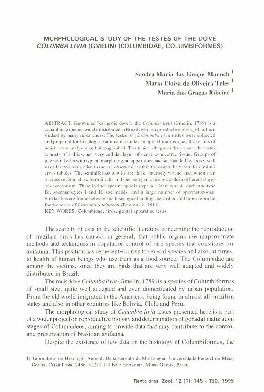
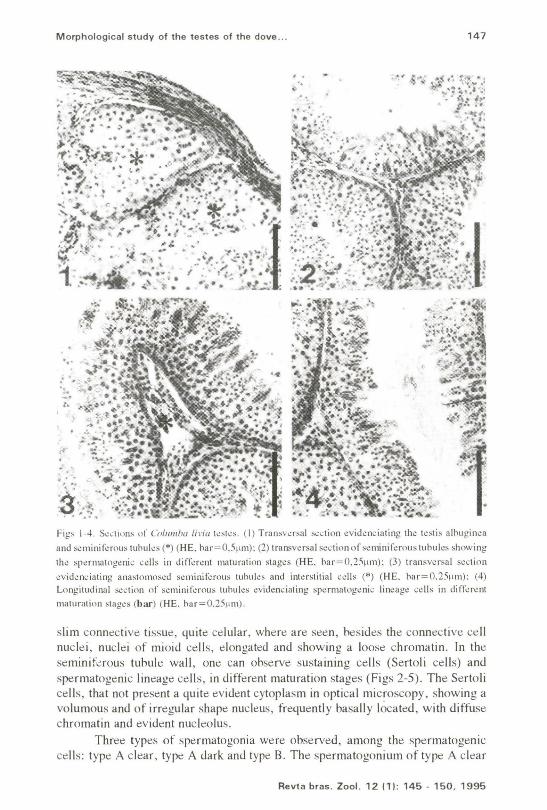
.Fil!~ 1-4. S~dions of Collimha lida kst~s. (I) Transv~rsal ~~lion t:vid~m.:iating tht: l~stis alhllgin~a
and s<:miniferou, tuhllle, (*) (HE. har=O.5flln): (2) transversal ,.:ction of ,eminiferou, tllhuk, showing
the spennalogwic cdl, in different maturation 'tages (HE. har=O.2Sf,m): (3) transversal section
evidenciating ana,tomoseu ,eminilerous tuhuks and int.:f,titial cclls (*) (HE. har=O.25~l\n): (4)Longitudinal ~cti()n of s~minif~rolls tuhuks ~vid~nciating sp~rIl1atog~nic lin~agt: cdls in dift~r~nt
maturation ,tages (bar) (HE. har=O.2Sf1m).
slim connective tissue, quite celular, where are seen, hesides the connective cellnuclei, nuclei of mioid cells, elongated and showing a loose chromatin. In theseminiferous tuoule wall, one can ooserve sustaining cells (Sertoli cells) andspermatogenic lineage cells, in different maturation stages (Figs 2-5). The Sertolicells, that not present a quite evident cytoplasm in optical microscopy, showing avolumous and of irre!,'lliar shape nucleus, frequently basally located, with diffusechromatin and evident nucleolus.
Three types of spermatogonia were observed, among the spermatogeniccells: type A clear, type A dark and type B. The spermatogonium of type A clear
Revta bras. Zool. 12 (1): 145 - 150, 1995
148 MARUCH et al.
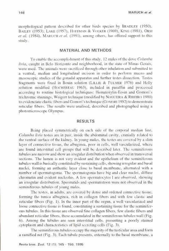
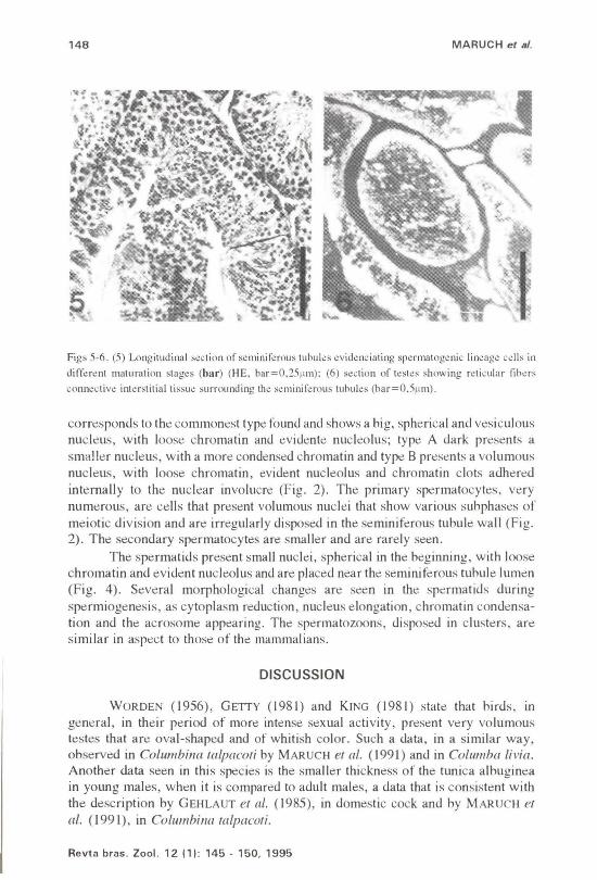
Figs 5-6. (5) Longitudinal s~~tion of s~minir~rouslubul~s ~vid~nciating sp~rl1latog~nic lin~ag~ ~~lIs in
diffa~nt maluration slag~s (bar) (HE. bar=O.25~un): (6) s~clion of ksl~s showing rdicular fih~rs
conn~cliw inl~rsliliallissu~surrounding lh~ s~minif~rous luhuks (har=O.511m).
corresponds to the commonest type found and shows a hig, spherical and vesiculousnucleus, with loose chromatin and evidente nucleolus; type A dark presents asmaller nucleus, with a more condensed chromatin and type B presents a volumousnucleus, with loose chromatin, evident nucleolus and chromatin clots adheredinternally to the nuclear involucre (Fig. 2). The primary spennatocytes, verynumerous, are cells that present vo]umous nuclei that show various suhphases ofmeiotic division and are irregularly disposed in the seminiferous tuhule wall (Fig.2). The secondary spermatocytes are smaller and are rarely seen.
The spermatids present small nuclei, spherical in the beginning, with loosechromatin and evident nucleolus and are placed near the seminiferous tuhule lumen(Fig. 4). Several morphological changes are seen in the spermatids duringspermiogenesis, as cytoplasm reduction, nucleus elongation, chromatin condensation and the acrosome appearing. The spermatozoons, disposed in clusters, aresimilar in aspect to those of the mammalians.
DISCUSSION
WORDEN (1956), GETTY (1981) and KING (1981) state that hirds, ingeneral, in their period of more intense sexual activity, present very volumoustestes that are oval-shaped and of whitish color. Such a data, in a similar way,observed in Columbilla falpacofi by MARUCH ef ai. (1991) and in Columba livia.Another data seen in this species is the smaller thickness of the tunica alhugineain young males, when it is compared to adult males, a data that is consistent withthe description by GEHLAUT ef ai. (1985), in domestic cock and hy MARUCH elai. (1991), in Columbilla {(llpacofi.
Revta bras. Zool. 12 (1): 145 - 150.1995
Morphological study of the testes of the dove ... 149
In Columha Ii via it was not observed the presence of connective tissue septathat come from tunica albuginea and divide the testis in lobules. Similar resultswere tound hy MARVAN (1969), LAKE (1971) and GEHLAUT er al. (1985) indomestic cock; MERCADANTE er al. (1983) in dove; HOFFMANN & VOLKER (1969)in other species of domestic birds and MARUCH er al. (1991) in the small doveColumhilla ralpacori.
In the seminiferous tubules of Columha Livia one can ohserve a slimconnective tissue, rich in blood vessels, showing interstitial cell groups that presentmorphological characteristics of steroid secretory cells. Such a results confirm thedescriptions by BRADLEY (1950), HOFFMANN & VOLKER (1969), GETTY (1981),KING (1981) and MARUCH er al. (1991), in different bird species.
In the seminiferous epithelium of dove, domestic cock and other birdspecies, one can see spermatogenic lineage cells in different maturation stages(LAKE 1957; KING 1981; ORSI el a/. 1984, and MARUCH er al. 1991). Such amorphological aspects were also ohserved in the seminiferous epithelium ofColumha li via.
For BRADLEY (1950), in domestic cock, the spermatogenic cells fromspermatogonia to spermatozoon, are disposed in layers, in a regular sequence,from the basal membrane up to the seminiferous tubule lumen. In Columba livia,the organization observed is similar to that described in Columbilla (([lpacori byMARUCH er al. (1991). Although there is a certain organization in the germinativecell distrihution, one can not notice the regularity (or synchronicity) descrihed hyBRADLEY (1950).
ACKNOWLEDGMENTS. Tho authors thank for the finanoial sponsoring from Pr6·Roitoriade Posquisa da Univorsidado Fodoral do Minas Gerais and from Funda<;ao do Amparo 11 Po,quisa doEstado do Minas Gorais (FAPEMIG) and also for tho valuabk oollaboration of tho translator Mariada GI6ria A. Forroira. of tho photographio laboratory loohnician Ivone do Carmo do Oliveira. of thelibrarian Maria Cooilia do Souza Lima and of tho biologist Sandra Roscnde Lima. in ohargo of thoproparation of tho matorialusod in this study.
REFERENCES
BAILEY, R. E. 1953. Accessory reproductive organs of male fringillid birds:seasonal variations and response to various sex hormones. Ant. Rec. 115:1-20.
BRADLEY, O.C. 1950. The reproductive organs, p. 57-70. III: O.e. BRADLEY(ED.). The structure of the fowl. London, J. B. Lippincott Company, 185p.
GEHLAUT, B.S; M.R. MALIK; A.M. SHRrvAsTAvA & I.e. DATTA. 1985.Comparative histomorphochemical studies on the testes of dwarf (dw) andnon-dwarf towl (Gallus domesticus). Ind. J. Anim. Sci. 55: 870-72.
GETTY, R. 1981. Anatomia dos animais domesticos. Rio de Janeiro, Interamericana, 1962p.
GOMORI, G. 1952. Microscopic histochemistry principles and practice. Chi-
Revta bras. Zoo!. 12 (1): 145 - 150. 1995
150 MARUCH et a/.
cago, University of Chicago Press, 277p.HOCHEREAU, M.T. 1963. Etude compar~e de la vague spermatog~tique chez Ie
taureau et chez Ie rat. Ann. BioI. Anim. Biochim. Biophys. 3: 5-20.HOFFMANN, G. & H. VOLKER. 1969. Anatomia Y tisiologia de las aves
domesticas. Zaragoza, Acrihia, 189p.HUBER, A. 1916. A note on the morphology of the seminiferous tuhules of birds.
Anat. Rec. 2: 177-180.KING, A.S. 1981. Aparelho urogenital das aves; 6rgaos genitais masculinos,
p.1805-1813. 111: R. GETTY (ed.). Anatomia dos animais domesticos. Rio deJaneiro, Interamericana, 743p.
LAKE, P.E. 1957. The male reproductive tract of the fowl. J. Anat. 91: 16-129.---. 1971. The male in reproduction, p.1411-1447. 111: D.J. BELL & B.M.
FREMAN (ed.). Physiology and biochemistry of the domestic fowl. London,Academic Press., vol. 3, 535p.
LILLIE, R.D. & H.M. FULMER. 1976. Histopathologic technic and practicalhistochemistry. New York, Mc Graw-Hill, 942p.
MARUCH, S.M.G.; M.E.O. TELES & M.G. RIBEIRO. 1991. Estudo morfologicodo testfculo de Columbilla talpacori (Temminck, 1811). Columhidae - Columbiforme. Rev. Bras. Cienc. Morfol. 8: 72-76.
MARVAN, F. 1969. Postnatal development of the male genital tract of the Gallusdomesricus. Anat. Anz. 124: 443-462.
MERCADANTE, M.CS.; A.M. ORSI; CA. VICENTINI; M.M. VALENTE & S.M.DIAS. 1983. Anatomical observations on the male reproductive system of thepigeon (Columba Livia). Rev. Cienc. Biomed. 4: 37-44.
NOGUEIRA, J .C. & R. D. RIBEIRO. 1980. A simplified Weigert's method forstaining elastic fibers. Arq. Esc. Vet. Univ. Fed. Minas Gerais 32: 333-335.
ORSI, A.M.; M.C.S. MERCADANTE; E. DIAS & CA. VICENTINI. 1984. Someobservations on the morphology of the pigeon's seminiferous epithelium cells.Anat. Histol. Emhryol. 13: 327-332.
WORDEN, A.N. 1956. Reproductive system, pA8-54. 111: A.N. WORDEN (ed.).Functional anatomy of hirds (Cage Birds). London, Academic Press, 136p.
Recebldo em 19.VII.1994; acelto em 08.111.1995.
Revta bras. Zool. 12 (1): 145 - 150, 1995