Embed Size (px)
Citation preview
Dentofasial, Vol.12, No.1, Februari 2013:60-63
ISSN:1412-8926
60
Sandwich osteotomy for vertical and transversal augmentation of the posterior mandible: a reviewSandwich osteotomy untuk augmentasi vertikal dan transversal pada posterior mandibula: sebuah tinjauan
1Sariatun Tawainela, 1Irma Drismayanti, 2A’la Unas B, 3Muh. Ruslin, 4Eri H. Jubhari1Dentist in Makassar2Student at Clinical Stage3Department of Oral and Maxillofacial Surgery4Department of ProsthodonticsFaculty of Dentistry, Hasanuddin UniversityMakassar, Indonesia
ABSTRAKPenggunaaan implan endoseous secara langsung berhubungan dengan topografi dan kualitas sisa tulang pasien. Beberapa teknik telah dicoba untuk memperluas penerapan melalui perubahan desain dan teknik bedah implan untuk augmentasi tulang. Artikel ini menjelaskan sandwich osteotomy yang dikombinasikan dengan autograft interposisi untuk augmentasi vertikal dan transversal pada mandibula posterior yang atrofik sebelum penempatan implan endoseous. Disimpulkan bahwa sandwich osteotomy segmental mandibula dapat direkomendasikan untuk memenuhi persyaratan dimensi augmentasi tulang preimplant pada mandibula posterior yang atrofik.Kata kunci: sandwich osteotomy, atrofi mandibula posterior, interpositional autograft, implan endoseous
ABSTRACT The use of endosseous implants is directly related to the topography and quality of the patient’s residual bone. Several techniques have been tried to expand its application through implant design alterations and surgical techniques for bone augmentation. This article reviews the sandwich osteotomy combined with an interpositional autograft for vertical and transversal augmentation in the atrophic mandible prior to endosseous implant placement. In conclusion, segmental mandibular sandwich osteotomy can be recommended to fulfill the dimensional requirements of preimplant bone augmentation in atrophic posterior mandible.Key words: sandwich osteotomy, posterior mandible atrophy, interpositional autograft, endosseous implant
Correspondence: Sariatun Tawainella, E-mail: [email protected]
INTRODUCTIONThe use of endoosseous implants for successful
restoration of patients with partial or total loss which has been well established is directly related to thetopographyandqualityof thepatient’s residual bone.
Several techniques have been tried to expand its use through implant design alterations and surgical techniques for bone augmentation process.1 Vertical augmentationofthemandibularor maxillary alveolar ridge to increase bone volume for implant has shown variable and controversial outcomes in comparison with horizontal augmentation.2
In edentulous patients, vertical resorption can progress to reach the basal bone. Horizontally, the resorption mayprogress to the extent that, even whenthere is enough bone height, the lack of bone width may render implant placement impossible.1 Posteriormandible may have thin alveolar bone after teeth lost, the facial cortical bone resorbs more than the lingual cortical,resulting ina 3 mm or less in width.3
Placement of dental implants is difficult in alveolar ridges with severe horizontal and vertical
bone resorption. To augment the severely atrophic ridge,grafting with bone blocks intraorally harvested has been recommended.4 Several techniques for bone augmentation,both vertically and horizontally, have been proposed.Different techniques have been used to achieve vertical augmentation, including onlayorinlay grafts,guided tissue regeneration,sinus floor grafting and transpositional or lateralization of the dental nerve.2 An alternative surgical procedure is the osteotomes technique.1
This article reviews the sandwich osteotomy combined with an interpositional autograft for vertical and transversal augmentation in the atrophic mandible prior to endosseous implant placement.
Endosseous implantDental implants are prosthetic devices of
alloplastic material implanted into the oral tissues beneath the mucosal and/or periosteal layer, and on/or within the bone, to provide retention and support fora fixedoraremovable prosthesis. An endosteal or endosseous dental implant isa dental implant placed
Sariatun Tawainela, dkk: Sandwich osteotomy for augmentation of the posterior mandible: a review
ISSN:1412-8926
61
into thealveolar and/or basal bone of themandible or maxilla and transecting onlay cortical plate.5
The endosseous dental implant is composed of an anchorage component,termed the endosseous dental implant body, which is ideally within the bone, and a retentive component, termed the endosseous dental implantabutment.Descriptionformssuchascylinder, conical, screw, or blade may be used as adjectives toenhancetheunderstandinggeometryofendosseous dental implant.5
Interpositional bone graftThe interpositional bone graft is placed between
a mobilized segmentalosteotomy and thebasal bone. A typical vertical gain is 4 or 5 mm in the maxilla but 5 to 10 mm in the mandible. The indication for the procedure is an alveolar defect where there is insufficient verticalheight for placement of implants such as in the anterior maxilla or posterior mandible when a stable vertical augmentation is required, usually over a three- or four-tooth segment.6
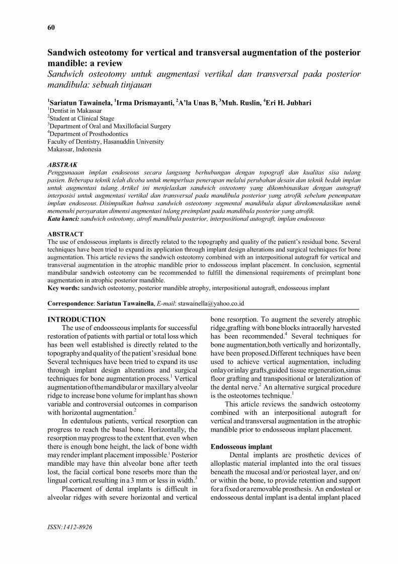
Fig 1A shows a posterior mandible deficiency with 6 mm of bone available above the inferior alveolar nerve. An osteotomy was performed (Fig.1B) through a vestibular incision to maintain both lingual and crestal blood supply. An interpositional cortical bone graft harvested from the ramus was placed at the osteotomy site, raising the alveolus about 7 mm (Fig. 1C). The raised segment rotated slightly lingually, but this was compensated for by using a bone plate to establish both the final vertical height and the crestal axis of the osteotomized segment.6
Procedures of sandwich osteotomySandwich osteotomy is a surgical technique to
repair the atrophic mandible, similar to the way it visors osteotomy, however, horizonal bone cutting, between the foramina mentalis is so limited that only the anterior portion of the cranial fragments are in the lift to the top.7
Bormann et al reported a sandwich osteotomy procedure which begins by making an elliptical
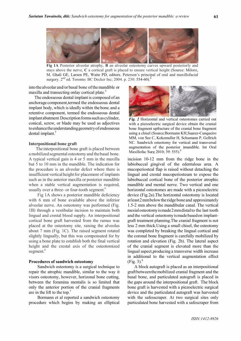
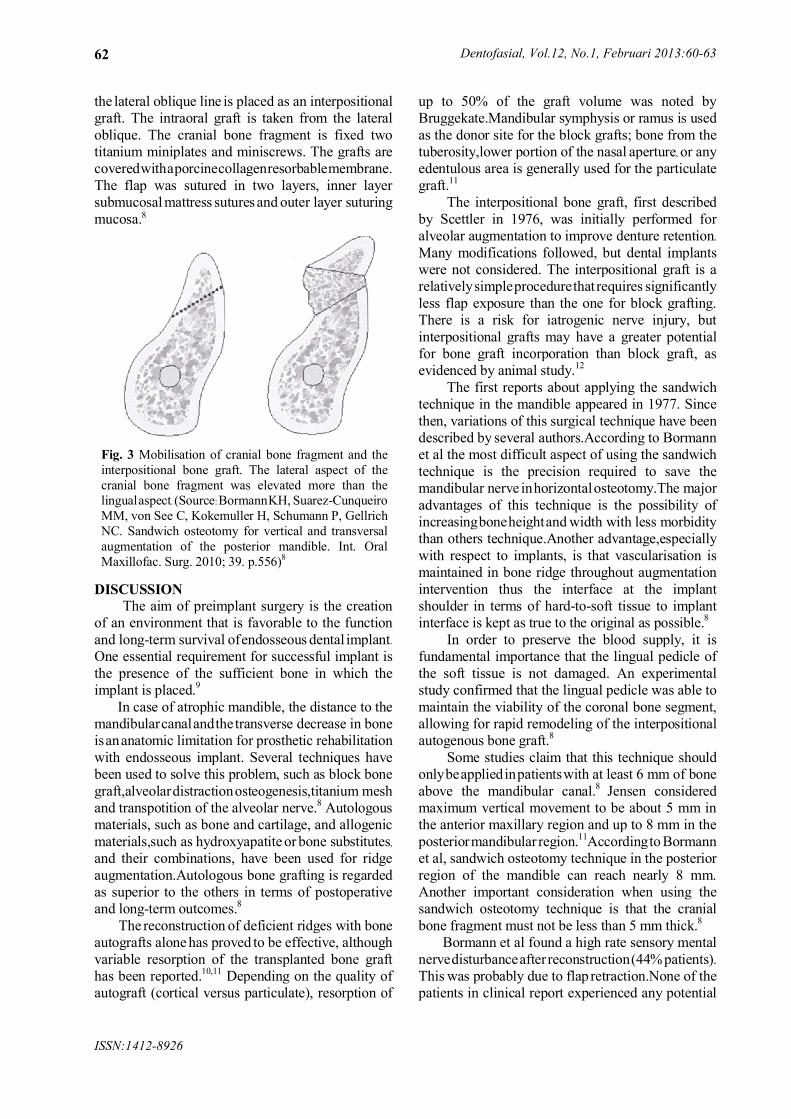
incision 10-12 mm from the ridge bone in the labiobuccal gingival of the edentulous area. A mucoperiosteal flap is raised without detaching the lingual and crestal mucoperiosteum to expose the labiobuccal cortical bone of the posterior atrophic mandible and mental nerve. Two vertical and one horizontal osteotomes are made with a piezoelectric device (Fig.2a).The horizontal osteotomy is located at least2mmbelowthe ridge boneand approximately 1.5-2 mm above the mandibular canal. The vertical mesialosteotomyismade2mmdistal to the last tooth and the vertical osteotomyismadebasedon implant-graft treatment planning.The cranial fragment is not less 2 mm thick.Using a small chisel, the osteotomy was completed by breaking the lingual cortical and the coronal bone fragment is carefully mobilized by rotation and elevation (Fig. 2b). The lateral aspect of the cranial segment is elevated more than the lingual aspect,producing a transverse width increase in additional to the vertical augmentation effect(Fig. 3).8
A block autograft is placed as an interpositional graftbetweenthemobilized cranial fragment and the basal bone, and particulated autograft is placed in the gaps around the interpositional graft. The block bone graft is harvested with a piezoelectric surgical device and the particulated autograft was harvested with the safesscraper. At two surgical sites only particulated bone harvested with a safescraper from
A B CFig 1A Posterior alveolar atrophy, B an alveolar osteotomy curves upward posteriorly and stays above the nerve, C a cortical graft is placed to ensure vertical height (Source: Miloro, M, Ghali GE, Larsen PE, Waite PD, editors. Peterson’s principal of oral and maxillofacial surgery. 2nd ed. Toronto: BC Decker Inc; 2004. p. 230: 554-60).6
A BFig. 2 Horizontal and vertical osteotomes carried out with a piezoelectric surgical device obtain the cranial bone fragment upfracture of the cranial bone fragment using a chisel (Source:Bormann KH,Suarez-Cunqueiro MM, von See C, Kokemuller H, Schumann P, Gellrich NC. Sandwich osteotomy for vertical and transversal augmentation of the posterior )mandible. Int Oral Maxillofac Surg 2010; 39: 555).8
Dentofasial, Vol.12, No.1, Februari 2013:60-63
ISSN:1412-8926
62
the lateral oblique line is placed as an interpositional graft. The intraoral graft is taken from the lateral oblique. The cranial bone fragment is fixed two titanium miniplates and miniscrews. The grafts arecoveredwithaporcinecollagenresorbablemembrane.The flap was sutured in two layers, inner layer submucosal mattress suturesand outer layer suturing mucosa.8
DISCUSSIONThe aim of preimplant surgery is the creation
of an environment that is favorable to the function and long-term survival ofendosseous dental implant.One essential requirement for successful implant is the presence of the sufficient bone in which the implant is placed.9
In case of atrophic mandible, the distance to the mandibularcanalandthetransverse decrease in bone isananatomic limitation for prosthetic rehabilitation with endosseous implant. Several techniques have been used to solve this problem, such as block bone graft,alveolardistractionosteogenesis,titanium mesh and transpotition of the alveolar nerve.8 Autologous materials, such as bone and cartilage, and allogenic materials,such as hydroxyapatite or bone substitutes,
and their combinations, have been used for ridgeaugmentation.Autologous bone grafting is regarded as superior to the others in terms of postoperative and long-term outcomes.8
The reconstruction of deficient ridges with bone autografts alone has proved to be effective, although variable resorption of the transplanted bone graft has been reported.10,11 Depending on the quality of autograft (cortical versus particulate), resorption of
up to 50% of the graft volume was noted by Bruggekate.Mandibular symphysis or ramus is used as the donor site for the block grafts; bone from the tuberosity,lower portion of the nasal aperture, or any edentulous area is generally used for the particulate graft.11
The interpositional bone graft, first described by Scettler in 1976, was initially performed for alveolar augmentation to improve denture retention.
Many modifications followed, but dental implants were not considered. The interpositional graft is arelativelysimpleprocedurethatrequires significantly less flap exposure than the one for block grafting. There is a risk for iatrogenic nerve injury, but interpositional grafts may have a greater potential for bone graft incorporation than block graft, as evidenced by animal study.12
The first reports about applying the sandwich technique in the mandible appeared in 1977. Since then, variations of this surgical technique have been described by several authors.According to Bormann et al the most difficult aspect of using the sandwich technique is the precision required to save the mandibular nerve inhorizontalosteotomy.The major advantages of this technique is the possibility of increasingboneheightand width with less morbidity than others technique.Another advantage,especially with respect to implants, is that vascularisation is maintained in bone ridge throughout augmentation intervention thus the interface at the implant shoulder in terms of hard-to-soft tissue to implant interface is kept as true to the original as possible.8
In order to preserve the blood supply, it is fundamental importance that the lingual pedicle of the soft tissue is not damaged. An experimental study confirmed that the lingual pedicle was able to maintain the viability of the coronal bone segment, allowing for rapid remodeling of the interpositional autogenous bone graft.8
Some studies claim that this technique should onlybeappliedinpatientswith at least 6 mm of bone above the mandibular canal.8 Jensen consideredmaximum vertical movement to be about 5 mm in the anterior maxillary region and up to 8 mm in the posteriormandibular region.11Accordingto Bormann et al, sandwich osteotomy technique in the posterior region of the mandible can reach nearly 8 mm.Another important consideration when using the sandwich osteotomy technique is that the cranial bone fragment must not be less than 5 mm thick.8
Bormann et al found a high rate sensory mental nervedisturbanceafterreconstruction(44%patients).This was probably due to flap retraction.None of the patients in clinical report experienced any potential
Fig. 3 Mobilisation of cranial bone fragment and the interpositional bone graft. The lateral aspect of the cranial bone fragment was elevated more than the lingualaspect. (Source:BormannKH, Suarez-Cunqueiro MM, von See C, Kokemuller H, Schumann P, Gellrich NC. Sandwich osteotomy for vertical and transversal augmentation of the posterior mandible. Int. Oral Maxillofac. Surg. 2010; 39. p.556)8
Sariatun Tawainela, dkk: Sandwich osteotomy for augmentation of the posterior mandible: a review
ISSN:1412-8926
63
morbidity at the donor site.8 Jensen found transient paraesthesia in all patients,lasting up to 6 weeks,and all patients in the report recoveredwithin this time.12
Although there are several bone augmentation techniques, osteotomy osteogenesis has moreadvantages. Alveolar distraction osteogenesis has the greatest potential of a 9.9 mm mean bone gain (range 4-15 mm).Sandwich osteotomy osteogenesis has the advantages of restoring vertical bone deficit together with transverse gain in dimension without undesired bone segment displacement, is less time-consumingand is less uncomfortable for the patient. Although nerve transposition maintains the vertical bone, it involves a high risk of permanent neurosensory disturbance.It would seem that the choice of technique is based more on evidence of efficacy.8
Marchetti et al report that in all the treated sites with interpositional bone graft (sandwich technique)
it was possible to place implants. None of the 21 implants placed failed,and minimal bone resorption was present 14 to 16 months after the prosthetic loading. These findings suggest that interpositional bone grafting in the posterior mandible could be a viable alternative to other surgical techniques.13
Bourmannetalconcluded that segmentalmandibular sandwich osteotomy is recommended to meet the dimensional requirements of preimplant bone augmentation in atrophic posterior mandible.8 Choi et al also have concluded that this procedure of augmentation is safe.14
In conclusion, segmental mandibular sandwich osteotomy can be recommended to meet the dimensional requirements of preimplant bone augmentation in atrophic posterior mandible. Because this techniques is safe and could be a viable alternative to other surgical techniques.
REFERENCES1. Cruzz Mauro, Reis CC, Flavio de Freitas Mattos. Implant-induced expansion of atrophic ridges for the placement
of implant. J Prosthet Dent 2001;85:377-81.2. González JMM, Sánchez JC, Trapero JC, Lafuente JCG, Regañón JD, Piñeiro MTV. Evaluation of minipigs as an
animal model for alveolar distraction. Oral Surg Oral Med Oral Pathol Oral Radiol Endod 2005;99:11-6.3. Block Michael S. Color atlas of dental implant suregery. Philadelphia: W.B. Saunders Company; 2001.4. Sohn DS, Ahn MR, Lee WH, Yeo DS, Lim SY. Piezoelectric osteotomy for intraoral harvesting of bone blocks. Int
J Periodont Restor Dent 2007: 27.5. Stellingsma C, Vissink A, Meijer HJA, Kuiper C, Raghoebar GM. Implantology and the severely resorbed
edentulous mandible. Crit Rev Oral Biol Med 2004; 15(4):240-8.6. Miloro M, Ghali GE, Larsen PE, Waite PD, editors. Peterson’s principal of oral and maxillofacial surgery. 2nd Ed.
Toronto: BC Decker Inc; 2004. p. 230.7. Dorland’s illustrated medical dictionary. 29th Ed. Philadelphia: W.B. Saunders; 2002. Sandwich osteotomy; p.1567.8. Bormann KH, Suarez-Cunqueiro MM, von See C, Kokemuller H, Schumann P, Gellrich NC. Sandwich osteotomy
for vertical and transversal augmentation of the posterior mandible. Int Oral Maxillofac Surg 2010; 39: 554-60. 9. Depprich RA, Handschel Jorg GK, Naujoks C, Hahn T, Meyer U, Kubler NR. Sinus lifting before Le Fort I
maxillary osteotomy: a suitable method for oral rehabilitation of edentulous patients with skeletal class-III conditions: review of the literature and report of a case. Head & face Medicine 2007; 3:2.
10. Fragiskos FD. Oral surgery. NewYork: Springer-Verlag Berlin Heidelberg; 2007.p.34611. Jovanic SA. Bone rehabilitation to achieve optimal aesthetics. Aesthetic Chronicle 1997: 9(1): 41-50.12. Jensen OT. Alveolar segmental “sandwich” osteotomies for posterior edentulous mandibular sites for dental
implant. J Oral Maxillofac Surg 2006: 64: 471-5.13. Marchetti C, Trasarti S, Corinaldesi G, Felice P. interpositional bone grafts in the posterior mandibular region: a
report on six patients. Int J Periodont Restor Dent 2007:27(6): 547-55.14. Choi BH, Lee SH, Huh JY, Han SG. Use of the sandwich osteotomy plus an interpositional allograft for vertical
augmentation of the alveolar ridge. J Craniomaxillofac Surg 2004: 32(1):51-4.