Embed Size (px)
Citation preview
8th International Symposium on Tilapia in Aquaculture 2008
SAPROLEGNIOSIS IN FRESHWATER CULTURED TILAPIA NILOTICA (ORECHROMIS NILOTICUS) AND TRIAL FOR
CONTROL BY USING BAFRY D50/500.
MOHAMED E. ABOU EL ATTA
Fish Health Department. Central Laboratory For Aquaculture Research,
Abbassa Abou Hammad, Sharkia Egypt
AbstractThis study was carried out on 100 cultured Tilapia
nilotica in floating cages with high stocking density suffered from saprolegniosis, which considered the most important causes of economic loses in freshwater fish second to bacterial disease in Egypt. The fish were subjected to full clinical, postmortem, bacteriological and histopathological examination, also trial for control using Bafry D50/500 and hematological examination of treated fish. Clinically, the infected fish showed loss of equilibrium, lethargy, unable to feed, hemorrhage at the base of fins may extended to cover all the body surface, the main characteristic lesions of saprolegniosis was appearance of cotton wool like tufts on the fins (dorsal, caudal, and pectoral fins); also, on eyes, the head and mouth and uni or bilateral blindness ended with death. Postmortemally, the infected fish showed pale grayish gills, the intestine were free from any food particles, dark enlarged liver, distended gallbladder and spleenomegaly. Bacteriologically, Aeromonas Spp. and Pseudomonas Spp. Were isolated from infected fish suffered from saprolegniosis. The wet mount from infected lesions showed the presence of large non sepetated hyphae, the colonies on SDA started with cysts of long hairs with cottony color, and then became gray, then black color colonies. From the microscopical and morphological finding were characteristic for saprolgnia. Saprolegnia parasitica were isolated with higher percentage from skin followed with fins, eyes, and mouth, but not isolated from liver nor kidney. Trial of using Bafry D50/500 for control in saprolegniosis in a dose of 0.75 ml/L (375ppm) for 10-12 minutes for 3 successive days as a bath gave a good results in treatment of the diseased fish in aquaria, this were indicated the rate of mortality reach 16.7% without any effect on the healthy stat of treated fish and also no effect on hematology and blood chemistry of treated fish with Bafry D50/500.Histopathological finding of the infected fish showed epithelial desquamation of the epidermis, erosion and ulceration of the infected area, edema, hyaline degeneration, and aggregation of M.M.C. and fragment of fungal hyphae occurred in the underling dermis of the skin, muscles; The gills of the infected fish showed complete desquamation of majority secondary lamellae, and gill arch was edematous with presence of aggregation of fungal spores and M.M.C., Histopathological finding of treated fish with Bafry D50/500:skin showed increase of M.M.C. and slight sloughing of most superficial layer of epidermis but the gills showed increasing and hyperplasia of epithelial cells covering of primary lamellae, this indicate that the infected fish was treated with BafryD50/500.
1403
SAPROLEGNIOSIS IN FRESHWATER CULTURED TILAPIA NILOTICA (ORECHROMIS NILOTICUS) AND TRIAL FOR CONTROL BY USING BAFRY D50/500.
Key word: Tilapia, Saprolegniosis, Bacteria, Histopathology, Bafry D50/500
INTRODUCTION
Fishes are the primary source of protein for human in many areas of the world and this is also in Egypt. Outbreaks of water born fungal infections of fish, Amphibians, and Reptiles are common problems especially in fish farms and hatcheries. Of particular concern is saprolegniosis, which is an infectious fungal disease that is wide spread in all stages of the life cycle of fish. Saprolegniosis infection may contribute to heavy mortality among fishes and are widespread in freshwater fish's ecosystem and affect wild and cultured fishes, also involving living, dead and eggs (Noga, 1996-Bruno and Wood, 1999-Hussein and Hatai, 1999-and Hussein et al, 2001). Saprolegniosis considered as single largest cause of economic losses in aquaculture (Meyer, 1991) second only to bacterial disease in economic importance. In Egypt, the mycotic disease constitute one of the most important disease causing troubles in fresh culture with several economical losses especially saprolegniosis (Easa, 1984, Shaheen ,1986, and El Zayat,1988).Saprolegnia considered agent of secondary infection arising from conditions as bacterial infections, poor husbandry including poor water quality, adverse water temperature , all of these factors increased occurrence of saprolegnia infections (Pickering and Willoughby,1982, and Bailey,1984). Saprolegniosis are generally restricted to chronic, steady losses(Bruno and Wood,1999-Pickering and Willoughby,1982) and cause 50% mortality in Salmon fish (Hatai and Hoshiai,1994), 50%losses in Anguilla anguilla (Bruono and Wood,1999), 50%losses in channel catfish in USA (Bruono and Wood,1999), and affect tilapia (Orechromis niloticus and Oreochromis mossambicus )in hatchery and fingerlings stages (Easa and Amin,1987,Ogbonna,1989, Aly et al., 1996, Aly and El Ashram,2000).The seasonal variation play an important role in spreading of the saprolegnia infections in freshwater fish especially during late autumn, common in winter months and early spring where the temperature was low (Hughes,1962) .Much work has been devoted to the examination of various types of fish fungicides and their effects either on aquatic animals or on the environment. Malachite green and formalin are the most potent fish fungicides although they have an acute impact on the aquatic ecosystems (Willoughby and Roberts, 1992, Schreier et al., 1996). Other problems with theses substances include an immune suppressive
1404
MOHAMED E. ABOU EL ATTA
effect on repeatedly treated fish (Prost and Spinska1989), terratogenic and mutagenic effect due to hazardous residues in fish tissues (Meyer and Jorgenson, 1983, Fitzpatrick et al., 1995, Meinert et al., 1995, and Bruono and Wood, 1999).Formalin, solution 37% formaldehyde is effective in treating saprolegnia but has effect on both environments personal who handle it (Fitzpatrick et al., 1995). For these reasons many researches have been investigating the use of safer compounds that have no harmful effect on fish and their eggs or on ecosystem (Hussein et al., 2001). So, the use of hydrogen peroxide (H2O2) is a potential chemotherapeutant compound looked upon aquaculture community with increasing interest. The compound is considered environmentally compatible as well as being an effective treatment for a variety of external fish disease, fungicide, bactericide, parasiticide on fish and fish eggs, (Marking et al., 1994, Mc Andrew et al., 1998, Derksen et al., 1999, Hawe et al.1999, Schreier et al.,
1996 and Speare & Arsenault, 1997). Hydrogen peroxide has a low regulatory priority classification by United States food and drug administration (FDA) for use as fungicide on fish and fish eggs (Schnick1994).The apparent lock of adverse environmental impact of hydrogen peroxide combined with its effectiveness as an antimicrobial agent (Rach et al., 1997).Therefore, the current investigation was planned to focus on saprolegniosis as a major fungal diseases affect freshwater fish (wild and cultured fishes )especially tilapia species as well as the optimal preventive trials to control saprolegniosis by using chemicals safe to both environment and personal who contact with these chemicals as Bafry D50/500 .
MATERIALS AND METHODS
І - MaterialsІ.1 Naturally infected fish A total number of 100 Orechromis niloticus fish showed skin lesions were collected from floating cages in El Bostan Damietta during the period November 2005 and January 2006, with average 75 ± 5 gm body weight and length 13 ± 2 cm. the collected fish were transported in ice box to the Central Laboratory for Aquaculture (CLAR) Abbassa Abou Hammad Sharkia, Egypt subjected to full clinical, bacteriological and mycological investigation.
1405
SAPROLEGNIOSIS IN FRESHWATER CULTURED TILAPIA NILOTICA (ORECHROMIS NILOTICUS) AND TRIAL FOR CONTROL BY USING BAFRY D50/500.
І.2 Fish for experimentA total number of 210 healthy fish with average body weight 70 ± 5gm Orechromis niloticus were collected from earthen pond of (CLAR) and transported to the wet laboratory and acclimatized to the conditions for 2 weeks, maintained at 25 ± 2 ºC in glass aquaria of 40 X 50 X 80 cm, supplied with chlorine free water, the temperature was thermostatically adjusted at 25 ± 2 ºC and fish were feed with 30% protein according to their body weight.І.3 MediaSabouraud dextrose agar (SDA) (Adwic SCG) was used for isolation of fungus and was prepared by dissolving 65 gm /liter of distilled water by gentle heating and sterilized in autoclave at 121 ºCfor15 mint and chloramphenicol was added by 50 mg / ml (Cruickshank et al., 1975).І.4 Chemicals used for treatment
a- Bafry D 50/500(H2O2+Silver ions) FMT: 106 Makram Obeidst., Nasr City, Cairo, Egypt.І.5 StainLacto phenol cotton blue stain was used (Leanor and Carey, 1978)ІІ - MethodsІІ-1 Clinical examination:-One hundred naturally infected living fish were examined for abnormal behaviors and external lesions on the skin, eye, and gills according to the method described by Amlacker, 1970.ІІ – 2- Postmortem examinationPostmortem examination was done on living or freshly dead fish and examination of internal organs was done according to Amlacker, 1970, and Lucky, 1977, to show any abnormalities.ІІ -3-Bacteriological examinationSamples were taken from skin, gills and internal organs of infected fish and streaked on tryptic soy agar and incubated at 27 ºC for 24-48 hrs, then purification and identification of isolated bacteria was done according to Austin and Austin (1993).ІІ -4- Mycological examinationІІ -4-a: Isolation of fungi:- was carried out from naturally infected fish, samples were taken from fish showing skin lesions, eye, fins, gills, mouth, spleen, liver and kidney lesions were collected and inoculated onto SDA
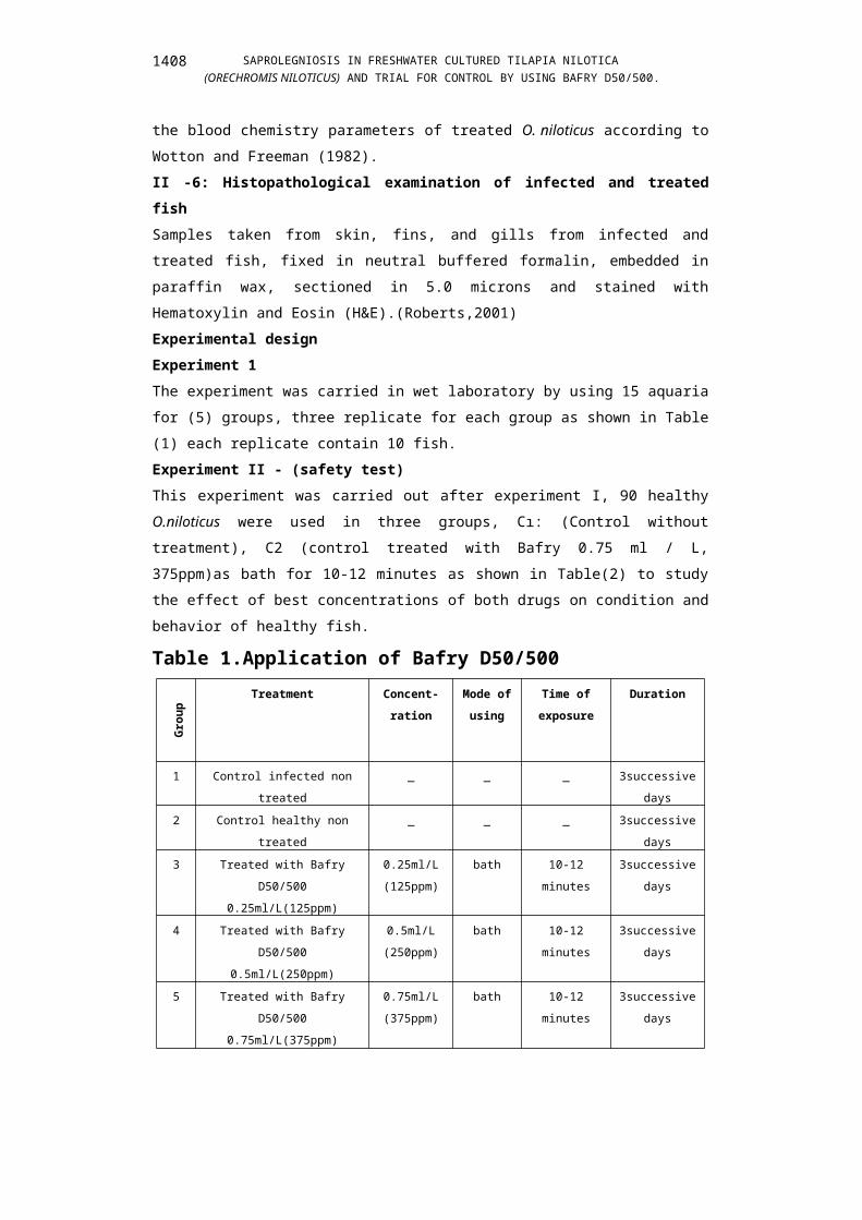
1406
MOHAMED E. ABOU EL ATTA
medium plates and incubated at 20 ± 2ºC for 3-4 days ,subculture on the same media was done for purification. ІІ -4- b: Identification of the isolatesAll positive cultures were examined for colonial growth, morphological features and microscopical characteristics.The morphological features include appearance of the cultures, rate of growth, texture of the surface colonies, colonies color according to Refai and Al Doory, 1986.Microscopical examination was done for wet preparations of the skin lesions and mycelia cultured on (SDA) to detect septation of hyphae according to Dvorak and Otcerasek, 1969.ІІ -5: Biochemical analysisBlood samples were taken from caudal vein under anathematic by using MS222 (200mg / L) to study the effect of bafry D50/500 on the blood chemistry parameters of treated O. niloticus according to Wotton and Freeman (1982). ІІ -6: Histopathological examination of infected and treated fishSamples taken from skin, fins, and gills from infected and treated fish, fixed in neutral buffered formalin, embedded in paraffin wax, sectioned in 5.0 microns and stained with Hematoxylin and Eosin (H&E).(Roberts,2001)Experimental design Experiment 1The experiment was carried in wet laboratory by using 15 aquaria for (5) groups, three replicate for each group as shown in Table (1) each replicate contain 10 fish. Experiment ІІ - (safety test)This experiment was carried out after experiment І, 90 healthy O.niloticus
were used in three groups, Cı: (Control without treatment), C2 (control treated with Bafry 0.75 ml / L, 375ppm)as bath for 10-12 minutes as shown in Table(2) to study the effect of best concentrations of both drugs on condition and behavior of healthy fish.
Table 1.Application of Bafry D50/500
Gro
up
Treatment Concent-ration
Mode of using
Time of exposure
Duration
1 Control infected non treated
_ _ _ 3successive days
1407
SAPROLEGNIOSIS IN FRESHWATER CULTURED TILAPIA NILOTICA (ORECHROMIS NILOTICUS) AND TRIAL FOR CONTROL BY USING BAFRY D50/500.
2 Control healthy non treated
_ _ _ 3successive days
3 Treated with Bafry D50/500
0.25ml/L(125ppm)
0.25ml/L(125ppm)
bath 10-12 minutes
3successive days
4 Treated with Bafry D50/500 0.5ml/L(250ppm)
0.5ml/L(250ppm)
bath 10-12 minutes
3successive days
5 Treated with Bafry D50/500
0.75ml/L(375ppm)
0.75ml/L(375ppm)
bath 10-12 minutes
3successive days
Table 2. safety test = tolerance test
Gro
up
TreatmentConcent-
ration
Mode of
using
Time of exposure
Duration
C1Control healthy without
treatment_ _ _
3successive days
C2Control healthy Treated with
Bafry D50/500 0.75ml/L(375ppm)
0.75ml/L(375ppm)
bath10-12
minutes3successiv
e days
RESULTS
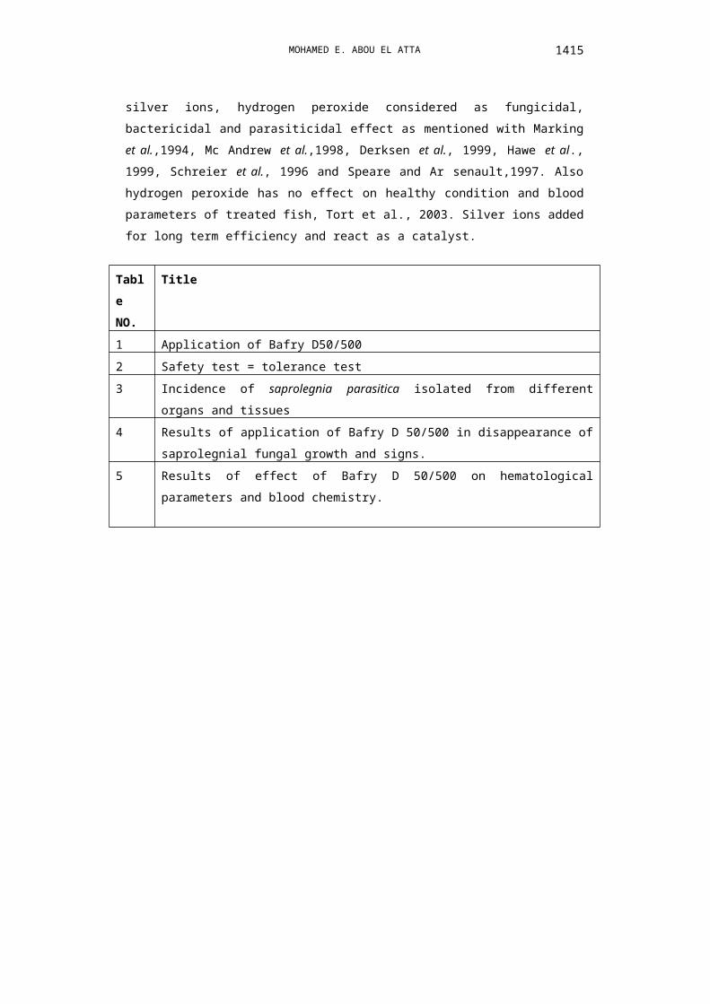
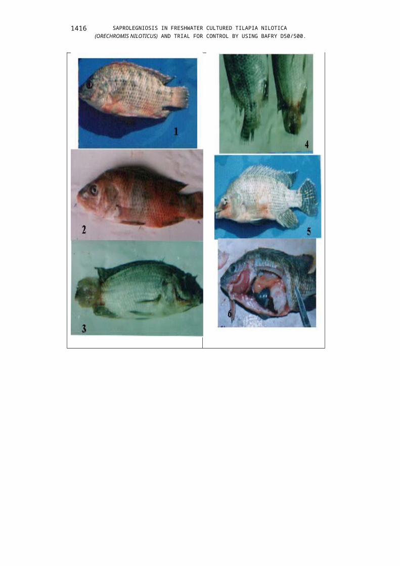
► Results of clinical examination The main clinical signs appeared on the infected fish were lethargy, loss of equilibrium, unable to feed, hemorrhage at the base of the fins, sometimes, the hemorrhages extended to cover all the body surface, as showing in photo(1),(2), the main characteristic lesions of saprolegniosis was appearance of cotton wool like tufts on the dorsal, tail (caudal), pectoral fins also appearance on the eye, head and mouth of fish as shown in photo.(3),(4) unilateral cloudy or opacity of the eye ended by blindness, emaciation and death occurred, as shown in photo(5).► Results of postmortem examination The main postmortem lesions are appearance of cotton wool tufts on caudal fin(tail), pale to grayish gills, serious fluid or exudates in the abdominal cavity, intestine free from any food particles, dark enlarged liver, distended gall bladder with bile, spleenomegaly and congested kidney as shown in photo(6).► Results of bacteriological examination
1408

MOHAMED E. ABOU EL ATTA
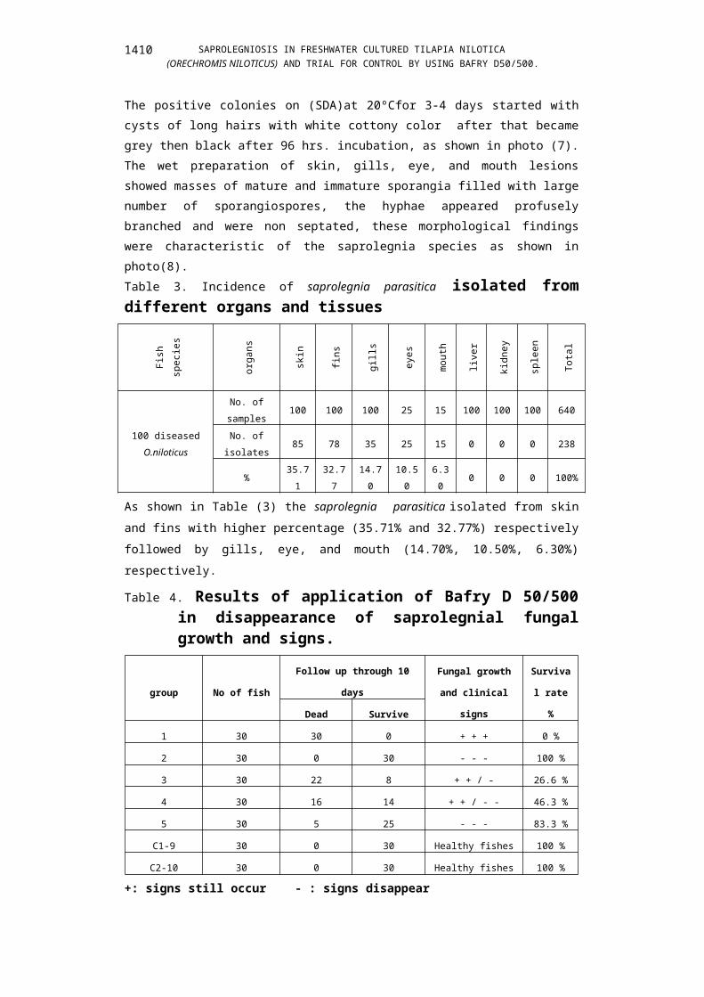
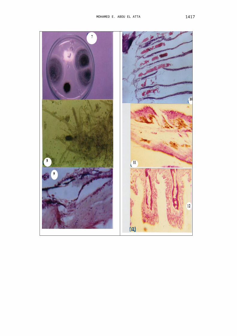
The bacteriological examination showed that isolation of Gram negative bacteria and identified as Aeromonas species and Pseudomonas species fro fish suffered saprolegniosis.► Results of mycological examination. The results of mycological examination showed that isolation of 238 isolates from 640 samples from 100 infected Oreochromis niloticus sampled from skin, gills, fins, eye, kidneys and mouth as shown in Table (3).The positive colonies on (SDA)at 20ºCfor 3-4 days started with cysts of long hairs with white cottony color after that became grey then black after 96 hrs. incubation, as shown in photo (7).The wet preparation of skin, gills, eye, and mouth lesions showed masses of mature and immature sporangia filled with large number of sporangiospores, the hyphae appeared profusely branched and were non septated, these morphological findings were characteristic of the saprolegnia species as shown in photo(8).Table 3. Incidence of saprolegnia parasitica isolated from different organs and tissues
Fish
spec
ies
orga
ns
skin
fins
gills
eyes
mou
th
liver
kidn
ey
sple
en
Tota
l100 diseased
O.niloticus
No. of samples
100 100 100 25 15100
100
100
640
No. of isolates
85 78 35 25 15 0 0 0 238
%35.7
132.7
714.7
010.5
06.30
0 0 0100%
As shown in Table (3) the saprolegnia parasitica isolated from skin and fins with higher percentage (35.71% and 32.77%) respectively followed by gills, eye, and mouth (14.70%, 10.50%, 6.30%) respectively.Table 4. Results of application of Bafry D 50/500 in
disappearance of saprolegnial fungal growth and signs.
group No of fish
Follow up through 10
days
Fungal growth
and clinical
signs
Surviva
l rate %Dead Survive
1 30 30 0 + + + 0 %
2 30 0 30 - - - 100 %
3 30 22 8 + + / - 26.6 %
4 30 16 14 + + / - - 46.3 %
1409
SAPROLEGNIOSIS IN FRESHWATER CULTURED TILAPIA NILOTICA (ORECHROMIS NILOTICUS) AND TRIAL FOR CONTROL BY USING BAFRY D50/500.
5 30 5 25 - - - 83.3 %
C1-9 30 0 30 Healthy fishes 100 %
C2-10 30 0 30 Healthy fishes 100 %
+: signs still occur - : signs disappearAs shown in Table (4) the Bafry D50/500 gave a good result in the treatment and disappearance of fungal growth and clinical signs in affected fish in aquaria especially in a dose of 0.75 ml/L(375ppm) as a bath for 10-12 minutes for 3 successive days and the mortality rate reach 16.7% and the survival rate reach 83.3% with no effect on healthy state of treated fish, this indicated with no significance in blood parameter and chemistry of treated healthy fish and non treated healthy fish except increasing in glucose level and AST as shown in Table(5)Table 5. Results of effect of Bafry D 50/500 on hematological parameters and blood chemistry.
Trea
tmen
t
PCV
Hb
Tota
l pro
tein
Albu
min
Glo
bulin
A/G
Ca+
+
gluc
ose
ALT
AST
C1
26.5
±1.4
5.2±0.1
5
4.5±0.2
1
1.41±0.1
5
3.09±0.1
8
0.456±0.02
1
7.6±0.5
2
66±3.3
32±4.2
75±4.5
a a a a a a b b a b
C2
26.3
±1.5
5.00±0.1
2
4.3±0.1
8
1.39±0.1
2
2.91±0.1
2
0.477±0.02
3
7.5±0.5
5
98±6.3
40±3.8
114±8.5
a a a a a a b a a a ► Results of histopathological examination of naturally infected fishThe skin showed epithelial desquamation in the epidermis which displayed either erosion to ulceration in the infected area. The other epidermal cells suffered vacuolar degeneration and focal necrosis. The underling dermis was edematous and contained fragments from the fungal hyphae with focal aggregation of melanomacrophages cells. The underlying muscles exhibited intramuscular edema, hyaline degeneration and Zinker‘s necrosis. The
1410

MOHAMED E. ABOU EL ATTA
necrotic muscles infiltrated with numerous mononuclear leukocytes and some melanomacrophages. Saprolegnia spp, appeared by H&E stain as non septated hyphae of variable size and lengths. As shown in photo (9). The gills showed desquamation to the majority of the secondary lamellae with few mononuclear leukocytes infiltrated the primary lamellae. The gill arch was edematous and contained the fungal spores and hyphae as well as some melanomacrophages and mononuclear cells. As shown in photo (10). ► Results of histopathological examination of treated fish Skin of Tilapia nilotica (O.niloticus) treated with BafryD50/500showed increase of M.M.C and slight sloughing of most superficial layer of epidermis. As shown in photo (11). Gill of Tilapia nilotica (O.niloticus)
treated with BafryD50/500showed hyperplasia of epithelial cells covering of primary lamellae. As shown in photo (12).
DISCUSSION
In Egypt the mycotic disease constitute one of the most important diseases causing troubles in fresh culture with several losses especially saprolegniosis Shaheen, 1986 and El Zayat, 1988. Saprolegniosis infection may contribute to heavy mortality among fish and are wide spread in freshwaters ecosystem and affect wild and cultured fishes ,also involving both living ,dead and eggs and was considered as single largest cause of economic losses in aquaculture, second only to bacterial disease in economic importance,Meyer,1991, Noga,1996, Bruno and Wood,1999,Hussein et al.,2001.Saprolegniosis considered as localized infection not systemic infection, generally are external and appear any where over the body surface especially fins, eyes, gills and ulcerated area on the body, the clinical signs appear on the fish suffered from saprolegniosis were developed of grayish white cotton like tufts on fins(dorsal, caudal and pectoral fins) also on eyes and mouth, emaciation and death occurred due to blindness and the affected fish unable to feed., also respectively distress occur due to fungal growth on gills so, the gills become very pale due to the excessive mucus secretion and the fungal growth. In some cases, the affected fish showing muscle lesions as erythema and ulceration due to the lytic action of bacteria, so saprolegnioisis considered as secondary invader to the bacteria, these results recorded also by Aly et al.,1996,Noga,1996,Bruno and Wood,1999,Hussein and Hatai,1999, Aly and El Ashram,2000,Hussein et al,
1411

SAPROLEGNIOSIS IN FRESHWATER CULTURED TILAPIA NILOTICA (ORECHROMIS NILOTICUS) AND TRIAL FOR CONTROL BY USING BAFRY D50/500.
2001, and El Ashram et al,2007.The main postmortem lesions appear were pale gills due to respiratory distress and excessive mucus secretion, this results observed by Plumo,1994, the enlarged liver, kidney, spleen and gallbladder may be due to systemic bacterial infection and not attributed to saprolegniosis, these results agree with Paperna et al.,1983,and Bailey,1984. Saprolegnia considered being secondary invader accompanied by opportunistic bacteria. The results of bacterial examination showed isolation of gram negative bacteria and were identified as Aeromonas
species and Pseudomonas species, these results observed by Paperna et
al., 1983, Bailey, 1984, Aly and El Ashram, 2000, and El Ashram et al., 2007. The results of mycological examination showed that isolation of Saprolegnia parasitica which was branched non septated tubular hyphae and isolated with higher percentage from skin (35.71%), followed by fins(32.77%), gills(14.70%), then mouth(6.30%) and not isolated from liver, kidney and spleen, these results agree with Marzouk et al.,1990 and Marzouk et al., 2003. The results of application of Bafry D50/500 for control of Saprolegnia parasitica revealed that using of Bafry D50/500 in a concentration of 0.75ml/L (375ppm) as a bath for10-12 minutes is the best concentration for elimination of saprolegnial growth with no affect on behavior of treated fish and also on healthy fish. Bafry D 50/500 is a mixture of hydrogen peroxide which is an oxidizing agent ant silver ions which is are added for long term efficiency, these results recorded by Marking et al., 1994, MC Andrew et al., 1998, Derksn et al., 1999 and Hawe et al., 1999. The results of effect of best concentration of Bafry D50/500 on blood parameters showed that no significance difference between control healthy non treated and the healthy treated fish , except glucose which is higher in healthy treated than non treated one due to H2O2 affect on cortisol level in the blood, also, AST is higher in treated healthy fish than healthy non treated, these results accepted with Tort et al., 2003.The results of histopathological examination of skin showed epithelial desquamation in the epidermis and other epidermal cells suffered vacuolar degeneration and focal degeneration thus due to sever spongiosis. Skin showed epithelial desquamation in the epidermis which displayed either erosion to ulceration in the infected area. The other epidermal cells suffered vacuolar degeneration and focal necrosis. The underling dermis was edematous and contained fragments from the fungal hyphae with focal aggregation of melanomacrophages cells. The underlying muscles exhibited
1412
MOHAMED E. ABOU EL ATTA
intramuscular edema, hyaline degeneration and Zinker‘s necrosis. The necrotic muscles infiltrated with numerous mononuclear leukocytes and some melanomacrophages. Saprolegnia spp, appeared by H&E stain as non septated hyphae of variable size and lengths. As shown in photo (9). The gills showed desquamation to the majority of the secondary lamellae with few mononuclear leukocytes infiltrated the primary lamellae. The gill arch was edematous and contained the fungal spores and hyphae as well as some melanomacrophages and mononuclear cells. As shown in photo (10),were similar to those observed by Amin et al., 1985, Ferguson,1989,Aly et al., 1986, Aly and El Ashram,2000, El genaidy et al., 2004, and El Ashram,2007.The results of histopathological examination of treated fish with Bafry D 50/500 showed increase of MMC and slight sloughing of most superficial layer of epidermis and presence of vaculation indicating the disappearance of causative agent, also, the gills showed hyperplasia or increasing of epithelial cells covering of the primary lamellae indicating slight healing of the infected gills of treated fish.
CONCLUSION
The most effective method for controlling and preventing saprolegniosis in fish ecosystem is a combination of good fish management as good water quality and avoid adverse water temperature and Bafry D50/500 used for controlling in a concentration of 0.75ml/L (375PPm) as a bath for 10-12 minutes, due to Bafry D50/500 is a mixture of hydrogen peroxide and silver ions, hydrogen peroxide considered as fungicidal, bactericidal and parasiticidal effect as mentioned with Marking et al.,1994, Mc Andrew et
al.,1998, Derksen et al., 1999, Hawe et al., 1999, Schreier et al., 1996 and Speare and Ar senault,1997. Also hydrogen peroxide has no effect on healthy condition and blood parameters of treated fish, Tort et al., 2003. Silver ions added for long term efficiency and react as a catalyst.
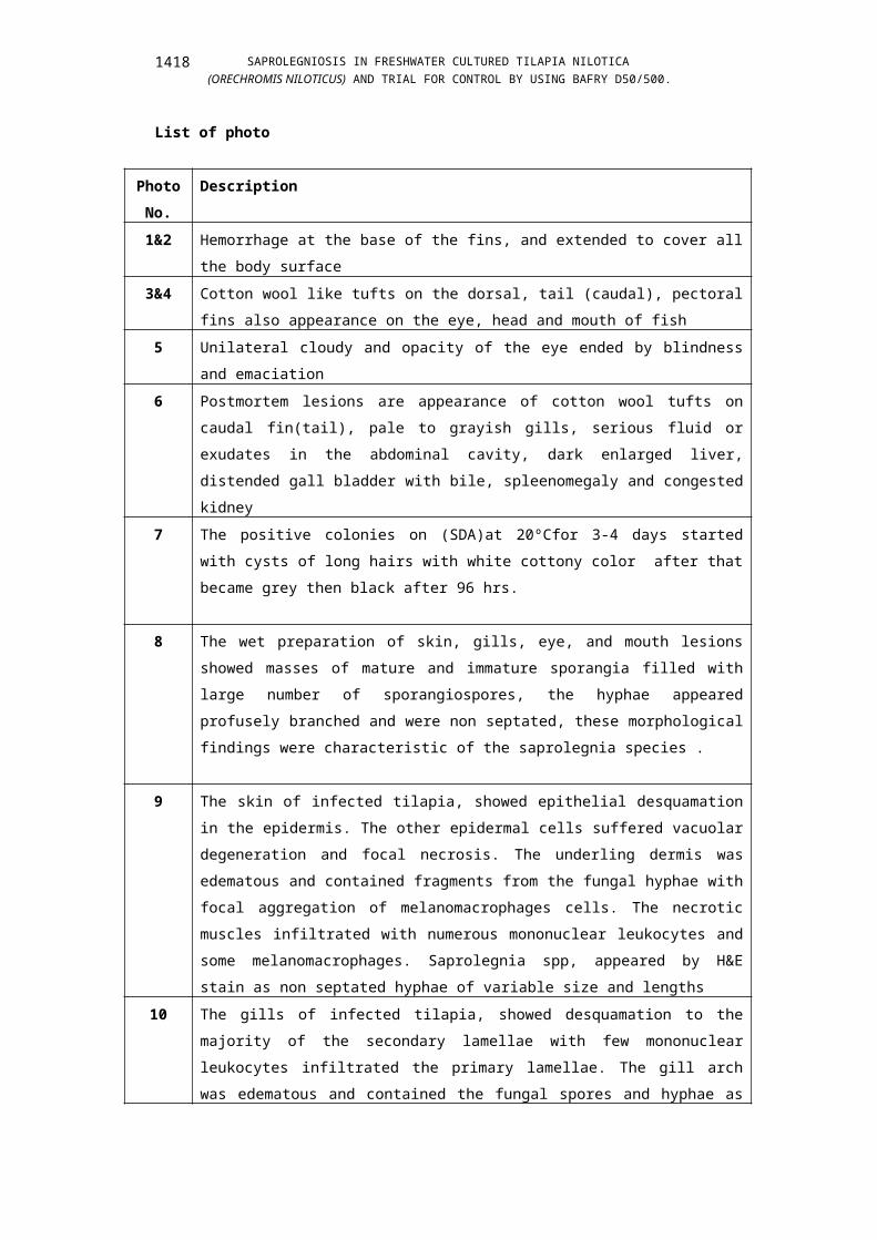
Table NO.
Title
1 Application of Bafry D50/5002 Safety test = tolerance test3 Incidence of saprolegnia parasitica isolated from different organs and
tissues
1413

SAPROLEGNIOSIS IN FRESHWATER CULTURED TILAPIA NILOTICA (ORECHROMIS NILOTICUS) AND TRIAL FOR CONTROL BY USING BAFRY D50/500.
4 Results of application of Bafry D 50/500 in disappearance of saprolegnial fungal growth and signs.
5 Results of effect of Bafry D 50/500 on hematological parameters and blood chemistry.
1414
MOHAMED E. ABOU EL ATTA 1415
SAPROLEGNIOSIS IN FRESHWATER CULTURED TILAPIA NILOTICA (ORECHROMIS NILOTICUS) AND TRIAL FOR CONTROL BY USING BAFRY D50/500.
List of photo
Photo No.
Description
1&2 Hemorrhage at the base of the fins, and extended to cover all the body surface
3&4 Cotton wool like tufts on the dorsal, tail (caudal), pectoral fins also appearance on the eye, head and mouth of fish
5 Unilateral cloudy and opacity of the eye ended by blindness and emaciation
6 Postmortem lesions are appearance of cotton wool tufts on caudal fin(tail), pale to grayish gills, serious fluid or exudates in the abdominal cavity, dark enlarged liver, distended gall bladder with bile, spleenomegaly and congested kidney
7 The positive colonies on (SDA)at 20ºCfor 3-4 days started with cysts of long hairs with white cottony color after that became grey then black after 96 hrs.
8 The wet preparation of skin, gills, eye, and mouth lesions showed masses of mature and immature sporangia filled with large number of sporangiospores, the hyphae appeared profusely branched and were non septated, these morphological findings were characteristic of the saprolegnia species .
9 The skin of infected tilapia, showed epithelial desquamation in the epidermis. The other epidermal cells suffered vacuolar degeneration and focal necrosis. The underling dermis was edematous and contained fragments from the fungal hyphae with focal aggregation of melanomacrophages cells. The necrotic muscles infiltrated with numerous mononuclear leukocytes and some melanomacrophages. Saprolegnia spp, appeared by H&E stain as non septated hyphae of variable size and lengths
10 The gills of infected tilapia, showed desquamation to the majority of the secondary lamellae with few mononuclear leukocytes infiltrated the primary lamellae. The gill arch was edematous and contained the fungal spores and hyphae as well as some melanomacrophages and mononuclear cells
1416
MOHAMED E. ABOU EL ATTA
11 Skin of Tilapia nilotica (O.niloticus) treated with BafryD50/500showed increase of M.M.C and slight sloughing of most superficial layer of epidermis
12 Gill of Tilapia nilotica (O.niloticus) treated with Bafry D50/500 showed hyperplasia of epithelial cells covering of primary lamellae.
REFERENCES1. Aly, S. M. and A. M. M. EL Ashram. 2000. Some factors contributing to
the development of Saprolegniosis in Nile tilapia (Oreochromis niloticus).Alex. J. Vet. Science 2000, Vol.16 NO.1:165-174.
2. Aly, S. M., L. F. Maberry and A. A. EL-Meleigy. 1996. Pathological studies on saprolegniosis among Oreochromis niloticus .Zag. Vet.J. Vol.24, NO.3 (1996) PP.51-56.
3. Amin, N. E., M. E. Essa and M. Saleh. 1985. Natural and experimental infection of Sartherdon niloticus (Tilapia nilotica) with saprolegniosis in Egypt. 2nd International Conference on warm water aquaculture Hawaii.
4. Austin, B. and D. A. Austin. 1993. Bacterial fish pathogens: Diseases in farmed and wild fish. 2nd ed. Ellis Horwood Ltd., Chichester, New York, London, England.
5. Bailey, T. A. 1984. Effect of twenty-five compounds on four species of aquatic fungi (Saprolegniasis) pathogenic to fish. Aquaculture 38, 97-104.
6. Bruno, D. W. and B. P. Wood. 1994. Saprolegnia and other Oomycetes. In Fish Diseases and Disorders, Volume 3, Viral, Bacterial and Fungal Infections. Edited by P.T.K. Woo and D.W. Bruno. CABI Publishing, Wallingford, Oxon, United Kingdom. pp. 599-659.
7. Bruno, D. W. and B. P. Wood. 1999. Saprolegnia and other Oomycetes. In Fish Disease and disorders, Volume 3, Viral, Bacterial and Fungal infections. Edited by P. T. K. Woo and D. W. Bruno. CABI publishing, Walling ford,Oxon,U.K.599-659 pp.
8. Chowdhury,M. B. R., U. A. Zahura, K. Z. A. Habib, M. D. Khatun and M. Muniruzzaman. 2003. Ulcer type of disease in the fishes of smallscale farmer‘s panel in Bangladesh, Asian Fisheries. Society (2001). P.67.
9. Derksen, J. A., V. E. Ostland and H. W. Feruson. 1999. Effects of hydrogen peroxide on clearance of formalin killed flavobacterium branchiophilum from the gills of rainbow trout, (Oncorhynchus mykiss)( Walbaum). Journal of fish diseases 22:59-67.
1417
SAPROLEGNIOSIS IN FRESHWATER CULTURED TILAPIA NILOTICA (ORECHROMIS NILOTICUS) AND TRIAL FOR CONTROL BY USING BAFRY D50/500.
10. Easa, M. E. and Amin N. E. 1987. Natural and experimental saprollegniosis of Tilapia (Oreochromis niloticus).Alex. J. Vet. Science.2000, Vol.16 NO.1:1165-174.
11. El Ashram, A. M. M., A. M. Abd El .Rhman and S. F. Sakr. 2007. Acontrobution to saprolegniosis in cultured Nile tilapia (Oreochromis niloticus) with special refernce to its controle.Egypt.J. Aquat. Biol. & fish. Vol. 11, No. 3:943-955 (2007) ISSN1110-1631.
12. El Genaidy, Halam, Zaki, S. Mona and S. M. Aly. 2004. Pathological and biochemical studies in Catfish infected with saprolegnia parasitica and treated with Potassium permanganate. Egypt. J. Basic Appl. Physiol. 3(1), 201-212.
13. El-Zayat, S. A .M. 1988.studies on freshwater fungi of Aswan high Dame lake. Ph.D. Thesis, Botany Dept. Faculty of Science (Aswan),Assiut University, Egypt.
14. Ferquson, H. W.1989. Textbook of systemic pathology of fish. 1st Ed. Iowa State Univ. Press. Ames, Iowa, Canada.
15. Fitzpatrick, M. S., C. B. Schreck and R. L. Chitwood. 1995. Evaluation of three candidate fungicides for treatment of adult spring chinook salmon. Prog. Fish-Cul. 57: 153-155.
16. Hatai, K., and G. I. Hoshiai. 1994. Pathogenicity of Saprolegnia parasitica coker. In Salmon Saprolegniasis. Edited by G. J. Mueller. U.S. Department of Energy, Bonneville Power Administration, Portland, Oregon. pp. 87-98
17. Howe, G. E., W. H. Gingerich, V. K. Dawson and J. J. Olson. 1999. Efficacy of hydrogen peroxide for treating Saprolegniasis in channel catfish. J. Aquat. Anim. Health, 11(3): 222-230.
18. Hughes, G. C. 1962. Seasonal periodicity of the saprolegniaceae in the south eastern United States. Transactions of the British Mycological Society 45:519-531.
19. Hussein, M. M. A., K. Hatai and Nomura 2001. Saprolegniois in salmonids and their eggs in Japan.J.Wildl; Dis.37:204-207.
20. Hussein, M. M. A. and K .Hatai 1999.Saprolegnia salmonis Sp.nov> isolated from sock eye salmon, Oncorhynchus nerka. Mycoscience, 40:385-389.
21. Marking, L. L., J. J. Rach and T. M. Schreier. 1994. Evaluayion of antifungal agents for fish culture. The progressive fish culturist.56:225-231.
22. Marzouk, M. S. M., F. El Far and M. A. Nawal. 1990. Some investigations on moulds and yeasts associated with tail and fun rot in freshwater fish in Egypt. Alex. J.Vet. Sci; Vol.6.No.1.193-203.
1418
MOHAMED E. ABOU EL ATTA
23. Marzouk, M. S. M., S. Rezeka, Samira and M. H. El Gamal. 2003. Some mycological investigations on cultured tilapia in Kafr El Sheikh Governorate. Kafr El Sheikh Vet. Med. J. Vol.1 No.2 (2003), 97-114.
24. Mc Andrew, K. J., C. Sommer ville, R. Wootten and J. E. Bron. 1998. The effect of hydrogen peroxide treatment on different life cycle stages of salmon louse, (Lepeophtheirrus salmonis) (Koyer, 1837). Journal of fish diseases 21:221-228.
25. Meinertz, J. R., G. R. Stehly, W. H. Gingerich and J. L. Allen. 1995. Residues of [14C] .Malachite green in eggs and fry of rainbow trout, Oncorhynchus mykiss [Walbaum], after treatment of eggs .J. Fish. Dis.18:239-247.
26. Meyer, F.P. (1991): Aquaculture disease and health management. J. Anim. Sci. 69: 4201-4208.
27. Meyer, F. P. and T. A. Jorgenson 1983.Teratological and other effects of malachite green on the development of rainbow trout and rabbits. Trans. Am. Fish.Soc., 112:818-824.
28. Noga, E.J. 1996. Fish Disease Diagnosis and Treatment. Mosby-Year Book, Inc. St. Louis, MO. 367 p.
29. Ogbonna, C. I. C. 1989. Fungi associated with disease of freshwater fishes in Plateau state, Nigeria, Journal of Aquatic Sciences, 4:59-62.
30. Paperna, J. G., A. S. Van and L. Basson. 1983. Review of diseases affecting cultured Cichlids. International Symposium on tilapia aquaculture. Pp.174-184.
31. Pickering, A. D. and L. G. Willoughby. 1982. In Microbial Diseases of Fish. Edited by R.J. Roberts. Academic Press, London, England. pp. 271-297.
32. Plumb, J. A. 1994. Health maintenance of cultured fishes principals Microbial diseases. CRC press Boca Raton F1.USA.
33. Prost, H., and A. Sopinska. 1989. Evaluation of the activity cellular protective process in carp with Saprolegnia infection and treatment with malachite green and immunostimulant. Medycyna Weterynaryjna,45: 603-605.
34. Rach, J. J., T. M. Schreier, G. E. Howe and S. D. Redman. 1997. Effect of species, life stage, and water temperature on the toxicity of hydrogen peroxide to fish. Prog. Fish-Cul. 59: 41-46.
35. Schnik, R. A. 1994. Low regulatory priority declared for hydrogen peroxide as an fungicide. Notice from National biological survey, National Fisheries Research Center, La Crosse, Wisconsin.
36. Schreier, T. M., J. j. Rach and G. E. Howe. 1996. Efficacy of formalin, Hydrogen peroxide, and sodium chloride on fungal infected Rainbow trout eggs .Aquaculture, 140:323-331.
1419
SAPROLEGNIOSIS IN FRESHWATER CULTURED TILAPIA NILOTICA (ORECHROMIS NILOTICUS) AND TRIAL FOR CONTROL BY USING BAFRY D50/500.
37. Shaheen, A. A. M. 1986. Mycoflora of some freshwater fish. M.V.Sc. Thesis, Zagazig University.
38. Speare, D. J. and Arsenault. 1997. Effects of intermittent hydrogen peroxide exposure on growth and columnaris disease of juvenile rainbow trout, Oncorhynchus mykiss. Canadian Journal of fisheries and Aquatic sciences 54:2653-2658.
39. Tort, J. Maria, Wooster, A. Gregory and Bowser, R. Paul 2003. Effect of hydrogen peroxide on hematology and blood chemistry parameters of Walleye Stizostedion vitreum. Journal of World Aquaculture Society, Vol. 34, No2 (pp.236-242).
40. Willoughby, L. G., and Roberts, R.J. 1992. Towards strategic use of fungicides against Saprolegnia parasitica in salmonid fish hatcheries. J. Fish Diseases 15: 1-13.
41. Roberts, R. J. 2001. Fish Pathology. 3rd Ed., W. B Sauders, U. K.42. Wotton, I. D. and H. Freeman. 1982. Microanalysis in medical
biochemistry. Churchill, Living stone, Edinburgh, London and New York.
1420