Embed Size (px)
Citation preview
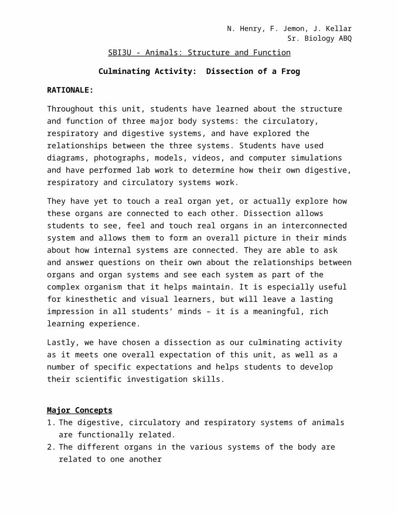
N. Henry, F. Jemon, J. KellarSr. Biology ABQ
SBI3U - Animals: Structure and Function
Culminating Activity: Dissection of a Frog
RATIONALE:
Throughout this unit, students have learned about the structure and function of three major body systems: the circulatory, respiratory and digestive systems, and have explored the relationships between the three systems. Students have used diagrams, photographs, models, videos, and computer simulations and have performed lab work to determine how their own digestive, respiratory and circulatory systems work.
They have yet to touch a real organ yet, or actually explore how these organs are connected to each other. Dissection allows students to see, feel and touch real organs in an interconnected system and allows them to form an overall picture in their minds about how internal systems are connected. They are able to ask and answer questions on their own about the relationships between organs and organ systems and see each system as part of the complex organism that it helps maintain. It is especially useful for kinesthetic and visual learners, but will leave a lasting impression in all students’ minds – it is a meaningful, rich learning experience.
Lastly, we have chosen a dissection as our culminating activity as it meets one overall expectation of this unit, as well as a number of specific expectations and helps students to develop their scientific investigation skills.
Major Concepts 1. The digestive, circulatory and respiratory systems of animals are functionally related.2. The different organs in the various systems of the body are related to one another
Curriculum Expectations: This culminating task will evaluate students’ ability to:
E2. Investigate, through laboratory inquiry the functional response of the respiratory and circulatory systems of animals, and the relationships between their respiratory, circulatory, and digestive systems;
E2.1 use appropriate terminology related to animal anatomy, including, but not limited to: systolic, diastolic, diffusion gradient, inhalation, exhalation, coronary, cardiac, ulcer, asthma, and constipation
E2.2 perform a laboratory or computer-simulated dissection of a representative animal, or use a mounted anatomical model, to analyse the relationships between the respiratory, circulatory, and digestive systems
N. Henry, F. Jemon, J. KellarSr. Biology ABQ
E3.1 explain the anatomy of the respiratory system and the process of ventilation and gas exchange from the environment to the cell (e.g., the movement of oxygen from the atmosphere to the cell; the roles of ventilation, hemoglobin, and diffusion in gas exchange)
E3.2 explain the anatomy of the digestive system and the importance of digestion in providing nutrients needed for energy and growth (e.g., the body’s mechanical and chemical processesdigest food, which provides the proteins needed to build muscle, and the fiber, water, vitamins, and minerals needed to regulate body processes)
E3.3 explain the anatomy of the circulatory system (e.g., blood components, blood vessels, the heart) and its function in transporting substances that are vital to health
Performance Objectives:
Investigate, through laboratory inquiry, relationships between the respiratory, circulatory, and digestive systems of a frog
Demonstrate an understanding of animal anatomy and physiology Identify the external and internal anatomy of a frog Use anatomical terms to describe both the external and internal structures of a frog
Levels of Reasoning Developed or Used: Knowledge, comprehension, application, analysis, synthesis, thinking and investigation
Modification for Gifted and ELL learners:
Dissection is an excellent learning opportunity for ELL students because they are able to see and manipulate the specimen and gain an understanding of the body systems without having to read a great deal of text. At this point in the unit they should already have an understanding of the systems – dissection allows them to confirm independently what they have learned. ELL students will work with students who are not ELL and pictures will be provided to go along side each step so that it is easier for them to follow. The instructional worksheet would be simplified by modifying the language requirements for written assessments so it is not too wordy but still addressing all levels of the achievement chart and providing a paragraph summary template (fill in the blank format). Students can receive bilingual support using word to word translation such as dictionaries, and glossaries and provide directions for the lab on tape. In addition, oral examination could be given instead of written and extended time for written lab report if needed.
Students who have demonstrated an excellent understanding of the body systems and good laboratory skills could act as group leaders and peer mentors throughout the dissection process. Following the dissection, all students will be required to complete a culminating assignment (as described later in this document). They will be given choice as to the format in which they will
N. Henry, F. Jemon, J. KellarSr. Biology ABQ
present their understanding. Students who are higher level could be required to choose one of the more complex formats available.
Lab Safety Rules and Precautions
Prior to beginning the lab, the following safety instructions should be discussed:
1) Extreme care must be taken when using dissecting instruments, particularly scalpels. To the extent possible, make cuts away from your body. The person who is cutting with the scalpel should be the only one whose hands are on the
specimen. Scalpels should remain at the dissection station and should not be carried throughout the
room. 2) The frogs are preserved in a chemical solution. Wear plastic gloves, eye protection and an
apron at all times and work in a well ventilated area. 3) If some chemical comes into contact with your skin, wash it off. At the end of the lesson,
wash your hands thoroughly. 4) Remind students of the location of eye wash stations – if chemical comes in contact with
their eyes it should be thoroughly flushed and medical attention sought. 5) Dispose of all materials as instructed by your teacher and clean your work area.6) Conduct yourself in a responsible manner at all times in the laboratory.7) Follow all written and verbal instructions carefully. If you do not understand a direction or
part of a procedure, please ASK YOUR TEACHER BEFORE PROCEEDING WITH THE ACTIVITY
8) If you or your lab partner is hurt, immediately (and loudly) call out the teacher's name to get the teacher's attention. Do not panic.
9) If a chemical should splash in your eye(s) or on your skin, immediately flush with running water for at least 20 minutes. Immediately (and loudly) yell out the teacher's name to get the teacher's attention.
Pre-lab Set-up/Activity:
N. Henry, F. Jemon, J. KellarSr. Biology ABQ
All materials and equipment will be checked to make sure that they are in working condition and that there are enough for each group of students.
Scalpel blades should be changed by the teacher prior to beginning the lab. If dissecting scopes are available, a few should be set up so that students can closely examine
organs they have removed from their specimen. All the materials and equipment will be in one area so that they are easily accessible to the
students. The lab will be clutter free so that students can work safely. Students should have thoroughly read the procedure and participated in the pre-lab talk prior
to beginning the lab.
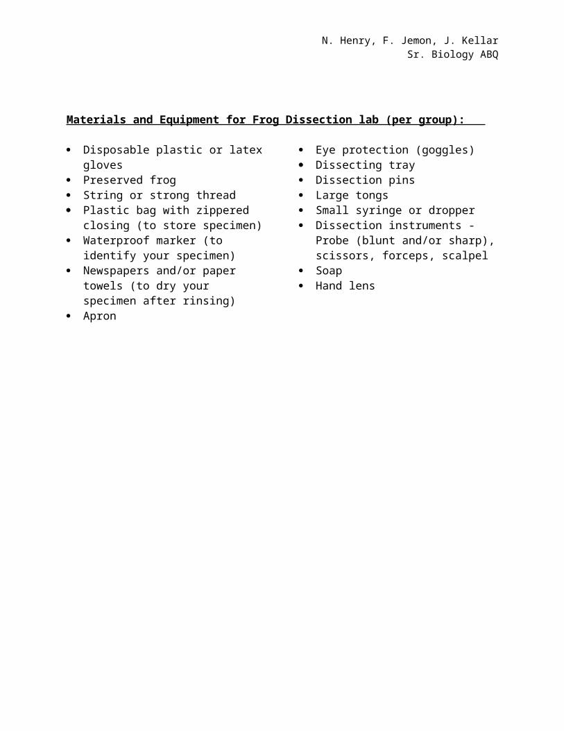
Materials and Equipment for Frog Dissection lab ( per group ):
Disposable plastic or latex gloves Preserved frog String or strong thread Plastic bag with zippered closing (to
store specimen) Waterproof marker (to identify your
specimen) Newspapers and/or paper towels (to dry
your specimen after rinsing) Apron
Eye protection (goggles) Dissecting tray Dissection pins Large tongs Small syringe or dropper Dissection instruments - Probe (blunt
and/or sharp), scissors, forceps, scalpel Soap Hand lens
N. Henry, F. Jemon, J. KellarSr. Biology ABQ
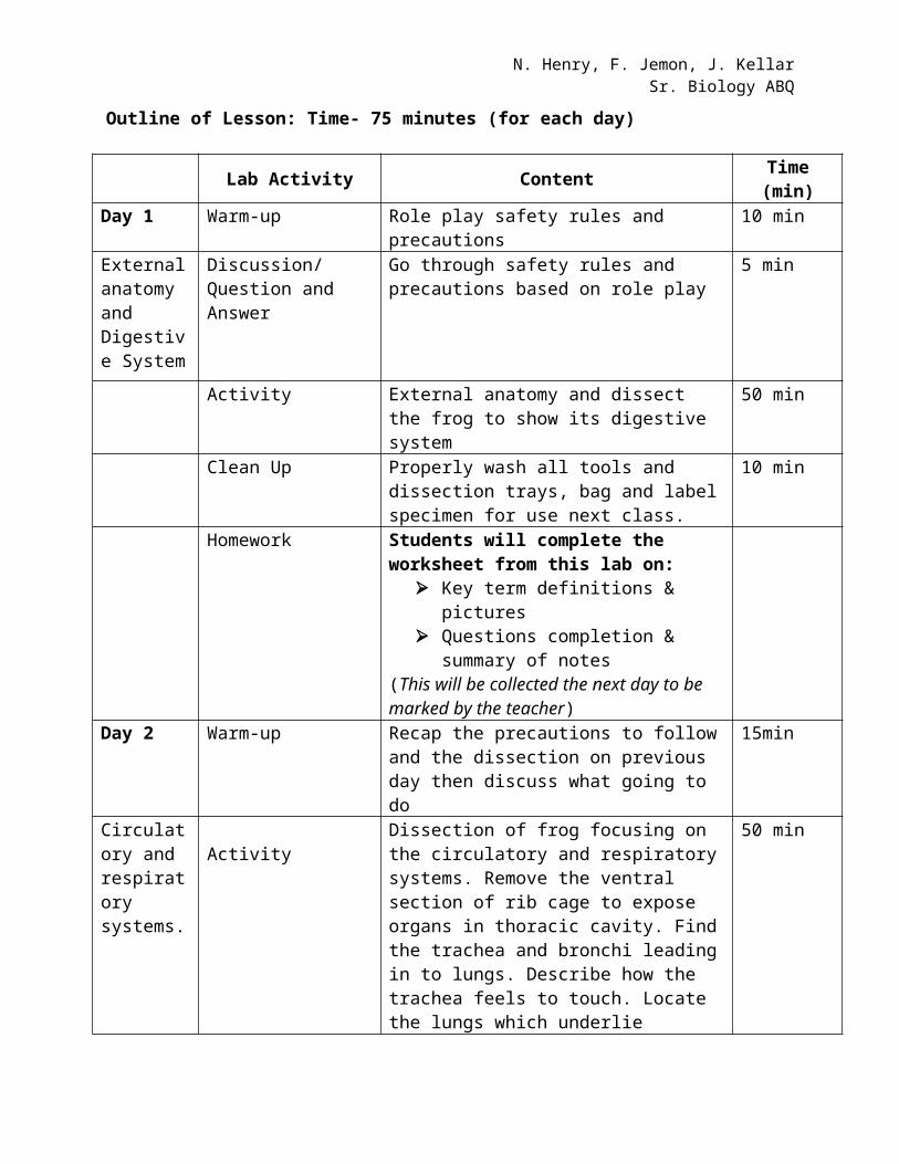
Outline of Lesson: Time- 75 minutes (for each day)
Lab Activity Content Time (min)
Day 1 Warm-up Role play safety rules and precautions 10 min External anatomy and Digestive System
Discussion/ Question and Answer
Go through safety rules and precautions based on role play
5 min
Activity External anatomy and dissect the frog to show its digestive system
50 min
Clean Up Properly wash all tools and dissection trays, bag and label specimen for use next class.
10 min
Homework Students will complete the worksheet from this lab on:
Key term definitions & pictures Questions completion & summary of
notes (This will be collected the next day to be marked by the teacher)
Day 2 Warm-up Recap the precautions to follow and the dissection on previous day then discuss what going to do
15min
Circulatory and respiratory systems.
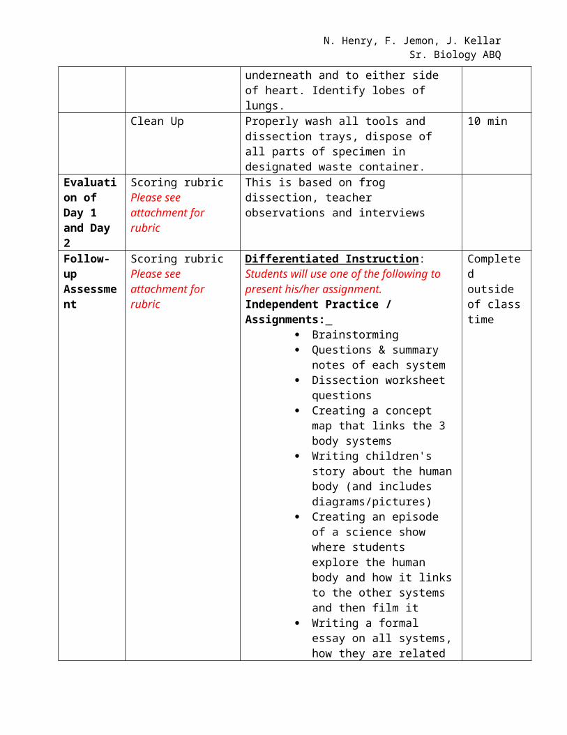
ActivityDissection of frog focusing on the circulatory and respiratory systems. Remove the ventral section of rib cage to expose organs in thoracic cavity. Find the trachea and bronchi leading in to lungs. Describe how the trachea feels to touch. Locate the lungs which underlie underneath and to either side of heart. Identify lobes of lungs.
50 min
Clean Up Properly wash all tools and dissection trays, dispose of all parts of specimen in designated waste container.
10 min
Evaluation of Day 1 and Day 2
Scoring rubricPlease see attachment for rubric
This is based on frog dissection, teacher observations and interviews
Follow-upAssessment
Scoring rubricPlease see attachment for rubric
Differentiated Instruction: Students will use one of the following to present his/her assignment.Independent Practice / Assignments:
Brainstorming Questions & summary notes of
Completed outside of class time
N. Henry, F. Jemon, J. KellarSr. Biology ABQ
each system Dissection worksheet questions Creating a concept map that
links the 3 body systems Writing children's story about
the human body (and includes diagrams/pictures)
Creating an episode of a science show where students explore the human body and how it links to the other systems and then film it
Writing a formal essay on all systems, how they are related and their disorders
Creating a power point presentation
Creating a detailed, labelled diagram of the human body systems
N. Henry, F. Jemon, J. KellarSr. Biology ABQ
Name:
Student Handout: Frog Dissection Culminating Activity
Introduction
Frogs belong to the class Amphibia. Amphibians have adaptations for living in both aquatic and terrestrial environments. They are among the most commonly studied organisms in biology. Although frogs and humans are different in many ways, they both have similar basic body plans. Humans and frogs belong to the phylum Chordata and by studying the anatomy of frogs we will be able to better understand our own body. In this two part investigation you will (a) examine and identify the external features of a frog and examine the organs of the digestive system and (b) examine the organs of the circulatory and respiratory systems.
Dissection involves the careful and systematic examination of the internal structures of an organism. To carry out a successful dissection students must know the vocabulary in the table below.
Term MeaningDorsal Upper or back surfaceVentral Under or belly surfaceLateral SideAnterior Toward the front (head) endPosterior Toward the back endSuperficial Near the surfaceProximal Close toDistal Far from
Materials and Equipment ( per group ):
Disposable plastic or latex gloves Preserved frog String or strong thread Plastic bag with zippered closing (to
store specimen) Waterproof marker (to identify your
specimen) Newspapers and/or paper towels (to dry
your specimen after rinsing) Apron
Eye protection (goggles) Dissecting tray Dissection pins Large tongs Small syringe or dropper Dissection instruments - Probe (blunt
and/or sharp), scissors, forceps, scalpel Soap Hand lens
N. Henry, F. Jemon, J. KellarSr. Biology ABQ
DAY 1: External Anatomy and Digestive System
Name: _________________________
Date: ___________________________
A. External Anatomy of the Frog1) Obtain a preserved frog, rinse the frog in the sink and place it in a dissecting tray.
2) Locate the forelegs and hindlegs. Each foreleg, or arm, is divided into four regions. Starting closest to the body, the parts are identified as: upper arm, forearm, wrist and hand. Each hindleg also has four regions: thigh, lower leg, ankle and foot. Identify the parts of the forelegs and hindlegs. Examine the hands and feet of the frog. If the hands have enlarged thumbs, the frog is male.
3) Locate the two large protruding eyes. Lift the lower eyelid using a probe: this lid is called the nictitating membrane. This protects the eye while the frog is under water because the membrane is translucent; the frog is able to see under water.
4) Posterior to each eye is a circular region of tight stretched skin. This region is the tympanic membrane or the eardrum. Locate the tympanic membrane on both sides of the head. Anterior to the eyes, locate two openings called the external nares (singular naris), or nostrils.
5) Pry the frog's mouth open and use scissors to cut the angles of the frog's jaws open. Cut deeply enough so that the frog's mouth opens wide enough to view the structures inside. Locate the tongue. Play with the tongue, (You may remove the tongue).
6) In the center of the mouth, toward the back is a single round opening. This is the esophagus. This tube leads to the stomach. Use a probe to poke into the esophagus. Look for two openings at the back of the floor of the mouth. These are the openings to the vocal sacs. They are present in males but not females.
7) Close to the angles of the jaw are two openings, one on each side. These are the Eustachian tubes. They are used to equalize pressure in the inner ear while the frog is swimming. Insert a probe into the Eustachian tube.
N. Henry, F. Jemon, J. KellarSr. Biology ABQ
8) Just behind the tongue and before you reach the esophagus is a slit like opening. You may need to use your probe to get it to open up. This slit is the glottis, and it is the opening to the lungs. The frog breathes and vocalizes with the glottis. Use your probe to open the glottis and compare that opening to the esophagus.
9) The frog has two sets of teeth. The vomarine teeth are found on the roof of the mouth. The maxillary teeth are found around the edge of the mouth. Both are used for holding prey, frogs swallow their meals whole and do NOT chew. Run you finger over both sets of teeth and note the differences between them.
10) On the roof of the mouth, you will find the two tiny openings of the nostrils, if you put your probe into those openings; you will find they exit on the outside of the frog.
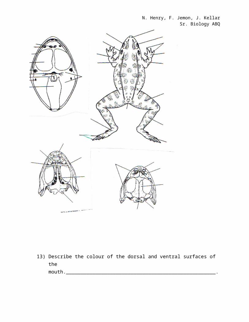
11) Label the following external structure on the diagram below. (8 marks)a) fore leg (limb) g) hindleg m) external nareb) upper arm h) thigh n) eyec) forearm I) lower leg o) nictitating membraned) wrist j) ankle p) mouthe) hand k) webbed footf) thumb l) tympanic membrane
12) In the figure below , label the following parts of the frog’s mouth. (5marks)
vomarine teeth internal nares maxillary teeth openings to Eustachian tubes tongue gullet openings
esophagus glottis opening openings to vocal sac palate
(Students will finish this worksheet for homework)
N. Henry, F. Jemon, J. KellarSr. Biology ABQ
Label each of the structures using the terms on the previous page.
A
N. Henry, F. Jemon, J. KellarSr. Biology ABQ
13) Describe the colour of the dorsal and ventral surfaces of the mouth.___________________________________________________.
14) How many digits are on each of the frog’s hand? ______________.
15) How many digits are on each of the frog’s feet? ______________.
16) Is your frog a male or a female? ___________. How do you know? _______________________________________________________.
17) Where is the nictitating membrane attached? _____________________________________________________________________________________________________________________ .
18) Where is the tongue attached in the mouth? _____________________________________________________________________.
Total (6marks).
B. Internal Anatomy of the Frog- Digestive System
1) Turn your preserved frog so that the ventral side is facing up. With dissecting pins, securely pin the frog’s feet and hands to the bottom of the dissecting tray. Angle the pins away from the body of the frog so that they will not interfere with your dissection.
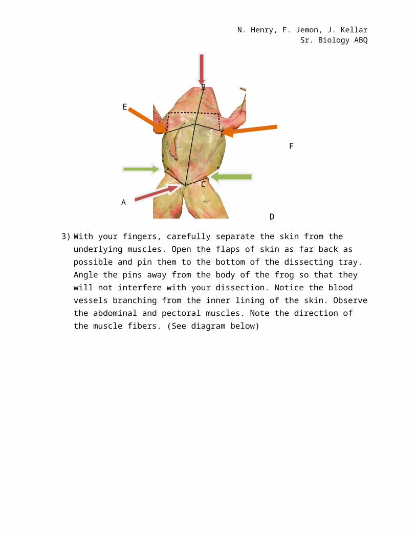
2) With forceps, lift the loose skin of the abdomen. Carefully insert the tip of a pair of scissors beneath the skin. Cut the skin along the line AB as seen in the diagram below, using forceps and scissors, cut the skin along line CD and EF.
B
E
F
N. Henry, F. Jemon, J. KellarSr. Biology ABQ
C
D
3) With your fingers, carefully separate the skin from the underlying muscles. Open the flaps of skin as far back as possible and pin them to the bottom of the dissecting tray. Angle the pins away from the body of the frog so that they will not interfere with your dissection. Notice the blood vessels branching from the inner lining of the skin. Observe the abdominal and pectoral muscles. Note the direction of the muscle fibers. (See diagram below)
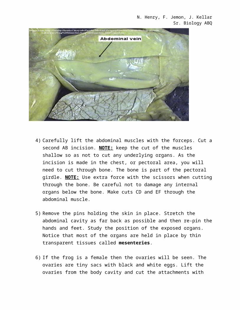
4) Carefully lift the abdominal muscles with the forceps. Cut a second AB incision. NOTE: keep the cut of the muscles shallow so as not to cut any underlying organs. As the incision is made in the chest, or pectoral area, you will need to cut through bone. The bone is part of the pectoral girdle. NOTE: Use extra force with the scissors when cutting through the bone. Be careful not to damage any internal organs below the bone. Make cuts CD and EF through the abdominal muscle.
5) Remove the pins holding the skin in place. Stretch the abdominal cavity as far back as possible and then re-pin the hands and feet. Study the position of the exposed organs. Notice that most of the organs are held in place by thin transparent tissues called mesenteries.
N. Henry, F. Jemon, J. KellarSr. Biology ABQ
6) If the frog is a female then the ovaries will be seen. The ovaries are tiny sacs with black and white eggs. Lift the ovaries from the body cavity and cut the attachments with scissors, then remove the ovaries. NOTE: Be careful not to rupture the ovaries as the eggs will spill out of them.
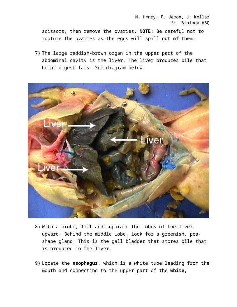
7) The large reddish-brown organ in the upper part of the abdominal cavity is the liver. The liver produces bile that helps digest fats. See diagram below.
8) With a probe, lift and separate the lobes of the liver upward. Behind the middle lobe, look for a greenish, pea-shape gland. This is the gall bladder that stores bile that is produced in the liver.
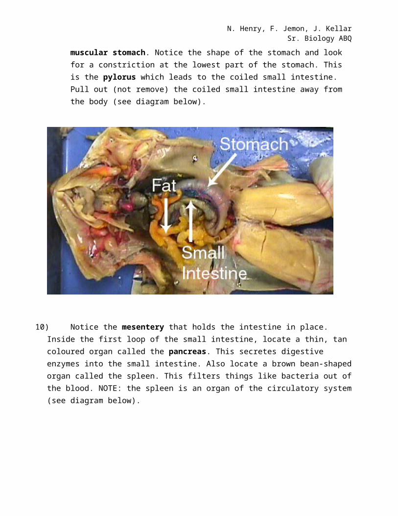
9) Locate the esophagus, which is a white tube leading from the mouth and connecting to the upper part of the white, muscular stomach. Notice the shape of the stomach and look for a constriction at the lowest part of the stomach. This is the pylorus which leads to the coiled small intestine. Pull out (not remove) the coiled small intestine away from the body (see diagram below).

N. Henry, F. Jemon, J. KellarSr. Biology ABQ
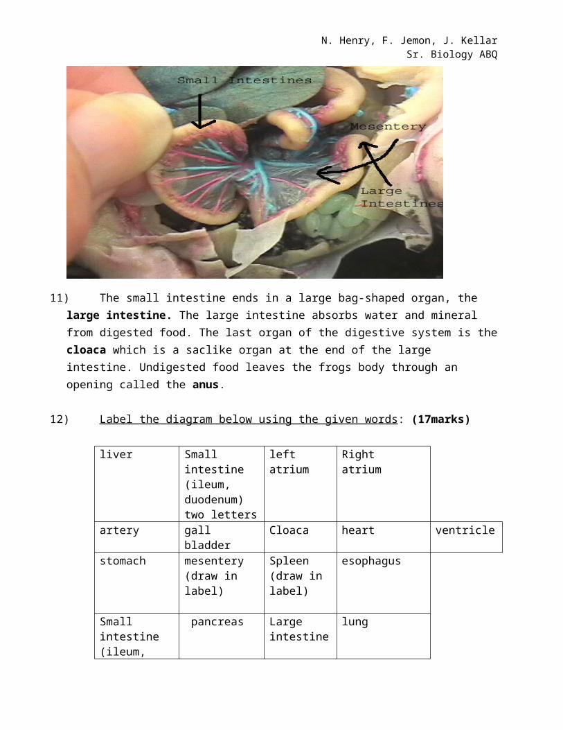
10) Notice the mesentery that holds the intestine in place. Inside the first loop of the small intestine, locate a thin, tan coloured organ called the pancreas. This secretes digestive enzymes into the small intestine. Also locate a brown bean-shaped organ called the spleen. This filters things like bacteria out of the blood. NOTE: the spleen is an organ of the circulatory system (see diagram below).
N. Henry, F. Jemon, J. KellarSr. Biology ABQ
11) The small intestine ends in a large bag-shaped organ, the large intestine. The large intestine absorbs water and mineral from digested food. The last organ of the digestive system is the cloaca which is a saclike organ at the end of the large intestine. Undigested food leaves the frogs body through an opening called the anus.
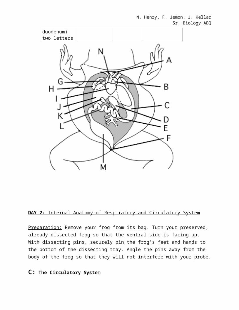
12) Label the diagram below using the given words : (17marks)
liver Small intestine (ileum, duodenum) two letters
left atrium Right atrium
artery gall bladder Cloaca heart ventricle
stomach mesentery (draw in label)
Spleen (draw in label)
esophagus
Small intestine (ileum, duodenum) two letters
pancreas Large intestine
lung
N. Henry, F. Jemon, J. KellarSr. Biology ABQ
DAY 2: Internal Anatomy of Respiratory and Circulatory System
Preparation: Remove your frog from its bag. Turn your preserved, already dissected frog so that the ventral side is facing up. With dissecting pins, securely pin the frog’s feet and hands to the bottom of the dissecting tray. Angle the pins away from the body of the frog so that they will not interfere with your probe.
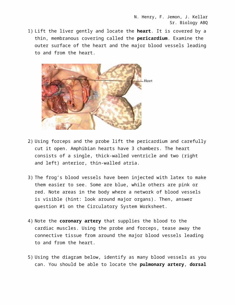
C: The Circulatory System1) Lift the liver gently and locate the heart. It is covered by a thin, membranous covering called
the pericardium. Examine the outer surface of the heart and the major blood vessels leading to and from the heart.
2) Using forceps and the probe lift the pericardium and carefully cut it open. Amphibian hearts have 3 chambers. The heart consists of a single, thick-walled ventricle and two (right and left) anterior, thin-walled atria.
3) The frog’s blood vessels have been injected with latex to make them easier to see. Some are blue, while others are pink or red. Note areas in the body where a network of blood vessels is visible (hint: look around major organs). Then, answer question #1 on the Circulatory System Worksheet.
4) Note the coronary artery that supplies the blood to the cardiac muscles. Using the probe and forceps, tease away the connective tissue from around the major blood vessels leading to and from the heart.
N. Henry, F. Jemon, J. KellarSr. Biology ABQ
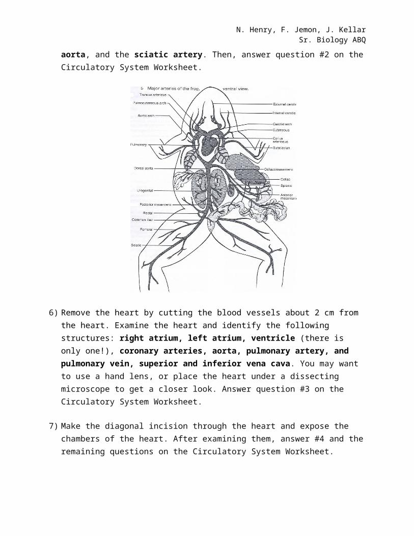
5) Using the diagram below, identify as many blood vessels as you can. You should be able to locate the pulmonary artery, dorsal aorta, and the sciatic artery. Then, answer question #2 on the Circulatory System Worksheet.
6) Remove the heart by cutting the blood vessels about 2 cm from the heart. Examine the heart and identify the following structures: right atrium, left atrium, ventricle (there is only one!), coronary arteries, aorta, pulmonary artery, and pulmonary vein, superior and inferior vena cava. You may want to use a hand lens, or place the heart under a dissecting microscope to get a closer look. Answer question #3 on the Circulatory System Worksheet.
7) Make the diagonal incision through the heart and expose the chambers of the heart. After examining them, answer #4 and the remaining questions on the Circulatory System Worksheet.
N. Henry, F. Jemon, J. KellarSr. Biology ABQ
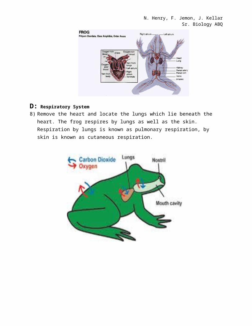
D: Respiratory System8) Remove the heart and locate the lungs which lie beneath the heart. The frog respires by lungs
as well as the skin. Respiration by lungs is known as pulmonary respiration, by skin is known as cutaneous respiration.
N. Henry, F. Jemon, J. KellarSr. Biology ABQ
9) Identify the lobes of lungs Describe the look and feel of lungs________________________________________________________________________________________________________________________________________________The lungs are open into a laryngo-tracheal chamber.
10) Trace backwards to locate bronchi and trachea. Describe how the trachea feels to touch.________________________________________________________________________________________________________________________________________________
The trachea is made up of cartilaginous plates, which is flexible.
11) Locate the larynx of the frog. The larynx opens to buccal cavity by the glottis. The buccal cavity communicates with outside by the mouth and external nostrils; and it is through the latter that the air passes in and out.
N. Henry, F. Jemon, J. KellarSr. Biology ABQ
12) Label the diagram given below. (2 marks)
N. Henry, F. Jemon, J. KellarSr. Biology ABQ
13) Identify the respiratory system from the diagram below and label it.(2 marks)
N. Henry, F. Jemon, J. KellarSr. Biology ABQ
Circulatory System Worksheet (20 marks)
As you complete the dissection, answer the following questions. They will be due in your next class period.
1. As you examined the specimen, you should have noticed that the blood vessels were either blue or red/pink. (4 marks)
a. Which type of blood vessels are blue? ________________b. Which type of blood vessels are red? _________________c. A network of blood vessels surrounds the lungs. Explain why.
d. A network of blood vessels surrounds the stomach and intestines. Explain why.
2. a) What is the function of the pulmonary artery? (1 mark)
b) What are the functions of the dorsal aorta and sciatic artery? (1 mark)
N. Henry, F. Jemon, J. KellarSr. Biology ABQ
3. Draw a diagram of the heart, labeling as many of the following parts as possible: right atrium, left atrium, ventricle (there is only one!), coronary arteries, aorta, pulmonary artery, pulmonary vein, superior and inferior vena cava. (6 marks)
4. a) Complete the table below. (3 marks)
Chamber Appearance Function
Right atrium
Left Atrium
Ventricle
b) The ventricle has thicker, more muscular walls than the atria. Relate this difference in wall structure to the functions of the 2 types of heart chamber. (2 marks)
5. What is the function of the circulatory system? How does its structure allow it to perform its function? Support your answer with evidence from your dissection. (3 marks)
N. Henry, F. Jemon, J. KellarSr. Biology ABQ
Concluding Questions (complete these questions independently) (16 marks)
6. How does the circulatory system interact with the digestive system? Support your answer with evidence from your dissection. (3 marks)
7. How does the circulatory system interact with the respiratory system? Support your answer with evidence from your dissection. (3 marks)
8. In what ways was your understanding of the digestive, circulatory, and respiratory systems enhanced by your observation of the real organs? (2 marks)
9. Describe the similarities and differences between the digestive, respiratory and circulatory systems of a frog and a human. (6 marks)
10. Do you think that the frog is a good model to use in training future doctors? Why or why not? (2 marks)
N. Henry, F. Jemon, J. KellarSr. Biology ABQ
TOTAL MARKS FOR ALL QUESTIONS: 76
References:
http://edweb.fdu.edu/folio/banborv/frogdissection/
http://www.slideshare.net/marglema9/frog-dissection-lab
http://www.aa.psu.edu/biology/frog/default.htm
http://www.biologycorner.com/myimages/frog-dissection/
http://www.altoona.psu.edu/academics/www/mns/bioal/Frog/resp.htm
Frog Dissection Guide: http://www.members.shaw.ca/jonesbiology1/bio11/units/ecology/frogdissection.PDF
Five major arteries of the Frog, ventral view: http://recipesforhealthy.tk/frog-dissection.html
Frog cross section: http://k-2.stanford.edu/InfoFrames/2-BioSys.5.0.html