Embed Size (px)
Citation preview
Washington University School of Medicine Washington University School of Medicine
Digital Commons@Becker Digital Commons@Becker
Open Access Publications
1-1-2021
Scalable bio marker combinations for early stroke diagnosis: A Scalable bio marker combinations for early stroke diagnosis: A
systematic review systematic review
Saiyet de la C Baez
Diana García Del Barco
Anette Hardy-Sosa
Gerardo Guillen Nieto
Maria Luisa Bringas-Vega
See next page for additional authors
Follow this and additional works at: https://digitalcommons.wustl.edu/open_access_pubs

Authors Authors Saiyet de la C Baez, Diana García Del Barco, Anette Hardy-Sosa, Gerardo Guillen Nieto, Maria Luisa Bringas-Vega, Jorge J Llibre-Guerra, and Pedro Valdes-Sosa
SYSTEMATIC REVIEWpublished: 28 May 2021
doi: 10.3389/fneur.2021.638693
Frontiers in Neurology | www.frontiersin.org 1 May 2021 | Volume 12 | Article 638693
Edited by:
Timo Uphaus,
Johannes Gutenberg University
Mainz, Germany
Reviewed by:
Svetlana A. Dambinova,
DeKalb Medical Center, United States
Johann Pelz,
University Hospital Leipzig, Germany
*Correspondence:
Pedro Valdes-Sosa
pedro.valdes@
neuroinformatics-collaboratory.org
Jorge J. Llibre-Guerra
Specialty section:
This article was submitted to
Stroke,
a section of the journal
Frontiers in Neurology
Received: 07 December 2020
Accepted: 29 March 2021
Published: 28 May 2021
Citation:
Baez SdlC, García del Barco D,
Hardy-Sosa A, Guillen Nieto G,
Bringas-Vega ML, Llibre-Guerra JJ
and Valdes-Sosa P (2021) Scalable
Bio Marker Combinations for Early
Stroke Diagnosis: A Systematic
Review. Front. Neurol. 12:638693.
doi: 10.3389/fneur.2021.638693
Scalable Bio Marker Combinationsfor Early Stroke Diagnosis: ASystematic ReviewSaiyet de la C. Baez 1,2, Diana García del Barco 2, Anette Hardy-Sosa 1,2,
Gerardo Guillen Nieto 1,2, Maria Luisa Bringas-Vega 1,3, Jorge J. Llibre-Guerra 4,5* and
Pedro Valdes-Sosa 1,3*
1 The Clinical Hospital of Chengdu Brain Sciences Institute, University Electronic Sciences and Technology of China UESTC,
Chengdu, China, 2Center for Genetic Engineering and Biotechnology, Havana, Cuba, 3Cuban Neurosciences Center,
Havana, Cuba, 4Department of Neurology, National Institute of Neurology and Neurosurgery of Cuba, Havana, Cuba,5Department of Neurology, Washington University School of Medicine in St. Louis, St. Louis, MO, United States
Background: Acute stroke treatment is a time-critical process in which every minute
counts. Laboratory biomarkers are needed to aid clinical decisions in the diagnosis.
Although imaging is critical for this process, these biomarkers may provide additional
information to distinguish actual stroke from its mimics and monitor patient condition and
the effect of potential neuroprotective strategies. For such biomarkers to be effectively
scalable to public health in any economic setting, these must be cost-effective and
non-invasive. We hypothesized that blood-based combinations (panels) of proteins might
be the key to this approach and explored this possibility through a systematic review.
Methods: We followed the PRISMA (Preferred Reporting Items for Systematic Reviews
and Meta-Analysis) guidelines for systematic review. Initially, the broader search for
biomarkers for early stroke diagnosis yielded 704 hits, and five were added manually.
We then narrowed the search to combinations (panels) of the protein markers obtained
from the blood.
Results: Twelve articles dealing with blood-based panels of protein biomarkers for
stroke were included in the systematic review. We observed that NR2 peptide (antibody
against the NR2 fragment) and glial fibrillary acidic protein (GFAP) are brain-specific
markers related to stroke. Von Willebrand factor (vWF), matrix metalloproteinase 9
(MMP-9), and S100β have been widely used as biomarkers, whereas others such as
the ischemia-modified albumin (IMA) index, antithrombin III (AT-III), and fibrinogen have
not been evaluated in combination. We herein propose the following new combination
of biomarkers for future validation: panel 1 (NR2 + GFAP + MMP-9 + vWF + S100β),
panel 2 (NR2+ GFAP+MMP-9+ vWF+ IMA index), and panel 3 (NR2+ GFAP+ AT-III
+ fibrinogen).
Conclusions: More research is needed to validate, identify, and introduce these panels
of biomarkers into medical practice for stroke recurrence and diagnosis in a scalable
manner. The evidence indicates that the most promising approach is to combine different
blood-based proteins to provide diagnostic precision for health interventions. Through
our systematic review, we suggest three novel biomarker panels based on the results in
the literature and an interpretation based on stroke pathophysiology.
Keywords: stroke, diagnosis, biomarker panels, serum biomarkers, neuroprotection
Baez et al. Biomarker Panels for Stroke Diagnosis
BACKGROUND
Stroke remains to be the second leading cause of deathworldwide, with a yearly death toll of 5.5 million (1, 2).Furthermore, approximately 116.4 million people are reportedlydisabled because of stroke, resulting in stroke being oneof the most important causes of disability in older people(3). Consequently, cerebrovascular diseases have substantialeconomic impact and significant social consequences. Thisimpact is exacerbated in lower- and middle-income countries.Evidence suggests that this situation is due to insufficient andnon-optimal strategies for the prevention of cerebrovasculardiseases and due to reduced availability of equipment for thediagnosis and treatment in medical centers (4).
Many of the shortcomings in managing stroke and relateddiseases are due to the heterogeneity of these pathologies. Themain subtypes of stroke are ischemic and hemorrhagic stroke.Ischemic stroke is characterized by a lack of blood supply toa part of the brain, whereas hemorrhagic stroke refers to acerebral bleed due to a blood vessel’s rupture (5). Ischemicstroke in turn comprises different subtypes such as transientischemic attack (TIA), which is transitory and reversible innature. We followed the classification system: Trial of Org 10172in Acute Stroke Treatment (TOAST) developed by Adams et al.(6), and we further distinguished large-artery atherosclerosis,cardioembolic (CE), lacunar, undetermined etiology, and otherdetermined etiology.
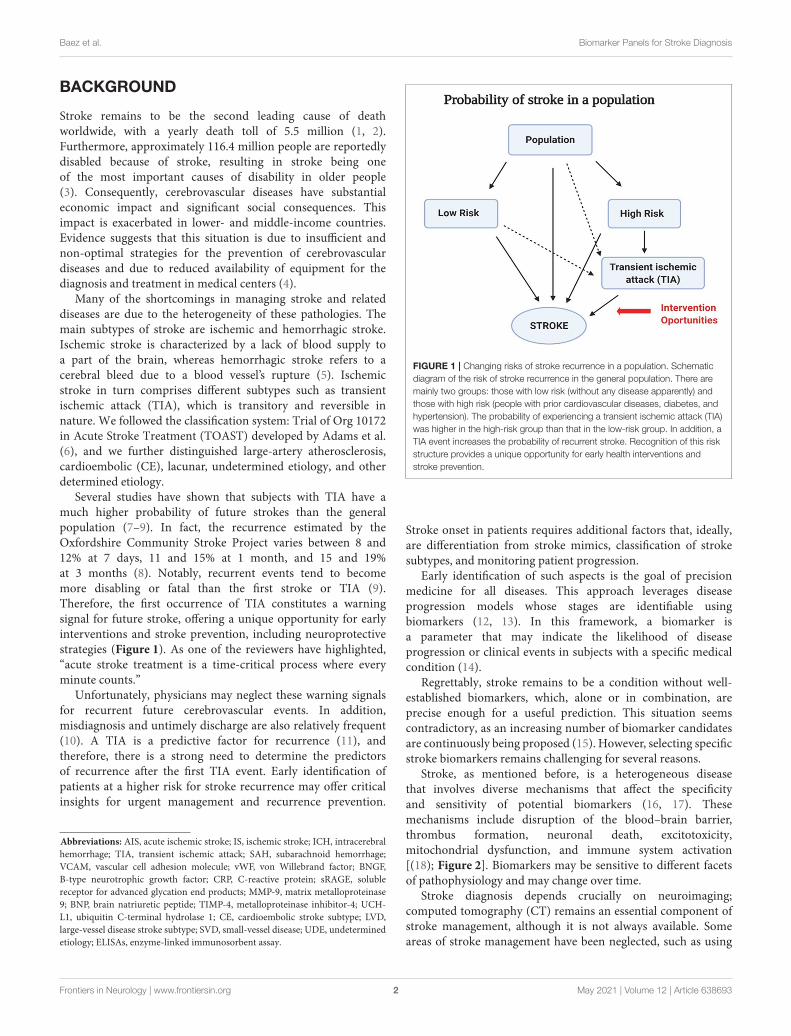
Several studies have shown that subjects with TIA have amuch higher probability of future strokes than the generalpopulation (7–9). In fact, the recurrence estimated by theOxfordshire Community Stroke Project varies between 8 and12% at 7 days, 11 and 15% at 1 month, and 15 and 19%at 3 months (8). Notably, recurrent events tend to becomemore disabling or fatal than the first stroke or TIA (9).Therefore, the first occurrence of TIA constitutes a warningsignal for future stroke, offering a unique opportunity for earlyinterventions and stroke prevention, including neuroprotectivestrategies (Figure 1). As one of the reviewers have highlighted,“acute stroke treatment is a time-critical process where everyminute counts.”
Unfortunately, physicians may neglect these warning signalsfor recurrent future cerebrovascular events. In addition,misdiagnosis and untimely discharge are also relatively frequent(10). A TIA is a predictive factor for recurrence (11), andtherefore, there is a strong need to determine the predictorsof recurrence after the first TIA event. Early identification ofpatients at a higher risk for stroke recurrence may offer criticalinsights for urgent management and recurrence prevention.
Abbreviations: AIS, acute ischemic stroke; IS, ischemic stroke; ICH, intracerebralhemorrhage; TIA, transient ischemic attack; SAH, subarachnoid hemorrhage;VCAM, vascular cell adhesion molecule; vWF, von Willebrand factor; BNGF,B-type neurotrophic growth factor; CRP, C-reactive protein; sRAGE, solublereceptor for advanced glycation end products; MMP-9, matrix metalloproteinase9; BNP, brain natriuretic peptide; TIMP-4, metalloproteinase inhibitor-4; UCH-L1, ubiquitin C-terminal hydrolase 1; CE, cardioembolic stroke subtype; LVD,large-vessel disease stroke subtype; SVD, small-vessel disease; UDE, undeterminedetiology; ELISAs, enzyme-linked immunosorbent assay.
FIGURE 1 | Changing risks of stroke recurrence in a population. Schematic
diagram of the risk of stroke recurrence in the general population. There are
mainly two groups: those with low risk (without any disease apparently) and
those with high risk (people with prior cardiovascular diseases, diabetes, and
hypertension). The probability of experiencing a transient ischemic attack (TIA)
was higher in the high-risk group than that in the low-risk group. In addition, a
TIA event increases the probability of recurrent stroke. Recognition of this risk
structure provides a unique opportunity for early health interventions and
stroke prevention.
Stroke onset in patients requires additional factors that, ideally,are differentiation from stroke mimics, classification of strokesubtypes, and monitoring patient progression.
Early identification of such aspects is the goal of precisionmedicine for all diseases. This approach leverages diseaseprogression models whose stages are identifiable usingbiomarkers (12, 13). In this framework, a biomarker isa parameter that may indicate the likelihood of diseaseprogression or clinical events in subjects with a specific medicalcondition (14).
Regrettably, stroke remains to be a condition without well-established biomarkers, which, alone or in combination, areprecise enough for a useful prediction. This situation seemscontradictory, as an increasing number of biomarker candidatesare continuously being proposed (15). However, selecting specificstroke biomarkers remains challenging for several reasons.
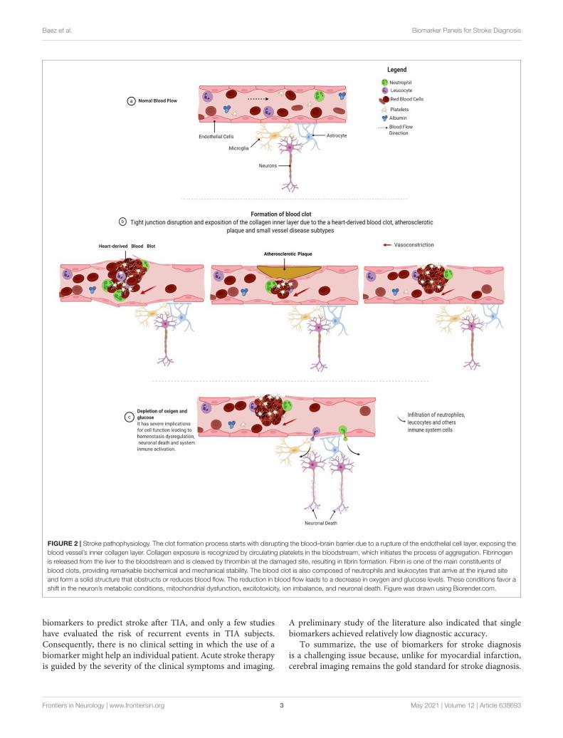
Stroke, as mentioned before, is a heterogeneous diseasethat involves diverse mechanisms that affect the specificityand sensitivity of potential biomarkers (16, 17). Thesemechanisms include disruption of the blood–brain barrier,thrombus formation, neuronal death, excitotoxicity,mitochondrial dysfunction, and immune system activation[(18); Figure 2]. Biomarkers may be sensitive to different facetsof pathophysiology and may change over time.
Stroke diagnosis depends crucially on neuroimaging;computed tomography (CT) remains an essential component ofstroke management, although it is not always available. Someareas of stroke management have been neglected, such as using
Frontiers in Neurology | www.frontiersin.org 2 May 2021 | Volume 12 | Article 638693
Baez et al. Biomarker Panels for Stroke Diagnosis
FIGURE 2 | Stroke pathophysiology. The clot formation process starts with disrupting the blood–brain barrier due to a rupture of the endothelial cell layer, exposing the
blood vessel’s inner collagen layer. Collagen exposure is recognized by circulating platelets in the bloodstream, which initiates the process of aggregation. Fibrinogen
is released from the liver to the bloodstream and is cleaved by thrombin at the damaged site, resulting in fibrin formation. Fibrin is one of the main constituents of
blood clots, providing remarkable biochemical and mechanical stability. The blood clot is also composed of neutrophils and leukocytes that arrive at the injured site
and form a solid structure that obstructs or reduces blood flow. The reduction in blood flow leads to a decrease in oxygen and glucose levels. These conditions favor a
shift in the neuron’s metabolic conditions, mitochondrial dysfunction, excitotoxicity, ion imbalance, and neuronal death. Figure was drawn using Biorender.com.
biomarkers to predict stroke after TIA, and only a few studieshave evaluated the risk of recurrent events in TIA subjects.Consequently, there is no clinical setting in which the use of abiomarker might help an individual patient. Acute stroke therapyis guided by the severity of the clinical symptoms and imaging.
A preliminary study of the literature also indicated that singlebiomarkers achieved relatively low diagnostic accuracy.
To summarize, the use of biomarkers for stroke diagnosisis a challenging issue because, unlike for myocardial infarction,cerebral imaging remains the gold standard for stroke diagnosis.
Frontiers in Neurology | www.frontiersin.org 3 May 2021 | Volume 12 | Article 638693
Baez et al. Biomarker Panels for Stroke Diagnosis
Therefore, expectations regarding the use of biomarkers in strokepatients should be realistic. We suggest that the primary use ofbiomarkers in stroke patients is to provide additional laboratoryinformation to effectively distinguish between actual stroke andits mimics and to monitor patient condition and the effect ofpotential neuroprotective strategies.
To highlight promising directions, we present a systematicreview of the literature on stroke biomarkers for the purposesmentioned above. This review comprises the following:
1. A preliminary review of the literature indicated thatcombinations of stroke biomarkers (“panels”) showedincreased diagnostic accuracy. Thus, we focused on panels ofbiomarkers instead of isolated determinations.
2. We limited our attention to only those studies thatreported the area under the receiver operating characteristic(ROC) curve (AUC). This choice allowed for quantitativecomparisons of accuracy.
3. Because of the paucity of studies reported in the literatureon subtype classification of stroke through biomarkercombinations, we narrowed our search to the small-vessel-disease subtype of stroke.
4. We also focused our review on blood-based biomarkers asthey seem to offer several advantages in terms of cost and easeof scalability (12).
As a consequence of our review, we propose new combinationsthat highlight the pathophysiological processes related to theselected biomarkers.
Overall, we adopted this scope for our review because ofthe geographical distribution of stroke. The highest incidenceof stroke has been reported in high-income countries. Betterreporting and shifting demographics place the onus on thedeveloping world, with an increase of 91.4 million disability-adjusted life-years and 4.85 million deaths in proportion to allglobal causes (4, 19). Thus, technologies that are deployablewithout advanced analytical or imaging technologies need tobe explored in more detail. Blood-based biomarker panels maytherefore contribute in providing valuable information for themanagement of stroke.
METHODS
Article SearchWe developed a search strategy with assistance from aresearch committee formed by neurologists, molecular biologists,mathematicians, and bioinformaticians. The search strategy wasestablished using a combination of standardized MeSH (MedicalSubject Headings) terms and keywords, including but not limitedto (-cerebrovascular disorder or brain vascular disorders orvascular diseases, intracranial or intracranial vascular diseaseor cerebrovascular occlusion or cerebrovascular accident orintracranial embolism, and thrombosis or cerebrovascularinsufficiencies) AND (- ischemia or Stroke or infarction orbrain infarction or hypoxia-ischemia or brain ischemia orischemic attack) AND (-intracerebral hemorrhage, cerebralhemorrhage, or intracranial hemorrhage) AND (- biologicalmarker or biomarker or biologic marker or marker, biological,
or biomarker panel) AND (- blood plasma sample, serumplasma sample, cerebrospinal fluid, blood proteins, plasma,blood, marker, serum, or serum marker or laboratory markers)AND (- diagnoses or diagnostic or examinations). The searchencompassed studies conducted between 1966 and June 2020for studies in patients with suspected stroke; the inclusion andexclusion criteria are provided below. The PubMed search wasconducted on October 10, 2020, at 12:48:21 P.M.
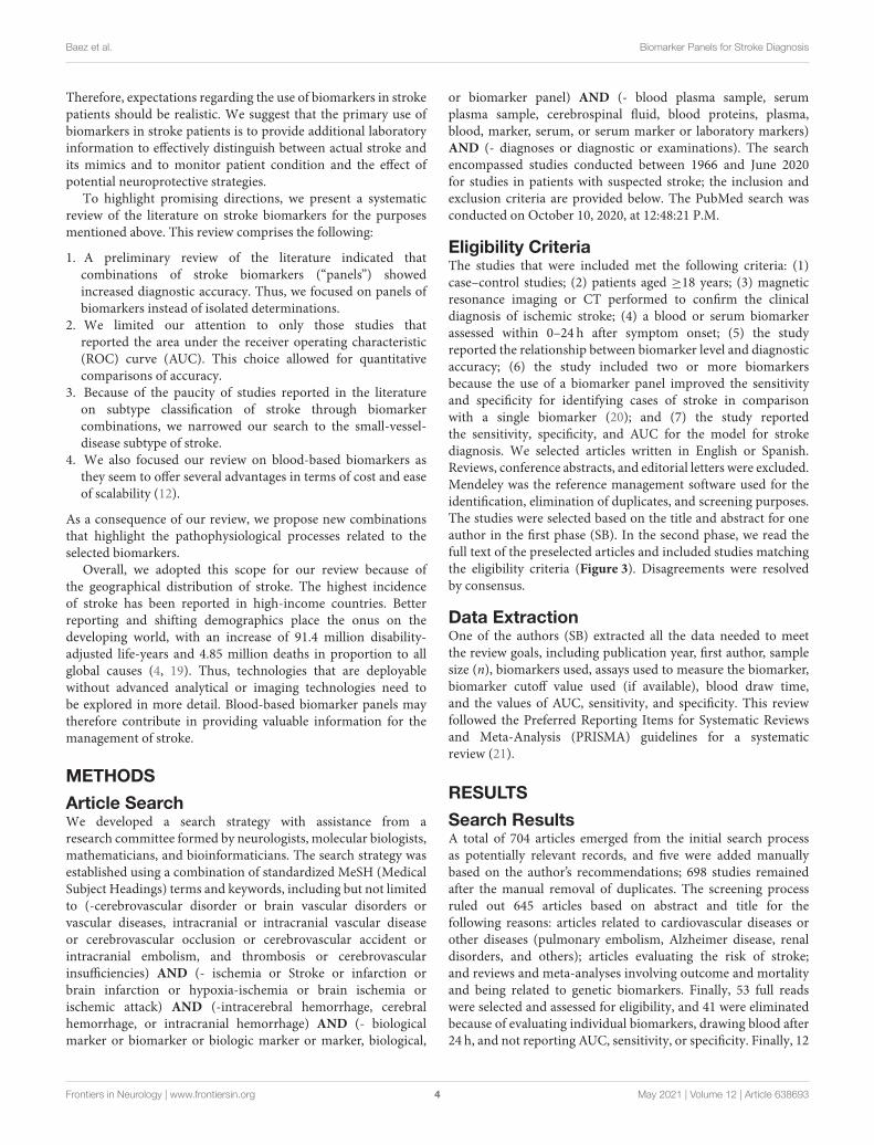
Eligibility CriteriaThe studies that were included met the following criteria: (1)case–control studies; (2) patients aged ≥18 years; (3) magneticresonance imaging or CT performed to confirm the clinicaldiagnosis of ischemic stroke; (4) a blood or serum biomarkerassessed within 0–24 h after symptom onset; (5) the studyreported the relationship between biomarker level and diagnosticaccuracy; (6) the study included two or more biomarkersbecause the use of a biomarker panel improved the sensitivityand specificity for identifying cases of stroke in comparisonwith a single biomarker (20); and (7) the study reportedthe sensitivity, specificity, and AUC for the model for strokediagnosis. We selected articles written in English or Spanish.Reviews, conference abstracts, and editorial letters were excluded.Mendeley was the reference management software used for theidentification, elimination of duplicates, and screening purposes.The studies were selected based on the title and abstract for oneauthor in the first phase (SB). In the second phase, we read thefull text of the preselected articles and included studies matchingthe eligibility criteria (Figure 3). Disagreements were resolvedby consensus.
Data ExtractionOne of the authors (SB) extracted all the data needed to meetthe review goals, including publication year, first author, samplesize (n), biomarkers used, assays used to measure the biomarker,biomarker cutoff value used (if available), blood draw time,and the values of AUC, sensitivity, and specificity. This reviewfollowed the Preferred Reporting Items for Systematic Reviewsand Meta-Analysis (PRISMA) guidelines for a systematicreview (21).
RESULTS
Search ResultsA total of 704 articles emerged from the initial search processas potentially relevant records, and five were added manuallybased on the author’s recommendations; 698 studies remainedafter the manual removal of duplicates. The screening processruled out 645 articles based on abstract and title for thefollowing reasons: articles related to cardiovascular diseases orother diseases (pulmonary embolism, Alzheimer disease, renaldisorders, and others); articles evaluating the risk of stroke;and reviews and meta-analyses involving outcome and mortalityand being related to genetic biomarkers. Finally, 53 full readswere selected and assessed for eligibility, and 41 were eliminatedbecause of evaluating individual biomarkers, drawing blood after24 h, and not reporting AUC, sensitivity, or specificity. Finally, 12
Frontiers in Neurology | www.frontiersin.org 4 May 2021 | Volume 12 | Article 638693
Baez et al. Biomarker Panels for Stroke Diagnosis
FIGURE 3 | PRISMA diagram. Flow diagram of the search and screening process. PubMed research yielded 704 articles at baseline, and five were added manually.
No other sources of article identification were identified.
articles were included in the systematic review. A PRISMA flowdiagram describing the search and screening process is shown inFigure 3.
An example of a systematic review that was not includedin our evaluation because of a lack of statistical measuresof diagnostic accuracy was a recent meta-analysis evaluatingseveral biomarkers (25). Most of the biomarkers evaluated inthis study are reported in the literature and are reviewed aspotential candidates and are added in several panels below;however, von Willebrand factor (vWF) and NR2 were omitted.Note that glial fibrillary acidic protein (GFAP) was the mostpromising biomarker in the study of separate ischemic stroke(IS), intracerebral hemorrhage (ICH), and healthy controls.The same study mentions D-dimer, matrix metalloproteinase 9(MMP-9), brain natriuretic peptide (BNP), and protein S100-β(S100β) derived from the meta-regression analysis as significantmarkers to be evaluated within 6 and 24 h of symptom onset (22).
Study CharacteristicsThe main features of the selected studies are listed in Table 1.Regarding sample characteristics, all the studies were case–control, which included control participants (without stroke),patients with acute IS (AIS), ICH, TIA, mimics, closed-head
injuries (CLHs), or subarachnoid hemorrhage (SAH). All thestudies involved subjects aged ≥18 years, and in the majority,immunoassays were used to evaluate the levels of biomarkers.Most of them reported their values of sensitivity, specificity,or AUC obtained within the first 6 h of symptom onset usingmultivariate or univariate regression logistic analyses.
Biomarker Analysis Based on SelectedStudiesOne of the biomarker panels frequently evaluated for theidentification of AIS is composed of four proteins: BNP, D-dimer,S100β, and MMP-9 (23–25). The results of previous studies havebeen mixed as follows:
• Laskowitz et al. (23) showed that combining these fourproteins outperformed other biomarkers in differentiatingmimics from intracranial hemorrhage cases, with c = 0.76.This result was validated in a study of 293 subjects, 361mimics, and 197 TIA with a validation cohort of 343suspected stroke cases. The study’s global results to classifystroke cases exhibited a high sensitivity of ∼90% but a lowspecificity of ∼45%, and 91% sensitivity and 45% specificityfor differentiating specific IS.
Frontiers in Neurology | www.frontiersin.org 5 May 2021 | Volume 12 | Article 638693
Baezetal.
Biomarke
rPanelsforStro
keDiagnosis
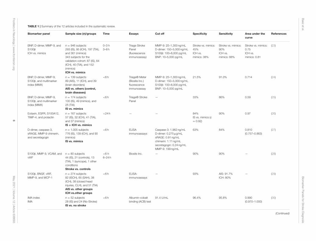
TABLE 1 | Summary of the 12 articles included in the systematic review.
Biomarker panel Sample size (n)/groups Time Essays Cut off Specificity Sensitivity Area under the
curve
References
BNP, D-dimer, MMP-9, and
S100β
ICH vs. mimics
n = 946 subjects
293 (IS), 95 (ICH), 197 (TIA),
and 361 (mimics)
343 subjects for the
validation cohort: 87 (IS), 64
(ICH), 40 (TIA), and 152
(mimics)
ICH vs. mimics
0–3 h
3–6 h
Triage Stroke
Panel
(fluorescence
immunoassay)
MMP-9: 25–1,300 ng/mL
D-dimer: 150–5,000 ng/mL
S100β: 100–8,000 pg/mL
BNP: 10–5,000 pg/mL
Stroke vs. mimics:
45%
ICH vs.
mimics: 38%
Stroke vs. mimics:
90%
ICH vs.
mimics: 88%
Stroke vs. mimics:
0.75
ICH vs.
mimics: 0.81
(23)
BNP, D-dimer, MMP-9,
S100β, and multimarker
index (MMX)
n = 139 subjects
89 (AIS), 11 (ICH), and 39
(brain disorders)
AIS vs. others (control,
brain diseases)
<6 h Triage® Meter
(Biosite Inc.)
fluorescence
immunoassay
MMP-9: 25–1,300 ng/mL
D-dimer: 150–5,000 ng/mL
S100β: 100–8,000 pg/mL
BNP: 10–5,000 pg/mL
21.5% 91.0% 0.714 (24)
BNP, D-dimer, MMP-9,
S100β, and multimarker
index (MMX)
n = 174 subjects
100 (IS), 49 (mimics), and
25 (TIA)
IS vs. mimics
<6 h Triage® Stroke
Panel
— 33% 86% 0.59 (25)
Eotaxin, EGFR, S100A12,
TIMP-4, and prolactin
n = 167 subjects
57 (IS), 32 (ICH), 41 (TIA),
and 37 (mimics)
IS + ICH vs. mimics
<24 h — — 84%
IS vs. mimics (c
= 0.92)
90% 0.97 (26)
D-dimer, caspase-3,
sRAGE, MMP-9 chimerin,
and secretagogin
n = 1,005 subjects
776 (IS), 139 (ICH), and 90
(mimics)
IS vs. mimics
<6 h ELISA
immunoassays
Caspase-3: 1.962 ng/mL
D-dimer: 0.275µg/mL
sRAGE: 0.91 ng/mL
chimerin: 1.11 ng/mL
secretagogin: 0.24 ng/mL
MMP-9: 199 ng/mL
63% 84% 0.810
(0.757–0.863)
(27)
S100β, MMP-9, VCAM, and
vWF
n = 80 subjects
44 (IS), 21 (controls), 13
(TIA), 1 (syncope), 1 other
conditions
Stroke vs. controls
<6 h
6–24 h
Biosite Inc. — 90% 90% — (28)
S100β, BNGF, vWF,
MMP-9, and MCP-1
n = 274 subjects
82 (ISCH), 65 (SAH), 38
(ICH), 38 (closed head
injuries, CLH), and 51 (TIA)
AIS vs. other groups
ICH vs.other groups
<6 h ELISA-
immunoassays
— 93% AIS: 91.7%
ICH: 80%
— (29)
IMA index
IMA
n = 52 subjects
28 (IS) and 24 (No-Stroke)
IS vs. no stroke
<6 h Albumin–cobalt
binding (ACB) test
91.4 U/mL 96.4% 95.8% 0.990
(0.970–1.000)
(30)
(Continued)
Frontiers
inNeurology|www.fro
ntiersin
.org
6May2021|Volume12|A
rticle638693
Baezetal.
Biomarke
rPanelsforStro
keDiagnosis
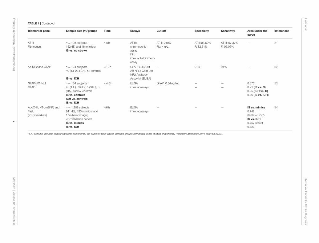
TABLE 1 | Continued
Biomarker panel Sample size (n)/groups Time Essays Cut off Specificity Sensitivity Area under the
curve
References
AT-III
Fibrinogen
n = 198 subjects
152 (IS) and 46 (mimics)
IS vs. no stroke
4.5 h AT-III:
chromogenic
assay
Fib:
immunoturbidimetry
assay
AT-III: 210%
Fib: 4 g/L
AT-III:93.62%
F: 82.61%
AT-III: 97.37%
F: 96.05%
— (31)
Ab NR2 and GFAP n = 124 subjects
49 (IS), 23 (ICH), 52 controls
IS vs. ICH
<12 h GFAP: ELISA kit
AB NR2: Gold Dot
NR2 Antibody
Assay kit (ELISA)
— 91% 94% — (32)
GFAP/UCH-L1
GFAP
n = 184 subjects
45 (ICH), 79 (IS), 5 (SAH), 3
(TIA), and 57 controls
IS vs. controls
ICH vs. controls
IS vs. ICH
<4.5 h ELISA
immunoassays
GFAP: 0.34 ng/mL —
—
—
—
0.875
0.71 (IS vs. C)
0.95 (ICH vs. C)
0.86 (IS vs. ICH)
(33)
ApoC-III, NT-proBNP, and
FasL
(21 biomarkers)
n = 1,308 subjects
941 (IS), 193 (mimics) and
174 (hemorrhagic)
767 validation cohort
IS vs. mimics
IS vs. ICH
<6 h ELISA
immunoassays
— — — IS vs. mimics
0.742
(0.686–0.797)
IS vs. ICH
0.757 (0.691–
0.823)
(34)
ROC analysis includes clinical variables selected by the authors. Bold values indicate groups compared in the studies analyzed by Receiver Operating Curve analysis (ROC).
Frontiers
inNeurology|www.fro
ntiersin
.org
7May2021|Volume12|A
rticle638693
Baez et al. Biomarker Panels for Stroke Diagnosis
• Kim et al. (24) validated the previous biomarker panel usingthe composite multimarker index (MMX), which combinesthe individual marker values into a single index value. Itexhibited good performance in the discrimination of patientswith acute infarction; at 6 h, it could differentiate AIS (p <
0.001) but with insufficient precision. With 91.0% sensitivity,21.3% specificity, and 71.4% AUC, the analysis exhibited amodest discriminatory power for acute stroke. According tothe MMX values, there was no significant difference betweenthe subjects with AIS and those with ICH (p= 0.884).
• These promising results should be considered with caution.Knauer et al. (25) evaluated MMX in a cohort of 174 casesin which 100 patients had stroke, 49 were mimics, and 25had TIA. They advised against the use of the panel BNP, D-dimer, S100β, and MMP-9 in this assay because of (1) thelow significance of MMX values to differentiate the IS group(MMX = 3.6 ± 2.0) from mimics (MMX = 4.2 ± 1.7) and(2) the low significance of MMX values when the analysisfor the individual biomarkers was replicated. The 2.3 cutoffvalue of MMX was reported to have 86% sensitivity with a lowspecificity of∼33% and an AUC of∼59%.
A panel comprising eotaxin, EGFR, S100A12, metalloproteinaseinhibitor-4 (TIMP-4), and prolactin was found to be elevatedin a study of 167 cases with neurologic deficits, allowing thedifferentiation of IS cases from mimics (c= 0.92) (26). The studyused a time window of 24 h and reported a high specificity of∼84%, sensitivity of∼90%, and AUC of∼97%.
Another panel, caspase-3, D-dimer, chimerin-II, MMP-9,secretagogin, and sRAGE, was assessed in a large cohort of1,005 cases where 915 had strokes and 90 had stroke-mimickingconditions, but only proteins could discriminate between the twogroups (27). At 6 h after symptom onset, these protein levelshad moderate sensitivity values of ∼84% but a low specificityof∼63%.
The panel S100β, MMP-9, vascular cell adhesion molecule(VCAM), and vWF proved to have good discriminatory poweras it differentiated 44 AIS cases from 21 controls within thefirst 6 h after symptom onset with high sensitivity (∼90%) andspecificity (∼90%).
A panel slightly modified from the previous one comprisingS100β, MMP-9, and vWF with two other markers includingmonocyte chemoattractant protein-1 (MCP-1) and B-typeneurotrophic growth factor (BNGF) had similar discriminatorypower (29). The levels for the panel in samples of 82 ISCH (ISwith ICH), 65 SAH, 38 ICH, 38 CLH, and 51 TIA patients, at 6 hfrom symptom onset and using multivariate logistic regressionmodel, had elevated sensitivity of ∼92% and specificity of ∼93%in the classification of ischemic events and a specificity of ∼93%and sensitivity of∼80% for the prediction of hemorrhagic stroke.
The protein GFAP has been of interest in combinationwith other biomarkers. The combination of antibodies (Ab)against NR2 and GFAP exhibited the best predictive power forcomparing 49 IS subjects from 23 ICH patients and 52 controls. Asensitivity of∼91% and specificity of∼94% were reported within12 h of symptom onset (32). The use of GFAP and UCH-L1 forthe identification of ICH vs. IS was tested in 129 stroke subjects,
three TIA patients, and 57 controls (33). Notably, GFAP alonewas capable of distinguishing between the condition with anAUC∼0.86, sensitivity of 61%, and specificity of 96% (33).
Recently, a panel consisting of apolipoprotein CIII (Apo C-III), NT-proBNP, and FasL was selected as the best combinationafter an extensive study of 21 biomarkers in 1,308 cases todifferentiate real stroke frommimics, within 6 h after stroke onset(34). This study was replicated with a smaller sample size fora different group of subjects, giving a modest accuracy of 0.742(0.686–0.797). Despite being one of the studies that screened thelargest number of biomarkers, this, in our opinion, has somelimitations. GFAP was not assayed, and the levels of MMP-9 werenot measured in the entire cohort because they were not deemeddiscriminative (34).
Finally, it is worth mentioning the two articles that screenedseveral biomarkers, although they did not combine them. Theycan be integrated into an optimized panel in the future. Thestudies and biomarkers are as follows:
• Ischemia-modified albumin (IMA): in a small number ofpatients (n = 28) with stroke compared to the no-strokegroup (n = 24) where the albumin-adjusted IMA index andIMA were measured within 6 h after symptom onset (30).Furthermore, the IMA index (98 U/mL) was even moresensitive (sensitivity, ∼95.8%; specificity, ∼96.4%; and AUC,∼99%) than the conventional IMA value (sensitivity,∼87.5%;specificity, 89.3%; and AUC, 92.8%) for the detection ofpatients with cerebral ischemia.
• The levels of antithrombin III (AT-III) in a study with 152stroke patients and 46 mimics reported the highest sensitivityof∼97.37% and specificity of∼93.62% using a cutoff of 210%,whereas 4 g/L of fibrinogen reached a sensitivity of ∼96.05%with a specificity of∼82.61% (31).
Observations From the Selected Studies(1) Only two biomarkers, NR2 peptide (Ab against NR2
fragment) and GFAP, have been reported as brain-specificmarkers linked to the progression of stroke, reaching thehighest predictive power when evaluated together (32).
(2) Two proteins that have been widely used as biomarkers arevWF and MMP-9; however, they are not specific to stroke.Although they were evaluated in a combined panel with higheraccuracy (28, 29), Reynolds et al. reported that vWF andMMP-9 alone could not be used for diagnosis. However, it has goodunivariate discrimination of non-diseased vs. diseased, with anadded discriminatory capacity to the logistic regression model[p < 0.0001; (29)].
(3) S100β is one of the most evaluated biomarkers; however, itis also not specific to stroke. Along with GFAP, it is one of thestrong candidates for the differentiation of hemorrhagic andischemic subtypes in the acute phase of stroke (35).
(4) An IMA index is the IMA value multiplied by individualserum albumin concentration/median albumin of the studypopulation (36). The IMA index seems to bemore specific thanIMA in the differentiation of IS, but it has not been used incombination in previous studies.
Frontiers in Neurology | www.frontiersin.org 8 May 2021 | Volume 12 | Article 638693
Baez et al. Biomarker Panels for Stroke Diagnosis
(5) According to previous studies, the use of AT-III andfibrinogen might help in distinguishing between conditionswith high accuracy in individual analyses (31).
(6) Usually, ROC curves are the statistical method to comparetwo groups of patients: ICH vs. mimics, AIS vs. other groups,IS vs. mimics, and IS+ ICH vs. mimics.
(7) Note that none of these results have been approved foradvanced clinical trials.
PROTEINS DERIVED FROM THESYSTEMATIC SEARCH AND THEIRRELATION TO THE PATHOPHYSIOLOGYOF STROKE
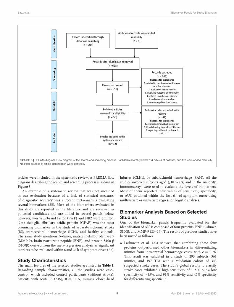
We have selectively outlined concepts of stroke pathophysiologythat justified our proposals for novel biomarker panels for clinicalprediction explained with graphical details in Figure 4:
(1) When rupture of the endothelial layer of the vesseloccurs, the inner collagen layer is exposed. The exposureof collagen with blood is recognized by platelets, forming asticky plug that initiates clot formation (37). Consequently,endothelial cells release vWF, MMP-9, P-selectin, E-selectin,and inflammatory mediators (38). vWF promotes plateletadhesion to the damaged site by forming a molecular bridgebetween the subendothelial collagen matrix and the platelet-surface receptor complex GPIb-IX-V (39). Fibrinogen isreleased from the liver to the bloodstream and is cleaved bythrombin at the damaged site, resulting in fibrin formation.Fibrin is one of the main constituents of blood clots andprovides remarkable biochemical and mechanical stability(40). The conversion of fibrinogen to fibrin by thrombinis inhibited by the enzyme AT-III, which is downregulatedduring an ischemic event. Neutrophils and leukocytes alsoadhere to the injury site and form a solid structure thatobstructs or reduces blood (Figure 2).
(2) Flow reduction leads to the depletion of oxygen andglucose, which has severe implications for cell function,resulting in dysregulation of neuronal homeostasis. Theprocess of intracellular medium acidification is reportedwhen metabolism changes to anaerobic conditions (41).Mitochondrial dysfunction plays a significant role in inducingneuronal death caused by an increase in enzymes that favorthe production of reactive oxygen species (ROS) and reactivenitrogen species (RNS), such as xanthine oxidase, NADPHoxidases, nitric oxide synthase, and a decrease in detoxifyingsystems (42, 43). Note that ROS and RNS pass into thebloodstream and are hypothesized to modify the N-terminalof albumin (44).
(3) Mitochondrial dysfunction affects ATP production, whichinduces a failure in the activity of the Na+–K+ pump. Na+–K+
pump activity depends on ATP hydrolysis (45), disappearingthe electrical gradient in the cellular membrane and causingthe influx of Na2+ into the neuron, resulting in membranedepolarization. Simultaneously, the activation of ASC1a, NCX,and TRP allows the influx of Ca2+ into the neuron, a process
known as calcium overloading (46). Calcium overloadingfavors glutamate release into the extracellular medium andcauses swelling due to the influx of water. NR2 is a subunit ofthe N-methyl-D-aspartate (NMDA) receptor and is a ligand-gated ion channel with high calcium permeability, which iscleaved by serine proteases under ischemic conditions (47).
(4) All these processes lead to neuronal death by necrosisor necroptosis, characterized by the loss of membraneintegrity, damage to cellular structures, swelling, and release ofcellular content, resulting in an acute inflammatory response(48). Consequently, there is an activation of astrocytesand microglia, which induces morphological changes andmediates the inflammatory response (49–51). These cellsrelease proteins such as S100β and GFAP, reflecting structuraland functional damage in the central nervous system (CNS)(35). MMP-9 is released by endothelial cells, astrocytes, andmicroglia and is activated by high nitric oxide concentrations.It degrades type IV collagen present in the endothelial blood–brain barrier, increasing parenchymal destruction, and isrelated to the inflammatory response after stroke (52).
PROPOSAL FOR NEW BIOMARKERPANELS
Based on our reviewed pathophysiology, we suggest furtherstudies of three different panels (Table 2). We have includedNR2 peptide and GFAP in all proposed panels because theyseem to be the most promising brain-specific biomarkersrelated to stroke. We have included other biomarkers in thesuggested panels under Observations From the Selected Studiesdescribed above because they seem promising in light of thepathophysiological process of stroke that have been previouslyevaluated (Table 3).
Panel 1: NR2+ GFAP+MMP-9+ vWF+ S100β
• NR2: Precisely, the NR2 subunit is the only biomarkerreported with the highest specificity (96%) and sensitivity(92%) at 12 h using a cutoff value of 1.0 µg/L in 101 ISand 91 no-stroke patients (60). When a lower cutoff valueof 0.5 ng/mL was tested within 0.5–4.5 h, it revealed highsensitivity and specificity of 88 and 99%, respectively, forthe differentiation between mild traumatic brain injury, AIS,ICH, healthy controls, and subjects at risk of TIA (vascularrisk factors) (https://www.ahajournals.org/doi/10.1161/str.44.suppl_1.A30). Additionally, the concentrations of NR2 werefound to be significantly elevated in IS subjects compared withpatients without cerebral damage and were also related to thesize of infarct and were used as a blood test for the validationof Cortexin treatment, a neurocytoprotector (61).
• GFAP: The predictive value of GFAP to determine the typesof stroke was assessed by Foerch, who used a cutoff valueof 2.9 ng/L, obtaining a sensitivity of 79% and a specificityof 98% (53). The same author, after several years, ratifiedGFAP as an efficient marker to differentiate ICH from IS,including stroke mimics [AUC = ∼0.915; 95% confidenceinterval (CI) = 0.847–0.982; p < 0.001; (62)]. Recently, thepotential of using a value of 0.43 ng/mL was found, achieving
Frontiers in Neurology | www.frontiersin.org 9 May 2021 | Volume 12 | Article 638693
Baez et al. Biomarker Panels for Stroke Diagnosis
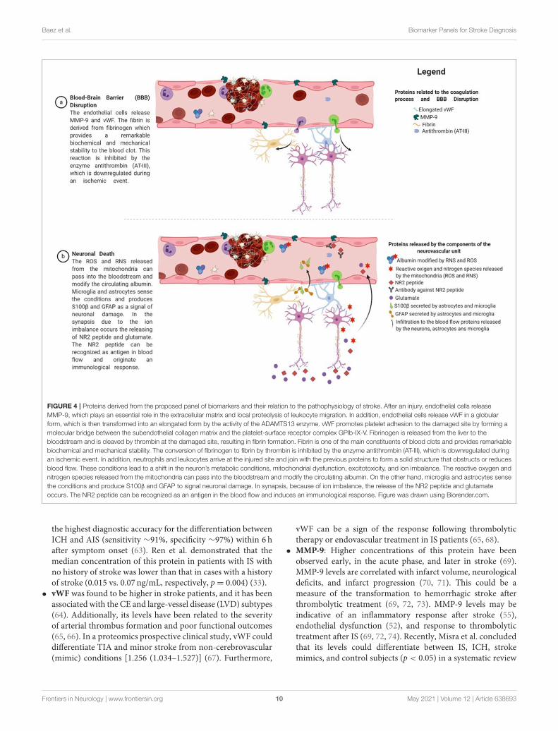
FIGURE 4 | Proteins derived from the proposed panel of biomarkers and their relation to the pathophysiology of stroke. After an injury, endothelial cells release
MMP-9, which plays an essential role in the extracellular matrix and local proteolysis of leukocyte migration. In addition, endothelial cells release vWF in a globular
form, which is then transformed into an elongated form by the activity of the ADAMTS13 enzyme. vWF promotes platelet adhesion to the damaged site by forming a
molecular bridge between the subendothelial collagen matrix and the platelet-surface receptor complex GPIb-IX-V. Fibrinogen is released from the liver to the
bloodstream and is cleaved by thrombin at the damaged site, resulting in fibrin formation. Fibrin is one of the main constituents of blood clots and provides remarkable
biochemical and mechanical stability. The conversion of fibrinogen to fibrin by thrombin is inhibited by the enzyme antithrombin (AT-III), which is downregulated during
an ischemic event. In addition, neutrophils and leukocytes arrive at the injured site and join with the previous proteins to form a solid structure that obstructs or reduces
blood flow. These conditions lead to a shift in the neuron’s metabolic conditions, mitochondrial dysfunction, excitotoxicity, and ion imbalance. The reactive oxygen and
nitrogen species released from the mitochondria can pass into the bloodstream and modify the circulating albumin. On the other hand, microglia and astrocytes sense
the conditions and produce S100β and GFAP to signal neuronal damage. In synapsis, because of ion imbalance, the release of the NR2 peptide and glutamate
occurs. The NR2 peptide can be recognized as an antigen in the blood flow and induces an immunological response. Figure was drawn using Biorender.com.
the highest diagnostic accuracy for the differentiation betweenICH and AIS (sensitivity ∼91%, specificity ∼97%) within 6 hafter symptom onset (63). Ren et al. demonstrated that themedian concentration of this protein in patients with IS withno history of stroke was lower than that in cases with a historyof stroke (0.015 vs. 0.07 ng/mL, respectively, p= 0.004) (33).
• vWF was found to be higher in stroke patients, and it has beenassociated with the CE and large-vessel disease (LVD) subtypes(64). Additionally, its levels have been related to the severityof arterial thrombus formation and poor functional outcomes(65, 66). In a proteomics prospective clinical study, vWF coulddifferentiate TIA and minor stroke from non-cerebrovascular(mimic) conditions [1.256 (1.034–1.527)] (67). Furthermore,
vWF can be a sign of the response following thrombolytictherapy or endovascular treatment in IS patients (65, 68).
• MMP-9: Higher concentrations of this protein have beenobserved early, in the acute phase, and later in stroke (69).MMP-9 levels are correlated with infarct volume, neurologicaldeficits, and infarct progression (70, 71). This could be ameasure of the transformation to hemorrhagic stroke afterthrombolytic treatment (69, 72, 73). MMP-9 levels may beindicative of an inflammatory response after stroke (55),endothelial dysfunction (52), and response to thrombolytictreatment after IS (69, 72, 74). Recently, Misra et al. concludedthat its levels could differentiate between IS, ICH, strokemimics, and control subjects (p < 0.05) in a systematic review
Frontiers in Neurology | www.frontiersin.org 10 May 2021 | Volume 12 | Article 638693
Baez et al. Biomarker Panels for Stroke Diagnosis
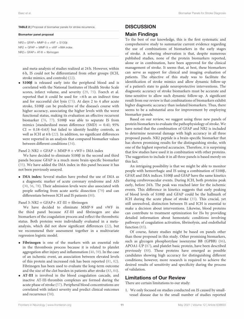
TABLE 2 | Proposal of biomarker panels for stroke recurrence.
Biomarker panel proposal
NR2+ GFAP+ MMP-9 + vWF + S100β
NR2 + GFAP + MMP-9 + vWF +IMA index
NR2+ GFAP+ AT-III + fibrinogen
and meta-analysis of studies realized at 24 h. However, within6 h, IS could not be differentiated from other groups (ICH,stroke mimics, and controls) (22).
• S100β is released early into the peripheral blood and iscorrelated with the National Institutes of Health Stroke Scalescores, infarct volume, and severity (29, 75). Foerch et al.reported that it could be used for <6 h as an indirect timeand for successful clot lysis (75). At days 2 to 4 after acutestroke, S100β can be predictive of the disease’s course withhigher accuracy, associating the higher levels with the worstfunctional status, making its evaluation an effective recurrentbiomarker (76, 77). S100β was able to separate IS frommimics [standardized mean difference (SMD) = 0.41; 95%CI = 0.18–0.63] but failed to identify healthy controls, aswell as ICH at 6 h (22). In addition, no significant differenceswere reported in an analysis that compared biomarker valuesbetween different conditions (34).
Panel 2: NR2+ GFAP+MMP-9+ vWF+ IMA indexWe have decided to eliminate S100β in the second and third
panels because GFAP is a much more brain-specific biomarker(35). We have added the IMA index in this panel because it hasnot been previously assayed.
• IMA index: Several studies have probed the use of IMA asa diagnostic marker of acute coronary syndrome and AIS(30, 56, 78). Their admission levels were also associated withpeople suffering from acute aortic dissection (79) and candifferentiate between ICH and IS patients (80).
Panel 3: NR2+ GFAP+ AT-III+ fibrinogenWe have decided to eliminate MMP-9 and vWF in
the third panel because AT-III and fibrinogen are alsobiomarkers of the coagulation process and reflect the thromboticstatus. Both proteins were individually evaluated in a meta-analysis, which did not show significant differences (22), butwe recommend their assessment together in a multivariateregression logistic model.
• Fibrinogen is one of the markers with an essential rolein the thrombosis process because it is related to plateletaggregation after injury and inflammation (40, 59). In the caseof an ischemic event, an association between elevated levelsof this protein and increased risk has been reported (81, 82).Fibrinogen has been used to evaluate the long-term outcomeand the size of the clot burden in patients after stroke (83, 84).
• AT-III is involved in the blood coagulation cascade, andinactive AT-III-thrombin complexes are formed during theacute phase of stroke (57). Peripheral blood concentrations arecorrelated with infarct severity and predict clinical outcomesand recurrence (58).
DISCUSSION
Main FindingsTo the best of our knowledge, this is the first systematic andcomprehensive study to summarize current evidence regardingthe use of combinations of biomarkers in the early stagesof stroke. A sobering observation is that, despite numerouspublished studies, none of the protein biomarkers reported,alone or in combination, have been approved for the clinicalmanagement of stroke. It seems that, at best, these biomarkerscan serve as support for clinical and imaging evaluation ofpatients. The objective of this study was to facilitate theidentification of stroke mimics and allow dynamic follow-upof a patient’s state to guide neuroprotective interventions. Thediagnostic accuracy of stroke biomarkers must be accurate andtime-sensitive to allow such dynamic follow-up. A significantresult from our review is that combinations of biomarkers exhibithigher diagnostic accuracy than isolated biomarkers. Thus, thereseems to be a substantial area for improvement by employingbiomarker panels.
Based on our review, we suggest using three new panels ofprotein biomarkers to evaluate the pathophysiology of stroke.Wehave noted that the combination of GFAP and NR2 is includedto determine neuronal damage with high accuracy in all threeproposed panels. NR2 peptide is a brain-specific biomarker thathas shown promising results for the distinguishing stroke, withone of the highest reported accuracies. Therefore, it is surprisingthat few studies have used it in combination with other proteins.The suggestion to include it in all three panels is based merely onthis fact.
An intriguing possibility is that we might be able to monitorpeople with hemorrhagic and IS using a combination of S100β,GFAP, and IMA indices. S100β and GFAP have the same kineticsduring cerebrovascular events. During ICH, both proteins peakearly, before 24 h. The peak was reached later for the ischemicevents. This difference in kinetics suggests that early peakingof blood levels of S100β and GFAP could be the hallmark ofICH during the acute phase of stroke (35). This crucial, yetstill unresolved, distinction between IS and ICH is essential tomake a decision about interventions. Likewise, blood proteinscan contribute to treatment optimization for ISs by providingdetailed information about hemostatic conditions involvingpathways of coagulation activation, fibrinolysis, and endothelialfunction (85).
Of course, future studies might be based on panels otherthan those proposed in this study. Other promising biomarkers,such as glycogen phosphorylase isoenzyme BB (GPBB) (86),APOA1-UP (87), and platelet basic protein, have been describedpreviously (88). These proteins have emerged as possiblecandidates showing high accuracy for distinguishing differentconditions; however, more research is required to achieve thedesired results of sensitivity and specificity during the processof validation.
Limitations of Our ReviewThere are certain limitations to our study:
1. We only focused on studies conducted on IS caused by small-vessel disease due to the small number of studies reported
Frontiers in Neurology | www.frontiersin.org 11 May 2021 | Volume 12 | Article 638693
Baez et al. Biomarker Panels for Stroke Diagnosis
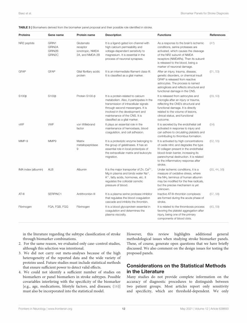
TABLE 3 | Biomarkers derived from the biomarker panel proposal and their possible role identified in stroke.
Proteins Gene name Protein name Description Functions References
NR2 peptide GRIN1
GRIN2A
GRIN2B
GRIN2C
Glutamate
receptor
ionotropic, NMDA
2A, and NMDA 2B
It is a ligand-gated ion channel with
high calcium permeability and
voltage-dependent sensitivity to
magnesium. It is essential in the
process of neuronal synapses.
As a response to the brain’s ischemic
conditions, serine proteases are
activated, which causes the cleavage
of the NR2 subunit of NMDA
receptors (NMDARs). Then its subunit
is released to the blood, being a
marker of neuronal damage.
(47)
GFAP GFAP Glial fibrillary acidic
protein
It is an intermediate filament class-III.
It is classified as a glial marker.
After an injury, trauma, disease,
genetic disorders, or chemical insult
GFAP is released from reactive
astrocytes. The process is named
astrogliosis and reflects structural and
functional damage in the CNS.
(51, 53)
S100β S100β Protein S100-β It is a protein-related to calcium
metabolism. Also, it participates in the
transmission of intracellular signals
through second messengers. It is
involved in the development and
maintenance of the CNS. It is
classified as a glial marker.
It is released from astrocytes and
microglia after an injury or trauma,
reflecting the CNS’s structural and
functional damage. It is directly
related to the volume of lesions,
clinical status, and functional
outcome.
(29, 50)
vWF VWF von Willebrand
factor
It plays an essential role in the
maintenance of hemostasis, blood
coagulation, and cell adhesion.
It is secreted by the endothelial cell
activated in response to injury and
can adhere to circulating platelets and
contributing to thrombus formation.
(54)
MMP-9 MMP9 Matrix
metallopeptidase
9
It is a proteolytic enzyme belonging to
the group of gelatinases. It has an
essential role in local proteolysis of
the extracellular matrix and leukocyte
migration.
It is activated by high concentrations
of oxide nitric and degrades the type
IV collagen present in the endothelial
blood–brain barrier, increasing its
parenchymal destruction. It is related
to the inflammatory response after
stroke.
(52, 55)
IMA index (albumin) ALB Albumin It is the major transporter of Zn, Ca2+,
Mg in plasma and binds water Na+,
K+, fatty acids, hormones, etc. It
regulates the colloidal osmotic
pressure of blood.
Under ischemic conditions, it is a
measure of oxidative stress, where
the NH2 terminus of human albumin
may be modified for the free radicals,
but the precise mechanism is yet
unknown.
(30, 44, 56)
AT-III SERPINC1 Antithrombin III It is a plasma serine protease inhibitor
that regulates the blood coagulation
cascade and inhibits the thrombin.
Inactive AT-III–thrombin complexes
are formed during the acute phase of
stroke.
(57, 58)
Fibrinogen FGA, FGB, FGG Fibrinogen It is a blood glycoprotein essential in
coagulation and determines the
plasma viscosity.
It is related to the thrombosis process
favoring the platelet aggregation after
injury, being one of the primary
components of blood clots.
(40, 59)
in the literature regarding the subtype classification of strokethrough biomarker combinations.
2. For the same reason, we evaluated only case–control studies,although this selection was intentional.
3. We did not carry out meta-analyses because of the highheterogeneity of the reported data and the wide variety ofproteins used. Future studies must include statistical methodsthat ensure sufficient power to detect valid effects.
4. We could not identify a sufficient number of studies onbiomarkers or panel biomarkers in stroke subtypes. Possiblecovariables interfering with the specificity of the biomarker[e.g., age, medications, lifestyle factors, and diseases; (16)]must also be incorporated into the statistical model.
However, this review highlights additional generalmethodological issues when studying stroke biomarker panels.These, of course, generate open questions that we have brieflydiscussed. We also comment on the design issues for testing theproposed panels.
Considerations on the Statistical Methodsin the LiteratureMany studies do not provide complete information on theaccuracy of diagnostic procedures to distinguish betweentwo patient groups. Most articles report only sensitivityand specificity, which are threshold-dependent. We only
Frontiers in Neurology | www.frontiersin.org 12 May 2021 | Volume 12 | Article 638693
Baez et al. Biomarker Panels for Stroke Diagnosis
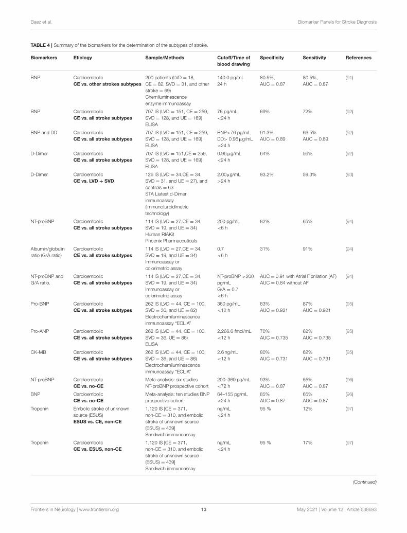
TABLE 4 | Summary of the biomarkers for the determination of the subtypes of stroke.
Biomarkers Etiology Sample/Methods Cutoff/Time of
blood drawing
Specificity Sensitivity References
BNP Cardioembolic
CE vs. other strokes subtypes
200 patients (LVD = 18,
CE = 82, SVD = 31, and other
stroke = 69)
Chemiluminescence
enzyme immunoassay
140.0 pg/mL
24 h
80.5%,
AUC = 0.87
80.5%,
AUC = 0.87
(91)
BNP Cardioembolic
CE vs. all stroke subtypes
707 IS (LVD = 151, CE = 259,
SVD = 128, and UE = 169)
ELISA
76 pg/mL
<24 h
69% 72% (92)
BNP and DD Cardioembolic
CE vs. all stroke subtypes
707 IS (LVD = 151, CE = 259,
SVD = 128, and UE = 169)
ELISA
BNP>76 pg/mL
DD> 0.96µg/mL
<24 h
91.3%
AUC = 0.89
66.5%
AUC = 0.89
(92)
D-Dimer Cardioembolic
CE vs. all stroke subtypes
707 IS (LVD = 151,CE = 259,
SVD = 128, and UE = 169)
ELISA
0.96µg/mL
<24 h
64% 56% (92)
D-Dimer Cardioembolic
CE vs. LVD + SVD
126 IS (LVD = 34,CE = 34,
SVD = 31, and UE = 27), and
controls = 63
STA Liatest d-Dimer
immunoassay
(immunoturbidimetric
technology)
2.00µg/mL
>24 h
93.2% 59.3% (93)
NT-proBNP Cardioembolic
CE vs. all stroke subtypes
114 IS (LVD = 27,CE = 34,
SVD = 19, and UE = 34)
Human RIAKit
Phoenix Pharmaceuticals
200 pg/mL
<6 h
82% 65% (94)
Albumin/globulin
ratio (G/A ratio)
Cardioembolic
CE vs. all stroke subtypes
114 IS (LVD = 27,CE = 34,
SVD = 19, and UE = 34)
Immunoassay or
colorimetric assay
0.7
<6 h
31% 91% (94)
NT-proBNP and
G/A ratio.
Cardioembolic
CE vs. all stroke subtypes
114 IS (LVD = 27,CE = 34,
SVD = 19, and UE = 34)
Immunoassay or
colorimetric assay
NT-proBNP >200
pg/mL
G/A = 0.7
<6 h
AUC = 0.91 with Atrial Fibrillation (AF)
AUC = 0.84 without AF
(94)
Pro-BNP Cardioembolic
CE vs. all stroke subtypes
262 IS (LVD = 44, CE = 100,
SVD = 36, and UE = 82)
Electrochemiluminescence
immunoassay “ECLIA”
360 pg/mL
<12 h
83%
AUC = 0.921
87%
AUC = 0.921
(95)
Pro-ANP Cardioembolic
CE vs. all stroke subtypes
262 IS (LVD = 44, CE = 100,
SVD = 36, UE = 86)
ELISA
2,266.6 fmol/mL
<12 h
70%
AUC = 0.735
62%
AUC = 0.735
(95)
CK-MB Cardioembolic
CE vs. all stroke subtypes
262 IS (LVD = 44, CE = 100,
SVD = 36, and UE = 86)
Electrochemiluminescence
immunoassay “ECLIA”
2.6 ng/mL
<12 h
80%
AUC = 0.731
62%
AUC = 0.731
(95)
NT-proBNP Cardioembolic
CE vs. no-CE
Meta-analysis: six studies
NT-proBNP prospective cohort
200–360 pg/mL
<72 h
93%
AUC = 0.87
55%
AUC = 0.87
(96)
BNP Cardioembolic
CE vs. no-CE
Meta-analysis: ten studies BNP
prospective cohort
64–155 pg/mL
<24 h
85%
AUC = 0.87
65%
AUC = 0.87
(96)
Troponin Embolic stroke of unknown
source (ESUS)
ESUS vs. CE, non-CE
1,120 IS [CE = 371,
non-CE = 310, and embolic
stroke of unknown source
(ESUS) = 439]
Sandwich immunoassay
ng/mL
<24 h
95 % 12% (97)
Troponin Cardioembolic
CE vs. ESUS, non-CE
1,120 IS [CE = 371,
non-CE = 310, and embolic
stroke of unknown source
(ESUS) = 439]
Sandwich immunoassay
ng/mL
<24 h
95 % 17% (97)
(Continued)
Frontiers in Neurology | www.frontiersin.org 13 May 2021 | Volume 12 | Article 638693
Baez et al. Biomarker Panels for Stroke Diagnosis
TABLE 4 | Continued
Biomarkers Etiology Sample/Methods Cutoff/Time of
blood drawing
Specificity Sensitivity References
D-Dimer SVD (lacunar)
SVD vs. CE + LVD
126 patients (LVD = 34,CE = 34,
SVD = 31, and UE = 27)
STA Liatest d-Dimer
immunoassay
(immunoturbidimetric technology)
0.54µg/mL
>24 h
96.2% 61.3% (93)
Homocysteine
(Hcy)
Lacunar (SVD)
SVD vs. controls
197 acute lacunar infarction
patients and 192 controls
–
15.5 µmol/L
<24 h
100%
AUC = 0.881
65%
AUC = 0.881
(98)
Fibrinogen Lacunar (SVD)
SVD vs. controls
197 acute lacunar infarction
patients and 192 controls
–
228.55 µg/dL
<24 h
58.3%
AUC = 0.688
83.2%
AUC = 0.688
(98)
Hcy/fibrinogen Lacunar (SVD)
SVD vs. controls
197 acute lacunar infarction
patients and 192 controls
–
15.5 µmol/L
228.55 µg/dL
<24 h
58.3%
AUC = 0.766
94.9%
AUC = 0.766
(98)
GFAP/d-dimer
preprint
LVD
LVD vs. other strokes
128 patients (LVD = 23,
non-LVD = 42, HS = 16, stroke
mimic = 31, and TIA = 16)
ELISA
d-dimer
+GFAP = 0.33
92%
AUC = 0.81
57%
AUC = 0.81
(99)
Bold values indicate groups compared in the studies analyzed by Receiver Operating Curve analysis (ROC).
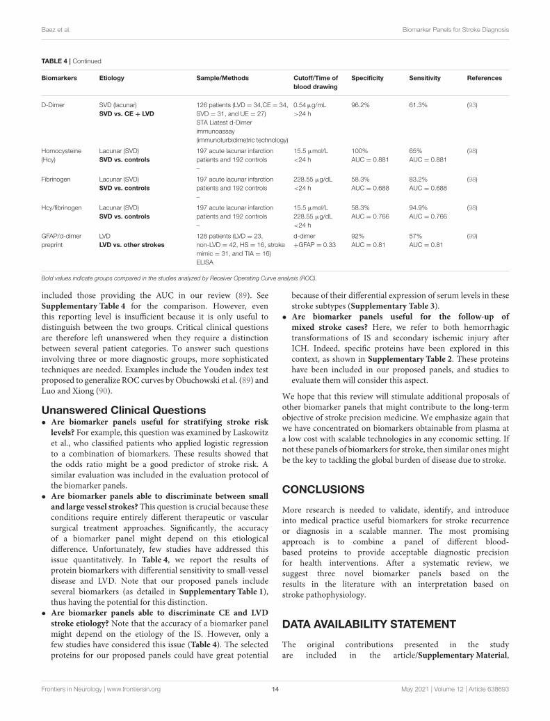
included those providing the AUC in our review (89). SeeSupplementary Table 4 for the comparison. However, eventhis reporting level is insufficient because it is only useful todistinguish between the two groups. Critical clinical questionsare therefore left unanswered when they require a distinctionbetween several patient categories. To answer such questionsinvolving three or more diagnostic groups, more sophisticatedtechniques are needed. Examples include the Youden index testproposed to generalize ROC curves by Obuchowski et al. (89) andLuo and Xiong (90).
Unanswered Clinical Questions• Are biomarker panels useful for stratifying stroke risk
levels? For example, this question was examined by Laskowitzet al., who classified patients who applied logistic regressionto a combination of biomarkers. These results showed thatthe odds ratio might be a good predictor of stroke risk. Asimilar evaluation was included in the evaluation protocol ofthe biomarker panels.
• Are biomarker panels able to discriminate between small
and large vessel strokes? This question is crucial because theseconditions require entirely different therapeutic or vascularsurgical treatment approaches. Significantly, the accuracyof a biomarker panel might depend on this etiologicaldifference. Unfortunately, few studies have addressed thisissue quantitatively. In Table 4, we report the results ofprotein biomarkers with differential sensitivity to small-vesseldisease and LVD. Note that our proposed panels includeseveral biomarkers (as detailed in Supplementary Table 1),thus having the potential for this distinction.
• Are biomarker panels able to discriminate CE and LVD
stroke etiology? Note that the accuracy of a biomarker panelmight depend on the etiology of the IS. However, only afew studies have considered this issue (Table 4). The selectedproteins for our proposed panels could have great potential
because of their differential expression of serum levels in thesestroke subtypes (Supplementary Table 3).
• Are biomarker panels useful for the follow-up of
mixed stroke cases? Here, we refer to both hemorrhagictransformations of IS and secondary ischemic injury afterICH. Indeed, specific proteins have been explored in thiscontext, as shown in Supplementary Table 2. These proteinshave been included in our proposed panels, and studies toevaluate them will consider this aspect.
We hope that this review will stimulate additional proposals ofother biomarker panels that might contribute to the long-termobjective of stroke precision medicine. We emphasize again thatwe have concentrated on biomarkers obtainable from plasma ata low cost with scalable technologies in any economic setting. Ifnot these panels of biomarkers for stroke, then similar ones mightbe the key to tackling the global burden of disease due to stroke.
CONCLUSIONS
More research is needed to validate, identify, and introduceinto medical practice useful biomarkers for stroke recurrenceor diagnosis in a scalable manner. The most promisingapproach is to combine a panel of different blood-based proteins to provide acceptable diagnostic precisionfor health interventions. After a systematic review, wesuggest three novel biomarker panels based on theresults in the literature with an interpretation based onstroke pathophysiology.
DATA AVAILABILITY STATEMENT
The original contributions presented in the studyare included in the article/Supplementary Material,
Frontiers in Neurology | www.frontiersin.org 14 May 2021 | Volume 12 | Article 638693
Baez et al. Biomarker Panels for Stroke Diagnosis
further inquiries can be directed to thecorresponding author/s.
AUTHOR CONTRIBUTIONS
SB, JL, and PV-S: study concept and design and draftedthe manuscript. AH-S, DG, and SB: acquisition, analysis,or interpretation of the data. PV-S and GG administeredthe project. PV-S and MB-V supervised the study. Allauthors critically revised the manuscript. All authorshad full access to all the data in the study. They takeresponsibility for the integrity and accuracy of the analysisand results.
ACKNOWLEDGMENTS
The authors would like to thank the NSFC (China-Cuba-Canada) project (No. 81861128001) and funds from the NationalNature and Science Foundation of China (NSFC) with fundingNos. 61871105 and 81871446, and CNS Program of UESTC(No. Y0301902610100201).
SUPPLEMENTARY MATERIAL
The Supplementary Material for this article can be foundonline at: https://www.frontiersin.org/articles/10.3389/fneur.2021.638693/full#supplementary-material
REFERENCES
1. Feigin VL. Primary stroke prevention needs overhaul. Int J Stroke. (2017)12:5–6. doi: 10.1177/1747493016669850
2. Gorelick PB. The global burden of stroke: persistent and disabling. LancetNeurol. (2019) 18:417–8. doi: 10.1016/S1474-4422(19)30030-4
3. GBD 2016 Stroke Collaborators. Global, regional, and nationalburden of stroke, 1990–2016: a systematic analysis for theGlobal Burden of Disease Study 2016. Lancet Neurol. (2019)18:439–58. doi: 10.1016/S1474-4422(19)30034-1
4. Feigin VL, Norrving B, George MG, Foltz JL, Roth GA, Mensah GA.Prevention of stroke: A strategic global imperative. Nat Rev Neurol. (2016)12:501–12. doi: 10.1038/nrneurol.2016.107
5. Yamada Y. Molecular basis of stroke. Clin Mol Med. (2020) 189–216. doi: 10.1016/b978-0-12-809356-6.00012-5
6. Adams HP, Bendixen BH, Kappelle LJ, Biller J, Love BB, Gordon DL, et al.Classification of subtype of acute ischemic stroke. Stroke. (1993) 2435–41. doi: 10.1161/01.str.24.1.35
7. Hankey GJ. Impact of treatment of people with transient ischaemic attackson stroke incidence and public health. Cerebrovasc. Dis. (1996) 6:26–33. doi: 10.1159/000108068
8. Coull AJ, Lovett JK, Rothwell PM. Population based study of early riskof stroke after transient ischaemic attack or minor stroke: Implications forpublic education and organisation of services. Br Med J. (2004) 328:326–8. doi: 10.1136/bmj.37991.635266.44
9. Lewsey J, Jhund PS, Gillies M, Chalmers JWT, Redpath A, BriggsA, et al. Temporal trends in hospitalisation for stroke recurrencefollowing incident hospitalisation for stroke in Scotland. BMC Med. (2010)8:23. doi: 10.1186/1741-7015-8-23
10. Newman-Toker DE, Moy E, Valente E, Coffey R, Hines AL. Misseddiagnosis of stroke in the emergency department: a cross-sectionalanalysis of a large population-based sample. Diagnosis. (2014) 1:155–66. doi: 10.1515/dx-2013-0038
11. Arsava EM, Kim GM, Oliveira-Filho J, Gungor L, Noh HJ, de JesusLordelo M, et al. Prediction of early recurrence after acute ischemicstroke. JAMA Neurol. (2016) 73:396–401. doi: 10.1001/jamaneurol.2015.4949
12. Simpkins AN, Janowski M, Oz HS, Roberts J, Bix G, Doré S, et al. Biomarkerapplication for precision medicine in stroke. Transl Stroke Res. (2020) 11:615–27. doi: 10.1007/s12975-019-00762-3
13. Schmidt-Richberg A, Guerrero R, Ledig C, Molina-Abril H, FrangiAF, Rueckert D. Multi-stage biomarker models for progressionestimation in Alzheimer’s disease. Lect Notes Comput. Sci. (2015)9123:387–98. doi: 10.1007/978-3-319-19992-4_30
14. Califf RM. Biomarker definitions and their applications. Exp Biol Med. (2018)243:213–21. doi: 10.1177/1535370217750088
15. Kamtchum-Tatuene J, Jickling GC. Blood biomarkers for strokediagnosis and management. NeuroMol Med. (2019) 21:344–68. doi: 10.1007/s12017-019-08530-0
16. Mayeux R. Biomarkers: potential uses and limitations.NeuroRx. (2004) 1:182–8. doi: 10.1602/neurorx.1.2.182
17. Ng GJL, Quek AML, Cheung C. Arumugam TV, Seet RCS. Stroke biomarkersin clinical practice: a critical appraisal. Neurochem. Int. (2017) 107:11–22. doi: 10.1016/j.neuint.2017.01.005
18. Li Y, Yang G-Y. Pathophysiology of ischemic stroke. Pathophysiol. Ischemic
Stroke. (2017) 3–25. doi: 10.1007/978-981-10-5804-2_419. Moran A, Forouzanfarb M, Uchechukwu S, Chughd S, Valery F, George M.
The epidemiology of cardiovascular diseases in Sub-Saharan Africa: the globalburden of diseases, injuries and risk factors 2010 study. Prog Cardiovasc Dis.(2013) 56:234–9. doi: 10.1016/j.pcad.2013.09.019
20. Jickling GC, Sharp FR. Biomarker panels in ischemic stroke. Stroke. (2015)46:915–920. doi: 10.1161/STROKEAHA.114.005604
21. Moher D, Shamseer L, Clarke M, Ghersi D, Liberati A, Petticrew M, et al.Preferred reporting items for systematic review and meta-analysis protocols(PRISMA-P) 2015 statement. Syst Rev. (2015) 4:1. doi: 10.1186/2046-4053-4-1
22. Misra S, Montaner J, Ramiro L, Arora R, Talwar P, Nath M,et al. Blood biomarkers for the diagnosis and differentiation ofstroke: a systematic review and meta-analysis. Int J Stroke. (2020)15:704–21. doi: 10.1177/1747493020946157
23. Laskowitz DT, Kasner SE, Saver J, Remmel KS, Jauch EC. Clinical usefulnessof a biomarker-based diagnostic test for acute stroke: the BiomarkerRapid Assessment in Ischemic Injury (BRAIN) study. Stroke. (2009) 40:77–85. doi: 10.1161/STROKEAHA.108.516377
24. Kim MH, Kang SY, Kim MC, Lee WI. Plasma biomarkers in the diagnosis ofacute ischemic stroke. Ann Clin Lab Sci. (2010) 40:336–41.
25. Knauer C, Knauer K, Müller S, Ludolph AC, Bengel D, Müller HP, et al.A biochemical marker panel in MRI-proven hyperacute ischemic stroke-aprospective study. BMC Neurol. (2012) 12:14. doi: 10.1186/1471-2377-12-14
26. Sharma R, Macy S, Richardson K, Lokhnygina Y, Laskowitz DT. A blood-based biomarker panel to detect acute stroke. J Stroke Cerebrovasc Dis. (2014)23:910–8. doi: 10.1016/j.jstrokecerebrovasdis.2013.07.034
27. Montaner J, Mendioroz M, Ribó M, Delgado P, Quintana M, Penalba A, et al.A panel of biomarkers including caspase-3 and d-dimer may differentiateacute stroke from stroke-mimicking conditions in the emergency department.J Intern Med. (2011) 270:166–74. doi: 10.1111/j.1365-2796.2010.02329.x
28. Lynch JR, Blessing R, White WD, Grocott HP, Newman MF,Laskowitz DT. Novel diagnostic test for acute stroke. Stroke. (2004)35:57–63. doi: 10.1161/01.STR.0000105927.62344.4C
29. Reynolds MA, Kirchick HJ, Dahlen JR, Anderberg JM, McPherson PH,Nakamura KK, et al. Early biomarkers of stroke. Clin Chem. (2003) 49:1733–9. doi: 10.1373/49.10.1733
30. Ahn JH, Choi SC, Lee WG, Jung YS. The usefulness of albumin-adjusted ischemia-modified albumin index as early detecting marker forischemic stroke. Neurol Sci. (2011) 32:133–8. doi: 10.1007/s10072-010-0457-4
31. Meng R, Li Z-Y, Ji X, Ding Y, Meng S, Wang X. Antithrombin III associatedwith fibrinogen predicts the risk of cerebral ischemic stroke. Clin Neurol
Neurosurg. (2011) 113:380–6. doi: 10.1016/j.clineuro.2010.12.016
Frontiers in Neurology | www.frontiersin.org 15 May 2021 | Volume 12 | Article 638693
Baez et al. Biomarker Panels for Stroke Diagnosis
32. Stanca DM, Marginean IC, Soritau O, Dragos C, Marginean M, MuresanuDF, et al. GFAP and antibodies against NMDA receptor subunit NR2as biomarkers for acute cerebrovascular diseases. J Cell Mol Med. (2015)19:2253–61. doi: 10.1111/jcmm.12614
33. Ren C, Kobeissy F, Alawieh A, Li N, Li N, Zibara K, et al. Assessmentof serum UCH-L1 and GFAP in acute stroke patients. Sci Rep. (2016)6:24588. doi: 10.1038/srep24588
34. Bustamante A, López-Cancio E, Pich S, Penalba A, Giralt D, García-BerrocosoT, et al. Blood biomarkers for the early diagnosis of stroke: the stroke-chipstudy. Stroke. (2017) 48:2419–25. doi: 10.1161/STROKEAHA.117.017076
35. Brunkhorst R, Pfeilschifter W, Foerch C. Astroglial proteins asdiagnostic markers of acute intracerebral hemorrhage-pathophysiologicalbackground and clinical findings. Transl Stroke Res. (2010)1:246–51. doi: 10.1007/s12975-010-0040-6
36. Mishra B, Pandey S, Niraula SR, Rai BK, Karki P, Baral N, et al.Utility of ischemia modified albumin as an early marker for diagnosisof acute coronary syndrome. J Nepal Health Res Counc. (2018) 16:16–21. doi: 10.3126/jnhrc.v16i1.19383
37. Sidelmann JJ, Gram J, Jespersen J, Kluft C. Fibrin clot formationand lysis: basic mechanisms. Semin Thromb Hemost. (2000) 26:605–18. doi: 10.1055/s-2000-13216
38. Kozuka K, Kohriyama T, Ikeda J, Nakamura S, Nomura E, Kajikawa H.Endothelial markers and adhesion molecules in acute ischemic stroke-Sequential change and differences in stroke subtype. Atherosclerosis. (2002)161:161–8. doi: 10.1016/S0021-9150(01)00635-9
39. Gragnano F, Sperlongano S, Golia E, Natale F, Bianchi R, Crisci M,et al. The role of von willebrand factor in vascular inflammation:from pathogenesis to targeted therapy. Mediators Inflamm. (2017)2017:5620314. doi: 10.1155/2017/5620314
40. Pieters M, Wolberg AS. Fibrinogen and fibrin: an illustrated review. Res PractThromb Haemost. (2019) 3:161–72. doi: 10.1002/rth2.12191
41. Magistretti PJ, Allaman I. Lactate in the brain: from metabolicend-product to signalling molecule. Nat Rev Neurosci. (2018)19:235–49. doi: 10.1038/nrn.2018.19
42. Liu F, Lu J, Manaenko A, Tang J, Hu Q. Mitochondria inischemic stroke: New insight and implications. Aging Dis. (2018)9:924–37. doi: 10.14336/AD.2017.1126
43. Chan PH. Mitochondria and neuronal death/survival signalingpathways in cerebral ischemia. Neurochem Res. (2004) 29:1943–9. doi: 10.1007/s11064-004-6869-x
44. Lippi G, Montagnana M, Guidi GC. Albumin cobalt binding and ischemiamodified albumin generation: an endogenous response to ischemia? Int J
Cardiol. (2006) 108:410–1. doi: 10.1016/j.ijcard.2005.03.04045. Pivovarov AS, Calahorro F, Walker RJ. Na+/K+-pump and
neurotransmitter membrane receptors. Invertebr Neurosci. (2019)19:1–16. doi: 10.1007/s10158-018-0221-7
46. Wu QJ, Tymianski M. Targeting nmda receptors in stroke:new hope in neuroprotection Tim Bliss. Mol Brain. (2018)11:1–14. doi: 10.1186/s13041-018-0357-8
47. GingrichMB, Traynelis SF. Serine proteases and brain damage - is there a link?Trends Neurosci. (2000) 23:399–407. doi: 10.1016/S0166-2236(00)01617-9
48. Liu C, Zhang K, Shen H, Yao X, Sun Q, Chen G. Necroptosis: a novelmanner of cell death, associated with stroke (review). Int J Mol Med. (2018)41:624–30. doi: 10.3892/ijmm.2017.3279
49. Yenari MA, Kauppinen TM, Swanson RA. Microglial activationin stroke: therapeutic targets. Neurotherapeutics. (2010) 7:378–91. doi: 10.1016/j.nurt.2010.07.005
50. Lamers KJB, Vos P, Verbeek MM, Rosmalen F, Van Geel WJA, Van EngelenMBG. Protein S-100B, neuron-specific enolase (NSE), myelin basic protein(MBP) and glial fibrillary acidic protein (GFAP) in cerebrospinal fluid(CSF) and blood of neurological patients. Brain Res Bull. (2003) 61:261–4. doi: 10.1016/S0361-9230(03)00089-3
51. Eng LF, Ghirnikar RS, Lee YL. Glial fibrillary acidic protein:GFAP-thirty-one years (1969-2000). Neurochem Res. (2000)25:1439–51. doi: 10.1023/a:1007677003387
52. Kurzepa J, Kurzepa J, Golab P, Czerska S, Bielewicz J. The significance ofmatrix metalloproteinase (MMP)-2 and MMP-9 in the ischemic stroke. IntJ Neurosci. (2014) 124:707–16. doi: 10.3109/00207454.2013.872102
53. Foerch C, Curdt I, Yan B, Dvorak F, Hermans M, Berkefeld J, et al. Serumglial fibrillary acidic protein as a biomarker for intracerebral haemorrhagein patients with acute stroke. J Neurol Neurosurg Psychiatry. (2006) 77:181–4. doi: 10.1136/jnnp.2005.074823
54. Buchtele N, Schwameis M, Gilbert JC, Schörgenhofer C, Jilma B. TargetingvonWillebrand factor in ischaemic stroke: focus on clinical evidence. Thromb
Haemost. (2018) 118:959–78. doi: 10.1055/s-0038-164825155. Turner RJ, Sharp FR. Implications of MMP9 for blood brain barrier
disruption and hemorrhagic transformation following ischemic stroke. FrontCell Neurosci. (2016) 10:56. doi: 10.3389/fncel.2016.00056
56. Abboud H, Labreuche J, Meseguer E, Lavallee PC, Simon O, Olivot JM,et al. Ischemia-modified albumin in acute stroke. Cerebrovasc Dis. (2007)23:216–20. doi: 10.1159/000097644
57. Hossmann V, Heiss WD, Bewermeyer H. Antithrombin IIIdeficiency in ischaemic stroke. Klin Wochenschr. (1983) 61:617–20. doi: 10.1007/BF01487340
58. Haapaniemi E, Tatlisumak T, Soinne L, Syrjälä M, Kaste M. Naturalanticoagulants (antithrombin III, protein C, and protein S) in patients withmild to moderate ischemic stroke. Acta Neurol Scand. (2002) 105:107–14. doi: 10.1034/j.1600-0404.2002.1o112.x
59. Drouet L. Fibrinogen : a t reatable risk factor? Cerebrovasc Dis. (1996) 6:2–6.60. Dambinova SA, BettermannK, Glynn T, TewsM,OlsonD,Weissman JD, et al.
Diagnostic potential of the NMDA receptor peptide assay for acute ischemicstroke. PLoS ONE. (2012) 7:e42362. doi: 10.1371/journal.pone.0042362
61. Dambinova S, Aliev KT, Bondarenko EV, Ponomarev GV, Skoromets AA,Skoromets AP, et al. Biomarkers for cerebral ischemia as a novel method forvalidating the efficacy of neurocytoprotectors. Neurosci Behav Physiol. (2019)49:142–6. doi: 10.1007/s11055-018-0707-0
62. Foerch C, Niessner M, Back T, Bauerle M, De Marchis GM, FerbertA, et al. Diagnostic accuracy of plasma glial fibrillary acidic proteinfor differentiating intracerebral hemorrhage and cerebral ischemia inpatients with symptoms of acute stroke. Clin Chem. (2012) 58:237–45. doi: 10.1373/clinchem.2011.172676
63. Katsanos AH, Makris K, Stefani D, Koniari K, Gialouri E,Lelekis M, et al. Plasma glial fibrillary acidic protein in thedifferential diagnosis of intracerebral hemorrhage. Stroke. (2017)48:2586–8. doi: 10.1161/STROKEAHA.117.018409
64. Hanson E, Jood K, Karlsson S, Nilsson S, Blomstrand C, Jern C. Plasma levelsof vonWillebrand factor in the etiologic subtypes of ischemic stroke. J Thromb
Haemost. (2011) 9:275–81. doi: 10.1111/j.1538-7836.2010.04134.x65. Tóth NK, Székely EG, Czuriga-Kovács KR, Sarkady F, Nagy O, Lánczi LI,
et al. Elevated factor VIII and vonWillebrand factor levels predict unfavorableoutcome in stroke patients treated with intravenous thrombolysis. FrontNeurol. (2018) 8:721. doi: 10.3389/fneur.2017.00721
66. Ancedy Y, Berthelot E, Lang S, Ederhy S, Boyer-Chatenet L, Di AngelantonioE, et al. Is vonWillebrand factor associated with stroke and death at mid-termin patients with non-valvular atrial fibrillation? Arch Cardiovasc Dis. (2018)111:357–69. doi: 10.1016/j.acvd.2017.08.004
67. Penn AM, Bibok MB, Saly VK, Coutts SB, Lesperance ML, Balshaw RF,et al. Verification of a proteomic biomarker panel to diagnose minor strokeand transient ischaemic attack: phase 1 of SpecTRA, large scale translationalstudy. Biomarkers. (2018) 23:392–405. doi: 10.1080/1354750X.2018.1434681
68. Schuppner R, Dirks M, Grosse GM, Böckmann M, Goetz F, Pasedag T, et al.ADAMTS-13 activity predicts outcome in acute ischaemic stroke patientsundergoing endovascular treatment. Thromb Haemost. (2018) 118:758–67. doi: 10.1055/s-0038-1637732
69. Montaner J, Alvarez-Sabín, J, Molina CA, Anglés, A, Abilleira S,Arenillas J, et al. Matrix metalloproteinase expression is related tohemorrhagic transformation after cardioembolic stroke. Stroke. (2001)32:2762–7. doi: 10.1161/hs1201.99512
70. Demir R, Ulvi H, Özel L, Özdemir G, Güzelcik M, Aygül R. Relationshipbetween plasma metalloproteinase-9 levels and volume and severity of infarctin patients with acute ischemic stroke. Acta Neurol Belg. (2012) 112:351–6. doi: 10.1007/s13760-012-0067-4
71. Rosell A, Alvarez-Sabín, J, Arenillas JF, Rovira A, Delgado P,Fernández-Cadenas I, et al. A matrix metalloproteinase proteinarray reveals a strong relation between MMP-9 and MMP-13 with
Frontiers in Neurology | www.frontiersin.org 16 May 2021 | Volume 12 | Article 638693
Baez et al. Biomarker Panels for Stroke Diagnosis
diffusion-weighted image lesion increase in human stroke. Stroke. (2005)36:1415–20. doi: 10.1161/01.STR.0000170641.01047.cc
72. Castillo J, Rodríguez I. Biochemical changes and inflammatory response asmarkers for brain ischaemia: molecular markers of diagnostic utility andprognosis in human clinical practice. Cerebrovasc Dis. (2004) 17(Suppl. 1):7–18. doi: 10.1159/000074791
73. Montaner J, Molina CA, Monasterio J, Abilleira S, Arenillas JF, Ribó M,et al. Matrix metalloproteinase-9 pretreatment level predicts intracranialhemorrhagic complications after thrombolysis in human stroke. Circulation.(2003) 107:598–603. doi: 10.1161/01.CIR.0000046451.38849.90
74. Montaner J, Rovira A, Molina CA, Arenillas JF, Ribó M, Chacón P,et al. Plasmatic level of neuroinflammatory markers predict the extentof diffusion-weighted image lesions in hyperacute stroke. J Cereb Blood
Flow Metab Off J Int Soc Cereb Blood Flow Metab. (2003) 23:1403–7. doi: 10.1097/01.WCB.0000100044.07481.97
75. Foerch C, du Mesnil de Rochemont R, Singer O, Neumann-Haefelin T,Buchkremer M, Zanella FE, et al. S100B as a surrogate marker for successfulclot lysis hyperacute middle cerebral artery occlusion. J Neurol NeurosurgPsychiatry. (2003) 74:322–5. doi: 10.1136/jnnp.74.3.322
76. Stroick M, Fatar M, Ragoschke-Schumm A, Fassbender K, Bertsch T,Hennerici MG. Protein S-100B–a prognostic marker for cerebral damage.Curr Med Chem. (2006) 13:3053–60. doi: 10.2174/092986706778521751
77. Wunderlich MT, Wallesch CW, Goertler M. Release of neurobiochemicalmarkers of brain damage is related to the neurovascular status on admissionand the site of arterial occlusion in acute ischemic stroke. J Neurol Sci. (2004)227:49–53. doi: 10.1016/j.jns.2004.08.005
78. Menon B, Ramalingam K, Krishna V. Study of ischemia modified albuminas a biomarker in acute ischaemic stroke. Ann Neurosci. (2019) 25:187–90. doi: 10.1159/000488188
79. Yang G, Zhou Y, He H, Pan X, Chai X. Ischemia-modified albumin, a novelpredictive marker of in-hospital mortality in acute aortic dissection patients.Front Physiol. (2019) 10:1253. doi: 10.3389/fphys.2019.01253
80. Gad MS, El-Din Zakaria NH, Al-MakarimElgaya NHA. Evaluation of the roleof ischemia modified albumin as a new biochemical marker for differentiationbetween ischemic and hemorrhagic stroke. Alexandria J Med. (2015) 51:213–7. doi: 10.1016/j.ajme.2014.08.006
81. Ridker PM, Brown NJ, Vaughan DE, Harrison DG, Mehta JL.Established and emerging plasma biomarkers in the prediction offirst atherothrombotic events. Circulation. (2004) 109(25 Suppl.1):IV6–19. doi: 10.1161/01.cir.0000133444.17867.56
82. Rothwell PM, Howard SC, Power DA, Gutnikov SA, Algra A, van Gijn J,et al. Fibrinogen concentration and risk of ischemic stroke and acute coronaryevents in 5113 patients with transient ischemic attack and minor ischemicstroke. Stroke. (2004) 35:2300–5. doi: 10.1161/01.STR.0000141701.36371.d1
83. Di Napoli M, Papa F. Inflammation, hemostatic markers, andantithrombotic agents in relation to long-term risk of newcardiovascular events in first-ever ischemic stroke patients. Stroke. (2002)33:1763–71. doi: 10.1161/01.STR.0000019124.54361.08
84. Pikija S, Trkulja V, Mutzenbach JS, McCoy MR, Ganger P, Sellner J.Fibrinogen consumption is related to intracranial clot burden in acuteischemic stroke: a retrospective hyperdense artery study. J Transl Med. (2016)14:1–9. doi: 10.1186/s12967-016-1006-6
85. Donkel SJ, Benaddi B, Dippel DWJ, Ten Cate H, De MaatMMP. Prognostic hemostasis biomarkers in acute ischemicstroke: a systematic review. Arterioscler Thromb Vasc Biol. (2019)39:360–72. doi: 10.1161/ATVBAHA.118.312102
86. Park KY, Ay I, Avery R, Caceres JA, Siket MS, Pontes-Neto OM,et al. New biomarker for acute ischaemic stroke: Plasma glycogen
phosphorylase isoenzyme BB. J Neurol Neurosurg Psychiatry. (2018) 89:404–9. doi: 10.1136/jnnp-2017-316084
87. Zhao X, Yu Y, XuW, Dong L,Wang Y, Gao B, et al. Apolipoprotein A1-uniquepeptide as a diagnostic biomarker for acute ischemic stroke. Int J Mol Sci.
(2016) 17:1–9. doi: 10.3390/ijms1704045888. George PM, Mlynash M, Adams CM, Kuo CJ, Albers GW, Olivot JM. Novel
TIA biomarkers identified by mass spectrometry-based proteomics. Int J
Stroke. (2015) 10:1204–11. doi: 10.1111/ijs.1260389. Obuchowski NA, Karlik S, Reinhold C. Fundamentals of clinical research for
radiologists - ROC analysis. Am J Roentgenol. (2005) 364–73.90. Luo J, Xiong C. Youden index and associated cut-points for three
ordinal diagnostic groups. Commun Stat Simul Comput. (2013) 42:1213–34. doi: 10.1080/03610918.2012.661906
91. Shibazaki K, Kimura K, Iguchi Y, Okada Y, Inoue T. Plasma brain natriureticpeptide can be a biological marker to distinguish cardioembolic stroke fromother stroke types in acute ischemic stroke. Intern Med. (2009) 48:259–64. doi: 10.2169/internalmedicine.48.1475
92. Montaner J, Perea-Gainza M, Delgado P, Ribó M, Chacón P, Rosell A, et al.Etiologic diagnosis of ischemic stroke subtypes with plasma biomarkers.Stroke. (2008) 39:2280–7. doi: 10.1161/STROKEAHA.107.505354
93. Ageno W, Finazzi S, Steidl L, Biotti MG, Mera V, d’Eril GM,et al. Plasma measurement of D-dimer levels for the earlydiagnosis of ischemic stroke subtypes. Arch Intern Med. (2002)162:2589–93. doi: 10.1001/archinte.162.22.2589
94. Zecca B, Mandelli C, Maino A, Casiraghi C, Bolla G, Consonni D, et al. Abioclinical pattern for the early diagnosis of cardioembolic stroke. Emerg Med
Int. (2014) 2014:242171. doi: 10.1155/2014/24217195. Rodríguez-Yáñez M, Sobrino T, Blanco M, de la Ossa NP, Brea D, Rodríguez-
González R, et al. High serum levels of pro-brain natriuretic peptide (proBNP) identify cardioembolic origin in undetermined stroke. Dis Markers.(2009) 26:189–95. doi: 10.3233/DMA-2009-0630
96. Bai J, Sun H, Xie L, Zhu Y, Feng Y. Detection of cardioembolicstroke with B-type natriuretic peptide or N-terminal pro-BNP:a comparative diagnostic meta-analysis. Int J Neurosci. (2018)128:1100–8. doi: 10.1080/00207454.2017.1408612
97. Yaghi S, Jayaraman MV, McTaggart RA, Hemendinger M,Narwal P, Dakay K, et al. Early elevated troponin levels afterischemic stroke suggests a cardioembolic source. Stroke. (2018)49:121–6. doi: 10.1161/STROKEAHA.117.019395
98. Fan H, Yang S, Li Y, Yin J, Qin W, Yang L, et al. Assessment of homocysteineas a diagnostic and early prognostic biomarker for patients with acute lacunarinfarction. Eur Neurol. (2018) 79:54–62. doi: 10.1159/000484893
99. Gaude E, Nogueira B, Graham S, Smith S, Shaw L, GraziadioS, et al. Combination of blood biomarkers and stroke scalesimproves identification of large vessel occlusions. Prepr medRxiv.
(2021). doi: 10.1101/2021.01.06.21249344
Conflict of Interest: The authors declare that the research was conducted in theabsence of any commercial or financial relationships that could be construed as apotential conflict of interest.
Copyright © 2021 Baez, García del Barco, Hardy-Sosa, Guillen Nieto, Bringas-Vega,
Llibre-Guerra and Valdes-Sosa. This is an open-access article distributed under the
terms of the Creative Commons Attribution License (CC BY). The use, distribution
or reproduction in other forums is permitted, provided the original author(s) and
the copyright owner(s) are credited and that the original publication in this journal
is cited, in accordance with accepted academic practice. No use, distribution or
reproduction is permitted which does not comply with these terms.
Frontiers in Neurology | www.frontiersin.org 17 May 2021 | Volume 12 | Article 638693