Embed Size (px)
Citation preview
7/30/2019 Scalp With All Details
http://slidepdf.com/reader/full/scalp-with-all-details 1/30
Scalp, face and lacrimal
apparatus
Dr. Sushil Kumar (MBBS,MD)
7/30/2019 Scalp With All Details
http://slidepdf.com/reader/full/scalp-with-all-details 2/30
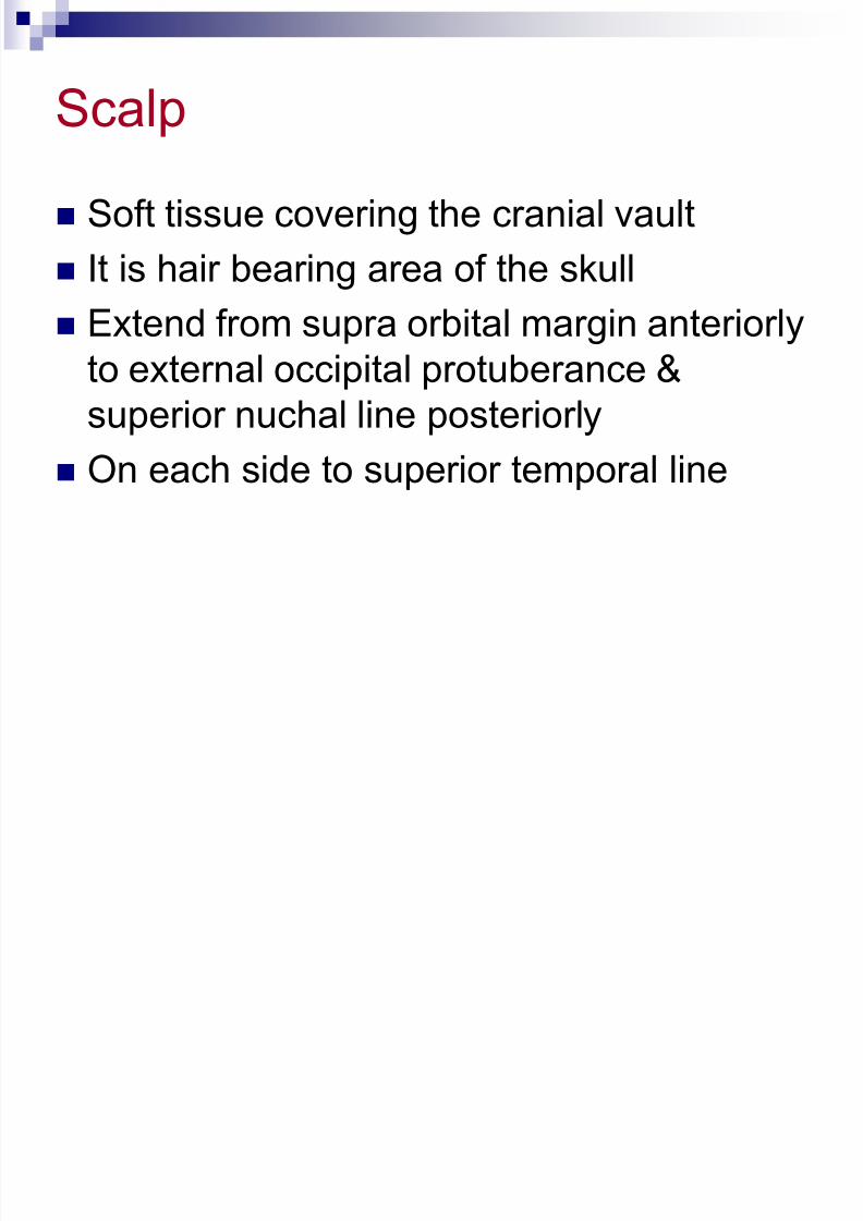
Scalp
Soft tissue covering the cranial vault
It is hair bearing area of the skull
Extend from supra orbital margin anteriorly
to external occipital protuberance &
superior nuchal line posteriorly
On each side to superior temporal line
7/30/2019 Scalp With All Details
http://slidepdf.com/reader/full/scalp-with-all-details 3/30
SCALP
S-Skin
C-connective tissue (superficial fascia)
A-aponeurosis (galea aponeurotica)
L-loose areolar tissue
P-pericranium
7/30/2019 Scalp With All Details
http://slidepdf.com/reader/full/scalp-with-all-details 4/30
Skin
Thick and hairy
Firmly attached to the epicranial
aponeurosis through dense fascia
Abundance sebaceous glands
Sebaceous cyst are common
7/30/2019 Scalp With All Details
http://slidepdf.com/reader/full/scalp-with-all-details 5/30
Connective tissue Fibrous and dense containing blood vessels and nerves
Binds skin to subjacent aponeurosis
Wounds bleed profusely as blood vessels are prevented
from retraction by fibrous tissue. Bleeding is stopped by
applying pressure against the bone
Subcutaneous hemorrhage are not extensive since
fascia is dense
Inflammation cause little swelling but are much painful
7/30/2019 Scalp With All Details
http://slidepdf.com/reader/full/scalp-with-all-details 6/30
Aponeurosis Anteriorly frontal belly and posteriorly occipital
belly of occipitofrontalis muscle
Frontal belly originate from skin of forehead and
mingled with orbicularis oculi muscle
Occipital belly originate from lateral 2/3 of
superior nuchal line
It gaps if cut transversely and should be stitched
7/30/2019 Scalp With All Details
http://slidepdf.com/reader/full/scalp-with-all-details 7/30
Loose areolar tissue
Extends anteriorly into the eyelids because frontalis hasno bony attachment
Posteriorly to superior nuchal line
On each side to superior temporal line
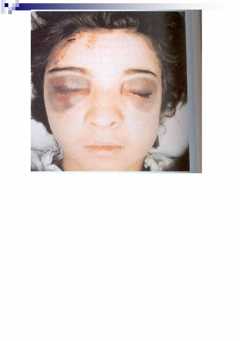
Bleeding cause generalized swelling of scalp
Called dangerous layer of scalp-emissary veins openhere and carry any infections inside the brain (venoussinus)
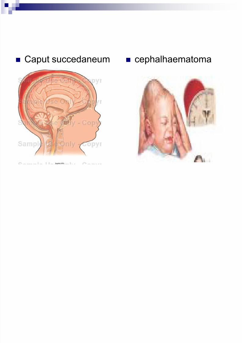
Bleeding lead to black eye Caput succedaneum in new born
7/30/2019 Scalp With All Details
http://slidepdf.com/reader/full/scalp-with-all-details 8/30
Pericranium
Is the periosteum of skull
Loosely attached to surface of bone but is
firmly adherent to the sutures
Injury deep to it take the shape of bone
(cephalhaematoma)
Scalping injury- should be replaced and
stitched because healing is better
7/30/2019 Scalp With All Details
http://slidepdf.com/reader/full/scalp-with-all-details 9/30
Caput succedaneum cephalhaematoma
7/30/2019 Scalp With All Details
http://slidepdf.com/reader/full/scalp-with-all-details 10/30
7/30/2019 Scalp With All Details
http://slidepdf.com/reader/full/scalp-with-all-details 11/30
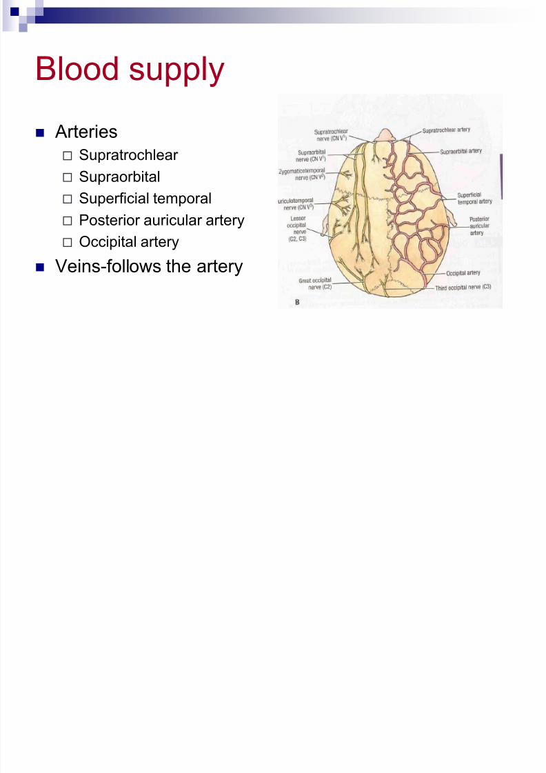
Blood supply
Arteries
Supratrochlear
Supraorbital
Superficial temporal
Posterior auricular artery
Occipital artery
Veins-follows the artery
7/30/2019 Scalp With All Details
http://slidepdf.com/reader/full/scalp-with-all-details 12/30
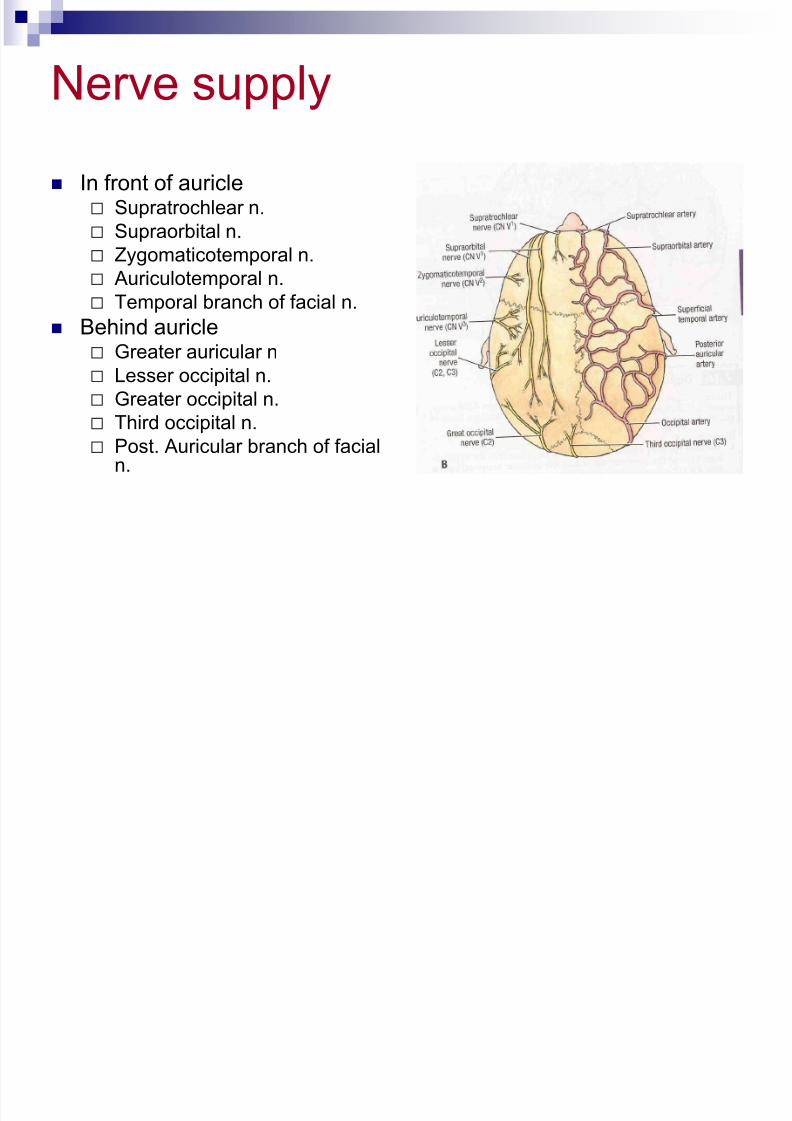
Nerve supply
In front of auricle Supratrochlear n.
Supraorbital n.
Zygomaticotemporal n.
Auriculotemporal n. Temporal branch of facial n.
Behind auricle Greater auricular n
Lesser occipital n.
Greater occipital n.
Third occipital n. Post. Auricular branch of facial
n.
7/30/2019 Scalp With All Details
http://slidepdf.com/reader/full/scalp-with-all-details 13/30
Lymphatics
Anterior part
Preauricular (parotid) gr. of lymph node
Posterior part
Posterior (mastoid) gr. of lymph node
&occipital gr. of lymph node
7/30/2019 Scalp With All Details
http://slidepdf.com/reader/full/scalp-with-all-details 14/30
Face
Boundaries
Extends superiorly to the hair line,
inferiorly to the chin and base of mandible,and on each side to auricle
Forehead is common to both scalp and
face
7/30/2019 Scalp With All Details
http://slidepdf.com/reader/full/scalp-with-all-details 15/30
Skin
Very vascular
Due to rich vascularity face blush and blanch
Wounds of face bleed profusely but heal rapidly Results of plastic surgery are excellent on face
Facial skin is rich in sebaceous gland and sweat
gland Sebaceous gland keep the skin oily but also
cause acne in adult
Sweat gland regulate body temperature
7/30/2019 Scalp With All Details
http://slidepdf.com/reader/full/scalp-with-all-details 16/30
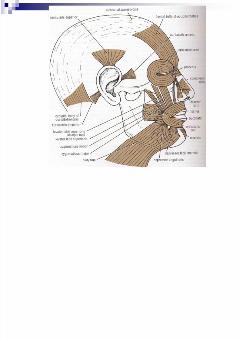
Facial muscle
Called muscle of facial expression and lie insuperficial fascia
Embryologically they develop from mesoderm of 2nd branchial arch, therefore supplied by facialnerve
7/30/2019 Scalp With All Details
http://slidepdf.com/reader/full/scalp-with-all-details 17/30
7/30/2019 Scalp With All Details
http://slidepdf.com/reader/full/scalp-with-all-details 18/30
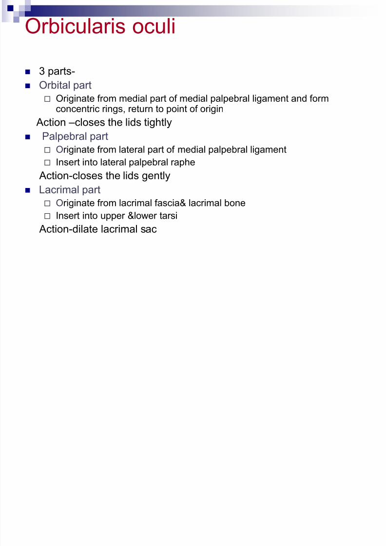
Orbicularis oculi
3 parts-
Orbital part
Originate from medial part of medial palpebral ligament and formconcentric rings, return to point of origin
Action –closes the lids tightly
Palpebral part Originate from lateral part of medial palpebral ligament
Insert into lateral palpebral raphe
Action-closes the lids gently
Lacrimal part
Originate from lacrimal fascia& lacrimal bone Insert into upper &lower tarsi
Action-dilate lacrimal sac

7/30/2019 Scalp With All Details
http://slidepdf.com/reader/full/scalp-with-all-details 19/30
Orbicularis auris
Originate from maxilla above incisor teeth andinsert into skin of lip.
Action –closes the mouth
7/30/2019 Scalp With All Details
http://slidepdf.com/reader/full/scalp-with-all-details 20/30
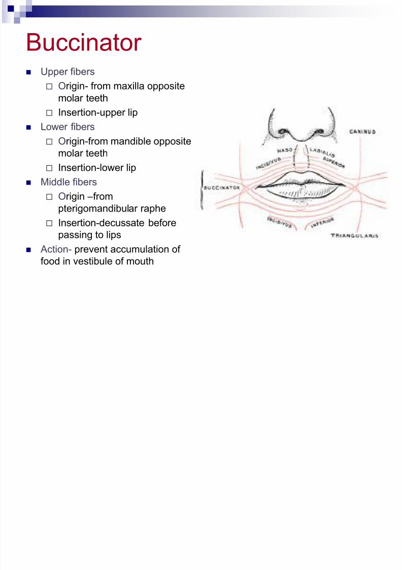
Buccinator Upper fibers
Origin- from maxilla opposite
molar teeth
Insertion-upper lip
Lower fibers
Origin-from mandible oppositemolar teeth
Insertion-lower lip
Middle fibers
Origin –from
pterigomandibular raphe Insertion-decussate before
passing to lips
Action- prevent accumulation of
food in vestibule of mouth
7/30/2019 Scalp With All Details
http://slidepdf.com/reader/full/scalp-with-all-details 21/30
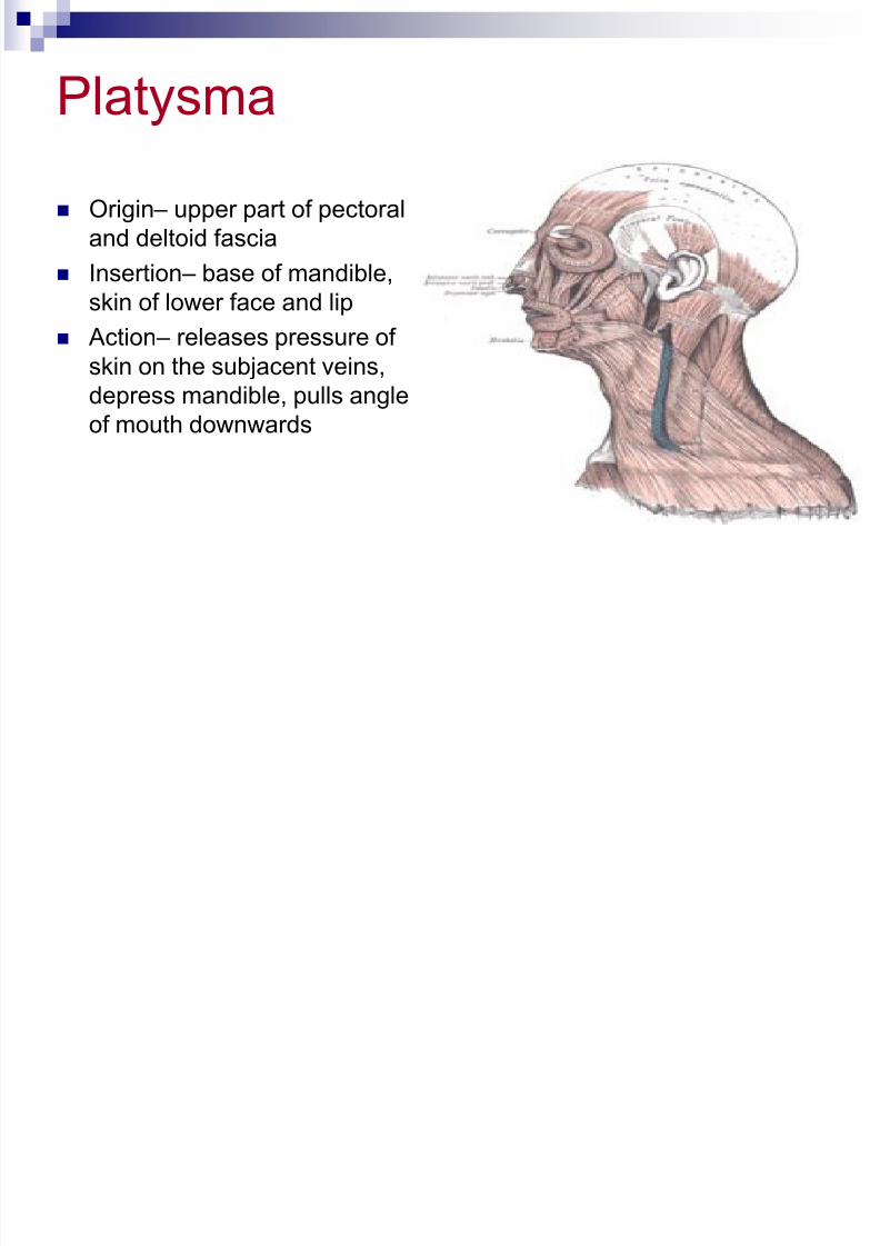
Platysma
Origin – upper part of pectoral
and deltoid fascia
Insertion – base of mandible,
skin of lower face and lip Action – releases pressure of
skin on the subjacent veins,
depress mandible, pulls angle
of mouth downwards
7/30/2019 Scalp With All Details
http://slidepdf.com/reader/full/scalp-with-all-details 22/30
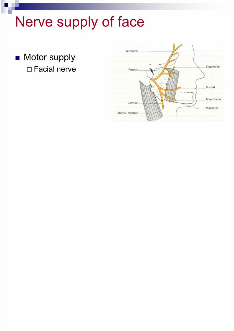
Nerve supply of face
Motor supply
Facial nerve
7/30/2019 Scalp With All Details
http://slidepdf.com/reader/full/scalp-with-all-details 23/30
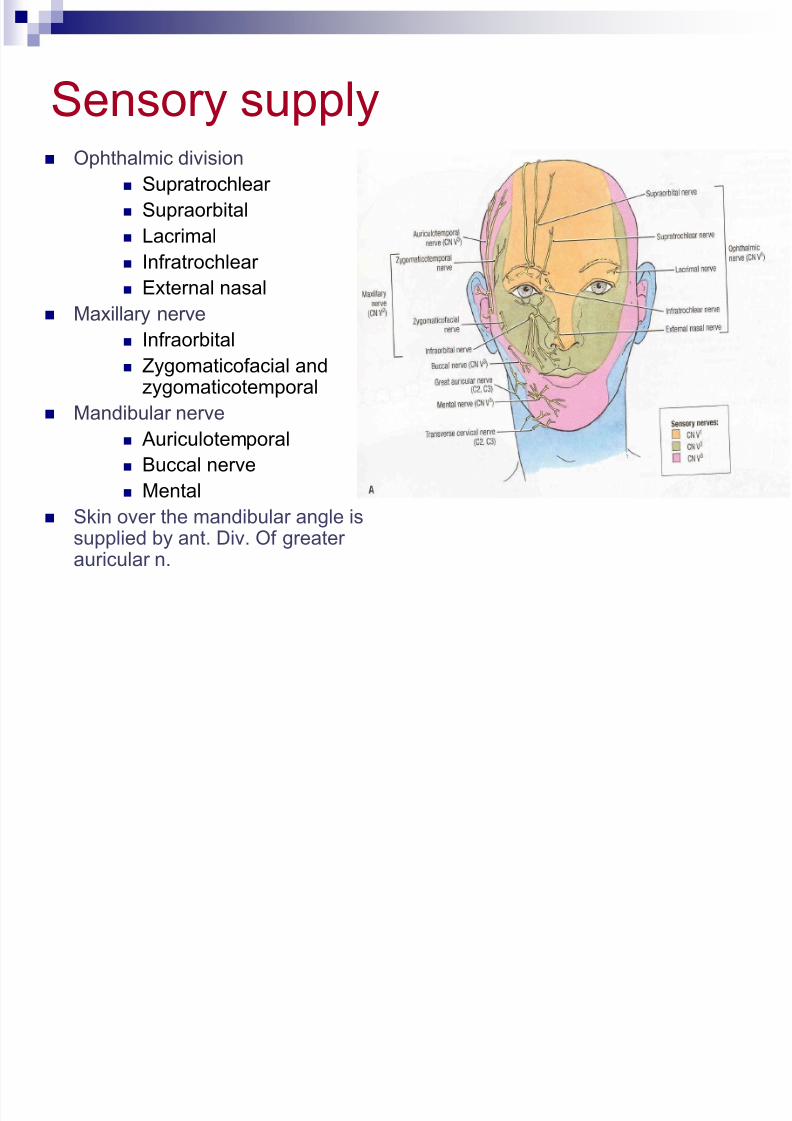
Sensory supply Ophthalmic division
Supratrochlear
Supraorbital
Lacrimal
Infratrochlear
External nasal
Maxillary nerve
Infraorbital
Zygomaticofacial andzygomaticotemporal
Mandibular nerve Auriculotemporal
Buccal nerve
Mental
Skin over the mandibular angle issupplied by ant. Div. Of greater
auricular n.
7/30/2019 Scalp With All Details
http://slidepdf.com/reader/full/scalp-with-all-details 24/30
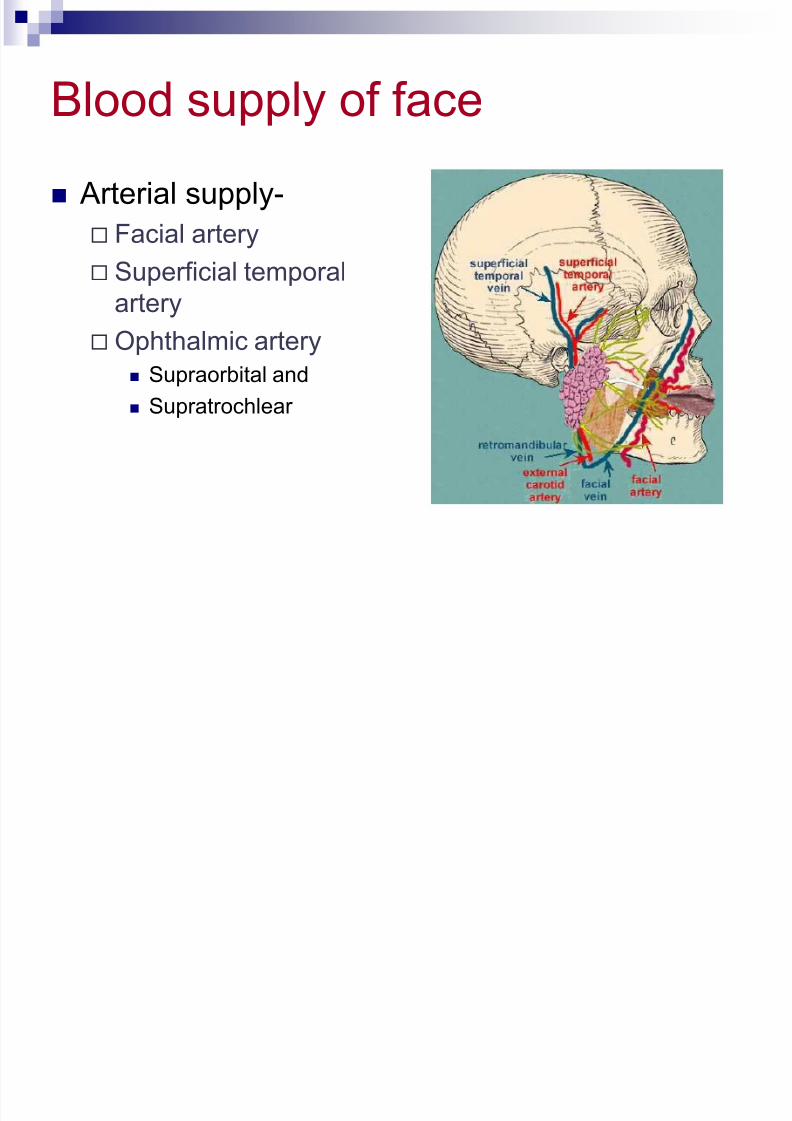
Blood supply of face
Arterial supply-
Facial artery
Superficial temporalartery
Ophthalmic artery
Supraorbital and
Supratrochlear
7/30/2019 Scalp With All Details
http://slidepdf.com/reader/full/scalp-with-all-details 25/30
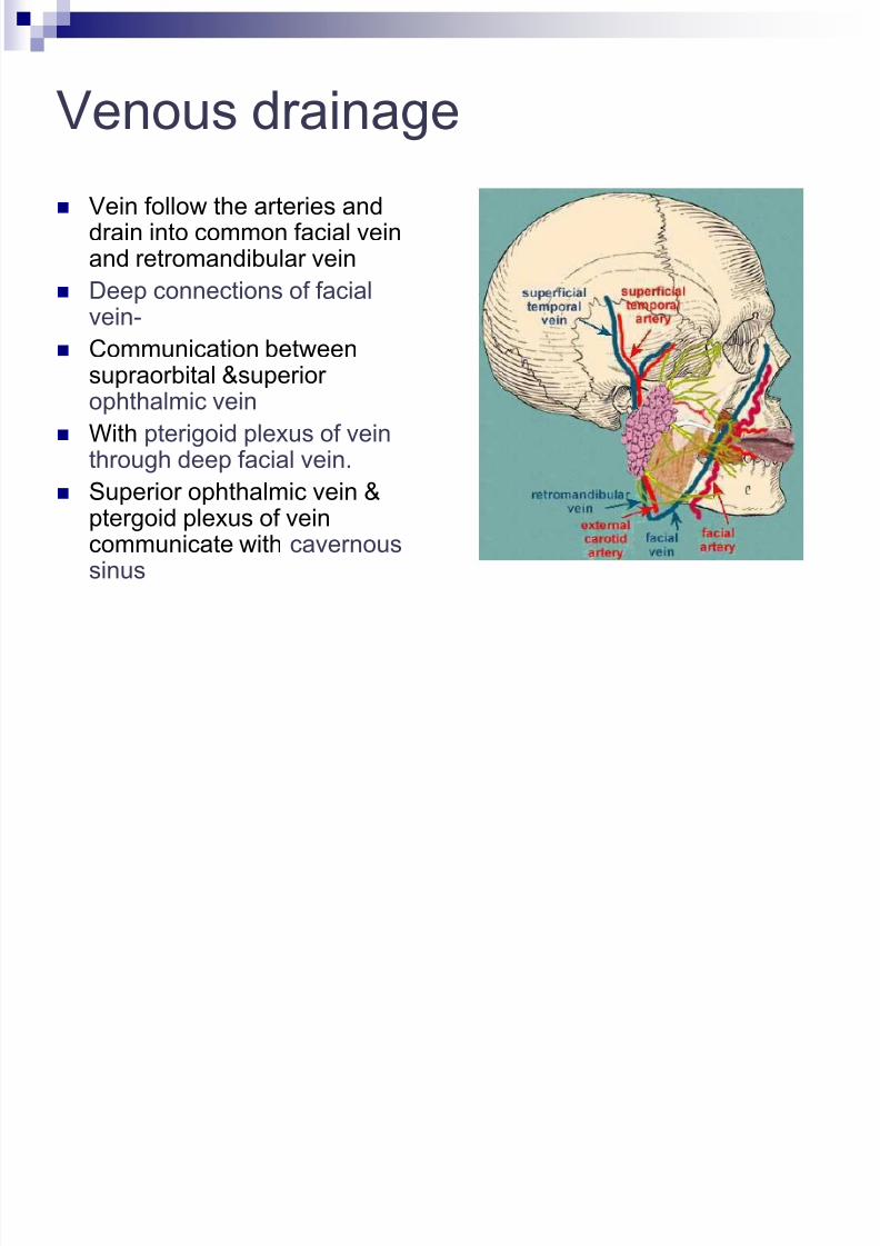
Venous drainage
Vein follow the arteries anddrain into common facial veinand retromandibular vein
Deep connections of facial
vein- Communication between
supraorbital &superior ophthalmic vein
With pterigoid plexus of vein
through deep facial vein. Superior ophthalmic vein &
ptergoid plexus of veincommunicate with cavernoussinus
7/30/2019 Scalp With All Details
http://slidepdf.com/reader/full/scalp-with-all-details 26/30
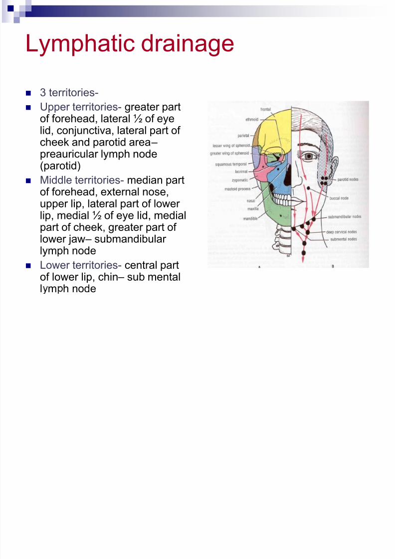
Lymphatic drainage
3 territories-
Upper territories- greater partof forehead, lateral ½ of eyelid, conjunctiva, lateral part of
cheek and parotid area – preauricular lymph node(parotid)
Middle territories- median partof forehead, external nose,
upper lip, lateral part of lower lip, medial ½ of eye lid, medialpart of cheek, greater part of lower jaw – submandibular lymph node
Lower territories- central part
of lower lip, chin – sub mentall m h node
7/30/2019 Scalp With All Details
http://slidepdf.com/reader/full/scalp-with-all-details 27/30
Applied
Trigeminal neuralgia Maxillary and mandibular nerve are involved
Excruciating pain in the region of distribution of these nerve
In infranuclear lesions of facial nerve (eg, bell’s palsy)- whole face is paralyzed c/f
Affected side is motionless
Loss of wrinkles
Eye cannot be closed
In smiling the mouth is drawn to normal side During mastication food accumulates in vestibule of mouth
In supranuclear lesions of facial nerve only the lower part of face is paralyzed. The upper part (frontalis &partof orbicularis oculi) escapes due to its bilateral
innervation
7/30/2019 Scalp With All Details
http://slidepdf.com/reader/full/scalp-with-all-details 28/30
7/30/2019 Scalp With All Details
http://slidepdf.com/reader/full/scalp-with-all-details 29/30
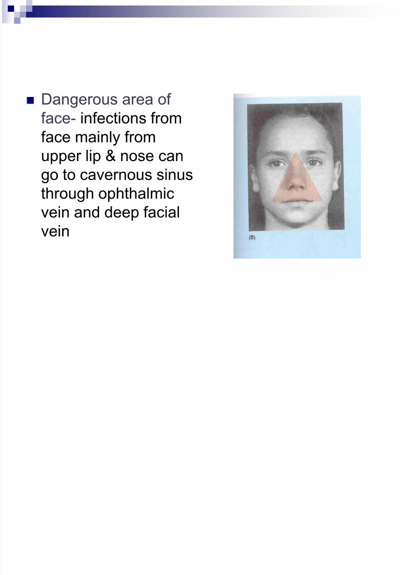
Dangerous area of
face- infections from
face mainly fromupper lip & nose can
go to cavernous sinus
through ophthalmic
vein and deep facialvein

7/30/2019 Scalp With All Details
http://slidepdf.com/reader/full/scalp-with-all-details 30/30