Embed Size (px)
Citation preview

1
SCANNING ELECTRON MICROSCOPE IDENTIFICATION OF FIBRES AND HAIRS
FROM THE COOK VOYAGE COLLECTIONS AT THE PITT RIVERS MUSEUM
CAROLINE CARTWRIGHT
(Senior Scientist, Department of Conservation and Scientific Research,
British Museum, London WC1B 3DG)
INTRODUCTION
Thirty-two fibre and hair samples from 19 objects in the Cook Voyage Collections at
the Pitt Rivers Museum were submitted by Jeremy Uden (Clothworkers' Foundation
Senior Conservation Fellow, Conserving "Curiosities" - Investigating the Cook Voyage
Collections at the Pitt Rivers Museum) for scanning electron microscope (SEM)
examination, imaging and identification at the British Museum (Figure 1 and also see
http://conserving-curiosities.blogspot.co.uk/2013_04_01_archive.html).
It should be stressed at the outset that particular questions have been addressed in
these targeted samples and that these objects often contain other hairs, fibres and
other plant remains that were not sampled in this study, as well as other organic
components. Consequently, it is the presence of a taxon (i.e. a taxonomic category or
group, such as a genus or species) that is significant.
METHODS
Selected sampling of the objects was carried out by Jeremy Uden and the author on
12 December 2012. Additional samples were brought to the British Museum on 19
June 2013 by Jeremy Uden. An effort was made to keep sample sizes to a minimum
and to try to avoid sampling any areas with macroscopically visible adhesives or
conservation consolidants that might affect fibre identification. Inevitably over the
years, objects can (and have) some adhering modern fibres on their surfaces
although these were not obvious macroscopically at the time of sampling. However,
as will be seen in the Figures (SEM images) accompanying this report, fortuitously
there were relatively few occurrences of this. In all instances care was taken to
establish (through examination of the objects and accompanying documentation) that
there were no areas that had been restored or replaced with different types of fibres
or hairs at a previous point in the object’s history, which could clearly lead to

2
confusing and misleading results, not only for the SEM fibre/hair identifications, but
also for the previous polarized light microscope (PLM) imaging carried out by Jeremy
Uden (see http://conserving-curiosities.blogspot.co.uk/2012/10/investigating-plant-
fibres.html ).
Examination of the samples and comparative reference specimens was undertaken in
a variable pressure (VP) SEM (Hitachi S-3700N) using the backscatter electron (BSE)
detector mostly at 15 kV but sometimes also at 12kV or 20 kV, depending on the
sample. Magnifications ranged from x23 to x900. The preferred working distance was
c.15 mm, but extended from 8.8 mm to 22.6 mm (as required). As the fibres/hairs
were in variable conditions, the SEM chamber was only partially evacuated (40 Pa,
sometimes 30 Pa or 25 Pa). With the BSE detector, 3D mode (rather than
Compositional) was preferentially selected to maximize the opportunity to reveal
diagnostic features for identification as well as traces of wear and abrasion due to
preparation and/or use of the materials and to show dirt, encrustations and fungal
hyphae.
Most of the material examined was placed on adhesive carbon discs mounted onto
aluminium SEM stubs; no other sample preparation was required. At a later stage,
selected samples from the objects as well as reference specimens were sputter-
coated with a platinum/palladium (Pt/Pd) alloy (using a Cressington 208 HR sputter
coater) to prevent charging with the electron beam in conventional SEM mode using
the secondary electron detector (SE) at high vacuum. The purpose was to try to
achieve higher resolution imaging and greater clarity of information from highly
deteriorated specimens. The Oxford Instruments energy-dispersive X-ray
spectroscopy (EDX) analyser attached to the SEM was used to provide elemental
identification and semi-quantitative compositional information where necessary e.g.
to determine whether crystals and inclusions were calcium oxalate or silicon (see
below).
To assist readers who might wish to refer to the conditions of VP-SEM operation
associated with the SEM images in the Figures accompanying this report, attention is
drawn to the information provided in the data bar at the foot of each image. Reading
left to right, the data bar information gives the model of the SEM and operator initials
(S3700CRC), accelerating voltage (kV), working distance (mm), electron detector and
mode (BSE3D or SE), partial evacuation status (Pa), magnification (x) and scale (in
micrometres or millimetres).

3
RESULTS
As full details of the objects are available on-line at the Pitt Rivers Museum databases,
http://databases.prm.ox.ac.uk/fmi/iwp/cgi?-db=objects_online&-loadframes, lengthy
descriptions are not included here (as agreed with Jeremy Uden on 19 November
2013). The results are summarised in Table 1.
The advantages and drawbacks of using fibre/hair atlases, on-line fibre/hair image
databases and texts as references have been discussed in detail elsewhere
(Cartwright and King 2012) but key points relevant to this study are reiterated here.
On-line and printed fibre/hair atlases frequently contain images using dark or bright
field PLM, or using differential interference contrast optical microscopy. Whilst these
are very useful for modern material, it is always difficult to try to compare these with
and attempt to match key features on historical, aged or archaeological fibres, many
of which have been altered through burial and/or through use, wear and tear, and the
natural processes of ageing and deterioration. ‘Textbook’ images of clean, recent
fibres and hairs, whether using PLM or SEM, cannot replicate the complex
characteristics exhibited by historical or archaeological fibres and hairs, many of
which are visible in the Figures (images) accompanying this report. In several
reference resources, such as in the atlas of plant material and fibres from New
Zealand and the Pacific (Carr and Cruthers 2007), Lowe et al. 2010 and Carr et al.
(2008) the authors themselves say that such databases may assist in identifying plant
materials but “should not be regarded as a substitute for a confirmed identification by
a plant scientist” (Carr et al. 2008; 252)
Table 1 provides the identifications of the selected material, but broader and deeper
interpretations that can be made in the future from these results are very much in the
realm of the specialist curators and conservators concerned. Consequently, selected
aspects relating to the SEM examination and identification phase will be highlighted in
this section, but placing these within a wider cultural framework of interpretation is
outside the brief of this part of the project.
The sample from the top of the pouch of the stringwork bag 1884.29.5.1 from New
Caledonia comprises Pteropus sp. (flying fox, fruit bat) hairs (Figures 1 – 4). Some of
the hairs are in a better condition than others (e.g. Figure 3) and are of varying
dimensions (Figure 4), presumably reflecting the presence of dense, fine underfur and
long guard hairs.

4
Three samples (Figures 5 – 7) were taken from 1886.1.876, a stringwork bag from
New Caledonia: (1) hair string from top of pouch; (2) mixed fibres at handle end; (3)
bark sample. Sample (1) proved not to be hair, but Phormium tenax, New Zealand
flax (Figure 5). Despite tantalising remnants of what appeared to be diagnostic
elements, identification of sample (2) was not ultimately possible due to its very poor
condition, some dirt and calcareous encrustations (Figure 6). Sample (3) was
Broussonetia papyrifera, paper mulberry (Figure 7).
Three samples were examined from 1886.1.1124, a Maori cloak from Polynesia / New
Zealand: (1) leaf end (Figures 8 – 9); (2) cord (Figure 10); (3) fine fibre (Figure 11). All
were Phormium tenax, New Zealand flax; sample (1) displayed non-active fungal
hyphae (Figure 9).
A sample of plant material from Maori cloak 1886.1.1134 from New Zealand was
identified as Cordyline australis, cabbage tree, tī kōuka (Figure 12).
A sample of barkcloth from Tonga, 1886.1.1239, was identified as Broussonetia
papyrifera, paper mulberry (Figure 13), as was a sample of barkcloth from Tahiti,
1886.1.1248 [.1 - 3] (Figure 14, also note on the image a few prismatic calcium
oxalate crystals, displaced from adjacent parenchyma cells). Figures 13 – 14 also
show the well-beaten nature of the fibres and other plant cells of the barkcloth.
Three samples were taken from 1886.1.1250, barkcloth from Easter Island. Sample 1,
from the barkcloth itself, was identified as Broussonetia papyrifera, paper mulberry
(Figures 15 – 16). These images not only show that the fibres have adjacent plant
cells (such as parenchyma) still attached to them in many instances, but also show
(particularly in Figure 15, but also in Figure 14 with 1886.1.1248) that some barkcloth
processing can result in fibres being ‘teased’ out and somewhat flattened – often
giving such processed Broussonetia papyrifera fibres a ‘cotton-like’ appearance. This
can sometimes be confusing when examining objects that may (a) ‘legitimately’ have
cotton as part of their textile, (b) have modern cotton fibres on their surface as more
recent contaminants. Samples 2 and 3 were taken from the parallel rows of stitching.
Figure 17 shows that sample 2, from the long decorated stripe, represents fibres (and
adjacent plant cells) taken from an area of Broussonetia papyrifera, paper mulberry
between the bark and the xylem. Figure 18 shows that sample 3, from the stitching,
includes ‘teased-out’ Broussonetia papyrifera fibres, but that some of the cell tissue
adjacent to the fibres is still present in places (including prismatic calcium oxalate
crystals, displaced from adjacent parenchyma cells).
5
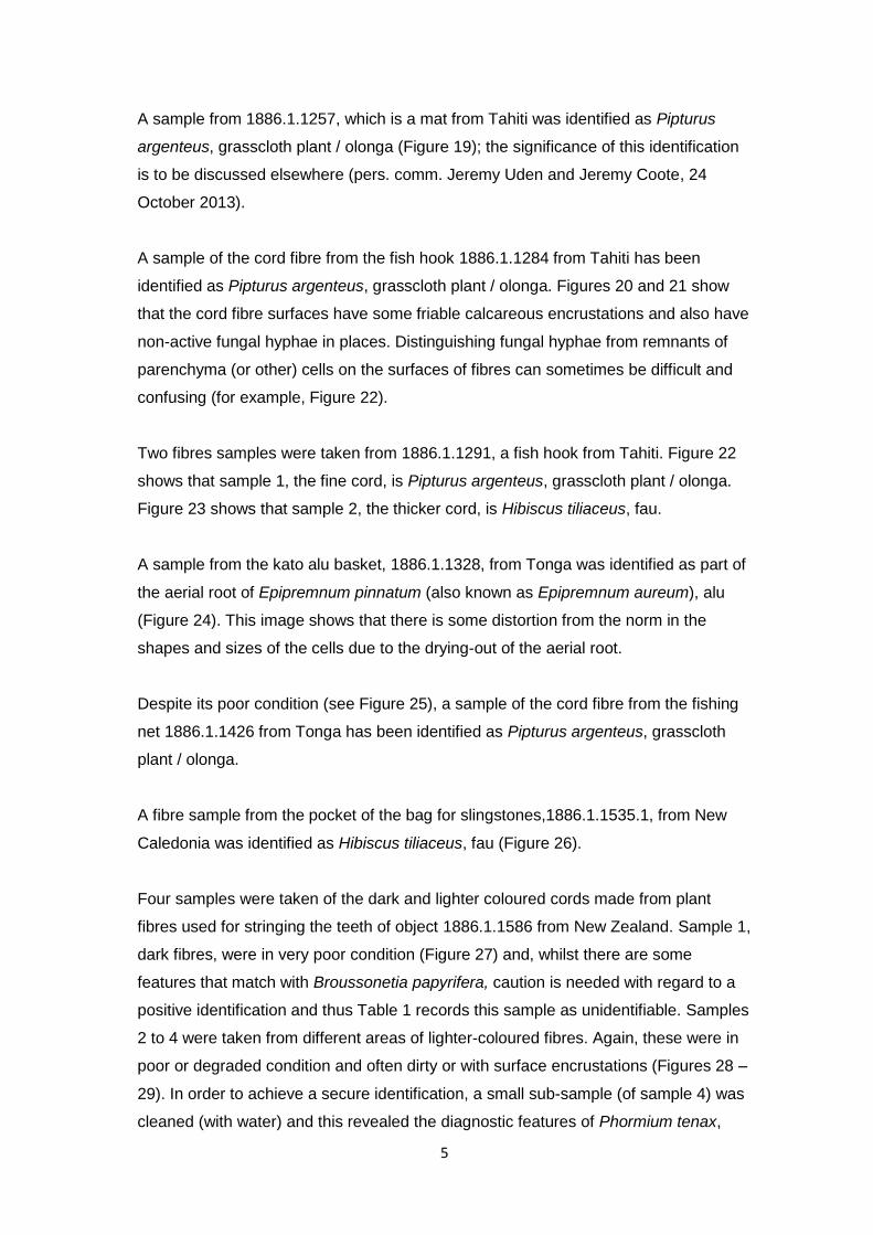
A sample from 1886.1.1257, which is a mat from Tahiti was identified as Pipturus
argenteus, grasscloth plant / olonga (Figure 19); the significance of this identification
is to be discussed elsewhere (pers. comm. Jeremy Uden and Jeremy Coote, 24
October 2013).
A sample of the cord fibre from the fish hook 1886.1.1284 from Tahiti has been
identified as Pipturus argenteus, grasscloth plant / olonga. Figures 20 and 21 show
that the cord fibre surfaces have some friable calcareous encrustations and also have
non-active fungal hyphae in places. Distinguishing fungal hyphae from remnants of
parenchyma (or other) cells on the surfaces of fibres can sometimes be difficult and
confusing (for example, Figure 22).
Two fibres samples were taken from 1886.1.1291, a fish hook from Tahiti. Figure 22
shows that sample 1, the fine cord, is Pipturus argenteus, grasscloth plant / olonga.
Figure 23 shows that sample 2, the thicker cord, is Hibiscus tiliaceus, fau.
A sample from the kato alu basket, 1886.1.1328, from Tonga was identified as part of
the aerial root of Epipremnum pinnatum (also known as Epipremnum aureum), alu
(Figure 24). This image shows that there is some distortion from the norm in the
shapes and sizes of the cells due to the drying-out of the aerial root.
Despite its poor condition (see Figure 25), a sample of the cord fibre from the fishing
net 1886.1.1426 from Tonga has been identified as Pipturus argenteus, grasscloth
plant / olonga.
A fibre sample from the pocket of the bag for slingstones,1886.1.1535.1, from New
Caledonia was identified as Hibiscus tiliaceus, fau (Figure 26).
Four samples were taken of the dark and lighter coloured cords made from plant
fibres used for stringing the teeth of object 1886.1.1586 from New Zealand. Sample 1,
dark fibres, were in very poor condition (Figure 27) and, whilst there are some
features that match with Broussonetia papyrifera, caution is needed with regard to a
positive identification and thus Table 1 records this sample as unidentifiable. Samples
2 to 4 were taken from different areas of lighter-coloured fibres. Again, these were in
poor or degraded condition and often dirty or with surface encrustations (Figures 28 –
29). In order to achieve a secure identification, a small sub-sample (of sample 4) was
cleaned (with water) and this revealed the diagnostic features of Phormium tenax,

6
New Zealand flax (Figure 30). It is suggested, therefore, that on this basis, samples 2
– 3 may also be Phormium tenax.
A sample of the binding, presumed to be woman’s hair, from a mourner’s costume
from Tahiti, 1886.1.1637, was confirmed as human hair, neatly and finely plaited
(Figures 31 – 32).
A sample was taken of the aerial root from the cloak of the mourner’s costume
1886.1.1637.4 from Tahiti. Figure 33 shows a transverse section of the aerial root,
identified as Freycinetia arborea. This image shows that there is some distortion from
the norm in the shapes and sizes of the cells due to the drying-out of the aerial root.
A sample of barkcloth from the mourner’s costume 1886.1.1637.7 from Tahiti was
identified as Broussonetia papyrifera; Figure 34 shows that some of the cell tissue
adjacent to the fibres is also present including druses (see Note 1 for a definition of
the term) and prismatic calcium oxalate crystals, both in situ within, and displaced
from, adjacent parenchyma cells.
Some of the plant material found inside the tamau headdress of plaited human hair,
1886.1.1685, from the Society Islands was sampled during conservation for
subsequent identification. Jeremy Uden notes (pers. comm. 4 December 2013) that it
was on a pandanus hat when found, but when it was worn for dancing the centre was
filled with flowers, thus the question was whether the remains sampled were of the
flowers, the pandanus hat or, more likely, material from the Tahitian mourner's
costume. Some parts of the sample were covered with dirt, encrustations and (non-
active) fungal hyphae, which masked any diagnostic features (Figure 35). A sub-
sample was split longitudinally; this revealed sufficient key features (Figure 36) to
enable the material to be identified as Pandanus, with a good match with the
reference collection specimen of Pandanus tectorius, Tahitian screwpine. Figure 36
shows the presence of (mostly displaced) fine, needle-like calcium oxalate crystals
(raphides).
Two samples were taken of the plant fibres from fish hook 1887.1.379 from New
Zealand. Sample 1, cord fibres were identified as Phormium tenax, New Zealand flax
(Figure 37). Sample 2, hook binding was in very poor condition with much dirt,
encrustations, contaminants, stray (possibly modern) surface fibre remnants and what
appeared to be adhesive or consolidant in some areas (Figure 38) – which ruled out
cleaning a tiny sub-sample with water. Initially it was thought that no identifications

7
were going to be possible as the features on the fibres were heavily camouflaged
(Figure 39), but persistent examination using the VP-SEM eventually revealed a few
identifiable fibres of Freycinetia banksii, kiekie (Figure 40).
A sample of the hair string from the ceremonial club 1923.74.5 from New Caledonia
was identified as Pteropus sp. (flying fox, fruit bat) guard hair and underfur, with some
hairs more deteriorated than others (Figure 41).
A sample from mat 1945.11.130 from Tahiti was examined particularly carefully in the
VP-SEM as parts of it were in poor condition with dirt, calcareous particles and
modern (synthetic) fibres on the surface, possibly displaced from other parts of the
object (Figure 42). Although the presence of calcium oxalate crystals must be used
with great caution when identifications are made (also see below) it was useful to
ultimately be able to detect the presence of calcium oxalate druses (see Note 1 for
definition) on a cleaned sub-sample (Figure 43). Parthasarathy (2009) noted that
calcium oxalate druses are abundant in the medullary ray cells of Thespesia populnea
and although the presence of druses alone did not determine the identification of
Thespesia populnea, purau, it was helpful nonetheless when comparing
characteristics visible on this sub-sample with diagnostic features on reference
collection specimens.
DISCUSSION AND CONCLUSIONS
In plant anatomy the term ‘fibre’ refers to a particular type of cell, which has the
function of support, but the term has acquired a more general usage (sensu lato) in
the literature, which can be confusing. Although the word ‘fibres’ has been used
throughout this report, in many instances the plant cells visible (in SEM examination
and in the resulting SEM images) also comprise cells that occur adjacent to fibres
such as parenchyma, collenchyma, phloem or xylem. When fibres (in the strict sense
of the term) are being extracted from a stem, adjacent cells (particularly parenchyma)
leave imprints or vestiges of their distinctly-shaped cell walls on the surface of the
fibres (sensu stricto). To the untrained eye, these can sometimes appear to represent
diagnostic features (hence the caution with which one must approach fibre
identifications). Figures 5 and 10 – 12 (to choose but a few examples) show areas in
which this can be seen.
Taking the decision to examine most of the fibres/hairs in the VP-SEM without first
cleaning and preparing them (for example, by obtaining casts or by thin-sectioning,

8
embedding in resin and polishing) has yielded significant additional information about
the condition of the fibres. Many display encrustation (possibly from the historical use
of pesticides), (non-active) fungal hyphae, loose particles (dirt), abrasion or
deterioration. Figures 9, 21, 35 and 38 show typical examples. The information is
useful for a number of reasons, which include adding to the body of knowledge about
the effects of preparation of the fibres (or skins) during the manufacture of the object,
use by its owner(s), as well as its subsequent storage. It can also inform active
conservation and care of the museum collections. In certain instances, a tiny sub-
sample was cleaned with water to enable crucial identifications, for example, the mat
1945.11.130 from Tahiti.
The presence of silica (silicon) and calcium in plants has long been of interest to plant
scientists, but the subject is a complex one, not least because the presence of both
calcium and silicon in plant tissue is directly related to the chemical composition of
soil in which the plant is growing and the rate at which the plant may take up these
elements. Nonetheless, many plant scientists and (archaeobotanists) have paid
attention to the presence and form (morphology) of silica bodies and calcium oxalate
crystals in their samples during the processes of identification. For example,
phytoliths, which are composed mainly of silicon dioxide (and are mostly defined as
silica bodies) can be representative of some plant families, sometimes of genus-level
morphology (Piperno 2006). Calcium oxalate crystals, whether prismatic in form, or
druses, styloids or raphides may be useful in characterising some plant families
(Prychid and Rudall 1999). But the presence or absence of calcium oxalate crystals
and/or silica bodies such as phytoliths should not be used as a prime identification
criterion on its own, but with discretion in conjunction with key cellular anatomical
features.
As noted above, this report has provided the identifications of the selected material as
a basis of reference for on-going research by the specialist curators and conservators
concerned from which new and challenging avenues of interest are emerging within a
wider cultural framework of interpretation
With suitable reference material, VP-SEM at the British Museum has been used
successfully to identify 30 out of the 32 selected fibre and hair samples from 19
objects in the Cook Voyage Collections at the Pitt Rivers Museum. While these
identifications have not always been straightforward, as many of the fibres have
markedly degraded over time, VP-SEM offered significant additional information
inasmuch as it permitted characterization of condition and the effects of use or wear.

9
ACKNOWLEDGEMENTS
Thanks are due to Jeremy Uden for sampling the objects and provision of object
information.
NOTES
1. Druses are compound crystals, usually roughly spherical with a star-like
appearance caused by the component crystals protruding from the surface. They
may be composed of calcium oxalate, silicates or carbonates.
REFERENCES
Carr, D. J. and Cruthers, N.M. 2007. He rārangi whakaaturanga o ngā taonga rākau.
www.otago.ac.nz/textiles/plantfibres/index.html [accessed 20/02/2009]
Carr, D., Cruthers, N., Girvan, E. and Scheele, S. 2008 ‘Approaches for conservators
to the identification of plant materials used in Māori artefacts’ Studies in Conservation
53; 252-263
Cartwright, C.R. and King, J.C.H. 2012 ‘Identifications of hairs and fibres in Great
Lakes objects from the eighteenth and nineteenth centuries using variable pressure
scanning electron microscopy’ British Museum Technical Research Bulletin 6; 69-81
Lowe, B.J., Carr, D.J., McCallum. R.E., Myers, T., Ngarimu-Cameron, R. and Niven,
B.E. 2010 ‘Understanding the variability of vegetable fibre: a case study from New
Zealand’ Textile Research Journal 80; 1138-1150
Parthasarathy, R. 2009 Pharmacognostical, phytochemical and pharmacological
evaluation of bark and leaf of Thespesia populnea. Soland ex.Correa. Unpublished D.
Phil thesis, Bharath University, India.
Piperno, D. R. 2006 Phytoliths: A Comprehensive Guide for Archaeologists and
Paleoecologists. AltaMira Press, San Diego
Prychid, C.J. and Rudall, P.J. 1999 ‘Calcium oxalate crystals in monocotyledons: a
review of their structure and systematics’ Annals of Botany 84; 725-739

1
TABLE 1
Fibre/hair identifications by Caroline Cartwright of Cook Voyage Collections samples
Accession number
Object Sample location Fibre / hair identification Comments Figure numbers (SEM images)
1884.29.5.1 stringwork bag, New Caledonia
Top of pouch Pteropus sp. (flying fox, fruit bat) hair guard hair and underfur; some hairs quite deteriorated
Figures 1 – 4
1886.1.876 stringwork bag, New Caledonia
Three samples: 1) hair string from top of pouch (2) mixed fibres at handle end (3) bark sample
(1) Phormium tenax, New Zealand flax (2) unidentifiable (3) Broussonetia papyrifera, paper mulberry
(2) very poor condition
(1) Figure 5 (2) Figure 6 (3) Figure 7
1886.1.1124 Maori cloak, Polynesia / New Zealand
(1) leaf end (2) cord (3) fine fibre
(1) Phormium tenax, New Zealand flax (2) Phormium tenax, New Zealand flax (3) Phormium tenax, New Zealand flax
(1) fungal hyphae present (non-active)
(1) Figures 8 – 9 (2) Figure 10 (3) Figure 11
1886.1.1134 Maori cloak, New Zealand
Plant material from cloak Cordyline australis (cabbage tree, tī kōuka)
Figure 12
1886.1.1239 barkcloth, Tonga Sample of barkcloth Broussonetia papyrifera, paper mulberry
Figure 13
1886.1.1248 [.1 - 3]
barkcloth, Tahiti Sample of barkcloth Broussonetia papyrifera, paper mulberry
Figure 14
1886.1.1250 barkcloth, Easter Island
Three samples of the plant material: (1) barkcloth (2) long decorated stripe (3) stitching
(2) (2) and (3) are from the parallel rows of stitching
(1) Broussonetia papyrifera, paper mulberry
(2) Broussonetia papyrifera, paper mulberry
(3) Broussonetia papyrifera, paper mulberry
1) Figures 15 – 16 2) Figure 17 3) Figure 18
1886.1.1257 mat, Tahiti Mat sample Pipturus argenteus, grasscloth plant, olonga
Figure 19
1886.1.1284 fish hook, Tahiti Cord fibre Pipturus argenteus, grasscloth plant, olonga
some fungal hyphae present (non-active)
Figures 20 – 21
1886.1.1291 fish hook, Tahiti Two fibre samples: (1) Fine cord (2) Thicker cord
(1) Pipturus argenteus, grasscloth plant, olonga
(2) Hibiscus tiliaceus, fau
(1) some fungal hyphae present (non-active)
(1) Figure 22 (2) Figure 23
1886.1.1328 kato alu basket, Tonga
A sample of the alu aerial root of Epipremnum pinnatum, alu
some cell distortion present due to drying-out of plant material
Figure 24

2
1886.1.1426 fishing net, Tonga Net fibre Pipturus argenteus, grasscloth plant, olonga
poor condition Figure 25
1886.1.1535.1 bag for slingstones, New Caledonia
Fibre from the pocket of the sling
Hibiscus tiliaceus, fau Figure 26
1886.1.1586 strung teeth, New Zealand
Four samples of the dark and lighter coloured cords made from plant fibres: (1) dark fibres (2) light fibres (3) light fibres (another area) (4) light fibres (another area)
(1) unidentifiable (2) – (4) Phormium tenax, New Zealand flax
(1) very poor condition (2) – (4) poor condition identification made on a cleaned sample
(1) Figure 27 (2) Figure 28 (3) Figure 29 (4) Figure 30
1886.1.1637 mourner’s costume, Tahiti
Woman’s hair from binding human hair Figures 31 – 32
1886.1.1637.4
cloak from mourner’s costume, Tahiti
Sample of the aerial root Freycinetia arborea Figure 33
1886.1.1637.7 barkcloth, part of mourner’s costume, Tahiti
Sample of barkcloth Broussonetia papyrifera, paper mulberry
Figure 34
1886.1.1685 tamau headdress of plaited human hair, Tahiti
Sample represents some of the plant material found inside the tamau
Pandanus tectorius, Tahitian screwpine
parts of the sample in poor condition with (non-active) fungal hyphae
Figures 35 – 36
1887.1.379 fish hook, New Zealand
Two samples of the plant fibres on the hook: 1) cord 2) hook binding
(1) Phormium tenax, New Zealand flax
(2) Freycinetia banksii, kiekie
(2) sample in very poor condition
(1) Figure 37 (2) Figures 38 – 40
1923.74.5 club, New Caledonia Hair string Pteropus sp. (flying fox, fruit bat) hair guard hair and underfur; some hairs deteriorated
Figure 41
1945.11.130 mat, Tahiti Mat sample Thespesia populnea, purau parts of the sample in poor condition
Figures 42 – 43


![Textiles ] What is textiles } Who was the first fashion designer ) Cotton > Inspiration - Fibres + The job of a fashion designer = Quiz](https://img.pdfslide.net/doc/110x75/56649f1f5503460f94c371c0/textiles-what-is-textiles-who-was-the-first-fashion-designer-cotton-.jpg)