Embed Size (px)
Citation preview

PHYSIOLOGICAL PSYCHOLOGY
Complementary Course of B.Sc. Counselling Psychology
II Semester
(CUCBCSS - 2014 Admission onwards)
UNIVERSITY OF CALICUT
SCHOOL OF DISTANCE EDUCATION
Calicut University P.O. Malappuram, Kerala, India 673 635

School of Distance Education
PHYSIOLOGICAL PSYCHOLOGY 2
UNIVERSITY OF CALICUT
SCHOOL OF DISTANCE EDUCATION
Complementary Course of B.Sc. Counselling Psychology
PHYSIOLOGICAL PSYCHOLOGY
Semester II
Prepared by: Smt. Abidha Kurukkan
M.Sc (Psychology), M.Ed
Research Scholar
University of Calicut
Layout: Computer Section, SDE
©
Reserved

School of Distance Education
PHYSIOLOGICAL PSYCHOLOGY 3
CONTENTS PAGES
MODULE – I 5
MODULE – II 17
MODULE – III 36

School of Distance Education
PHYSIOLOGICAL PSYCHOLOGY 4

School of Distance Education
PHYSIOLOGICAL PSYCHOLOGY 5
Module 1
PERIPHERAL NERVOUS SYSTEM
The Nervous System
The nervous system can be simply described as collection of neurons which are arranged to
work in a coordinated function. One of the most important functions of the nervous system is
to process incoming information in such a way that appropriate mental and motor responses
will occur. The nervous system is composed of three major parts: the sensory input portion,
the central nervous system (or integrative portion), and the motor output portion. Sensory
receptors detect the state of the body or the state of the surroundings. For example, the eyes
are sensory organs that give one a visual image of the surrounding area. The ears also are
sensory organs. The central nervous system is composed of the brain and spinal cord. The
brain can store information, generate thoughts, create ambition, and determine reactions that
the body performs in response to the sensations. Appropriate signals are then transmitted
through the motor output portion of the nervous system to carry out one’s desires. More than
99 per cent of all sensory information is discarded by the brain as irrelevant and unimportant.
But, when important sensory information excites the mind, it is immediately channeled into
proper integrative and motor regions of the brain to cause desired responses. This channeling
and processing of information is called the integrative function of the nervous system. Thus,
if a person places a hand on a hot stove, the desired instantaneous response is to lift the hand.
And other associated responses follow, such as moving the entire body away from the stove,
and perhaps even shouting with pain.
Classification of nervous system
The human nervous system can be divided into two main parts: the central nervous system
(CNS) and the peripheral nervous system (PNS). The former consists of the brain and spinal
cord, while the latter is the rest of the nervous system (all the neural (nerve) tracts that lie
outside these central tissues and connect to the rest of the body). The brain and spinal cord
carry out the bulk of the complex processing, while the peripheral acts as a sort of buffer
between the central nervous system and the outside world. The PNS is connected to the CNS
and most of these connections are made via the spinal cord. The PNS is further subdivided
into two parts, the somatic nervous system and the autonomic nervous system (ANS), the
former responsible for somato sensation and conscious/purposeful action, while the latter is
responsible for "vegetative" processes. The somatic nervous system consists of nerves that
serve the muscles and sensory receptors, and the ANS is made up of nerves that serve the
smooth muscles of the internal organs. The autonomic division can also be divided into two
systems, the sympathetic and parasympathetic, which carry out the opposing processes of
arousal and relaxation.

School of Distance Education
PHYSIOLOGICAL PSYCHOLOGY 6
PERIPHERAL NERVOUS SYSTEM
For all its power, the brain still depends on the peripheral nervous system to enable it to
perceive the outside world and to tell the body to carry out its commands. The role of the
peripheral nervous system is to carry sensory information from the body to the spinal cord
and brain and bring back to the body commands for appropriate responses; that are relay
between the central nervous system on one hand and the body surface, skeletal muscles, and
internal organs on the other hand.
The peripheral nervous system contains three structural divisions: the cranial nerves, the
spinal nerves, and the autonomic nervous system. Together, the cranial nerves and spinal
nerves comprise the somatic nervous system. The somatic nervous system includes the
motor neurons that operate the skeletal muscles and the sensory neurons that bring
information into CNS from the body and the outside world and returns commands to the
muscles. The autonomic nervous system controls the actions of many glands and organs and
that controls and regulates the internal organs without any conscious recognition or effort.
The autonomic nervous system controls smooth muscle (stomach, blood vessels, etc.), the
glands and the heart and other organs. It is made up of two antagonistic (opposing) sets of
neuronal tracts, known as the sympathetic and parasympathetic nervous systems, as well as
a third neuronal network, known as the enteric nervous system, which involves the digestive
organs. From the functional perspective, the PNS can be divided into the somatic nervous
system and the autonomic nervous system.
Nerve pair Functions
I Olfactory nerve
Carrying information about smell to the brain
II Optic nerve
Carrying information from the eyes to the brain
III Oculomotor nerve
Controls muscles of the eye
IV Trochlear nerve Controls the muscles of the eye
V Trigeminal nerve
Controls chewing movements and provides feedback
regarding facial expression
VI Abducens nerve Controls the muscles of the eye
VII Facial nerve
Produces muscle movement in facial expressions and
that carries taste information back to the brain
VIII Auditory nerve
Carries information from the inner ear to the brain
IX Glossopharyngeal
nerve
Manages both sensory and motor functions in the
throat
X Vagus nerve
Serves the heart, liver, and digestive tract

School of Distance Education
PHYSIOLOGICAL PSYCHOLOGY 7
The Cranial Nerves
The spinal cord mediates information transmission between the brain and regions of body
below the neck. However, communication between the brain and other regions of the head is
not via this route. Rather, information travels via a series of special nerves, termed cranial
nerves. Cranial nerves are twelve pairs of nerves that exit the brain as part of the peripheral
nervous system. They serve the region of the head and neck. Three of the cranial nerves carry
only sensory information. These are the olfactory nerve (I), the optic nerve (II), and the
auditory nerve (VIII). Five of the nerves carry only motor information. The muscles of the
eyes are controlled by the oculomotor nerve (III), the trochlear nerve (IV), and the abducens
nerve (VI). The spinal accessory nerve (XI) controls the muscles remaining nerves have
mixed sensory and motor functions. The trigeminal nerve (V) controls chewing movements
but also provides some feedback regarding facial expression. The facial nerve (VII) produces
facial expressions and carries the sensation of taste. The glossopharyngeal nerve (IX)
performs both sensory and motor functions for the throat. Finally, the long-distance fibers of
the vagus nerve (X) provide input and receive sensation from the heart, liver, and digestive
tract.
The Spinal Nerves
31 pairs of spinal nerves exit the spinal cord to provide sensory and motor pathways to the
torso, arms, and legs. Each spinal nerve is also known as a mixed nerve, because it contains
a sensory, or afferent, nerve (a means toward the CNS in this case, as in access) and a motor,
or efferent, nerve (e means away from the CNS, as in exit). The mixed nerves travel together
to the part of the body they serve. This makes a great deal of practical sense. The nerves that
are bringing you sensory information from your hand are adjacent to the nerves that tell your
hand to move. Damage to a mixed nerve is likely to reduce both sensation and motor control
for a particular part of the body.
Upon leaving the spinal cord itself, the spinal nerves enjoy the protection of only two layers
of meninges: the dura mater and pia mater. CSF does not surround the spinal nerves. Afferent
roots arise from the dorsal part of the spinal cord, whereas efferent roots arise from the
ventral part. Once outside the cord, the dorsal afferent root swells into the dorsal spinal
ganglion (A collection of cell bodies of afferent nerves located just outside the spinal cord),
which contains the cell bodies of the afferent nerves that process information about touch,
temperature, and other body senses from the periphery. Beyond the dorsal spinal ganglion,
the dorsal and ventral roots join to form a mixed nerve. Afferent (sensory) nerves contain
both myelinated and unmyelinated fibers, whereas efferent (motor) nerves are all myelinated
in the adult. Myelin is a substance that insulates nerve fibers and increases the speed with
which they can transmit messages. Myelinated fibers in both systems tend to be very large
and very fast. Among the sensations carried by myelinated afferent fibers is the first, sharp
experience of pain. Small unmyelinated afferent fibers are responsible for that dull, achy
feeling that follows injury.
XI Spinal accessory
nerve
Controls
The muscles of the neck
XII Hypoglossal nerve
Responsible for movement of the tongue

School of Distance Education
PHYSIOLOGICAL PSYCHOLOGY 8
The Autonomic Nervous System
The autonomic nervous system (ANS) is a branch of the nervous system which is concerned
with regulating the internal state of the organism. The main reason for considering it within a
psychology text is because it plays an important role in the control of emotional behaviours.
For example, it has a major role in the ‘fight or flight’ response that occurs when we are
faced with a dangerous situation.
ANS regulates the basic visceral processes needed for the maintenance of normal bodily
functions. It operates independently of voluntary control, although certain events, such as
stress, fear, sexual excitement, and alterations in the sleep-wake cycle, change the level of
autonomic activity. The autonomic system usually is defined as a motor system that
innervates three major types of tissue: cardiac muscle, smooth muscle, and glands. However,
it also relays visceral sensory information to the central nervous system and processes it so
that alterations can be made in the activity of specific autonomic motor outflows, such as
those that control the heart, blood vessels, and other visceral organs. It also stimulates the
release of certain hormones involved in energy metabolism (e.g., insulin, glucagon, and
epinephrine [also called adrenaline]) or cardiovascular functions (e.g., renin and vasopressin).
These integrated responses maintain the normal internal environment of the body in an
equilibrium state called homeostasis. The autonomic system consists of two major divisions:
the sympathetic nervous system and the parasympathetic nervous system. These often
function in antagonistic ways.
Structure of the ANS
Sympathetic and parasympathetic branches
The ANS serves those functions that are not under voluntary control. The ANS serves many
of the internal organs of our body. It is composed of two subsystems, the sympathetic
nervous system and the parasympathetic nervous system. These two branches work in a
complementary way to regulate the balance of the internal environment. For example, activity
in the sympathetic nervous system serves to increase the heart rate, whereas activity in the
parasympathetic nervous system serves to reduce the heart rate. The sympathetic nervous
system mainly activates systems. The parasympathetic nervous system mainly calms things
down. At all preganglionic synapses of both systems the neurotransmitter is acetylcholine. At
the target organ synapses the neurotransmitter substances in the sympathetic system is
predominantly norepinephrine. In the parasympathetic system it is predominantly
acetylcholine.
Sympathetic nervous system
It is the division of the autonomic nervous system that coordinates arousal. Sympathetic
(thoracolumbar) division activates the body under conditions of stress and emergency and is
called the fight-or-flight system. Sympathetic responses include dilated pupils, increased
heart and respiratory rates, increased blood pressure, dilation of the bronchioles of the lungs,
increased blood glucose levels, and sweating. During exercise or flight, sympathetic
vasoconstriction shunts blood from the skin and digestive viscera to the heart, brain, and
skeletal muscles. The sympathetic nervous system has been elegantly designed to cope with
emergencies. It prepares the body for action. Human beings have two basic ways of dealing
with an emergency. We can run, or we can fight. As a result, the sympathetic nervous system
is known as our fight-or-flight system. You probably know all too well what this feels like
because you probably have had a close call or two while driving your car. In this type of
emergency, our hearts race, our breathing is rapid, the palms of our hands get sweaty, our
faces are pale, and we are mentally alert and focused. All of these behaviors have been

School of Distance Education
PHYSIOLOGICAL PSYCHOLOGY 9
refined through millions of years of evolution to keep you alive when faced with an
emergency. The sympathetic nervous system prepares the body for fighting or fleeing by
shutting down low-priority systems and putting blood and oxygen into the most necessary
parts of the body. Salivation and digestion are put on standby. If you’re facing a hungry lion
on the Serengeti Plain, you don’t need to worry about digesting your lunch unless you survive
the encounter. Your heart and lungs operate to provide extra oxygen, which is fed to the
large-muscle groups. Blood vessels near the skin’s surface are constricted to channel blood to
the large-muscle groups. Aside from giving you that pale look, you enjoy the added benefit of
not bleeding very badly should you be cut. With the increased blood flow to the brain, mental
alertness is at a peak.
The sympathetic nervous system is configured for a simultaneous, coordinated response to
emergencies. Axons from neurons in the thoracic and lumbar segments of the spinal cord
communicate with a series of ganglia just outside the cord known as the sympathetic chain.
Fibers from cells in the sympathetic chain then communicate with target organs. Because the
messages from the spinal neurons reach the sympathetic chain through fibers of equal length,
they arrive at about the same time. Consequently, input from the sympathetic chain arrives at
all of the target organs simultaneously. This coordinated response is essential for survival. It
wouldn’t be efficient for the heart to get a delayed message in the case of an emergency.
Because the same organs receive input from both the sympathetic and parasympathetic
systems, it is important for the organs to have a way to identify the source of the input.
This is accomplished through the types of chemical messengers used by the two systems.
Both the sympathetic and parasympathetic systems communicate with cells in ganglia outside
the spinal cord, which then form a second connection with a target organ. Both systems use
the chemical messenger acetylcholine (ACh) to communicate with their ganglia. At the target
organ, the parasympathetic nervous system continues to use acetylcholine. The sympathetic
nervous system, however, switches to another chemical messenger, norepinephrine, to
communicate with target organs. The only exception is the connection between the
sympathetic nerves and the sweat glands, where acetylcholine is still used. This system of
two chemical messengers provides a clear method of action at the target organ. If the heart,
for instance, is stimulated by acetylcholine, it will react by slowing. If it receives stimulation
from norepinephrine, it will speed up. Survival depends on not having any ambiguities,
mixed messages, or possibility of error.
The Parasympathetic Nervous System
It is the division of the autonomic nervous system responsible for rest and energy storage.
Parasympathetic (cranio sacral) division (resting and digesting system) is active under
normal, ordinary (nonstressful) conditions. It conserves body energy and maintains body
activities at basal levels. The effects include pupillary constriction, glandular secretion,
increased digestive tract mobility, and muscle actions leading to elimination of feces and
urine. During times of sympathetic nervous system activity, the body is expending rather than
storing energy. Obviously, the sympathetic nervous system can’t run continuously, or the
body would run out of resources. The job of the parasympathetic nervous system is to provide
rest, repair, and energy storage.
Whereas the neurons for the sympathetic nervous system are found in the thoracic and lumbar
regions of the spinal cord, the neurons for the parasympathetic nervous system are found
above and below these regions, in the brain and sacral divisions of the spinal cord,
specifically. This is the origin of the name parasympathetic. Para means around, and the
neurons of the parasympathetic nervous system are around those of the sympathetic nervous
system, like brackets or parentheses. After exiting the brain and sacral spinal cord,

School of Distance Education
PHYSIOLOGICAL PSYCHOLOGY 10
parasympathetic axons do not synapse with a chain, as was the case with the sympathetic
axons. Instead, they travel some distance to locations near their target organs, where the
parasympathetic ganglia are located. Because timing is not as important to parasympathetic
activity as it is to sympathetic activity, the coordination provided by a chain is not necessary.
The table shows the differences in actions of sympathetic and parasympathetic divisions of
autonomic nervous system.
Actions of the ANS
ORGAN SYMPATHETIC ACTION PARASYMPATHETIC
ACTION
Eye
Mouth
Lungs
Heart
Sweat glands
Intestines
Stomach
Liver
Adrenal glands
Skin
Bladder
Penis
Dilates pupils
Inhibits salivation
Relaxes airways
Increases heart rate
Increases sweating
NO ACTION
Inhibits digestion
Stimulates glucose release
Stimulates adrenaline release
Constricts blood vessels
Relaxes bladder
Stimulates ejaculation
Constricts pupils
Stimulates salivation
Constricts airways
Decreases heart rate
NO ACTION
Dilates blood vessels
Stimulates digestion
NO ACTION
NO ACTION
Dilates blood vessels
Contracts bladder
Stimulates erection
Central control of the ANS
The brain structure that plays the greatest role in managing the autonomic nervous system is
the hypothalamus. The pathways to and from the hypothalamus are exceedingly complex.
Many structures involved with emotion have the potential to affect the hypothalamus and,
indirectly then, the autonomic nervous system. As a result, the responses of our internal
organs are tightly connected with our emotional behaviors, leading to the many common
physical symptoms we experience as a result of our emotions.
The hypothalamus, in turn, connects with the midbrain tegmentum and to the reticular
formation in particular. Damage to the midbrain in the vicinity of the red nucleus produces a
wide variety of autonomic disturbances, probably due to interruptions to large fiber pathways
that descend from these areas to the autonomic neurons of the lower brainstem and spinal
cord.
Functions and psychology of ANS
The contraction (or relaxation) of muscles and the secretion of glands is an overt expression
of the functional activity of the nervous system. These actions are mediated through the
somatic motor system and the autonomic (visceral) nervous system. The somatic motor
system innervates the voluntary (skeletal, striated) muscles, whereas the autonomic nervous
system influences the activities of involuntary (smooth) muscles, cardiac (heart) muscle, and
glands. The autonomic nervous system is often called the general visceral efferent system or
School of Distance Education
PHYSIOLOGICAL PSYCHOLOGY 11
vegetative motor system because the effectors are associated with the visceral systems over
which only minimal, if any, direct conscious control can be exerted. The general role of the
autonomic nervous system (ANS) is to influence those visceral activities that are directed
toward maintaining a relatively stable internal environment. For example, functional
expressions of the activity of the ANS are the maintenance of (1) blood pressure
commensurate with the demands of the organism and (2) a constant body temperature. These
two systems are not independent; they interact. With a drop in body temperature, the somatic
motor system responds by generating heat through contraction of voluntary muscles, and the
ANS simultaneously stimulates the constriction of cutaneous blood vessels to reduce
radiational heat loss.
In this section we will examine the role of the ANS in two differing scenarios. The first ishow
the ANS contributes to the formation of ulcers. The second is the role of the ANS in the
‘fight or flight’ situation.
‘Executive stress’ and the formation of ulcers
Most people are familiar with the view that high-powered executives are more likely to
develop stomach ulcers due to the stresses of their job. A study that tried to address this was
conducted by Brady et al. (1958). In this experiment two monkeys were put in chairs. Both
monkeys had one foot attached to an electrode that could deliver an electric shock. One of the
monkeys (the passive monkey) had no way of avoiding the shock but the other monkey (the
executive monkey) could press a lever to prevent a shock being delivered. If the executive
monkey pressed the lever then both monkeys were spared a shock on that trial. If the monkey
failed to press the lever in time then both monkeys received a shock. In this way the
executive monkey was in total control of shock avoidance.
Brady et al. found that the executive monkey developed ulcers whereas the passive monkey
did not. They had predicted this because they claimed that the executive monkey was under
greater stress due to having the responsibility not just for its own shock but also for the other
monkey’s shock. However, this conclusion may be an oversimplification, and a number of
criticisms of the experiment suggest why. In the study the monkeys used as executives were
all proven good learners and so they mastered the association between pressing the lever and
preventing the shock in just a few hours or less. Once learned neither monkey then ever
received a shock. One might argue, therefore, that the only difference between the monkeys
was the high level of physical exertion endured by the executive monkey. Perhaps this caused
the ulcers.
In another experiment by Foltz and Millett (1964) the executive monkey experiment was
repeated but this time a new, naive executive was introduced every few weeks. It was noticed
that when a new executive was brought in it took a few hours for it to learn the task and
during this time the passive monkey would become highly agitated. In this scenario it was the
passive monkey that developed stomach ulcers. Foltz and Millett suggested that the passive
monkeys developed ulcers because of the high arousal levels associated with the unavoidable
shock.
So how might we explain the association between stress and the development of stomach
ulcers? Recent evidence suggests that it is not what happens during the stressful period that
causes ulcer formation but what happens during the period immediately afterwards. During
the stressful period levels of sympathetic nervous system activity are high. After the stress is
over, the parasympathetic nervous system rebounds with a high level of activity. One of the
functions of the parasympathetic nervous system is to cause a release of digestive juices. If
there is nothing in the stomach to be digested then the digestive juices will damage the walls

School of Distance Education
PHYSIOLOGICAL PSYCHOLOGY 12
of the stomach and intestines and cause ulcers. It would seem, therefore, that one good thing
to do after a stressful situation has passed is to eat so that the digestive juices will be used up.
Fight or flight
A theory advanced by Walter B. Cannon, according to which animal and human organisms in
situations requiring that they either fight or flee are provided with a check and drive
mechanism that put them in readiness to respond with undivided energy output; the
mechanism is characterized by increased sympathetic nervous system activity,
including increased catecholamine production withassociated increases in blood pressure, hea
rt and respiratory rates, and skeletal muscle blood flow. Thus an internal reaction makes
possible external behavior in response to danger. For example, imagine that you are a little
mouse and you spot a cat in the near distance. Imagine also that it has spotted you and starts
to give chase. You have two choices. You can either stand, and try to defend yourself or you
can run like crazy. If you are a sensible mouse you will choose the latter but either way your
sympathetic nervous system will be called into action. Recollecting the outcomes of
sympathetic activity described in the table, we can take each one in turn and analyze whether
or not it is a useful function to aid running away.
• The pupils dilate. This allows plenty of light to enter the eye so that the animal can see
where it is going.
• The mouth dries out. The last thing you need to worry about is anything to do with digesting
your food. If you don’t survive it will hardly matter. Hence you must make all sources of
energy available for fleeing.
• The airways are relaxed. This helps you to breath more easily so that you can get more
oxygen into the blood. When you are doing a strenuous activity (like running) your muscles
will use up oxygen more quickly.
• Heart rate increases. Again, this enables blood to be pumped around the body more quickly
to speed up the supply of oxygen to where it is needed.
• Increased sweating. The increase in activity will increase the heat that the body is
generating. To counter this sweating deposits fluid onto the skin’s surface where it will
evaporate and cool the body down.
• Digestion by the stomach ceases. As with salivating, the last place you need to expend
energy at a time like this is in digesting food.
• Glucose release from the liver is stimulated. Once again this is about energy. As glucose is
the major source of energy for cells there needs to be a plentiful supply in the blood. That
which has previously been stored in the liver is now released.
• Adrenaline is released from the adrenal medulla. This is a hormone that is released into
the blood where it has a general mobilising effect.
• Blood vessels to the skin constrict. If any injury is sustained whilst fleeing the lowered
blood supply to the skin will prevent excessive bleeding.
• The bladder relaxes. The bladder is a muscle that is usually contracted until you wish to
urinate. The energy for this is more useful elsewhere and so the bladder relaxes and urination
takes place.
• Ejaculation. Obviously, the sympathetic nervous system’s control over ejaculation is not of
functional value at this time.
We can see that all except one of the actions of the sympathetic nervous system help the
animal to spring into action. If the scenario called for standing and fighting (e.g. during a
territorial battle) then these same considerations would hold true. For this reason, whether

School of Distance Education
PHYSIOLOGICAL PSYCHOLOGY 13
fighting or fleeing, the body shuts down unwanted processes and mobilises oxygen and
energy to where it might be needed.
The polygraph
The polygraph was invented in 1921 by John Augustus Larson, a medical student at the
University of California at Berkeley and a police officer of the Berkeley Police Department in
Berkeley, California. Many members of the scientific community consider polygraphy to be
pseudoscience.
Polygraphy (popularly referred to as a lie detector) is the process which is used in medical
practice for comprehensive study of functioning of different body systems with particular
reference to circulation, respiration and peripheral nervous response. A polygraph measures
and records several physiological indices such as blood pressure, pulse, respiration and skin
conductivity while the subject is asked and answers a series of questions. This technology has
been attempted in forensic investigation process. The basis of its application is the fact that
mental excitation or stimulation there is alteration of these body functions due to autonomic,
particularly sympathetic excitation; that is the deceptive answers will produce physiological
responses that can be differentiated from those associated with non-deceptive answers.
Basing on this principle, polygraph, which indicates the functioning levels of the above noted
systems, has been used to know whether a suspect or an accused of a case is deceptive while
facing interrogations during the investigation, so that subsequent investigation process can be
channeled through right way. For this purpose, the persons to be so examined with the help of
a polygraph should be so done in his complete physical and mental relaxation stage, without
any factor acting on him to influence the responses, except which should naturally occur
while giving a deceiving or false reply.
Procedure of interrogation and questioning to the subject:
The subject to be examined is to be prepared without any premedication. The preparation is
more a mental preparation than otherwise. Certain subjects are naturally unsuitable for this
test, for instance, subjects with psychotic personality, over reactive personality, drug addicts;
persons suffering from gross abnormality of any of these three conditions and persons who
are by nature deceptive, restless and non co-operative. These subjects require special
preparation and need time to be fit for the test. They are not suitable for ready examination.
Preparation of the subject (who is suitable for ready examination): the person is subjected to
pre-examination interview during which its purpose, aim, the process of polygraph
examination to be followed, should be explained to him to his optimum understanding. For
satisfactory result of the test, the tester should have the knowledge of the incident. The
subject should be informed that, he would be asked certain questions, and he is to answer the
questions as „yes‟ or „no‟. For this questions will be of suggestive in nature. The subject has
nothing to be apprehensive about any wrong study and interpretation of the polygraphic test.
But if he deceives then, that will be reflected in the test. In the second stage he should be
made acquainted with the questions and he has to understand the questions well so as to give
„yes‟ or „no‟ answers. Ideally, not more than 10 questions should be asked to him in the
same sitting.
Materials & Methods
The person is made to sit on a chair and the accessories of the instrument are properly
attached on different parts of the body. An arm cuff is placed around the arm for recording
blood pressure and pulse rate and pulse features. An elastic belt is placed around the chest to
measure the rate and amplitude of respiration with deviations and an electrode connection is
placed, one on the tip of one side index finger for recording galvanic skin reaction (Galvanic

School of Distance Education
PHYSIOLOGICAL PSYCHOLOGY 14
current is used for the purpose). The response is recorded graphically on a single paper from
where different adverse responses, the intensity of responses, and the time and extent of
exciting reaction, can be studied. All these measurements are recorded simultaneously in the
form of traces on a graph paper individually. These recordings on a graph paper, collectively,
are known as PolyGram. It is evaluated to find out whether during the lie detection test the
subject experienced emotional stress from any of the questions asked, or showed no reaction.
Application and utility:
Since the development of polygraph, it has been widely applied in criminal investigation by
the police. However, of late, the polygraph has also been used elsewhere and for other
purposes: Recruitment of police and other personal. Apart from the police department the
federal bureau of investigation and the department of defence, banks and other organizations
are also utilizing the lie detector as an aid for investigation undertaken by them.
The big business and industrial concerns use the lie detector for checking the honesty of their
employees. Specific quality of polygraph and allied deception tests can briefly be
summarized as follows: 1. It can detect deception.
2. The guilty can be induced to confess to his crime.
3. It can discriminate between the innocent and the guilty.
4. It can replace the third degree methods used in interrogations.
5. It can narrow down the field of inquiry for the police.
6. It can check the veracity of the statement of a witness.
7. It is an effective tool to ascertain and check the honesty of candidates or employees.
Autonomic balance/ homeostasis
The complementary and reciprocal interactions of the sympathetic and parasympathetic
branches of the autonomic nervous system, which together, create a state of homeostasis or
balance. The human organism consists of trillions of cells all working together for the
maintenance of the entire organism. While cells may perform very different functions, all the
cells are quite similar in their metabolic requirements. Maintaining a constant internal
environment with all that the cells need to survive (oxygen, glucose, mineral ions, waste
removal, and so forth) is necessary for the well-being of individual cells and the well-being of
the entire body. The varied processes by which the body regulates its internal environment
are collectively referred to as homeostasis.
Homeostasis in a general sense refers to stability, balance or equilibrium. It is the body's
attempt to maintain a constant internal environment. Maintaining a stable internal
environment requires constant monitoring and adjustments as conditions change. This
adjusting of physiological systems within the body is called homeostatic regulation.
Homeostatic regulation involves three parts or mechanisms: 1) the receptor, 2) the control
center and 3) the effector. The receptor receives information that something in the
environment is changing. The control center or integration center receives and processes
information from thereceptor. And lastly, the effector responds to the commands of
the control center by either opposing or enhancing the stimulus. This is an ongoing process
that continually works to restore and maintain homeostasis. For example, in regulating body
temperature there are temperature receptors in the skin, which communicate information to
the brain, which is the control center, and the effector is our blood vessels and sweat glands
in our skin.

School of Distance Education
PHYSIOLOGICAL PSYCHOLOGY 15
Because the internal and external environment of the body is constantly changing and
adjustments must be made continuously to stay at or near the set point, homeostasis can be
thought of as a synthetic equilibrium.
Since homeostasis is an attempt to maintain the internal conditions of an environment by
limiting fluctuations, it must involve a series of negative feedback loops.
Positive and Negative Feedback:
When a change of variable occurs, there are two main types of feedback to which the system
reacts:
Negative feedback: a reaction in which the system responds in such a way as to
reverse the direction of change. Since this tends to keep things constant, it allows the
maintenance of homeostasis. For instance, when the concentration of carbon dioxide
in the human body increases, the lungs are signaled to increase their activity and expel
more carbon dioxide. Thermoregulation is another example of negative feedback.
When body temperature rises, receptors in the skin and the hypothalamus sense a
change, triggering a command from the brain. This command, in turn, effects the
correct response, in this case a decrease in body temperature.
1. Positive feedback: a response is to amplify the change in the variable. This has a
destabilizing effect, so does not result in homeostasis. Positive feedback is less
common in naturally occurring systems than negative feedback, but it has its
applications. For example, in nerves, a threshold electric potential triggers the
generation of a much larger action potential. Blood clotting in which the platelets
process mechanisms to transform blood liquid to solidify is an example of positive
feedback loop. Another example is the secretion of oxytocin which provides a
pathway for the uterus to contract, leading to child birth.
Review Questions
1. The PNS is connected to CNS via ------
a) Limbic system
b) ANS
c) Spinal cord
d) Medulla
2. One division of ANS is
a) Somatic nervous system
b) Sympathetic nervous system
c) Cranial nerves
d) Spinal nerves
3. Function of Trochlear nerve is
a) Carrying information about smell to the brain
b) Carries information from the inner ear to the brain
c) Controls the muscles of the eye
d) Manages both sensory and motor functions in the throat
4. -------nerve serves the heart, liver and digestive tract.
a) Glossopharyngeal nerve
b) Vagus nerve
c) Spinal accessory nerve
d) Hypoglossal nerve

School of Distance Education
PHYSIOLOGICAL PSYCHOLOGY 16
5. --------- has the major role in fight or flight response
a) Sympathetic nervous system
b) Parasympathetic nervous system
c) Somatic nervous system
d) Spinal cord
6. Which functions are controlled by the sympathetic nervous system?
7. Explain the role of ANS in regulating the balance of the internal environment.
8. Briefly explain the functions of twelve pairs of cranial nerves.
9. What is the principle behind polygraph test? How it works? 10. What are the physiological reactions found in an individual during an emergency
situation?
----------------------------
School of Distance Education
PHYSIOLOGICAL PSYCHOLOGY 17
Module II
The Central Nervous System The central nervous system includes the brain and spinal cord. The peripheral nervous system
contains all the nerves that exit the brain and spinal cord, carrying sensory and motor
messages to and from the other parts of the body. The tissue of the CNS is encased in bone,
but the tissue of the PNS is not.
Although the neurons in both the CNS and PNS are essentially similar, there are some
differences between the two systems. As we saw previously, the CNS is covered by three
layers of membranes, whereas the PNS is covered by only two. Cerebrospinal fluid circulates
within the layers covering the CNS but not within the PNS. In addition, damage to the CNS is
considered permanent, whereas recovery can occur in the PNS.
The reason that psychologists need to know about the workings of the central nervous system
is that most of our psychological behaviour is underpinned by our physiological makeup
(most notably the workings of our brains). The central nervous system is the most important
part of our nervous system and is involved in all psychological activity.
The Spinal Cord The spinal cord is a long cylinder of nerve tissue that extends from the medulla, the most
caudal structure of the brain, down to the first lumbar vertebra (vertebral column, the bones
of the spinal column that protect and enclose the spinal cord). The neurons making up the
spinal cord are found in the upper two thirds of the vertebral column. The spinal cord is
shorter than the vertebral column because the cord itself stops growing before the bones in
the vertebral column do. Running down the center of the spinal cord is the central canal.
The spinal cord functions primarily in the transmission of neural signals between the brain
and the rest of the body but also contains neural circuits that can independently control
numerous reflexes and central pattern generators.
The spinal nerves exit between the bones of the vertebral column. The bones are cushioned
from one another with disks. If any of these disks degenerate, pressure is exerted on the
adjacent spinal nerves, producing a painful pinched nerve. Based on the points of exit, we
divide the spinal cord into 31 segments, it controls head, neck, and arms. We refer to the neck
brace used after a whiplash injury as a cervical collar. Below the eight cervical nerves are
the 12 thoracic nerves, which serve most of the torso. Five lumbar nerves come next,
serving the lower back and legs. The five sacral nerves serve the backs of the legs and the
genitals. Finally, we have the single coccygeal nerve. Although the spinal cord weighs only 2
percent as much as the brain, it is responsible for several essential functions. The spinal cord
is the original information superhighway.
When viewed in a horizontal section much of the cord appears white. White matter
(An area of neural tissue primarily made up of myelinated axons) is made up of nerve fibers
known as axons, the parts of neurons that carry signals to other neurons. The tissue looks
white due to a fatty material known as myelin, which covers most human axons. When the
tissue is preserved for study, the myelin repels staining and remains white, looking much like
the fat on a steak. These large bundles or tracts of axons are responsible for carrying
information to and from the brain. Axons from sensory neurons that carry information about
touch, position, pain, and temperature travel up the dorsal parts of the spinal cord. Axons
from motor neurons, responsible for movement, travel in the ventral parts of the cord.
School of Distance Education
PHYSIOLOGICAL PSYCHOLOGY 18
There appears to be a gray butterfly or letter H shape in the center of the cord. Gray
matter consists of areas primarily made up of cell bodies. The tissue appears gray because
the cell bodies absorb some of the chemicals used to preserve the tissue, which stains them
gray. The neurons found in the dorsal horns of the H receive sensory input, whereas neurons
in the ventral horns of the H pass motor information on to the muscles. These ventral horn
cells participate in either voluntary movement or spinal reflexes.
Without any input from the brain, the spinal cord neurons are capable of some
important reflexes. The knee jerk, or patellar reflex, that your doctor checks by tapping your
knee, is an example of one type of spinal reflex. This reflex is managed by only two neurons.
One neuron processes sensory information coming to the cord from muscle stretch receptors.
This neuron communicates with a spinal motor neuron that responds to input by contracting a
muscle, causingyour foot to kick. Spinal reflexes also protect us from injury. If you touch
something hot or step on something sharp, your spinal cord produces a withdrawal reflex.
You immediately pull your body away from the source of the pain. This time, three neurons
are involved: a sensory neuron, a motor neuron, and an interneuron between them. Because
so few neurons are involved, the withdrawal reflex produces very rapid movement. The
spinal cord also manages a number of more complex postural reflexes that help us stand and
walk. These reflexes allow us to shift our weight automatically from one leg to the other.
Damage to the spinal cord results in loss of sensation (of both the skin and internal organs)
and loss of voluntary movement in parts of the body served by nerves located below the
damaged area. Some spinal reflexes are usually retained. Muscles can be stimulated, but they
are not under voluntary control. A person with cervical damage is a quadriplegic (quad
meaning “four,” indicating loss of control over all four limbs). All sensation and ability to
move the arms, legs, and torso are lost. A person with lumbar-level damage is a paraplegic.
Use of the arms and torso is maintained, but sensation and movement in the lower torso and
legs are lost. In all cases of spinal injury, bladder and bowel functions are no longer under
voluntary control, as input from the brain to the sphincter muscles does not occur. Currently,
spinal damage is considered permanent, but significant progress is being made in repairing
the spinal cord. The main functions of spinal cord are conveying sensory information to
brain, conveying motor information to PNS and reflexively integrates sensory and motor
information (i.e. decides what to do without asking the brain for help).
Reflex behavior A reflex forms the basis of a relatively straightforward and automatic reaction (or ‘response’)
that is triggered by a stimulus. Each reflex is found in all members of the species, unless there
is malfunction, e.g. every dog salivates to meat in its mouth. We automatically move a limb
away from a damagingly hot object. We close our eyes when an object comes rapidly towards
us. As a result of how the nervous system is constructed, reflexes ‘just happen’ when an
appropriate stimulus is presented. We do not need to think about producing them. The genes
of an animal help to determine a nervous system that is equipped with a number of fitness-
enhancing reflexes.
From functional and evolutionary perspectives, reflexes provide ready-made ‘built-in’
answers to common problems that have presented themselves throughout the evolution of the
species. They are an economical means of operating. For example, all animals have a reflex
that reacts rapidly to damaging stimuli, such as sharp objects touching the skin. Humans
cannot afford to engage sophisticated but very slow conscious processing with finding
creative and original solutions to such a problem. This would not be cost-effective. By

School of Distance Education
PHYSIOLOGICAL PSYCHOLOGY 19
contrast, other problems cannot be solved on the basis of ‘ready-made’ solutions and they
need to engage our conscious processing.
Without any input from the brain, the spinal cord neurons are capable of some important
reflexes. The knee jerk, or patellar reflex is an example of one type of spinal reflex. This
reflex is managed by only two neurons. One neuron processes sensory information coming to
the cord from muscle stretch receptors. This neuron communicates with a spinal motor
neuron that responds to input by contracting a muscle, causing your foot to kick. Spinal
reflexes also protect us from injury. If you touch something hot or step on something sharp,
your spinal cord produces a withdrawal reflex. You immediately pull your body away from
the source of the pain. This time, three neurons are involved: a sensory neuron, a motor
neuron, and an interneuron between them. Because so few neurons are involved, the
withdrawal reflex produces very rapid movement. The spinal cord also manages a number of
more complex postural reflexes that help us stand and walk. These reflexes allow us to shift
our weight automatically from one leg to the other.
Damage to the spinal cord results in loss of sensation (of both the skin and internal organs)
and loss of voluntary movement in parts of the body served by nerves located below the
damaged area. Some spinal reflexes are usually retained. Muscles can be stimulated, but they
are not under voluntary control.
SPINAL REFLEX ARCS
Reflex responses are mediated by neuronal linkages called reflex arcs or loops. The structure
of a spinal somatic reflex arc can be summarized in the following manner. (1) A sensory
receptor responds to an environmental stimulus. (2) An afferent fiber conveys signals through
the peripheral nerves to the gray matter of the spinal cord. (3a) In the simplest reflex arc, the

School of Distance Education
PHYSIOLOGICAL PSYCHOLOGY 20
afferent root enters the spinal cord and synapses directly with lower motoneurons
(monosynaptic reflex). (3b) In more complex, and more common, reflex arcs, the afferent
root synapses with interneurons, which, in turn, synapse with lower motoneurons
(polysynaptic reflex). (4) A lower motoneuron transmits impulses to effectors striated
voluntary (skeletal) muscles.
Spinal reflexes are also classified as (1) segmental, (2) intersegmental, or (3) suprasegmental
reflexes. A segmental reflex comprises neurons associated with one or even a few spinal
segments. An intersegmental reflex consists of neurons associated with several to many
spinal segments. A suprasegmental reflex involves neurons in the brain that influence the
reflex activity in the spinal cord.
Reflexes in which the sensory receptor is in the muscle spindle of any muscle group are
known as myotatic, stretch, or deep tendon reflexes (DTR). These are intrasegmental reflexes.
Examples are (1) the biceps reflex tapping the biceps brachii tendon results in flexion of the
forearm at the elbow, (2) the triceps reflex—tapping the triceps tendon results in extension of
the forearm at the elbow, (3) the quadriceps reflex (knee jerk—tapping of the quadriceps
tendon results in extension of the leg at the knee, and (4) the triceps sural reflex (ankle
jerk)—tapping of the Achilles tendon results in plantar flexion of the foot.
Reflexes in which the sensory receptor is the Golgi tendon organ (GTO), located in a tendon
at its junction with a muscle, are known as Golgi tendon reflexes. A third kind of reflex, with
sensory receptors variously located, is a flexor reflex. In this reflex, for example, the upper
extremity withdraws from a noxious stimulus such as a hot stove. The reflex comprises (1)
sensory receptors, (2) afferent neurons, (3) spinal interneurons, (4) alpha motoneurons, and
(5) voluntary muscles. The flexor reflex is a protective reflex initiated by a diverse group of
receptors in the skin, muscles, joints, and viscera and conveyed by A-delta and C pain fibers,
as well as group III and IV fibers (called flexor reflex afferents [FRA]s). Intense stimulation
can elevate the level of excitability within the spinal cord to a point at which a crossed reflex
is evoked, with such responses as leaning or jumping away from a stimulus.
Brain
The brain essentially serves as the body’s information processing centre. It receives signals
from sensory neurons (nerve cell bodies and their axons and dendrites) in the central and
peripheral nervous systems, and in response it generates and sends new signals that instruct
the corresponding parts of the body to move or react in some way. It also integrates signals
received from the body with signals from adjacent areas of the brain, giving rise to perception
and consciousness.
The brain weighs about 1,500 grams (3 pounds) and constitutes about 2 percent of total body
weight. It consists of three major divisions: (1) the massive paired hemispheres of the
cerebrum, (2) the brainstem, consisting of the thalamus, hypothalamus, epithalamus,
subthalamus, midbrain, pons, and medulla oblongata, and (3) the cerebellum.

School of Distance Education
PHYSIOLOGICAL PSYCHOLOGY 21
Structural outline of brain:
Early in embryological development, the brain divides into three parts: the hindbrain,
midbrain (or mesencephalon), and forebrain. Together, the hindbrain and midbrain make
up the brainstem. Later in embryological development, the midbrain makes no further
divisions, but the hindbrain divides into the myelencephalon, or medulla (The most caudal
part of the hindbrain), and the metencephalon. Cephalon refers to the head. We will begin
our study of the brain with the hindbrain, which is located just above the spinal cord.
The Hindbrain: It is the most caudal division of the brain, including the medulla, pons, and cerebellum.
The Myelencephalon (Medulla)
The gradual swelling of tissue above the cervical spinal cord marks the most caudal portion
of the brain, the myelencephalon, or medulla. The medulla, like the spinal cord, contains
large quantities of white matter. The vast majority of all information passing to and from
higher structures of the brain must still pass through the medulla. Instead of the butterfly
appearance of the gray matter in the spinal cord, the medulla contains a number of nuclei, or
collections of cell bodies with a shared function. These nuclei are suspended within the white
matter of the medulla. Some of these nuclei contain cell bodies whose axons make up several
of the cranial nerves serving the head and neck area. Other nuclei manage essential functions
such as breathing, heart rate, and blood pressure. Damage to the medulla is typically fatal due
to its control over these vital functions. Along the midline of the upper medulla, we see the
caudal portion of a structure known as the reticular formation. The reticular formation is a
complex collection of nuclei that runs along the midline of the brainstem from the medulla up
into the midbrain. The structure gets its name from the Latin reticulum, or network. The
reticular formation plays an important role in the regulation of sleep and arousal.
The Metencephalon
Pons and Cerebellum
The metencephalon contains two major structures, the pons and the cerebellum. The ponslies
immediately rostral to the medulla. Pons means “bridge” in Latin, and one of the roles of the
School of Distance Education
PHYSIOLOGICAL PSYCHOLOGY 22
pons is to form connections between the medulla and higher brain centers as well as with the
cerebellum.
As in the medulla, large fiber pathways with embedded nuclei are found in the pons. Among
the important nuclei found at this level of the brainstem are the cochlear nucleus and the
vestibular nucleus. The fibers communicating with these nuclei arise in the inner ear. The
cochlear nucleus receives information about sound, and the vestibular nucleus receives
information about the position and movement of the head. This vestibular input helps us keep
our balance (or makes us feel motion sickness on occasion).
The reticular formation, which begins in the medulla, extends through the pons and on into
the midbrain. The pons contains a number of other important nuclei that have wide-ranging
effects on the activity of the rest of the brain. Nuclei located within the pons are necessary for
the production of rapid-eye-movement (REM) sleep. The raphe nuclei and the locus
coeruleus project widely to the rest of the brain and influence mood, states of arousal, and
sleep. The functions of pons include sensory roles in hearing, equilibrium, and taste, and in
facial sensations such as touch and pain, as well as motor roles in eye movement, facial
expressions, chewing, swallowing, and the secretion of saliva and tears.
The second major part of the metencephalon is the cerebellum. The cerebellum looks almost
like a second little brain attached to the dorsal surface of the brainstem. Its name, cerebellum,
actually means little brain” in Latin. The use of “little” is misleading because the cerebellum
actually contains more nerve cells (neurons) than the rest of the brain combined. When
viewed with a sagittal section, the internal structure of the cerebellum resembles a tree. White
matter, or axons, forms the trunk and branches, while gray matter, or cell bodies, forms the
leaves. The traditional view of the cerebellum emphasizes its role in coordinating voluntary
movements, maintaining muscle tone, and regulating balance. Input from the spinal cord tells
the cerebellum about the current location of the body in three- dimensional space. Input from
the cerebral cortex, by way of the pons, tells the cerebellum about the movements you intend
to make. The cerebellum then processes the sequences and timing of muscle movements
required to carry out the plan.
Considerable data support this role for the cerebellum in movement. Damage to the
cerebellum affects skilled movements, including speech production. Because the cerebellum
is one of the first structures affected by the consumption of alcohol, most sobriety tests, such
as walking a straight line or pointing in a particular direction, are actually tests of cerebellar
function. Along with the previously mentioned vestibular system, the cerebellum contributes
to the experience of motion sickness.
More contemporary views see the cerebellum as responsible for much more than balance and
motor coordination. In spite of its lowly position in the hindbrain, the cerebellum is involved
in some of our more sophisticated processing of information. In the course of evolution, the
size of the cerebellum has kept pace with increases in the size of the cerebral cortex. One of
the embedded nuclei of the cerebellum, the dentate nucleus, has become particularly large in
monkeys and humans. A part of the dentate nucleus, known as the neodentate, is found only
in humans. In addition to language difficulties, patients with cerebellar damage also
experience subtle deficits in cognition and perception. The cerebellum also participates in
learning. Also functional imaging studies have shown cerebellar activation in relation to
language, attention, and mental imagery; correlation studies have shown interactions between
the cerebellum and non-motor areas of the cerebral cortex; and a variety of non-motor
symptoms have been recognized in people with damage that appears to be confined to the
cerebellum. In cases of autism, a disorder in which language, cognition, and social awareness
are severely afflicted, the most reliable anatomical marker is an abnormal cerebellum.

School of Distance Education
PHYSIOLOGICAL PSYCHOLOGY 23
Although neuroscientists do not agree on its exact function, most theories propose a
cerebellum that can use past experience to make corrections and automate behaviors, whether
they involve motor systems or not.
The Midbrain It is the division of the brain lying between the hindbrain and forebrain. The midbrain, or
mesencephalon has a dorsal or top half known as the tectum, or “roof,” and a ventral, or
bottom half, known as the tegmentum, or “covering.” In the midbrain, cerebrospinal fluid is
contained in a small channel at the midline known as the cerebral aqueduct. The cerebral
aqueduct separates the tectum from the tegmentum and links the third and fourth ventricles.
Although the midbrain is relatively small compared with the other portions of the brainstem,
it still contains a complex array of nuclei. Surrounding the cerebral aqueduct are cell bodies
known as periaqueductal gray (peri means around). Periaqueductal gray appears to play an
important role in our perception of pain. There are large numbers of receptors in the
periaqueductal gray that respond to opiates such as morphine and heroin. Electrical
stimulation of this area provides considerable relief from pain.
The midbrain also contains the most rostral portion of the reticular formation and a number of
nuclei associated with cranial nerves. Several important motor nuclei are also found at this
level of the brainstem, including the red nucleus and the substantia nigra. The red nucleus,
which is located within the reticular formation, communicates motor information between the
spinal cord and the cerebellum. The substantia nigra, whose name literally means “black
stuff” due to the pigmentation of the structure, is closely connected with the basal ganglia of
the forebrain. Degeneration of the substantia nigra occurs in Parkinson’s disease, which is
characterized by difficulty moving.
On the dorsal surface of the midbrain are four prominent bumps. The upper pair is known as
the superior colliculi. The superior colliculi receive input from the optic nerves leaving the
eye. Although the colliculi are part of the visual system, they are unable to tell you what
you’re seeing. Instead, these structures allow us to make visually guided movements, such as
pointing in the direction of a visual stimulus. They also participate in a variety of visual
reflexes, including changing the size of the pupils of the eye in response to light conditions.
The other pair of bumps is known as the inferior colliculi. These structures are involved with
hearing, or audition. The inferior colliculi are one stop along the pathway from the ear to the
auditory cortex. These structures are involved with auditory reflexes such as turning the head
in the direction of a loud noise. The inferior colliculi also appear to participate in the
localization of sounds in the environment by comparing the timing of the arrival of sounds at
the two ears.
Some Important Structures in the Brainstem
Brainstem
Location
Important Structures Functions
Medulla
Reticular formation Arousal
Cranial nerve nuclei Various
Pons
Reticular formation (continuing) Arousal
Cranial nerve nuclei Various
Cochlear nucleus Audition

School of Distance Education
PHYSIOLOGICAL PSYCHOLOGY 24
Vestibular nucleus
Balance,
position
Raphe nucleus Sleep and arousal
Locus coeruleus
Sleep and arousal
Cerebellum
Balance, motor
coordination,
cognition
Midbrain
Reticular formation (continuing) Arousal
Cranial nerve nuclei Various
Periaqueductal gray Pain
Red nucleus Motor
Substantia nigra Motor
Superior colliculi Vision
Inferior colliculi Audition
The Forebrain It is the division of the brain containing the diencephalon and the telencephalon.
The forebrain contains the most advanced and most recently evolved structures of the brain.
Like the hindbrain, the forebrain divides again later in embryological development. The two
resulting divisions are the diencephalon and the telencephalon. The diencephalon contains the
thalamus and hypothalamus, which are located at the midline just above the mesencephalon
or midbrain. The telencephalon contains the bulk of the symmetrical left and right cerebral
hemispheres.
The Thalamus and Hypothalamus The diencephalon is located at the rostral end of the brainstem. The upper portion of the
diencephalon consists of the thalamus. We actually have two thalamic nuclei, one on either
side of the midline. These structures appear to be just about in the middle of the brain, as
viewed in a midsagittal section. Inputs from most of our sensory systems converge on the
thalamus, which then forwards the information on to the cerebral cortex for further
processing. It appears that the thalamus does not change the nature of the sensory
information; so much as it filters the information passed along to the cortex, depending on the
organism’s state of arousal. The cerebral cortex, in turn, forms large numbers of connections
with the thalamus.
The exact purpose of this cortical input to the thalamus remains a mystery. In addition to its
role in sensation, the thalamus is also involved with states of arousal and consciousness.
Damage to the thalamus typically results in coma, and disturbances in circuits linking the
thalamus and cerebral cortex are involved in some seizures. The thalamus has also been
implicated in learning and memory.
Functions of thalamus:
School of Distance Education
PHYSIOLOGICAL PSYCHOLOGY 25
The thalamus has multiple functions. It may be thought of as a kind of hub of information. It
is generally believed to act as a relay between different subcortical areas and the cerebral
cortex. In particular, every sensory system (with the exception of the olfactory system)
includes a thalamic nucleus that receives sensory signals and sends them to the associated
primary cortical area. For the visual system, for example, inputs from the retina are sent to
the lateral geniculate nucleus of the thalamus, which in turn projects to the visual cortex in
the occipital lobe. The thalamus is believed to both process sensory information as well as
relay it—each of the primary sensory relay areas receives strong feedback connections from
the cerebral cortex. Similarly the medial geniculate nucleus acts as a keyauditory relay
between the inferior colliculus of the midbrain and the primary auditory cortex, and
the ventral posterior nucleus is a key somatosensory relay, which sends touch
and proprioceptive information to the primary somatosensory cortex.
The thalamus also plays an important role in regulating states of sleep and wakefulness.
Thalamic nuclei have strong reciprocal connections with the cerebral cortex,
formingthalamo-cortico-thalamic circuits that are believed to be involved with consciousness.
The thalamus plays a major role in regulating arousal, the level of awareness, and activity.
Damage to the thalamus can lead to permanent coma.
The role of the thalamus in the more anterior pallidal and nigral territories in the basal
ganglia system disturbances is recognized but still poorly understood. The contribution of the
thalamus to vestibular or to tectal functions is almost ignored. The thalamus has been thought
of as a "relay" that simply forwards signals to the cerebral cortex. Newer research suggests
that thalamic function is more selective. Many different functions are linked to various
regions of the thalamus. This is the case for many of the sensory systems (except for the
olfactory system), such as the auditory, somatic, visceral, gustatory and visual systems where
localized lesions provoke specific sensory deficits. A major role of the thalamus is devoted to
"motor" systems. The thalamus is functionally connected to the hippocampus as part of the
extended hippocampal system at the thalamic anterior nuclei with respect to spatial memory
and spatial sensory datum they are crucial for human episodic memory and rodent event
memory. There is support for the hypothesis that thalamic regions connection to particular
parts of the mesio-temporal lobe provide differentiation of the functioning of recollective and
familiarity memory.
Just below the thalamus is the hypothalamus. The name hypothalamus literally means
“below the thalamus.” The hypothalamus is a major regulatory center for such behaviors as
eating, drinking, sex, biorhythms, and temperature control. Rather than being a single,
homogeneous structure, the hypothalamus is a collection of nuclei. For example, the
ventromedial nucleus of the hypothalamus (VMH) participates in the regulation of feeding
behavior. The suprachiasmatic nucleus receives input from the optic nerve and helps set daily
rhythms according to the rising of the sun. The hypothalamus is directly connected to the
pituitary gland, from which many important hormones are released. Finally, the
hypothalamus directs the autonomic nervous system, the portion of the peripheral nervous
system that controls our glands and organs.
The hypothalamus is the structure where most of our homeostatic control takes place. For
example, nuclei within the hypothalamus are critically involved in eating and satiety,
drinking, and temperature regulation. In addition to its role in homeostasis, the hypothalamus
has a major role in the control of biological rhythms. We have an internal body clock that
operates on a circadian rhythm of approximately 24 hours. This biological clock synchronizes
our sleep–waking cycle and also the release of a wide range of hormones. The clock is
located in the suprachiasmatic nucleus (SCN) of the hypothalamus. Since destruction of the

School of Distance Education
PHYSIOLOGICAL PSYCHOLOGY 26
SCN completely destroys the circadian rhythm, it is safe to say that this is a clearly localized
function. Sexual functioning comprises a large range of separate, but coordinated, elements.
These include hormonal control, olfaction, and other sensory systems, as well as a number of
higher functions.
Nevertheless, we can still consider localization of function here as each of these elements is
relatively localized to a small region of the brain. For example, whilst hormonal control is
localized in the hypothalamus and the pituitary gland, male sexual behavior is controlled by
the medial preoptic area (just rostral to the hypothalamus) and female sexual behavior is
controlled by the ventromedial nucleus of the hypothalamus.
The hypothalamus plays an important role in controlling the release of hormones. It has direct
control over the release of many pituitary hormones and also contains a number of detectors
for circulating hormones. The hypothalamus sends neuronal projections to the posterior
pituitary gland. These come from two hypothalamic nuclei called the supraoptic nucleus and
the paraventricular nucleus. The hormones released by the posterior pituitary are actually
synthesized in the hypothalamic neurons and are transported down the axon to the posterior
pituitary. Once there, the release of these hormones is controlled by neuronal activity in these
hypothalamic nuclei.
The connection between the hypothalamus and the anterior pituitary is by way of a rich
vascular system called the hypothalamic–pituitary portal system. Neuronal cells in the
hypothalamus are called neurosecretory cells because they produce and release hormones that
are secreted directly into the blood from the ends of their axons. Many of the hormones
released by the hypothalamus are called releasing hormones as they, in turn, cause the release
of hormones from the anterior pituitary. Examples of these are corticotropinreleasing
hormone (CRH) and growth-hormone-releasing hormone (GRH).
The Basal Ganglia
Several nuclei make up the basal ganglia, which participate in motor control. A ganglion
(ganglia is plural) is a general term for a collection of cell bodies. These nuclei include the
caudate nucleus, the putamen, the globus pallidus, and the subthalamic nucleus (which gets
its name from its location “sub,” or below, the thalamus). Because these structures are so
closely connected with the substantia nigra of the midbrain, some anatomists include the
substantia nigra as part of the basal ganglia. Also associated with the basal ganglia is the
nucleus accumbens, which plays an important role in the experience of reward.
The basal ganglia are an important part of our motor system. Degeneration of the basal
ganglia, which occurs in Parkinson’s disease and in Huntington’s disease, produces
characteristic disorders of movement. The basal ganglia have also been implicated in a
number of psychological disorders, including attention deficit/ hyperactivity disorder
(ADHD) and obsessive-compulsive disorder (OCD).
Although located in the diencephalon, the hypothalamus is often included in the limbic
system. We are obviously emotional when it comes to eating, drinking, and sex. The
hypothalamus also produces our so-called fightor- flight response to emergencies. Electrical
stimulation to parts of the hypothalamus can produce pleasure, rage, and fear as well as
predatory behavior.
Corpus striatum
The striatum, also known as the neostriatum or striate nucleus, is a subcortical part of
the forebrain. It receives input from the cerebral cortex and is the primary input to the basal
ganglia system. In all primates, the striatum is divided by a white matter tract called
the internal capsule into two sectors called the caudate nucleus and the putamen.

School of Distance Education
PHYSIOLOGICAL PSYCHOLOGY 27
The lenticular nucleus refers to the putamen together with the globus pallidus. Functionally,
the striatum helps coordinate motivation with body movement. Body movements can be as
simple as controlling fine-motor functions or as complex as inhibiting one's behavior
depending upon social interactions.
Functions:
The striatum is best known for its role in the planning and modulation of movement
pathways, but is also potentially involved in a variety of other cognitive processes
involving executive function, such as working memory. Metabotropic dopamine receptors are
present both on spiny neurons and on cortical axon terminals. Second messenger cascades
triggered by activation of these dopamine receptors can modulate pre- and postsynaptic
function, both in the short term and in the long term. In humans, the striatum is activated by
stimuli associated with reward, but also by aversive, novel, unexpected, or intense stimuli,
and cues associated with such events. fMRI evidence suggests that the common property
linking these stimuli, to which the striatum is reacting, is salienceunder the conditions of
presentation. A number of other brain areas and circuits are also related to reward, such as
frontal areas. The striatum is also associated with novelty-related decision-making behaviors.
Functional maps of the striatum reveal interactions with widely distributed regions of the
cerebral cortex important to a diverse range of functions.
The ventral tegmental dopaminergic neurons that innervate portions of the striatum are the
primary site of rewarding feeling. Intracranial stimulation studies first done by James
Olds and collaborators in the 1950s showed that implants in this brain area will elicit bar
pressing from rats for many hours at a time. Interference with dopamine neurotransmission
impairs behavioral reward processes and their underlying neuronal mechanisms.
The Limbic System
Different anatomists propose different sets of forebrain structures for inclusion in the limbic
system. Limbic means border and describes the location of these structures on the margins of
the cerebral cortex. The limbic system (or paleomammalian brain) is a complex set of brain
structures located on both sides of the thalamus, right under the cerebrum. It is not a separate
system but a collection of structures from the telencephalon, diencephalon,
and mesencephalon. It includes the olfactory bulbs, hippocampus, amygdala, anterior
thalamic nuclei, fornix, columns of fornix, mammillary body, septum pellucidum, habenular
commissure, cingulate gyrus, parahippocampal gyrus, limbic cortex, and
limbic midbrain areas.
The limbic system supports a variety of functions
including adrenaline flow, emotion, behavior, motivation, long-term memory, and olfaction.
Emotional life is largely housed in the limbic system, and it has a great deal to do with the
formation of memories.
The hippocampus, named after the Greek word for “seahorse,” curves around within the
cerebral hemispheres from close to the midline out to the tip of the temporal lobe. The
hippocampus participates in learning and memory. Damage to the hippocampus in both
hemispheres produces a syndrome known as anterograde amnesia. People with this type of
memory loss have difficulty forming new long-term declarative memories, which are
memories for facts, language, and personal experience. In studies of patients with
hippocampal damage, it was found that memories formed prior to the damage remained
relatively intact; however, the patients were able to learn and remember procedures for
solving a puzzle requiring multiple steps.
School of Distance Education
PHYSIOLOGICAL PSYCHOLOGY 28
The amygdala plays important roles in fear, rage, and aggression. In addition, the amygdala
interacts with the hippocampus during the encoding and storage of emotional memories.
Damage to the amygdala specifically interferes with an organism’s ability to respond
appropriately to dangerous situations.
In laboratory studies, rats with damaged amygdalas were unable to learn to fear tones that
reliably predicted electric shock. Rhesus monkeys with damaged amygdalas were overly
friendly with unfamiliar monkeys, a potentially dangerous way to behave in a species that
enforces strict social hierarchies. Stimuli that normally elicit fear in monkeys, such as rubber
snakes or unfamiliar humans, failed to do so in monkeys with lesions in their amygdalas. In
humans, autism, which produces either extreme and inappropriate fear and anxiety or a
complete lack of fear, might involve abnormalities of the amygdale.
The cingulate cortex is a fold of cortical tissue on the inner surface of the cerebral
hemispheres. “Cingulum” means “belt” in Latin. The cingulate cortex contains an unusual
and possibly recently evolved class of nerve cells known as Von Economo neurons. Von
Economo neurons are found only in the great apes and humans and might, therefore, have
considerable significance for the recent evolution of intelligent behavior. The cingulate cortex
is further divided into anterior and posterior sections. The anterior cingulate cortex (ACC)
exerts some influence over autonomic functions but has received the greatest attention from
neuroscientists for its apparent roles in decision-making, error detection, emotion,
anticipation of reward, and empathy.
The septal area is located anterior to the thalamus and hypothalamus. Electrical stimulation
of this area is usually experienced as pleasurable, whereas lesions in this area produce
uncontrollable rage and attack behaviors. On one unforgettable occasion, a rat with a septal
lesion jumped at my face when I leaned over to pick it up (my apologies to those of you who
are phobic about rodents).
Other structures often included in the limbic system are the olfactory bulbs, which are
located at the base of the forebrain. These structures receive and process information about
smell. If our sense of smell were not at all emotional, the perfume industry would probably
go out of business.
The Cortex
The outer covering of the cerebral hemispheres is known as the cortex, from the Latin word
for “bark.” Like the bark of a tree, the cerebral cortex is a thin layer of gray matter that varies
from 1.5 mm to 4 mm in thickness in different parts of the brain. Unlike the spinal cord, the
cerebral hemispheres are organized with gray matter on the outside and white matter on the
inside. Below the thin layers of cortical cell bodies are vast fiber pathways that connect the
cortex with the rest of the nervous system.
The cerebral cortex has a wrinkled appearance somewhat like the outside of a walnut. The
hills of the cortex are referred to as gyri (plural of gyrus), and the valleys are known as sulci
(plural of sulcus). A particularly large sulcus is usually called a fissure. Why is the cerebral
cortex so wrinkled? This feature of the cortex provides more surface area for cortical cells.
We have limited space within the skull for brain tissue, and the wrinkled surface of the cortex
allows us to pack in more neurons than we could otherwise. If stretched out flat, the human
cortex would cover an area of about 2½ square feet. Just as we ball up a piece of paper to
save space in our wastebasket, the sulci and gyri of the brain allow us to fit more tissue into
our heads. The degree of wrinkling, or convolution, is related to how advanced a species is.
Our brains are much more convoluted than a sheep’s brain, for instance, and the sheep’s brain
is more convoluted than a rat’s brain.

School of Distance Education
PHYSIOLOGICAL PSYCHOLOGY 29
The cells of the cerebral cortex are organized in layers. The number, organization, and size of
the layers vary somewhat throughout the cortex. In most parts of the cortex, there are six
distinct layers, which are numbered from the outermost layer toward the center of the brain.
Layer I has no cell bodies at all. Instead, it is made up of the nerve fibers of cells forming
connections with other layers. Layers II and IV contain large numbers of small cells known
as granule cells. Layers III and V are characterized by large numbers of the triangular-
shaped pyramidal cells. These layers usually provide most of the output from an area of
cortex to other parts of the nervous system.Layer VI has many types of neurons, which merge
into the white matter that lies below the cortical layers.
There are a number of systems for dividing the cerebral cortex. A simpler approach divides
the cortex into four sections known as lobes. The lobes are actually named after the skull
bones that lie above them. The most rostral of the lobes is the frontal lobe. The caudal
boundary of the frontal lobe is marked by the central sulcus. On the other side of the central
sulcus, we find the parietal lobe. In the ventral direction, the frontal lobe is separated from
the temporal lobe by the lateral sulcus. At the very back of the cortex is the occipital lobe.
Separating the two cerebral hemispheres along the dorsal midline is the longitudinal fissure.
These areas of the cortex are so large that many different functions are located in each lobe.
Broca’s area, located in the left frontal lobe, is concerned with speech production, whereas
Wernicke’s area, located in the left temporal lobe, is concerned with speech comprehension.
The frontal lobes, especially the orbitofrontal cortex, receive inputs from various brain
locations involved in emotion. It sends fibers to the hippocampus, the amygdala, and the
lateral hypothalamus, amongst other structures. Destruction of the frontal lobes has a calming
effect. This led to the development of lobotomy, cutting the fiber tracts connecting the frontal
lobes with other parts of the brain, as a treatment for anxiety, depression, and obsessive-
compulsive behavior. The orbitofrontal cortex plays a role in reward mechanisms, and seems
to mediate the of use emotion to direct judgments. The parietal lobe contains much of what is
referred to as the association cortex. It contains many of the regions of the brain where
information is integrated after it has been processed for its initial perceptual qualities. The
temporal lobes are implicated in hearing and memory functions. Indeed, damage to the
temporal lobes is associated with a form of amnesia.
In general, we can divide the functional areas of the cortex into three categories:
sensory cortex, motor cortex, and association cortex. The sensory cortex processes incoming
information from the sensory systems. Different areas of the sensory cortex are found in the
occipital, temporal, and parietal lobes. The occipital lobe contains the primary visual cortex.
The primary auditory cortex is located in the temporal lobe. The postcentral gyrus of the
parietal lobe contains the primary somatosensory cortex, which is the highest level of
processing for information about touch, pain, position, and temperature. The postcentral
gyrus gets its name from its location directly caudal (“post” means after) to the central sulcus,
which divides the frontal and parietal lobes. The motor areas of the cortex provide the highest
level of command for voluntary movements. The primary motor cortex is located in the
precentral gyrus of the frontal lobe.
Some areas of the cortex have neither specific motor nor specific sensory functions. These
areas are known as association cortex. Association means connection. In other words, these
are the areas we have available for connecting and integrating sensory and motor functions.
The right and left cerebral hemispheres are linked by a special branch of white matter known
as the corpus callosum and by the much smaller anterior commissure.
Localization of Function in the Cortex

School of Distance Education
PHYSIOLOGICAL PSYCHOLOGY 30
In addition to the sensory and motor functions identified earlier, we can localize a number of
specific functions in areas of the cerebral cortex. In many cases, these functions appear to be
managed by cortex on either the left or right hemisphere. In addition to being the location of
the primary motor cortex, the frontal lobe participates in a number of higher-level cognitive
processes such as the planning of behavior, attention, and judgment. Two important
structures within the frontal lobes are the dorsolateral prefrontal cortex, located to the top and
side of the frontal lobes, and the orbitofrontal cortex, located above and behind the eyes.
These areas of the frontal lobes maintain extensive, reciprocal connections with the limbic
system, the basal ganglia, and other parts of the cortex. The dorsolateral prefrontal cortex is
involved in executive functions such as attention and working memory and the planning of
behavior, whereas the orbitofrontal cortex is involved in impulse control.
One of the classical methods for identifying brain functions is to consider cases in which the
area of interest has been damaged. Possibly the most dramatic case of frontal lobe damage is
that of the unfortunate Phineas Gage, a railroad worker in the middle 1800s. While Gage was
preparing to blow up some rock, a spark set off his gunpowder and blew an iron tamping rod
through his head, entering below his left eye and exiting through the top of his skull. A
reconstruction of the rod’s pathway through Gage’s skull. Miraculously, Gage survived the
accident. He was not the same man, however, according to his friends. Prior to his accident,
Gage appears to have been responsible, friendly, and polite. After his accident, Gage had
difficulty holding a job and was profane and irritable. His memory and reason were intact,
but his personality was greatly changed for the worse.
Gage’s results are consistent with modern findings of frontal lobe damage. People with
damage to the dorsolateral prefrontal cortex experience apathy, personality change, and the
lack of ability to plan. People with damage to the orbitofrontal cortex experience emotional
disturbances and impulsivity. As we will see in our discussion of mental disorders, several
types of psychopathology involve the frontal lobes. Some people with schizophrenia show
lower than normal activity in the frontal lobe. Because children with attention
deficit/hyperactivity disorder are usually very impulsive and have short attention spans, it has
been suggested that they, too, suffer from underactivity of the frontal lobes. Finally, people
who show extreme antisocial behavior, including serial murderers, frequently show damage
to the orbitofrontal cortex.
In 1935, Yale researchers Carlyle Jacobsen and John Fulton reported evidence indicating that
chimpanzees with frontal lobe damage experienced a reduction in negative emotions. After
listening to a presentation by Fulton, Portuguese neurologist Egaz Moniz advocated the use
of frontal lobotomies (A surgical procedure in which a large portion of the frontal lobe is
separated from the rest of the brain) with human patients. During the 1940s and 1950s, more
than 10,000 frontal lobotomies were performed to reduce fear and anxiety in mental patients
and in some people without major disorders. The physicians stopped doing lobotomies (very
few are done today) because they recognized the tremendous negative side effects of the
procedure, but that would not be entirely accurate. Lobotomies were largely discontinued
when major antipsychotic medications were discovered. With the new drugs, the lobotomies
were no longer considered necessary.
Supporting Cells The neuroglia (also known as glia or glial cells) protect, surround and nourish the neurons.
There are several types of glia, each with specialized functions. Unlike most neurons, which
lose their ability to divide after maturity, glia can reproduce freely and therefore be replaced
regularly.

School of Distance Education
PHYSIOLOGICAL PSYCHOLOGY 31
Neuroglia (glia) Neuroglia (glia) are the supportive cells that surround the cell bodies, dendrites, and axons of
neurons in both the CNS and PNS. In mammals, glia outnumber neurons by 10–50 times
depending on the region of the CNS. They constitute about one-half of the total volume of the
human brain. Neuroglia (nerve glue) were originally considered to have relatively passive
and limited roles within the CNS, but now are known to function at high rates of metabolic
activity. During development, neuroglia guide migrating neuronal precursors from the
neuroepithelium to their destinations, where patterns of neuronal circuitry are formed
Throughout life, they are important in the maintenance and sustenance of neurons and their
circuits and the release of trophic factors.
Classification of Glial Cells
Like neurons, glial cells resist a rigid classification. Those of the CNS include astrocytes
(astroglia), oligodendrocytes (oligodendroglia), microglia, and ependymal cells. The astroglia
and ligodendroglia are called macroglia. The“glial” cells of the PNS consist of Schwann cells
that surround nerve fibers and perineuronal satellite cells surrounding the cell body. These
cell types, now considered to be functionally indistinguishable, are collectively called
neurolemma cells. All of these cells except microglia are derived from ectoderm. The
Schwann cells and satellite cells originate from the neural crests derived from ectoderm.
Microglia are mainly of mesodermal origin. Glial cells can divide mitotically throughout life
in contrast to neurons, which do not. In the CNS, the neurons and glial cells are separated
from each other by an extracellular fluid in the 10- to 20-nm-wide intercellular
space comprising 15–20% of the brain’s volume. The glial cells have no synapses, do not
generate action potentials, and presumably are not directly involved in information
processing.
Multiple Roles of Glia
The following are some of the essential roles in which glial cells are actively involved: (1)
Glia provide the organized scaffolds that give the CNS structural support for neurons and
their circuitry. (2) Certain glial cells (radial glia) are critical in guiding the developing
neurons during migration from their sites of origin to their correct destination and in directing
the paths for the outgrowth of their axons. (3) Glial cells produce growth and trophic factors
that are key elements in CNS regeneration and plasticity. (4) Oligodendrocytes and Schwann
cells produce myelin sheaths. (5) Microglia function (a) as scavengers removing debris
produced following injury or neuronal death and (b) in the immune surveillance of the CNS.
(6) Astrocytes are important for maintaining homeostasis of the microenvironment of the
extracellular fluid for neuronal function by buffering the pH and regulating the potassium ion
concentrations. They act as bridges that shuttle nutrients from the capillaries to the neurons.
(7) Glial cells are involved in the production of cerebrospinal fluid (CSF) and of extracellular
fluids that coat, support and protect neurons. (8) Glia proliferate to form astrocytic scars to
repair nervous tissue following injury (reactive gliosis).
Astrocytes
Astroglia constitute a heterogeneous morphologic and functional population occupying the
spaces surrounding each CNS neuron. There are protoplasmic astrocytes in the gray matter
and fibrous astrocytes in white matter. Others include Bergmann cells in the cerebellum,
Muller’s cells in the retina, pinealocytes in the pineal gland, and pituicytes located in the
posterior lobe of the pituitary gland. These cells contain 8- to 10-nmwide microfilaments
composed of polymerized strands of glia fibrillary acetic protein (GFAP), a specific
School of Distance Education
PHYSIOLOGICAL PSYCHOLOGY 32
biochemical marker for astrocytes that can be revealed through immunohistochemistry. Thus,
astroglia can be distinguished from neurons for diagnostic purposes.
The cell bodies and processes of astrocytes are interconnected by gap junctions to form a
matrix in which the neurons are embedded and separated from each other. In addition,
astrocytes can act synergistically as a functional syncytium, allowing for the interchange of
ions and molecules between the astrocytes and the extracellular fluids.
Both glial cells and neurons have negative membrane potentials, indicating that their cell
membranes are permeable to potassium ions. Because their cell membranes have only a few
potassium channels, astrocytes do not generate action potentials. Each astrocyte could have
several processes that extend among the neurons before terminating in different places as
expansions called end-feet that (1) form a jacket in contact with the basement membrane of a
capillary, (2) are juxta posed with the free surfaces of the cell bodies and dendrites and
envelop the synapses, thereby insulating synapses from each other, (3) come into contact with
the pia mater of the pial-glial limiting membrane adjacent to the subarachnoid space, and (4)
make contact with ependymal cells of the ventricular system. Astrocytes store and transfer
metabolites such as glucose from the capillaries to the neurons, and they take up excess
potassium from the extracellular potassium sinks via potassium channels. Additionally,
following intense neuronal activity, they take up glutamate and neurotoxins that accumulate
in the extracellular spaces and synaptic clefts. Following the uptake of excess potassium ions
from focal high-concentration sinks, the astrocytes can then transfer the excess ions via their
gap junctions to regions within the astrocytic syncytium, where the potassium ion
concentration is lower (known as spatial buffering). This prevents the spreading depression
that results from the presence of high extracellular concentrations of potassium ions that can
trigger excessive neuronal depolarization. In essence, astrocytes have roles in regulating and
maintaining the homeostatic composition of the extracellular fluid (ionic microenvironment
and pH) essential to the normal functioning of the neurons of the CNS. The ability of these
cells to divide throughout life could explain, in part, why tumors of astrocytic origin are the
most common CNS tumors. Astrocytes might secrete such neurotrophins as NGF and BDNF,
which are important in promoting the survival of some neurons.
Oligodendrocytes
These CNS cells are the equivalents of Schwann cells of the PNS. They are the cells that
make and maintain CNS myelin. There are two types of oligodendroglia: perineuronal
satellite cells, which are closely associated with cell bodies and dendrites in the gray matter,
and (2) interfascicular cells, which are involved in myelination of axons in white matter. The
numerous processes of Individual oligodendrocytes form the myelinated internodes for as
many as 70 axons. Asnoted earlier for peripheral nerve axons, the myelin sheath is a
continuous layering of spiral lamellae of the oligodendroglial plasma membrane. Myelination
of many axons commences prenatally. Most pathways in the human brain are not fully
myelinated until 2 years after birth. Oligodendrocytes can participate in the remyelination
that can occur following acute or chronic demyelination. This so-called spontaneous
remyelination takes place in such diseases as multiple sclerosis and could explain the clinical
improvement observed in different demyelinating diseases.
Microglia
Microglia exist as (1) resting microglial cells in normal CNS (called resident brain
macrophages), which can become converted into (2) activated or reactive nonphagocytic
microglia capable of producing cytokines that become (3) phagocytic microglia
(macrophages). Other sources of macrophages are monocytes (its precursor cell in the blood)
and meningial and perivascular cells of CNS blood vessels. They become scavengers after

School of Distance Education
PHYSIOLOGICAL PSYCHOLOGY 33
being activated by foreign bodies, brain injury, degradation products, or inflammation and
thus function as phagocytes to remove debris from the CNS. The resident microglia are small
cells that comprise from 5% to 10 % of all glial cells. They contain lysosomes and vesicles
characteristic of macrophages, only sparse ER, and a few cytoskeletal fibers. They are found
in the CNS as parenchymal microglia, in the choroid plexus, and in the circumventricular
organs. Microglia could participate in shaping of neuronal circuits by actively eliminating
“extra” axon collateral branches without affecting the viability of the neuron itself.
Microglia are the representatives of the immune system, with activated microglia having a
key role in immune processing in the CNS. They are the cornerstones for the interaction
between the domains of the CNS (neurological) and the peripheral immune system (non-
neurological). Microglia join astrocytes in responding to immune factors and are associated
with the synthesis of growth factors and adhesion molecules. They can produce and secrete
cytokines—the soluble proteins associated with the magnitude of the inflammatory and
immune response. Glia can participate in autoimmune disease process by being able to act as
antigen-presenting cells and, thus, they serve in the immune surveillance of the CNS.
In brief, microglia are dynamic immunocompetent cells that function as vigilant ever-present
guardians protecting the vital and vulnerable brain and spinal cord. Microglia can be imaged
in vivo by positron-emitting tomography (PET) as a means of evaluating microglial
activation following a stroke in humans. In this procedure, PET scanning images
benzodiazepine receptors of the activated microglia.
Ependyma
The ependymal cells are simple cuboidal glial cells that line the central canal of the spinal
cord and the ventricles of the brain, including a layer of the tela choroidea. They are involved
in the production of cerebrospinal fluid (CSF). These cells are ciliated in the embryonic
stages of humans. The ependymal cells and the adjacent astrocyte end-feet comprise a brain–
CFS interface The cellular elements of this interface along with that of the pial–glial
membrane on the surface of the brain and spinal cord permit (are not barriers) the exchange
of substances between the CSF and the CNS. In the floor of the third ventricle are patches of
specialized ependymal cells called tanycytes (elongated cell) with basal processes that extend
through the neuropil to terminate with end-feet on blood vessels and neurons. They have a
role in transporting substances between the ventricles and blood. One suggestion is that they
transport molecules from the CSF to hypothalamic neurons involved with the regulation of
gonadotropic hormone release from the pituitary gland.
Schwann Cells and Satellite Cells
Schwann cells of peripheral nerves and perineuronal satellite cells of sensory and autonomic
ganglia in the PNS are the equivalent of the three types of CNS supportive cell (astroglia,
oligodendroglia, and microglia). Except for differences in location, Schwann cells and
satellite cells are indistinguishable from each other and, hence, are collectively called
neurolemma cells. Like astroglia, neurolemma cells both (1) enclose and separate
unmyelinated nerve fibers from each other and (2) are located in the interneuronal space
between neurons. Like oligodendroglia, they produce myelin sheaths around axons and a few
cell bodies of ganglia. Like microglia, Schwann cells can become phagocytes in response to
nerve injury and inflammation. Unlike glial cells, the Schwann cells secrete collagen,
laminin, and fibronectin (extracellular adhesive proteins). These proteins are the main
constituents of the basal lamina and extraneuronal matrix and also of the basement lamina
that surrounds the cell membrane of axons. The Schwann cells invest the peripheral neurons
and, thus, are effective in isolating their immediate environment from the extraneuronal space
of about 20 mm intercalated between a Schwann cell and a neuron.

School of Distance Education
PHYSIOLOGICAL PSYCHOLOGY 34
FLUID ENVIRONMENT OF THE BRAIN
The brain and spinal cord can only function in a chemically stable homeostatic fluid
environment. This comprises (1) the interstitial fluid bathing the neurons, glia, and blood
vessels within the central nervous system and (2) the CSF. These two fluids are essentially
similar in composition.
Cerebrospinal fluid (CSF) The meninges are three layers of connective tissue that surround and protect the soft brain
and spinal cord. Cerebrospinal fluid (CSF) passes between two of the layers of the meninges
and, thus, slowly circulates over the entire perimeter of the central nervous system (CNS).
CSF also flows through the ventricles (One of four hollow spaces within the brain that
contain cerebrospinal fluid). Within the lining of the ventricles, the choroid plexus (The
lining of the ventricles, which secretes the cerebrospinal fluid ) converts material from the
nearby blood supply into cerebrospinal fluid. CSF is very similar in composition to the clear
plasma of the blood. It is a crystal clear, colorless solution that looks like water and is found
in the ventricular system and the subarachnoid space. It consists of water, small amounts of
protein, gases in solution (oxygen and carbon dioxide), sodium, potassium, magnesium, and
chloride ions, glucose, and a few white cells (mostly lymphocytes). Because of its weight and
composition,
CSF essentially floats the brain within the skull. This has several advantages. If you bump
your head, the fluid acts like a cushion to soften the blow to your brain. In addition, neurons
respond to appropriate input, not to pressure on the brain. Pressure can often cause neurons to
fire in maladaptive ways, such as when a tumor causes seizures by pressing down on a part of
the brain. By floating the brain, the cerebrospinal fluid prevents neurons from responding to
pressure and providing false information.
The CSF, formed primarily by a combination of capillary filtration and active epithelial
secretion, serves two major functional roles:
1. Physical Support. By acting as a “water jacket” surrounding the brain and by providing
buoyancy for it, the CSF protects, supports, and keeps the brain afloat in a sea of fluid.
2. Homeostasis. The CSF of the ventricles and the subarachnoid space comprises a pool to
which some of the endogenous water-soluble products, including unwanted substances, drain
by diffusion from extracellular fluids of the brain to the ventricles and subarachnoid space.
CSF circulates through the central canal (The small midline channel in the spinal cord that
contains cerebrospinal fluid) of the spinal cord and four ventricles in the brain: the two lateral
ventricles, one in each hemisphere, and the third and fourth ventricles in the brainstem. The
fourth ventricle is continuous with the central canal of the spinal cord, which runs the length
of the cord at its midline.
Below the fourth ventricle, there is a small opening that allows the CSF to flow into the
ubarachnoid space that surrounds both the brain and spinal cord. New CSF is made
constantly, with the entire supply being turned over about three times per day. The old CSF is
reabsorbed into the blood supply at the top of the head. Because there are several narrow
sections in this circulation system, blockages sometimes occur. This condition, known as
hydrocephalus, is apparent at birth in affected infants. Hydrocephalus literally means water
on the brain. Left untreated, hydrocephalus can cause mental retardation, as the large quantity
of CSF prevents the normal growth of the brain. Currently, however, hydrocephalus can be
treated by the installation of a shunt to drain off excess fluid. When the baby is old enough,
surgery can be used to repair the obstruction. Some adults also experience blockages of the
CSF circulatory system. They, too, must be treated with shunts and/or surgery.

School of Distance Education
PHYSIOLOGICAL PSYCHOLOGY 35
CSF moves through a completely self-contained and separate circulation system that never
has direct contact with the blood supply. Because the composition of the cerebrospinal fluid
is often important in diagnosing diseases, a spinal tap is a common, though extremely
unpleasant, procedure. In a spinal tap, the physician withdraws some fluid from the
subarachnoid space through a needle.
Review questions
1. Cerebellum is a part of
a) Midbrain
b) Hindbrain
c) Parietal lobe
d) Temporal lob
2. ------------means little brain
a) Medulla
b) Hypothalamus
c) Cerebellum
d) Thalamus
3. Circadian rhythm is controlled by
a) Thalamus
b) Neuroglia
c) Hypothalamus
d) amygdala
4. Explain the spinal reflex arc.
5. What are the two structures contained in metencephalon?, describe their function.
6. Give a short note on neuroglia.
7. What are the functional roles of CSF.
8. Explain the differing roles of four lobes of cortex in human functioning.

School of Distance Education
PHYSIOLOGICAL PSYCHOLOGY 36
Module III
SENSORY PROCESSES
Primates, including human beings, experience a dramatically colorful world. In contrast, the
visual world of dogs features only the blues, yellows, and grays. Some nocturnal mammal
species probably do not see color at all. Our ability to see many colors helps us to find better
food such as younger or tastier leaves of fruits.
Visual perception involves active processes, which depend upon bottom-up factors (signals
arising from light falling on the eye) and top-down factors (e.g. memories and expectations).
A set of cells at the back of the eye converts light energy into electrical signals. This provides
the bottom-up factor common to all visual perception and action. Cells in the eye also do
some processing of information as well as transmitting it towards the brain.
The figure exemplifies some processing that the CNS does on raw sensory input. It
distinguishes between what is detected by the early stages of the visual system and perception
of the world. In the case of the Kanizsa triangle people perceive a white triangle but it is
illusory. If you examine the physical stimulus, you will see that there are no full sides to the
triangle. Rather, any sides ‘seen’ are extrapolations by the brain. This illustrates that
perception is much more than seeing exactly what stimulates the eye. It is also dependent
upon context and involves extrapolation beyond the physical image.
Light is the stimulus in case of vision. Light and sound are characterized by
wavelength and frequency. (However, whereas sound needs a medium through which to pass,
e.g. air, light can pass through a vacuum.) Corresponding to variations in wavelength of light
(physical stimulus) is the spectrum of colours that we perceive (the psychological
dimension). For example, we usually describe light having a wavelength of 690 nanometres
(nm) as red. Strictly speaking, red is a psychological quality, albeit one usually associated
with a particular physical stimulus. The light emitted by, or reflected from, an object in part
determines perception. By exploiting this input and stored information, the visual system
extracts what is invariant about the world.
Characteristics of Light
Visible light, or the energy we can see, is one form of electromagnetic radiation produced
by the sun. Electromagnetic radiation can be described as moving waves of energy.
Wavelength, or the distance between successive peaks of waves, is decoded by the visual
system either as color or as shades of gray. The amplitude of light waves refers to the height
of each wave, which is translated by the visual system as brightness. Large-amplitude waves
are perceived as bright, and low amplitude waves are perceived as dim. Electromagnetic
radiation can also be described as the movement of tiny, indivisible particles known as
photons. Photons always travel at the same speed (the so-called speed of light), but they can
vary in the amount of energy they possess. It is this variation in energy levels among photons
that gives us waves with different wavelengths and amplitudes.
The Advantages of Light as a Stimulus

School of Distance Education
PHYSIOLOGICAL PSYCHOLOGY 37
Electromagnetic energy and visible light in particular, has features that make it a valuable
source of information. First, electromagnetic energy is abundant in our universe. Second,
because electromagnetic energy travels very quickly, there is no substantial delay between an
event and an organism’s ability to see the event. Finally, electromagnetic energy travels in
fairly straight lines, minimizing the distortion of objects. What we see is what we get,
literally.
The Electromagnetic Spectrum
The light from the sun contains a mixture of wavelengths and appears white to the human
eye. Placing a prism in sunlight will separate the individual wavelengths, which we see as
individual colors. Light shining through water droplets is affected the same way, producing
the rainbows we enjoy seeing after a rainstorm. Light that is visible to humans occupies a
very small part of the electromagnetic spectrum. The range of electromagnetic energy visible
to humans falls between 400 and 700 nanometers (nm). Shorter wavelengths, approaching
400 nm, are perceived by humans as violet and blue, whereas longer wavelengths,
approaching 700 nm, are perceived as red.
Absorption, Reflection, and Refraction
Objects can absorb, reflect, or refract electromagnetic radiation. In some cases, an object’s
physical characteristics will absorb or retain certain wavelengths. In other cases, light is
reflected from the surface of objects, or bent back toward the source. Most of the light energy
entering the eye has been reflected from objects in the environment. Absorption and
reflection determine the colors we see. The color of an object is not some intrinsic
characteristic of the object but, rather, the result of the wavelengths of light that are
selectively absorbed and reflected by the object. Instead of saying that my sweater is red, it is
more accurate to say that my sweater has physical characteristics that reflect long
wavelengths of visible light (perceived as red) and absorb shorter wavelengths. “Light-
colored” clothing keeps us cooler because materials perceived as white or light-colored
reflect more electromagnetic energy. “Dark” clothing keeps us warmer because these
materials absorb more electromagnetic energy. You can easily demonstrate this concept by
timing the melting of ice cubes in sunlight when one ice cube is covered by a white piece of
cloth and the other by a black piece of cloth.
Air and water refract, or change the direction of, traveling waves of light in different ways.
Because human eyes developed for use in air, they don’t work as well underwater. To see
clearly underwater, we need goggles or a face mask to maintain a bubble of air next to the
eye. Consequently, even though our bodies are underwater, our eyes remain exposed to light
that has been refracted by air, and they function normally.
Features of Light as a Stimulus:
Feature Significance
Wavelength Distance between peaks of the waves; determines the perceived color of objects.
Amplitude Height of the wave; determines brightness.
Absorption Objects that absorb more visible light energy appear dark-colored.
Reflection
Objects that reflect more visible light energy appear light-colored. We perceive the
reflected wavelengths as the color of an object.
Refraction Refraction, as by air and water molecules, changes the direction of light.

School of Distance Education
PHYSIOLOGICAL PSYCHOLOGY 38
General Principles of Perception
Each receptor is specialized to absorb one kind of energy and transduce (convert) it into an
electrochemical pattern in the brain. For example, visual receptors can absorb and sometimes
respond to as little as one photon of light and transduce it into a receptor potential, a local
depolarization or hyperpolarization of a receptor membrane. The strength of the receptor
potential determines the amount of excitation or inhibition the receptor delivers to the next
neuron on the way to the brain. After all the information from millions of receptors reaches
the brain, the brain make sense to it.
From Neuronal Activity to Perception
The main point is that our brain’s activity does not duplicate the objects that we see. For
example, when we see a table, the representation of the top of the table does not have to be on
the top of our retina or on the top of our head. Consider an analogy to computers: When a
computer stores a photograph, the top of the photograph does not have to be toward the top of
the computer’s memory bank.
Visual Attention
Of all the stimuli striking your retina at any moment, you attend to only a few. A stimulus can
grab our attention by its size, brightness, or movement, but we can also voluntarily direct our
attention to one stimulus or another in what is called a “top-down” process that is, one
governed by other cortical areas, principally the frontal and parietal cortex. The difference
between attended and unattended stimuli pertains to the amount and duration of activity in a
cortical area. While we are increasing our brain’s response to the attended stimulus, the
responses to other stimuli decrease. Also, if we are told to pay attention to color or motion,
activity increases in the areas of our visual cortex responsible for color or motion perception.
In fact, activity increases in those areas even before the stimulus. Somehow the instructions
prime those areas, so that they can magnify their responses to any appropriate stimulus.
Law of Specific Nerve Energies
An important aspect of all sensory coding is which neurons are active. Impulses in one
neuron indicate light, whereas impulses in another neuron indicate sound. In 1838, Johannes
Müller described this insight as the law of specific nerve energies. Müller held that whatever
excites a particular nerve establishes a special kind of energy unique to that nerve. In modern
terms, any activity by a particular nerve always conveys the same kind of information to the
brain. We can state the law of specific nerve energies another way: No nerve has the option
of sending the message “high C note” at one time, “bright yellow” at another time, and
“lemony scent” at yet another. It sends only one kind of message—action potentials. The
brain somehow interprets the action potentials from the auditory nerve as sounds, those from
the olfactory nerve as odors, and those from the optic nerve as light. Admittedly, the word
“somehow” glosses over a deep mystery, but the idea is that some experiences are given. You
don’t have to learn how to perceive green; a certain pattern of activity in particular produces
that experience automatically.
Here is a demonstration: If you rub your eyes, you may see spots or flashes of light even in a
totally dark room. You have applied mechanical pressure, but that mechanical pressure
excited visual receptors in your eye; anything that excites those receptors is perceived as
light.

School of Distance Education
PHYSIOLOGICAL PSYCHOLOGY 39
Role of Arousal and Attention in Visual Perception
It is well established that we continuously filter “the wheat from the chaff” of incoming
sensory information, selectively allocating attention to what is important to our well-being,
and suppressing distracting information. By continuously focusing and refocusing our
“attentional spotlight”, we prioritize what we process. Research over the past decade has
demonstrated that emotion is an important factor in focusing attention. Researches have
contributed to a growing body of evidence suggesting that, from early childhood onward,
emotionally arousing stimuli require less attention in initial stages of processing, and
subsequently capture and maintain more attentional resources for sustained processing, than
neutral stimuli. Recent research have also demonstrated that one’s emotional state can
modulate the extent of perceptual processing in the visual cortex, and that producing
emotional facial expressions literally reduces or increases what we take in from the world.
Attention is generally viewed as a limited resource that must be allocated just as physical
resources are allocated to facilitate an organism’s survival. Indeed, attentional and metabolic
resources may be linked, as information capturing attention and awareness should be of
sufficient biological importance to also harness metabolic resources. Such allocation of
resources requires integration of incoming information with explicit goals and the ongoing
requirements of bodily systems. Indeed, attentional filtering has often been discussed in terms
of either “top-down” or “bottom-up” processes. Top-down processes involve a prioritizing of
visual information that is shaped by expectations, effortful attentional processes, and explicit
goals. For example, in the context of a laboratory experiment, top-down processing might
involve holding the task rules in mind and, based on these rules, attending to one type of
stimulus while ignoring another. In daily life, top-down processing allows us attend to traffic
while driving, ignoring an otherwise interesting conversation or beautiful scenery. It allows
us to attend to a boring lecture for the sake of a grade or collegiality rather than more
immediately rewarding thoughts of lunch or the attractive person seated nearby.
Top-down and bottom-up processes typically interact with each other, so that strongly salient
stimuli can capture attentional resources at the expense of explicit goals, yet explicit goals
modulate the capture of attention. Indeed, evidence suggests that processing of even the most
basic qualities of a stimulus (orientation, colour, motion) is reduced during inattention, and
the most motivationally salient stimulus will require some degree of attention for processing.
Research suggests that the amygdala is a key hub for integration of topdown and bottom-up
processes – mediating the interface between the internal state of the organism and the
incoming stream of visual information from the world.
A body of research on “motivated attention” has investigated the hypothesis that emotionally
salient stimuli enjoy privileged perceptual processing – that emotionally arousing images are
processed more rapidly and more vividly than neutral images, and require less top-down
attention to reach awareness. The body of research has consistently demonstrated that
emotionally salient images elicit higher levels of activation in the visual cortices than neutral
stimuli. Functional magnetic resonance imaging (fMRI) and positron emission tomography
(PET) studies have found that, in several regions of visual cortex, positive and negative
emotionally arousing scenes elicit greater activation than neutral ones. Event-related potential
(ERP) studies suggest that the effect of motivational salience – the importance or relevance of
an image to one’s well-being – on visual processing is also rapid, occurring within 170 ms of
stimulus onset for faces, and between 200 and 300 ms for complex scenes.

School of Distance Education
PHYSIOLOGICAL PSYCHOLOGY 40
The attentional blink studies established that emotional stimuli have privileged access to
awareness when attentional resources are stressed. Further, this advantage is dependent on the
amygdala. The fMRI studies provided evidence for an alternate route model of visual
processing, including a fast route for rapid transmission of salience-related visual information
to the amygdala, and showed that the amygdala is more sensitive to arousal/intensity than
valence. Also emotionally arousing images elicit greater visual cortex activation, enjoy
privileged access to awareness, and are associated with the subjective experience of sensory
vividness, there is also evidence of individual and developmental differences in biases
towards positive vs. negative stimuli.
The Structures and Functions of the Visual System
Animals have different solutions for the exact placement of the eyes in the head, in case of
human, having eyes in the front of the head provides superior depth perception that is
advantageous for hunting.
Protecting the Eye
A number of mechanisms are designed to support and protect the eye. Eyes are located in the
bony orbit of the skull, which can deflect many blows. In addition, the eye is cushioned by
fat. When people are starving, they show a characteristic hollow-eyed look due to the loss of
this important fat cushion.
A second line of defense is provided by the eyelids. The eyelids can be opened and closed
either voluntarily or involuntarily. Involuntary closure of the eyelids, or a blink, both protects
the eye from incoming objects and moistens and cleans the front of the eye. Under most
circumstances, we blink about once every four to six seconds.
Tears, another feature of the eyes’ protective system, are produced in the lacrimal gland at the
outer corner of each eye. The fluid is composed primarily of water and salt but also contains
proteins, glucose, and substances that kill bacteria. Tears flush away dust and debris and
moisten the eye so that the eyelids don’t scratch the surface during blinks. Tears that are shed in
response to emotional events contain about 24 percent more protein than tears responding to irritants,
such as onions, but the exact purposes of this difference remain unknown.
The Anatomy of the Eye
The human eye is roughly a sphere with a diameter of about 24 mm, just under one inch, and
individual variations are very small, no more than 1 or 2 mm. Newborns’ eyes are about 16–
17 mm in diameter (about 6/10 of an inch) and attain nearly their adult size by the age of 3
years. The “white” of the eye, or sclera, provides a tough outer covering that helps the fluid-
filled eyeball maintain its shape. The major anatomical features of the eye are illustrated in
Figure.

School of Distance Education
PHYSIOLOGICAL PSYCHOLOGY 41
Light entering the eye first passes through the outer layer, or cornea. Because the cornea is
curved, it begins the process of bending or refracting light rays to form an image in the back
of the eye. The cornea is actually a clear, blood vessel–free extension of the sclera. Special
proteins on the surface of the cornea discourage the growth of blood vessels. The lack of a
blood supply and the orderly alignment of the cornea’s fiber structure make it transparent. As
living tissue, the cornea still requires nutrients, but it obtains them from the fluid in the
adjacent anterior chamber rather than from blood. This fluid is known as the aqueous
humor. The cornea has the dubious distinction of having a greater density of pain receptors
than nearly any other part of the body.
After light travels through the cornea and the aqueous humor of the anterior chamber, it next
enters the pupil. The pupil is actually an opening formed by the circular muscle of the iris,
which comes from the Greek word for rainbow. The iris adjusts the opening of the pupil in
response to the amount of light present in the environment. Pupil diameter is also affected by
your emotional state through the activity of the autonomic nervous system. The color of the
iris is influenced primarily by its amount of melanin pigment, which varies from brown to
black, in combination with the reflection and absorption of light by other elements in the iris
such as its blood supply and connective tissue. The irises of people with blue or gray eyes
contain relatively less melanin than the irises of people with brown eyes. Consequently, some wavelengths are reflected and scattered from the blue or gray iris in ways that are similar to
light in the atmosphere, which is also perceived as blue. Green eyes contain a moderate
amount of melanin, and brown or black eyes contain the greatest amounts. “Amber” eyes,
brown eyes with a golden look, contain an additional yellowish pigment.
Directly behind the iris is the lens. The lens helps focus light on the retina in the back of the
eye and functions very much like the lens of a camera. Like the cornea, the lens is transparent
due to its fiber organization and lack of blood supply. It, too, depends on the aqueous humor
School of Distance Education
PHYSIOLOGICAL PSYCHOLOGY 42
for nutrients. Muscles attached to the lens allow us to adjust our focus as we look at objects
near to us or far away. This process is called accommodation.
The major interior chamber of the eye, known as the vitreous chamber, is filled with a
jellylike substance called vitreous humor. Unlike the aqueous humor, which circulates and is
constantly renewed, the vitreous humor we have today is the same vitreous humor with which
we were born. Under certain circumstances, we can see floaters, or debris, in the vitreous
humor, especially as we get older. Finally, light will reach the retina at the back of the eye.
The image that is projected on the retina is upside down and reversed relative to the actual
orientation of the object being viewed. We can duplicate this process by looking at our image
in both sides of a shiny spoon. In the convex, or outwardly curving side, we will see our
image normally. If we look at the concave side, we will see our image as our retina sees it.
The visual system has no difficulty decoding this image to give us a realistic perception of the
actual orientation of objects.
The word retina comes from the Latin word for “fisherman’s net.” As the name implies, the
retina is a thin but complex network containing special light-sensing cells known as
photoreceptors. The photoreceptors are located in the deepest layer of the retina. Before
light can reach the photoreceptors, it must pass through the vitreous humor, numerous blood
vessels, and a number of neural layers. We don’t normally see the blood vessels and neural
layers in our eyes due to an interesting feature of our visual system. Our visual system
responds to change and tunes out stimuli that remain constant. Because the blood vessels and
neural layers are always present, we don’t “see” them. The blood vessels serving the eye and
the axons forming the optic nerve exit the back of the eye in a place known as the optic disk.
This area does not contain any photoreceptors at all, which gives each eye a blind spot. Under
normal conditions, we don’t notice these blind spots. Toward the middle of the retina, there is
a yellowish area about 6 mm in diameter that is lacking large blood vessels. This area is
known as the macula, from the Latin word for “spot.” When we stare directly at an object,
the image of that object is projected by the cornea and lens to the center of the macula. As a
result, we say that the macula is responsible for central vision as opposed to peripheral
vision. Peripheral vision is our ability to see objects that are off to the side while looking
straight ahead.
In the very center of the macula, the retina becomes thin and forms a pit. The pit is known as
the fovea, which is about 1.8 mm in diameter. In humans, the fovea is particularly specialized
for detailed vision and contains only one type of photoreceptor, the cones, which permit
vision in bright light. Primates, including humans, are the only mammals whose foveas
contain only cones. Other mammals, such as cats, have retinal areas that are similar to a
fovea, but these contain both cones and the photoreceptors known as rods, which allow vision
in dim light.
The retina is embedded in a pigmented layer of cells called the epithelium. These cells
support the photoreceptors and absorb random light. Because of this absorption of random
light, the interior of the eye looks black when seen through the pupil. When a bright light
source, such as a camera flash, is pointed directly at the eye, we see the reflection of the true
red color of the retina that results from its rich blood supply. The shine we see reflected from
the eyes of some animals at night has a different origin. Although it is normally advantageous
to reduce reflection in the eye, the epithelium of some nocturnal animals, such as the cat,
contains a white compound that acts more like a mirror. By reflecting light through the eye a
second time, the odds of perceiving very dim lights at night are improved.
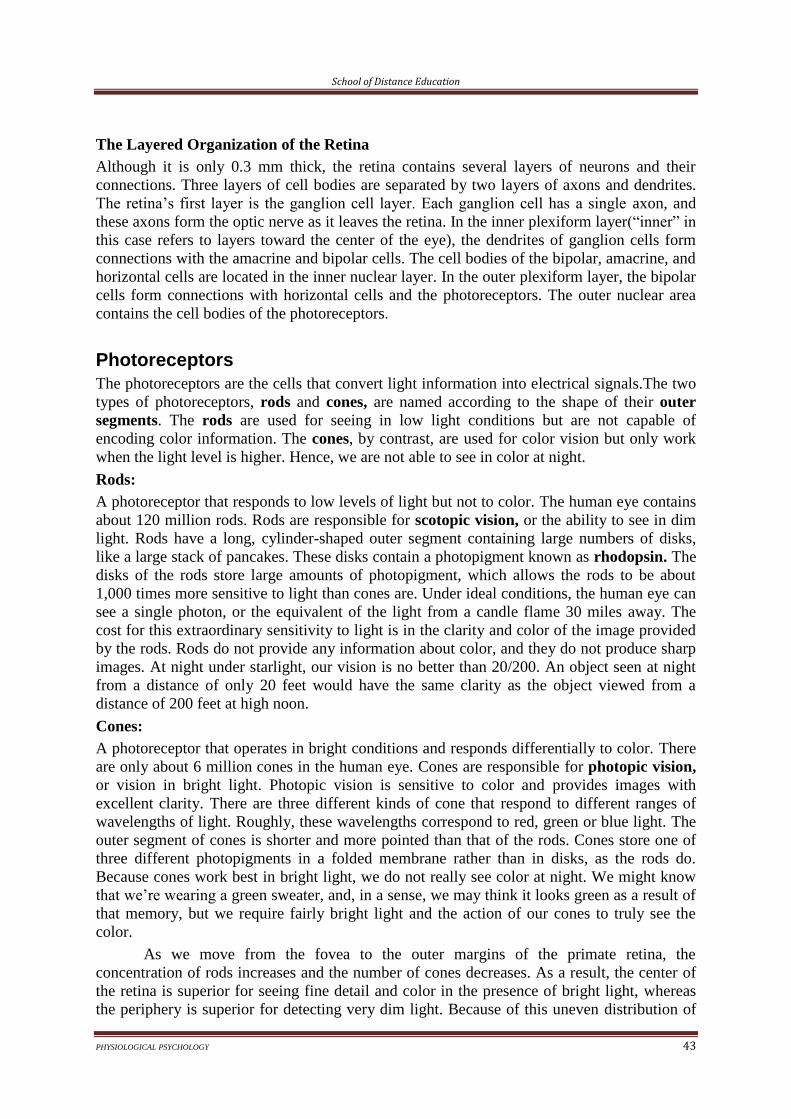
School of Distance Education
PHYSIOLOGICAL PSYCHOLOGY 43
The Layered Organization of the Retina
Although it is only 0.3 mm thick, the retina contains several layers of neurons and their
connections. Three layers of cell bodies are separated by two layers of axons and dendrites.
The retina’s first layer is the ganglion cell layer. Each ganglion cell has a single axon, and
these axons form the optic nerve as it leaves the retina. In the inner plexiform layer(“inner” in
this case refers to layers toward the center of the eye), the dendrites of ganglion cells form
connections with the amacrine and bipolar cells. The cell bodies of the bipolar, amacrine, and
horizontal cells are located in the inner nuclear layer. In the outer plexiform layer, the bipolar
cells form connections with horizontal cells and the photoreceptors. The outer nuclear area
contains the cell bodies of the photoreceptors.
Photoreceptors
The photoreceptors are the cells that convert light information into electrical signals.The two
types of photoreceptors, rods and cones, are named according to the shape of their outer
segments. The rods are used for seeing in low light conditions but are not capable of
encoding color information. The cones, by contrast, are used for color vision but only work
when the light level is higher. Hence, we are not able to see in color at night.
Rods:
A photoreceptor that responds to low levels of light but not to color. The human eye contains
about 120 million rods. Rods are responsible for scotopic vision, or the ability to see in dim
light. Rods have a long, cylinder-shaped outer segment containing large numbers of disks,
like a large stack of pancakes. These disks contain a photopigment known as rhodopsin. The
disks of the rods store large amounts of photopigment, which allows the rods to be about
1,000 times more sensitive to light than cones are. Under ideal conditions, the human eye can
see a single photon, or the equivalent of the light from a candle flame 30 miles away. The
cost for this extraordinary sensitivity to light is in the clarity and color of the image provided
by the rods. Rods do not provide any information about color, and they do not produce sharp
images. At night under starlight, our vision is no better than 20/200. An object seen at night
from a distance of only 20 feet would have the same clarity as the object viewed from a
distance of 200 feet at high noon.
Cones:
A photoreceptor that operates in bright conditions and responds differentially to color. There
are only about 6 million cones in the human eye. Cones are responsible for photopic vision,
or vision in bright light. Photopic vision is sensitive to color and provides images with
excellent clarity. There are three different kinds of cone that respond to different ranges of
wavelengths of light. Roughly, these wavelengths correspond to red, green or blue light. The
outer segment of cones is shorter and more pointed than that of the rods. Cones store one of
three different photopigments in a folded membrane rather than in disks, as the rods do.
Because cones work best in bright light, we do not really see color at night. We might know
that we’re wearing a green sweater, and, in a sense, we may think it looks green as a result of
that memory, but we require fairly bright light and the action of our cones to truly see the
color.
As we move from the fovea to the outer margins of the primate retina, the
concentration of rods increases and the number of cones decreases. As a result, the center of
the retina is superior for seeing fine detail and color in the presence of bright light, whereas
the periphery is superior for detecting very dim light. Because of this uneven distribution of

School of Distance Education
PHYSIOLOGICAL PSYCHOLOGY 44
rods and cones across the retina, we see better in dim light when we do not look directly at an
object.
Differences between Rods and Cones
Rods and cones respond to a wide range of wavelengths, but their photopigments each have
different peak sensitivities. There are three classes of cones. The so-called blue or short-
wavelength cones, which contain the photopigment cyanolabe, respond maximally to
wavelengths of 419 nm (violet). The green, or middle-wavelength cones, containing
chlorolabe, have peak responses to 531 nm (green), and the red or long-wavelength cones,
containing erythrolabe, peak at 558 nm (yellow). The rhodopsin in rods absorbs photons most
effectively at wavelengths of 502 nm (a bluish-green). Rods and cones need different
amounts of light to respond. Rhodopsin breaks apart when relatively little light has been
absorbed, which explains in part the rods’ great sensitivity to low levels of light. The cone
photopigments aremuch more resistant to breaking apart and will do so only in the presence
of bright light. This is one of the reasons that cones are active in daylight rather than during
the night.
Activity in the photoreceptors:
The way in which the rods and cones convert light into electrical signals is by way of
complex chemical processes. Rods contain a pigment called rhodopsin that becomes
bleached when exposed to light. This bleaching leads to a cascade of events that result in a
reduced release of glutamate (a neurotransmitter) by the rod cell. This is the signal for the
presence of light. Note that this signal is transmitted through inhibition rather than excitation.
The process in the cones is very similar, except that the opsins in each cone are different and
this probably accounts for the different wavelengths of light responded to. As well as being
activated by light, the rods and cones can activate each other. One way in which this can
occur is via activation of the horizontal cells. This system serves to reduce the responses of
neighboring cells exposed to diffuse stimulation and to heighten the responses of cells at
places where the light level changes.
Blind spot:
The ganglion cell axons band together to form the optic nerve (or optic tract), an axon bundle
that exits through the back of the eye. The point at which it leaves (which is also where the
major blood vessels leave) is called the blind spot. . As this region is densely packed with
neurons, there is no room for photoreceptors. Hence, this region of the retina is literally blind.
The brain interpolates the blind spot based on surrounding detail and information from the
other eye, so the blind spot is not normally perceived.
The first documented observation of the phenomenon was in the 1660s by Edme Mariotte in
France. At the time it was generally thought that the point at which the optic nerve entered
the eye should actually be the most sensitive portion of the retina; however, Mariotte's
discovery disproved this theory. The blind spot is located about 12–15° nasal and 1.5° below
the horizontal and is roughly 7.5° high and 5.5° wide.
Every person is therefore blind in part of each eye. You can demonstrate your own blind spot
by following the test.
Demonstration of the blind spot
R
L

School of Distance Education
PHYSIOLOGICAL PSYCHOLOGY 45
Instructions: Close one eye and focus the other on the appropriate letter (R for right or L for left). Place your
eye a distance from the screen approximately equal to 3× the distance between the R and the L. Move your eye
towards or away from the screen until you notice the other letter disappear. For example, close your right eye,
look at the "L" with your left eye, and the "R" will disappear.
Some people have a much larger blind spot because glaucoma has destroyed parts of the optic
nerve. Generally, they do not notice it any more than you notice your smaller one.
The Fovea
Perhaps the most important structural area on the retina is the fovea. The fovea is the area on
the retina where we have the best spatial and color vision. When we look at, or fixate, an
object in our visual field, we move our head and eyes such that the image of the object falls
on the fovea. As you are reading this text, you are moving your eyes to make the various
words fall on your fovea as you read them. To illustrate how drastically spatial acuity falls off
as the stimulus moves away from the fovea, try to read preceding text in this paragraph while
fixating on the period at the end of this sentence. It is probably difficult, if not impossible, to
read text that is only a few lines away from the point of fixation. The fovea covers an area
that subtends about two degrees of visual angle in the central field of vision. To visualize two
degrees of visual angle, a general rule is that the width of your thumbnail, held at arm’s
length, is approximately one degree of visual angle.
Visual pathways:
The visual pathway starts at the eye with the photoreceptors. These send an input to cells
called retinal ganglion cells in the retina, at the back of the eye. The bipolar and ganglion
cells provide a direct pathway for information from the photoreceptors to the brain that is
modified by input from the horizontal and amacrine cells. The horizontal and amacrine cells
integrate information across the surface of the retina. You can think of the
photoreceptorbipolar- ganglion connections as running perpendicular to the back of the eye,
whereas the horizontal and amacrine connections run parallel to the back of the eye. From
here, the pathway goes to the lateral geniculate nucleus and then to the visual cortex.

School of Distance Education
PHYSIOLOGICAL PSYCHOLOGY 46
The most important thing to note about the figure is the way in which light travels from the
two eyes to the visual cortex. If we trace light that comes in from the left of center (the left
visual field) when we are looking straight ahead, we see that the light hits the inside (nasal)
part of the left retina and the outside (lateral) part of the right retina. If we now follow the
nasal pathway, we see that it crosses the midline at the optic chiasma. From here, the
information travels to the right lateral geniculate nucleus and on to the right visual cortex.
The path of the nasal visual field is described as contralateral. If we now follow the lateral
pathway, we see that it does not cross the midline at the optic chiasma but continues on the
same side to the right lateral geniculate nucleus. From here, the information travels on to the
right visual cortex. This path is described as ipsilateral. The net result is that information
from the left of center when we are looking straight ahead (the left visual field), all goes only
to the right hemisphere, even though the light goes to both eyes. The same principle is true
for information from the right of center (the right visual field), only all of the information
goes to the left hemisphere. The exception to this is that information from objects we are
directly focusing on (called foveal vision) goes to both hemispheres. The following sessions
discuss these in detail.
Horizontal Cells
The horizontal cells are located in the inner nuclear layer. They receive input from the
photoreceptors and provide output to another type of cell in the inner nuclear layer, the
bipolar cell. The major task of horizontal cells is to integrate information from photoreceptors
that are close to one another. The spreading structure of the horizontal cell is well suited to
this task. Like the photoreceptors, horizontal cells communicate through the formation of
graded potentials rather than of action potentials. About half the bipolar cells in the human
retina are on-center (Cells that depolarize when light hits the center of their receptive field are
called on-center. Cells that hyperpolarize when light hits the center of their receptive field are
called off-center.), and the other half are off-center. This arrangement of the receptive fields

School of Distance Education
PHYSIOLOGICAL PSYCHOLOGY 47
is referred to as an antagonistic centersurround organization. The response of a bipolar
cell depends on the amount of light falling on its center relative to the amount of light falling
on its surround. It is called antagonistic because light falling on the center of the receptive
field always has the opposite effect on the cell’s activity from light falling on the surround.
Photoreceptors and horizontal cells serving the center (doughnut hole) and surround (the
doughnut) compete with each other to activate the bipolar cell in a process known as lateral
inhibition. Lateral means that the process occurs across the surface of the retina. In lateral
inhibition, active photoreceptors and horizontal cells limit the activity of neighboring, less
active cells. This produces a sharpening, or exaggeration, of the bipolar cells’ responses to
differences in light falling on adjacent areas. Through lateral inhibition, bipolar cells begin to
identify the boundaries of a visual stimulus by making comparisons between light levels
falling in adjacent areas of the retina. The message sent by the bipolar cells is “I see an edge
or boundary.”
Amacrine Cells
Amacrine cells, also located in the inner nuclear layer, form connections with bipolar cells,
ganglion cells, and other amacrine cells. In addition to integrating visual messages, amacrine
cells process movement.
Ganglion Cells
Ganglion cells receive input from bipolar and amacrine cells. Unlike the interneurons and
photoreceptors discussed so far, ganglion cells form conventional action potentials. However,
ganglion cells are never completely silent. The presence of light simply changes the ganglion
cells’ spontaneous rate of signaling. The axons of ganglion cells leave the eye and form the
optic nerve, which travels to higher levels of the brain. The human eye has approximately 1
million ganglion cells, yet they must accurately communicate input from about 126 million
photoreceptors. The ganglion cells accomplish this editing task through the organization of
their receptive fields.
Ganglion Receptive Fields
Ganglion receptive fields show the same antagonistic center-surround organization that we
observed in the receptive fields of bipolar cells. Ganglion cells replicate the information
passed to them by the bipolar cells. On-center bipolar cells connect to on-center ganglion
cells, whereas off-center bipolar cells connect to off-center ganglion cells. Ganglion cell
receptive fields vary in size. Receptive fields vary from 0.01 mm in diameter in the macula to
0.5 mm (50 times larger) in the periphery. Cells with small receptive fields respond best to
fine detail.
The Three Types of Ganglion Cells
About 90 percent of human ganglion cells are P cells (P stands for parvocellular, or small
cells), 5 percent are M cells (M stands for magnocellular, or big cells), and the remaining 5
percent are K cells (K stands for koniocellular). M cells are larger than P cells and have
thicker, faster axons. M cells have larger receptive fields than P cells. M cells respond to
smaller differences in light between the center and surround, whereas P cells require a greater
difference. This implies that M cells respond to subtle differences of contrast such as when
viewing gray letters on a black background. P cells respond to larger differences in contrast
such as when viewing black letters on a white background. M cells, but not P cells, respond
to stimuli that are turned on and off rapidly, such as the flicker of a monitor or television
screen.
A final difference is that P cells respond only to lights of a particular color, whereas M cells
respond to light regardless of its color. K cells share most of the characteristics of P cells. M

School of Distance Education
PHYSIOLOGICAL PSYCHOLOGY 48
cells are primarily responsible for providing information about large, lowcontrast, moving
objects, whereas P cells are responsible for information about smaller, high-contrast, colorful
objects. This distinction between magnocellular pathways and parvocellular pathways is
preserved up through some of the highest levels of cortical visual processing.
Optic Nerve Connections The ganglion cell axons exit the eye through the optic disk, forming an optic nerve leaving
each eye. The optic nerves preserve the organization of the retina. In other words, axons from
adjacent ganglion cells remain next to one another in the optic nerves. each human optic
nerve divides in half, with the outer half continuing to travel to the same side of the brain
(ipsilaterally) while the inner half crosses to the other side of the brain (contralaterally). This
partial crossing ensures that information from both eyes regarding the same part of the visual
field will be processed in the same places in the brain. If you hold your eyes steady by
looking at a focal point, information from the visual field to the left of the focal point will be
transmitted to the right hemisphere. Information from the visual field to the right of the focal
point will be transmitted to the left hemisphere. In humans, about 50 percent of the fibers
from each eye cross to the opposite hemisphere. In rabbits and other animals with eyes placed
on the side of the head, 100 percent of the fibers cross the midline to the opposite side.
Because each of a rabbit’s eyes sees a completely different part of the rabbit’s visual field,
there is no need for the rabbit to reorganize the input.
The optic nerves cross at the optic chiasm (named after its X shape, or chi in Greek). The
nerves continue past the optic chiasm as the optic tracts. Most of the axons in the optic tract
proceed to the thalamus, which in turn projects to the primary visual cortex of the brain.
However, a few axons leave the optic tract and synapse in the suprachiasmatic nucleus of the
hypothalamus, providing the light information used to regulate daily rhythms. About 10
percent of the axons in the optic tract project to the superior colliculus in the midbrain.
The Superior Colliculus
In many species, including frogs and fish, the superior colliculus is the primary brain
structure for processing visual information. Because humans have a cerebral cortex for this
purpose, they use the superior colliculus to guide movements of the eyes and head toward
newly detected objects in the visual field.
The Lateral Geniculate Nucleus of the Thalamus
Most of the remaining 90 percent of optic tract axons form synapses in the lateral geniculate
nucleus (LGN), located in the dorsal thalamus. the LGN is a layered structure that is bent in
the middle. In primates, including humans, the LGN has about the same area as a credit card,
but is about three times thicker. The LGN features six distinct stacked layers, numbered from
ventral to dorsal. Layers 1 and 2 (the most ventral layers) contain larger neurons than the
other four layers. These magnocellular layers receive input from the M cells in the retina.
The other four are referred to as parvocellular layers, which receive input from the P cells.
Between each of the six layers are very small neurons making up the koniocellular layers,
which receive input from the K cells. The LGN keeps information from the two eyes
completely separate.
Neurons in the LGN show the same antagonistic center-surround organization of receptive
fields that we observed in the retinal bipolar and ganglion cells. In LGN neurons, however,
the lateral inhibition between center and surround is much stronger than we observed among
retinal cells. This greater inhibition causes cells in the LGN to amplify or boost the contrast
between areas of light and dark. Surprisingly, the retina is not the major source of input to the
LGN. About 80 percent of the input to the LGN comes from the primary visual cortex,

School of Distance Education
PHYSIOLOGICAL PSYCHOLOGY 49
located in the occipital lobe. The function of this input remains unclear. The LGN also
receives input from the brainstem reticular formation. This input allows an animal to modify
the flow of information to the cortex from the LGN based on its level of arousal and
alertness. The exact role of the LGN in visual processing is not well understood. The LGN
might modify input to the cortex based on arousal. The LGN might also organize or sort
information prior to sending it to the cortex.
Primary visual cortex Primary visual cortex is often referred to as striate cortex, due to its striped appearance.
Striate cortex, located in the occipital lobe, contains approximately 250 million neurons as
opposed to the 1 million neurons found in the LGN. The cortex in this area ranges from 1.5 to
2 mm in thickness, or about the height of the letter m on this page. Like other areas of cortex,
the striate cortex has six distinct layers. Compared with other areas of cortex, striate cortex is
relatively thicker in layers II and IV, which receive most of the input from other parts of the
brain. Layer IV receives input from the LGN. Striate cortex is thinner in layers III, V, and VI,
which contain output neurons that communicate with other parts of the brain.
Visual Analysis Beyond the Striate Cortex
The striate cortex begins, but by no means finishes, the task of processing visual input. At
least a dozen additional areas of the human cerebral cortex participate in visual processing.
Because these areas are not included in the striate cortex, they are often referred to as
extrastriate areas. These areas are also referred to as secondary visual cortex.
Next to the striate cortex is an area known as V2. If you stain V2 for cytochrome oxidase, a
pattern of stripes emerges. Alternating thick and thin stripes are separated by interstripe
regions. The thick stripes form part of the magnocellular pathway and project to a visual
pathway known as the dorsal stream. The dorsal stream travels from the primary visual
cortex toward the parietal lobe and then proceeds to the medial temporal lobe. The dorsal
stream, commonly referred to as the “where” pathway, specializes in the analysis of
movement, object locations, and the coordination of eyes and arms in grasping or reaching.
The thin stripes and interstripe regions of V2 project to another visual region known as V4,
continuing the parvocellular pathway. Area V4 participates in a second major pathway, the
ventral stream, which proceeds from the primary visual cortex to the inferior temporal lobe.
This second pathway, commonly referred to as the “what” pathway, specializes in object
recognition.
The Dorsal Stream Area MT
MT stands for the medial temporal lobe, appears to play an important role in the processing
of motion. Input to Area MT is primarily from the magnocellular pathways. Recall that the
cells in this pathway have large receptive fields and often show responses to rapidly changing
light conditions and direction of movement. Most of the cells in Area MT respond to
movement in a particular direction. Unlike previous motion detectors, however, Area MT
cells respond to movement across large regions of the visual field. Further processing of
motion occurs adjacent to Area MT in Area MST, which stands for the medial superior
temporal lobe. Tanaka and Saito (1989) found that Area MST neurons respond to stimulus
rotation, stimulus expansion, and stimulus contraction. These are very large, global types of
movement that do not produce consistent responses in other areas. Area MST helps us use
vision to guide our movements. Melvyn Goodale and his colleagues suggested that the dorsal
stream would be more accurately characterized as a “how” stream than as a “where” stream.
According to this view, not only does the dorsal stream tell us an object’s location, but it also
provides information about how to interact with an object. Patients with damage to the dorsal

School of Distance Education
PHYSIOLOGICAL PSYCHOLOGY 50
stream can judge the orientation of an object, such as lining up a card with a slot, but are
unable to combine orientation and action to push the card through the slot.
The Ventral Stream
As the information from the primary cortex and Area V2 travels ventrally toward the
temporal lobe, we come to Area V4. The cells in this area have large receptive fields and
respond to both shape and color. Cells in Area V4 project to the inferior temporal lobe, or
Area IT. Cells in Area IT respond to many shapes and colors. In humans and monkeys, a
small section of Area IT known as the fusiform face area (FFA).
Visual acuity Visual acuity (VA) is acuteness or clearness of vision. It depends on optical and neural
factors, i.e., (i) the sharpness of the retinal focus within the eye, (ii) the intactness and
functioning of the retina, and (iii) the sensitivity of the interpretative faculty of the brain. Snellen chart is frequently used for visual acuity testing.
A common cause of low visual acuity is refractive error (ametropia), or errors in how the
light is refracted in the eyeball. Causes of refractive errors include aberrations in the shape of
the eyeball, the shape of the cornea, and reduced flexibility of the lens. In the case
of pseudomyopia, the aberrations are caused by muscle spasms. Too high or too low
refractive error (in relation to the length of the eyeball) is the cause of nearsightedness
(myopia) or farsightedness (hyperopia) (normal refractive status is referred to
as emmetropia). Other optical causes are astigmatism or more complex corneal irregularities.
These anomalies can mostly be corrected by optical means (such as eyeglasses, contact
lenses, laser surgery, etc.).
Neural factors that limit acuity are located in the retina or the brain (or the pathway leading
there). Examples for the first are a detached retina and macular degeneration, to name just
two. A common impairment amblyopia caused by incorrect nerve pathway function
connecting eye with brain is involved. In some cases, low visual acuity is caused by brain
damage, such as from traumatic brain injury or stroke. When optical factors are corrected for,
acuity can be considered a measure of neural well-functioning.
Visual acuity is typically measured while fixating, i.e. as a measure of central (or foveal)
vision, for the reason that it is highest there. However, acuity in peripheral vision can be of
equal (or sometimes higher) importance in everyday life. Acuity declines towards the
periphery in an inverse-linear (i.e. hyperbolic) fashion.
Normal visual acuity is commonly referred to as 20/20 vision (even though acuity in
normally sighted people is generally higher), the metric equivalent of which is 6/6 vision. At
20 feet or 6 meters, a human eye with nominal performance is able to separate contours that
are approximately 1.75 mm apart. A vision of 20/40 corresponds to lower
than nominal performance, a vision of 20/10 to better performance.
20/20 is normal (daylight) vision. In low light (i.e., scotopic) vision, spatial resolution is
much lower. This is due to spatial summation of rods, i.e. a number of rods merge into a
bipolar cell, in turn connecting to a ganglion cell, and the resulting unit for resolution is large
(and acuity small). Visual acuity is much better in bright light than dim light, the former
reaching 2 with a bright center and surrounding, the latter perhaps being 0.4 (25 arc minutes).
In this case, the stimulus is 1.7 inches (4.3 cm) seen at a distance of 20 feet (6.1 m).
The maximum angular resolution of the human eye at a distance of 1 km is typically 30 to
60 cm. This gives an angular resolution of between 0.02 to 0.03 degrees, which is roughly 1.2
- 1.8 arc minutes per line pair, which implies a pixel spacing of 0.6-0.9 arc minutes. 20/20

School of Distance Education
PHYSIOLOGICAL PSYCHOLOGY 51
vision is defined as the ability to resolve two points of light separated by a visual angle of one
minute of arc, or about 320-286 pixels per inch for a display on a device held 10 to 12 inches
from the eye (since by the Nyquist theorem two points are needed to resolve a space).
Thus, visual acuity, or resolving power (in daylight, central vision), is the property of cones.
To resolve detail, the eye's optical system has to project a focused image on the fovea, a
region inside themacula having the highest density of cone photoreceptor cells (the only kind
of photoreceptors existing on the fovea), thus having the highest resolution and best color
vision. Acuity and color vision, despite being mediated by the same cells, are different
physiologic functions that do not interrelate except by position. Acuity and color vision can
be affected independently.
As in a photographic lens, visual acuity is affected by the size of the pupil. Optical
aberrations of the eye that decrease visual acuity are at a maximum when the pupil is largest
(about 8 mm), which occurs in low-light conditions. When the pupil is small (1–2 mm),
image sharpness may be limited by diffraction of light by the pupil (see diffraction limit).
Between these extremes is the pupil diameter that is generally best for visual acuity in
normal, healthy eyes; this tends to be around 3 or 4 mm.
If the optics of the eye were otherwise perfect, theoretically, acuity would be limited by pupil
diffraction, which would be a diffraction-limited acuity of 0.4 minutes of arc (minarc) or 20/8
acuity. The smallest cone cells in the fovea have sizes corresponding to 0.4 minarc of the
visual field, which also places a lower limit on acuity. The optimal acuity of 0.4 minarc or
20/8 can be demonstrated using a laser interferometer that bypasses any defects in the eye's
optics and projects a pattern of dark and light bands directly on the retina. Laser
interferometers are now used routinely in patients with optical problems, such as cataracts, to
assess the health of the retina before subjecting them to surgery.
Any relatively sudden decrease in visual acuity is always a cause for concern. Common
causes of decreases in visual acuity are cataracts(Clouding of the lens) and scarredcorneas,
which affect the optical path, diseases that affect the retina, such as macular
degeneration and diabetes, diseases affecting the optic pathway to the brain such
as tumors and multiple sclerosis, and diseases affecting the visual cortex such as tumors
and strokes.
Color Vision Color vision is the ability of an organism or machine to distinguish objects based on
the wavelengths (or frequencies) of the light they reflect, emit, or transmit. Colors can be
measured and quantified in various ways; indeed, a person's perception of colors is a
subjective process whereby the brain responds to the stimuli that are produced when
incoming light reacts with the several types of cone cells in the eye. In essence, different
people see the same illuminated object or light source in different ways. In the human visual
system, the shortest visible wavelengths, about 350 nm, are perceived as violet; progressively
longer wavelengths are perceived as blue, green, yellow, orange, and red, near 700 nm. The
“visible” wavelengths vary depending on a species’ receptors. For example, birds’ receptors
enable them to see shorter wavelengths than humans can. That is, wavelengths that we
describe as “ultraviolet” are simply violet to birds. (Of course, we don’t know what their
experience looks like.) In general, small songbirds see further into the ultraviolet range than
do predatory birds such as hawks and falcons. Many songbird species have taken advantage
of that tendency by evolving feathers that strongly reflect very short wavelengths, which can
be seen more easily by their own species than by predators.
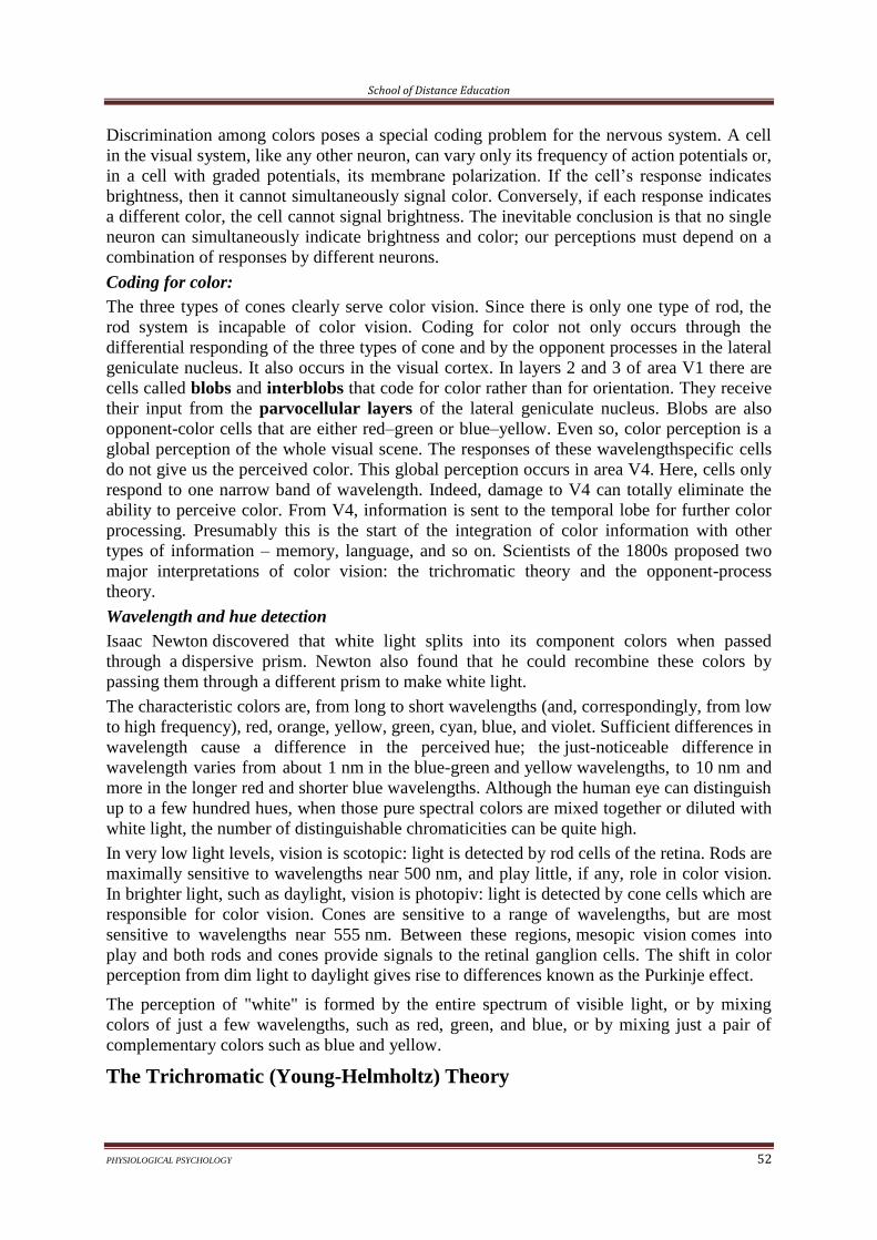
School of Distance Education
PHYSIOLOGICAL PSYCHOLOGY 52
Discrimination among colors poses a special coding problem for the nervous system. A cell
in the visual system, like any other neuron, can vary only its frequency of action potentials or,
in a cell with graded potentials, its membrane polarization. If the cell’s response indicates
brightness, then it cannot simultaneously signal color. Conversely, if each response indicates
a different color, the cell cannot signal brightness. The inevitable conclusion is that no single
neuron can simultaneously indicate brightness and color; our perceptions must depend on a
combination of responses by different neurons.
Coding for color:
The three types of cones clearly serve color vision. Since there is only one type of rod, the
rod system is incapable of color vision. Coding for color not only occurs through the
differential responding of the three types of cone and by the opponent processes in the lateral
geniculate nucleus. It also occurs in the visual cortex. In layers 2 and 3 of area V1 there are
cells called blobs and interblobs that code for color rather than for orientation. They receive
their input from the parvocellular layers of the lateral geniculate nucleus. Blobs are also
opponent-color cells that are either red–green or blue–yellow. Even so, color perception is a
global perception of the whole visual scene. The responses of these wavelengthspecific cells
do not give us the perceived color. This global perception occurs in area V4. Here, cells only
respond to one narrow band of wavelength. Indeed, damage to V4 can totally eliminate the
ability to perceive color. From V4, information is sent to the temporal lobe for further color
processing. Presumably this is the start of the integration of color information with other
types of information – memory, language, and so on. Scientists of the 1800s proposed two
major interpretations of color vision: the trichromatic theory and the opponent-process
theory.
Wavelength and hue detection
Isaac Newton discovered that white light splits into its component colors when passed
through a dispersive prism. Newton also found that he could recombine these colors by
passing them through a different prism to make white light.
The characteristic colors are, from long to short wavelengths (and, correspondingly, from low
to high frequency), red, orange, yellow, green, cyan, blue, and violet. Sufficient differences in
wavelength cause a difference in the perceived hue; the just-noticeable difference in
wavelength varies from about 1 nm in the blue-green and yellow wavelengths, to 10 nm and
more in the longer red and shorter blue wavelengths. Although the human eye can distinguish
up to a few hundred hues, when those pure spectral colors are mixed together or diluted with
white light, the number of distinguishable chromaticities can be quite high.
In very low light levels, vision is scotopic: light is detected by rod cells of the retina. Rods are
maximally sensitive to wavelengths near 500 nm, and play little, if any, role in color vision.
In brighter light, such as daylight, vision is photopiv: light is detected by cone cells which are
responsible for color vision. Cones are sensitive to a range of wavelengths, but are most
sensitive to wavelengths near 555 nm. Between these regions, mesopic vision comes into
play and both rods and cones provide signals to the retinal ganglion cells. The shift in color
perception from dim light to daylight gives rise to differences known as the Purkinje effect.
The perception of "white" is formed by the entire spectrum of visible light, or by mixing
colors of just a few wavelengths, such as red, green, and blue, or by mixing just a pair of
complementary colors such as blue and yellow.
The Trichromatic (Young-Helmholtz) Theory

School of Distance Education
PHYSIOLOGICAL PSYCHOLOGY 53
People can distinguish red, green, yellow, blue, orange, pink, purple, greenish-blue, and so
forth. If we don’t have a separate receptor for every distinguishable color, how many types do
we have? The first person to approach this question fruitfully was an amazingly productive
man named Thomas Young (1773–1829). Young was the first to decipher the Rosetta stone,
although his version was incomplete. He also founded the modern wave theory of light,
defined energy in its modern form, founded the calculation of annuities, introduced the
coefficient of elasticity, discovered much about the anatomy of the eye, and made major
contributions to many other fields. Previous scientists thought they could explain color by
understanding the physics of light. Young was among the first to recognize that color
required a biological explanation. He proposed that we perceive color by comparing the
responses across a few types of receptors, each of which was sensitive to a different range of
wavelengths.
This theory, later modified by Hermann von Helmholtz, is now known as the trichromatic
theory of color vision, or the Young-Helmholtz theory. According to this theory, we
perceive color through the relative rates of response by three kinds of cones, each kind
maximally sensitive to a different set of wavelengths (Trichromatic means “three colors.”).
For deciding the number three, he collected psychophysical observations, reports by
observers concerning their perceptions of various stimuli. He found that people could match
any color by mixing appropriate amounts of just three wavelengths. Therefore, he concluded
that three kinds of receptors—we now call them cones—are sufficient to account for human
color vision.
According to the trichromatic theory, we discriminate among wavelengths by the ratio of
activity across the three types of cones. For example, light at 550 nm excites the medium-
wavelength and long wavelength receptors about equally and the short-wavelength receptor
almost not at all. This ratio of responses among the three cones determines a perception of
yellow. More intense light increases the activity of all three cones without much change in
their ratio of responses. As a result, the color appears brighter but still yellow. When all three
types of cones are equally active, we see white or gray. Note that the response of any one
cone is ambiguous. For example, a low response rate by a middle wavelength cone might
indicate low-intensity 540-nm light or brighter 500-nm light or still brighter 460- nm light. A
high response rate could indicate either bright light at 540 nm or bright white light, which
includes 540 nm. The nervous system can determine the color and brightness of the light only
by comparing the responses of the three types of cones.
Given the desirability of seeing all colors in all locations, we might suppose that the three
kinds of cones would be equally abundant and evenly distributed. In fact, they are not. Long-
and medium-wavelength cones are far more abundant than short-wavelength (blue) cones,
and consequently, it is easier to see tiny red, yellow, or green dots than blue dots.
Furthermore, the three kinds of cones are distributed randomly within the retina. The
trichromatic (three-receptor) nature of color vision was not in doubt, but the idea of three
images being transmitted to the brain is both inefficient and fails to explain several visually
observed phenomena.
The Opponent-Process Theory
The trichromatic theory correctly predicted the discovery of three kinds of cones, but it is
incomplete as a theory of color vision. Ewald Hering, a 19th-century physiologist, proposed
the opponent-process theory: We perceive color in terms of paired opposites: red versus

School of Distance Education
PHYSIOLOGICAL PSYCHOLOGY 54
green, yellow versus blue, and white versus black. That is, there is no such thing as reddish
green, greenish red, or yellowish blue. The brain has some mechanism that perceives color on
a continuum from red to green and another from yellow to blue. Hering also observed that
there was a distinct pattern to the color of the after images we see. For example if one looks
at a unique red patch for about a minute and then switches the gaze to a homogeneous white
area they will see a greenish patch in the white area.
Hering hypothesized that trichromatic signals from the cones fed into subsequent neural
stages and exhibited two major opponent classes of processing. 1. Spectrally opponent
processes which were red vs. green and yellow vs. blue. 2. Spectrally non-opponent processes
which was black vs. white. This opponent process model lay relatively dormant for many
years until a pair of visual scientists working at Eastman Kodak at the time, conceived of a
method for quantitatively measuring the opponent processes responses. Leo Hurvich and
Dorothea Jameson invented the hue cancellation method to psychophysically evaluate the
opponent processing nature of color vision.
Due in large measure to the efforts of Hurvich and Jameson the opponent processes theory
attained a central position shared with the the trichromatic theory. One very fortuitous
scientific event to that also took place in the 1950s was the discovery of electrophysiological
responses that emulated opponent processing. Consequently, with the quantitative data
provided by the psychophysics and direct neurophysiological responses provided by
electrophysiology opponent processing is no longer questioned
The Retinex Theory
The trichromatic theory and the opponent-process theory have limitations, especially in
explaining color constancy. Color constancy is the ability to recognize the color of an object
despite changes in lighting. If you put on green-tinted glasses or replace your white light bulb
with a green one, you will notice the tint, but you still identify bananas as yellow, paper as
white, walls as brown (or whatever), and so forth. You do so by comparing the color of one
object with. The color of another, in effect subtracting a fixed amount of green from each.
Color constancy requires you to compare a variety of objects.
To account for color and brightness constancy, Edwin Land proposed the retinex theory (a
combination of the words retina and cortex): The cortex compares information from various
parts of the retina to determine the brightness and color for each area. For example, if the
cortex notes a constant amount of green throughout a scene, it subtracts some green from
each object to determine its true color.
Dale Purves and colleagues have expressed a similar idea in more general terms: Whenever
we see anything, we make an inference or construction. For example, when we look at the
objects, we ask our self, “On occasions when I have seen something that looked like this,
what did it turn out to be?” We go through the same process for perceiving shapes, motion, or
anything else: and calculate what objects probably produced the pattern of stimulation we just
had. That is, visual perception requires a kind of reasoning process, not just retinal
stimulation.
Colorblindness
Occasional errors occur in the chromosomes that carry the genes that encode the cone
photopigments. As a result, individuals with these genes show several kinds of atypical
responses to color, known as colorblindness.
Dichromacy (having two cone photopigments) is the most common type of abnormality and
results from a missing or abnormal cone pigment. Because genes for the red and green
photopigments appear on the X chromosome, this type of dichromacy is sex-linked. Men are

School of Distance Education
PHYSIOLOGICAL PSYCHOLOGY 55
about ten times more likely to be colorblind than women. There are very rare cases in which
the blue photopigment is missing. The gene for the blue photopigment is located on
Chromosome 7, so these cases are not sex-linked and appear equally in males and females.
Rarer still are cases of monochromacy. This condition occurs when a person has only one
type of cone or a complete absence of cones. In either case, the person can’t see color at all. Some individuals have three cone pigments, but their peak response occurs at slightly
different wavelengths than is typical. This leads to a condition known as anomalous
trichromacy. These individuals match colors in a slightly different way than most people,
but they might not even know that they are unusual. As many as 50 percent of all women
may be tetrachromats, or people with four different color pigments. These individuals match
colors in a manner that would be predicted by their having four color pigments rather than
three.
Review Questions
1. -----------determines the perceived color of objects
a) Frequency
b) Wavelength
c) Amplitude
d) Refraction
2. ---------fills the space between the lens and the retina of the eyeball
a) Vitreous humour
b) Aqueous humour
c) Ciliary body
d) Optic nerve
3. ----- helps to see in dim light
a) Cons
b) Rods
c) Brightness
d) Wavelength
4. Fovea contains ------
a) More rods
b) More cones
c) No cones
d) Less cones
5. Retinex theory is proposed by
a) Thomas Young
b) Edwin Land
c) Ewald Hering
d) Hurvich and Jameson
6. Briefly explain the visual pathway.
7. Differentiate the roles of rods and cons.
8. Give a short note on blind spot
9. Write two theories of color vision.
Bibliography
Freberg, L. (2010). Discovering Biological Psychology (2nd
edition). USA: Wadsworth.
Kalat, J.W. (2007). Biological Psychology. Canada: Thomson Wadsworth.
Rogers, K. (2011). The Brain and the Nervous System. NY: Britannica Educational
Publishing.

School of Distance Education
PHYSIOLOGICAL PSYCHOLOGY 56
Silber, K. (2005). The physiological basis of behavior: Neural and hormonal processes.
London: Routledge.
Wagner, H., & Silber, K. (2004). Instant notes: Physiological Psychology. London: BIOS
Scientific Publishers.