Embed Size (px)
Citation preview

A STUDY TO DETERMINE THE ASSOCIATION BETWEEN PATTERN OF
CALCIFICATION IN CT SCAN AND STAGING OF RETINOBLASTOMA IN HOSPITAL
UNIVERSITY SCIENCE MALAYSIA (HUSM).
Dr Rozita Goon
MMed Radiology
Department of Radiology
School of Medical Sciences, University Sains Malaysia
Health Campus, 16150 Kelantan, Malaysia
Introduction: Retinoblastoma is one of the most common intraocular malignancy in
children under 15 years old. About 80% of cases occurs in patients under 3 years old. The
incidence varies form countries to countries. In United States of America, it occurs in 1 of every
15000 live birth. Most incidence of retinoblastoma is unilateral, bilateral involvement is seen in
approximately 30% of cases and it is detected earlier. Physical and radiological examination
helps in diagnosing retinoblastoma. CT scan of the orbit is one of the important tools in
diagnosing retinoblastoma. Intraocular calcification can be detected in about 80% of the CT scan
of orbit. Many researches have been performed since 1980s to determine association between
intraocular calcification in retinoblastoma with prognosis and size of the tumour.

Objectives: To determine frequency of intraocular calcification in retinoblastoma and
association between the pattern of calcification with histopathological examination, clinical data
and staging.
Patients and methods: This was a retrospective descriptive study. All patients had
undergone pretreatment CT scan of the orbit and enucleation. Characteristic of calcification on
CT scan images which were presence of calcification, size, site and Hounsfield Unit of
calcification were recorded. CT scan images were reviewed via GE Centricity PACS- IW
(Intregrated web) version 3.71. Histological findings which were presence of calcification, size,
site of tumour and optic nerve involvement were recorded. History of presenting illness, family
history, demographic data and clinical classification were sought form medical record and
recorded. Association between characteristic of calcification and histological findings, clinical
data and staging were determined.
Results: There was 95% intraocular CT calcification seen in retinoblastoma in this study.
There was significant association between presence of calcification on CT and presence of
calcification on HPE(p=0.042). There was also significant association between presence of HPE
calcification and CT calcification site(p=0.016). Significant association noted between CT
calcification size and strabismus(p=0.035). However. there was no significant association
between the patterns of calcification on CT with staging of retinoblastoma.
Conclusion: Although presence of calcification on CT scan was used as a criteria to
diagnosed retinoblastoma, there is no significant association between patterns of the calcification
with staging in retinoblastoma.
Prof Madya Noreen Nurfaraheen Lee Abdullah: Supervisor

1
1. INTRODUCTION
Retinoblastoma is one of the most common intraocular malignancy in children and
one of commonest in infancy. It accounts for more than 80% of all primary intraocular
tumour in children up to 15 years old (Mahoney et al. 1990). The incidence varies from
countries to countries. In United State of America, it accounts in 1of every 15000 live
birth. 200 cases are diagnosed per year in the United States. The average age at diagnosis is
18 months with 80% of cases occurring before 3 years old. Approximately 30% are
bilateral and are typically diagnosed earlier than unilateral cases ( S.C.Kaste et al. 2000).
Main clinical presentation is leukocoria which can be seen in almost 60% of cases.
Other sign of retinoblastoma is strabismus or squint which is present in 20% of cases. It
has a good prognosis and a fair chance of preservation of the vision ( Balmer and Munier,
2002). Other signs include atypical features that account of 20% of cases including
inflammation such as inflammation of the eyes, proptosis and eye discharge. These are late
sign and carry poorer prognosis.
Direct funduscopy is the first clinical examination done by ophthalmologist. It is
stated that direct funduscopy with proper pupil dilatation can improved ability to observe
leukocoria resulting from small retinoblastoma lesions in the posterior pole and larger
lesions located outside of the posterior pole ( John C. Canzano and James T. Handa,1999).
Orbital sonography help in delinating good intraocular detail and provide sequential study.
Retinoblastoma appears as an echogenic soft-tissue mass with various degrees of
calcification. CT detects intraocular, extraocular, and intracranial disease extension. It

2
provide great delineation of bony abnormalities and tumoural calcifications. MRI helps
the characterization of complex structure depiction of ocular, orbital, and intracranial
abnormality.
It is important to detect the tumour early since it can be treated effectively with a
survival rate of 90% with current treatment ( Balmer and Munier, 2002). Treatment
depends on the ocular status of the disease (Constantino Sábado Álvareza et al. 2005).
Enucleation is still a mainstay treatment for large tumor that requires an aggressive
approach especially in unilateral retinoblastoma where the vision of the eye is severed.
Other treatment includes, bachytherapy, chemotheraphy and external beam therapy.
The Reese – Ellsworth classification has been since the 1960s the most popular
grouping system. It was devised to evaluate the response to external beam radiotherapy.
New classifications are being developed as is the case of the International Classification of
Retinoblastoma ( Salvadore Alvarez et al. 2005) which is more concentrated on
chemotherapy regime.
For our study, we would like to see association between calcification patterns with
the histopathological findings and clinical data. Tumour calcification was seen much more
common in tumour confined to the globe and therefore may be a favourable diagnosis for
retinoblastoma (Alan Danziger et al. 1979). Degree of calcification appeared to depend
upon tumour size. Only small tumours were devoid of calcification. This correlate with the
theory that DNA released from necrotic retinoblastoma tumour cell has propensity to form
a DNA calcium complex (Mafee et al. 2005). Hence, it is important to study the depicted
pattern of calcification in relation to the histopathology to determine that some pattern of
calcification represent the degree of malignancy in retinoblastoma.

3
SECTION TWO: LITERATURE REVIEW:
2.1 Epidemiology of retinoblastoma
Retinoblastoma is the most common malignancy of the retina in childhood. Although it is a
rare disease, it accounts for 80% of all primary ocular malignancies in children up to 15
years of age (Mahoney et al. 1990) and 5% of malignant solid tumors in children (Shah et
al. 2000). It is estimated that 1 case of retinoblastoma per 18,000-30,000 live births,
depending on the country. In developed countries, retinoblastoma are detected early. This
may be due to the good screening system in patient with family history and well educated
parents. Where as in developing and underdeveloped countries, retinoblastoma is often
detected late after is has invaded the orbit or brain (Bunin and Orjuela, 2007).
For eg, in England about 1/30000 live birth, 1/18000 live birth in United State and
1/16000 in Finland. In Riyadh, Saudi Arabia the incidence reported as high as 1/ 11580 (H.
Olivecrona et al. 1994).
Retinoblastoma may develop in utero and up to 4 years of age. First signs appear at
a median age of 7 months in bilateral cases and 24 months in unilateral cases (Aubin et al.
2002)
2.2 HISTORY OF RETINOBLASTOMA:
It is first described by Dr Peter Pawius Dutch who is an anatomist and botanist, in an
autopsy report in 1597 in Amsterdam who made the discovery of tumour resembling
retinoblastoma. In 1836, Langenbech, Robin, and Nystin of Paris confirmed by
pathological studies that the tumor microscopically arising from the retina. Retinoblastoma
4
was first described as a specific entity by James Wardrop in 1809, with enucleation as his
suggested treatment. Histologic studies including those of Flexner and Verhoeff and
subsequent electron microscopy have given insights into its pathogenesis. Wintersteiner
reported the resemblance of the tumor rosettes to photoreceptors of the adult retina. (Albert
DM, 1987). In the 1920s, Verhoeff believed that the tumor arose from embryonic retinal
cells and proposed the name retinoblastoma.
2.3 SECONDARY PRIMARY MALIGNANCIES
Persons with bilateral retinoblastoma are at higher risk of developing second
primary malignancies throughout life. The cumulative incidence of second cancer 50 years
after diagnosis is 51%. The mean latency between retinoblastoma and second malignancy
is approximately 13 years. External-beam radiation increases the risk of second
malignancy and shows a radiation dose-response relationship for all sarcomas. Most
second malignancies are high-grade tumors and having poor prognosis. Osteogenic
sarcoma, the most common second tumor, often arises due to radiation. Other reported
cases of malignancies include neuroblastoma, chondrosarcoma, rhabdomyosarcoma,
glioma, leukemia, sebaceous carcinoma, squamous cell carcinoma, and cutaneous
melanoma ( Smith and Donaldson, 1991 ).
2.4 RISK FACTORS
Genetic factor has been found as one of the precursor for retinoblastoma. Alfred Knudson
formed his famous ‘two hit’ hypothesis of tumor suppression. He studied inherited and

5
noninherited retinoblastoma cases. He proposed that inherited children with
retinoblastoma must be borned with mutation or one hit, already in the germline and later
on only required a second tumour activating hit to formally formed retinoblastoma. In
patients with nonheritable form of retinoblastoma, the first hit comes later on after the
patient was borned in already differentiated retinal cells. Once hit cells accumulates a
second hit later in childhood and explain why the presence of retinoblastoma later on in
children with non heritable form of retinoblastoma (P.H. Fitzegerald et al. 1983). In
inheritable cases, mutation of the tumor-suppressor gene RB1 responsible for
retinoblastoma also increases the risk of neuroectodermal lesions and other primary
nonocular tumors.
Patients who inherited an RB1 mutation would develop tumors earlier, and they
would often develop more than one tumor. In contrast, individuals who did not inherit a
mutation would almost always be affected by a single tumor. This statement, which
Knudson called the two-mutation hypothesis, is now known as the two-hit hypothesis.
Based on the predicted mutation rate, Knudson expected that many individuals in the
general population would acquire a single somatic mutation in theRB1 gene over their
lifetime, and that the retinas of most people would therefore likely contain small groups of
retinoblasts that had received one "hit" in the RB1 gene. In order to become cancerous,
each retinoblast with one mutant copy of the RB1 gene would need to acquire a mutation in
the remainingwild-type copy of the gene. Most individuals who had one hit did not
develop retinoblastoma, however, because most of their mutated cells had already
differentiated and quit dividing before they could receive a second hit ( Heidi Chial,2008).
6
Other nonheritable factors that are concern with risks of retinoblastoma are poor
living conditions, maternal diet deficiency, and human papilloma virus infections. Some
regional variations may be linked to genetic environmental susceptibility or specific
population behavioral patterns. In vitro fertilization has been reported as an increased risk
of retinoblastoma (Moll et al. 2003). There is no reported predilection concerning sex or
race.
2.5 PATHOLOGY
Most retinoblastomas are composed of undifferentiated cells with hyperchromatic nuclei
and very scant cytoplasm. The mitotic rate is high and tumors often outgrow their blood
supply, resulting in patches of necrosis 100 to 200 microns from nutrient vessels. The most
important prognostic finding is the status of the optic nerve. The depth and extent of tumor
invasion of the nerve strongly correlate with survival. Tumor present at the surgical margin
of the optic nerve or tumor infiltration of the subarachnoid space has a poor prognosis.
Focal signs of retinal differentiation (eg, tumor rosettes and fleurettes) are common but
have little prognostic importance.
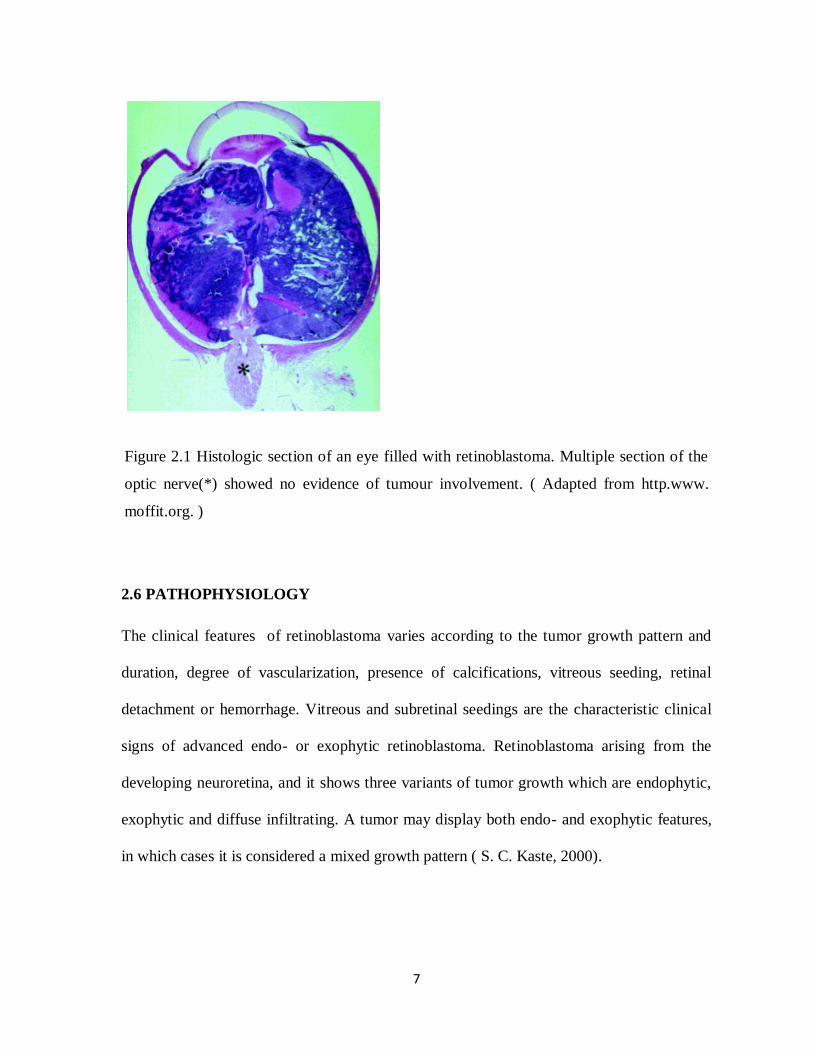
7
Figure 2.1 Histologic section of an eye filled with retinoblastoma. Multiple section of the
optic nerve(*) showed no evidence of tumour involvement. ( Adapted from http.www.
moffit.org. )
2.6 PATHOPHYSIOLOGY
The clinical features of retinoblastoma varies according to the tumor growth pattern and
duration, degree of vascularization, presence of calcifications, vitreous seeding, retinal
detachment or hemorrhage. Vitreous and subretinal seedings are the characteristic clinical
signs of advanced endo- or exophytic retinoblastoma. Retinoblastoma arising from the
developing neuroretina, and it shows three variants of tumor growth which are endophytic,
exophytic and diffuse infiltrating. A tumor may display both endo- and exophytic features,
in which cases it is considered a mixed growth pattern ( S. C. Kaste, 2000).

8
2.6.1Endophytic
It occurs when the tumour growth breaks through the internal limiting membrane and a
whitish mass, either showing an absence of tumor vessels or showing minute but irregular
vessels, becomes visible. This growth pattern is characteristic of vitreous seeding in which
tissue fragments become separated from the main tumor. Endophytic form manifests as
one or more isolated or coalesced tumors of variable size, round or oval-shaped, yellowish-
white (calcifications) or pinkish (vascularized) in color, with a marked tendency to vitreous
seeding. In some aggressive cases tumor cells, resembling spheroid masses, may be seen
floating in the vitreous and anterior chamber. These floating masses may obscure the view
of the primary mass.
2.6.2Exophytic
This growth pattern occurs in the subretinal space (behind the retinal layer) and is
associated with the accumulation of subretinal fluid and retinal detachment. The tumor
cells may enter the choroid layer of the eye and then infiltrate blood vessels or ciliary
nerves. Retinal detachment will caused masking of the underlying tumors, and secondary
seeding in the subretinal space.
2.6.3Diffuse infiltrating
This is a rare subtype which makes up for 1.5% of all the cases of retinoblastoma. Here, a
distinct tumor mass is absent although tumor cells infiltrate the retina. Diffuse infiltrating
retinoblastoma is a progressive lesion, often of greyish plaque-like appearance, that may
progressively infiltrate the anterior segment and lead to sedimentation of tumoral cells in
9
the anterior chamber (pseudohypopyon). At this stage, the clinical appearance mimics that
of severe inflammation. No obvious calcification seen.
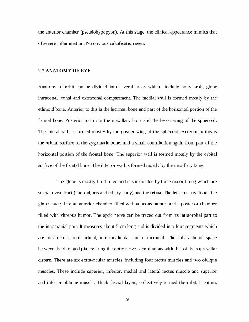
2.7 ANATOMY OF EYE
Anatomy of orbit can be divided into several areas which include bony orbit, globe
intraconal, conal and extraconal compartment. The medial wall is formed mostly by the
ethmoid bone. Anterior to this is the lacrimal bone and part of the horizontal portion of the
frontal bone. Posterior to this is the maxillary bone and the lesser wing of the sphenoid.
The lateral wall is formed mostly by the greater wing of the sphenoid. Anterior to this is
the orbital surface of the zygomatic bone, and a small contribution again from part of the
horizontal portion of the frontal bone. The superior wall is formed mostly by the orbital
surface of the frontal bone. The inferior wall is formed mostly by the maxillary bone.
The globe is mostly fluid filled and is surrounded by three major lining which are
sclera, uveal tract (choroid, iris and ciliary body) and the retina. The lens and iris divide the
globe cavity into an anterior chamber filled with aqueous humor, and a posterior chamber
filled with vitreous humor. The optic nerve can be traced out from its intraorbital part to
the intracranial part. It measures about 5 cm long and is divided into four segments which
are intra-ocular, intra-orbital, intracanalicular and intracranial. The subarachnoid space
between the dura and pia covering the optic nerve is continuous with that of the suprasellar
cistern. There are six extra-ocular muscles, including four rectus muscles and two oblique
muscles. These include superior, inferior, medial and lateral rectus muscle and superior
and inferior oblique muscle. Thick fascial layers, collectively termed the orbital septum,
10
situated anterior to the globe divide the more anterior skin and subcutaneous tissues to
preseptal compartment from postseptal compartment.
Arterial supply to orbit is mainly via the three large terminal branches of the
ophthalmic artery, which are lacrimal, nasociliary and frontal branches, in addition to
smaller branches, mainly the central artery of the retina which supplies the inner aspect of
the optic nerve and short and long ciliary branches. Veins in the orbit tend to follow
arteries but demonstrate greater interconnections until they form the superior ophthalmic
vein, which drains through the superior orbital fissure into the cavernous sinus, and the
inferior ophthalmic vein, which drains through the inferior orbital fissure to the cavernous
sinus and the pterygoid plexus.(Grainger Allison et al, 2008)
Figure 2.2 Gross anatomy of the eye. (Adapted from http://www.eyes and
eyesight.com/2009/02/anatomy-of-the-eye )

11
2.8 FUNCTION OF RETINA
Retina is a light-sensitive tissue lining the inner surface of the eye. The optics of the eye
create an image on the retina, which serves much the same function as the film in a
camera. Light striking the retina initiates a cascade of chemical and electrical events that
ultimately trigger nerve impulses. These are sent to various visual centers of the brain
through the fibers of the optic nerve.
The retina is a complex, layered structure with several layers of neurons
interconnected by synapses. The only neurons that are directly sensitive to light are the
photoreceptor cells. These are mainly of two types: the rods and cones. Rods function
mainly in dim light and provide black-and-white vision, while cones support daytime
vision and the perception of colours. A third, much rarer type of photoreceptor, the
photosensitive ganglion cell, is important for reflexive responses to bright daylight.
Neural signals from the rods and cones undergo complex processing by other neurons
of the retina. The output takes the form of action potentials in retinal ganglion cells whose
axons form the optic nerve. Several important features of visual perception can be traced to
the retinal encoding and processing of light.
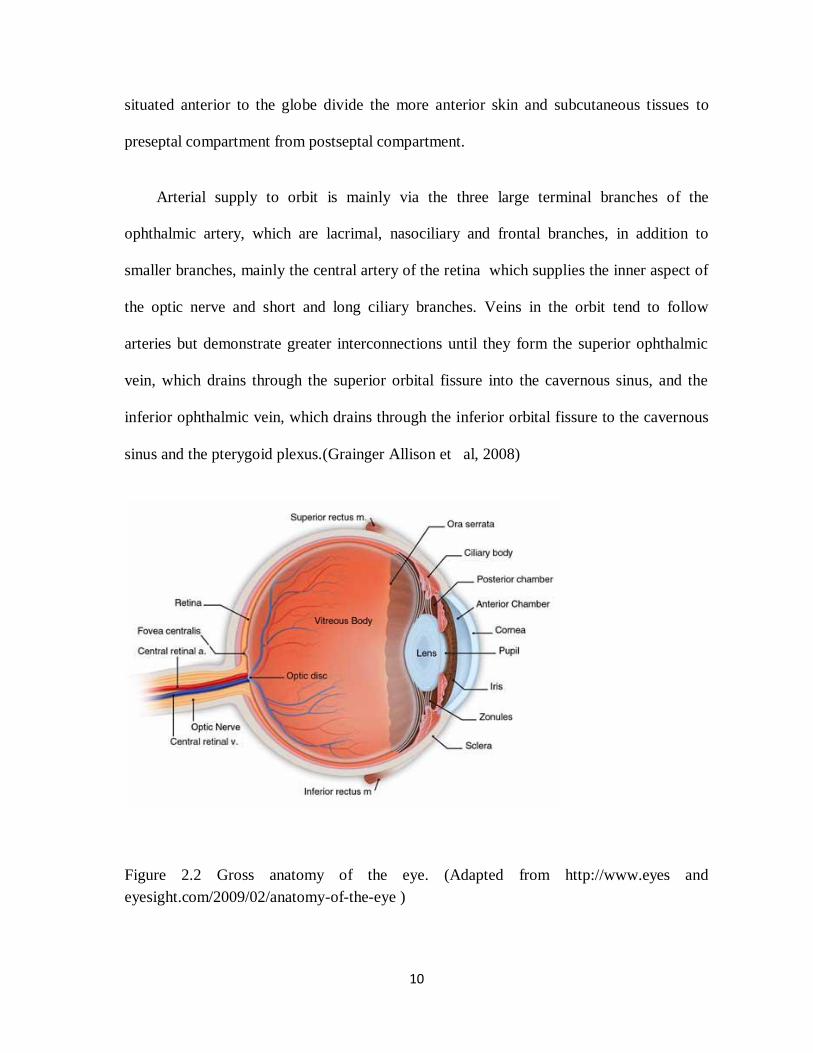
2.9 PRESENTING SYMPTOMS
The primary sign is leukocoria in 60% of cases. It is also known as cat reflex or white
papillary reflex. It is indicative of reflection of light from a tumoural lesion within the
12
pupillary area when the fundus is directly illuminated. This white pupillary reflex is
commonly seen on flash photography or torch by the light. Initially it is intermittent,
depending on the lighting, degree of pupil dilation, direction of gaze, angle of observation
and, most importantly the location and size of the tumor. Other differential diagnosis for
leukocoria are The two most frequent are Coats’ disease and persistent hyperplastic
primary vitreous (PHPV), followed by ocular toxocariasis.
Second most important sign of retinoblastoma is strabismus which can be found in
about 20% of cases. It is caused by the tumour growth that extends to the macula and
caused difficulty in seeing the object. In other word, strabismus is an invaluable early sign
carrying an excellent prognosis and a good chance of eye preservation.
Other sign of retinoblastoma are atypical and can be manifested as inflammatory
reaction in about 20 %. These include redness, swelling and proptosis of the eye. These are
late signs and it usually indicate that the eye has loss its function. Prognosis is also poor if
there is development of these symptoms.
Figure 2.3 Advanced intraocular retinoblastoma present as bilateral leukocoria and
strabismus. (Adapted from en.wikipedia available from: http:/// Wikipedia.com )

13
2.10 EXAMINATIONS
Diagnosis of retinoblastoma can be made on indirect ophthalmoscopy in about 90% of
cases. Pupillary dilation is an excellent tool that help to increased the ability of the
examiner to detect leukocoria (John C. Canzano and James T. Handa, 1999 ). Early lesions
appear as flat, transparent, or slightly white placoid tumors in the neurosensory retina. As
tumors enlarge, they have a white color with chalky, fleck- like deposits of calcium.
Figure 2.4 Fundus of the left eye revealing a small retinoblastoma just outside the
posterior pole with early calcification. Avalable from: H. Lee Moffitt Cancer Center &
Research Institute web site)
2.10 RADIOLOGICAL EXAMINATION
2.10.1 Ultrasound of the orbit
Orbital sonography provides good intraocular detail and allows sequential study
without ionizing radiation. It is also cost effective and rapid investigation. The prerequisite

14
for HRSG is high frequency transducers ranging from 5- 15 MHz with short focus. No
specific preparation or sedation is required. The mobility of sonographic equipment is
advantageous for intraoperative imaging performed in conjuction with ophthalmologic
examination. Warm sterile gel is applied to a closed eyelid. Tranducer was put on to the
eye and light pressure is used. Excessive ocular pressure can cause bradycardia and induce
pain. Color and power Doppler capability, power up to –3 dB was used. Transverse and
longitudinal planes provide excellent tumor detail. Retinoblastoma appears as an echogenic
soft-tissue mass with various degrees of calcification. Tumors tend to outgrow their blood
supply resulting in areas of necrosis within the tumour. The vitreous may have echogenic
debris from hemorrhage, increased globulin content, or tumor seeding. However, this
technique is limited to study of the globe and anterior orbit, if there is intraocular
calcification, the area posterior to it cannot be assessed due to hampered ultrasound wave
signal, and is degraded by patient motion. It is recommended that routine clinical screening
of all parents and sibling of retinoblastoma patient to provide earlier detection of
retinoblastoma and treatment at presymptomatic disease stage.
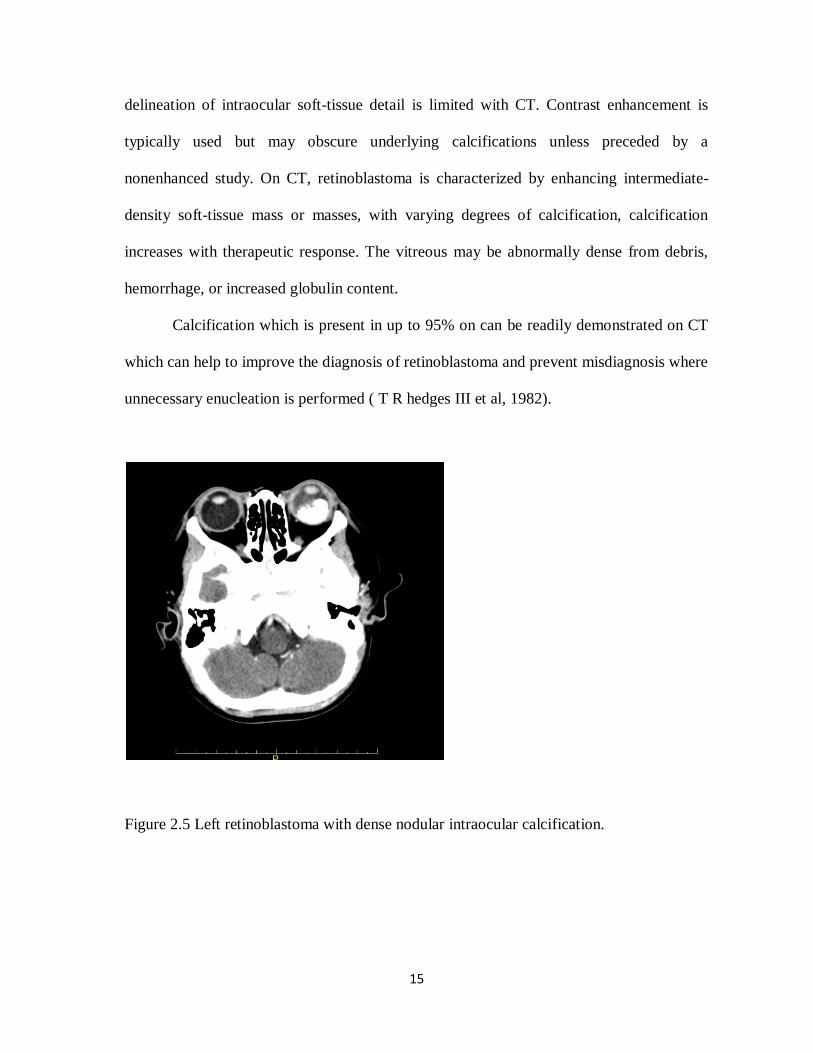
2.10.2 CT scan orbit.
CT detects intraocular, extraocular, and intracranial disease extension. It provide
great delineation of bony abnormalities and tumoural calcifications. Calcification is one of
the most important findings in diagnosing retinoblastoma. A lesion with calcification
within an orbit is which is seen in children younger than 3 years old is highly suspicious of
retinoblastoma. Calcification in retinoblastoma is seen in almost 80% of cases. However,
15
delineation of intraocular soft-tissue detail is limited with CT. Contrast enhancement is
typically used but may obscure underlying calcifications unless preceded by a
nonenhanced study. On CT, retinoblastoma is characterized by enhancing intermediate-
density soft-tissue mass or masses, with varying degrees of calcification, calcification
increases with therapeutic response. The vitreous may be abnormally dense from debris,
hemorrhage, or increased globulin content.
Calcification which is present in up to 95% on can be readily demonstrated on CT
which can help to improve the diagnosis of retinoblastoma and prevent misdiagnosis where
unnecessary enucleation is performed ( T R hedges III et al, 1982).
Figure 2.5 Left retinoblastoma with dense nodular intraocular calcification.x
16
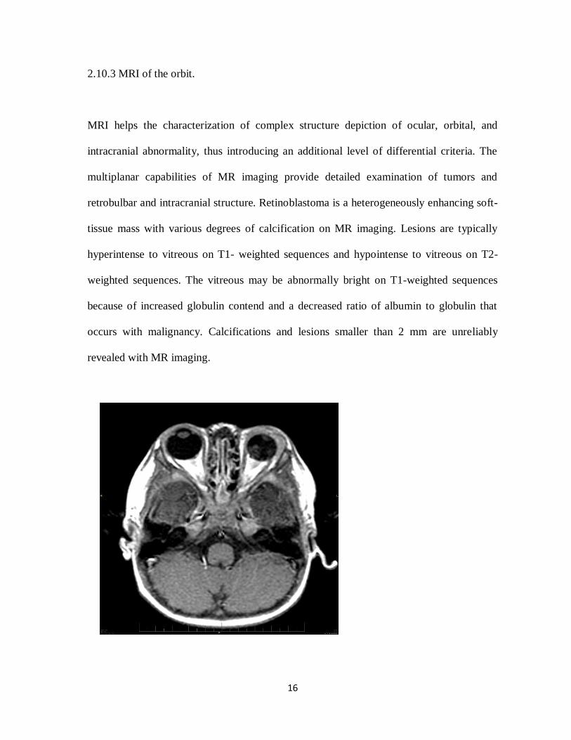
2.10.3 MRI of the orbit.
MRI helps the characterization of complex structure depiction of ocular, orbital, and
intracranial abnormality, thus introducing an additional level of differential criteria. The
multiplanar capabilities of MR imaging provide detailed examination of tumors and
retrobulbar and intracranial structure. Retinoblastoma is a heterogeneously enhancing soft-
tissue mass with various degrees of calcification on MR imaging. Lesions are typically
hyperintense to vitreous on T1- weighted sequences and hypointense to vitreous on T2-
weighted sequences. The vitreous may be abnormally bright on T1-weighted sequences
because of increased globulin contend and a decreased ratio of albumin to globulin that
occurs with malignancy. Calcifications and lesions smaller than 2 mm are unreliably
revealed with MR imaging. Orbit
al

17
Figure 2.6 Left retinoblastoma with hypointense area in all sequence seen within
suggestive of calcification.
2.11. CLASSIFICATION OF RETINOBLASTOMA.
Several staging has been developed to assist in management of the retinoblastoma.
Previously retinoblastoma was staged according to Reese Ellsworth staging system. The
Reese – Ellsworth classification has been devised in 1960s. It was devised to evaluate the
response to external beam radiotherapy (EBRT).
TABLE 2.1 Reese – Ellsworth Classification for conservative treatment of
retinoblastoma (Constantino Sábado Álvarez et al. 2005).
Group I: Very
favourable
(a) Solitary tumor, less than 4 disc diameters in size, at or
behind the equator
(b) Multiple tumors, none over 4 disc diameters in size, all at
or
behind the equator
Group II: Favorable
(a) Solitary tumor, 4 to 10 disc diameters in size, at or behind
the equator
(b) Multiple tumors, 4 to 10 disc diameters in size, behind the
equator
Group III: Doubtful
(a) Any lesion anterior to the equator
(b) Solitary tumors larger than 10 disc diameters behind the
equator
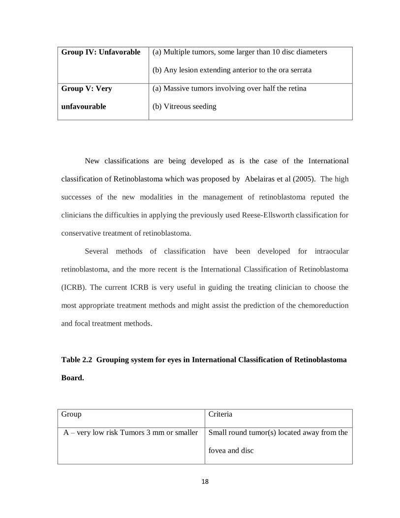
18
Group IV: Unfavorable
(a) Multiple tumors, some larger than 10 disc diameters
(b) Any lesion extending anterior to the ora serrata
Group V: Very
unfavourable
(a) Massive tumors involving over half the retina
(b) Vitreous seeding
New classifications are being developed as is the case of the International
classification of Retinoblastoma which was proposed by Abelairas et al (2005). The high
successes of the new modalities in the management of retinoblastoma reputed the
clinicians the difficulties in applying the previously used Reese-Ellsworth classification for
conservative treatment of retinoblastoma.
Several methods of classification have been developed for intraocular
retinoblastoma, and the more recent is the International Classification of Retinoblastoma
(ICRB). The current ICRB is very useful in guiding the treating clinician to choose the
most appropriate treatment methods and might assist the prediction of the chemoreduction
and focal treatment methods.
Table 2.2 Grouping system for eyes in International Classification of Retinoblastoma
Board.
Group Criteria
A – very low risk Tumors 3 mm or smaller
Small round tumor(s) located away from the
fovea and disc

19
≥3 mm from the fovea
≥1.5 mm from the optic disc
No vitreous seeding
No vitreous seeding
Group B – low risk
All eyes without tumor dissemination not in
Group A
Tumor dissemination is defined to include
vitreous seeding
and the presence of subretinal fluid, even if
subretinal
seeding is not clinically apparent
Any size, shape or location
No vitreous or subretinal seeding
Group C – moderate risk
Eyes with only local tumor dissemination
Vitreous or subretinal seeding no more than
3 mm from tumor
Group D – high risk
Eyes with diffuse tumor dissemination
endophytic or exophytic disease.
Vitreous seeding large, diffuse and/or
greasy
Avascular masses of tumor may be present
in the vitreous
Subretinal dissemination may consist of fine
seeds,large avascular
plaques on the underneath of the detached
retina, or extensive subretinal masses
(exophytic disease)

20
Group E – very high risk eyes
Unsalvageable eyes
Neovascular glaucoma
Massive intraocular hemorrhage
Blood-stained cornea
Massive tumor necrosis associated with
aseptic orbital cellulitis
Phthisis or prephthisis
Tumor anterior to anterior vitreous face
Anterior segment tumor
Tumor touching the lens
Diffuse infiltrating retinoblastoma
2.12 Treatment:
The primary goal of retinoblastoma therapy is to save life and preserve useful
vision, and thus needs to be individualized. Factors that need to be considered include
unilaterality or bilaterality of the disease, potential for vision, and intraocular and
extraocular staging. Retinoblastoma rarely metastasizes, and the chance of cure remains
excellent (C. Rodriguez-Galindo, 2010). There are multidisciplinary approach in treating
retinoblastoma involving the oncologist, ophthalmologist and radiologist team to make
sure that prompt and appropriate treatment can be given.
Standard treatments for retinoblastoma have yielded excellent results when
measured in terms of survival and preservation of vision. With conventional therapies,
survival exceeds 90%. Standard therapy for unilateral retinoblastoma has traditionally been

21
enucleation. For bilateral retinoblastoma, enucleation of the more advanced eye and
external-beam radiotherapy for the less affected eye have been standard therapies. Over the
last few decades, this general approach has witnessed an expanding role of eye-salvaging
therapy, particularly external-beam radiation for medium-sized tumors and radioplaque
therapy for smaller tumors. Some small tumors can be destroyed with cryotherapy or laser,
depending on their location and thickness.
Despite the excellent cure rates, there are several drawbacks to the current arsenal
of therapies. Enucleation sacrifices all vision and causes some degree of cosmetic
deformity. External-beam radiotherapy is highly effective in destroying most tumors that
fill less than half the eye, but failure rates increase with more advanced tumors. Most
importantly, nearly 51% of patients with heritable retinoblastoma will develop a second
malignancy within five decades of radiotherapy. These undesirable risks and outcomes
have prompted a search for alternative therapies to salvage eyes and avoid the risk of
radiotherapy.
Chemotherapy has been historically regarded as ineffective for intraocular
retinoblastoma, and its use has been restricted to treatment of extraocular disease. Pilot
studies using drug regimens that may cross the blood-ocular barrier combined with
supplemental laser and cryotherapy have reported favorable results.
2.12.1 External beam Radiotherapy
If retinoblastoma is multifocal, close to the macula or optic nerve, and vision is
well preserved, It is inadequate to apply only cryotherapy, photocoagulation, or plaque
22
therapy as a monotherapy. Enucleation is too aggressive since the vision is still preserved.
For these cases, the usage of external-beam irradiation or chemotherapy with focal therapy
is recommended. External- beam radiation therapy and chemotherapy are also indicated for
large tumors and vitreous seeding. The treatment for external beam therapy was
documented in 1910, when Hilgartner reported that treatment of a case of bilateral
retinoblastoma with X-rays in 1910. This has encouraged other oncologist to further
explore X- ray treatment. In 1918, Verhoeff cured a case of retinoblastoma with X-ray
treatment (Marcus et al. 1990).
2.12.2 Chemotherapy
Chemotherapy has been used for many years in the treatment of extended
retinoblastoma, to treat or prevent metastatic spread. Combined chemotherapy with
cyclophosphamide, doxorubicin, methotrexate and actinomycin-D, as in other embryonic
neural tumors has been employed (Constantino Sábado Álvareza et al. 2005).
2.12.3 Enucleation
Enucleation is indicated for majority of the children present with a large tumor that
requires an aggressive approach especially in unilateral retinoblastoma where the vision of
the eye is severed. In bilateral retinoblastoma when both eyes are blind, a bilateral
enucleation is done. This occur in 20-40% of cases. The optic nerve is cut near its exit
from the socket to make sure that a long segment of the nerve is obtained. This is
important especially when the tumour has already infiltrate the nerve to make sure that the

23
surgical margin is free from tumour cell. In young children, orbital growth slows after
enucleation. As the child grows, the orbit appears small. For better orbit growth and
cosmetic purpose, orbital prosthesis can be used to be fit within the globe.
2.13 Prognosis
Prognostic factors depend on the ocular status. The staging system for
retinoblastoma must fulfill at least two requirements. First, it must predict likelihood of
cure, a requirement of all malignancy staging systems. However, an important goal of
retinoblastoma treatment is preservation of sight in the affected eye. Previously, the most
widely used staging grouping system for retinoblastoma was proposed by Reese (1976)
and Ellsworth (1969). This system does not predict survival probability. However, it
predicts the chance of visual preservation with conservative therapy (Constantino Sábado
Álvareza et al. 2005).
Eye enucleation and external beam radiation were the mainstays of therapy in the
past but in recent years they are being displaced by combined chemotherapy followed by
focal treatments as cryotherapy, phototherapy or brachytherapy with plaques. These
therapeutic plan encompasses survival rates over 90%, making second cancers instead of
progressive disease the most frequent cause of death nowadays in patients with hereditary
retinoblastoma.
When retinoblastoma has spread outside the eye globe prognosis impairs notably
and it is a great challenge to achieve complete remission especially in case of central
nervous system involvement.

24
2.14 Genetic counseling.
The risk to the offspring of an individual with retinoblastoma depends on whether
patient has a germline mutation. Risk assessment is accomplished by obtaining a family
history and determining if the patient has unilateral, bilateral or multifocal tumour
involvement. Parents and siblings of persons with retinoblastoma shpuld be examined for
occult retinocytoma or spontaneously regressed retinoblastoma. The presence of
retinocytoma or a regressed retinoblastoma has the same genetic implications as
retinoblastoma. Penetrance of RB1 mutations are high, meaning that approximately 90% of
individuals with a germline RB1 mutation will develop retinoblastoma.
Laboratory techniques to identify RB1 are not routinely available. However,
predictive testing for retinoblastoma has great potential for improving the effectiveness of
genetic counseling by positively identifying germline mutations in persons with unilateral
involvement and asymptomatic carriers ( Retinoblastoma, Curtis E Margo et. al. available
at http://www.moffit.org, ) [Accessed 4/10/2010]