Embed Size (px)
Citation preview
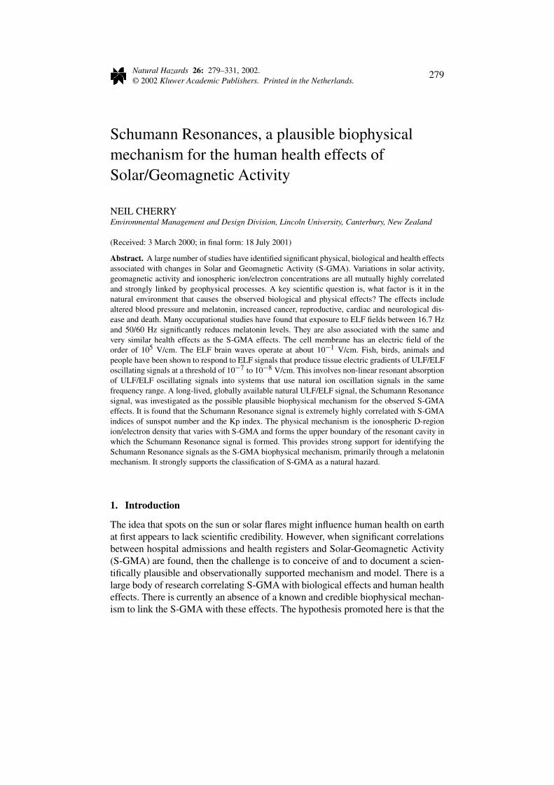
Natural Hazards 26: 279–331, 2002.© 2002 Kluwer Academic Publishers. Printed in the Netherlands.
279
Schumann Resonances, a plausible biophysicalmechanism for the human health effects ofSolar/Geomagnetic Activity
NEIL CHERRYEnvironmental Management and Design Division, Lincoln University, Canterbury, New Zealand
(Received: 3 March 2000; in final form: 18 July 2001)
Abstract. A large number of studies have identified significant physical, biological and health effectsassociated with changes in Solar and Geomagnetic Activity (S-GMA). Variations in solar activity,geomagnetic activity and ionospheric ion/electron concentrations are all mutually highly correlatedand strongly linked by geophysical processes. A key scientific question is, what factor is it in thenatural environment that causes the observed biological and physical effects? The effects includealtered blood pressure and melatonin, increased cancer, reproductive, cardiac and neurological dis-ease and death. Many occupational studies have found that exposure to ELF fields between 16.7 Hzand 50/60 Hz significantly reduces melatonin levels. They are also associated with the same andvery similar health effects as the S-GMA effects. The cell membrane has an electric field of theorder of 105 V/cm. The ELF brain waves operate at about 10−1 V/cm. Fish, birds, animals andpeople have been shown to respond to ELF signals that produce tissue electric gradients of ULF/ELFoscillating signals at a threshold of 10−7 to 10−8 V/cm. This involves non-linear resonant absorptionof ULF/ELF oscillating signals into systems that use natural ion oscillation signals in the samefrequency range. A long-lived, globally available natural ULF/ELF signal, the Schumann Resonancesignal, was investigated as the possible plausible biophysical mechanism for the observed S-GMAeffects. It is found that the Schumann Resonance signal is extremely highly correlated with S-GMAindices of sunspot number and the Kp index. The physical mechanism is the ionospheric D-regionion/electron density that varies with S-GMA and forms the upper boundary of the resonant cavity inwhich the Schumann Resonance signal is formed. This provides strong support for identifying theSchumann Resonance signals as the S-GMA biophysical mechanism, primarily through a melatoninmechanism. It strongly supports the classification of S-GMA as a natural hazard.
1. Introduction
The idea that spots on the sun or solar flares might influence human health on earthat first appears to lack scientific credibility. However, when significant correlationsbetween hospital admissions and health registers and Solar-Geomagnetic Activity(S-GMA) are found, then the challenge is to conceive of and to document a scien-tifically plausible and observationally supported mechanism and model. There is alarge body of research correlating S-GMA with biological effects and human healtheffects. There is currently an absence of a known and credible biophysical mechan-ism to link the S-GMA with these effects. The hypothesis promoted here is that the
280 NEIL CHERRY
Schumann Resonance (SR) signal is the plausible biophysical mechanism to linkthe S-GMA levels to biological and human health effects. This operates by beingresonantly absorbed by brain systems and altering the serotonin/melatonin balance.Confirmation of this hypothesis will strengthen the proposal that the S-GMA is anatural hazard for humans, animals and other species.
This study is in a context of fundamental biological concepts relating tohomeostasis and adaptation. On one hand the survival of organisms in changingenvironments requires adaptation. On the other hand mammals have very advancedneurological and physiological systems that must be maintained within narrowactivity ranges because of homeostatic requirements. Homeostasis is partly main-tained in variable environments, such as daily climate cycles, through the use ofexternal reference signals called Zeitgebers (time givers). The daily solar cycle isdetected by mammal’s eyes and brains. This induces a diurnal cycle of endocrinehormones that regulate a whole body system of diurnal changes. Isolating peopleor birds from the daily solar/climate signals leads to a significant lengthening ofthe circadian period (Wever, 1973, 1974). Wever also showed that there is a naturalULF/ELF electromagnetic signal that also acts as a circadian Zeitgeber. The char-acteristics of this signal are contained in the Schumann Resonance signal and thereis no other known natural signal with the appropriate characteristics.
2. Schumann Resonance Signal
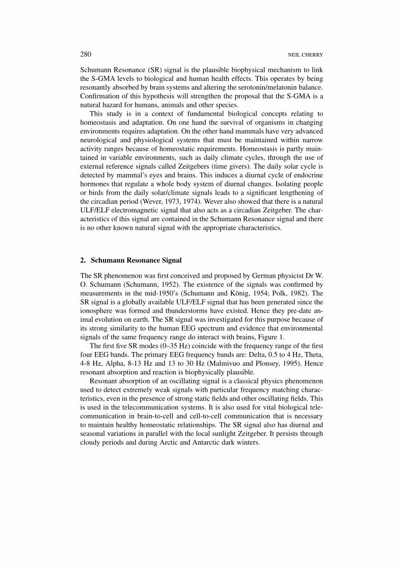
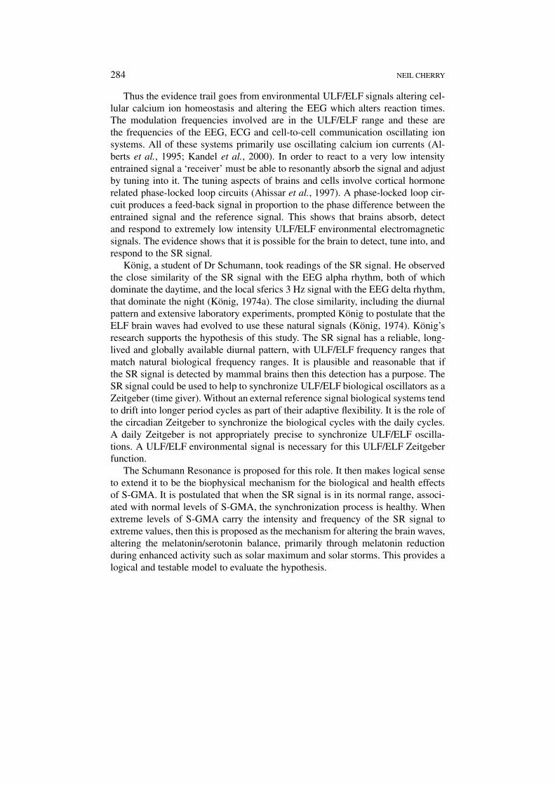
The SR phenomenon was first conceived and proposed by German physicist Dr W.O. Schumann (Schumann, 1952). The existence of the signals was confirmed bymeasurements in the mid-1950’s (Schumann and König, 1954; Polk, 1982). TheSR signal is a globally available ULF/ELF signal that has been generated since theionosphere was formed and thunderstorms have existed. Hence they pre-date an-imal evolution on earth. The SR signal was investigated for this purpose because ofits strong similarity to the human EEG spectrum and evidence that environmentalsignals of the same frequency range do interact with brains, Figure 1.
The first five SR modes (0–35 Hz) coincide with the frequency range of the firstfour EEG bands. The primary EEG frequency bands are: Delta, 0.5 to 4 Hz, Theta,4-8 Hz, Alpha, 8-13 Hz and 13 to 30 Hz (Malmivuo and Plonsey, 1995). Henceresonant absorption and reaction is biophysically plausible.
Resonant absorption of an oscillating signal is a classical physics phenomenonused to detect extremely weak signals with particular frequency matching charac-teristics, even in the presence of strong static fields and other oscillating fields. Thisis used in the telecommunication systems. It is also used for vital biological tele-communication in brain-to-cell and cell-to-cell communication that is necessaryto maintain healthy homeostatic relationships. The SR signal also has diurnal andseasonal variations in parallel with the local sunlight Zeitgeber. It persists throughcloudy periods and during Arctic and Antarctic dark winters.
SCHUMANN RESONANCES 281
Figure 1. A typical daytime spectrum for the vertical electric field measured near Kingston,Rhode Island, showing the first five Schumann Resonances modes (Polk, 1982).
3. SR Signal Frequency
The Schumann Resonance signal is generated by tropical thunderstorms and isa set of resonant modes within the resonant cavity formed between the earth’ssurface and the D-Region of the ionosphere. It consists of a spectrum of ULF/ELFresonant peaks with a fundamental frequency of about 7.8 Hz and broad resonantpeaks typically at 14, 20, 26, 33, 39, 45 and 51 Hz. An example of the measureddaytime spectrum of the first five modes is given in Figure 1. The frequencies varysystematically diurnally primarily with local D-region height, but also with tropicalthunderstorm activity.
4. SR Signal Strength
Balser and Wagner (1960) recorded the SR signal over several days in June 1960in Boston, USA. They measured a mean RMS vertical electric field strength ofthis ELF signal of 0.6 mV/m. Polk (1982) summarized several measurement pro-grammes, covering the first three resonant peaks. He gives the vertical electric fieldrange as 0.22–1.12 mV/m (0.013–0.33 pW/cm2). König (1974a) gives the typicalelectric field strength as 1 mV/m (0.27 pW/cm2) and the magnetic field as 10−5
A/m (12.6 pT). Williams (1992) reports 5 years of SR magnetic field intensitymeasurements from Rhode Island with monthly mean 8 Hz mode intensities in therange 1.3 to 6.3 pT.
282 NEIL CHERRY
5. Diurnal and D-Region Effects
Readings from M.I.T. in Boston were the first to show the frequency spectrum ofthe SR Signal (Balser and Wagner, 1960). They found that there was a frequencyand intensity shift between day and night. The first five modes dominated thedaytime. At night their intensity and frequency decreased and a large proportionof signals were less than 4 Hz. This frequency and intensity shift is from increas-ing the depth of the resonant cavity in the nocturnal hemisphere. The ion/electrondensity in the D-Region decreases rapidly after sunset as the solar production ofions ceases and recombination dominates. The dependence of the SR signal onthe D-Region was established by initial theoretical models (Tran and Polk, 1979).They showed that the Q-value of the resonant cavity depended on the conductivityof the atmosphere between 40 and 100 km, most strongly between 40 and 60 kmaltitude. Sentman and Fraser (1991) confirmed the sensitivity of the SR signal tothe local height of the D-Region. The D-Region correction increases the correlationcoefficient from r = 0.39 to r = 0.82, a highly significant improvement.
6. Role of Tropical Thunderstorms
The dominant diurnal pattern in the SR signal frequency and intensity are primarilythe result of the D-Region diurnal electron density variation. It is also modulatedby the diurnal incidence of tropical thunderstorms (Polk, 1982). These producepeaks of intensity as the peak of daily solar heating passes progressively aroundthe world from east to west (Nickolaenko et al., 1996). This produces a singlepeak in January (southern summer) and three peaks at 0800, 1400 and 2200 USTin August (northern summer) (Sentman and Fraser, 1991). The close correlationbetween the monthly tropical temperature anomaly and 8Hz SR signal intensitywas shown by Williams (1992). His data also reveals the strong influence of the ElNino/La Nina events. El Nino produces hotter mean conditions and La Nina coolerconditions. There are corresponding increases and decreases in SR signal intensity.
7. D-Region Characteristics
The D-Region of the ionosphere has electron density profiles that vary significantlywith diurnal, 27-day, seasonal and sunspot cycles, and with solar flares and storms(Nicolet and Aikin, 1960; King and Lawden, 1962; Titheridge, 1962; Craig, 1965;Matsushita and Campbell, 1967; Akasofu and Chapman, 1972; Coyne and Belrose,1972; Mitre, 1974; Rawer, 1984; Craven and Essex, 1986; Hargreaves, 1992).
Following a solar flare there is a prompt enhancement of the D-Region throughthe enhanced ionization from the arrival of cosmic rays. These events are calledSudden Ionospheric Disturbances (SID). A SID increases the ion density of theD-Region by a factor of 10 compared with quiet solar days (Belrose and Cetiner ,1962). SID monthly incidence is very closely correlated with Solar Flares and theSolar X-Ray flux (Davies, 1996).
SCHUMANN RESONANCES 283
Prolonged enhancement of the D-Region electron density was observed for atleast 5 days (Craven and Essex, 1987; Balon and Rao, 1990), and for at least 6days (Belrose, 1968). The enhancement was particularly strong at night. This effecthas been called the post storm effect (PSE). The most probable explanation is theinduced precipitation of electrons from the Van Allen Radiation Belt (Hargreaves,1992).
The dependency of the SR signal on the D-Region and the sensitivity of the D-Region to the S-GMA strongly indicates that the SR signal should closely followthe changes in solar and geomagnetic activity. This predicts that the SR signal willbe highly correlated with the solar cycles and the S-GMA events. The solar cyclesinclude the diurnal, 3.5 day, weekly, 13.5 day, 27–28 day solar rotation, semian-nual, annual, 11 year and 22 year cycles, and a number of harmonics (Chapman,1936; Cliver et al., 1996; Cornelissen et al., 1998). During solar flares the electronpattern in the D-Region predicts that there will be a prompt enhancement for aday or two and then a prolonged enhancement for 6 to 7 days and then fallingoff quickly. If a second or subsequent S-GMA events occur within this period theeffects should be cumulative.
8. ULF/ELF Resonant Absorption
It is noted above that the brain waves and SR signal share a ULF/ELF frequencyrange making resonant absorption possible. Extensive research shows that it ishighly likely. Adey (1990) summarized observations of cellular level electric fieldstrengths. The cell membrane potential, a static DC field across the cell membrane,is of the order of 105 V/cm. The brain waves have a typical amplitude of 10−1
V/cm. The brain successfully operates using oscillating signals a million timessmaller than the membrane potential. Fish, birds, primates and humans have beenshown to detect and react to ULF/ELF signals in the range 10−7 to 10−8 V/cm,more than a million times less than the EEG electric field. A recent study involvingflat worms (Planarian Dugesia tigrina) identified a threshold for 60 Hz electricfields of 5 × 10⁻⁸ V/cm for induced reproductive anomalies (Jenrow et al., 1996).
The biophysical mechanism for these effects was found when seeking to un-derstand why ULF/ELF signals alter primate and human reactions times and theirbrain wave signals (Adey, 1981). It was shown that environmental electromagneticfields in this frequency range significantly altered the cellular calcium ion fluxesand EMR waves in brain tissue (Bawin et al., 1973; Bawin and Adey, 1976; Adey,1980). The field strength involved was 10−7 V/cm. Since that time the calcium ionefflux/influx effect has been observed in many independent laboratories. The effectis taken as established by overwhelming evidence in a review (Blackman, 1990).The effect is a function of the modulation frequency more than the signal intensitysince it is a resonant phenomenon involving non-linear, non-equilibrium reactions(Adey, 1993).
284 NEIL CHERRY
Thus the evidence trail goes from environmental ULF/ELF signals altering cel-lular calcium ion homeostasis and altering the EEG which alters reaction times.The modulation frequencies involved are in the ULF/ELF range and these arethe frequencies of the EEG, ECG and cell-to-cell communication oscillating ionsystems. All of these systems primarily use oscillating calcium ion currents (Al-berts et al., 1995; Kandel et al., 2000). In order to react to a very low intensityentrained signal a ‘receiver’ must be able to resonantly absorb the signal and adjustby tuning into it. The tuning aspects of brains and cells involve cortical hormonerelated phase-locked loop circuits (Ahissar et al., 1997). A phase-locked loop cir-cuit produces a feed-back signal in proportion to the phase difference between theentrained signal and the reference signal. This shows that brains absorb, detectand respond to extremely low intensity ULF/ELF environmental electromagneticsignals. The evidence shows that it is possible for the brain to detect, tune into, andrespond to the SR signal.
König, a student of Dr Schumann, took readings of the SR signal. He observedthe close similarity of the SR signal with the EEG alpha rhythm, both of whichdominate the daytime, and the local sferics 3 Hz signal with the EEG delta rhythm,that dominate the night (König, 1974a). The close similarity, including the diurnalpattern and extensive laboratory experiments, prompted König to postulate that theELF brain waves had evolved to use these natural signals (König, 1974). König’sresearch supports the hypothesis of this study. The SR signal has a reliable, long-lived and globally available diurnal pattern, with ULF/ELF frequency ranges thatmatch natural biological frequency ranges. It is plausible and reasonable that ifthe SR signal is detected by mammal brains then this detection has a purpose. TheSR signal could be used to help to synchronize ULF/ELF biological oscillators as aZeitgeber (time giver). Without an external reference signal biological systems tendto drift into longer period cycles as part of their adaptive flexibility. It is the role ofthe circadian Zeitgeber to synchronize the biological cycles with the daily cycles.A daily Zeitgeber is not appropriately precise to synchronize ULF/ELF oscilla-tions. A ULF/ELF environmental signal is necessary for this ULF/ELF Zeitgeberfunction.
The Schumann Resonance is proposed for this role. It then makes logical senseto extend it to be the biophysical mechanism for the biological and health effectsof S-GMA. It is postulated that when the SR signal is in its normal range, associ-ated with normal levels of S-GMA, the synchronization process is healthy. Whenextreme levels of S-GMA carry the intensity and frequency of the SR signal toextreme values, then this is proposed as the mechanism for altering the brain waves,altering the melatonin/serotonin balance, primarily through melatonin reductionduring enhanced activity such as solar maximum and solar storms. This provides alogical and testable model to evaluate the hypothesis.
SCHUMANN RESONANCES 285
9. The Circadian Cycle
The body has a highly regulated and strongly integrated system that has de-veloped to produce healthy living in the face of diurnal and seasonal climaticvariations. Melatonin plays a central role. Environmental factors that alter themelatonin/serotonin balance have the ability to influence all of the functions andorgans that the circadian melatonin/serotonin cycle uses for thermal homeostasis.This includes blood pressure, breathing, altering the immune system, cardiac,neurological and reproductive processes.
A primary endocrine process involved in the diurnal (circadian) cycle is themelatonin/serotonin system. It initially operates between the pineal gland and thehypothalamus. A particular part of the hypothalamus is the suprachiamatic nucleus(SCN). It contains the ‘biological clock’. In order to mediate the daily cycle thereare high affinity melatonin and serotonin receptors in the brain and throughoutthe central nervous system (CNS). This includes the autonomic and sympatheticnervous systems (Chabot et al., 1998; Beresford et al., 1998; Naitoh et al., 1998;Andrade, 1998; Al-Ghoul et al., 1998; Verge and Calas, 2000; Hunt et al., 2001).Melatonin modulates the functional sensitivity of the serotonin receptors (Dugovicet al., 1991).
The cardiovascular system is daily mediated with melatonin through recept-ors in the heart, arteries and lungs (Pang et al., 1993; Viswanathan et al., 1993;Guardiola-Lemaitre,1997). Heart rate variability (HRV) is used to monitor theautonomic nervous system (Salo et al., 2000), and reduced HRV is a risk factorfor heart disease (Carney et al., 2000; Savitz et al., 1999a) quote a conclusionfrom a recent National Heart, Lung and Blood Institute workshop that “epidemi-ological evidence strongly implicates activation of the autonomic nervous systemas a causal link in the onset of cardiovascular disease”.
There are also melatonin receptors in the vital organs throughout the bodythat are part of the diurnal cycle system. This includes the pituitary gland whichregulates the production of Growth Hormone and Thyroid Stimulating Hormone(Vriend et al., 1987).
Melatonin has direct action in the immune system through the T-Lymphocytes(T-Helper Cells), interleukin-2 and -6 (IL-2. IL-6) and natural killer cells (NK-cells) through melatonin receptors on the T-Cells. This enhances the natural andacquired immunity (Poon et al., 1994; Maestroni, 1995; Garcia-Maurino et al.,1999; Currier et al., 2000). Melatonin receptors have been identified in a numberof peripheral organs and tissues. For example, melatonin receptors maintain in-traocular pressure (IOP) in the eye (Osborne, 1994). The reproductive organs alsohave melatonin receptors in the testes, prostate, ovary, mammary gland and otherreproductive organs (Pang et al., 1998). The fetus has many melatonin receptorsso that the maternal melatonin can communicate daily and seasonal cycles in utero(Naitoh et al., 1998; Thomas et al., 1998).
286 NEIL CHERRY
Through receptors melatonin regulates the diurnal and seasonal activity. This in-volves metabolism, body temperature, blood pressure, heart beat, peripheral bloodflow, respiratory activity, sleep-wake cycle, reaction times, hormone levels andimmune system blood cells. It also involves the humoral organs of the lung, heart,kidney, spleen, liver and lymphocytes of the immune system (Wever, 1974; Ishidaet al., 1999).
Melatonin is also a highly potent antioxidant that scavenges free radicals fromcells (Reiter, 1994). This implicates that reduced melatonin is associated withneurological, cardiac, reproductive and carcinogenic illness and death (Reiter andRobinson, 1995).
10. The Schumann Resonance Hypothesis
The hypothesis of this study is that:
The Schumann Resonance signal is the biophysical mechanism for the healtheffects of S-GMA because it is detected by the brain where it interacts withthe ELF brain waves by resonant interaction with neurons calcium ions and italters the melatonin/serotonin balance.
As a corollary, the SR provides an ULF/ELF, diurnal and seasonal synchroniz-ation reference signal, a Zeitgeber (time giver).
A systematic evaluation of the SR Hypothesis and the GMA Melatonin mech-anism will be carried out in the context of a model. The model outlines all of thelinking elements, from the solar, geomagnetic and ionospheric activity to the bio-logical and health effects associated with S-GMA, with the SR signal and inducedmelatonin reduction at its core.
11. The Model
This model was conceived with the Schumann Resonance Hypothesis at its core,assisted by the Melatonin Mechanism. It involves the following elements:
a. Solar activity is highly variable. The level of activity is transferred to the earthpromptly through cosmic radiation and over several days through large cloudsof ionised plasma transported in the solar wind. These produce significantcharacteristic changes to the earth’s magnetosphere and ionosphere, includingthe lowest layer, the D-Region.
b. The Schumann Resonances are a spectrum of ULF/ELF radio signals generatedby tropical thunderstorms, radiating around the world at the speed of light,ducted within the resonant cavity formed between the lowest layers of theionosphere (D-Region) and the earth’s surface.
c. A combination of seasonal and diurnal changes in the D-Region and in tropicalthunderstorm activity, induces strong diurnal and seasonal changes in the SRsignal. Solar sunspot cycles, solar rotation and solar flares and storms produce
SCHUMANN RESONANCES 287
characteristic changes in the D-Region which cause characteristic changes inthe SR signal. This leads to highly significant correlations between S-GMAindices and SR signals.
d. Human and animal brains naturally use highly reactive, non-linear alterationsof ULF/ELF oscillating calcium ions in neurons and other cells to regu-late the basic cell functions, such as neurotransmitter release and cell-to-cellcommunication.
e. Environmental electromagnetic fields in the ULF/ELF frequency range, includ-ing the SR signal, resonantly interact with the natural signals, inducing changesin the cellular calcium ion signals, brain waves patterns and reaction times.This produces altered melatonin/serotonin production.
f. By altering the neurohormone regulation of the natural cycles, as demonstratedby the circadian cycle, a wide range of body organs and processes are altered.Melatonin reduction alters blood pressure and heart rate with cardiovascularand cardiopulmonary consequences. It also produces neurological, reproduct-ive and carcinogenic effects and impairment of the immune system. The vastmajority of people cope with these induced changes but the exacerbation ofeffects in the weak and vulnerable people causes detectable increases in illnessand death rates.
g. Significant alterations in S-GMA cause significant changes in the intensity ofthe SR signal. This takes the SR signal strength and frequency outside the nor-mal range, inducing alteration of brain and heart synchronization and changesthe melatonin levels. Many of the biological and health effects of alteredmelatonin are produced by these extreme changes, showing that S-GMA isa natural hazard.
If all the above can be demonstrated, justified and confirmed then there is strongevidence that the SR signal is the plausible biophysical mechanism for the biolo-gical and health effects of S-GMA, mainly through altered melatonin. This wouldconfirm that S-GMA is a natural hazard for human beings. The Model Elements(a) to (d) are established by published research cited above.
Two corollaries arise from the hypothesis and model. The first relates to thepublic health implications of residential, occupational and military exposures toELF and ELF modulated signals. If extreme variations of natural extremely lowintensity ELF signals causes human health effects, then it is highly probable thathumanly produced ELF fields that are many orders of magnitude higher thanthe naturally occurring signals, are also causing significant similar health effects.Since residential and occupational studies are available, this evidence is used toindependently test the hypotheses.
The second corollary arises because of the strong dependence of the SR in-tensity on the tropical temperature anomaly (Williams, 1992). This means thatthe more extreme weather associated with Global Warming, including extremesof El Nino/La Nina events, will cause stronger extremes of SR signals. This occurs
288 NEIL CHERRY
especially around sunspot maximum and during periods of strong solar storms, andis predicted to accentuate the adverse health effects associated with S-GMA.
12. Methods
The steps taken to evaluate the hypothesis and the model are:
1. Seek correlation between S-GMA indices Ns and Kp and the SR signal.2. Review of the studies relating ULF/ELF signals, including SR signals and
human reaction times and circadian rhythms.3. Review of the evidence that ULF/ELF electromagnetic fields reduce melatonin.4. Summarize the biological and clinical studies relating reduced melatonin to
health effects.5. Give an example of sunspot data relationship to human health effects to illus-
trate the principles and problems of environmental epidemiological studies inan EMR polluted world.
6. Summarize the studies relating S-GMA to biological and health effects underthe headings of reproductive, cardiac, neurological and cancer effects in orderto evaluate the role of melatonin and homeostatic patterns.
7. The prediction from the hypothesis that similar effects should occur in residen-tial and occupational studies is evaluated to determine conflict or confirmationof the hypothesis.
8. Conclusions are drawn and further clarifying research is suggested.
13. Correlation to Causation
If variable A is correlated with variable B, and variableB is correlated with vari-able C, then variable A is correlated with variable C. However, none of theserelationships necessarily imply a causal relationship. The S-GMA indices arehighly correlated with biological and health effects. The observed and establishedcharacteristics of the atmosphere predict that through the S-GMA effects on the D-Region, the SR signal should be closely correlated with the S-GMA indices. Henceif this correlation is confirmed then the SR signal will also be correlated with thebiological and health effects.
A plausible and supported mechanism that explains the connective processesis crucial in moving towards establishing a causal link. The Schumann Resonancesignal is favoured for this purpose because of the matched frequency range allow-ing resonant absorption in the brain. This involves the alteration of calcium ions inneurons (Adey, 1981; Blackman, 1990). It is supported by the existence of a naturalULF/ELF Zeitgeber (Wever, 1974), for which the SR signal characteristics comply.The close dependence of the SR signal on the D-Region implies that there shouldbe a correlation with the biological and health effects correlated with S-GMA.
SCHUMANN RESONANCES 289
14. Statistical Methods
Sunspot Number (Ns) and GMA indices Ap and Kp, and the Schumann Resonancefrequency and intensity data for two US sites, were downloaded from publicly ac-cessible web sites in the United States. Statistical analysis involves several standardmethods. Time series cycle frequency analysis uses the Maximum Entropy Spec-trum method. Trend analysis uses the least squares fit (LSF), linear correlationcoefficient, and the Mantel–Haenszel t-test using a two-tail method to estimate theprobability, p-value. For a sample size = N there are N − 2 degrees of freedom.The t-value is calculated from the correlation coefficient (r) and the degrees offreedom, t = r[(N − 2)/(1 − r2)]0.5.
For large samples of N > 120 elements, using the Student t-distribution, thethreshold for significance is p = 0.05 and t = 1.96. When p = 0.01 it is highlysignificant and has t = 2.576. The very highly significant, p = 0.005 has t = 2.795and the extremely significant, p = 0.001 has t = 3.291 (Robson, 1967; Chou, 1972;Sprott, 1990). A sample with 1002 data points, N = 1000, and if r = 0.4 then t =13.8 and p < 10−12.
For some samples in this analysis the t-value exceeds 5 and occasionally even>10. A log/linear graphic method was used to estimate the p-values for largesamples with higher t-values. For t = 4.85, p = 10−6; for t = 6.3, p = 10−9;and for t = 7.8, p = 10−12. Any value more significant than this is referred to asp < 10−12.
14.1. DATA-SETS USED FOR THIS ANALYSIS
The geomagnetic field strength is measured at a large number of stations. A globalnetwork of 12 stations are integrated to produce 3-hour averaged indices. One isa linear index, Ap, and another is a pseudo-logarithmic index, Kp. The subscript‘p’ refers to a planetary index. A common index of solar activity is the sunspotnumber, Ns. Sunspots are indicative of long-term changes in the solar activity andthe Ap and Kp indices of the short-term activity experienced at the surface of theearth, such as the effects of solar flares or solar storms (Campbell, 1997).
Solar and Geomagnetic Indexes were down-loaded from the U.S. NGDC-NOAA web-site [ftp://ftp.ngdc.noaa.gov/STP/GEOMAGNETIC_DATA/INDICES].The 3-hourly global index Kp, a semi-logarithmic measure of GMA was used formost calculations. Daily sunspot number was also used.
The SR data for two sites in California was downloaded from the Universityof California, Berkeley web-site [ftp://quake.geo.berkeley.edu/pub/em/]. A three-year data-set was assembled covering the period 13th January 1997 to 31 December1999.
The two SR recording sites were Parkfield (PKB) (lat. 35.8892; long.−120.4249) and Hollister (SAO) (lat. 36.765, long. −121.445). Two sets of datafiles were available. The first contained 15-minute measurements of the east-westand north-south components of the 0–20 Hz field intensity in pT and the center
290 NEIL CHERRY
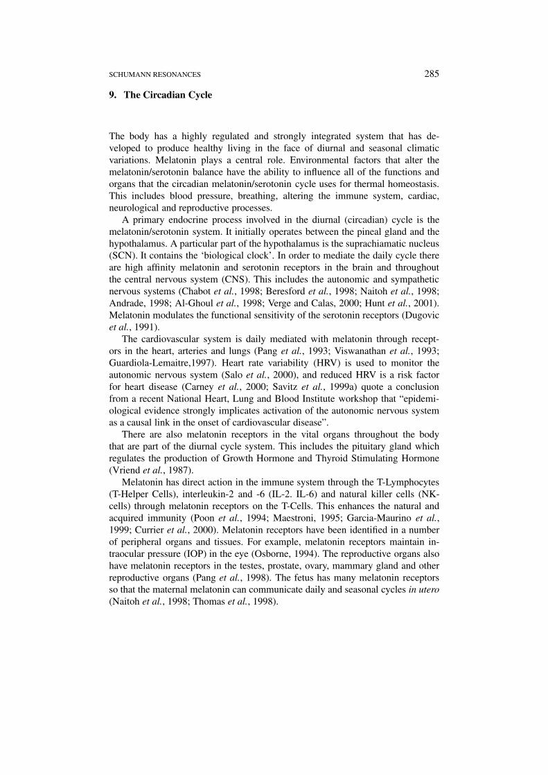
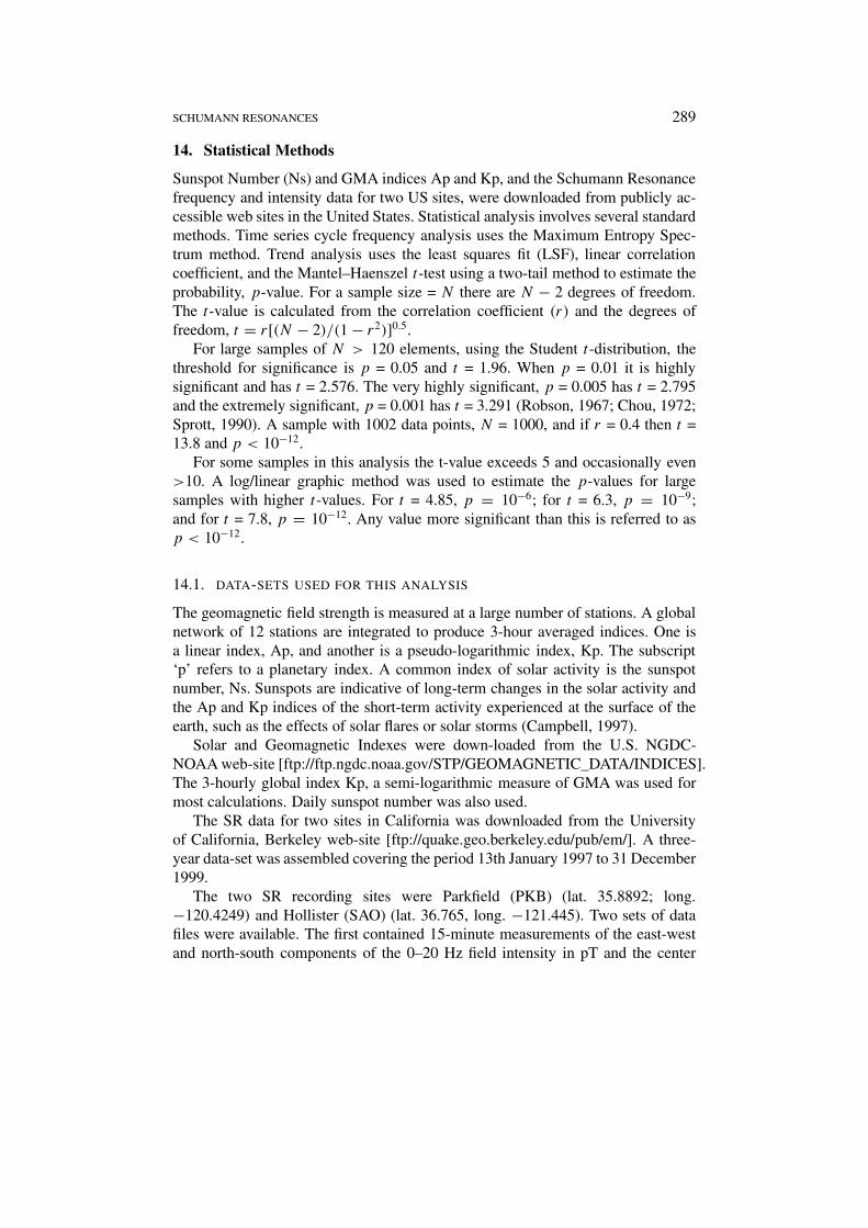
Figure 2. The time sequence of 20-day averaged Schumann Resonance signal intensity in the0-20 Hz range. From the UC Berkeley dataset, for the three years used in this study. The trendis highly significant, p < 0.0001.
frequency of the 8 Hz peak. The intensity data was vectorially added and thefrequency was averaged.
The PKB and SAO hourly data were extremely strongly correlated, p < 10−12.Hence missing data was replaced through a linear least squares fit relationship.Some signal instability was detected with large changes in single 15-minute datapoints. These were removed using a linear interpolation filter. Smoothing using1/4-1/2-1/4 was applied prior to averaging to a form an hourly mean data set forPKB for the period 1997–1999. The first 12 days of 1997 were rejected because ofclear data errors.
The second SR set of data involved three orthogonal components with the sig-nals analysed into 13 frequency ranges. This data was used to characterize the waysin which diurnal frequency changes occurred. The 11th column contained the 1–2 Hz SR intensity. The three components were combined vectorially. The unit ofpT/Hz1/2 was converted to pT by multiplying by
√2. This data was compiled to
form a 3-year data set of hourly mean observations to carry out correlations withKp and Sunspot numbers.
Figure 2 shows the 20-day mean values of the measured 0–20 Hz SR intensityfrom the PKB site for the three years of the study period.
The SR 20-day mean signal is very highly correlated with the 20-day meansunspot number, r = 0.881, N = 54, t = 13.4, p < 0.00001. When the daily mean
SCHUMANN RESONANCES 291
sunspot numbers and SR intensity and frequency are compared, very significantcorrelations are found. For the SR intensity r = 0.376, t = 13.33, p < 0.000001,for the SR frequency r = 0.436, t = 15.91, p < 0.000001.
The trend in Figure 2 is primarily due to the mean rise in sunspots over thisperiod toward the sunspot maximum in the 2000 year. The seasonal pattern isrelated to tropical thunderstorm activity. It is lower during the southern summerand higher during the northern summer.
Annual variations relate to the El Nino/La Nina oscillation. The year 1997 wasan El Nino year with higher than average tropical temperatures. By May 1998 it hadswitched to a La Nina event with lower than average tropical mean temperatures.The La Nina was weak during 1999. Hence the temperature effect raised the 1997SR intensity and lowered the 1998 SR intensity. Adjusting for this would increasethe significant correlation with the sunspot number. Figure 2 reveals the prime offactors, sunspot activity and tropical temperature, that change the long-term meanSR signal intensity.
14.2. GRAPHICAL AND STATISTICAL COMPARISON BETWEEN KP AND SR
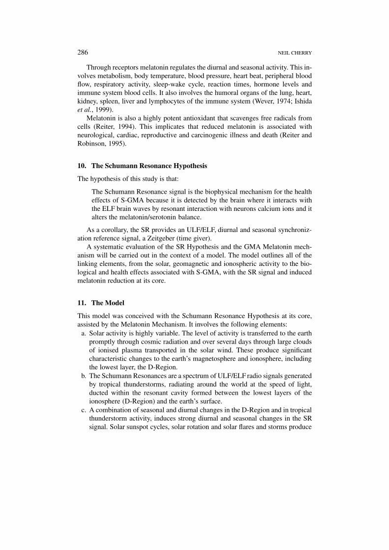
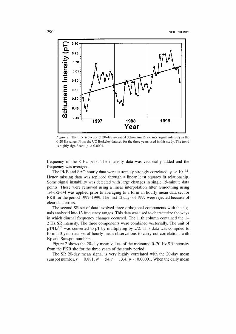
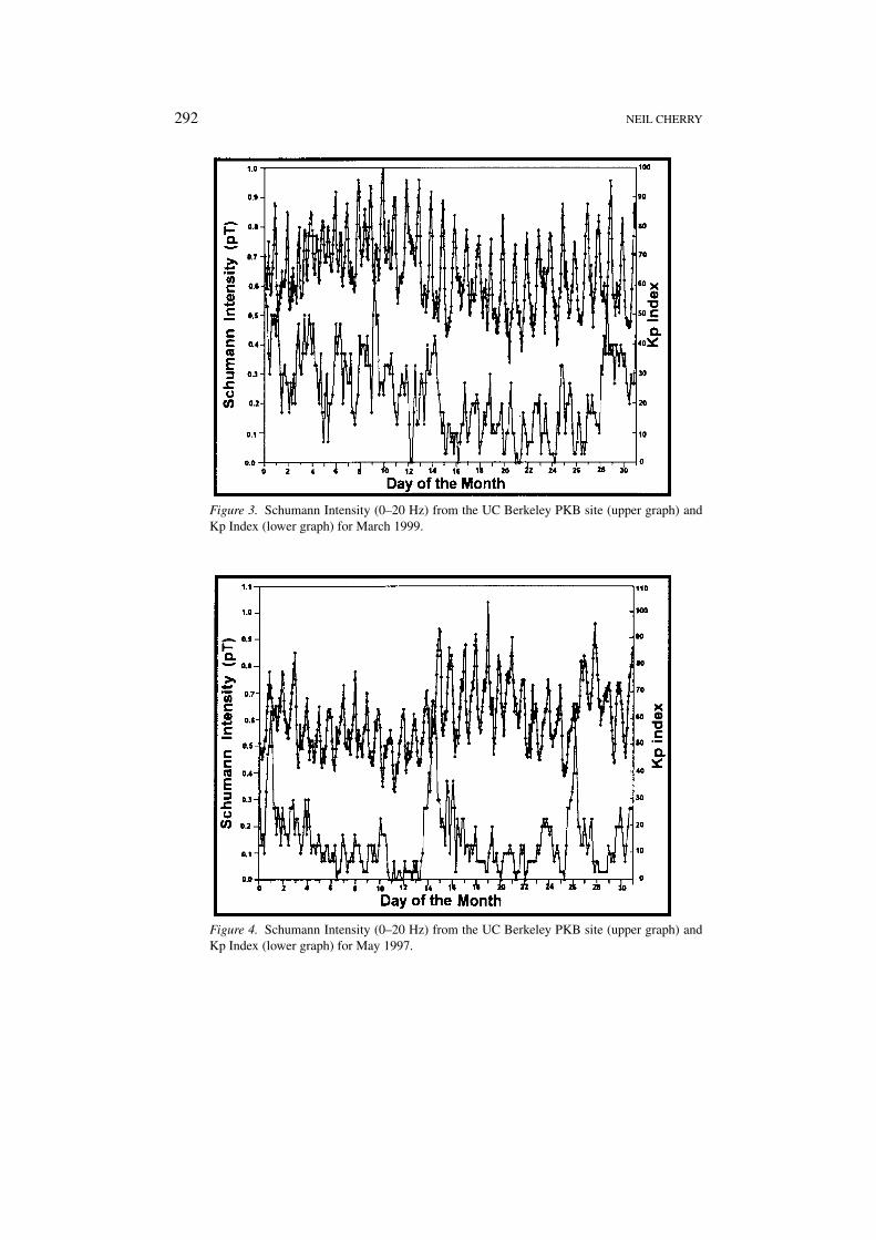
A GMA storm was shown to significantly increase the frequency of the primary SRmodels (Cannon and Rycroft, 1982). Consistent with the effect of the D-Region,this also implies that the SR intensity was significantly increased. This is evaluatedwith the data set available comparing the Kp index with the SR 0–20 Hz intensity.An initial relationship between the 3-hour Kp GMA index and the 0–20 Hz SRintensity from the PKB site shows some vital features and confirms the earlierobservations. Figure 3 shows the 0–20 Hz SR signal and the Kp Index for March1999.
The SR signal, Figure 3, shows the distinctive regular diurnal oscillation. Kpshows strong GMA in the first half of the month, weak GMA from the 15th to 27thand then a GMA event starting on the 28th. The overall SR signal reflects thesebroad changes. The first 10 days show an upward trend suggesting a cumulativeeffect. Individual GMA events, e.g. on the 4th, 10th, 14th appear to show a timedelay. A lagged linear correlation analysis of the 3-hourly mean data, reveals thehighest correlation with a 6-hour lag, r = 0.312, t = 5.123, p < 0.00001.
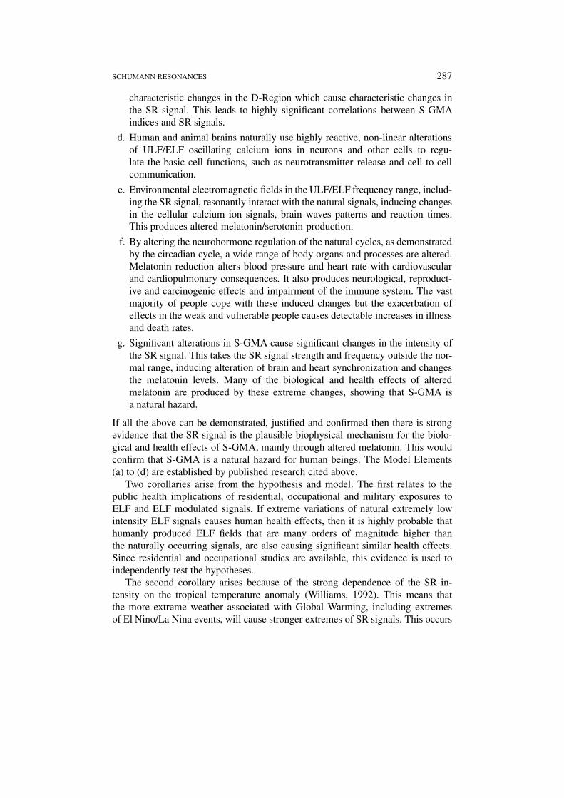
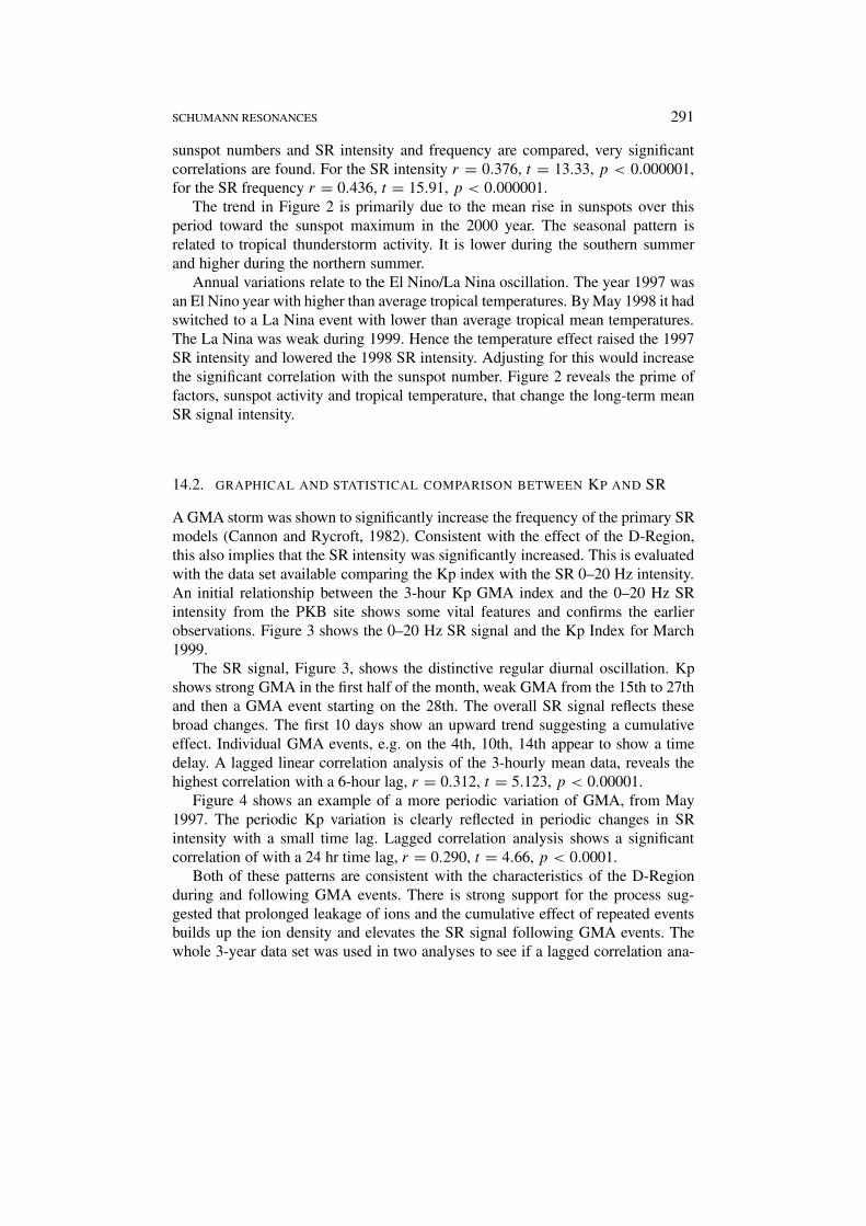
Figure 4 shows an example of a more periodic variation of GMA, from May1997. The periodic Kp variation is clearly reflected in periodic changes in SRintensity with a small time lag. Lagged correlation analysis shows a significantcorrelation of with a 24 hr time lag, r = 0.290, t = 4.66, p < 0.0001.
Both of these patterns are consistent with the characteristics of the D-Regionduring and following GMA events. There is strong support for the process sug-gested that prolonged leakage of ions and the cumulative effect of repeated eventsbuilds up the ion density and elevates the SR signal following GMA events. Thewhole 3-year data set was used in two analyses to see if a lagged correlation ana-
292 NEIL CHERRY
Figure 3. Schumann Intensity (0–20 Hz) from the UC Berkeley PKB site (upper graph) andKp Index (lower graph) for March 1999.
Figure 4. Schumann Intensity (0–20 Hz) from the UC Berkeley PKB site (upper graph) andKp Index (lower graph) for May 1997.
SCHUMANN RESONANCES 293
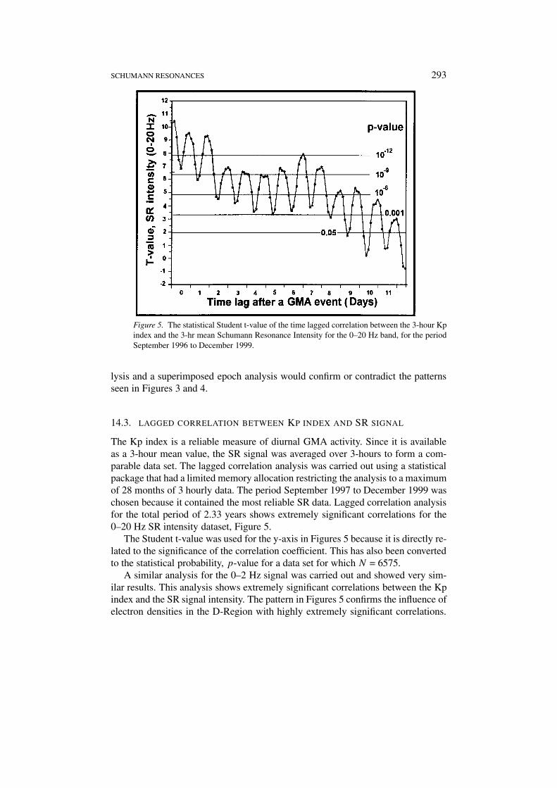
Figure 5. The statistical Student t-value of the time lagged correlation between the 3-hour Kpindex and the 3-hr mean Schumann Resonance Intensity for the 0–20 Hz band, for the periodSeptember 1996 to December 1999.
lysis and a superimposed epoch analysis would confirm or contradict the patternsseen in Figures 3 and 4.
14.3. LAGGED CORRELATION BETWEEN KP INDEX AND SR SIGNAL
The Kp index is a reliable measure of diurnal GMA activity. Since it is availableas a 3-hour mean value, the SR signal was averaged over 3-hours to form a com-parable data set. The lagged correlation analysis was carried out using a statisticalpackage that had a limited memory allocation restricting the analysis to a maximumof 28 months of 3 hourly data. The period September 1997 to December 1999 waschosen because it contained the most reliable SR data. Lagged correlation analysisfor the total period of 2.33 years shows extremely significant correlations for the0–20 Hz SR intensity dataset, Figure 5.
The Student t-value was used for the y-axis in Figures 5 because it is directly re-lated to the significance of the correlation coefficient. This has also been convertedto the statistical probability, p-value for a data set for which N = 6575.
A similar analysis for the 0–2 Hz signal was carried out and showed very sim-ilar results. This analysis shows extremely significant correlations between the Kpindex and the SR signal intensity. The pattern in Figures 5 confirms the influence ofelectron densities in the D-Region with highly extremely significant correlations.
294 NEIL CHERRY
They both show a strong and extremely significant initial increase, a prompt re-sponse for the first night and day and a persistent relationship that extends out for 6to 7 days after the day of the event (Day 0) and then it drops off significantly. Thecorrelation also shows a 24-hour diurnal oscillation.
These analyses give strong confirmation of the part of the model proposing thatthe SR intensity is strongly dependent on the D-Region electron density. It showsthat through slow electron leakage, the solar storm events last for about 7 days andthis is shown by the SR signal.
15. Superimposed Epoch Analysis
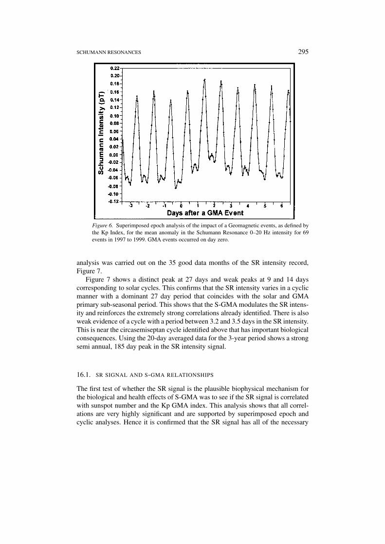
The above analysis uses the continuous time series of data over 28 months. Analternative approach is to identify isolated GMA events and to carry out a superimposed epoch analysis to produce the average signal before, during and after themoderate to strong GMA events. In the 3-year period being studied 69 relativelyisolated GMA events were identified. The intensity of the 0-20 Hz SR signal andthe frequency of the 8 Hz signal were averaged over the 3 days prior to and the 6days following each event, Figure 6. The overall variance of the hourly data overthis period was 0.017 pT for the SR intensity and 0.0026 Hz for the SR frequency.
Figure 6 shows prompt response of an elevated 0-20Hz SR intensity, especiallyover the first two days. The SR signal is still elevated at nighttime for at least 6 daysafter the GMA event. The SR 8 Hz band frequency shows a similar, but slightlysmaller response. Both the signal intensity and frequency are consistent with thelagged correlation analysis above showing a prompt response and a prolongedrecovery period of 6 to 7 days.
Thus the individual months, the lagged correlation and superimposed epochanalyses confirm that the SR intensity reliably, consistently and extremely signi-ficantly varies with the GMA as indicated by the Kp index. The results are fullyconsistent with a prompt and a prolonged D-Region electron density effect. Hencethe biological and health effects that are correlated with GMA indices are alsocorrelated with the GMA related changes in the SR signal.
16. Correlations with Sunspot Cycles
Sunspot number (Ns) is a direct indicator of solar activity. On a monthly basis Ns ishighly correlated with GMA over a 100 year period from 1830 to 1930 (Chapman,1936). Chapman also shows that sunspot activity peaks at about 3.5 days prior to amagnetic disturbance and that there is a 27 to 28 day cycle in the GMA index, andthat there is a semi-annual cycle in the GMA.
A cyclic analysis has been used in other studies to identify causative relation-ships through closely matched cyclic factors (Komarov et al., 1998). The sun’smean rotation period of about 27–28 days has a series of recognised sub-harmonicsof about 18, 14, 9, 7, 5.5 and 3.5 days. Hence a Maximum Entropy Power Spectrum
SCHUMANN RESONANCES 295
Figure 6. Superimposed epoch analysis of the impact of a Geomagnetic events, as defined bythe Kp Index, for the mean anomaly in the Schumann Resonance 0–20 Hz intensity for 69events in 1997 to 1999. GMA events occurred on day zero.
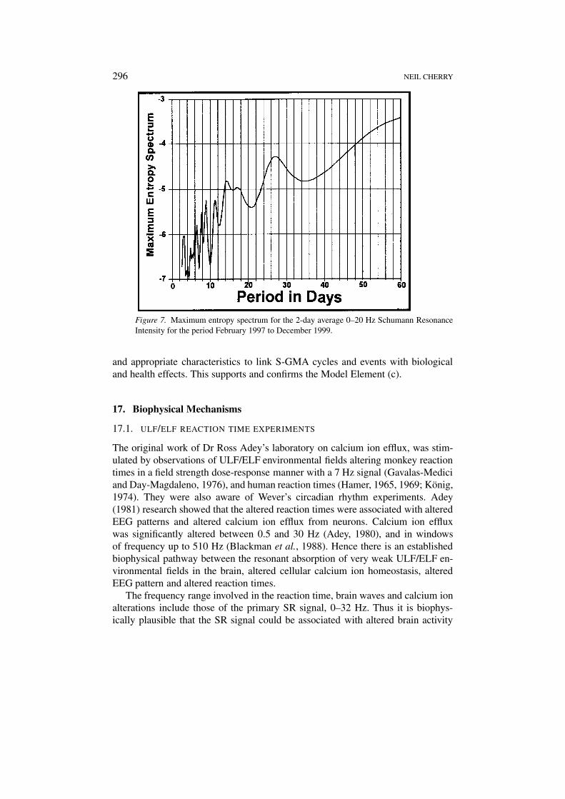
analysis was carried out on the 35 good data months of the SR intensity record,Figure 7.
Figure 7 shows a distinct peak at 27 days and weak peaks at 9 and 14 dayscorresponding to solar cycles. This confirms that the SR intensity varies in a cyclicmanner with a dominant 27 day period that coincides with the solar and GMAprimary sub-seasonal period. This shows that the S-GMA modulates the SR intens-ity and reinforces the extremely strong correlations already identified. There is alsoweak evidence of a cycle with a period between 3.2 and 3.5 days in the SR intensity.This is near the circasemiseptan cycle identified above that has important biologicalconsequences. Using the 20-day averaged data for the 3-year period shows a strongsemi annual, 185 day peak in the SR intensity signal.
16.1. SR SIGNAL AND S-GMA RELATIONSHIPS
The first test of whether the SR signal is the plausible biophysical mechanism forthe biological and health effects of S-GMA was to see if the SR signal is correlatedwith sunspot number and the Kp GMA index. This analysis shows that all correl-ations are very highly significant and are supported by superimposed epoch andcyclic analyses. Hence it is confirmed that the SR signal has all of the necessary
296 NEIL CHERRY
Figure 7. Maximum entropy spectrum for the 2-day average 0–20 Hz Schumann ResonanceIntensity for the period February 1997 to December 1999.
and appropriate characteristics to link S-GMA cycles and events with biologicaland health effects. This supports and confirms the Model Element (c).
17. Biophysical Mechanisms
17.1. ULF/ELF REACTION TIME EXPERIMENTS
The original work of Dr Ross Adey’s laboratory on calcium ion efflux, was stim-ulated by observations of ULF/ELF environmental fields altering monkey reactiontimes in a field strength dose-response manner with a 7 Hz signal (Gavalas-Mediciand Day-Magdaleno, 1976), and human reaction times (Hamer, 1965, 1969; König,1974). They were also aware of Wever’s circadian rhythm experiments. Adey(1981) research showed that the altered reaction times were associated with alteredEEG patterns and altered calcium ion efflux from neurons. Calcium ion effluxwas significantly altered between 0.5 and 30 Hz (Adey, 1980), and in windowsof frequency up to 510 Hz (Blackman et al., 1988). Hence there is an establishedbiophysical pathway between the resonant absorption of very weak ULF/ELF en-vironmental fields in the brain, altered cellular calcium ion homeostasis, alteredEEG pattern and altered reaction times.
The frequency range involved in the reaction time, brain waves and calcium ionalterations include those of the primary SR signal, 0–32 Hz. Thus it is biophys-ically plausible that the SR signal could be associated with altered brain activity
SCHUMANN RESONANCES 297
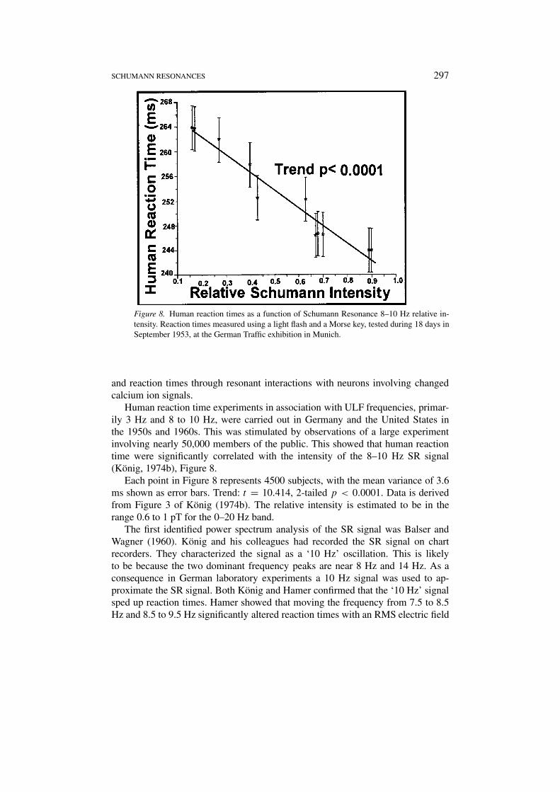
Figure 8. Human reaction times as a function of Schumann Resonance 8–10 Hz relative in-tensity. Reaction times measured using a light flash and a Morse key, tested during 18 days inSeptember 1953, at the German Traffic exhibition in Munich.
and reaction times through resonant interactions with neurons involving changedcalcium ion signals.
Human reaction time experiments in association with ULF frequencies, primar-ily 3 Hz and 8 to 10 Hz, were carried out in Germany and the United States inthe 1950s and 1960s. This was stimulated by observations of a large experimentinvolving nearly 50,000 members of the public. This showed that human reactiontime were significantly correlated with the intensity of the 8–10 Hz SR signal(König, 1974b), Figure 8.
Each point in Figure 8 represents 4500 subjects, with the mean variance of 3.6ms shown as error bars. Trend: t = 10.414, 2-tailed p < 0.0001. Data is derivedfrom Figure 3 of König (1974b). The relative intensity is estimated to be in therange 0.6 to 1 pT for the 0–20 Hz band.
The first identified power spectrum analysis of the SR signal was Balser andWagner (1960). König and his colleagues had recorded the SR signal on chartrecorders. They characterized the signal as a ‘10 Hz’ oscillation. This is likelyto be because the two dominant frequency peaks are near 8 Hz and 14 Hz. As aconsequence in German laboratory experiments a 10 Hz signal was used to ap-proximate the SR signal. Both König and Hamer confirmed that the ‘10 Hz’ signalsped up reaction times. Hamer showed that moving the frequency from 7.5 to 8.5Hz and 8.5 to 9.5 Hz significantly altered reaction times with an RMS electric field
298 NEIL CHERRY
intensity of 3.8 mV/m. König (1974) notes that this level is close to the natural SRsignal strength (about 1 mV/m).
König also found that a superimposed epoch analysis related to the arrival of3 Hz signals from locally generated thunderstorms, significantly slowed reactiontimes. This was tested and confirmed in a series of laboratory experiments usinghuman volunteers. König found that with a range of field strengths, 1 to 5 V/m, the‘3 Hz’ signal consistently slowed people’s reactions and a ‘10 Hz’ signal consist-ently accelerated people’s reaction times. Reactions were also correlated with themore objective test for galvanic skin response (GSR), using a 5 V/m 3 Hz signal,König (1974b).
17.2. CIRCADIAN ELF ZEITGEBER EXPERIMENTS
Support for the role of the SR signal as a diurnal Zeitgeber came from the MaxPlanck Institute’s long-term isolation experiments (Wever, 1974). The Max PlanckInstitute set up an elaborate, large and careful experiment to investigate the hy-pothesis that there was a natural electromagnetic field, such as the SchumannResonance, that acted as a circadian Zeitgeber. This was promoted because ofKönig’s experiments. Because the daily sunlight cycle is a very dominant Zeit-geber, the hypothesis was tested by constructing two identical rooms in whichall signs of the usual daily variation, sunlight, temperature, humidity etc., wereremoved. The second room (Room 2) was also shielded to significantly reduce allexternal oscillating electromagnetic fields by over 40 db.
From introductory experiments, rectal temperature was identified as a reliableobjective measure of the daily circadian rhythm. In the isolation rooms the freerunning day length was significantly lengthened in Room 1 from 24 to 24.87 hr. InRoom 2 it was significantly longer than Room 1, 25.26 hr, p < 0.01. The standarddeviation of the variation of day lengths was also significantly higher in Room2 compared with Room 1, p < 0.01. An additional feature was a phenomenontermed internal desynchronization. In this case individuals showed much longerand highly erratic daylengths. While 15 of 50 subjects in the shielded Room 2internally desynchronized, none of the 34 subjects did in Room 1, p < 0.001. Thedesynchronized subjects were followed up because of their evident sensitivity.
König had characterized the primary mode of the SR signal as a ‘10 Hz’ signal.Hence Wever’s team used a square-wave 10 Hz, 2.5 V/m signal in Room 2 to seewhat would happen if a Schumann-like signal was introduced. Without the subjectsknowledge, a 10 Hz signal was turned on and off at varying intervals. This signalconsistently removed the desynchronization and reduced the mean daylength forthose subjects (Wever, 1967, 1968). The longer the circadian cycle in the absenceof the field, the greater was the shortening by the 10 Hz signal, r = 0.928, n =10, p < 0.001. This showed that not only was a highly significant phenomenonoccurring, but it also varied in an very significant and reasonable manner.
SCHUMANN RESONANCES 299
Wever (1974) concludes that his team found “significant proof that electromag-netic fields in the ELF range influence human circadian rhythms, and therefore,human beings”. They proved that there is a ULF/ELF electromagnetic signal thatis a circadian Zeitgeber. By design the corrective signal mimics an aspect of the SRsignal. While it could be another signal but there is no other known signal has theappropriate characteristics. Hence it is highly likely that the ULF/ELF circadianZeitgeber is the SR signal.
Taken together, the experiments of König, Hamer and Wever give very strongsupport for the hypothesis that the SR signal is involved as a diurnal Zeitgeber,that it is detectable by the human brain and that it causes induced alterations inreaction times. This is highly consistent and gives considerable support for the SRhypothesis.
Laboratory evidence that ULF/ELF signals alter reaction times, animal andhuman brain waves, neuron cell calcium ions and neurotransmitters gives furthersupport. This makes the hypothesis biophysically plausible and observationallysupported. It is therefore largely, but not finally, confirmed.
17.3. EVIDENCE OF ULF/ELF INDUCED MELATONIN REDUCTION
The SR signal has ULF/ELF frequencies. Hence evidence that ULF/ELF electro-magnetic fields reduce melatonin or increase serotonin in animals and people isrelevant to the Hypothesis and its GMA Melatonin Mechanism.
There is extensive evidence that ELF signals increase serotonin and reducemelatonin in animals and in people. Rosen et al. (1998) state that seven differ-ent laboratories have reported suppression of nighttime rise in pineal melatoninproduction in laboratory animals. They show that a 50 µT, 60 Hz field with a0.06 µT DC field, over 10 experiments, averages a significant 46% reductionin melatonin production from pinealocytes. Yaga et al. (1993) showed that ratpineal response to pulsed ELF magnetic fields varied significantly during the light-dark-cycle. They showed that magnetic field exposure significantly suppressed therate-limiting enzyme in melatonin synthesis, N-acetyltransferase (NAT) activity,during the mid-to-late-dark phase.
Suppression of melatonin in rodents is frequently observed when they are ex-posed to weak electromagnetic fields. A question has been raised as to whether thepineal gland or the eyes are the sensors of the fields. Brendel et al. (2000) carriedout an experiment to resolve this question using hamsters. They concluded thatthere was significant suppression of melatonin with 16.7 Hz, p < 0.01, and with50 Hz, p < 0.001 as a result of primarily mechanisms in the pineal gland.
Stark et al. (1997) observed a significant increase in salival melatonin in a groupof 5 cows when the short-wave radio transmitter at Schwarzenberg, Switzerland,was turned off for three days, compared to 5 cows that had much lower RF expos-ure. Hence, despite the high natural levels of variability of melatonin, there are now
300 NEIL CHERRY
at least ten independent observations of significant melatonin reduction in animalsfrom ULF/ELF and RF exposure.
Many human studies that show significant alteration of the melatonin/serotoninbalance by electromagnetic fields. Wang (1989) observed a dose-response increasein serotonin in workers exposed to ELF fields and Davis (1997) a dose-response de-crease in melatonin in workers. Human melatonin reduction studies from ULF/ELFelectromagnetic fields include: Wilson et al. (1990), Graham et al. (1994, 2000),Pfluger and Minder (1996), Arnetz et al. (1996), Davis (1997), Wood et al. (1998),Karasek et al. (1998), Burch et al. (1998, 1999a, 2000) and Juutilainen et al.(2000). For a residential population exposed to a SW radio signal in Switzerlandthe melatonin rose significantly after the tower signal was turned off (Abelin,1999). Two studies directly involve correlation of melatonin reduction with GMAvariation (Burch et al., 1999b; Rapoport et al., 1998).
Thus there is very strong evidence that ULF/ELF electromagnetic fields reducemelatonin in people and in animals. This occurs down to very low mean intensitylevels, with dose- response relationships, and in association with GMA. Undernormal criteria this level of evidence would be assessed as a causal biologicaleffect.
17.4. S-GMA AND MELATONIN REDUCTION
In addition to Burch et al. (1999b) and Rapoport et al. (1998), additional dir-ect evidence that reduced melatonin is significantly correlated with S-GMA isavailable. Bardasano et al. (1989) observed an extremely significant reduction(p < 0.001) in synaptic ribbons of pinealocytes of rats during geomagnetic stormscompared with quiet solar days. Thyroxine levels in a single limbic epileptic patientwere highly correlated (r = 0.66) in a dose-response manner, with daily GMA(O’Connor and Persinger, 1996). The strongest association (r = 0.76) was foundbetween thyroxine levels and the Kp index during the previous night (2 am to5 am). These analyses were carried out specifically to test the GMA Melatoninmechanism and they support it.
Seasonal melatonin variation in rats was correlated with the seasonal variationin the earth’s geomagnetic field, Bartsch et al. (1994). A group of hospital pa-tients with cardiovascular pathology and a control group of healthy people wereboth monitored during magnetic storms. In both groups magnetic storms increasedthe cortisone, activated the sympathoadrehenal system and reduced the melatonin(Rapoport et al., 1998).
Burch et al. (1999a, b) measured urinary melatonin metabolite in 149 work-ers exposed to 60 Hz magnetic fields. Reduced melatonin was correlated with3-phase conductors exposure, cellphone use and overall magnetic fields, with adose-response decrease in workers exposed to low light levels. When all of theseeffects were removed from the data, it also showed a highly significant reductionof melatonin for GMA fields above 35 nT, p < 0.01. When the data was stratified
SCHUMANN RESONANCES 301
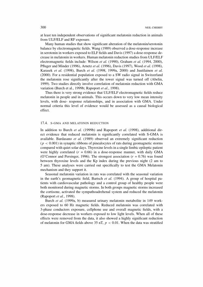
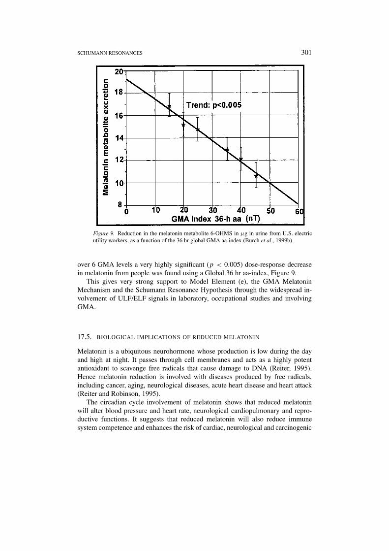
Figure 9. Reduction in the melatonin metabolite 6-OHMS in µg in urine from U.S. electricutility workers, as a function of the 36 hr global GMA aa-index (Burch et al., 1999b).
over 6 GMA levels a very highly significant (p < 0.005) dose-response decreasein melatonin from people was found using a Global 36 hr aa-index, Figure 9.
This gives very strong support to Model Element (e), the GMA MelatoninMechanism and the Schumann Resonance Hypothesis through the widespread in-volvement of ULF/ELF signals in laboratory, occupational studies and involvingGMA.
17.5. BIOLOGICAL IMPLICATIONS OF REDUCED MELATONIN
Melatonin is a ubiquitous neurohormone whose production is low during the dayand high at night. It passes through cell membranes and acts as a highly potentantioxidant to scavenge free radicals that cause damage to DNA (Reiter, 1995).Hence melatonin reduction is involved with diseases produced by free radicals,including cancer, aging, neurological diseases, acute heart disease and heart attack(Reiter and Robinson, 1995).
The circadian cycle involvement of melatonin shows that reduced melatoninwill alter blood pressure and heart rate, neurological cardiopulmonary and repro-ductive functions. It suggests that reduced melatonin will also reduce immunesystem competence and enhances the risk of cardiac, neurological and carcinogenic
302 NEIL CHERRY
disease and death through reducing its antioxidant activity. These predictions arechecked against clinical studies.
17.6. MELATONIN REDUCTION AND HEALTH EFFECTS
Reiter and Robinson (1995) and Brzezinski (1997) reviewed the clinical studiesinvolved with reduced melatonin. Brzezinski identifies roles for melatonin in sleepand circadian rhythm, mood, sexual maturation, reproduction, cancer, immune sys-tem response and aging. Reiter and Robinson confirm all of the effects identifiedby Brzezinski, and add arthritis, asthma, diabetes, hypertension, blood clotting andstroke, cardiac arrhythmia, ischemic heart disease, heart attack, epilepsy, manicdepression, suicide, sudden infant death syndrome (SIDS), Alzheimer’s and Par-kinson’s Diseases. Published papers are cited to justify each of these health effects.Most of the associated conditions relate to the oxidative damage by free radicalsand melatonin’s multiple roles as a potent antioxidant, sleep enhancer and immunesystem booster. Melatonin also acts as a neurohormone and a cellular messengerwith receptors in the nuclei of many cells.
In his conclusion Brzezinski favours high clinical doses for melatonin-relatedtherapy. However he cites three trials where 0.1 to 0.3 mg/day was effective atenhancing sleep. Reiter also cites the M.I.T. study of Dollins et al. (1994). He usesit to confirm the long known effect of melatonin on sleep. In the M.I.T. study, thebiggest incremental effect of melatonin on sleep onset latency and sleep durationwas the first 0.1 mg dose. With at least two other studies confirming the M.I.T.study this confirms that very low daily doses of melatonin have very significantclinical effects.
Since S-GMA significantly reduces melatonin in a dose-response manner, allof these health effects have the potential to be associated with S-GMA. There areseveral primary effects that are of particular interest in S-GMA related studies.They are cancer, SIDS, cardiac, neurological disease and death, including heartattack and suicide. These are all related to reduced melatonin in clinical studiesand through the diurnal regulatory mechanisms.
18. S-GMA Related Health Effects
18.1. PRINCIPLES AND PROBLEMS WITH ENVIRONMENTAL EPIDEMIOLOGY
STUDIES
In seeking to identify and confirm environmental disease agents human populationsare studied in attempts to correlate health effects with exposures to agents. Oftenother factors produce complex variations in the health effects. These are calledconfounders. Methods have been developed to deal with complex situations andto identify individual agents where possible. Time series analysis where cyclicfrequencies can identify common features can be helpful. Multiple regression ana-
SCHUMANN RESONANCES 303
lysis can also assist. When erratic events are involved the problem can be reducedsignificantly.
In seeking to identify the effects of natural electromagnetic fields generated bysolar activity through the Geomagnetic System, the ‘disease agent’, extremely lowintensity electromagnetic fields, are being masked and interfered with in developedsocieties with power supply systems, telecommunications, appliances, computers,etc. This means that earlier records and records from less developed populationscould well have ‘cleaner’ relationships with S-GMA indices. This effect is referredto by Villoresi et al. (1998) who found significant correlations between heart attackincidence rate and GMA storm events in the data from 14 large hospitals in StPetersburg for 1989–1990. Weekly analysis showed a rather constant rate duringMonday to Friday and sharp decrease in rates over the weekend and during mid-week festivities. They associated this with different EMR exposures during workdays and holidays, mainly due to the electrified public transport. This is confirmedby studies showing significantly higher hypertension and coronary heart disease inelectric train drivers (Ptitsyna et al., 1996).
To illustrate the principles and problems, an analysis of annual time series datafor suicide in Southeast Asia was carried out for the period 1974–1992. The datawas extracted from Table II-3-24-1 from a report of the Southeast Asian MedicalInformation Centre, SEAMIC (1997). Suicide is a good bio-indicator for this pur-pose because of its clear diagnosis and its direct connection to clinical depressionand altered melatonin homeostasis. Data is given for the Philippines, Thailand,Singapore, and Japan. The period and the range of countries, give a range of de-velopment, stress levels, health recording systems and EMR exposures. The orderof the countries reflects increasing development, health recording systems and ex-posures to artificial EMR. The data shows an increase in the recorded annual meanmale suicide rate from 0.66 in the Philippines, 6.9 in Thailand, 12.4 in Singapore,and 23.1 in Japan (per 100,000 population).
The period covers just less than two solar cycles with two sunspot maximumsin 1979/80 and 1989/90 and sunspot minimum in 1986. The suicide rates are gen-erally higher in the later half of the period than the earlier years. When lineartrends are removed it is found that Thailand and the Philippines are positivelycorrelated with the sunspot cycle. In the highly developed Singapore and Japan itis weakly negatively correlated. The principle of signal noise level associated withmore EMR exposures in developed societies, and the more complete and betteraccurate data recording, promotes Thailand as the most likely country to detect asunspot cycle in the annual suicide data. This relationship has been expressed asannual sunspot number vs the annual male suicide rate in Figure 10.
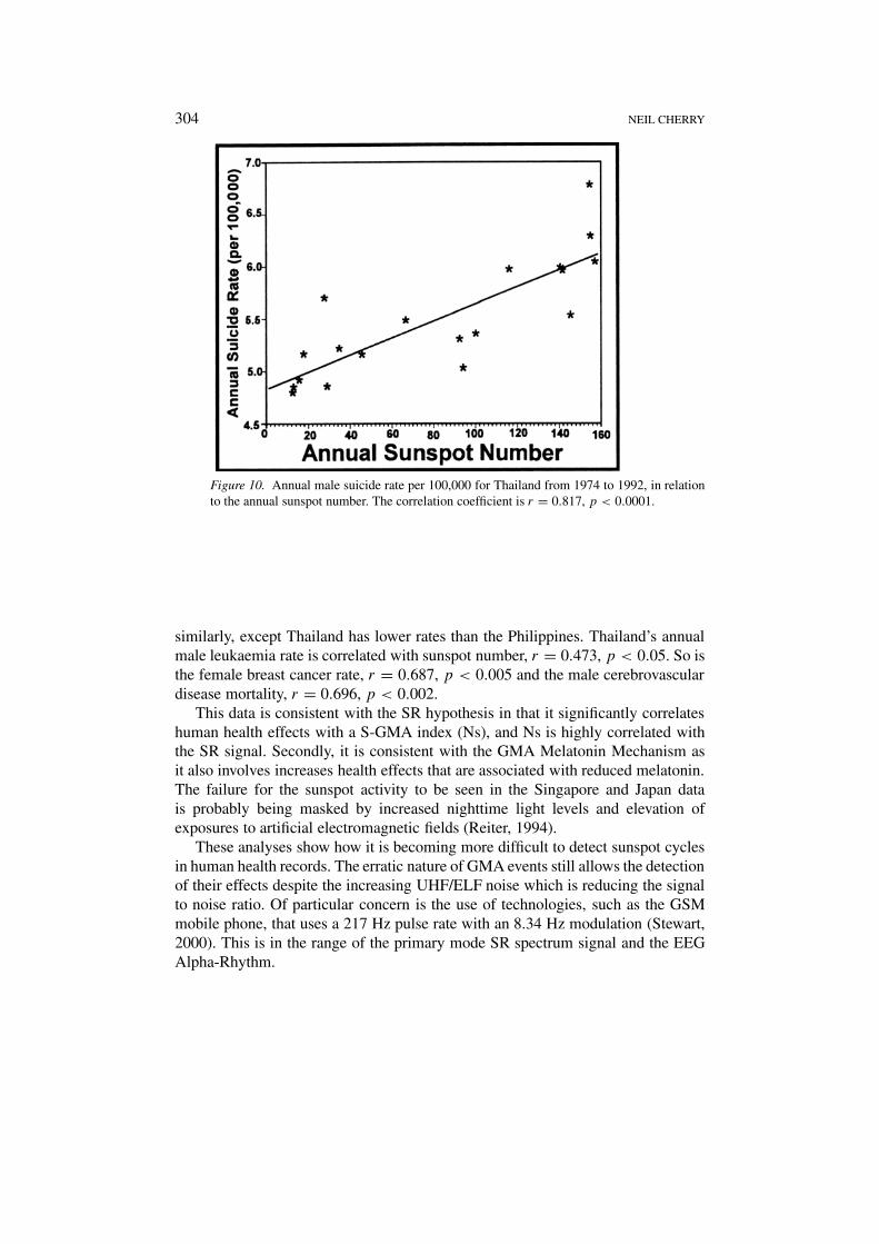
Figure 10 shows an extremely significant correlation between annual sunspotnumber and annual male suicide rate in Thailand over the period 1974–1992. Thisis consistent with the S-GMA melatonin mechanism.
Risk of cancer, especially leukaemia and breast cancer, is associated withmelatonin reduction. In this data-set the four countries cancer rates are ranked
304 NEIL CHERRY
Figure 10. Annual male suicide rate per 100,000 for Thailand from 1974 to 1992, in relationto the annual sunspot number. The correlation coefficient is r = 0.817, p < 0.0001.
similarly, except Thailand has lower rates than the Philippines. Thailand’s annualmale leukaemia rate is correlated with sunspot number, r = 0.473, p < 0.05. So isthe female breast cancer rate, r = 0.687, p < 0.005 and the male cerebrovasculardisease mortality, r = 0.696, p < 0.002.
This data is consistent with the SR hypothesis in that it significantly correlateshuman health effects with a S-GMA index (Ns), and Ns is highly correlated withthe SR signal. Secondly, it is consistent with the GMA Melatonin Mechanism asit also involves increases health effects that are associated with reduced melatonin.The failure for the sunspot activity to be seen in the Singapore and Japan datais probably being masked by increased nighttime light levels and elevation ofexposures to artificial electromagnetic fields (Reiter, 1994).
These analyses show how it is becoming more difficult to detect sunspot cyclesin human health records. The erratic nature of GMA events still allows the detectionof their effects despite the increasing UHF/ELF noise which is reducing the signalto noise ratio. Of particular concern is the use of technologies, such as the GSMmobile phone, that uses a 217 Hz pulse rate with an 8.34 Hz modulation (Stewart,2000). This is in the range of the primary mode SR spectrum signal and the EEGAlpha-Rhythm.
SCHUMANN RESONANCES 305
18.2. S-GMA RELATED REPRODUCTIVE EFFECTS
The fetus has melatonin receptors to detect its mother’s diurnal cycle. Sparks andHunsaker (1988) reported the results of necropsy samples of infants who died ofSudden Infant Death Syndrome (SIDS). They found that their pineal gland wassignificantly smaller than aged-matched controls, p < 0.005. In follow up meas-urements of 111 SIDS infant’s showed smaller pineal glands in 106 infants, raisingthe significant to p < 0.0001. This strongly implied that reduced melatonin wasassociated with SIDS. This suggests that since GMA is strongly correlated withreduced melatonin then it could well enhance the rate of SIDS.
O’Connor and Persinger (1997) showed the GMA is correlated with SIDS. Theyused monthly SIDS statistics from 1960–1961 in Ontario, Canada. They comparedthe monthly SIDS rate with the monthly average aa GMA index. The data wasstratified within the levels of aa in 10 nT intervals. This showed that there was ahighly significant increase in SIDS for the number of days with aa in the 11 to 20nT range, r = 0.91, p < 0.001, and in the 31 to 40 nT range, r = 0.65, p < 0.01.For the intermediate range, 21 to 30 nT there was a highly significant reduction ofSIDS, r = −0.79, p < 0.001. Their strongest correlation occurred for a reductionof SIDS between days with 21 to 25 nT, r = −0.96, p < 0.0001. This showsa homeostatic relationship between the GMA level and the rates of SIDS with farlower SIDS rates near the mean GMA level 21–30 nT and significantly higher ratesfor lower and higher GMA levels.
The possible role of micropulsations (PC1: 0.2–5 Hz) on SIDS rates duringperiods of very low GMA was then studied, O’Connor and Persinger (1999). Us-ing a similar method they found that stratifying the data into 11, 5 nT intervallevels, there was a marginally significant correlation with increased micropulsa-tions levels, r = 0.63, p < 0.05. Consistent with O’Connor and Persinger (1997),they also found a negative correlation r = −0.79 at an intermediate level of PC1,15 to 20 nT, suggesting a homeostatic effect. They noted that the PC1’s usually oc-cur in 1 to 4 hour periods over 5 to 7 quiet days following an isolated geomagneticstorm. This shows that the effects would also be correlated with the SR signal thathas the same pattern of lasting up to 7 days after a solar storm, Figure 5.
18.3. S-GMA RELATED CARDIAC EFFECTS
A 35-year old cardiologist, with a family history of hypertension and stroke, usedan electronic blood pressure monitor to record his blood pressure every 15 minutesfor 3 years. This revealed a significant periodicity of 27.7 days in systolic anddiastolic blood pressure and heart rate, which was coherent with the GMA Kp-index (Watanabe et al., 1994).
An Italian study of 447 patients with hypertension also found very significantcorrelations between systolic and diastolic blood pressure and GMA indices overa 5-year period (Ghione et al., 1998). A multiple correlation with potential con-founding factors, such as age and date, confirmed the significant correlation with
306 NEIL CHERRY
GMA. Stratifying the days into quiet, disturbed and highly disturbed GMA daysconsistently showed significantly higher values in the highly disturbed days for allblood pressure parameters, except for systolic night-time pressure. The differencebetween quiet and highly disturbed GMA days was 6 to 8 mm for the 24 hoursystolic and diastolic blood pressure. The GMA indices and the blood pressuremeasurements contain the 27-day period. The authors concluded that these res-ults seem to reflect a real relation between geomagnetic disturbances and bloodpressure.
Two independent studies show that human blood pressure is significantly cor-related with GMA. Melatonin is a diurnal blood pressure regulator and S-GMAmodulates human melatonin levels, therefore these studies confirm that bloodpressure change is a melatonin-related biological effect of S-GMA. Hence it isbiologically plausible that extreme levels of S-GMA will cause a wide range ofcardiac health effects and death.
Reduced melatonin produces arrhythmic cardiac activity. The cardiac activityof rabbits was monitored during two GMA storms (Chibisov et al., 1995). At theinitial and main phase of the storm the normal circadian structure of the cardi-ovascular parameter was lost. Desynchronization grew during the storm, leadingto an abrupt drop of cardiac activity during the main phase of the storm. This wasfollowed by the destruction and degradation of cardiomyocytes. The parametersof cardiac activity became significantly synchronized and the circadian rhythmrestored during the storm’s recovery period.
Human patients with ischemic heart disease (47-men and 33-women) weremonitored for cardiac parameters daily over for 2–3 weeks (Gurfinkel et al., 1995).Changes in their microcirculations were related to GMA and to changes of atmo-spheric pressure. In the first day of a GMA storm pathological changes of capillaryflow were detected in 71.5% of patients with acute myocardial infarction (men:73.7%, women: 69.2%). They also observed perivascular edema, red blood aggreg-ation, delay and slowing down of capillary flow. Similar changes were detected in64.8% of patients with angina pectoris, (men: 73.3%, women: 56.3%). The reac-tions of these patients to GMA disturbances were over 2.5-times higher than theeffects of atmospheric pressure changes.
GMA events are significantly correlated with increased blood coagulation andplatelet aggregation (Pikin et al., 1998). Blood pressure, capillary flow, blood co-agulation and aggregation changes are observed during GMA events, consistentwith the effect expected with reduced melatonin in people with heart disease.Therefore, it is reasonably predicted that GMA will be associated with observablechanges in cardiac disease and death when large human populations are studied.
18.4. GMA RELATED HUMAN CARDIAC DISEASE AND DEATH
Early correlations between S-GMA and heart attacks were assumed by some au-thors to be spurious, inaccurate and inconsistent (Malin and Srivastava, 1979, 1980;
SCHUMANN RESONANCES 307
Knox et al., 1979). Results found in India were not confirmed in populations in theU.S.. These were seen as inconsistent. The lack of a plausible mechanism alsomade these results not credible to some researchers. Artificial EMR exposures indeveloped countries masking the natural signals effects is a plausible explanationof the results. In the 1990’s many other studies identified relationships that arehighly significant and consistent with the original results.
With clinical measurement being able to identify highly significant changes inblood pressure, blood flow, aggregation and coagulation during GMA events, theseresults are highly plausible. They are mediated by melatonin in the normal diurnaland seasonal cycles. Since melatonin is also significantly correlated with levels ofGMA during solar storms this will also have cardiac effects. Reduced melatoninis associated with cardiac arrhythmia and heart rate variability in clinical studiescited above.
De Bruyne et al. (1999) studied older heart patients (>55 years) and com-pared their heart rate variability (HRV) with their increased risk or mortality frommyocardial infarction. They found that both decreased and increased HRV weresignificant risk factors, with increased HRV being the greater risk factor. Thisshows a timing related homeostatic relationship and GMA events are related todesynchronization of cardiac rhythms. Measured HRV also demonstrates anom-alies in myocardial infarction, sudden death, heart failure, autonomic neuropathyand hypertension (Kerut et al., 1999).
The EEG pattern, pulse rate, blood pressure and rate of sensomotor reactionwas measured in a group of people. The parameters significantly correlated thesephysiological variables with the Kp-index (Doronin et al., 1998). They noted thatthe oscillations in the Kp-index had identical periods in the monitored EEG Alpha-Rhythm. This confirms the whole-body changes that occur in conjunction withGMA alteration by changing the brain and heart patterns. This supports the modelthat suggests that the brain wave pattern is changed, involving alteration of ELFbrain signals, and this is transferred through melatonin receptors and the autonomicnervous system to the cardiovascular system.
18.5. CARDIAC EFFECTS OF HIGH GMA
During periods of Active Sun and increased GMA the following statisticallysignificant effects have been observed:
• Cardiac arrhythmia in children (Markarov, 1998).• Novikova and Ryvkin (1977) observed a consistent and significant increase in
heart attack incidence and death between active and quiet GMA conditions for1961–66 at Sverdlovsk, USSR.
• GMA is highly correlated with daily myocardial infarction incidence ratesduring big GMA storms (Villoresi et al., 1998).
• GMA activity is also correlated with sudden cardiovascular death (Sitar, 1990),and ischaemic heart disease mortality (Otto et al., 1982).
308 NEIL CHERRY
• Monthly solar activity was highly significantly correlated with monthly hos-pital admissions for cardiovascular disease (Stoupel and Shimshoni, 1991).Solar activity is highly correlated with GMA and SR intensity.
• Stoupel et al. (1997) observed that during periods of low solar and geomagneticactivity, solar proton fluxes were correlated with cardiovascular deaths.
• Oraevskii et al. (1998a) found that 75% of GMA storms caused an increasedof the hospitalization of patients with myocardial infarction by 30 to 80%.
• Oraevskii et al. (1998b) report that MIR space orbital station staff experienceda significantly increased heart rate, reduced heart rate variability and decreasedrespiratory waves, corresponding with a specific adaptation of stress-reaction.At the same time hospital patients with ischemic heart disease had a similarreaction including deterioration of the physiological status, rheologic bloodcharacteristics and heart rate disturbances, associated with GMA disturbances.
• Breus et al. (1998) report disturbance of cardiovascular activity among MIRastronauts during the main phase of solar storms compared to the recoveryphase. Similar effects were observed in rabbits.
18.6. CARDIAC EFFECTS OF LOW GMA
Periods of Quiet Sun activity are significantly associated with:• Stoupel et al. (1990) found a highly significant correlation (p = 0.01) for
higher pregnancy induced hypertension for monthly periods of low GMA.• Sudden death from cardiac arrhythmia, especially paroxymal atrial fibrillation,
and stroke (Stoupel, 1993; Stoupel et al., 1995a; Stoupel et al., 1994). Stoupelet al. conclude that their results are consistent with previous studies show-ing increased heart electrical instability during periods of lowest geomagneticactivity.
• Ischaemic heart disease for ages > 70 years (Stoupel et al., 1995b).• Stoupel et al. (1999) found an very highly significant inverse correlation
(r = −0.64, p = 0.0001) for a 72 month period between solar activityand stroke/ischemic heart disease death. They concluded that monthly ratio ofdeaths from stroke/ischemic heart disease is related to environmental physicalactivity.
The cardiac studies are consistent with the Schumann Resonance Hypothesis andadd considerable weight to the melatonin, homeostatic and arrhythmic factors inthe Hypothesis. Blood pressure, blood coagulation, heart attack, cardiac arrhythmiaand sudden cardiac death are highly correlated with GMA in a homeostatic (Ushaped) manner. This data is consistent with the involvement of melatonin. Beingdirectly supported by clinical cardiovascular monitored changes of blood pressure,capillary flow and blood aggregation, multiple studies and very highly significantcorrelations with solar activity and GMA. This gives robust evidence supportinga causal relationship between GMA and ischemic and arrhythmic cardiovasculardisease, heart attack and death. The highly significant correlation between S-GMA
SCHUMANN RESONANCES 309
and the SR signal intensity gives additional support for the SR Hypothesis througha Melatonin Mechanism.
18.7. NEUROLOGICAL EFFECTS
The brain is a very sensitive electromagnetic organ. The model and hypothesisproposes a direct mechanism for SR signals to interact with the brain, altering thebrain waves and neurohormone responses. Altered reaction time is a prompt andacute indication of this interaction. It has been shown that exogenous ELF signalsaffect melatonin/serotonin, dopamine and opiate systems (Frey, 1995). This effectis plausible through interference with endogenous ELF systems and the vital roleof calcium ion signalling. Melatonin reduction is directly correlated with S-GMAlevels that are highly correlated with a natural exogenous ELF field, the SR signal.Given the biological effects of reduced melatonin, this predicts that there couldwell be a wide range of melatonin related neurological effects correlated with solarcycles and GMA events. A large number of studies have been carried out and aresummarized below.
People with epilepsy are primary subjects for the detection of acute effectsof GMA on human neurological functions because of the frequency-based dys-function that they suffer from. Karlov et al. (1996) compared the reactions of 18healthy individuals and 20 epileptic patients to magnetic fields modulated in theEEG frequency range. They showed that alteration of the magnetic field elevatedthe functional activity of the brain synchronizing structures and increased eitherthe epileptic activity or activation of the epileptic focus. Sandyk and Anninos(1992a, b) report success in reducing epileptic seizures with picoTesla ULF mag-netic fields with SR-like frequencies. Sandyk (1992) relates these results to thealteration of the EEG alpha rhythm (8–13 Hz), pineal melatonin and magnetic fieldaltered circadian seizure incidences.
Belisheva et al. (1995) concluded that their observations showed that localGMA variations can be a principle reason for modulation of the brain’s func-tional state. They also conclude that this means that an optimal level of GMA,manifested in periodic oscillations in certain amplitude frequency range is de-manded for steady brain’s function state. The decrease of optimal GMA activitylevel and the appearance of aperiodic disturbances of GMA are associated with theunsteady brain state. This gives strong support for the role of the SR spectrum inthe homeostasis of brain activity.
The EEG rhythms, pulse rate, blood pressure and rate of sensomotor reactionon a group of people were significantly correlated with the Kp index (Doronin etal., 1998). They noted that the oscillations in the Kp index had identical periodsto the monitored EEG alpha-rhythm. This confirms a GMA-related modulation ofthe EEG alpha rhythm, which could well be the SR signal. This was independentlyconfirmed by Belov et al. (1998), who monitored the EEG rhythms in 26 humansubjects.
310 NEIL CHERRY
A positive correlation between the EEG data and GMA was revealed. It wasmost obvious in the frontal and central lobes of the brain. A negative correlationbetween some local EEG synchronization and indices of solar activity was alsoobserved. Belov et al. (1998) concluded that the degree of synchronization of thespontaneous EEG pattern seemed to reflect sensitivity of the human nervous systemto the earth’s magnetic field. This confirms a role for a S-GMA-related mechanism,most likely involving the SR signal. They observed a stressor response to strongGMA events and a sedative effect from low frequency magnetic oscillations. Theseresults are consistent with the experiments of König and of Hamer who showed thatthe alpha-rhythm related 10 Hz signal stimulated people and the 3 Hz delta-rhythmrelated signal slows people.
18.8. NEUROLOGICAL EFFECTS OF HIGH GMA
• Tambiev et al. (1995) found a significant correlation between GMA andmemory and attention. Aviation accidents are positively correlated with solarstorms (Komarov et al., 1998). Through cyclic analysis the authors concludedthat there was a causal relationship because of strong frequency matchingbetween aviation accidents and GMA event cycles.
• Neurological effects were confirmed by Selitskii et al. (1999) who, by arti-ficially reducing GMA, were able to increase the synchronization of alpha-rhythm EEG and generalized slow-wave discharges in people with epilepsy.This shows that increased GMA produces de-synchronization of the EEGalpha-rhythm and results in significantly more frequent convulsive seizures.
• GMA disturbances suppress the cortical reactivity to external receptive stimuliand raise the functional activity of the cerebral inhibitory components, such asthe caudate nucleus in awake cats (Makarova, 2000).
• A total of 40 men and women were tested for the effects on their writing ofpleasant and unpleasant stories and changes in the local geomagnetic field(Persinger et al., 1994). A double-blind method was used in the applicationof 10 mG fields over the right and left temporal lobes both separately andtogether. Men and women receiving the stimulation showed significantly morepleasantness when the right hemisphere was exposed than the left hemisphere.Covariance with changes in the natural geomagnetic field was negativelycorrelated with pleasantness, increasing the significance of the experimentaltreatment.
• A group of 127 human volunteers were studied over a 4-year period and wereobserved to show a step-like increase in partial sensory deprivation (mainlyhearing and seeing) for GMA levels above 15 nT, r = 0.33. The strongestcorrelation was found for the GMA during the 3-hr period at the beginning ofthe previous nights sleep (Persinger and Richards, 1995).
SCHUMANN RESONANCES 311
• Rats being studied for chronic limbic epileptic effects showed significantlymore aggression (r = 0.5) during GMA events (Persinger, 1997). Intenseaggression was observed for aa > 40 nT.
• St. Pierre and Persinger (1998) found that aggressive biting in rats was posit-ively correlated with GMA. Persinger (1999) identified a moderate associationbetween annual GMA and the probability of war in the same year.
• Avdonina and Samovichev (1995) evaluated relationships between S-GMAand the days that serial crimes were committed during 1980–1990 in USSR.The analysis of 150 episodes (ten separate series) shows that the crimes aremost probable immediately or 1–3 days after sharp decrease of solar activity(Wolf Numbers), geomagnetic activity (A3 and K indices) and of the lowestdaily surface and air temperature.
• Bureau and Persinger (1995) investigated the time taken to initiate an epilepticseizure as a function of the level of GMA. They found that the latency de-creased in a dose-response manner with GMA level. A 20-25 nT level reducedthe latency by 12%. Bureau et al. (1996) were studying rats with inducedlimbic seizure hypothermia. During high GMA, >35 nT, depth of hypothermiawas significantly reduced (p < 0.001). In the subsequent 5 days mortality wasincreased when GMA >20 nT.
• In three separate experiments, Michon et al. (1996) tried to clarify the meansby which GMA levels affected spontaneous epileptic seizures in rats. A 700 nTconstant intensity signal inhibited the seizures. At lower levels, in the range ofnatural GMA a 7 Hz signal, exposure for 5 mins, once per hour, in the periodmidnight to 8 am, increased the incidence of seizures the following morningduring the presentation of food. This appears to explain persistent effects ofGMA on epileptic seizures.
• Rajaram and Mitra (1981) found a very significant, p < 0.01, correlationbetween annual percentage of patients with convulsive seizures in Calcutta andannual mean GMA over the period 1955–1971.
• Epileptic seizures and dizziness are correlated with monthly and annual solaractivity. This revealed significant differences between males and females(Stoupel et al., 1991), Epileptic attacks were highly correlated jointly withGMA and lunisolar tides (Mikulecky et al., 1996).
• Persinger (1995) identified a significant correlation between sudden epilepticdeath following periods high daily average GMA, >50 nT. Monthly incidenceof sudden unexplained death was correlated with the monthly incidence ofGMA events over 50 nT. They concluded that these results supported the roleof the ‘anticonvulsant’ melatonin.
• Persinger (1995a) observed a 35% variance (multiple r = 0.59) for the portionof seizures related to increased GMA at the same time as the seizure and duringthe previous overnight period.
312 NEIL CHERRY
• Renton and Persinger (1998) monitored the partial epileptic-like experiences of12 women over two months. Five of the women displayed significant increaseswhen the daily GMA level exceeded 40 nT.
• Nikolaev et al. (1976) compared a daily variation of a psychopathological in-dex for a group in a Moscow mental hospital with the Ap index over a periodof 9 months. There was a very high correlation.
• Urgent hospitalization for mental disorders and suicide rise significantly withsolar activity, Oraevskii et al. (1998a). Psychiatric admissions increase withsolar radio flux activity (p < 0.05) and sudden magnetic storms (p < 0.01)and decrease with GMA (p < 0.05), Raps et al. (1991). Admission fordepression increases significantly after solar storms (Kay, 1994).
• GMA can be considered as a trigger of migraine attacks (De Matteis et al.,1994). They confirmed this by finding a significant correlation between mi-graine attack and GMA in a small geographic area chosen to minimize theclimatic differences.
• Conesa (1995, 1997) associated high GMA with isolated sleep paralysis.• The geographic incidence of multiple sclerosis is strongly correlated with the
geomagnetic field rather than latitude (Resch, 1995).
18.9. NEUROLOGICAL EFFECTS OF LOW GMA
• Highly anxious pilots have greater anxiety on days of quiet sun (Usenko,1992).
• Conesa (1995, 1997) associated low GMA with vivid dreaming.• Serial crime in Moscow increased 3 days after a low solar activity period
(Avdonina and Samovichev, 1995).• Tunyi and Tesarova (1991) found that suicide, sports injuries, fatal work
injuries, alcoholism are more prevalent during periods of low solar activity.• Stoupel et al. (1995a) found that suicide in those older than 70 years was
greater during periods of low solar activity. Stoupel et al. (1995b) found ona monthly mean basis that suicide was negatively correlated (r = −0.22,p = 0.03) with GMA levels.
• Raps et al. (1992) found negative correlations for psychiatric admissions in-volving 1829 patients between 1977 and 1987, against sudden ionosphericdisturbances (r = −0.274), GMA index (r = −0.216) and hours of positiveionisation in the ionosphere (r = −0.262). All of these indices are positivelycorrelated with the SR signal strength.
18.10. NEUROLOGICAL SUMMARY
Acute clinical effects of altered EEG rhythms have been observed in several stud-ies. Geomagnetic storms are strongly related to human health effects and death.
SCHUMANN RESONANCES 313
Increased GMA, consistent with reduced melatonin, causes arrhythmic brain activ-ity in the form of epilepsy. The occurrence of epilepsy and sudden unexpecteddeath is highly correlated with increased solar activity and GMA storm events inmany studies involving rats and people. Suicide is also strongly correlated withenhanced GMA and solar cycles.
There is weak evidence of effects for low GMA activity. For the elderly, > 70years, the risk is increased for low levels of S-GMA. Low S-GMA has also beenassociated with anxiety, vivid dreaming, injuries and alcoholism.
This adds very strong support for the SR hypothesis that elevated S-GMA altersbrain waves and induces reduced melatonin. Weak support is given for the ‘Ushaped’ homeostatic relationship.
18.11. S-GMA RELATED MORTALITY AND BIRTH RATE
If the above arguments are true then GMA would be correlated with mortalityamongst the very old and very young.
• Zaitseva and Pudovkin (1995) used a multiple regression analysis involvingthe annual sunspot number (Ns) and deviation of the Kp index from averagewith a 30-year mortality and birth rate record, 1867–1897 from Russia. Theydetermined that mortality rate rose significantly (r = 0.78) and birth ratedropped significantly (r = −0.67) with changes in the S-GMA indices Nsand �Kp.
• Shumilov et al. (1998) observed high correlations between traumas, deaths andsudden diseases with both enhanced and very low S-GMA levels, for minersworking at high latitudes in the Spitsbergen mines.
• Kopanev et al. (1979) experimented with rabbits to test the effect of shieldingfrom the geomagnetic field. They found significantly higher mortality in theanimals shielded from GMF, compared to unshielded controls, supporting ahomeostatic factor.
• A 100-fold shielding of guinea-pigs from the earth’s GM field produced areduction in ferment antioxidizing activity and non-ferment mechanisms inthe heart, along with reduced carbohydrate metabolism for lipid metabolism(Babych, 1995). A similar field reduction caused an inhibitory effect, downto complete stoppage, of the ciliate neuronal development of new-born rats(Svanidze et al., 1994; Sandodz et al., 1995).
18.12. CANCER INCIDENCE WITH SUNSPOT CYCLES
SR signals and S-GMA vary significantly with sunspot activity, and S-GMA ishighly correlated with reduced melatonin. Melatonin is a highly potent antioxidantand therefore reduced melatonin causes enhanced DNA damage that can initiate,promote and progress towards cancer. Childhood cancer develops very quickly.Most adult cancer has long latencies. Hence a sunspot cycle related cancer could
314 NEIL CHERRY
cluster increased childhood cancer and cancer initiation around sunspot maximum.Solar cycles could plausibly influence long-term cancer cycles. In a similar mannerto acute air pollution events in relation to cardiovascular death, sunspot maximum,a period of years with reduced melatonin, could cluster and ‘harvest’ those withterminal cancer, accelerating their death. The following studies report some results.
• Riabykh and Bodrova (1992) found a significant correlation between WolfSunspot Numbers and benign breast cancer for women in the middle and latemenarche.
• Dimitrov (1993) found some significant lagged-correlations with malignantmelanoma in Czechoslovakia and solar 7.5 and 11 year cycles.
• Dimitrov (1999) identified significant solar cycle related Malignant Melanomaand Non-Hodgkin’s Lymphoma (NHL) in adult U.S. populations, and Dimitrov(1999a) correlated childhood NHL in the U.S. with solar cycles, p < 0.05.
This study above found significant correlation with male leukaemia and femalebreast cancer in Thailand with annual sunspot numbers. There is consistent butlimited data to correlate cancer with sunspot activity. The possibility that this re-lated to UV radiation was considered. Malignant Melanoma (MM) is linked to UVradiation, which varies very slightly with sunspot activity, 0.16%, Haigh (1996).MM is also significantly elevated in the telecommunications industry in Sweden(Vagero et al., 1985). MM is also associated with the review of ELF exposures inelectrical occupations, Hardell et al. (1995) and with the highest ELF exposuresin Tynes et al. (1994). Hence it is highly probable that the relationships found byDimitrov (1993, 1999) can be related to the SR signal and are unlikely to be causedby sunspot cycle related UV changes.
18.13. OTHER EFFECTS
• Reproductive capacity and survival rates of micro-organisms, Escherichia (EColi), Shigella and Salmonella, were highly correlated with GMA events. Theexperiments that started 2–4 days prior to a GM storm showed suppressionof reproductive capacity followed by its stimulation. If the storm started onthe first day of the experiment then there was a sharp drop in reproductivecapacity and population death occurred within 1 to 5 days. In periods oflow GMA the suppression of reproductive capacity was commonly recorded(Chernoshchekov, 1989).
• Petrichuk et al. (1992) present results of a study of 35 premature and 35 healthychildren over a period of 11 years. GMA was correlated with a range of signi-ficant factors, including a progressive increase in lymphocytes. If intrauterinedevelopment took place during a period of high solar activity, then early devel-opment resulted. For both groups the energetic age-dependent factors vary withthe GMA. Lymphocytes that are associated with fetal and newborn hypoxia areperturbed by a combination of high atmospheric pressure and GMA (Petrichuket al., 1998).
SCHUMANN RESONANCES 315
• Stoupel et al. (1993) found that intra-ocular pressure in relation to four levelsof daily GMA was lowest on stormy days and highest on quiet solar days. Thisis a melatonin-related effect.
• A range of mouse, hamster and pisces cell lines were studied in situations ofchanging GMA (Belisheva and Popov, 1995). Morpho-functional characterist-ics, including the percentage of polynuclear cells, were estimated. GMA causesan abrupt, step-wise change in the state of the cell cultures. Changes in proper-ties of the cell surface related to cell fusion, the appearance of heterokaryons,enlargement of cellular adhesion and aggregation.
• Polikarpov (1996) studied the biological activity in soil, including DNA syn-thesis. The study found that during periods of increased solar and GMA thecell culture S. aureus 209 showed significantly greater biological activity.
18.14. SUMMARY ABOUT S-GMA BIOLOGICAL AND HEALTH EFFECTS
The above studies consistently show that GMA events produce significant biolo-gical changes in micro-organisms (E. Coli and Salmonella) as well as effects onsoil organisms, animal cell-lines, human lymphocytes and eyes. This is in addi-tion to the above studies showing cancer, reproductive factors, cardiac events andextensive neurological effects in people that are largely consistent with melatoninreduction by S-GMA.
There is very firm evidence for extreme variations of S-GMA to be classifiedas a natural hazard for all forms of life on earth. There is strong evidence for theGMA Melatonin Mechanism and robust evidence for the Schumann ResonanceHypothesis. The hypothesis and mechanism are supported by ULF/ELF studiesshowing reduced melatonin in animals and people.
18.15. RESIDENTIAL AND OCCUPATIONAL EXPOSURES PREDICTION
Evaluation of the SR Hypothesis is advanced by evaluating its prediction that resid-ential ELF and RF/MW fields should significantly increase in cancer, reproductive,cardiac and neurological effects in human populations. Typically these fields areabout a thousand times higher in terms of the electric field and a million timeshigher in terms of exposure intensity and the magnetic field. For example a 1 mV/mSR signal converts to 0.27 pW/cm2. A survey of 15 U.S. cities in 1979/80 (Telland Mantiply, 1980), found the median exposure level was 5 nW/cm2. Around 1%of the population was exposed to more than 1 µW/cm2. This is over 1 milliontimes higher than the SR intensity. Living near high voltage powerlines chronicallyexposes people to ELF magnetic fields in the range 0.1 to 0.5 µT, about a milliontimes higher than the 0–20 Hz SR signal at 0.5 pT. If these health effects are foundthen this strengthens both the SR Hypothesis and the GMA Melatonin Mechanism.
316 NEIL CHERRY
19. Electrical Occupation and Residential Health Studies
19.1. CANCER STUDIES
Hardell et al. (1995) reviewed almost 100 studies carried out up to 1994 and de-scribed significant increases in cancer as possible associations with ELF fields.They concluded that there were associations with many cancer types. These in-cluded an increased risk of leukaemia in children and the existence of, or distanceto, powerlines in the vicinity of their residence. Occupation exposure to ELF elec-tromagnetic fields showed an increased risk of chronic lymphatic leukaemia, breastcancer, malignant melanoma of the skin, nervous system tumours, non-Hodgkinlymphoma, acute lymphatic leukaemia or acute myeloid leukaemia. Since 1995many other studies have confirmed, extended and strengthened these conclusions.
For example, Cantor et al. (1995) found that occupational exposure to RF fieldssignificantly increased female breast cancer. In a very large study (Szmigielski,1996), showed that RF/MW exposure from radar and radio in Polish military per-sonnel caused significant increases in cancer mortality across many organs in thebody, including brain tumours (p < 0.05) and leukaemia (p < 0.001). When theleukaemia cases were divided into various types, lymphoma, chronic myelocyticand acute myeloblastic leukaemia were all individually very highly significant,p < 0.001. Feychting et al. (1995) found a dose response increase in childhoodleukaemia with exposure to high voltage powerlines, ≥0.5 µT RR= 5.1 (95% CI:2.1–12.6) and Savitz et al. (2000) found a dose-response increase in brain cancer inelectrical workers with the highest exposure giving RR = 2.5 (95% CI = 1.0–6.3).
19.2. REPRODUCTIVE STUDIES
Pregnant physiotherapists experienced a dose-response increase in early preg-nancy miscarriage having used microwaves for diathermy (Ouellet-Hellstrom andStewart, 1993). Later pregnancy miscarriage was found in physiotherapists usingshortwave diathermy (Kallen et al., 1982; Larsen et al., 1991; Taskinen et al.,1990). Kallen et al. and Larsen et al. also showed significant congenital malform-ation. Miscarriage is also associated with exposed electronic technicians (Vaughanet al., 1990), as well as electric blanket use (Wertheimer and Leeper, 1986; Be-langer et al., 1998). It is also associated with powerline exposure (Juutilainen etal., 1993).
Electrical workers also pass on higher cancer risks to their children (Hicks etal.,1984; Spitz and Johnson, 1985; Johnson and Spitz, 1989; Wilkins and Hund-ley, 1990) Bunin et al., 1990; Wilkins and Wellage, 1996; Olshan et al., 1999;Smulevich et al., 1999; Feychting et al., 2000). This is likely to be from geneticdamage of DNA in the sperm that could be direct, Lai and Singh (1996, 1997), orassociated with reduced melatonin.
SCHUMANN RESONANCES 317
19.3. CARDIAC STUDIES
Cardiac effects include reduced heart rate variability with 60 Hz fields from RFexposure in TV transmission stations (Sastre et al., 1998; Bortkiewicz et al., 1995,1996, 1997). Male physiotherapists had significant dose-response increases inischemic heart disease from both short-wave and microwave exposure (Hamburgeret al., 1983). Naval technicians exposed to radar during the Korean War werediagnosed with significantly higher cardiovascular disease, p < 0.001 (Robinetteet al., 1980). Savitz et al. (1999) carried out the most extensive exposure relatedstudy and identified significantly increased and significant dose-response increasesin cardiovascular disease mortality in electric utility workers. This was particularlyrelated to cases of arrhythmia and acute myocardial infarction. Significantly elev-ated risk occurred for all cumulative exposures above 0.6 µT-years. The consistentelevation and exposure gradient effects were found for arrhythmia and heart at-tack but not for atherosclerosis and chronic coronary heart disease. The latter twowere elevated inconsistently. In contrast, Ptitsyna et al. (1996) found a doubling ofchronic heart diseases in train drivers chronically exposed to ULF fields (0.001–10Hz), consistent with SR frequencies.
19.4. NEUROLOGICAL STUDIES
Children chronically exposed to a radar pulsed at 24.4 Hz showed significantimpairment of learning and physical performance (Kolodynski and Kolodynska,1996). ELF exposed people show significantly more clinical depression (Verkasaloet al., 1997); Amyotropic lateral sclerosis (Deapen and Henderson, 1986; Davani-pour et al., 1997; Savitz et al., 1998; Johansen et al., 1999); Alzheimer’s disease(Sobel et al., 1995, 1996); and senile dementia, epilepsy, and Motor Neuron Dis-ease (Johansen, 2000). Significant increases in suicide from ELF exposures wasreported by Perry et al. (1981) and Baris and Armstrong (1990). This was con-firmed by Van Wijngaarden et al. (2000). They found a significant dose-responseincrease relative to recent ELF exposures with a zero exposure threshold. Theyrelate this to melatonin reduction.
19.5. PUBLIC HEALTH CONCLUSIONS
Multiple published studies show that residential and occupational exposures toelectromagnetic fields and radiation results in significant increases, including dose-response increases in cancer, reproductive, cardiac and neurological health effectsand mortality. The prediction from the SR Hypothesis is very well confirmed. Thereis considerable consistency between the S-GMA studies and the residential andoccupational ELF/RF/MW exposures. The very recent dose-response studies ofELF exposure and heart disease death, suicide and brain cancer all point towards anear zero threshold of ELF exposure for the level of no effect.
318 NEIL CHERRY
19.6. RECOMMENDED RESEARCH TO TEST THE HYPOTHESIS
At the core of the Hypothesis is the postulate that human brains detect and re-spond to the SR signal. The response involves altered EEG rhythms, reaction times,melatonin production, blood pressure and heart rate. These need to be tested dir-ectly in relation to actual or experimental SR signals. Experimental design mustdeal with possible confounders. The physiological parameters could be monitoredduring isolated GMA events using local measurements of the SR signal. Tests needto be developed to detect whether the physiological parameters are more closelyrelated to Kp or the SR signal. Isolation experiments, as used by Wever (1974), maybe necessary. Correlation of SR signals with acute health effects, such as SIDS,cardiac arrhythmia, heart attack and suicide could also be evaluated for indirect butsupportive or challenging evidence.
20. Summary and Conclusions
S-GMA indices are significantly correlated with the biological and health effects.Confirmation of the Schumann Resonance Hypothesis first required that the SRsignal be highly correlated with the S-GMA indices. This would show that the SRsignal was also correlated with the observed effects. The SR signal is shown to bevery highly correlated with sunspot number and the GMA Kp-index. The GMAevent analysis confirms that the characteristics of the SR signal closely follow theKp-index and the electrons and ions density patterns in the D-Region. Hence thisaspect of the hypothesis is confirmed.
The second aspect was the biological plausibility of the SR signal interactingwith the brain and altering the melatonin production. This is theoretically predictedand plausible through resonant absorption that is made possible through frequencymatching and tuned circuits. It is shown conclusively that environmental ULF/ELFelectromagnetic fields at extremely low intensity do interact with brain tissue,changing the calcium ion fluxes, the EEG rhythms and reaction times. Brains andcells use phase-locked loop circuits to detect and respond to natural ULF/ELF elec-tric signals. Laboratory experiments show that ‘Schumann-like’ signals do changehuman and monkey reaction times using ULF to ELF electric field levels thatapproach the SR intensity. Human reaction times are also highly correlated, in adose-response manner, with SR intensity, confirming this interaction.
The Melatonin Mechanism is strongly supported by multiple independent stud-ies showing that ULF/ELF fields reduce melatonin in animals and people. TheS-GMA connection is confirmed directly and with significant dose-response rela-tionships. The S-GMA health studies and the occupational and residential healthstudies are almost all associated with reduced melatonin. This strongly supportsthe SR Hypothesis and the Melatonin Mechanism.
Mammals have advanced physiological systems to deal with diurnal and sea-sonal climate variations. This involves the melatonin/serotonin system communic-ating with all the vital organs in the body in order to maintain thermal homeostasis.
SCHUMANN RESONANCES 319
External ULF/ELF signals help to regulate this through providing a Zeitgeber sig-nal. It is highly probable that the Schumann Resonances provide this signal. It isthus plausible that alteration of the SR signal by S-GMA is transferred to mammalpineal glands and can affect their vital organs, especially their brains, CNS, hearts,reproduction and immune systems.
Taken together, the large body of research and the SR/S-GMA correlationsshown in this paper, provides robust support for the hypothesis that the SchumannResonance signal provides a Zeitgeber for human brains and acts as the plaus-ible biological mechanism, including a melatonin mechanism, for the observedhealth effects caused by Solar/Geomagnetic Activity, including cancer, reproduct-ive, neurological and cardiac health effects and mortality. This gives strong supportfor the hypothesis that S-GMA is a natural hazard.
References
Abelin, T.: 1999, Sleep disruption and melatonin reduction from exposure to a shortwave radio signalin Switzerland, Seminar at Environment Canterbury, New Zealand. August 1999.
Adey, W. R.: 1980, Frequency and power windowing in tissue interactions with weak electromagneticfields’ Proc IEEE 68(1), 119–125.
Adey, W. R.: 1981, Tissue interactions with nonionizing electromagnetic fields, Physiol. Rev. 61(2),435–514.
Adey, W. R.: 1990, Joint actions of environmental nonionizing electromagnetic fields and chemicalpollution in cancer promotion, Environ Health Perspectives 86, 297–305.
Adey, W. R.: 1993, ‘Biological effects of electromagnetic fields’, J. Cell. Biochem. 51(4), 410–416.Ahissar, E., Haidarliu, S., and Zacksenhouse, M.: 1997, Decoding temporally encoded sensory input
by cortical oscillations and thalamic phase comparators, Proc. Natl. Acad. Sci. (USA) 94(21),11633–11638.
Alberts, B., Bray, D., Lewis, J., Raff, M., Roberts, K., and Watson, J. D.: 1994, Molecular Biologyof the Cell, 3rd edition, Garland Publishing, New York.
Al-Ghoul, W. M., Herman, M. D., and Dubocovich, M. L.: 1998, Melatonin receptor expression inhuman cerebellum, Neuroreport 9(18), 4063–4068.
Akasofu, S. I. and Chapman, S.: 1972, Solar and Terrestrial Physics, Oxford University Press,London.
Andrade, R.: 1998, Regulation of membrane excitability in the central nervous system by serotoninreceptor subtypes, Ann. NY Acad. Sci. 861, 190–203.
Arnetz, B. B. and Berg, M.: 1996, Melatonin and andrenocorticotropic hormone levels in videodisplay unit workers during work and leisure, J. Occup. Med. 38(11), 1108–1110.
Avdonina, E. N. and Samovichev, E. G.: 1995, Some heliogeophysical characteristics of a series ofespecially dangerous crimes, Biofizika 40(5), 1060–1063.
Babych, V. I.: 1995, The characteristics of tissue lipid peroxidation in the internal organs and the lipidmetabolic indices of the blood plasma in a low geomagnetic field, Fiziol Zh. 41(5–6), 44–49.
Balon, N. and Rao, P. B.: 1990, Dependence of ionospheric response on the local time of suddencommencement and the intensity of geomagnetic storms, J. Atmospheric and Terrestrial Physics52(4), 269–275.
Balser, M. and Wagner, C. A.: 1960, Observations of earth-ionosphere cavity resonances, Nature 188(Nov 19), 638–641.
320 NEIL CHERRY
Bardasano, J. L., Cos, S., and Picazo, M. L.: 1989, Numerical variation in synaptic ribbons of ratpinealocytes under magnetic storm conditions and on calm days, [In German], J. Hirnforsch.30(60), 639–643.
Baris, D. and Armstrong, B.: 1990, Suicide among electric utility workers in England and Wales, Br.J. Indust. Med. 47, 788–789.
Bartsch, H., Bartsch, C., Mecke, D., and Lippert, T. H.: 1994, Seasonality of pineal melatonin pro-duction in the rat: possible synchronization by the geomagnetic field, Chronobiol. Int. 11(1),21–26.
Bawin, S. M., Gavalas-Medici, R., and Adey, W. R.: 1973, Effects of modulated very high frequencyfields on specific brain rhythms in cats, Brain Res. 58, 365–384.
Bawin, S. M. and Adey, W. R.: 1976, Sensitivity of calcium binding in cerebral tissue toweak environmental oscillating low frequency electric fields, Proc. Nat. Acad. Sci. USA 73,1999–2003.
Belanger, K., Leaderer, B., Hellenbrand, K., Holford, T. R., McSharry, J-E., Power, M-E., andBracken, M. B.: 1998, Spontaneous abortion and exposure to electric blankets and heated waterbeds, Epidemiology 9, 36–42.
Belisheva, N. K. and Popov, A. N.: 1995, Dynamics of the morphofunctional status of cell culturesexposed to variations in the geomagnetic field at high latitudes, Biofizika 40(4), 755–764.
Belisheva, N. K., Popov, A. N., Petukhova, N. V., Pavlova, L. P., Osipov, K. S., Tkachenko, S. E., andVaranova, T. I.: 1995, Qualitative and quantitative assessment of exposure to geomagnetic fieldvariations on the functional status of the human brain [in Russian], Biofizika 40(5), 1005–1012.
Belov, D. R., Kanunikov, I. E., and Kiselev, B. V.: 1998, Dependence of human EEG synchronizationon the geomagnetic activity on the day of experiment, Ross Fiziol. Zh Im I M Sechenova, 84(8),761–774.
Belrose, J. S.: 1968, AGARD Report 29, 1968, cited in J. K. Hargreaves, 1992, The Solar-terrestrialEnvironment, Cambridge University Press, Cambridge, UK.
Belrose, J. S. and Cetiner, C.: 1962, Measurement of electron densities in the ionospheric D-Regionat the time of a 2+ solar flare, Nature 195, 688–690.
Beresford, I. J., Browning, C., Starkley, S. J., Brown, J., Foord, S. M., Coughlan, J., North, P. C.,Dubocovich, M. L., and Hagan, R. M.: 1998, GR196429: A nonindolic antagonist at high-affinitymelatonin receptors, J. Pharmocol. Exp. Ther. 285(3), 1239–1245.
Blackman, C. F., Benane, S. G., Elliott, D. J., and Pollock, M. M.: 1988, Influence of electromagneticfields on the efflux of calcium ions from brain tissue in vitro: A three-model analysis consistentwith the frequency response up to 510 Hz, Bioelectromagnetics 9, 215–227.
Blackman, C. F.: 1990, ELF effects on calcium homeostasis. In: B. W. Wilson, R. G. Stevens, L.E. Anderson (eds), Extremely Low Frequency Electromagnetic Fields: The Question of Cancer,Battelle Press Columbus, pp. 187–208.
Bortkiewicz, A., Zmyslony, M., Palczynski, C., Gadzicka, E., and Szmigielski, S.: 1995, Dys-regulation of autonomic control of cardiac function in workers at AM broadcasting stations(0.738–1.503 MHz), Electro- and Magnetobiology 14(3), 177–191.
Bortkiewicz, A., Gadzicka, E., and Zmyslony, M.: 1996, Heart rate in workers exposed to medium-frequency electromagnetic fields, J. Auto. Nerv. Sys. 59, 91–97.
Bortkiewicz, A., Zmyslony, M., Gadzicka, E., Palczynski, C., and Szmigielski, S.: 1997, AmbulatoryECG monitoring in workers exposed to electromagnetic fields, J. Med. Eng. and Tech. 21(2),41–46.
Brendel, H., Niehaus, M., and Lerchl, A.: 2000, Direct suppression effects of weak magnetic fields(50 Hz and 16 2/3 Hz) on melatonin synthesis in the pineal gland of Djungarian hamsters(Phodopus sungorus), J. Pineal. Res. 29(4), 228–233.
Breus, T. K., Baevskii, R. M., Nikulina, G. A., Chibisov, S. M., Chernikova, A. G., Pukhlianko, M.,Oraevskii, V.N., Halberg, F., Cornelissen, G., and Petrov, V. M.: 1998, Effect of geomagnetic
SCHUMANN RESONANCES 321
activity on the human body in extreme conditions and correlation with data from laboratoryobservations, Biofizika 43(5), 811–818.
Brzezinski, A.: 1997, Melatonin in humans, N. Engl. J. Med. 336(3), 186195.Bunin, G. R., Ward, E., Kramer, S., Rhee, C. A., and Meadows, A. T.: 1990, Neuroblastoma and
parental occupation, Am. J. Epidemiol. 131(5), 776–780.Burch, J. B., Reif, J. S., Pittrat, C. A., Keefe, T. J., and Yost, M. G.: 1997, Cellular telephone use and
excretion of a urinary melatonin metabolite. In: Annual Review of Research in Biological Effectsof Electric and Magnetic Fields from the Generation, Delivery and Use of Electricity, San Diego,CA, Nov. 9–13, P-52.
Burch, J. B., Reif, J. S., Yost, M. G., Keefe, T. J., and Pittrat, C. A.: 1998, Nocturnal excretionof urinary melatonin metabolite among utility workers, Scand. J. Work Environ. Health. 24(3),183–189.
Burch, J. B., Reif, J. S., Yost, M. G., Keefe, T. J., and Pittrat, C. A.: 1999a, Reduced excretion ofa melatonin metabolite among workers exposed to 60 Hz magnetic fields, Am. J. Epidemiology150(1), 27–36.
Burch, J. B., Reif, J. S., and Yost, M. G.: 1999b, Geomagnetic disturbances are associated withreduced nocturnal excretion of melatonin metabolite in humans, Neurosci. Lett. 266(3), 209–212.
Burch, J. B., Reif, J. S., Noonan, C. W., and Yost, M. G.: 2000, Melatonin metabolite levels inworkers exposed to 60-Hz magnetic fields: work in substations and with 3-phase conductors, J.of Occupational and Environmental Medicine 42(2), 13-142.
Bureau, Y. R. and Persinger, M. A.: 1995, Decreased latencies for limbic seizures induced in ratsby lithium-pilocarpine occur when daily average geomagnetic activity exceeds 20 nanoTesla,Neurosci. Lett. 192(2), 142–144.
Bureau, Y. R., Persinger, M. A., and Parker, G. H.: 1996, Effect of enhanced geomagnetic activity onhypothermia and mortality in rats, Int. J. Biometeorology 39(4), 197–200.
Campbell, W. H.: 1997, Introduction to Geomagnetic Fields, Cambridge University Press, Cam-bridge, U.K.
Cannon, P. S. and Rycroft, M. J.: 1982, Schumann resonance frequency variations during suddenionospheric disturbances, J. Atmos. Terrest. Physics 44(2), 201–206.
Cantor, K. P., Stewart, P. A., Brinton, L. A., and Dosemeci, M.: 1995, Occupational exposures andfemale breast cancer mortality in the United States, Journal of Occupational Medicine 37(3),336–348.
Carney, R. M., Freedland, K. E., Stein, P. K., Skala, J. A., Hoffman, P., and Jaffe, A. S.: 2000, Changein heart rate and heart rate variability during treatment for depression in patients with coronaryheart disease, Psychosom. Med. 62(5), 639–647.
Chabot, V., Caldani, M., de Reviers, M. M., and Pelletier, J.: 1998, Localization and quantification ofmelatonin receptors in the diencephalon and posterior telencephalon of the sheep brain, J. Pineal.Res. 24(1), 50–57.
Chapman, S.: 1936, The Earth’s Magnetism, Methuen Co. Ltd., London.Chapman, F. W. and Jones, D. L.: 1964, Earth-Ionosphere resonances and the propagation of
extremely low frequency radio waves, Nature 202, 654–657.Chernoshchekov, K. A.: 1989, A method for studying the effect of the geomagnetic field on the
vital activities of microorganisms in the enteric family, Zh Mikrobiol. Epidemiol. Immunobiol. 9,28–34.
Chibisov, S. M., Breus, T. K., Levitin, A. E., and Drogova, G. M.: 1995, Biological effects ofplanetary magnetic storms, Biofizika 40(5), 959–968.
Chou, Y. L.: 1972, Probability and Statistics for Decision Making, Holt, Rinehart and Winston, NewYork.
Cliver, E. W., Boriakoff, V., and Bounar, K. H.: 1996, The 22-year cycle of geomagnetic and solarwind activity, J. Geophys. Res. 101(A12), 27091–27109.
322 NEIL CHERRY
Conesa, J.: 1995, Relationship between isolated sleep paralysis and geomagnetic influences: a casestudy, Percept. Mot. Skills 80(3 Pt2), 1263–1273.
Conesa, J.: 1997, Isolated sleep paralysis, vivid dreams and geomagnetic influences: II, Percept. Mot.Skills 8(2), 579–584.
Cornelissen, G., Halberg, F., Obridko, V. N., and Breus, T. K.: 1998, Quasi-eleven year modulationof global and spectral features of geomagnetic disturbances, Biofizika 43(4), 677–680.
Coyne, T. N. R. and Belrose, J. S.: 1972, The diurnal and seasonal variation of electron densities inthe midlatitude D region under quiet conditions, Radio Science 7(1), 163–174.
Craig, R. A.: 1965, The Upper Atmosphere: Meteorology and Physics, International GeophysicsSeries, Vol. 8, Academic Press, New York and London.
Craven, M. and Essex, E. A.: 1987, Diurnal, seasonal and storm-time variability of the total electroncontent of the atmosphere north of Macquarie Island. Australian National Antarctic ResearchExpeditions, Research Note 56.
Currier, N. L., Sun, L. Z., and Miller, S. C.: 2000, Exogenous melatonin: quantitative enhancementin vivo of cells mediating non-specific immunity, J. Neuroimmunol 104(2), 101–108.
Davanipour, Z., Sobel, E., Bowman, J. D., Qian, Z., and Will, A. D.: 1997, Amyotropic lateralsclerosis and occupational exposure to electromagnetic fields, Bioelectromagnetics 18(1), 28–35.
Davies, K.: 1996, Sudden ionospheric disturbances, Chapter III.3.11. In: W. Dieminger, G. K.Hartmann and R. Leitinger (eds), The Upper Atmosphere: Data Analysis and Interpretation,Spriner-Verlag, Berlin, Heidelberg and New York.
Davis, S.: 1997, Weak residential magnetic fields affect melatonin in Humans, Microwave News,Nov/Dec 1997.
Deapen, D. M. and Henderson, B. E.: 1986, A case-control study of Amyotrophic Lateral Sclerosis,Am. J. Epidemiol. 123(5), 790–799.
De Bruyne, M. C., Kors, J. A., Hoes, A. W., Klootwijk, P., Dekker, J. M., Hofman, A., van Bemmel, J.H., and Grobbee, D. E., 1999, Both decreased and increased heart rate variability on the standard10-second electrocardiogram predict cardiac mortality in the elderly, Am. J. Epidemiol. 150(12),1282–1288.
De Matteis, G., Vellante, M., Marrelli, A., Villante, U., Santalucia, P., Tuzi, P., and Prencipe, M.:1994, Geomagnetic activity, humidity, temperature and headache: is there any correlation?,Headache 34(1), 41–43.
Dimitrov, B. D.: 1993, Heliophysical activity and incidence variations of skin malignant melanomain Czechoslovakia: a regional study, Int. J. Biometeorology 37(2), 68–71.
Dimitrov, B. D.: 1999, Malignant Melanoma of the skin and Non-Hodgkin’s lymphoma in USA: acomparative epidemiological study, Folia Med. (Plovdiv) 41(1), 121–125.
Dimitrov, B. D.: 1999a, Non-Hodgkin’s lymphoma in US children: biometeorological approach,Folia Med. (Plovdiv) 41(1), 29–33.
Dollins, A. B., Zhdanova, I. V., Wurtman, R. J., Lynch, H. J., and Deng, M. H.: 1994, Effect ofinducing nocturnal serum melatonin concentrations in daytime on sleep, mood, body temperatureand performance, Proc. Nat. Acad. Sci. USA 91(5), 1824–1828.
Doronin, V. N., Parfentev, V. A., Tleulin, S. Zh, Namvar, R. A., Somsikov, V. M., Drobzhev, V. I., andChemeris, A. V.: 1998, Effect of variations of the geomagnetic field and solar activity on humanphysiological indicators, Biofizika 43(4), 647–653.
Dugovic, C., Leyson, J. E., and Wauquier, A.: 1991, Serotonin and sleep in the rat: the role of 5HT2receptors. In: C. Idzikowski and P. J. Cowen (eds), Serotonin and Sleep Disorder, WrightsonBiomedical Publishing Co., Petersfield, U.K.
Elliot, W. H. and Elliot, D. C.: 1997, Biochemistry and Molecular Biology, Oxford University Press,Oxford.
Feychting, M., Schulgen, G., Olsen, J. H., and Ahlbom, A.: 1995. Magnetic fields and childhoodcancer – a pooled analysis of two Scandinavian studies, European J. of Cancer 12, 2035–2039.
SCHUMANN RESONANCES 323
Feychting, M., Floderous, B., and Ahlbom, A.: 2000, Parental occupational exposure to magneticfields and childhood cancer (Sweden)’, Cancer Causes Control 11(2), 151–156.
Frey, A. H.: 1995, On the Nature of Electromagnetic Field Interactions with Biological Systems,Springer-Verlag, New York and R. G. Landes Co., Austin.
Garcia-Maurino, S., Pozo, D., Carrillo-Vico, A., Calvo, J. R., and Guerrero, J. M.: 1999, Melatoninactivates Th1 lymphocytes by increasing IL-12 production, Life Sci. 65(20), 2143–2150.
Gavalas-Medici, R. and Day-Magdaleno, S. R.: 1976, Extremely low frequency, weak electric fieldsaffect schedule controlled behaviour of monkeys, Nature (London) 261, 256–258.
Ghione, S., Mezzasalma, L., Del Seppia, C., and Papi, F.: 1998, Do geomagnetic disturbances ofsolar origin affect arterial blood pressure?, J. Hum. Hypertension 12(11), 749–754.
Graham, C., Cook, M. R., Cohen, H. D., and Gerkovich, M. M.: 1994, A dose response study ofhuman exposure to 60 Hz electric and magnetic fields, Bioelectromagnetics 15, 447–463.
Graham, C., Cook, M. R., Sastre, A., Riffle, D. W., and Gerkovich, M. M.: 2000, Multi-night expos-ure to 60 Hz magnetic fields: effects on melatonin and its enzymatic Metabolite, J. Pineal Res.28(1), 1–8.
Guardiola-Lemaitre, B.: 1998, Development of animal models for the chronobiotics of melatoninanalogs, Therapie 53(5), 439–444.
Gurfinkel’ IuI, Liubimov, V. V., Oraevskii, V. N., Parfenova, L. M., and Iur’ev, A. S.: 1995, The effectof geomagnetic disturbances in capillary blood flow in ischemic heart disease patients, Biofizika40(4), 793–799.
Haigh, J. D.: 1996, The impact of solar variability on climate, Science 272, 981–984.Hamburger, S., Logue, J. N., and Sternthal, P. M.: 1983. Occupational exposure to non-ionizing
radiation and an association with heart disease: an exploratory study, J. Chronic Diseases 36,791–802.
Hamer, J. R.: 1965, Biological entrainment of the human brain by low frequency radiation’, NSL65-199, Northrop Space Labs.
Hamer, J. R.: 1969, Effects of low level, low frequency electric fields on time judgement, Fifth Intern.Biometeorological Congress, Montreaux, Switzerland.
Hardell, L., Holmberg, B., Malker, H., and Paulsson, L. E.: 1995, Exposure to extremely lowfrequency electromagnetic fields and the risk of malignant diseases – an evaluation of epidemi-ological and experimental findings, Eur. J. Cancer Prevention, Suppl. 1, 3–107.
Hargreaves J. K.: 1992, The Solar-terrestrial Environment, Cambridge University Press, Cambridge,U.K.
Hicks, N., Zack, M., Caldwell, G. G., Fernbach, D. J., and Falletta, J. M.: 1984, Childhood cancerand occupational radiation exposure in parents, Cancer 53(8), 1637–1643.
Hunt, A. E., Al-Ghoul, W. M., Gillette, M. U., and Dubocovich, M. L.: 2001, Activation of MT(2)melatonin receptors in rat suprachiasmatic nucleus phase advances the circadian clock, Am. J.Physiol. Cell. Physiol. 280(1), C110–118.
Ishida, N., Kaneka, M., and Allada, R.: 1999, Biological clocks, Proc. Nat. Acad. Sci. USA 96,8819–8820.
Jenrow, K. A., Smith, C. H., and Liboff, A. R.: 1996, Weak extremely low frequency magnetic fields-induced regeneration anomalies in Planarian Dugesia tigrina, Bioelectromagnetics 17, 467–474.
Johansen, C., Kock-Henriksen, N., Rasmussen, S., and Olsen, J. H.: 1999, Multiple Sclerosis amongutility workers, Neurology 52, 1279–1282.
Johansen, C.: 2000, Exposure to electromagnetic fields and risk of central nervous system disease inutility workers, Epidemiology 11(5), 539–543.
Johnson, C. C. and Spitz, M. R.: 1989, Childhood nervous system tumors: an assessment of riskassociated with parental occupations involving the use, repair or manufacture of electrical andelectronic equipment, Int. J. Epidemiol. 18(4), 756–762.
Juutilainen, J., Matilainen, P., Saarikoski, S., Laara, E., and Suonio, S.: 1993, Early pregnancy lossand exposure to 50 Hz magnetic fields, Bioelectromagnetics 14(3), 229–236.
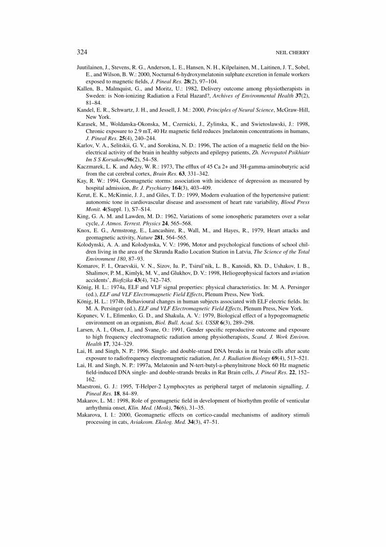
324 NEIL CHERRY
Juutilainen, J., Stevens, R. G., Anderson, L. E., Hansen, N. H., Kilpelainen, M., Laitinen, J. T., Sobel,E., and Wilson, B. W.: 2000, Nocturnal 6-hydroxymelatonin sulphate excretion in female workersexposed to magnetic fields, J. Pineal Res. 28(2), 97–104.
Kallen, B., Malmquist, G., and Moritz, U.: 1982, Delivery outcome among physiotherapists inSweden: is Non-ionizing Radiation a Fetal Hazard?, Archives of Environmental Health 37(2),81–84.
Kandel, E. R., Schwartz, J. H., and Jessell, J. M.: 2000, Principles of Neural Science, McGraw-Hill,New York.
Karasek, M., Woldanska-Okonska, M., Czernicki, J., Zylinska, K., and Swietoslawski, J.: 1998,Chronic exposure to 2.9 mT, 40 Hz magnetic field reduces ]melatonin concentrations in humans,J. Pineal Res. 25(4), 240–244.
Karlov, V. A., Selitskii, G. V., and Sorokina, N. D.: 1996, The action of a magnetic field on the bio-electrical activity of the brain in healthy subjects and epilepsy patients, Zh. Nevropatol PsikhiatrIm S S Korsakova96(2), 54–58.
Kaczmarek, L. K. and Adey, W. R.: 1973, The efflux of 45 Ca 2+ and 3H-gamma-aminobutyric acidfrom the cat cerebral cortex, Brain Res. 63, 331–342.
Kay, R. W.: 1994, Geomagnetic storms: association with incidence of depression as measured byhospital admission, Br. J. Psychiatry 164(3), 403–409.
Kerut, E. K., McKinnie, J. J., and Giles, T. D.: 1999, Modern evaluation of the hypertensive patient:autonomic tone in cardiovascular disease and assessment of heart rate variability, Blood PressMonit. 4(Suppl. 1), S7–S14.
King, G. A. M. and Lawden, M. D.: 1962, Variations of some ionospheric parameters over a solarcycle, J. Atmos. Terrest. Physics 24, 565–568.
Knox, E. G., Armstrong, E., Lancashire, R., Wall, M., and Hayes, R., 1979, Heart attacks andgeomagnetic activity, Nature 281, 564–565.
Kolodynski, A. A. and Kolodynska, V. V.: 1996, Motor and psychological functions of school chil-dren living in the area of the Skrunda Radio Location Station in Latvia, The Science of the TotalEnvironment 180, 87–93.
Komarov, F. I., Oraevskii, V. N., Sizov, Iu. P., Tsirul’nik, L. B., Kanoidi, Kh. D., Ushakov, I. B.,Shalimov, P. M., Kimlyk, M. V., and Glukhov, D. V.: 1998, Heliogeophysical factors and aviationaccidents’, Biofizika 43(4), 742–745.
König, H. L.: 1974a, ELF and VLF signal properties: physical characteristics. In: M. A. Persinger(ed.), ELF and VLF Electromagnetic Field Effects, Plenum Press, New York.
König, H. L.: 1974b, Behavioural changes in human subjects associated with ELF electric fields. In:M. A. Persinger (ed.), ELF and VLF Electromagnetic Field Effects, Plenum Press, New York.
Kopanev, V. I., Efimenko, G. D., and Shakula, A. V.: 1979, Biological effect of a hypogeomagneticenvironment on an organism, Biol. Bull. Acad. Sci. USSR 6(3), 289–298.
Larsen, A. I., Olsen, J., and Svane, O.: 1991, Gender specific reproductive outcome and exposureto high frequency electromagnetic radiation among physiotherapists, Scand. J. Work Environ.Health 17, 324–329.
Lai, H. and Singh, N. P.: 1996. Single- and double-strand DNA breaks in rat brain cells after acuteexposure to radiofrequency electromagnetic radiation, Int. J. Radiation Biology 69(4), 513–521.
Lai, H. and Singh, N. P.: 1997a, Melatonin and N-tert-butyl-a-phenylnitrone block 60 Hz magneticfield-induced DNA single- and double-strands breaks in Rat Brain cells, J. Pineal Res. 22, 152–162.
Maestroni, G. J.: 1995, T-Helper-2 Lymphocytes as peripheral target of melatonin signalling, J.Pineal Res. 18, 84–89.
Makarov, L. M.: 1998, Role of geomagnetic field in development of biorhythm profile of venticulararrhythmia onset, Klin. Med. (Mosk), 76(6), 31–35.
Makarova, I. I.: 2000, Geomagnetic effects on cortico-caudal mechanisms of auditory stimuliprocessing in cats, Aviakosm. Ekolog. Med. 34(3), 47–51.
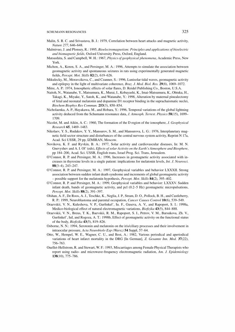
SCHUMANN RESONANCES 325
Malin, S. R. C. and Srivastava, B. J.: 1979, Correlation between heart attacks and magnetic activity,Nature 277, 646–648.
Malmivuo, J. and Plonsey, R.: 1995, Bioelectromagnetism: Principles and applications of bioelectricand biomagnetic fields, Oxford University Press, Oxford, England.
Matsushita, S. and Campbell, W. H.: 1967, Physics of geophysical phenomena, Academic Press, NewYork.
Michon, A., Koren, S. A., and Persinger, M. A.: 1996, Attempts to simulate the association betweengeomagnetic activity and spontaneous seizures in rats using experimentally generated magneticfields, Percept. Mot. Skills 82(2), 619–626.
Mikulecky, M., Moravcikova, C., and Czanner, S.: 1996, Lunisolar tidal waves, geomagnetic activityand epilepsy in the light of multivariate coherence, Braz. J. Med. Biol. Res. 29(8), 1069–1072.
Mitre, A. P.: 1974, Ionospheric effects of solar flares, D. Reidel Publishing Co., Boston, U.S.A.Naitoh, N., Watanabe, Y., Matsumura, K., Murai, I., Kobayashi, K., Imai-Matsumura, K., Ohtuka, H.,
Takagi, K., Miyake, Y., Satoh, K., and Watanabe, Y.: 1998, Alteration by maternal pinealectomyof fetal and neonatal melatonin and dopamine D1 receptor binding in the suprachaismatic neclei,Biochem Biophys Res Commun. 253(3), 850–854.
Nicholaenka, A. P., Hayakawa, M., and Hobara, Y.: 1996, Temporal variations of the global lightningactivity deduced from the Schumann resonance data, J. Atmosph. Terrest. Physics 58(15), 1699–1709.
Nicolet, M. and Aikin, A. C.: 1960, The formation of the D region of the ionosphere, J. GeophysicalResearch 65, 1469–1483.
Nikolaev, Y. S., Rudakov, Y. Y., Mansurov, S. M., and Mansurova, L. G.: 1976, Interplanetary mag-netic field sector structure and disturbances of the central nervous system activity, Reprint N 17a,Acad. Sci USSR, 29 pp, IZMIRAN, Moscow.
Novikova, K. F. and Ryvkin, B. A.: 1977, Solar activity and cardiovascular diseases, In: M. N.Gnevyshev and A. I. Ol’ (eds), Effects of solar Activity on the Earth’s Atmosphere and Biosphere,pp 184–200, Acad. Sci. USSR, English trans, Israel Prog. Sci. Trans, Jerusalem.
O’Connor, R. P. and Persinger, M. A.: 1996, Increases in geomagnetic activity associated with in-creases in thyroxine levels in a single patient: implications for melatonin levels, Int. J. Neurosci.88(3–4), 243–247.
O’Connor, R. P. and Persinger, M. A.: 1997, Geophysical variables and behavior LXXXII. Strongassociation between sudden infant death syndrome and increments of global geomagnetic activity– possible support for the melatonin hypothesis, Percept. Mot. Skills 84(2), 395–402.
O’Connor, R. P. and Persinger, M. A.: 1999, Geophysical variables and behavior: LXXXV. Suddeninfant death, bands of geomagnetic activity, and pcl (0.2–5 Hz) geomagnetic micropulsations,Percept. Mot. Skills 88(2), 391–397.
Olshan, A. F., De Roos, A. J., Teschke, K., Neglia, J. P., Stram, D. O., Pollock, B. H., and Castleberry,R. P.: 1999, Neuroblastoma and parental occupation, Cancer Causes Control 10(6), 539–549.
Oraevskii, V. N., Kuleshova, V. P., Gurfinkel’, Iu. F., Guseva, A. V., and Rapoport, S. I.: 1998a,Medico-biological effect of natural electromagnetic variations, Biofizika 43(5), 844–888.
Oraevskii, V. N., Breus, T. K., Baevskii, R. M., Rapoport, S. I., Petrov, V. M., Barsukova, Zh. V.,Gurfinkel’, IuI, and Rogoza, A. T.: 1998b, Effect of geomagnetic activity on the functional statusof the body, Biofizika 43(5), 819–826.
Osborne, N. N.: 1994, Serotonin and melatonin on the iris/ciliary processes and their involvement inintraocular pressure, Acta Neurobiolo Exp (Warsz) 54 Suppl, 57–64.
Otto, W., Hempel, W. E., Wagner, C. U., and Best, A.: 1982, Various periodical and aperiodicalvariations of heart infarct mortality in the DRG [In German], Z. Gesamte Inn. Med. 37(22),756–763.
Ouellet-Hellstrom, R. and Stewart, W. F.: 1993, Miscarriages among Female Physical Therapists whoreport using radio- and microwave-frequency electromagnetic radiation, Am. J. Epidemiology138(10), 775–786.
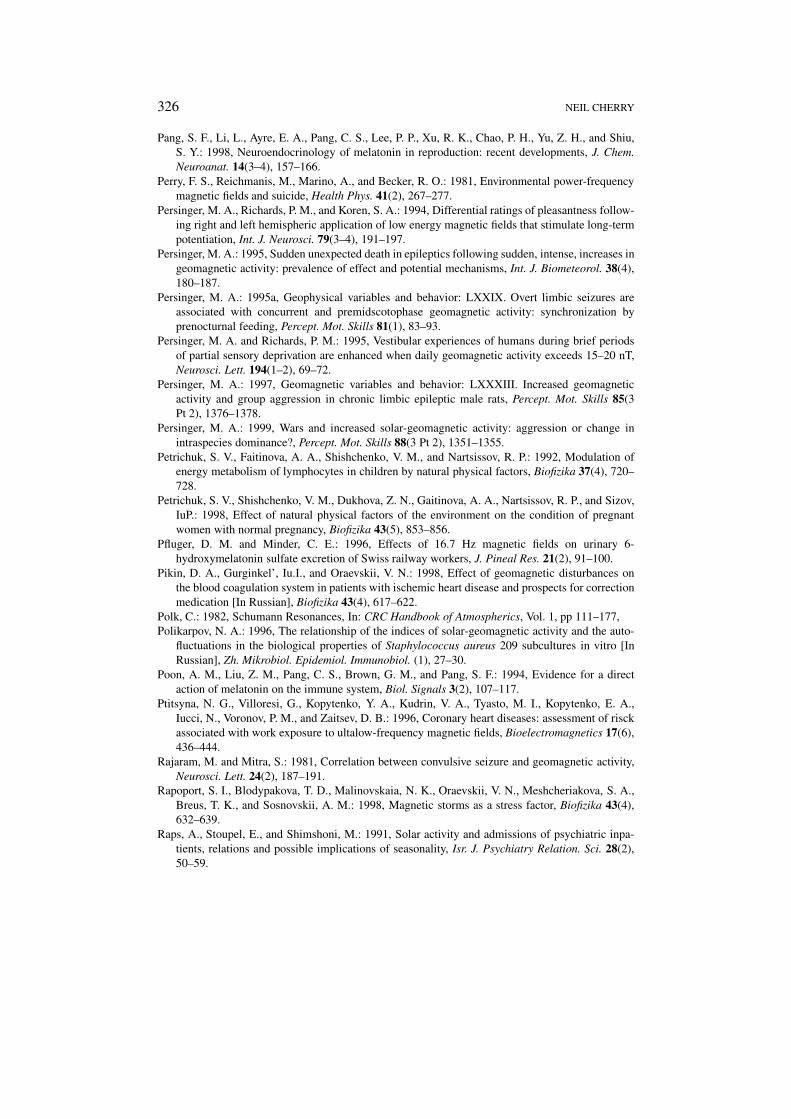
326 NEIL CHERRY
Pang, S. F., Li, L., Ayre, E. A., Pang, C. S., Lee, P. P., Xu, R. K., Chao, P. H., Yu, Z. H., and Shiu,S. Y.: 1998, Neuroendocrinology of melatonin in reproduction: recent developments, J. Chem.Neuroanat. 14(3–4), 157–166.
Perry, F. S., Reichmanis, M., Marino, A., and Becker, R. O.: 1981, Environmental power-frequencymagnetic fields and suicide, Health Phys. 41(2), 267–277.
Persinger, M. A., Richards, P. M., and Koren, S. A.: 1994, Differential ratings of pleasantness follow-ing right and left hemispheric application of low energy magnetic fields that stimulate long-termpotentiation, Int. J. Neurosci. 79(3–4), 191–197.
Persinger, M. A.: 1995, Sudden unexpected death in epileptics following sudden, intense, increases ingeomagnetic activity: prevalence of effect and potential mechanisms, Int. J. Biometeorol. 38(4),180–187.
Persinger, M. A.: 1995a, Geophysical variables and behavior: LXXIX. Overt limbic seizures areassociated with concurrent and premidscotophase geomagnetic activity: synchronization byprenocturnal feeding, Percept. Mot. Skills 81(1), 83–93.
Persinger, M. A. and Richards, P. M.: 1995, Vestibular experiences of humans during brief periodsof partial sensory deprivation are enhanced when daily geomagnetic activity exceeds 15–20 nT,Neurosci. Lett. 194(1–2), 69–72.
Persinger, M. A.: 1997, Geomagnetic variables and behavior: LXXXIII. Increased geomagneticactivity and group aggression in chronic limbic epileptic male rats, Percept. Mot. Skills 85(3Pt 2), 1376–1378.
Persinger, M. A.: 1999, Wars and increased solar-geomagnetic activity: aggression or change inintraspecies dominance?, Percept. Mot. Skills 88(3 Pt 2), 1351–1355.
Petrichuk, S. V., Faitinova, A. A., Shishchenko, V. M., and Nartsissov, R. P.: 1992, Modulation ofenergy metabolism of lymphocytes in children by natural physical factors, Biofizika 37(4), 720–728.
Petrichuk, S. V., Shishchenko, V. M., Dukhova, Z. N., Gaitinova, A. A., Nartsissov, R. P., and Sizov,IuP.: 1998, Effect of natural physical factors of the environment on the condition of pregnantwomen with normal pregnancy, Biofizika 43(5), 853–856.
Pfluger, D. M. and Minder, C. E.: 1996, Effects of 16.7 Hz magnetic fields on urinary 6-hydroxymelatonin sulfate excretion of Swiss railway workers, J. Pineal Res. 21(2), 91–100.
Pikin, D. A., Gurginkel’, Iu.I., and Oraevskii, V. N.: 1998, Effect of geomagnetic disturbances onthe blood coagulation system in patients with ischemic heart disease and prospects for correctionmedication [In Russian], Biofizika 43(4), 617–622.
Polk, C.: 1982, Schumann Resonances, In: CRC Handbook of Atmospherics, Vol. 1, pp 111–177,Polikarpov, N. A.: 1996, The relationship of the indices of solar-geomagnetic activity and the auto-
fluctuations in the biological properties of Staphylococcus aureus 209 subcultures in vitro [InRussian], Zh. Mikrobiol. Epidemiol. Immunobiol. (1), 27–30.
Poon, A. M., Liu, Z. M., Pang, C. S., Brown, G. M., and Pang, S. F.: 1994, Evidence for a directaction of melatonin on the immune system, Biol. Signals 3(2), 107–117.
Ptitsyna, N. G., Villoresi, G., Kopytenko, Y. A., Kudrin, V. A., Tyasto, M. I., Kopytenko, E. A.,Iucci, N., Voronov, P. M., and Zaitsev, D. B.: 1996, Coronary heart diseases: assessment of risckassociated with work exposure to ultalow-frequency magnetic fields, Bioelectromagnetics 17(6),436–444.
Rajaram, M. and Mitra, S.: 1981, Correlation between convulsive seizure and geomagnetic activity,Neurosci. Lett. 24(2), 187–191.
Rapoport, S. I., Blodypakova, T. D., Malinovskaia, N. K., Oraevskii, V. N., Meshcheriakova, S. A.,Breus, T. K., and Sosnovskii, A. M.: 1998, Magnetic storms as a stress factor, Biofizika 43(4),632–639.
Raps, A., Stoupel, E., and Shimshoni, M.: 1991, Solar activity and admissions of psychiatric inpa-tients, relations and possible implications of seasonality, Isr. J. Psychiatry Relation. Sci. 28(2),50–59.
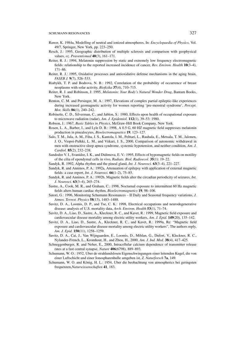
SCHUMANN RESONANCES 327
Rawer, K. 1984a, Modelling of neutral and ionized atmospheres, In: Encyclopaedia of Physics, Vol.49/7, Springer, New York, pp. 223–250.
Resch, J.: 1995, Geographic distribution of multiple sclerosis and comparison with geophysicalvalues, oz. Praventivmed 40(3), 161–171.
Reiter, R. J.: 1994, Melatonin suppression by static and extremely low frequency electromagneticfields: relationship to the reported increased incidence of cancer, Rev. Environ. Health 10(3–4),171–86.
Reiter, R. J.: 1995, Oxidative processes and antioxidative defense mechanisms in the aging brain,FASEB J. 9(7), 526–533.
Riabykh, T. P. and Bodrova, N. B.: 1992, Correlation of the probability of occurrence of breastneoplasms with solar activity, Biofizika 37(4), 710–715.
Reiter, R. J. and Robinson, J: 1995, Melatonin: Your Body’s Natural Wonder Drug, Bantam Books,New York.
Renton, C. M. and Persinger, M. A.: 1997, Elevations of complex partial epileptic-like experiencesduring increased geomagnetic activity for women reporting ‘pre-menstral syndrome’, Percept.Mot. Skills 86(1), 240–242.
Robinette, C. D., Silverman, C., and Jablon, S.: 1980, Effects upon health of occupational exposureto microwave radiation (radar), Am. J. Epidemiol. 112(1), 39–53: 1980.
Robson, J.: 1967, Basic Tables in Physics, McGraw-Hill Book Company, New York.Rosen, L. A., Barber, I., and Lyle D. B.: 1998, A 0.5 G, 60 HZ magnetic field suppresses melatonin
production in pinealocytes, Bioelectromagnetics 19, 123–127.Salo, T. M., Jula, A. M., Fiha, J. S., Kantola, I. M., Pelttari, L., Rauhala, E., Metsala, T. M., Jalonen,
J. O., Viopoi-Pulkki, L. M., and Viikari, J. S., 2000, Comparison of autonomic withdrawal inmen with onstructive sleep apnea syndrome, systemic hypotension, and neither condition, Am. J.Cardiol. 85(2), 232–238.
Sandodze V. I., Svanidze, I. K., and Didimova, E. V.: 1995, Effects of hypomagnetic fields on motilityof the cilia of ependymal cells in vivo, Radiats. Biol. Radioecol. 35(1): 19–22.
Sandyk, R. 1992, Alpha rhythm and the pineal gland, Int. J. Neurosci. 63(3–4), 221–227.Sandyk, R. and Anninos, P. A.: 1992a, Attenuation of epilepsy with application of external magnetic
fields: a case report, Int. J. Neurosci. 66(1–2), 75–85.Sandyk, R. and Anninos, P. A.: 1992b, Magnetic fields alter the circadian periodicity of seizures, Int.
J. Neurosci. 63(3–4), 265–274.Sastre, A., Cook, M. R., and Graham, C.: 1998, Nocturnal exposure to intermittent 60 Hz magnetic
fields alters human cardiac rhythm, Bioelectromagnetics 19, 98–106.Satori, G.: 1996, Monitoring Schumann Resonances – II Daily and Seasonal frequency variations, J.
Atmos. Terrest. Physics 58(13), 1483–1488.Savitz, D. A., Loomis, D. P., and Tse, C. K.: 1998, Electrical occupations and neurodegenerative
disease: analysis of U.S. mortality data, Arch. Environ. Health 53(1), 71–74.Savitz, D. A., Liao, D., Sastre, A., Kleckner, R. C., and Kavet, R.: 1999, Magnetic field exposure and
cardiovascular disease mortality among electric utility workers, Am. J. Epid. 149(20), 135–142.Savitz, D. A., Liao, D., Sastre, A., Kleckner, R. C., and Kavet, R.: 1999a, Re: “Magnetic field
exposure and cardiovascular disease mortality among electric utility workers”, The authors reply,Am. J. Epid. 150(11), 1258–1259.
Savitz, D. A., Cai, J., Van Wijngaarden, E., Loomis, D., Mihlan, G., Dufort, V., Kleckner, R. C.,Nylander-French, L., Kromhout, H., and Zhou, H., 2000, Am. J. Ind. Med. 38(4), 417–425.
Schneggenburger, R. and Neher, E., 2000, Intracellular calcium dependence of transmitter releaserates at a fast central synapse, Nature 406(6798), 889–893.
Schumann, W. O.: 1952, Uber de strahlundslosen Eigenschwingungen einer leitenden Kugel, die voneiner Luftschicht und einer Ionospharenhulle umgeben ist, Z. Naturforsch 7a, 149.
Schumann, W. O. and König, H. L.: 1954, Uber die beobachtung von atmospherics bei geringstenfrequenzen,Naturwissenschaften 41, 183.
328 NEIL CHERRY
Schwartz, J. L., House, D. E., and Mealing, A. R.: 1990, Exposure of frog hearts to CW or amplitudemodulated VHF fields: selective efflux of calcium ions at 16 Hz, Bioelectromagnetics 11, 349–358.
SEAMIC: 1997, Recent trends in health statistics in Southeast Asia 1974–1993, Publication No. 77,Southeast Asian Medical Information Center, Tokyo.
Selitskii, G. V., Karlov, V. A., and Sorokina, N. D.: 1999, The influence of hypogeomagnetic field onthe bioelectric activity of the brain in epilepsy, Zh. Nevrol. Psikhiatr. Im. S.S. Korsakova. 99(4),48–50.
Sentman, D. D. and Fraser, B. J.: 1991, Simultaneous observations of Schumann Resonances inCalifornia and Australia: Evidence for intensity modulation by local height of the D Region, J.Geophys. Res. 96(A9), 15973–15984.
Sitar, J.: 1990, The causality of lunar changes on cardiovascular mortality [In Czech], Cas. Lek. Cesk.129(45), 1425–1430.
Smulevich, V. B., Solionova, L. G., and Belyakova, S. V.: 1999, Parental occupation and other factorsand cancer risk in children II, Int. J. Cancer 83(60), 718–722,
Shumilov, O. I., Kasatkina, E. A., and Raspopov, O. M.: 1998, Heliomagnetic activity and level ofextreme situations at the Polar cap [In Russian], Biofizika 43(4), 670–676.
Sobel, E., Davanipour, Z., Sulkava, R., Erkinjuntti, T., Wikstrom, J., Henderson, V. W., Bucjwalter,G., Bowman, D., and Lee, P-J.: 1995, Occupations with exposure to electromagnetic fields: apossible risk factor for Alzheimer’s Disease, Am. J. Epidemiol 142(5), 515–524.
Sobel, E., Dunn, M., Davanipour, D. V. M., Qian, M. S., and Chui, M. D.: 1996, Elevated riskof Alzheimer’s disease among workers with likely electromagnetic field exposure, Neurology47(12), 1477–1481.
Sparks, D. L. and Hunsaker, J. C.: 1988, The pineal gland in sudden infant death syndrome:preliminary observations, J. Pineal Res. 5, 111–118.
Spitz, M. R. and Johnson, C. C.: 1985, Neuroblastoma and parental occupation. A case-controlanalysis, Am. J. Epidemiol. 121(6), 924–929.
Sprott, J. C.: 1990, Numerical Recipes: Routines and Examples in Basic, Cambridge UniversityPress, Cambridge.
Stark, K. D. C., Krebs, T., Altpeter, E., Manz, B., Griol, C., and Abelin, T.: 1997, Absence of chroniceffect of exposure to short-wave radio broadcast signal on salivary melatonin concentrations indairy cattle, J. Pineal Res. 22, 171–176.
Stewart, W., 2000, Mobile phones and health, Independent Expert Group on Mobile Phones,(IEGMP) U.K. Parliamentary Enquiry, London.
Stoupel, E., Hod, M., Shimshoni, M., Friedman, S., Ovadia, J., and Keith, L.: 1990, Monthly cosmicactivity and pregnancy induced hypertension, Clin. Exp. Obstet. Gynecol. 17(1), 7–12.
Stoupel, E., Martfel, J., and Rotenberg, Z.: 1991, Admissions of patients with epileptic seizures (E)and dizziness (D) related to geomagnetic and solar activity levels: differences in female and malepatients, Med. Hypothesis 36(4), 384–388.
Stoupel, E. and Shimshoni, M.: 1991, Hospital cardiovascular deaths and total distribution of deathsin 180 consecutive months with difference cosmic physical activity: a correlation study (1974–1988), Int. J. Biometeorology 35(1), 6–9.
Stoupel, E.: 1993, Sudden cardiac deaths and ventricular extrasystoles on days of four levels ofgeomagnetic activity, J. Basic Physiol. Pharmacol. 4(4), 357–366.
Stoupel, E., Goldenfeld, M., Shimshoni, M., and Siegel, R.: 1993, Intraocular pressure (IOP) in rela-tion to four levels of daily geomagnetic and extreme yearly solar activity, Int. J. Biometeorology37(1), 42–45.
Stoupel, E., Martfel, J. N., and Rotenberg, Z.: 1994, Paroxysmal atrial fibrulation and stroke(cerebrovasular accidents) in males and females above and below the age 65 on days of differentgeomagnetic activity levels, J. Basic. Clin. Physiol. Pharmacol. 5(3–4), 315–329.
SCHUMANN RESONANCES 329
Stoupel, E., Abramson, E., Sulkes, J., Martfel, J., Stein, N., Handelman, M., Shimshoni, M., Zadka,P., and Gabbay, U.: 1995a, Relationship between suicide and myocardial infarction with regardto changing physical environmental conditions, Int. J. Biometeorol. 38(4), 199–203
Stoupel, E., Petrauskiene, J., Kalediene, R., Abramson, E., and Sulkes, J.: 1995b, Clinical cosmobi-ology: the Lithuanian study 1990–1992, Int. J. Biometeorology 38(4), 204–208.
Stoupel, E., Petrauskiene, J., Kalediene, R., Domarkiene, S., Abramson, E., and Sulkes, J.: 1996, Dis-tribution of deaths from ischemic heart disease and stroke. Environmental and aging influencesin men and women, J. Basic. Clinical Physiol. Pharmacol. 7(4), 303–319.
Stoupel, E., Abramson, J., Domarkiene, S., Shimshoni, M., and Sulkes, J.: 1997, Space proton fluxand the temporal distribution of cardiovascular deaths, Int J Biometeorol 40(2), 113–116.
Stoupel, E., Petrauskiene, J., Abramson, E., Kalediene, R., Israelovich, P., and Sulkes, J.: 1999, Re-lationship between deaths from stroke and ischemic heart disease: Environmental implications,J. Basic. Clinical Physiol. Pharmacol. 10(2), 135–145.
St Pierre, L. and Persinger, M. A.: 1998, Geophysical variables and behavior: LXXXIV. Quantitativeincreases in group aggression in male epileptic rats during increases in geomagnetic activity,Percept. Mot. Skills 86(3 Pt 2), 1392–1394.
Svanidze, I. K., Sandodze, V. Ia., Didimova, E. V., Chkhikvadze, T. I., Portnoi, V. N., and Razdol’skii,A. S.: 1994, The effect of hypo- and hypermagnetic fields on the motor activity of the ciliaryapparatus of the ependymal cells [In Russian], Radiats. Biol. Radioecol. 34(1), 100–104 .
Szmigielski, S.: 1996, Cancer morbidity in subjects occupationally exposed to high frequency (ra-diofrequency and microwave) electromagnetic radiation, Science of the Total Environment 180,9–17.
Tambiev, A. E., Medvedev, S. D., and Egorova, E. V.: 1995, The effect of geomagnetic disturbanceson the functions of attention and memory [In Russian], Aviakosm. Ekolog. Med. 29(3), 43–45.
Tang, Y. and Othmer, H. G.: 1995, Frequency encoding in excitable systems with applications tocalcium oscillations, Proc Natl. Acad. Sci. USA 92(17), 7869–7873.
Taskinen, H., Kyyronen, P., and Hemminki, K.: 1990, Effects of ultrasound, shortwaves and physicalexertion on pregnancy outcome in physiotherapists, J. Epidemiol, Commun. Health 44, 196–210.
Tell, R. A. and Mantiply, E. D.: 1980, Population exposure to VHF and UHF broadcast radaition inthe United States, Proc IEEE 68(1), 4–10.
Tran, A. and Polk, C.: 1979, Schumann resonances and electrical conductivity of the atmosphere andlower ionosphere – I. Effects of conductivity at various altitudes on resonance frequencies andattenuation, J. Atmos. Terrest. Physics 41, 1241–1248.
Thomas, L., Drew, J. E., Abramovich, D. R., and Williams, L. M.: 1998, The role of melatonin in thehuman fetus, Int. J. Mol. Med. 1(3), 539–543.
Titheridge, J. E.: 1962, The electron density in the lower ionosphere, J. Atmos. Terr. Phys. 24, 269–282
Tunyi, I. and Tesarova, O.: 1991, Suicide and geomagnetic activity [In Slovak], Soud. Lek. 36(1–2),1–11.
Tynes, T., Reitan, J. B., and Andersen, A.: 1994, Incidence of cancer among workers in Norwegianhydroelectric power companies, Scan. J. Work Environ. Health 20(5), 339–344.
Usenko, G. A.: 1992, Psychosomatic status and the quality of the piloting in fliers duringgeomagnetic disturbances, Aviakosm. Ekolog. Med. 26(4), 23–27.
Vagero, D., Ahlbom, A., Olin, R., and Sahlsten, S.: 1985, Cancer morbidity among workers in thetelecommunication industry, Br. J. Ind. Med. 42, 191–195.
Van Wijngaarden, E., Savitz, D. A., Kleckner, R. C., Cai, J. and Loomis, D., 2000, Exposure toelectromagnetic fields and suicide among electric utility workers: a nested case-control study,Occup. Environ. Med. 57, 258–263.
Vanecek, J.: 1998, Cellular mechanisms of melatonin action, Physiol. Rev. 78(3), 687–721.Vaughan, T. L., Daling, J. R., and Starzyk, P. M.: 1984, Fetal death and maternal occupation, J.
Occup. Med., 676–678.
330 NEIL CHERRY
Verge, D. and Calas, A., 2000, Serotoninergic neurons and serotonin receptors: gains fromcytochemical approaches, J. Chem. Neuroanat. 18(1–2), 41–56.
Verkasalo, P. K., Kaprio, J., Varjonen, J., Romanov, K., Heikkila, K., and Koskenvuo, M.: 1997,Magnetic fields of transmission lines and depression, Am. J. Epidemiology 146(12), 1037–1045.
Vignati, M. and Giuliani, L.: 1997, Radiofrequency exposure nerar high-voltage lines, EnvironmentalHealth Perspectives 105(6), 1569–1573.
Villoresi, G., Ptitsyna, N. G., Tiasto, M. I., and Iucci, N.: 1998, Myocardial infarct and geomagneticdisturbances: analysis of data on morbidity and mortality [In Russian], Biofizika 43(4), 623–632.
Viswanathan, M., Laitinen, J. T., and Saavedra, J. M.: 1993, Vascular melatonin receptors, Biol.Signals 2(4), 221–227.
Vriend, J., Borer, K. T., and Thliveris, J. A.: 1987, Melatonin, its antagonism of thyroxine’santisomatotrophic activity in male Syrian hamsters, Growth 51(1), 35–43.
Wang, S. G. 1989, 5-HT contents change in peripheral blood of workers exposed to microwave andhigh frequency radiation, Chung Hua Yu Fang I Hsueh Tsa Chih 23(4), 207–210.
Walleczek, J: 1992, Electromagnetic field effects on cells of the immune system: the role of calciumsignalling, FASEB J. 6(13), 3177–3185.
Watanabe, Y., Hillman, D. C., Otsuka, K., Bingham, C., Breus, T. K., Cornelissen, G., and Halberg,F.: 1994, Cross-spectral coherence between geomagnetic disturbance and human cardiovascularvariables at non-societal frequencies, Chronobiologia 21(3–4), 265–272.
Wertheimer, N. and Leeper, E.: 1986, Possible effects of electric blankets and heated waterbeds onfetal development, Bioelectromagnetics 7, 13–22.
Wever, R.: 1967, Uber die Beeinflussung der circadianen Periodik ees Menschen durch schwacheelektromagnetische Felder, Z. vergl Physiol 56, 111–128.
Wever, R.: 1968, Einfluss schwacher elektro-magnetischer Felder auf die circadiane Periodik desMenschen, Naturwissenschaften 55, 29–32.
Wever, R.: 1973, Human circadian rhythms under the influence of weak electric fields and differentaspects of these studies, Int. J. Biometeorology 17, 227–232.
Wever, R.: 1974, ELF-effects on Human Circadian Rhythms, pp. 101–144, In: M. A. Persinger (ed.),ELF and VLF Electromagnetic Field Effects, Plenum Press, New York.
Whitfield, J. F.: 1992, Calcium signals and cancer, Crit. Rev. Oncolog. 3(1–2), 55–90.Wilkins, J. R. and Hundley, V. D.: 1990, Parental occupational exposure to electromagnetic fields
and neuroblastoma in offspring, Am. J. Epidemiol. 131(6), 995–1008.Wilkins, J. R. and Wellage, L. C.: 1996, Brain tumor risk in offspring of men occupationally exposed
to electric and magnetic fields, Scand. J. Work Environ. Health 22(5), 339–345.Williams, E. R.: 1992, The Schumann Resonance: A global tropical thermometer, Science 256, 1184–
1187.Wilson, B. W., Wright, C. W., Morris, J. E., Buschbom, R. L., Brown, D. P., Miller, D. L., Sommers-
Flannigan, R., and Anderson, L.E.: 1990, Evidence of an effect of ELF electromagnetic fields onhuman pineal gland function, J. Pineal Res. 9(4), 259–269.
Wood, A. W., Armstrong, S. M., Sait, M. L., Devine, L., and Martin, M. J.: 1998, Changes in humanplasma melatonin profiles in response to 50 Hz magnetic field exposure, J. Pineal Res. 25(2),116–127.
Yaga, K., Reiter, R. J., Manchester, L. C., Nieves, H., Sun, J. H., and Chen L. D.: 1993, Pinealsensitivity to pulsed static magnetic fields changes during the photoperiod, Brain Res. Bull. 30(1–2), 153–156.
Zaitseva, S. A. and Pudovkin, M. I.: 1995, Effect of solar and geomagnetic activity on populationdynamics among residents of Russia [In Russian], Biofizika 40(4), 861–864.
Zatz, M.: 1989, Relationship between linght, calcium influx and cAMP in the acute regulation ofmelatonin production in cultured chick pineal cells, Brain Res. 477, 14–18.
SCHUMANN RESONANCES 331
Zawilska, J. B. and Nowak, J. Z.: 1990, Calcium influx through voltage-sensitive calcium channelsregulates in vivo serotonin N-acetyltransferase (NAT) activity in hen retina and pineal gland,Neurosci. Lett. 118(1), 17–20.
Zawilska, J. B., Woldan-Tambor, A., and Nowak, J. Z.: 1998, Effects of calcium ions and sub-stances affecting Ca 2+-regulated mechanisms on histamine-evoked stimulation of cyclic AMPformation in chick pineal gland, Pol. J. Pharmacol. 50(4–5), 307–314.