Embed Size (px)
Citation preview
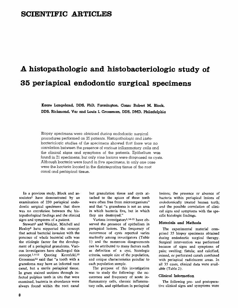
SCIENTIFIC ARTICLES
A histopathologic and histobacteriologic s tudy of
35 periapical endodontic surgica l spec imens
Kaare Langeland, DDS, PhD, Farm/nqton. Conn" Robert M. Block,
DDS, Richmond, Va; and Louis L Grossman~ DDS, DMD, Philadelphia
Biopsy spec imens were obta ined dur ing endodontic surgical p rocedures pe r fo rmed on 35 patients. Histopathologic a n d histo- bacter iologic studies of the spec imens showed that there w a s no correlation be t ween the p resence of var ious inf lammatory cells a n d the clinical s igns a n d symptoms of the patients. Epithelium w a s found in 21 spec imens , but only nine lesions were d i agnosed as cysts. Although bac te r i a were found in five specimens , in only one c a s e were the bac te r i a located in the disirlteqratinq tissue of the root cana l a n d per iap ica l tissue.
In a previous study, Block and as- sociates 1 have demonstrated by an examination of 230 periapical endo- dontic surgical specimens that there was no correlation between the his- topathological findings and the clinical signs and symptoms of a patient.
Stewart2 and Winkler, Mitchell and Healey ~ have supported the concept that actual bacterial invasion with the presence of whole bacterial ceils was the etiologic factor for the develop- ment of a periapical granuloma. Vari- ous investigators have challenged this concept. 1,4-1~ Quoting Kronfeld, 1~ Grossman ~2 said that "a tooth with a granuloma may have an infected root canal, but a sterile periapical tissue. In gram stained sections through in- fected putpless teeth in situ that were examined, bacteria in abundance were always found within the root canal
but granulation tissue and cysts at- tached to the apices of these teeth were often free from microorganisms" and that "a granuloma is not an area in which bacteria live, but in which they are destroyed."
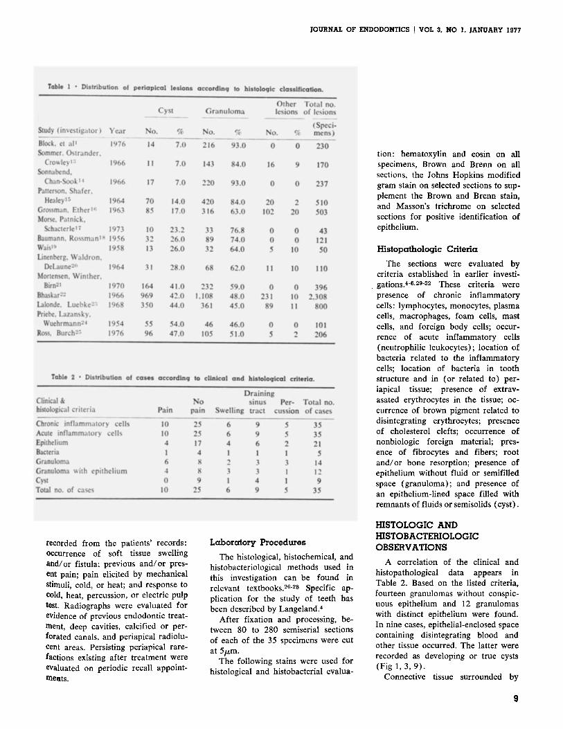
Various investigators1,13-25 have ob- served the presence of epithelium in periapical lesions. The frequency of occurrence of cysts reported varies markedly among investigators (Table 1) and the numerous disagreements can be attributed to many factors such as definition of a cyst, histologic criteria, sample size of the population, and unique characteristics peculiar to each population sample.
The purpose of this investigation was to study the following: the oc- currence and frequency of acute in- flammatory cells, chronic inflamma- tory cells, and epithelium in periapicaI
lesions; the presence or absence of bacteria within periapical lesions of endodontically treated human teeth; and the possible correlation of clini- cal signs and symptoms with the spe- cific histologic findings.
Materials and Methods
The experimental material com- prised 35 biopsy specimens obtained during endodontic surgical therapy. Surgical intervention was performed because of signs and symptoms of pain; swelling; fistula; and calcified, missed, or perforated canals combined with periapical radiolucent areas. In all 35 cases, clinical data were avail- able (Table 2).
Clinical Information
The following pre- and postopera- tive clinical signs and symptoms were
JOURNAL OF ENDODONTICS I VOL 3, NO 1, JANUARY 1977
- i o Distribution of perlapical lesions accordinq to histoloqic classification.
Other Total no. Cyst Granuloma lesions of lesions
(Speci- :or) Year No. % No. % No. % mens)
1976 14 7.0 216 93.0 0 0 230 ier,
1966 11 7.0 143 84.0 16 9 170
1966 17 7.0 220 93.0 0 0 237
1964 70 14.0 420 84.0 20 2 510 16 1963 85 17.0 316 63.0 102 20 503
1973 t0 23.2 33 76.8 0 0 43 lanlS 1956 32 26.0 89 74.0 0 0 12I
1958 13 26.0 32 64.0 5 10 50 ['on,
1964 31 28.0 68 62.0 11 10 110 her,
1970 164 41.0 232 59.0 0 0 396 1966 969 42.0 1,108 48.0 231 10 2,308
�9 2a 1968 350 44.0 361 45.0 89 11 800
1954 55 54.0 46 46.0 0 0 101 1976 96 47.0 105 51.0 5 2 206
2 �9 Distribution of cases according to clinical and histological criteria.
criteria Pain
Draining No sinus Per- Total no.
pain Swelling tract cussion of cases
~lammatory cells 10 25 6 9 5 35 mmatory cells 10 25 6 9 5 35
4 17 4 6 2 21 1 4 1 1 1 5 6 8 2 3 3 14
with epithelium 4 8 3 3 1 12 0 9 1 4 1 9
f cases 10 25 6 9 5 35
recorded from the patients' records: occurrence of soft tissue swelling and/or fistula; previous a n d / o r pres- ent pain; pain elicited by mechanical stimuli, cold, or heat; and response to cold, heat, percussion, or electric pulp test. Radiographs were evaluated for evidence of previous endodontic treat- ment, deep cavities, calcified or per- forated canals, and periapical radiolu- cent areas. Persisting periapical rare- factions existing after treatment were evaluated on periodic recall appoint- meats.
L a b o r a t o r y P r o c e d u r e s
The histological, histochemical, and histobacteriological methods used in this investigation can be found in relevant textbooks. ~6-2s Specific ap- plication for the study of teeth has been described by Langeland. 4
After fixation and processing, be- tween 80 to 280 semiserial sections of each of the 35 specimens were cut at 5/zm.
The following stains were used for histological and histobacterial evalua-
tion: hematoxylin and eosin on all specimens, Brown and Brenn on all sections, the Johns Hopkins modified gram stain on selected sections to sup- plement the Brown and Brenn stain, and Masson's trichrome on selected sections for positive identification of epithelium.
H i s t o p a t h o l o q i c C r i t e r i a
The sections were evaluated by criteria established in earlier investi-
�9 gations. 4-6.29-82 These criteria were presence of chronic inflammatory cells: lymphocytes, monocytes, plasma cells, macrophages, foam cells, mast cells, and foreign body cells; occur- rence of acute inflammatory cells (neutrophilic leukocytes); location of bacteria related to the inflammatory cells; location of bacteria in tooth structure and in (or related to) per- iapical tissue; presence of extrav- asated erythrocytes in the tissue; oc- currence of brown pigment related to disintegrating erythrocytes; presence of cholesterol clefts; occurrence of nonbiologic foreign material; pres- ence of fibrocytes and fibers; root and /o r bone resorption; presence of epithelium without fluid or semifllled space (granuloma) ; and presence of an epithelium-lined space filled with remnants of fluids or semisolids (cyst) .
H I S T O L O G I C A N D H I S T O B A C T E R I O L O G I C O B S E R V A T I O N S
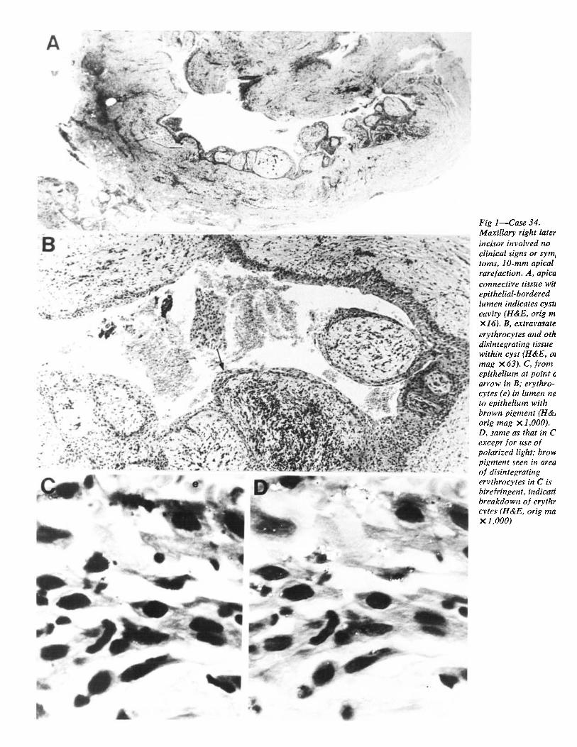
A correlation of the clinical and histopathological data appears in Table 2. Based on the listed criteria, fourteen granulomas without conspic- uous epithelium and 12 granulomas with distinct epithelium were found. In nine cases, epithelial-enclosed space containing disintegrating blood and other tissue occurred. The latter were recorded as developing or true cysts (Fig 1, 3, 9) .
Connective tissue surrounded by
It.lt " ... " ' . -'~I. ~. - ": .,~.'~Ik ," ,-. "' '.*'" .,~1(~, ~' .~','," r = ~ , ~ . ~ . , ; , . ~ , . , ; , ~ �9 . . . . ,~ .
, .,~ .~.. ,~ :~-. ~. ... ~.,~.~,~ . .. ,~, . �9 , , ,
' " . , " 7 , w e ~ ' . " l i e " " % d ~ l ~ " ' * "" " '~ " qW " "~ :% . - �9 " . . . . , L '
- ~ ' , " ~ - : . ~ 1 ~ . ~ , . ~ . " . " , .~ , , ~ .'. . ,~. ~.: ,, .~.. :. -.,. "~.: ... . .. .,i, ~
, , ' ~ : . . ,," ~ . ~ ,. - ,. ~ .... ..
" �9 ~ d [ . 'nil'- " , I " ~~ ~ . , - ~
:" ~ ~ "?,' " : , " ' - ~ 4 .
,~ . . . , . ~ , ~ , , : , .
,, iL
, �9
. . ~ , �9
e
:. 5 "~
�9 , ~ , . " . . ~ " ~
. . , -.. . , . . - _.
" " d " , , D ~ ' , "
' t l " f l ~ ~ "
, , i t
~ o
q
~'-,;
Fig 1---Case 34. Maxillary right later incisor involved no clinical signs or sym~ toms, l O-mm apical rare]action. A, apica connective tissue wit epithelial-bordered lumen indicates cyst~ cavity (H&E, or igm • 16). B, extravasate erythrocytes and oth disintegrating tissue within cyst (H&E, oJ mag • 63). C, ]rom epithelium at point arrow in B; erythro- cytes (e) in lumen ne to epithelium with brown pigment (H&~ orig mag • 1,000). D, same as that in C except for use o] polarized light; brow pigment seen in area of disintegrating ervthrocytes in C is birefringent, indicati breakdown o/erythr cvtes (H&E, orig ma • 1,000)
. . . ,. , . : . . . . , , ~ , . . . , _ ~ ! ~
�9 . . . . . . . . . . ~ i ~ . , . T ~ . ~ . . . . . r 7 , .
" ' . qlr �9 �9 P ~ ~ ~ l l a ] l l l h ~ 5 1 p l E
f i r
r
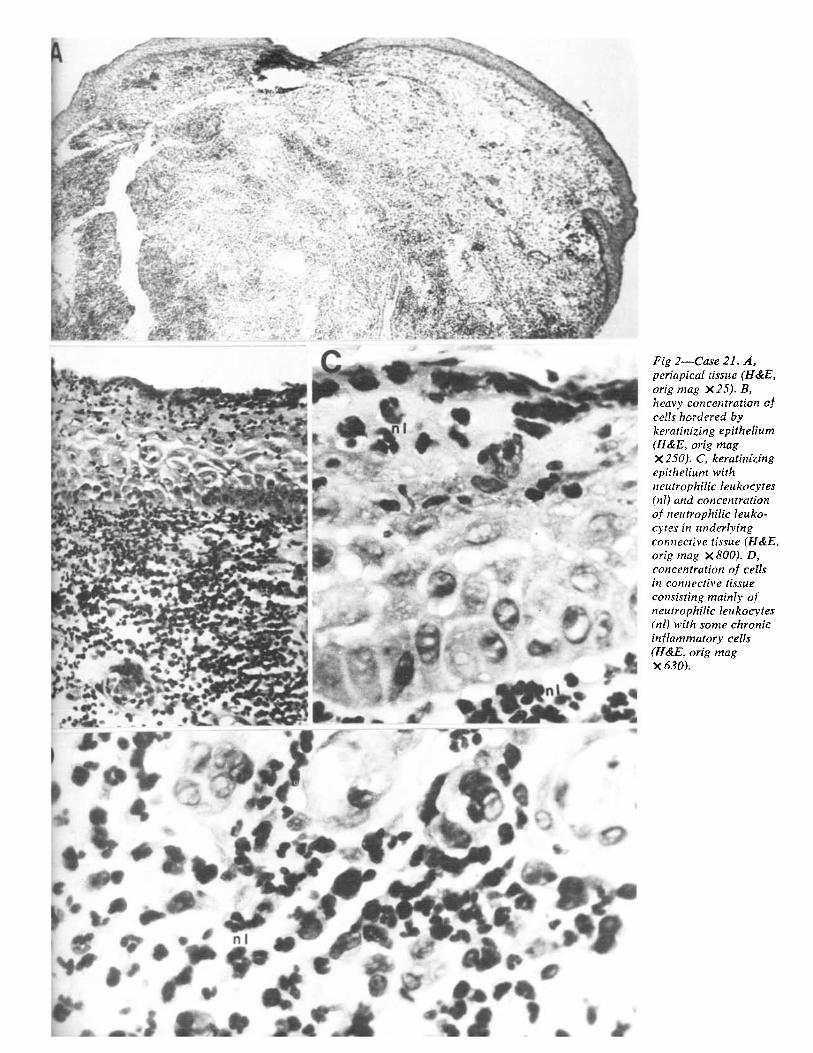
Fig 2--Case 21. A, periapical tissue (H&E, orig mag • 25). B, heavy concentration o/ cells bordered by keratinizing epithelium (H&E, orig mag • 250). C, keratinizing epithelium with neutrophilic leukocytes (nl) and concentration of neutrophilic leuko- cytes in underlying connective tissue (H&E, orig mag • 800). D, concentration of cells in connective tissue consisting mainly of neutrophilic Ieukocytes (nt) with some chronic in[lammatory cells (H&E, orig mag X 630).
A P , . k i B _ _ . . . ~ . ~
==.,~,,
:~i I - ~ ,~ W -%
i~_ ~I~ b c V'" .~ ~,=~
- ~ = -,
JOURNAL OF ENDODONTICS ] VOL 3, NO 1, JANUARY 1977
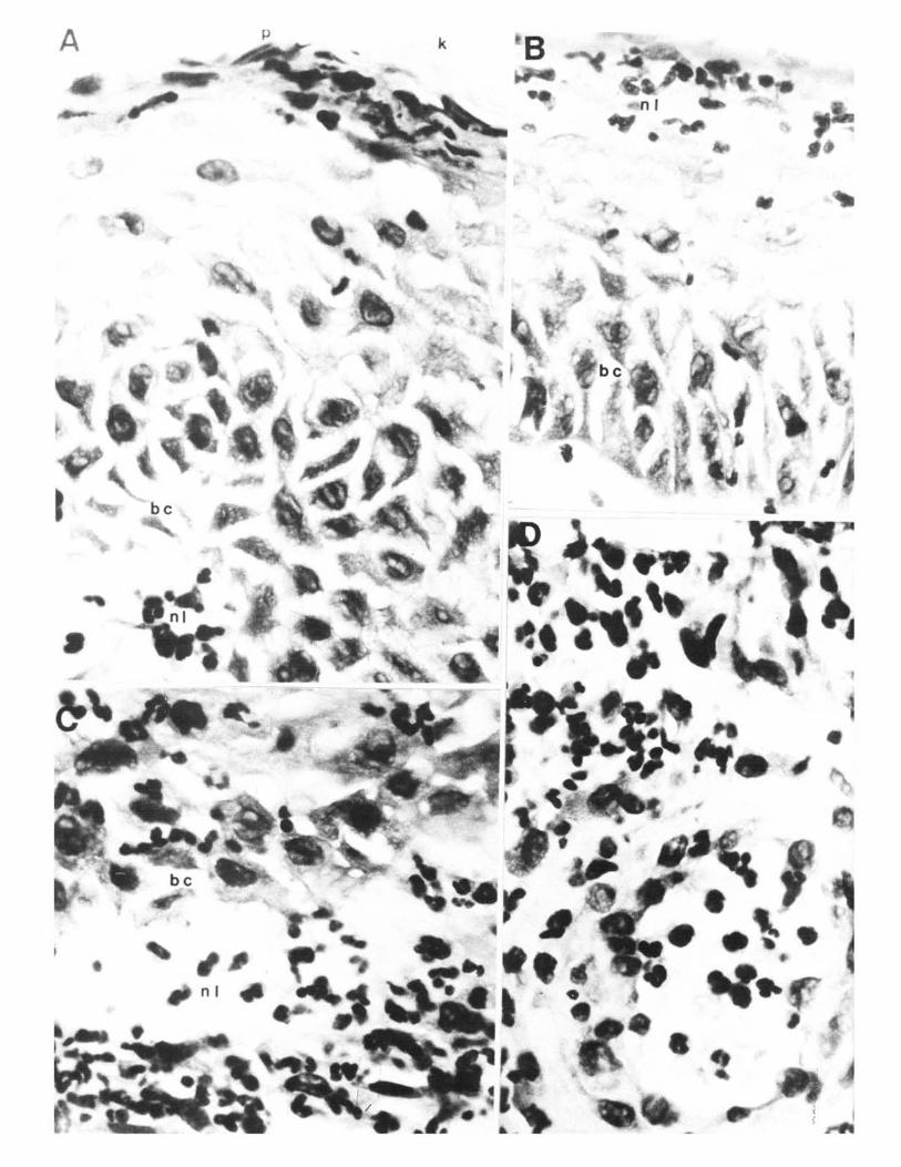
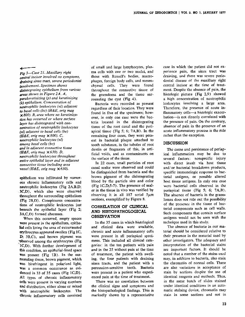
Fig 3---Case 21. Maxillary right central incisor involved no symptoms, draining sinus tract, severe periodontal involvement. Specimen shows disintegrating epithelium from various areas shown in Figure 2A. A, parakeratinizing (p) and keratinizing (k) epithelium. Concentration of neutrophilic leukocytes (nl) adjacent to basal cells (bc) (H&E, orig mag X 800). B, area where no keratiniza- tion has occurred or where surface layer has disintegrated with con- centration of neutrophilic leukocytes (nl) adjacent to basal cells (bc) (It&E, orig mag • 800). C, neutrophilic leukocytes (nl) among basal cells (bc) and in adjacent connective tissue (It&E, orig mag • 630). D, neutrophilic leukocytes throughout entire epithelial layer and in adjacent connective tissue including area vessel (H&E, orig mag • 630).
epithelium was infiltrated by numer- ous chronic inflammatory cells and neutrophilic leukocytes (Fig 2A,B,D;
3C ,D) , which also were observed throughout the surrounding epithelium (Fig 2B,D). Conspicuous concentra- tions of neutrophilic leukocytes just beneath the epithelial layer (Fig 2; 3A,C,D) formed abscesses.
When this occurred, empty spaces were present in the epithelium, epithe- lial cells lining the area of extravasated erythrocytes appeared swollen (Fig 1C, D; 3B,C), and brown pigment was
"~bserved among the erythrocytes (Fig ~'IC,D). With further development of this condition, an epithelial-lined space was present (Fig 1B). In the sur- rounding tissue, brown pigment, which was birefringent in polarized light, was a common occurrence as evi- denced in 33 of 35 cases (Fig 1C, D). All types of chronic inflammatory cells were present in varying numbers and distribution, either alone or mixed with neutrophilic leukocytes. The chronic inflammatory cells consisted
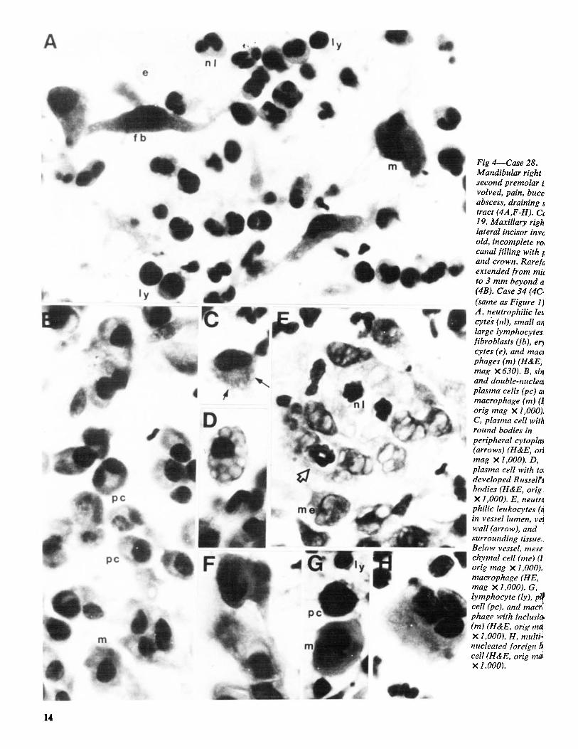
of small and large lymphocytes, plas- ma cells with one or two nuclei, and those with Russell's bodies, macro- phages, foreign body cells, and mesen- chymal cells. They were found throughout the connective tissue of the granuloma and the tissue sur- rounding the cyst (Fig 4).
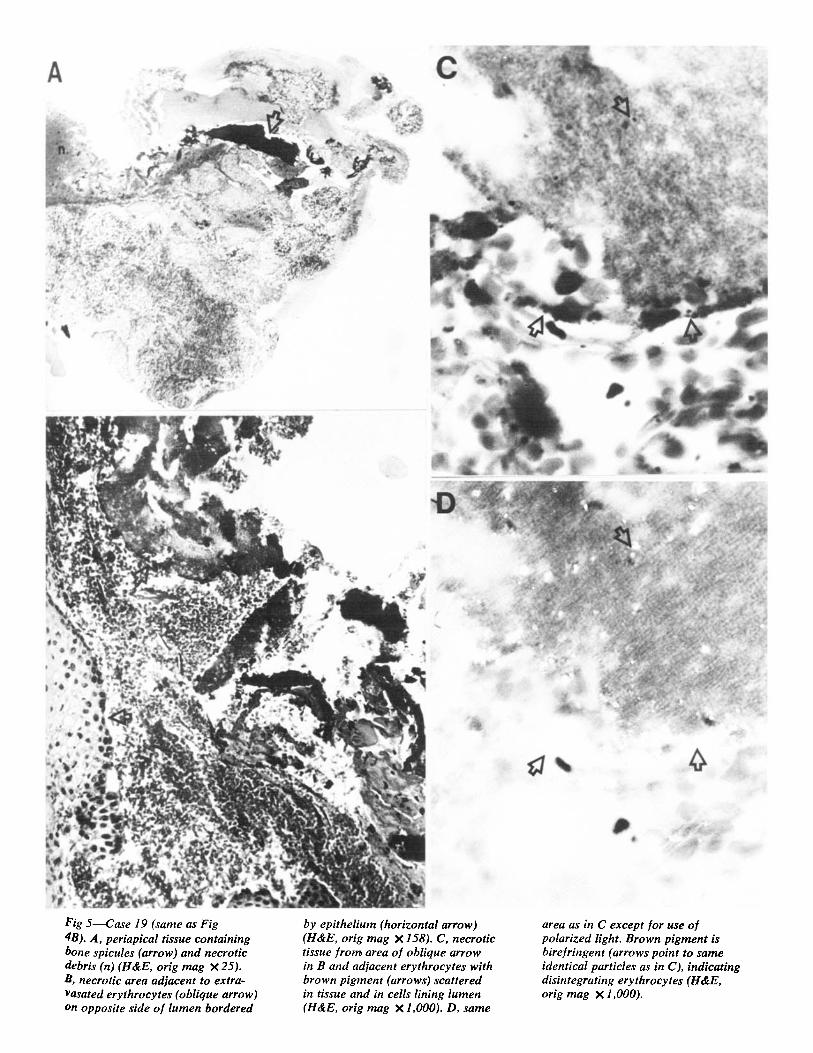
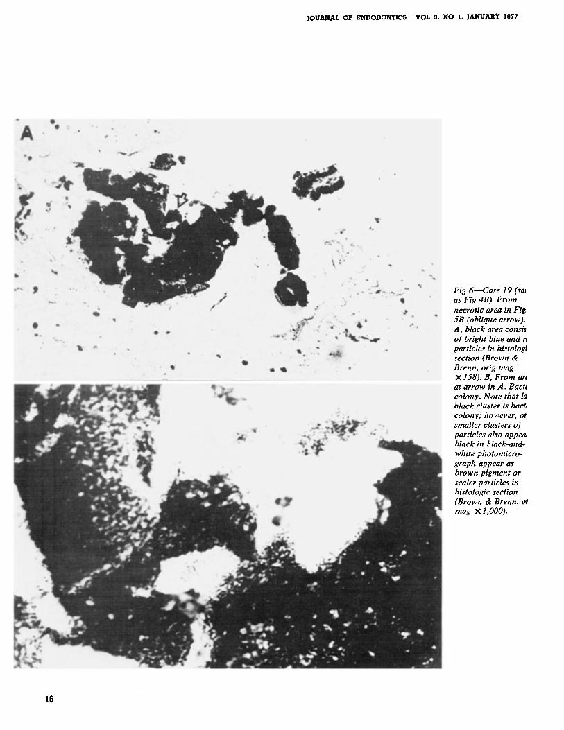
Bacteria were recorded as present regardless of their location. They were found in five of the specimens; how- ever, in only one case were the bac- teria located in the disintegrating tissue of the root canal and the peri- apical tissue (Fig 5; 6; 7A,B). In the remaining four cases, they were pres- ent in bacterial plaque attached to tooth substance, in the tubules of root dentin or fragments of this, in arti- ficial voids, and as contaminants on the surface of the tissue.
In 22 cases, small particles of root canal sealer were observed and could be distinguished from bacteria and the brown pigment of the disintegrating blood by the particle size and color (Fig 1C,D;5-7). The presence of seal- er in the tissue in vivo was verified by observing it in all 187 serial 5/xm sections, exemplified by Figure 8.
CORRELATION OF CLINICAL AND HISTOPATHOLOGICAL OBSE RVATION
In the 35 cases in which histological and clinical data were available, chronic and acute inflammatory ceils were present in all periapical speci- mens. This included all clinical cate- gories: in the ten patients with pain and in the 25 without pain at the time of treatment, the patient with swell- ing, the four patients with draining sinus tracts, and the patient with a percussion-sensitive tooth. Bacteria were present in a patient who experi- enced pain at the time of treatment.
There was no correlation between the clinical signs and symptoms and the histopathological findings. This is markedly shown by a representative
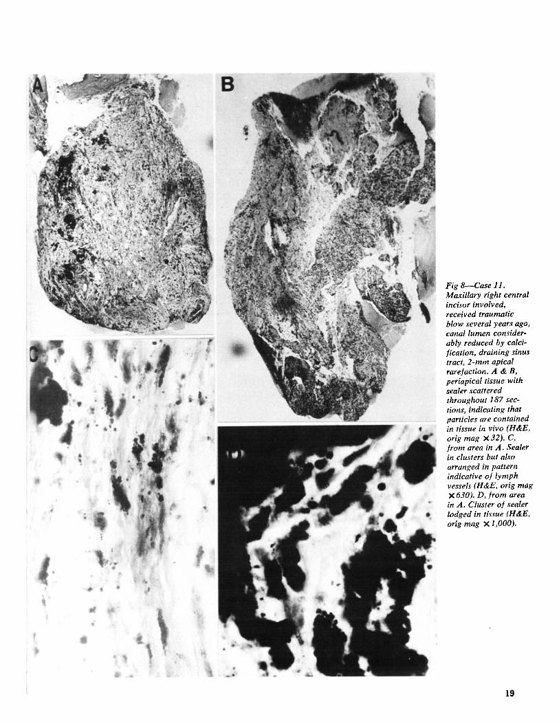
case in which the patient did not ex- perience pain, the sinus tract was draining, and there was severe perio- dontal disease of the maxillary right central incisor at the time of treat- ment. Despite the absence of pain, the histologic picture (Fig 2,9) showed a high concentration of neutrophilic leukocytes involving a large area. Therefore, the presence of acute in- flammatory cells--a histologic exacer- b a t i o n - i s not directly correlated with the presence of pain. On the contrary, absence'0f pain in the presence of an acute inflammatory process is the rule rather than the exception.
DISCUSSION
The cause and persistence of periap- ical inflammation may be due to several factors: nonspecific injury with direct insult via host tissue and/or bacterial breakdown products, specific immunologic response to bac- terial antigens, or possible altered host tissue antigens. In only one case were bacterial cells observed in the periapical tissue (Fig 5; 6; 7A,B). The absence of bacteria in the granu- lomas does not rule out the possibility of the presence in the tissue of bac- terial components such as cell walls. Such components that contain surface antigens would not be seen with the technique used in this study.
The absence of bacteri~ in our ma- terial should be considered relative to their presence in the material used by other investigators. The adequacy and interpretation of the bacterial stains are important factors. It should be noted that a number of the stains used may, in addition to bacteria, also stain the chromatin of normal cells. There are also variations in acceptance of stain by sections despite the use of identical reagents and methods. Thus, in the same batch of slides stained under identical conditions in an auto- matic staining device, chromatin may stain in some sections and not in
13
A n! $
f b
t,e P
Fig 4---Case 28. Mandibular right second premolar i, volved, pain, bucc
~ abscess, draining tract (4A,F-H). Ct 19. Maxillary righ lateral incisor invc old, incomplete ro~ canal [illing with and crown. Rare]~ extended ]rom mit
~ . ~ . ~ ~ A, neutrophilic let cytes (nl), small at large lymphocytes
:j . [ibroblasts ([b), er: cytes (e), and mac phages (m) (H&E,
8 mag • 630). B, sit, and double-nuclea
$ ~ l ~ " plasmacells(pc) a ~ A macrophage (m) 0
V ~ . ~ orig mag • l,O00) C, plasma cell witl round bodies in
" " i peripheral cytoplc~ (arrows) (H&E, or
~" .f~ii" ~ mag X 1,000). D, . plasma cell with to
de eloped Russell ~ bodies (H&E, orig X 1,000). E, neutrt
f , .
Oe
to 3 mm beyond a~ ~'~ (4B). Case 34 (4C.
- - (same as Figure 1)
r y
F
i
philic leukocytes 0 in vessel lumen, ve. wall (arrow), and surrounding tissue.~ Below vessel, mese, chymal cell (me) (~ orig mag • 1,000). ~ macrophage (HE, mag • 1,000). G, lymphocyte (ly), pl# cell (pc), and macr~ phage with inclusio~ (m) (H&E, orig mat X 1,000), H. multi~ nucleated foreign a~ cell (H&E, orig ma X 1.000).
1 4
+ , + ' y " , : ~ . ,
�9 ' "+ ' " ,',. ' ~ ' ~ ., ' ; ' " i ~ +P" �9 ,: ~'..~....',..+' ,.~ +.+
�9 " �9 "4~ ", ~,o ~ " - , ~ ram+: - ~ . P ~ + L . . . " , ~ J l ,+.., -.,,,,.,. l p ' . . , : - . . , + , ~ \ ~ . : ~ �9 ..,,r -. L . 4 - .+" +'
�9 + . , . . . . . . ;, . . . . . . ..'.. . . . . . . , . , :,~: 6 . . . _
;:~JtlS~'+"3+". , " , + ' . + ; , , : ~ ? " ~ ' " ~ : "" " " r ' ' . 3 1 1 , ~ " ~ " ' ~ : ".~. - ;- "~+~ " " " -,;"" ~'+, + , l + r ' " " �9 " i " +~ '~ ' " + " + . _.,._ , . ' . ' ,. �9 . -. ; , , . ,~.. , . - ' , . , ; . ~ . . . ~ " .,,.'"
'Dr ~' + ~ . <,.." ~ ., , ..,..'+.j o . , ~ , . ' ; : . ' . . ~ : . : . , I , : . . + . - ,."+++'-';,.
. . . . . �9 , - . . . . . ~ . ~ ~.- .,_.,:.~. "- ...' .. "...+,.-'r?,,"
' "~!~'.;..::. �9 " " '4"; " ,-, " ~ . . , . . ~ , , : , : . . ~ . .
- . :~ ~.. �9 ::~,, ~ . . ~
v ".~" ' , ~ L ' I ; ~1' ~ ' " " ' . " , ; , , ,3 .~ ~,..~,, .. ,
, "."+ b [ ' : ~ " " P ~ t ! ~ '"
, r ~ 4P . . . . . �9 . r 41'
~ ~d[ ' , t . + . .
' ';+--:,,~,.-,+ -.'" %,'r" :
~,' ~+ , ~ : : * , + ' ~ i
~ . . ":~.- . ,
~++ + +
0
Fig 5--Case 19 (same as Fig 41t). A, periapical tissue containing bone spicules (arrow) and necrotic debris (n) (H&E, orig mag • 25). B, necrotic area adjacent to extra- Vasated erythrocytes (oblique arrow) on opposite side of lumen bordered
by epithelium (horizontal arrow) (H&E, orig mag X 158). C, necrotic tissue ]rorr~ area of oblique arrow in B and adjacent erythroeytes with brown pigment (arrows) scattered in tissue and in cells lining lumen (H&E, orig mag • 1,000). D, same
area as in C except for use of polarized light. Brown pigment is birefringent (arrows point to same identical particles as in C), indicating disintegrating erythrocytes (H&E, orig mag • 1,000).
JOURNAL OF ENDODONTICS I VOL a, NO 1, JANUARY 1977
A ~D
b
O
Q
O e
I
LAI
"D ~ q b
I
r
%
L ~
Fig 6--Case 19 (saJ as Fig 4B). From necrotic area in Fig 5B (oblique arrow). ,4, black area consis of bright blue and r~ particles in histolo~ section (Brown & Brenn, orig mag • 158). B, From ar~ at arrow in ,4. Bact~ colony. Note that la black cluster is bact~ colony; however, otJ smaller clusters of particles also appetu black in black-and- white photomicro- graph appear as brown pigment or sealer pag"ticles in histologic section (Brown & Brenn, or mag • 1,000).
16
~i[ ~*
~
#* Ir
0 4~
".~ "~" ~ ~ ' i~
' I
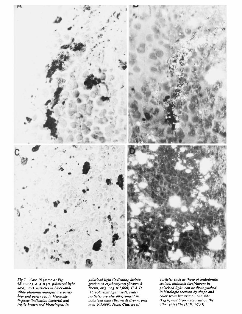
Fig 7--Case 19 (same as Fig 4B and 6)..4 & B (B, polarized light used), dark particles in black-and- white photomicrographs are partly bl~e and partly red in histologic seqtions (indicating bacteria) and pa~'tly brown and birefringent in
polarized light (indicating disinte- gration of erythrocytes) (Brown & Brenn, orig mag • 1,000). C & D, (D, polarized light used), sealer particles are also birefringent in polarized light (Brown & Brenn, orig mag • 1,000). Note: Clusters of
particles such as those of endodontic sealers, although bire/ringent in polarized light, can be distinguished in histologic sections by shape and color ]rom bacteria on one side (Fig 6) and brown pigment on the other side (Fig 1C, D; 5C, D).
JOURNAL OF ENDODONTICS I VOL 3. NO 1, JANUARY 1977
others; however, if the chromatin stains, it does so in areas where no bacteria could be present.
In control material of intact teeth, the chromatin may stain and could ap- pear as engulfed bacteria as reported by Boyle. 33 The finding by Winkler, Mitchell, and Healey 3 of bacteria evenly dispersed throughout the granu- loma is not corroborated by our study, although we have used the identical reagents and the same staining meth- ods. The only particles that were evenly dispersed in our sections were brown pigment and particles of the endodontic sealer (Fig 1, 5-8). More importantly, in cases where root remnants were enclosed in the speci- men, bacteria did stain in the necrotic tissue but did not in the remaining vital part of the pulp nor in the periapical granuloma or cyst. There- fore, the demonstration of bacteria in the necrotic part of the canal, in ad- jacent dentinal tubules, and in bac- terial plaque shows the efficacy of the staining method used. The absence of bacteria in the periapical tissue of the same sections cannot, therefore, be a false-negative. However, the dis- tinction between bacteria, brown pig- ment, and sealer particles may create some confusion particularly when based on black and white photo- micrographs. This corroborates the findings of a number of other inves- tigators.l.r-12
The location of bacteria related to specific cells is an important consider- ation. The fact that bacteria appear in the necrotic region of the pulp or periapical tissue only (Fig 5,6) and that neutrophilic leukocytes gather in a heavy concentration next to this area indicates that these leukocytes operate as macrophages. By using the root canal as a confined tissue, the sequelae of the tissue destruction can be observed. The alteration in the cell picture as the root apex is approached - -namely , the decrease in the number
of neutrophilic leukocytes and the ap- pearance of lymphocytes, plasma cells, macrophages, foreign body cells, and mast cells in relatively unaltered re- maining pulp tissue--indicates that the acuteness of the inflammation de- creases in the apical direction with increasing distance from the bacterial colony. The appearance of a con- siderable periapical involvement under these conditions in the presence of a remaining vital root pulp has been the subject of considerable disagreement, but the explanation seems simple in terms of inflammation and immunol- ogy. It is the tissue disintegration products and bacterial toxins that travel from the place of origin through the lymph vessels of the re- maining pulp and gather in the peri- apical tissue. This should explain the seemingly illogical appearance of re- maining, nearly unaltered pulp tissue between two areas of severe inflam- mation.34-36
Although Eleazer, Farber, and Selt- zer 37 found no cell-mediated immune response when adding venous blood to a culture prepared from the in- flamed pulp or the periapical inflamed tissue, the presence of particularly small and large lymphocytes, plasma cells, and macrophages indicates an immune response of the periapical tissue to the deteriorating pulp tissue. Specifically, when plasma cells are observed, active antibody synthesis is most likely occurring and these cells may become overloaded with their synthetic product to produce homo- geneous, acidophilic, large inclusions --Russell 's bodies (Fig 4D). These develop whenever the endoplasmic re- ticulum becomes so overloaded and distended as to become visible under the light microscope (Fig 4C). 3s It has been shown that the root canal offers a pathway for immuniza- tion. 3a-41 Not until there is an area of necrosis in the apical tissue will there be any colonization of bacteria.
The vast disagreement regarding in- cidence of periapical granulomas and cysts 1,1a'25 (Table 1) should be con- sidered relative to the criteria used by each investigator. Dorland 42 defined a cyst as "any sac, normal or abnormal, especially one which contains a liquid or semisolid material." According to this definition, the appearance of epithelium alone in the absence of a lumen filled with a disintegrated liquified tissue could not be defined as a cyst. It should be noted that epithelium in the form of rests of Malassez appear in the normal peri- apical tissue and that proliferation is a normal occurrence during periapical inflammation, for example, that caused by irritant endodontic proce- dures or materials. 34 A definition spe- cifically relevant to this investigation has been given by Sharer, Hine, and Levy44: "a cyst is defined as a patho- logic epithelium-lined cavity usually containing fluid or semisolid material." By using this definition in our investi- gation, only 9 (Fig 1-3) of 35 cases could be recorded as cysts; 12 addi- tional cases had epithelium present but no diseernable fluid-filled space.
It appears from the literature (Table 1 ) that depending on the def- inition, histologic criteria, type of population, and specific characteristics of the population sample, the occur- rence of periapical lesions that are cystic varies from 7% 1-18,14 to 54%.24 Some of the reported discrepancies may be related to the surgical tech- nique. If, as in most periapical sur- gery, fragments rather than a whole continuous lesion are removed, fluid- filled spaces could collapse and the fluid would escape. This was con- sidered in our laboratory evaluation in the present study as well as in our past investigations. Particular attention is directed toward epithelial cells ad- jacent to an empty space. Any altera- tion in the cells' morphologic structure or in adhering tissue remnants such
18
O
, ~
k
,d
"4
~ ......
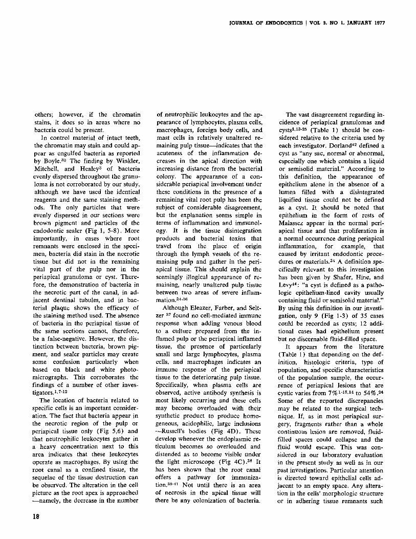
Fig 8--Case 11. Maxillary right central incisor involved, received traumatic blow several years ago, canal lumen consider- ably reduced by calci- fication, draining sinus tract, 2-mm apical rarefaction. A & B, periapical tissue with sealer scattered throughout 187 sec- tions, indicating that particles are contained in tissue in vivo (H&E, orig mag • 32). C, from area in A. Sealer in clusters but also arranged in pattern indicative of lymph vessels (H&E, orig mag • 630). D, from area in A. Cluster of sealer lodged in tissue (H&E, orig mag • 1,000).
19
,r
O
b
;I ii
4'
Fig 9--Case 21. A keratinizing epithe with large number, neutrophilic leuko~ and accumulation, neutrophilic leukol in abscess in subja~ connective tissue (H&E, orig mag • 158). B, [rom ce concentration in ct nective tissue in A. Mostly neutrophili leukocytes mixed a /ew lymphocytes macrophages (H&J orig mag • 1,000). neutrophilic leukoc and macrophages. presence o[ severe acute in[lammatiot total absence o/pa (H&E, orig mag • 1,000).
20
JOURNAL OF ENDODONTICS ] VOL 3, NO I, JANUARY 1977
as those shown in Figures 1 B-D; 2 B,C; 3; 5; and 9 permits their clas- sification as developing or true cysts. It should be noted that this is repre- sentative of the entire series of sec- tions in each case and that examina- tion of additional sections in any in- vestigation could alter the incidence.
The type of population from which the biopsy sample was obtained is especially a key issue. If, for example, all the endodontic surgical specimens are obtained from patients in whom nonsurgical endodontics has failed, and most of the cases are from an- terior teeth, which are easily retreated, the sample population is quite biased. Bhaskar's findings ~2 that 42% of all periapical lesions are cysts had the aforementioned limiting factors.
Lalonde and Luebke 2s and Morten- sen, Winther, and Birn 21 have re- ported a different sample population bias. In both of these studies, the periapical radiolucent area was iden- tified by radiograplis in an oral sur- gery screening clinic or by private practitioners. Their specimens are gen- erally of nonendodontically treated teeth. From their populations, it would be difficult to deduce any valuable in- formation concerning the incidence of cysts because endodontic therapy was not performed on any of the teeth.
By comparing the data of the vari- ous investigators, it is impossible to eliminate the bias and differences be-
tween the population samples. There- fore, to conclude that cysts occur in a certain incidence really depends on the investigator's definition of a cyst, histologic criteria, and characteristics of the population sample.
In addition, because the occurrence of periapical lesions that are cystic is still questionable, there does not seem to be any scientific evidence to SUpport the concept of Bhaskar, 45 Bender, 46 and Morse 4r to recommend instrumentation beyond the apex to
puncture the cyst and elicit nonsur- gical resolution.
The cells present in the periapical area--particularly the neutrophilic leukocytes, which are present in all sections in varying concentrations (Fig 2-4,9) may indicate a possible mechanism of the development of cystic cavities. When the neutrophilic leukocytes disintegrate, they release enzymes capable of dissolving cells and ground substance. Confluence of the microsacs occurs and forms small fluid-filled spaces. When these occur in or adjacent to the epithelium, a cyst has begun to develop. Other fluid- filled spaces may appear where there may be an accumulation of foam cells. When these cells disintegrate and their lipid content 48 is released into the tissue, a semisolid-filled sac occurs. Only when it is surrounded by epithe- lium does such a space fit the defini- tion of a cyst, although the deteri- orating masses might later form a part of the content of a cyst. The disagree- ment among investigators can only be resolved by a comparison of photo- micrographs at sufficiently high mag- nification and quality to allow exact differential diagnosis.
The histologic term acute in[lam- mation is based on the presence of considerable numbers of neutrophilic leukocytes in the tissue and in afferent vessels. However, it is a common mis- conception to associate the presence of the neutrophilic leukocytes with pain. This investigation confirms our earlier finding of noncorrelation between neutrophilic leukocytes that appeared in about equal numbers and distribu- tion in tissue taken from patients with and without pain (Fig 3). 1,a~ A better association could possibly be found provided an actual count of cells were performed in all serial sections taken through the entire lesion. However, because most laboratories take very few semiserial sections through what is considered a representative part of
the lesion, such counts would be meaningless. Even with the relatively high number of sections taken in this s tudy--80 to 280 sections from each specimen--a quantitation of the re- suits would be misleading.
Other clinical signs and symptoms such as swelling, draining sinus tract, or percussion sensitivity also could not be correlated with a specific histo- logic picture (Table 1). It is con- firmed in this study that histologic evidence of acute inflammation may frequently occur in the total absence of pain.
Based on the presence of neutro- philic leukocytes in all specimens, re- gardless of pain or other clinical con- ditions, there is nothing in our ma- terial that supports Bhaskar's recom- mendation 45 of instrumentation be- yond the apex with the intent of caus- ing acute inflammation as discussed by Morse. 4r The neutrophilie leuko- cytes are already there, and the sequelae of their life cycle is known. To instrument beyond the apex causes tissue damage in addition to that which already exists. The inflamma- tory cells are there as an inflamma- tory and immunologic response to the various noxious products, antigens, and toxins derived from the disinte- gration of the pulp. Instrumentation beyond the apex will push these toxic products out into the periapical tis- sues, as demonstrated by Figure 3-19 in Ingle's text 49 and aggravate the tissue disintegration, compounding the damaging effect of the mechanical ir- ritation. Clinically, this results in a flare-up of iatrogenic origin.
A biologic approach to endodontic therapy dictates that the source of the periapical inflammation be removed, that is, the noxious products that have accumulated in the root canal. When these are successfully removed, the periapical lesion, in most cases, heals without surgery. If, on the other hand,
9.1
JOURNAL OF ENDODONTICS I VOL 3, NO 1, JANUARY 1977
surgery is performed without retreat- ment or without apical obturation of the canal, the periapical lesion will persist or recur. Thus, there is neither theoretical nor practical support for the idea of instrumentation beyond the apical foramen. 45
C O N C L U S I O N S
Of 35 biopsy specimens of periapi- cal tissue examined, the following observations were made:
- - W h o l e bacterial cells were seen in five specimens.
- -Bac t e r i a were present in the periapical tissue in only one case.
- - B o t h acute and chronic inflam- matory cells occurred in all periapical lesions.
- -Ep i the l i um was observed in 21 of 35 periapical lesions, but only nine cysts were present.
- - T h e r e was no correlation between the presence of inflammatory cells and the clinical signs and symptoms.
- -Because the exact incidence of cysts is still undeterminable, there is no practical support for the claim that overinstrumentation will contribute to nonsurgical resolution.
S O b l M / E R Y
A total of 35 biopsy specimens was obtained during endodontic surgical procedures for the treatment of pain, swelling, draining sinus tract, and cal- cified, missed, or perforated root canals combined with periapical radio- lucent areas. The specimens were examined histologically, using his- topathologic and histobacteriologic methods.
Epithelium was found in 21 speci- mens, but only nine lesions were com- patible with the definition of a cyst. Both chronic and acute inf lammatory cells were found in all the specimens. Although bacteria were found in five specimens, in only one case were the bacteria located in the disintegrating
tissue of the root canal and the periapical tissue. Small particles of root canal sealer were found in 22 specimens. These sealer particles could be distinguished from bacteria and disintegrating blood cells by means of special stains and polarized light.
There was no correlation between the presence of various inflammatory cells and the clinical signs and symp- toms of the patients.
The concept of instrumentation be- yond the root apex with the intent of causing an acute inflammation to re- solve a cyst was not supported by the findings of this study.
This study was supported by the Office of Naval Research, N-00014-71-C-0180. The opinions expressed herein are those of the authors and cannot be construed as reflecting the views of the navy de- partment or the naval service at large.
The authors acknowledge the skillful and technical assistance of Mr. Michael Purcell, research assistant.
Dr. Langeland is professor and chair- man, department of endodontics, School of Dental Medicine, University of Con- necticut Health Center, Farmington; he is also visiting professor, department of endodontics, School of Graduate Dentis- try, Boston University Medical Center. Dr. Block is a former general dentistry resident, School of Dental Medicine, University of Connecticut Health Center; he is currently an endodontic graduate student, School of Dentistry, Medical College of Virginia, Virginia Common- wealth University, Richmond. Dr. Gross- man is professor emeritus, department of oral medicine, School of Dental Medi- cine, University of Pennsylvania, Phila- delphia. Requests for reprints should be directed to: Dr. Kaare Langeland, Pro- fessor and Chairman, Department of Endodontics, School of Dental Medicine, University of Connecticut Health Center, Farmington, 06032.
References
1. Block, R.M.; Bushell, A.; Rodrigues, H.; and Langeland, K. A histopatho- logical, histobacteriologic and radio- graphic study of periapical endodontic
surgical specimens. Oral Surg 42:656 Nov 1976.
2. Stewart, G.G. A study of bacteria found in root canals of anterior teeth and the probable mode of ingress. J Endodontia 2:8 Sept 1947.
3. Winkler, T.F. III; Mitchell, D.F.; and Healey, H.J. A bacterial study of human periapical pathosis employing a modified Gram tissue stain. Oral Surg 34:109 July 1972.
4. Langeland, K. Tissue changes in the dental pulp. An experimental histo- logic study. Odontol Tidskr 65:239 Aug 1957.
5. Langeland, K. Prevention of pulpal damage. Dent Clin North Am 16:709 Oct 1972.
6. Spangberg, L.; EngstrSm, B.; and Langeland, K. Biologic effects of dental materials. 3. Toxicity and antimicrobial effect of endodontic antiseptics in vitro. Oral Surg 36:856 Dec 1973.
7. Andreasen, J.O., and Rud, J. A histobacteriologic study of dental and periapical structures after endodontic surgery. Int J Oral Surg 1:272, 1972.
8. Rud, J.; Andreasen, J.O.; M611er- Jensen, J.E. A multivariate analysis of the influence of various factors upon healing after endodontic surgery. Int J Oral Surg 1:258, 1972.
9. Rud, J.; Andreasen, J.O.; M611er- Jensen, J.E. Radiographic criteria for the assessment of healing after endodon- tic surgery. Int J Oral Surg 1:195, 1972.
10. Kronfeld, R. Histopathology of the teeth and their surrounding structures, ed 2. Philadelphia, Lea & Febiger, 1939, p 209.
11. Grossman, L.I. Bacteriologic sta- tus of periapical tissue in 150 cases of infected pulpless teeth. J Dent Res 38: 101 Jan-Feb 1959.
12. Grossman, L.I. Endodontic prac- tice, ed 8. Philadelphia, Lea & Febiger, 1974, p 86.
13. Sommer, R.F., and Kerr, D.A. Quoted by Sommer, R.F.; Ostrander, F.D.; and Crowley, M.C. Clinical en- dodontics; a manual of scientific endo- dontics, ed 3. Philadelphia, W. B. Saunders Co., 1966, p 410.
14. Sonnabend, E., and Chan-Sook, O.H. Zur Frage des Epithels im apicalen Granulationsgewebe (Granulom) men- schlicher Ziihne. Deutsch Zahniirztl Z 21:627 May 1, 1966.
15. Patterson, S.S.; Shafer, W.G.i and Healey, H.J. Periapical lesions associated
22
JOURNAL OF ENDODONTICS t VOL 3, NO 1, JANUARY 1977
with endodontically treated teeth. JADA 68:191 Feb 1964.
16. Grossman, L.I., and Ether, S. Estudo comparativo clinico e histologico- pathologico de reacoes periapicalis cronicas. Rev Brasileira Odontol 22:226, 286, 1963.
17. Morse, D.R.; Patnick, J.W.; and Schacterle, G.R. Electrophoretic differ- entiation of radicular cysts and granu- lomas. Oral Surg 35:249 Feb 1973.
18. Baumann, L., and Rossman, S.R. Clinical roentgenologic, and histopatho- logic findings in teeth with apical radio- lucent areas. Oral Surg 9:1330 Dec 1956.
19. Wais, F.T. Significance of findings following biopsy and histologic study of 100 periapical lesions. Oral Surg 11:650 June 1958.
20. Linenberg, W.B.; Waldron, C.A.; .and DeLaune, G.F., Jr. A clinical, roentgenographic, and histopathologic evaluation of periapical lesions. Oral Surg 17:467 April 1964.
21. Mortensen, H.; Winther, J.E.; and Birn, H. Periapical granulomas and cysts. An investigation of 1,600 cases. Seand J Dent Res 78:241, 1970.
22. Bhaskar, S.N. Periapical lesions--- types, incidence, and clinical features. Oral Surg 21:657 May 1966.
23. Lalonde, E.R.~' and Luebke, R.G. The frequency and distribution of peri- apical cysts and granulomas. An evalua- tion of 800 specimens. Oral Surg 25:861 June 1968.
24. Priebe, W.A.; Lazansky, J.P.; and Wuehrmann, A.H. The value of the roentgenographic film in the differential diagnosis of periapical lesions. Oral Surg 7:979 Sept 1954.
25. Ross, P.N., and Burch, B.S. A clinical histopathologic study of conserva- tive endodontic failures. J Dent Res 55 (special issue B) : abstract no. 271, 1976.
26. Lillie, R.D. Histopathologic tech- nique and practical histochemistry, ed 3.
New York, McGraw-Hill Book Co., 1965, p 32.
27. Sheehan, D.C., and Hrapchak, B.B. Theory and practice of histotech- nology. St. Louis, C. V. Mosby Co., 1973, p 3.
28. Luna, L.G., ed. Manual of histo- logic staining methods of the Armed Forces Institute of Pathology, ed 3. New York, McGraw-Hill Book Co., 1968, p 1.
29. Langeland, K. Pulpal response to caries and operative procedures. J Dent Assoc South Africa 18:101 April 1963.
30. Langeland, K. Biologic considera- tions in operative dentistry. Dent Clin North Am, March 1967, p 125.
31. Langeland, K. Histologic evalua- tion of pulp reactions to operative pro- cedures. Oral Surg 12:1235 Oct 1959.
32. Langeland, K. The histopathologic basis in endodontic treatment. Dent Clin North Am, Nov 1967, p 491.
33. Boyle, P.E. Intracellular bacteria in a dental granuloma. J Dent Res 14: 297, 1934.
34. Langeland, K., Dowden, W.E.; Tronstad, L.; and Langeland, L.K. Hu- man pulp changes of iatrogenic origin. Oral Surg 32:943 Dec 1971.
35. Langeland, K.; Anderson, D.M.; Cotton, W.R.; and Shklair, I.L. Micro- biological aspects of dentin caries and their pulpal sequelae. Proceedings. Nij- megen, Holland, University of Nijmegen, 1976, p 173.
36. Langeland, K. Pulp histology and physiology. In Cohen, S., and Burns, R., eds. Pathways of the pulp. St. Louis, C. V. Mosby Co., 1976, p 203.
37. Eleazer, P.D.; Farber, P.A.; and Seltzer, S. Lack of lymphocytes stimula- tion by root canal products. J Endod 1:388 Dec 1975.
38. Gray, A., and Doniach, I. Ultra- structure of plasma cells containing Rus- sell bodies in human stomach and thy- roid. J Clin Pathol 23:608 Oct 1970.
39. Barnes, G.W., and Langeland, K. Antibody formation in primates follow- ing introduction of antigens into the root canal. J Dent Res 45:1111 July-Aug 1966.
40. Okada, H.; Aono, M.; Yoshida, M.; Munemoto, K.; Nishida, O.; and Yokomizo, I. Experimental study on focal infection in rabbits by prolonged sensitization through dental pulp canals. Arch Oral Biol 12:1017 Sept 1967.
41. Block, R.M.; Lewis, R.D.; Sheats, J.B.; and Burke, S.H. Antibody forma- tion to dog pulp tissue altered by para- formaldehyde-containing paste within the root canal. J Endod, to be published.
42. Dorland's illustrated medical dic- tionary, ed 24. Philadelphia, W. B. Saunders Co., 1965, p 376.
43. Langeland, K. Is N2 an acceptable method of treatment? In Grossman, L.I., ed. Transactions of the 5th International Conference on Endodontics. Philadel- phia, University of Pennsylvania Press, 1973, p 205.
44. Sharer, W.G.; Hine, M.K.; and Levy, B.M. A textbook of oral pathology, ed 3. Philadelphia, W. B. Saunders Co., 1974, p 236.
45. Bhaskar, S.N. Nonsurgical resolu- tion of radicular cysts. Oral Surg 34:458 Sept 1972.
46. Bender, I.B. A commentary on General Bhaskar's hypothesis. Oral Surg 34:469 Sept 1972.
47. Morse, D.R.; Wolfson, E.; and Schacterle, G.R. Nonsurgical repair of electrophoretically diagnosed radicular cysts. J Endod 1:158 May 1975.
48. Zegarelli, D.J.; Zegarelli-Schmidt, E.C.; and Zegarelli, E.V. Verruciform xanthoma. A clinical, light microscope, and electron microscope study of two cases. Oral Surg 38:725 Nov 1974.
49. Ingle, J.I., and Beveridge, E.E. Endodontics. Philadelphia, Lea & Febiger, 1976, p 173.
2 3