Embed Size (px)
Citation preview
Scientific Framework for
Small Cell Lung Cancer (SCLC)
National Cancer Institute
June 2014
1
Table of Contents
Executive Summary 2
Introduction 4
Background 4
Summary of the Literature and Recent Advances 5
SCLC Research Supported by the National Cancer Institute 11
Scientific Opportunities for SCLC Research and Plans to Implement 11
Recommended Initiatives
Training a Highly Competent Research Workforce 17
Oversight and Benchmarks for Progress 17
Summary of Scientific Recommendations and Implementation Timelines 18
Conclusion 19
Links and References 21
Addendum 28
Table 1: Genes of Interest in Small Cell Lung Cancer
Appendices 30
Appendix 1: Investment in Small Cell Lung Cancer Research – NCI/NIH
Appendix 2: Report from Small Cell Lung Cancer Workshop 2013
2
Scientific Framework For Small Cell Lung Cancer
Executive Summary
The Recalcitrant Cancer Research Act of 2012 (H.R. 733) requires the National Cancer Institute
(NCI) to “develop scientific frameworks” that will assist in making “progress against recalcitrant
or deadly cancers.” Small cell lung cancer (SCLC) is a recalcitrant cancer as defined by its five-
year relative survival rate of less than 7 percent and the loss of approximately 30,000 lives per
year. While it is true that the outcomes for the other common forms of lung cancer (squamous
cell and adenocarcinoma) need to be greatly improved, each of the three major types of cancer
that originate in the lung present very different problems, requiring different solutions. As the
Scientific Framework describes, treatment of SCLC has not changed in the last 30 years;
avoidance of the use of tobacco is the only known way to prevent the disease; no screening
method has proved effective; responses to chemotherapy are not durable and are difficult to
understand; and life expectancy after diagnosis tends to be very short. Consensus within the
scientific community regarding the limited early diagnostic and therapeutic approaches available
for patients with SCLC, as well as the limited availability of materials for research, have
provided an impetus for the evaluation of new and missed opportunities that could build upon the
existing portfolio of SCLC research to make additional progress against this disease.
Scientific progress has been made in the last decade in understanding some of the molecular and
cellular biology characteristics of SCLC. This progress includes: identifying critical oncogenic
changes found in nearly all SCLCs (such as inactivation of the TP53 and RB genes;
overexpression of Bcl-2 and Myc family members); developing genetically-engineered mouse
models that closely mimic human SCLC, leading to the identification of a pulmonary
neuroendocrine cell as the probable cell of origin for SCLC; creating patient-derived SCLC
xenografts and tumor lines for preclinical studies and drug testing; and identifying essential stem
cell signaling pathways (Hedgehog and Notch) and lineage oncogenes (ASCL1) driving the
growth of SCLC with its characteristic neuroendocrine phenotype.
In contrast to these basic research findings, there have been few therapeutic advances in the
systemic treatment of this cancer over the past 30 years. Standard therapeutic interventions used
today, which combine chemotherapy (etoposide plus platinum compounds) and radiation
therapy, reflect the prevailing state-of-the-art from the early 1980s.
SCLC is highly associated with cigarette smoking; the mutations that occur in SCLCs display a
mutational signature consistent with exposure to carcinogens found in tobacco, and the decrease
in cigarette smoking in the US population is reflected in the substantial decrease in the incidence
of SCLC over the past 30 years. Additional public health efforts to prevent smoking initiation
and to increase smoking cessation will decrease the incidence of this disease further.
Many patients who develop SCLC today stopped smoking years before their diagnosis. Thus,
the first major obstacle to progress against SCLC is the continuing risk of developing the disease
that remains for decades after smoking cessation. The second obstacle to progress is that for
most patients the tumor is already widely metastatic at the time SCLC is diagnosed; therefore,
curative surgery is rarely an option. Indeed, while SCLCs have been identified by computed
3
tomography (CT)-based screening efforts, including those of the NCI’s National Lung Screening
Trial (NLST), the vast majority of SCLCs found during these efforts were already metastatic,
severely limiting any benefit CT screening might have on patient survival. Therefore, earlier
detection methods are urgently needed. SCLCs are initially quite sensitive to chemotherapy and
radiation therapy to a degree unlike most other common solid tumors (prostate cancer, colon
cancer, or adenocarcinoma of the lung). However, the third obstacle to progress is the rapid
development of resistance to chemotherapy in more than 95% of SCLC patients. Median
survival from the time of diagnosis is less than 2 years. Because patients with SCLC rarely
undergo surgical resection of their tumors, compared with patients with other forms of lung
cancer, the histologic diagnosis of SCLC is most often made using needle biopsies that yield
only small numbers of tumor cells. Hence, the fourth obstacle to progress is the lack of tumor
tissue for clinical, molecular, and cell biologic studies, and in particular the lack of SCLC tissue
obtained when resistance to chemotherapy develops, which would permit studies of the
molecular mechanisms of acquired resistance.
In view of the obstacles limiting progress toward understanding the biology, early diagnosis, and
treatment of SCLC, the NCI assembled an expert panel of extramural scientists to identify new
scientific opportunities, resources, or technologies
that could advance our knowledge of SCLC, and, in so doing, improve outcomes for patients
with this disease or for those individuals at risk of developing SCLC. To expand the scope of
SCLC research, five initiatives were developed by this group of experts for consideration by the
NCI. It was recommended that the NCI: 1) develop better research tools for the study of SCLC
by optimizing the collection of tumor tissue specimens representing distinct phases of SCLC
(from initial diagnosis to disease recurrence following radio-chemotherapy) and by developing
new tumor models (conditionally-reprogrammed cell lines, patient-derived xenografts, and
genetically-engineered mouse models) that reflect the phases of SCLC found in the clinic; 2)
expand comprehensive genomic profiling studies of clinically-annotated SCLC specimens to
improve basic understanding of the frequency, distribution, and range of molecular abnormalities
that exist both at diagnosis and following therapeutic relapse; 3) investigate new diagnostic
approaches for populations at high risk of developing SCLC; 4) focus therapeutic development
efforts on specific molecular vulnerabilities of SCLC (tumor suppressor genes, unique genetic
drivers and their pathways, neuronal characteristics, and immunotherapy); and 5) investigate the
mechanisms underlying both the high initial rate of response to primary SCLC therapy and the
rapid emergence of drug and radiation resistance following completion of treatment.
The NCI is developing plans to implement these recommendations. In addition, the means to
evaluate progress and provide oversight of the NCI’s SCLC research portfolio will be
implemented to meet the goals of the Recalcitrant Cancer Research Act (H.R. 733) of 2012.
4
Introduction
H.R. 733, The Recalcitrant Cancer Research Act of 2012 (RCRA), enacted on January 2, 2013,
calls upon the National Cancer Institute (NCI) to “develop scientific frameworks that will help
provide the strategic direction and guidance needed to make true progress against recalcitrant or
deadly cancers.” Recalcitrant cancers are defined as those cancers with a five-year relative
survival rate below 50 percent. The Act requires the NCI to identify two or more recalcitrant
cancers that have a five-year relative survival rate of less than 20% and are estimated to cause at
least 30,000 deaths per year in the United States.
This report, prepared by the NCI, National Institutes of Health (NIH), for submission to
Congress focuses on the NCI’s approach to small cell lung cancer (SCLC). The report reviews
the current state-of-the-art in SCLC research and incorporates recommendations for new
scientific initiatives provided by experts in the disease who met at a workshop held at the NCI on
July 8-9, 2013 (Appendix 2). The workshop was attended by the NCI Director and senior NCI
scientific staff; it was conducted to evaluate research opportunities that could improve the
scientific understanding and medical control of SCLC. This report fulfills, in part, the provision
of the RCRA that the NCI develop a scientific framework for two identified recalcitrant cancers
(the other is pancreatic ductal adenocarcinoma) within 18 months of enactment of the RCRA (by
July 2, 2014). Each scientific framework will be sent to Congress and made available publicly
on the Department of Health and Human Services (DHHS) website within 30 days of
completion.
Background
As our knowledge about cancer biology has increased over the last several years, scientists have
come to understand that the classification of cancers used in the past – grouping all cancers that
arise in one organ, such as the lung, as one kind of cancer with multiple subtypes – is no longer
appropriate. There are three relatively common forms of lung cancer. Of these, SCLC arises
from neuroendocrine cells; squamous cell lung cancer from the squamous epithelium in the
large, central airways of the bronchial system; and adenocarcinoma of the lung from the
pneumocytes in the lung periphery. It is recognized that these are three distinct kinds of cancer
arising from three distinct cell lineages. These tumor types exhibit mostly different mutational
profiles and have different clinical histories and treatment options; and often different scientific
investigators. In addition, the amount of research progress has varied for these three cancers. As
a consequence, each of the three major types of cancer that originate in the lung present very
different problems and require different solutions.
Fifty years after issuance of the Report on Smoking and Health by the United States Surgeon
General, tobacco use has changed dramatically: In the United States, the prevalence of current
cigarette smoking among adults has declined from 42% in 1965 to 18% in 20121. However, the
consequences of smoking or ever-having smoked still affect many individuals. SCLC is one of
the diseases that is directly related to smoking2. Currently, SCLC accounts for approximately
13-15% of all lung cancers diagnosed in the United States2, 3
. With changes in cigarette filter
design, and with a steady decline in the number of smokers in the United States, an encouraging
5
decline (of 20-25% of all lung cancers) in SCLC incidence has been observed. Nonetheless,
approximately 30,000 individuals are diagnosed with SCLC yearly in this country. Median
survival for patients with limited stage disease (tumor that has not spread beyond the hemi-
thorax and related supraclavicular lymph nodes) is approximately 18 months; while survival for
the two thirds of patients who are diagnosed with more advanced (extensive stage) disease is 9
months4, 5
. Accordingly, only 3 to 6% of patients are alive 5 years following diagnosis of
SCLC3.
Despite positive epidemiologic trends in the United States, the worldwide incidence of lung
cancer, including SCLC, is still rising6. The highest incidence and mortality rates among males
are currently in Central and Eastern Europe. In many developing countries, lung cancer rates are
predicted to increase because of tobacco usage patterns that continue to broaden.
While recent progress has occurred in the understanding of SCLC biology, the clinical
application of this knowledge is still in its infancy. Thus, there is currently an opportunity to
consider how to hasten the control of SCLC by expanding our comprehension of SCLC at the
molecular level, by fostering new diagnostic approaches for individuals at high risk of
developing SCLC, by focusing the development of new treatments on recently-discovered
molecular vulnerabilities in SCLC, and by understanding why rapid, initial responses to systemic
therapy are almost always followed by a drug- and radiation-refractory state that cannot be
ameliorated with further treatment.
Summary of the Literature and Recent Advances
Etiology, Epidemiology, Prevention:
Based on considerable evidence from population and experimental studies, it is clear that
smoking is the most frequent cause of SCLC. Heavy smokers (for example, those who have
smoked on average at least one pack of cigarettes a day for 30 years) have approximately a 110
times greater chance of developing SCLC than individuals who have never smoked tobacco.
Although refraining from ongoing tobacco smoking reduces the risk of developing SCLC, the
relative risk of developing the disease does not equal that of a non-smoker despite 35 years of
smoking abstinence7. Still, within 2 years of smoking cessation a lowered risk can be observed,
and a rapid decline in the risk of developing SCLC continues over the next 10 years8. However,
the declining risk of SCLC slows thereafter; 35 years following smoking cessation, depending on
the amount smoked before quitting, an approximate 3-fold higher risk remains. Other less
prevalent risk factors include occupational or environmental exposures to uranium, radon,
beryllium, cadmium, silica, vinyl chloride, nickel compounds, and diesel exhaust9.
The age-adjusted incidence rate for SCLC in the US peaked in the early 1990s (11.29 cases per
100,000) and is currently 6.74 cases per 100,000 (2010 data10
). The decrease is primarily due to
reduced rates of smoking and to the use of filtered cigarettes. Today’s filters absorb larger
carcinogenic particles, but still allow smaller particles to enter the lungs. However, filter use
also prompts smokers to inhale more deeply. The result is a shift toward a higher incidence of
peripheral lung adenocarcinomas (arising from alveolar tissues deeper in the lung) rather than the
6
previously more prevalent squamous cell cancers and SCLCs that arise centrally11
. Among all
patients diagnosed with SCLC, the proportion of women with the disease increased from 28% in
1973 to 50% in 20022. Most cases of SCLC occur in individuals aged 60-80 years
12, and the
estimated overall death rate for SCLC is 30,000 per year13-15
. The best known preventive
measure against SCLC is smoking abstinence. A small percentage of SCLC patients have never
smoked, and their disease may be caused by other environmental factors or as yet unknown
causes16
. A healthy lifestyle that includes a balanced diet and adequate physical activity can also
decrease the risk of SCLC17-19
.
Biology and Genetics:
The unique histologic features of SCLC were first described by Barnard in 1926 as “oat cell
sarcoma” because of the appearance of the cells under the microscope20
. The biology of SCLC
differs from that of adenocarcinoma or squamous cell carcinoma of the lung in that its cell of
origin has neuroendocrine characteristics: the cells express features of neuronal as well as
endocrine cells21, 22
. These features, such as the expression of the ASCL1 molecule, assist in the
diagnosis of the disease23
. Patients with SCLC may also display symptoms derived from an
increased release of certain hormones (for example, antidiuretic hormone) produced by SCLC
cells24
.
According to the current World Health Organization classification, SCLC belongs in the larger
group of neuroendocrine tumors of the lung. This group consists of low-grade typical carcinoid
tumors, intermediate-grade atypical carcinoid tumors, and high-grade neuroendocrine tumors
including large-cell neuroendocrine carcinoma and SCLC25, 26
. Typical carcinoid tumors are
relatively benign with 90% 5-year survival rates; atypical carcinoid tumors can be more
aggressive with a 50-60% 5-year survival rate. Large cell neuroendocrine carcinomas and SCLC
represent the most aggressive of this class of malignancies with 5-year survival rates of 27% and
3-6%, respectively. While differing from adenocarcinomas, which are epithelial in origin, SCLC
has, in some cases, been demonstrated to develop in parallel with adenocarcinoma, or may
emerge in a patient treated for adenocarcinoma of the lung (in this case the tumor is classified as
a combined SCLC/adenocarcinoma). The simultaneous presence of two histologic types of lung
cancer may be due to a shared carcinogenic insult27
, and may be related to field cancerization,
i.e., histologically normal-appearing tissue adjacent to neoplastic tissue that displays molecular
abnormalities, some of which are the same as those of the tumor28
.
Two reports published in 2012 provided a detailed characterization of the genomic landscape of
SCLC using next generation sequencing approaches, including full exome sequencing,
transcriptome profiling by RNASeq, copy number analyses, and limited whole genome
sequencing to identify translocations29, 30
; these investigations expanded understanding of the
range of genetic alterations in this disease. In large part because of its association with smoking,
SCLC has one of the highest densities of mutation per tumor (an average of 7.4 mutations per
mega base pair), surpassing another highly mutated cancer, melanoma, with a density of 6.2929
.
Most of the mutations are of the passenger type, which means that they do not (necessarily)
contribute to the initiation or progression of the disease. More important are driver mutations
that directly contribute to carcinogenesis. These two reports confirmed what had been
previously proposed in studies that examined a smaller number of tumors, namely that the most
7
prevalent inactivated tumor suppressor genes in SCLC are TP53 and RB-131-33
. Concomitant
inactivation of these tumor suppressors is nearly universal in SCLC. Novel mutations were also
found, such as those in genes controlling epigenetic regulators, stem cell genes, as well as other
driver mutations within established proto-oncogene and tumor suppressor gene families (Myc
family genes, Bcl-2, PTEN, CREBBP, FGFR1, SLIT2, EPHA7, for example; see Table 1 in the
addendum for a current list). The number of primary SCLCs for which data have been reported
at the level of full exome sequencing comprises only 82 samples (compared with the baseline
number of 500 specimens per disease used in The Cancer Genome Atlas [TCGA] initiative) and
is inadequate to characterize the spectrum of potential oncogenic driver mutations in SCLC to
include those alterations with a frequency of occurrence below 10% with statistical significance.
To highlight this, FGFR1 amplification was detected at a rate of 6% in one study29
, while such
alterations were not observed at all in the other30
. Another limitation of these investigations is
that many of the samples analyzed were from surgically resected early stage and chemo-naïve
patients, and do not represent the full natural history of the disease with regard to development of
metastases and changes induced by therapeutic intervention. As noted previously, the use of
needle biopsies as the initial diagnostic approach for patients with presumed SCLC has limited
the availability of tumor specimens for genetic analyses. A renewed effort to obtain such
specimens will be needed to fully assess genomic changes in this malignancy.
Recently, considerable attention has been devoted to evaluating epigenetic changes in SCLC,
including altered methylation patterns and the discovery of variations in microRNA expression34,
35. One study found that 73 genes were methylated in >77% of primary SCLCs; most of the
methylated genes had not been previously linked to aberrant methylation in other tumor types.
Gene ontology analyses indicated a significant enrichment of methylated genes encoding
transcription factors related to the process of neuronal differentiation. Another investigation
demonstrated that expression of the microRNA, miR-886-3p, is down-regulated by DNA
hypermethylation in SCLC; its downregulation is closely correlated with shorter patient survival.
miR-866-3p is a potent repressor of cell proliferation, migration, and cancer cell invasion.
Finally, another investigation suggested that overexpression of the polycomb repressive complex
2 (PRC2) may contribute to poorer prognosis for patients with SCLC36
. PRCs are known to
modify the epigenetic status of tumors and repress target genes that establish and maintain cell
fate. An improved understanding of these epigenetic alterations may facilitate the development
of new biomarkers for early detection and prognosis, and may lead to the development of better
therapies for patients with SCLC.
The search for prognostic markers for patients with SCLC led to the discovery of the
neuroendocrine marker, pro-opiomelanocortin (POMC), the expression of which has been shown
to predict liver metastases and poor survival in SCLC22
. Other therapeutic targets and prognostic
markers [such as poly-(ADP-ribose)-polymerases (PARP) —critical DNA repair proteins] have
been discovered by proteomic approaches37
. In addition, the identification and characterization
of SCLC stem cells, detectable by expression of CD44 and CD90 surface markers, may reveal
hidden vulnerabilities in this disease38
. SCLC cells expressing these surface proteins have
increased expression of the mesenchymal markers N-cadherin and vimentin, increased mRNA
levels of the embryonic stem cell-related genes NANOG and OCT4, and increased resistance to
irradiation compared to other sub-populations studied.
8
Models of SCLC:
Human SCLC-derived cell lines and animal models of SCLC have played an important role in
SCLC research. Over the last decade, much has been learned about the nature of SCLC from
studies based on cell lines39-41
. This work revealed new molecular abnormalities in SCLC cells
that have been used in diagnosis and therapy, as well as in the establishment of mutational
profiles for this disease29, 30
. SCLC cell lines demonstrate expression of cell surface receptors
that play an important role in neuronal function, such as the vasopressin receptor and three
bombesin receptors, as well as expression of transcription factors that play a role in the biology
of tumor stem cells, such as SOX2, ASCL1, OCT4, and NANOG. Thus, cell models exist that,
at least in part, mimic the molecular abnormalities observed in SCLC tumor specimens in vivo41
.
However, many currently available SCLC cell lines grow slowly in vitro, which is not
representative of the proliferation rate of this tumor in vivo; slow growth in cell culture also
limits the use of SCLC cell lines for high throughput drug screening41
.
Animal models employed to study SCLC include: xenografts produced from the subcutaneous
implantation of established human SCLC cell lines into immune-deficient mice; patient-derived
xenografts (PDXs) in which human tumors are implanted directly from a biopsy specimen into
an immune-incompetent mouse without having been established first as a cell line in vitro; and
genetically-engineered mouse models (GEMMs) in which gene knock-out and knock-in
technologies have made it possible to reflect the genetic and biological evolution of human
SCLC. Xenograft models derived from established SCLC cell lines, unfortunately, often do not
resemble primary SCLCs at the molecular level. PDXs, on the other hand, mirror the
heterogeneity of the primary tumor and can mimic some factors in the tumor microenvironment,
excluding those of the immune system42
. PDX models have facilitated next-generation DNA
sequencing of SCLC specimens by allowing the expansion in vivo of a small, initial tumor
specimen43
. PDX models have also assisted in the proteomic characterization of SCLCs by
establishing molecular signatures that reflect the neuroendocrine origin of this cancer44
.
Unfortunately, because of the difficulty obtaining SCLC specimens appropriate for direct
xenografting, the number of SCLC PDX models available for investigation is quite modest45
.
GEMMs can develop spontaneous, autochthonous tumors and have the microenvironment
required for tumor progression, including degradation of the extracellular matrix and
angiogenesis; however, they may lack the genetic complexity that is typical of SCLC. SCLC
GEMMs are usually developed based on the inactivation of 2 or 3 critical tumor suppressor
genes (mimicking the situation in SCLC patients). The gene combinations include TP53, RB1,
p130, and PTEN46-49
. These models have been used to explore the biology of SCLC, including
cell of origin studies and examinations of the development of metastases in SCLC50
. For
example, GEMMs were used to confirm the importance of the Hedgehog cell signaling pathway
in SCLC. However, in view of the time required for the development of tumors in GEMMs,
drug testing in these models, although valuable, can be laborious and expensive.
Another way to define critical proteins and signaling pathways in SCLC is through the use of
synthetic lethality siRNA/shRNA screens in cell culture that can efficiently explore cooperating
pathways and networks involved in the development of this cancer51
. Two genes convey
synthetic lethality if mutations in either, alone, are compatible with cell viability, but mutations
9
in both lead to cell death52
. This technique can also be adapted for the selection of drugs that
could be used in combination therapies or for targeting “undruggable” targets53
.
Risk Assessment and Screening:
Although SCLC is, in most cases, a disease associated with tobacco use, little is known about
predisposing genetic or non-genetic factors that lead to the development of the disease in certain
current or former smokers, but not in others. Somatic mutations accumulate during the lifetime
of an individual exposed to the carcinogens in tobacco smoke. There is a need for further study
of the germline (i.e., heritable) traits that may contribute to the development of SCLC. In rare
instances, an inherited p53 deficiency (Li-Fraumeni syndrome) may lead to the development of
SCLC after radiation exposure54
, but additional studies will be required to fully understand the
interactions between environmental exposures and individual inherited predispositions to SCLC.
Screening for SCLC is also a challenge. There are currently no validated biomarkers that can be
measured in blood or other tissues to detect SCLC at an early stage. Furthermore, the recent
NCI-sponsored National Lung Screening Trial55-57
that proved the value of screening individuals
at high risk of developing lung cancer with low-dose helical computed tomography (CT) also
demonstrated that screening did not improve survival for the subset of SCLC patients detected by
CT screening, unlike those with adenocarcinoma or squamous cell cancer of the lung55, 57
. The
majority of patients with SCLC detected by CT screening (86% of the 125 patients) were
diagnosed with advanced stage disease, similar to the percent seen in the absence of dedicated
screening. Consistent with this distribution of stages, subsequent therapy did not significantly
prolong the survival of screened patients. These results suggest that metastatic dissemination
and/or resistance to systemic therapy may develop early in the natural history of SCLC.
Diagnosis, Staging, and Monitoring:
The diagnosis of SCLC, whether the patient is symptomatic or not, usually begins with histologic
confirmation of an abnormality detected on imaging studies, typically by fine needle aspirate
biopsy. Immunohistochemical evaluation employing a variety of neuroendocrine or other
markers confirms the diagnosis of SCLC. Medical history, physical examination, routine
laboratory tests, and computed tomographic scans of the chest and abdomen with infusion of
contrast material, and magnetic resonance imaging (MRI) of the brain complete the initial
evaluation. For patients without evidence of disease outside one hemithorax on these studies, 18
Fluoro-deoxyglucose positron emission tomography (PET) is useful for optimal staging, and
can detect bone metastases. Staging for patients with SCLC is most commonly categorized
using the Veterans Administration Lung Study Group system; limited-stage disease (LD), which
occurs in approximately one third of patients, is defined as SCLC confined to the hemithorax of
origin, the mediastinum, or the supraclavicular nodes, which can be encompassed within a
tolerable radiation therapy port. Extensive-stage disease (ED) SCLC has spread beyond the
supraclavicular areas and is too widespread to be included within the definition of LD. Patients
with distant metastases by definition have ED58
.
Monitoring of response to therapy is usually performed by imaging techniques capable of
providing accurate measurements of tumor size; these size measurements are interpreted by
10
Response Evaluation Criteria In Solid Tumors (RECIST) criteria that define categories of
response to treatment58
. PET staging now approaches a 100% level of sensitivity and greater
than 90% specificity59-62
. The use of PET scanning to both stage and follow the effect of
treatment for patients with SCLC has enhanced the accuracy by which the effectiveness of new
treatment modalities can be examined.
Therapy and Resistance:
Current therapeutic approaches for SCLC are of modest long-term benefit despite the
exceptionally good response to first-line therapy. Treatment for LD includes a standard first line
chemotherapy regimen63, 64
with concomitant radiation that can be encompassed in a single
radiation port65, 66
. Treatment for ED includes the same chemotherapy options, without
concomitant radiation64
. In some instances, particularly for small peripheral lung nodules,
surgery can also be considered67
.
Treatment programs for SCLC have changed little over the past three decades; the most
important advances have improved the precision of radiation therapy and have introduced better
supportive care measures, such as more effective antiemetic regimens. The generally accepted
standard for first-line systemic therapy, etoposide combined with either cisplatin or carboplatin,
has been in use since the early 1980s65, 68-70
. An alternative first-line chemotherapy regimen,
cisplatin and irinotecan, appeared to be superior in a Phase III study conducted in Japan71
, but
these results could not be confirmed in subsequent US comparative trials64
. SCLC is an
unusually chemosensitive and radiosensitive disease, at least initially, resulting in objective
response rates of 60 to 80% in patients without substantive co-morbid conditions. However,
essentially all patients with ED, and most patients with LD, experience disease progression
within months of completing first-line therapy. A recent genome-wide association study
suggested that germline genetic variations may affect resistance to irinotecan, and thus may be
associated with decreased overall survival of SCLC patients treated with chemotherapy72
.
Certain single nucleotide polymorphisms (SNPs) that were associated with shorter overall SCLC
survival may affect the expression of transcription factors involved in the epithelial-to-
mesenchymal transition (EMT), a process by which epithelial cells lose their cell polarity and
cell-cell adhesion, and gain migratory and invasive properties that may be involved in the
development of metastases.
There is only one FDA-approved therapy for recurrent SCLC: topotecan, a topoisomerase 1
inhibitor73-75
. Recurrent SCLC is substantially less responsive to therapy than primary disease.
Response rates for topotecan are approximately 25% for relapses occurring at least 3 months
after completion of first-line therapy, and as low as 3 – 6% for progressive disease occurring at
the time of or shortly after completion of first-line therapy. Objective responses to a third line of
chemotherapy are uncommon76
. Hence, no consensus has been reached on treatment regimens
for patients whose disease has progressed after first- and second-line therapy.
Prophylactic whole brain irradiation, in the absence of detectable brain metastases, is an
important component of therapy for most limited stage, and some extensive stage, patients with
SCLC. It is typically administered to those individuals who respond well to initial treatment
shortly after completion of first-line combined modality therapy77, 78
. Prophylactic cranial
11
radiation therapy decreases the risk of subsequent, clinically significant brain metastases and
improves survival in patients with LD and ED66, 79
.
Approximately 100 SCLC interventional clinical trials have been registered in the
ClinicalTrials.gov database since December 2007; about one-third of which are supported by the
NCI80
. These studies include efforts to target the neuroendocrine character of SCLC, its
dependence on the PARP pathway81
, and the use of immunological interventions including
therapeutic vaccines82
, antibody radio-immunoconjugates83
, or checkpoint inhibitors intended to
stimulate anti-cancer immune responses70
.
SCLC Research Supported By The NCI
The NCI supports a range of research activities that are relevant to improving outcomes for
patients with SCLC. A review of the portfolio of NCI and NIH grants that support basic and
translational studies, clinical trials, and training programs related to SCLC is presented in
Appendix 1.
Scientific Opportunities For SCLC Research And Plans To Implement Recommended Initiatives
Recent advances in SCLC research were examined during a multidisciplinary workshop
convened to develop new scientific opportunities for this recalcitrant disease. The workshop
report, Small Cell Lung Cancer: Seizing on Opportunities to Translate Recent Research into the
Clinic for New Diagnostics and Interventions, was presented to and accepted by the NCI Clinical
Trials and Translational Research Advisory Committee on June 18, 2014, and is available as
Appendix 2 of this Report. Based on the unique features of SCLC and the urgent need to
improve early diagnosis and treatment, as well as the recommendations of the experts attending
the SCLC Workshop, the NCI plans to focus on five scientific opportunities that could improve
the outlook for patients with SCLC:
1. Building better research tools for the study of SCLC by (a) optimizing the collection of tumor
tissue specimens representing distinct phases of SCLC (from initial diagnosis to disease
recurrence following radio-chemotherapy) and (b) developing new tumor models
(conditionally-reprogrammed cell lines, patient-derived xenografts, and genetically-
engineered mouse models) that reflect the phases of SCLC found in the clinic;
2. Expanding comprehensive genomic profiling studies of clinically-annotated SCLC specimens
to improve the basic understanding of the frequency, distribution, and range of molecular
abnormalities that exist both at diagnosis and following therapeutic relapse;
3. Investigating new diagnostic approaches for populations at high risk of developing SCLC;
4. Focusing therapeutic development efforts on specific molecular vulnerabilities of SCLC
(tumor suppressor genes, unique genetic drivers and their pathways, neuronal
characteristics, and immunotherapy); and
5. Examining the mechanisms underlying both the high initial rate of response to primary SCLC
therapy and the rapid emergence of drug and radiation resistance following completion of
treatment.
12
1. Better Research Tools for the Study of SCLC:
(A) Optimizing Collection of Tumor Tissues: The diagnosis of SCLC is frequently made by
cytological examination of biopsy material obtained by fine needle aspiration; repeat biopsies,
performed during distinct stages of disease progression, are rarely attempted. The paucity of
available biospecimens for this disease is striking, and is a primary barrier to progress in SCLC
research. Newer image-guided diagnostic approaches, such as endoscopic bronchial ultrasound-
guided core biopsies, can be safely performed and yield substantially more tumor for molecular
characterization. The use of these newer biopsy approaches underscores the importance of
incorporating specialists in pulmonary medicine, cardiothoracic surgeons, and interventional
radiologists (who perform the diagnostic procedures) as active members of the multidisciplinary
team of health care professionals who care for patients with SCLC.
Beyond changing standard of care approaches to diagnostics, investigators in the field should be
encouraged to implement biopsy protocols to ensure that good quality biospecimens are obtained
under optimized conditions for banking, molecular profiling, creating xenografts, and/or cell line
derivation. Research protocols to permit well-controlled and standardized repeat biopsies over
time (and during the multiple phases of SCLC disease progression) should also be strongly
encouraged. These will provide the tumor tissues with which to answer critical questions about
SCLC regarding the range of driver mutations involved, mechanisms of progression, acquired
resistance to therapeutics, and factors promoting metastasis. While there are ethical
considerations related to performing biopsies strictly for research purposes, patients may, in fact,
benefit from interim analyses of the spectrum of mutations in their tumors that could uncover
clinically-actionable targets.
Recommendations for next steps: The NCI has recently re-issued an RFA (Support for Human
Specimen Banking in NCI-Supported Clinical Trials) that will provide assistance for the
collection of human tumor tissues from patients entered on the full range of NCI-supported
clinical trials (both early therapeutics studies and comparative studies). The NCI’s new Early
Therapeutics Clinical Trials Network, furthermore, routinely collects formalin-fixed diagnostic
specimens as well as fresh biopsy tissues during proof-of-mechanism studies of new agents.
New resources from the Specimen Banking RFA can be focused to increase the number of SCLC
specimens gathered at specific stages of SCLC including pre- and post-treatment samples from
the same patient. Tailored administrative supplements to Specialized Programs of Research
Excellence in Lung Cancer, translational programs that are already funded for disease-specific
tumor banking, could also be developed to support the collection of SCLC specimens under
defined clinical and tumor banking conditions, including optimization of protocols for collecting
specimens from primary and metastatic sites as well as under informed consent conditions that
would permit sharing of relevant pre-treatment and genomic information.
(B) New SCLC Models: The complex biology of SCLC could be understood at greater depth by
developing new tumor models that better mirror the human disease. SCLC cell lines currently
used for tissue culture studies suffer from a number of potential deficiencies, including low
growth fractions and a tendency to proliferate as multi-cell tumor aggregates, making their use
for drug screening difficult. Furthermore, many SCLC lines do not have germline DNA
available to permit certain identification of somatic mutations, and most SCLC lines have been
continuously propagated for years using standard methods that may drastically alter their
13
molecular composition compared with the primary tumors from which they were derived. New
techniques, including the development of conditionally-reprogrammed tumor cell lines
(developed with Rho kinase inhibitors), initiated from small tumor biopsies or circulating tumor
cells, offer the possibility of rapid establishment of SCLC cell lines with molecular pedigrees
much closer to primary tumors84
. These models, especially if well-annotated clinically and
developed using sequential tumor biopsies from individual patients, could be used to study
mechanisms underlying the early evolution of drug resistance, a phenomenon that occurs
regularly following initial therapy in patients with SCLC.
In addition to new, clinically-annotated cell lines from patients with SCLC, the need also exists
for development of a larger collection of PDX models that have been derived from paired
biopsies obtained before combined modality therapy is initiated, and then at the time of disease
progression in the same patients. Such models would be of value for understanding mechanisms
of both primary and acquired drug resistance. An SCLC PDX collection sized to provide
sufficient genetic heterogeneity to reflect the variation in therapeutic response observed in the
clinic could provide an attractive platform for in vivo studies of molecularly-guided, single agent
and combination targeted therapies.
Current GEMMs have elucidated the cell of origin for SCLC and essential driver mutations for
this disease; however, the long latency period required for the development of SCLCs in
GEMMs has limited the broad applicability of these models, in particular for drug screening.
There is a need to improve such models by: 1) incorporating a greater degree of genetic
heterogeneity during their elaboration; 2) producing GEMMs that integrate acquisition of drug
resistance into the model development process (which would be useful for screening second line
therapies); and, 3) evaluating the effects of tobacco smoke on the carcinogenic process in
GEMMs. Recently, other models have been developed that may be suitable to study SCLC
metastases85
. In these systems, newly-developed mouse strains that lack functional B-, T-, and
NK cells (Pfp/Rag2 double-knockout) have been used to facilitate the production of mice
carrying SCLC xenografts that undergo spontaneous metastases; this model more clearly mirrors
the clinical course of SCLC.
Recommendations for next steps: The NCI’s Division of Cancer Treatment and Diagnosis began
a Patient-Derived Models (PDM) Program approximately 12 months ago to develop a library of
clinically- and genetically-characterized PDXs and conditionally-reprogrammed cell lines in
support of the extramural community. Over 1000 human tumor specimens will be collected
from biopsies of metastatic solid tumors of patients undergoing screening for eligibility in an
NCI-supported clinical trial that matches targeted anticancer agents with specific mutations.
Supplemental grants to 15 NCI-supported Cancer Centers in fiscal year 2013 were also placed to
increase the number of surgical specimens and CTCs available for model development. Thus,
the facilities and resources are in hand to conduct a focused effort to increase the number of PDX
models and tumor cell lines from patients with SCLC. With further support from and to the
NCI-supported Cancer Center community, it is likely that the collection of annotated SCLC
specimens can be facilitated; based on the current tissue collection and PDX ‘take’ rate,
approximately 50-75 new PDX and cell line models could be established over 3 years from 150
to 200 SCLC biopsies and/or surgical specimens.
14
2. Comprehensive Genomic Profiling:
The small number of SCLCs that have been analyzed by exome or whole genome sequencing is
inadequate to define the full spectrum and distribution of driver mutations in this disease. Efforts
to characterize a much larger set of tumors from patients with SCLC, particularly from patients
entered on clinical trials, for genomic, epigenetic, and transcriptome alterations, should be
strongly encouraged. Furthermore, comparative analyses of paired biospecimens from single
individuals, obtained from chemo-sensitive and chemo-resistant disease, or from primary and
metastatic sites, should permit a more focused description of the driver alterations associated
with changes in disease state. A comprehensive molecular analysis of specimens from the small
subset of patients with long-term survival from SCLC would also be of substantial interest86
.
Studies of SCLC genomics should be accompanied by an evaluation of genetic changes in the
germline of SCLC patients as well as individuals at high risk of developing SCLC to identify
possible heritable predispositions to this disease. Finally, coordination of these complementary
efforts with a comprehensive proteomic characterization of SCLC is necessary for the validation
of novel diagnostic and therapeutic targets appropriate for intervention.
Recommendations for next steps: Assuming the attempts described above to improve collection
of clinically-annotated SCLC tissues from both the NCI Early Therapeutics Network and NCI-
supported Cancer Centers are successful, it is likely that approximately 200 SCLCs could be
accumulated (half from clinical trial activities and half directly from tissue acquisition efforts
through the NCI Cancer Centers Program). Although not equivalent to the standard approach of
studying approximately 500 tumors in any one tumor histology used by TCGA, comprehensive
molecular profiling of 200 SCLCs would substantively increase (by a factor of 2-3) the number
of these tumors evaluated to date; furthermore, it is likely that the approach outlined herein
would provide a greater range of clinical information associated with the tumor tissues than has
routinely been available for efforts of the TCGA consortium. Resources to perform the
sequencing and molecular profiling of these specimens and the associated analyses of the
correlative data would be made available through supplementation of ongoing activities at
currently-funded TCGA sites.
3. New Diagnostic Approaches:
In view of the need for new approaches to the diagnosis and prevention of SCLC, the unique
genetic dependencies that underlie the pathogenesis of SCLC, and the multiple genetic
alterations found in the histologically “normal” lung epithelia of patients with SCLC, there is an
opportunity to expand understanding of the critical molecular changes in the lung that precede
the development of frank SCLC. Assessment of field cancerization in the normal epithelium
surrounding tumors is already ongoing in patients with adenocarcinomas of the lung; preliminary
data indicate a distinction between a noncancerous smoker’s transcriptome signature in bronchial
epithelial cells and adenocarcinoma cells from smokers87
. Further, the failure of spiral CT
screening to detect SCLC early enough for successful intervention has focused attention on the
potential to develop early tissue- or blood-based molecular predictors of SCLC; hence, molecular
profiling efforts as described above should also include studies of tobacco-exposed but non-
malignant lung tissues, including tissues adjacent to SCLCs.
15
Recent improvements in non-invasive diagnostic techniques that can use circulating tumor cells
(CTCs) or DNA from blood to characterize genetic alterations specific for an individual patient’s
tumor88, 89
suggest that more sensitive screening tests for SCLC, perhaps incorporating
assessments of mutant RB and TP53 in CTCs or circulating DNA, are possible. Validation of
non-invasive methods to detect early stage SCLC or to more clearly identify molecular risk
factors in individuals with a long history of smoking could provide critical insights into the
natural history of SCLC. Using another non-invasive technique, preliminary studies indicate that
measurement of volatile compounds and DNA abnormalities in the breath of patients with lung
cancer may enable early diagnosis90
. Establishing the relevance of these tests for the early
detection and/or monitoring of SCLC will require validation in prospective clinical studies.
Another opportunity to improve the early detection of SCLC lies in the use of improved
quantitative and functional imaging with multi–detector CT, dynamic contrast-enhanced (DCE)
MRI, and combined PET and CT imaging60
. These techniques allow more reliable detection and
staging of SCLCs; for example, PET-based staging appears to be superior to conventional
staging, and can significantly alter patient management, particularly with regard to the design of
radiotherapy fields62
. Major advances in the early diagnosis of SCLC may result from
complementary combinations of molecular and imaging tests designed for use in high-risk
populations.
Recommendations for next steps: New studies are needed for individuals at high risk of
developing SCLC to ascertain, for example, whether molecular profiling of bronchial epithelial
cells or sequencing circulating DNA from blood for the hallmarks of SCLC (such as mutations in
RB or TP53) might permit early diagnosis of a pre-invasive stage of small cell neoplasia of the
lung. The NCI’s Early Detection Research Network has a strong lung cancer program that
currently explores the use of non- and semi-invasive early diagnostic tools for lung cancers; it
could serve as a platform for obtaining blood for DNA mutational analyses, as well as sputum
and bronchoscopic biopsies for molecular profiling of pre-invasive disease. The molecular
characterization of lesions that arise prior to frank SCLC in high-risk populations will be the
focus of a new Program Announcement to stimulate improved early diagnostic approaches for
SCLC.
4. Therapeutic Development Efforts:
The nearly universal loss of functional TP53 and RB tumor suppressor genes is a hallmark of
SCLC. GEMMs developed by combined knockout of these tumor suppressor genes effectively
mimic the pathologic features of this disease. Research to examine targetable vulnerabilities
associated with loss of these two genes could lead to new therapeutic approaches focused on
molecular pathways that are altered by the loss of RB and TP53 function. While it is currently
not possible to restore the activity of malfunctioning tumor suppressor genes, synthetic lethality
approaches could target multiple proteins that these suppressor genes regulate91, 92
, potentially
renewing control of cancer cell growth. An additional experimental approach involves local
delivery of tumor suppressor genes via gene therapy93
. MYC, ASCL1, and Hedgehog signaling
pathways represent other important therapeutic targets in SCLC; preclinical models suggest that
SCLCs demonstrate dramatic “addiction” to the function of these pathways. Despite prior
difficulties developing therapies directed against transcription factors such as MYC and ASCL1,
16
renewed efforts to target these critical dependencies in SCLC may be appropriate because of
recent advances in chemical biology and drug screening94
.
In addition to small molecule therapeutics, new immunotherapy strategies, such as the use of
checkpoint inhibitors targeting immune suppressor mechanisms in the tumor microenvironment,
as well as therapeutic vaccine approaches, have recently been applied to the treatment of some
types of lung cancer95, 96
. Recent results from Phase II studies suggest that the human anti-
CTLA-4 monoclonal antibody ipilimumab adds to the therapeutic benefit of chemotherapy in
SCLC97
. An ongoing Phase III clinical trial that compares the etoposide/platinum combination
plus or minus ipilimumab will help to define the role of immune suppressors in SCLC patients
with extensive disease98
; results from this and other studies could begin to broaden the range of
therapeutic approaches applicable to patients with SCLC. As part of this it will be important to
define the targets of cytotoxic immune responses after breaking tolerance including whether the
immune targets include oncopeptide mutations, and also defining mechanisms of escape from
such immune surveillance.
Recommendations for next steps: The NCI has nearly completed screening a library of
investigational single agents and targeted combinations against one of the largest panels of
SCLC cell lines yet assembled. However, as described previously, the lack of clinical annotation
of currently available human SCLC lines limits, in part, the general utility of these studies. New
synthetic lethal approaches of specific relevance to the transcription factors and suppressor gene
mutations known to control SCLC growth should be employed with the new SCLC model
systems under development to advance the field of SCLC therapeutics. To expand pre-clinical
studies of synthetic lethal screens against tumor suppressor pathways and small molecule
targeting of the transcription factors or neuronal genes involved in the proliferation of SCLCs by
individual investigators and multi-institutional teams, the NCI will announce its interest in
supporting a Program Announcement focusing on SCLC therapeutics. As described below, this
Program Announcement would also support studies to understand the rapid development of
chemo-radioresistance in SCLC.
5. Mechanisms Underlying Both High Initial Rate of Response and the Rapid Emergence of Drug and Radiation Resistance:
Patients with SCLC often respond very well to first-line chemo-radiotherapy; however, disease
progression almost invariably occurs within months of achieving an initial remission64
.
Recurrence is usually characterized by rapidly progressive, treatment-resistant disease.
Understanding the mechanisms underlying early therapeutic sensitivity for most SCLC patients
and the rapid molecular changes involved in the acquisition of resistance to drug and radiation
treatment are critical to improving long-term outcomes. Recent studies suggest that the
mechanisms of therapeutic response and resistance to chemo-radiotherapy for SCLCs are
pleiotropic, and include: 1) altered mRNA expression levels of several genes (ERCCI, BRCA1,
ATP7B, PKM2, TOPOI, TOPOIIA, TOPOIIB, and C-MYC)99
; 2) the expression of certain cancer
stem cell markers (CD133) that are associated with the overexpression of mitogenic
neuropeptide receptors100, 101
; 3) elevated levels of DNA repair proteins and/or activation of the
PI3K/mTOR pathway102
; and 4) overexpression of ATP-binding cassette transporters103
among
many. However, definitive studies to elucidate molecular mechanisms of resistance, including
the genetic evolution of drug resistance patterns, await the ready availability of clinical SCLC
17
tumor samples obtained before and after treatment, and the development of model systems more
reflective of acquired drug and radiation resistance in patients. Until such tumor tissues and
models are available, definitive interventions to overcome SCLC resistance, and predictive
biomarkers to guide those interventions, will remain difficult to develop.
Recommendations for next steps: Although multiple experimental hypotheses to explain the
development of drug resistance in SCLC have been advanced, these have primarily been derived
from cell line models of acquired resistance that utilize step-wise, increasing exposures of tumor
cells to chemotherapeutic agents in vitro. The relationship between resistance mechanisms
discovered in this fashion to therapeutic resistance observed in the clinic is often incomplete.
Thus, the development of new approaches to understanding the rapid emergence of drug and
radiation resistance in SCLC using new, clinically-annotated SCLC models is of central
importance if the outcome for patients with this disease is to be improved. The NCI will develop
a Program Announcement devoted to elaborating new therapeutic approaches to SCLC. This
call for grant applications will seek studies that interrogate specific molecular vulnerabilities in
SCLC by synthetic lethal screening, and the discovery of new approaches to understanding and
overcoming the mechanisms behind the rapid emergence of therapeutic resistance, using model
systems developed from patient specimens obtained before and after the occurrence of drug-
resistant, progressive disease.
Training a Highly Competent Research Workforce
As NCI implements the five recommended initiatives, progress in the development of a
competent research workforce will be an intentional byproduct; these initiatives will serve as
incentives for scientists to join the SCLC research community. NCI understands that a dedicated
workforce is necessary to undertake SCLC research across a broad range of investigational
topics, including basic and translational research involving the epidemiology, etiology, biology,
genetics, and environmental factors that could lead to SCLC malignancies and/or its prevention.
NCI also recognizes the importance of research training to sustain a workforce that takes
advantage of the best research opportunities in the area of SCLC by supporting pre-doctoral and
post-doctoral investigators, as well as independent early-career scientists and clinicians.
Barriers to entry into this field of study have been an issue in the past, and ideas to attract more
scientists to study SCLC were proposed by the SCLC Working Group, including the
establishment of dedicated funding opportunities (for the full report, see Appendix 2). Based on
the recommendations developed by the Working Group, the Program Announcements noted
above (recommendations four and five) and the joint workshop being co-sponsored with
International Association of the Study of Lung Cancer (IASLC; further described below) intend
to draw scientists to this field of study. Additionally, NCI is supporting many researchers in
cross-cutting fields that aim to significantly impact SCLC, such as immunotherapy, radiology,
genomics, and prevention research.
18
Oversight and Benchmarks for Progress
Lack of complete understanding of the biology of the SCLC has slowed the development of
successful therapeutic interventions. Limited access to adequate tumor specimens and the
aggressive character of SCLC has also played a role in slowing progress. Recognizing the
importance of the disease, the NCI will establish a SCLC Action Planning Group (SCLCAPG)
composed of NCI staff scientists and extramural advisors to oversee the implementation of the
recommendations proposed in this report The NCI also plans to co-sponsor, with the IASLC, a
joint workshop on SCLC early in 2015. The workshop will be built around the proposed
research opportunities highlighted in this report; it will serve as a platform to conceptualize
opportunities to support SCLC investigations in each of the research areas that have been
described. Reports to the Clinical Trials and Translational Research Advisory Committee
(CTAC) at regular intervals will inform the public of progress in this disease and fulfill
requirements of the RCRA.
Summary and Scientific Recommendations and Implementation Timelines
Recommendations:
1. Better Research Tools for the Study of SCLC
Build better research tools for the study of SCLC by (a) optimizing the collection of tumor tissue
specimens representing distinct phases of SCLC (from initial diagnosis to disease recurrence
following radio-chemotherapy) and (b) developing new tumor models (conditionally-
reprogrammed cell lines, patient-derived xenografts, and genetically-engineered mouse models)
that reflect the phases of SCLC found in the clinic.
2. Comprehensive Genomic Profiling of SCLC
Expand comprehensive genomic profiling studies of clinically-annotated SCLC specimens to
improve the basic understanding of the frequency, distribution, and range of molecular
abnormalities that exist both at diagnosis and following therapeutic relapse.
3. New Diagnostic Approaches for SCLC
Investigate new diagnostic approaches for populations at high risk of developing SCLC.
4. Therapeutic Development Efforts
Focus therapeutic development efforts on specific molecular vulnerabilities of SCLC (tumor
suppressor genes, unique genetic drivers and their pathways, neuronal characteristics, and
immunotherapy).
5. Mechanisms Underlying Both High Rate of Initial Response and Rapid Emergence
of Drug and Radiation Resistance
19
Examine the mechanisms underlying both the high initial rate of response to primary SCLC
therapy and the rapid emergence of drug and radiation resistance following completion of
treatment.
Implementation Timelines
1. Support infrastructure for SCLC specimen collection via collaborative projects over the next
3 years across NCI’s research networks to expand the generation of PDX and
conditionally-reprogrammed cell lines from biopsies of SCLC patients enrolled on
clinical trials or for whom detailed clinical information is available.
2. Characterize the genetic and molecular features of the SCLC specimens that have been
collected at diagnosis and relapse over the next 3 to 5 years.
3. Issue a Program Announcement in the second half of 2015 supporting studies focused on
discovering early molecular changes in histologically normal lung, blood (including
circulating DNA), and other relevant tissues that could be applied in subsequent
screening studies for populations at high risk of developing SCLC.
4. Issue a Program Announcement in the second half of 2015 to improve SCLC therapeutics
focusing on understanding how the molecular vulnerabilities of this cancer could be used
to develop targeted agent combinations as well as a better understanding of the rapid
development of clinical resistance to drug and radiation therapy.
Oversight Timelines
1. Establish SCLC Action Planning Group (SCLCAPG) in 2014 to oversee implementation of
recommendations.
2. Co-sponsor a joint workshop on SCLC with the International Association of the Study of
Lung Cancer (IASLC) in early 2015.
3. Report implementation progress to the Clinical Trials and Translational Research Advisory
Committee (CTAC) at least annually beginning in 2015.
Conclusion
The 2013 workshop, Small Cell Lung Cancer: Seizing on Opportunities to Translate Recent
Research into the Clinic, provided the NCI with expert assistance in defining major research
opportunities that could expand NCI’s investigational program for SCLC. New research
resources, technologies, and approaches with the potential to improve outcomes for patients with
this disease include: 1) Building better research tools for the study of SCLC; 2) Expanding
comprehensive genomic profiling of clinically-annotated SCLC specimens; 3) Investigating new
diagnostic approaches for populations at high risk of developing SCLC; 4) Focusing therapeutic
efforts on specific molecular vulnerabilities of SCLC; and 5) Examining the mechanism
underlying both the high rate of initial rate of response to primary SCLC therapy and the rapid
emergence of drug and radiation resistance. The NCI has developed an implementation plan
with timelines to capitalize on these research opportunities. Reports to CTAC (the Clinical and
Translational Research Advisory Committee) at regular intervals will inform the public of
progress in this difficult disease and fulfill requirements of the Recalcitrant Cancer Research Act.
20
For several decades, SCLC patients treated with “state-of-the-art” therapy have experienced
temporary remissions with rapid evolution of drug and radiation resistant disease. However,
there is now an opportunity to expand understanding of SCLC at the genetic level and to use new
technologies to generate GEMM and PDX mouse models and cell lines that reflect the clinical
course of SCLC. Broader availability of these models and their incorporation into future drug
development efforts could speed the elaboration of more effective, molecularly targeted
interventions, improving the outlook for patients with SCLC.
21
Links and References
Links:
1. Report of the NCI Workshop: Small Cell Lung Cancer: Seizing on Opportunities to
Translate Recent Research into the Clinic (July 2013)
http://deainfo.nci.nih.gov/advisory/ctac/0614/SCLCworkshopReport.pdf
2. The Health Consequences of Smoking – 50 years of Progress: A Report of the Surgeon
General, 2014 http://www.surgeongeneral.gov/library/reports/50-years-of-progress/50-
years-of-progress-by-section.html
References:
1. U.S. Department of Health and Human Services. The Health Consequences of Smoking –
50 Years of Progress: A Report of the Surgeon General. Atlanta (GA): U.S. Department
of Health and Human Services, Centers for Disease Control and Prevention, National
Center for Chronic Disease Prevention and Health Promotion, Office on Smoking and
Health; 2014.
2. Govindan R, Page N, Morgensztern D, Read W, Tierney R, Vlahiotis A, et al. Changing
epidemiology of small-cell lung cancer in the United States over the last 30 years:
analysis of the surveillance, epidemiologic, and end results database. J Clin Oncol.
2006;24:4539-44.
3. American Cancer Society. Cancer Facts & Figures 2013. Atlanta (GA): American Cancer
Society; 2013.
4. Jackman DM, Johnson BE. Small-cell lung cancer. Lancet. 2005;366:1385-96.
5. Janne PA, Freidlin B, Saxman S, Johnson DH, Livingston RB, Shepherd FA, et al.
Twenty-five years of clinical research for patients with limited-stage small cell lung
carcinoma in North America. Cancer. 2002;95:1528-38.
6. Youlden DR, Cramb SM, Baade PD. The International Epidemiology of Lung Cancer:
geographical distribution and secular trends. J Thorac Oncol. 2008;3:819-31.
7. Pesch B, Kendzia B, Gustavsson P, Jockel KH, Johnen G, Pohlabeln H, et al. Cigarette
smoking and lung cancer--relative risk estimates for the major histological types from a
pooled analysis of case-control studies. Int J Cancer. 2012;131:1210-9.
8. Khuder SA, Mutgi AB. Effect of smoking cessation on major histologic types of lung
cancer. Chest. 2001;120:1577-83.
9. Cancer Treatment Centers of America. Small cell lung cancer risk factors 2014 [cited
2014 June 25]; Available from: http://www.cancercenter.com/lung-cancer/risk-
factors/tab/small-cell-lung-cancer-risk-factors/.
10. Howlader N, Noone AM, Krapcho M, Garshell J, Miller D, Altekruse SF, et al. SEER
Cancer Statistics Review, 1975-2011: National Cancer Institute, Bethesda (MD),
http://seer.cancer.gov/csr/1975_2011/, based on November 2013 SEER data submission,
posted to the SEER web site, April 2014. Table 15.7: Small Cell Cancer of the Lung and
Bronchus (Invasive), Age-adjusted SEER Incidence Rates by Year, Race and Sex.
22
11. Marugame T, Sobue T, Nakayama T, Suzuki T, Kuniyoshi H, Sunagawa K, et al. Filter
cigarette smoking and lung cancer risk; a hospital-based case--control study in Japan. Br
J Cancer. 2004;90:646-51.
12. Tan WW. Small Cell Lung Cancer. 2014 26 March 2014 [cited 2014 25 June]; Available
from: http://emedicine.medscape.com/article/280104-overview
13. Howlader N, et al., ed. SEER Cancer Statistics Review. National Cancer Institute,
Bethesda (MD), Table 15.13; Small Cell Cancer of the Lung and Bronchus (Invasive), 5-
Year Relative and Period Survival by Race, Sex, Diagnosis Year, Age and Stage at
Diagnosis.
14. Ibid. Table 15.11: Small Cell Cancer of the Lung and Bronchus (Invasive) and Non-
Small Cell Cancer of the Lung and Bronchus (Invasive), SEER Incidence Rates, Age-
Adjusted and Age-Specific Rates, by Race and Sex.
15. Ibid. Table 15.4: Small Cell Cancer of the Lung and Bronchus (Invasive) and Non-Small
Cell Cancer of the Lung and Bronchus (Invasive), Trends in SEER 9 Observed Incidence
Using the Joinpoint Regression Program, 1975-2011 With up to Five Joinpoints, By Race
and Sex.
16. Couraud S, Zalcman G, Milleron B, Morin F, Souquet PJ. Lung cancer in never smokers-
-a review. Eur J Cancer. 2012;48:1299-311.
17. Kubik A, Zatloukal P, Tomasek L, Pauk N, Havel L, Dolezal J, et al. Interactions
between smoking and other exposures associated with lung cancer risk in women: diet
and physical activity. Neoplasma. 2007;54:83-8.
18. Kubik A, Zatloukal P, Tomasek L, Pauk N, Petruzelka L, Plesko I. Lung cancer risk
among nonsmoking women in relation to diet and physical activity. Neoplasma.
2004;51:136-43.
19. Mao Y, Pan S, Wen SW, Johnson KC, Canadian Cancer Registries Epidemiology
Research G. Physical activity and the risk of lung cancer in Canada. Am J Epidemiol.
2003;158:564-75.
20. Barnard WG. The nature of the “oat-celled sarcoma” of the mediastinum. J Pathol
Bacteriol. 1926;29:241-4.
21. Franklin WA, Noguchi M, Gonzalez A. Molecular and Cellular Pathology of Lung
Cancer: Small Cell Lung Carcinoma. In: Pass HI, Carbone DP, Johnson DH, Minna JD,
Scagliotti GV, Turrisi AT, 3rd, editors. Principles & Practice of Lung Cancer: The
Official Reference Text of the IASLC, 4th ed. Philadelphia (PA): Lippincott Williams &
Wilkins; 2010. p. 299-302.
22. Stovold R, Meredith SL, Bryant JL, Babur M, Williams KJ, Dean EJ, et al.
Neuroendocrine and epithelial phenotypes in small-cell lung cancer: implications for
metastasis and survival in patients. Br J Cancer. 2013;108:1704-11.
23. Westerman BA, Neijenhuis S, Poutsma A, Steenbergen RD, Breuer RH, Egging M, et al.
Quantitative reverse transcription-polymerase chain reaction measurement of HASH1
(ASCL1), a marker for small cell lung carcinomas with neuroendocrine features. Clin
Cancer Res. 2002;8:1082-6.
24. Bartter FC, Schwartz WB. The syndrome of inappropriate secretion of antidiuretic
hormone. Am J Med. 1967;42:790-806.
25. Travis WD, Brambilla E, Muller-Hermelink HK, Harris CC, editors. World Health
Organization Classification of Tumours. Pathology and Genetics of Tumours of the
Lung, Pleura,Thymus and Heart. Lyon (France): IARC Press; 2004.
23
26. Brambilla E, Travis WD, Colby TV, Corrin B, Shimosato Y. The new World Health
Organization classification of lung tumours. Eur Respir J. 2001;18:1059-68.
27. Sequist LV, Waltman BA, Dias-Santagata D, Digumarthy S, Turke AB, Fidias P, et al.
Genotypic and histological evolution of lung cancers acquiring resistance to EGFR
inhibitors. Sci Transl Med. 2011;3:75ra26.
28. Gomperts BN, Walser TC, Spira A, Dubinett SM. Enriching the molecular definition of
the airway "field of cancerization:" establishing new paradigms for the patient at risk for
lung cancer. Cancer Prev Res (Phila). 2013;6:4-7.
29. Peifer M, Fernandez-Cuesta L, Sos ML, George J, Seidel D, Kasper LH, et al. Integrative
genome analyses identify key somatic driver mutations of small-cell lung cancer. Nat
Genet. 2012;44:1104-10.
30. Rudin CM, Durinck S, Stawiski EW, Poirier JT, Modrusan Z, Shames DS, et al.
Comprehensive genomic analysis identifies SOX2 as a frequently amplified gene in
small-cell lung cancer. Nat Genet. 2012;44:1111-6.
31. D'Amico D, Carbone D, Mitsudomi T, Nau M, Fedorko J, Russell E, et al. High
frequency of somatically acquired p53 mutations in small-cell lung cancer cell lines and
tumors. Oncogene. 1992;7:339-46.
32. Heighway J, Betticher DC. Lung: small cell cancer. Atlas of Genetics and Cytogenetics
in Oncology and Haematology. June 2004 URL :
http://atlasgeneticsoncology.org/Tumors/LungSmallCellID5142.html.
33. Yuan J, Knorr J, Altmannsberger M, Goeckenjan G, Ahr A, Scharl A, et al. Expression of
p16 and lack of pRB in primary small cell lung cancer. J Pathol. 1999;189:358-62.
34. Cao J, Song Y, Bi N, Shen J, Liu W, Fan J, et al. DNA methylation-mediated repression
of miR-886-3p predicts poor outcome of human small cell lung cancer. Cancer Res.
2013;73:3326-35.
35. Kalari S, Jung M, Kernstine KH, Takahashi T, Pfeifer GP. The DNA methylation
landscape of small cell lung cancer suggests a differentiation defect of neuroendocrine
cells. Oncogene. 2013;32:3559-68.
36. Sato T, Kaneda A, Tsuji S, Isagawa T, Yamamoto S, Fujita T, et al. PRC2 overexpression
and PRC2-target gene repression relating to poorer prognosis in small cell lung cancer.
Sci Rep. 2013;3:1911.
37. Byers LA, Wang J, Nilsson MB, Fujimoto J, Saintigny P, Yordy J, et al. Proteomic
profiling identifies dysregulated pathways in small cell lung cancer and novel therapeutic
targets including PARP1. Cancer Discov. 2012;2:798-811.
38. Wang P, Gao Q, Suo Z, Munthe E, Solberg S, Ma L, et al. Identification and
characterization of cells with cancer stem cell properties in human primary lung cancer
cell lines. PLoS One. 2013;8:e57020.
39. Campbell PJ, Stephens PJ, Pleasance ED, O'Meara S, Li H, Santarius T, et al.
Identification of somatically acquired rearrangements in cancer using genome-wide
massively parallel paired-end sequencing. Nat Genet. 2008;40:722-9.
40. Pleasance ED, Stephens PJ, O'Meara S, McBride DJ, Meynert A, Jones D, et al. A small-
cell lung cancer genome with complex signatures of tobacco exposure. Nature.
2010;463:184-90.
41. Teicher BA. Targets in small cell lung cancer. Biochem Pharmacol. 2014;87:211-9.
24
42. Daniel VC, Marchionni L, Hierman JS, Rhodes JT, Devereux WL, Rudin CM, et al. A
primary xenograft model of small-cell lung cancer reveals irreversible changes in gene
expression imposed by culture in vitro. Cancer Res. 2009;69:3364-73.
43. Rossello FJ, Tothill RW, Britt K, Marini KD, Falzon J, Thomas DM, et al. Next-
generation sequence analysis of cancer xenograft models. PLoS One. 2013;8:e74432.
44. Taguchi A, Politi K, Pitteri SJ, Lockwood WW, Faca VM, Kelly-Spratt K, et al. Lung
cancer signatures in plasma based on proteome profiling of mouse tumor models. Cancer
Cell. 2011;20:289-99.
45. Gardner EE, Connis N, Poirier JT, Cope L, Dobromilskaya I, Gallia GL, et al.
Rapamycin rescues ABT-737 efficacy in small cell lung cancer. Cancer Res.
2014;74:2846-56.
46. Dooley AL, Winslow MM, Chiang DY, Banerji S, Stransky N, Dayton TL, et al. Nuclear
factor I/B is an oncogene in small cell lung cancer. Genes Dev. 2011;25:1470-5.
47. Meuwissen R, Linn SC, Linnoila RI, Zevenhoven J, Mooi WJ, Berns A. Induction of
small cell lung cancer by somatic inactivation of both Trp53 and Rb1 in a conditional
mouse model. Cancer Cell. 2003;4:181-9.
48. Schaffer BE, Park KS, Yiu G, Conklin JF, Lin C, Burkhart DL, et al. Loss of p130
accelerates tumor development in a mouse model for human small-cell lung carcinoma.
Cancer Res. 2010;70:3877-83.
49. Sutherland KD, Proost N, Brouns I, Adriaensen D, Song JY, Berns A. Cell of origin of
small cell lung cancer: inactivation of Trp53 and Rb1 in distinct cell types of adult mouse
lung. Cancer Cell. 2011;19:754-64.
50. Park KS, Liang MC, Raiser DM, Zamponi R, Roach RR, Curtis SJ, et al.
Characterization of the cell of origin for small cell lung cancer. Cell Cycle.
2011;10:2806-15.
51. Minna JD, Gazdar A, Augustyn A, Britt R, Carstens R, Dospoy P, et al. Developing a
new functional classification of lung cancer based on tumor acquired vulnerabilities.
[abstract]. In: Proceedings of the AACR-IASLC Joint Conference on Molecular Origins
of Lung Cancer; 2014 Jan 6-9; San Diego, CA. Clin Cancer Res. 2014;20(2Suppl).
52. Kaelin WG, Jr. The concept of synthetic lethality in the context of anticancer therapy.
Nat Rev Cancer. 2005;5:689-98.
53. Kaelin WG, Jr. Synthetic lethality: a framework for the development of wiser cancer
therapeutics. Genome Med. 2009;1:99.
54. Limacher JM, Frebourg T, Natarajan-Ame S, Bergerat JP. Two metachronous tumors in
the radiotherapy fields of a patient with Li-Fraumeni syndrome. Int J Cancer.
2001;96:238-42.
55. Aberle DR, DeMello S, Berg CD, Black WC, Brewer B, Church TR, et al. Results of the
two incidence screenings in the National Lung Screening Trial. N Engl J Med.
2013;369:920-31.
56. The National Lung Screening Trial Research Team, Aberle DR, Adams AM, Berg CD,
Black WC, Clapp JD, et al. Reduced lung-cancer mortality with low-dose computed
tomographic screening. N Engl J Med. 2011;365:395-409.
57. The National Lung Screening Trial Research Team, Church TR, Black WC, Aberle DR,
Berg CD, Clingan KL, et al. Results of initial low-dose computed tomographic screening
for lung cancer. N Engl J Med. 2013;368:1980-91.
25
58. Kalemkerian GP. Staging and imaging of small cell lung cancer. Cancer Imaging.
2011;11:253-8.
59. Faivre-Finn C, Lorigan P. Efficacy of positron emission tomography staging for small-
cell lung cancer: a systematic review and cost analysis in the Australian setting. J Thorac
Oncol. 2012;7:e25.
60. Nishino M, Jackman DM, Hatabu H, Janne PA, Johnson BE, Van den Abbeele AD.
Imaging of lung cancer in the era of molecular medicine. Acad Radiol. 2011;18:424-36.
61. Oh JR, Seo JH, Hong CM, Jeong SY, Lee SW, Lee J, et al. Extra-thoracic tumor burden
but not thoracic tumor burden on (18)F-FDG PET/CT is an independent prognostic
biomarker for extensive-disease small cell lung cancer. Lung Cancer. 2013;81:218-25.
62. Ruben JD, Ball DL. The efficacy of PET staging for small-cell lung cancer: a systematic
review and cost analysis in the Australian setting. J Thorac Oncol. 2012;7:1015-20.
63. Jiang L, Yang KH, Guan QL, Mi DH, Wang J. Cisplatin plus etoposide versus other
platin-based regimens for patients with extensive small-cell lung cancer: a systematic
review and meta-analysis of randomised, controlled trials. Intern Med J. 2012;42:1297-
309.
64. Levy B, Saxena A, Schneider BJ. Systemic therapy for small cell lung cancer. J Natl
Compr Canc Netw. 2013;11:780-7.
65. Amini A, Byers LA, Welsh JW, Komaki RU. Progress in the management of limited-
stage small cell lung cancer. Cancer. 2014;120:790-8.
66. Videtic GM. The role of radiation therapy in small cell lung cancer. Curr Oncol Rep.
2013;15:405-10.
67. Anraku M, Waddell TK. Surgery for small-cell lung cancer. Semin Thorac Cardiovasc
Surg. 2006;18:211-6.
68. Aisner J, Whitacre M, Abrams J, Propert K. Doxorubicin, cyclophosphamide, etoposide
and platinum, doxorubicin, cyclophosphamide and etoposide for small-cell carcinoma of
the lung. Semin Oncol. 1986;13:54-62.
69. Turrisi AT, 3rd, Glover DJ, Mason BA. A preliminary report: concurrent twice-daily
radiotherapy plus platinum-etoposide chemotherapy for limited small cell lung cancer. Int
J Radiat Oncol Biol Phys. 1988;15:183-7.
70. William WN, Jr., Glisson BS. Novel strategies for the treatment of small-cell lung
carcinoma. Nat Rev Clin Oncol. 2011;8:611-9.
71. Noda K, Nishiwaki Y, Kawahara M, Negoro S, Sugiura T, Yokoyama A, et al. Irinotecan
plus cisplatin compared with etoposide plus cisplatin for extensive small-cell lung cancer.
N Engl J Med. 2002;346:85-91.
72. Han JY, Lee YS, Shin ES, Hwang JA, Nam S, Hong SH, et al. A genome-wide
association study of survival in small-cell lung cancer patients treated with irinotecan
plus cisplatin chemotherapy. Pharmacogenomics J. 2014;14:20-7.
73. GlaxoSmithKline. GSK receives approval for Hycamtin® (topotecan) capsules for the
treatment of relapsed small cell lung cancer [Press Release]. 2007. Retreived from
http://www.gsk.com/media/press-releases/2007/gsk-receives-approval-for-hycamtin-
topotecan-capsules-for-the-treatment-of-relapsed-small-cell-lung-cancer.html.
74. O'Brien ME, Ciuleanu TE, Tsekov H, Shparyk Y, Cucevia B, Juhasz G, et al. Phase III
trial comparing supportive care alone with supportive care with oral topotecan in patients
with relapsed small-cell lung cancer. J Clin Oncol. 2006;24:5441-7.

26
75. Perez-Soler R, Glisson BS, Lee JS, Fossella FV, Murphy WK, Shin DM, et al. Treatment
of patients with small-cell lung cancer refractory to etoposide and cisplatin with the
topoisomerase I poison topotecan. J Clin Oncol. 1996;14:2785-90.
76. Simos D, Sajjady G, Sergi M, Liew MS, Califano R, Ho C, et al. Third-line
chemotherapy in small-cell lung cancer: an international analysis. Clin Lung Cancer.
2014;15:110-8.
77. Slotman B, Faivre-Finn C, Kramer G, Rankin E, Snee M, Hatton M, et al. Prophylactic
cranial irradiation in extensive small-cell lung cancer. N Engl J Med. 2007;357:664-72.
78. Slotman BJ, Mauer ME, Bottomley A, Faivre-Finn C, Kramer GW, Rankin EM, et al.
Prophylactic cranial irradiation in extensive disease small-cell lung cancer: short-term
health-related quality of life and patient reported symptoms: results of an international
Phase III randomized controlled trial by the EORTC Radiation Oncology and Lung
Cancer Groups. J Clin Oncol. 2009;27:78-84.
79. Sas-Korczynska B, Sokolowski A, Korzeniowski S. The influence of time of radio-
chemotherapy and other therapeutic factors on treatment results in patients with limited
disease small cell lung cancer. Lung Cancer. 2013;79:14-9.
80. U.S. National Library of Medicine (NLM). In: ClinicalTrials.gov [Internet]. [accessed
June 3, 2014]
81. National Cancer Institute. Temozolomide With or Without Veliparib in Treating Patients
With Relapsed or Refractory Small Cell Lung Cancer In: ClinicalTrials.gov [Internet].
Bethesda (MD): National Library of Medicine (US). 2000 - [cited 2014 May 15].
Available from http://clinicaltrials.gov/show/NCT01638546 NLM Identifier:
NCT01638546.
82. Antonia SJ, Mirza N, Fricke I, Chiappori A, Thompson P, Williams N, et al.
Combination of p53 cancer vaccine with chemotherapy in patients with extensive stage
small cell lung cancer. Clin Cancer Res. 2006;12:878-87.
83. Yoshida C, Tsuji AB, Sudo H, Sugyo A, Kikuchi T, Koizumi M, et al. Therapeutic
efficacy of c-kit-targeted radioimmunotherapy using 90Y-labeled anti-c-kit antibodies in
a mouse model of small cell lung cancer. PLoS One. 2013;8:e59248.
84. Liu X, Ory V, Chapman S, Yuan H, Albanese C, Kallakury B, et al. ROCK inhibitor and
feeder cells induce the conditional reprogramming of epithelial cells. Am J Pathol.
2012;180:599-607.
85. Muller I, Ullrich S. The PFP/RAG2 double-knockout mouse in metastasis research:
small-cell lung cancer and prostate cancer. Methods Mol Biol. 2014;1070:191-201.
86. Lassen U, Osterlind K, Hansen M, Dombernowsky P, Bergman B, Hansen HH. Long-
term survival in small-cell lung cancer: posttreatment characteristics in patients surviving
5 to 18+ years--an analysis of 1,714 consecutive patients. J Clin Oncol. 1995;13:1215-20.
87. Kadara H, Fujimoto J, Yoo SY, Maki Y, Gower AC, Kabbout M, et al. Transcriptomic
architecture of the adjacent airway field cancerization in non-small cell lung cancer. J
Natl Cancer Inst. 2014;106:dju004.
88. Igawa S, Gohda K, Fukui T, Ryuge S, Otani S, Masago A, et al. Circulating tumor cells
as a prognostic factor in patients with small cell lung cancer. Oncol Lett. 2014;7:1469-73.
89. Newman AM, Bratman SV, To J, Wynne JF, Eclov NC, Modlin LA, et al. An
ultrasensitive method for quantitating circulating tumor DNA with broad patient
coverage. Nat Med. 2014;20:548-54.
27
90. Dent AG, Sutedja TG, Zimmerman PV. Exhaled breath analysis for lung cancer. J Thorac
Dis. 2013;5:S540-S50.
91. Sos ML, Dietlein F, Peifer M, Schottle J, Balke-Want H, Muller C, et al. A framework
for identification of actionable cancer genome dependencies in small cell lung cancer.
Proc Natl Acad Sci U S A. 2012;109:17034-9.
92. Weidle UH, Maisel D, Eick D. Synthetic lethality-based targets for discovery of new
cancer therapeutics. Cancer Genomics Proteomics. 2011;8:159-71.
93. Kamat CD, Shmueli RB, Connis N, Rudin CM, Green JJ, Hann CL. Poly(beta-amino
ester) nanoparticle delivery of TP53 has activity against small cell lung cancer in vitro
and in vivo. Mol Cancer Ther. 2013;12:405-15.
94. Fletcher S, Prochownik EV. Small-molecule inhibitors of the Myc oncoprotein. Biochim
Biophys Acta. 2014:DOI: 10.1016/j.bbagrm.2014.03.005. Epub 2014 Mar 19.
95. Brahmer JR. Harnessing the immune system for the treatment of non-small-cell lung
cancer. J Clin Oncol. 2013;31:1021-8.
96. Forde PM, Kelly RJ, Brahmer JR. New strategies in lung cancer: translating
immunotherapy into clinical practice. Clin Cancer Res. 2014;20:1067-73.
97. Spigel DR, Socinski MA. Rationale for chemotherapy, immunotherapy, and checkpoint
blockade in SCLC: beyond traditional treatment approaches. J Thorac Oncol.
2013;8:587-98.
98. Bristol-Myers Squibb. Trial in Extensive-Disease Small Cell Lung Cancer (ED-SCLC)
Subjects Comparing Ipilimumab Plus Etoposide and Platinum Therapy to Etoposide and
Platinum Therapy Alone. In: ClinicalTrials.gov [Internet]. Bethesda (MD): National
Library of Medicine (US). 2000 - [cited 2014 May 15]. Available from
http://clinicaltrials.gov/show/NCT01450761 NLM Identifier: NCT01450761.
99. Karachaliou N, Papadaki C, Lagoudaki E, Trypaki M, Sfakianaki M, Koutsopoulos A, et
al. Predictive value of BRCA1, ERCC1, ATP7B, PKM2, TOPOI, TOPOmicron-IIA,
TOPOIIB and C-MYC genes in patients with small cell lung cancer (SCLC) who
received first line therapy with cisplatin and etoposide. PLoS One. 2013;8:e74611.
100. Kubo T, Takigawa N, Osawa M, Harada D, Ninomiya T, Ochi N, et al. Subpopulation of
small-cell lung cancer cells expressing CD133 and CD87 show resistance to
chemotherapy. Cancer Sci. 2013;104:78-84.
101. Sarvi S, Mackinnon AC, Avlonitis N, Bradley M, Rintoul RC, Rassl DM, et al. CD133+
cancer stem-like cells in small cell lung cancer are highly tumorigenic and chemoresistant
but sensitive to a novel neuropeptide antagonist. Cancer Res. 2014;74:1554-65.
102. Cardnell RJ, Feng Y, Diao L, Fan YH, Masrorpour F, Wang J, et al. Proteomic markers
of DNA repair and PI3K pathway activation predict response to the PARP inhibitor BMN
673 in small cell lung cancer. Clin Cancer Res. 2013;19:6322-8.
103. Minami T, Kijima T, Otani Y, Kohmo S, Takahashi R, Nagatomo I, et al. HER2 as
therapeutic target for overcoming ATP-binding cassette transporter-mediated
chemoresistance in small cell lung cancer. Mol Cancer Ther. 2012;11:830-41.
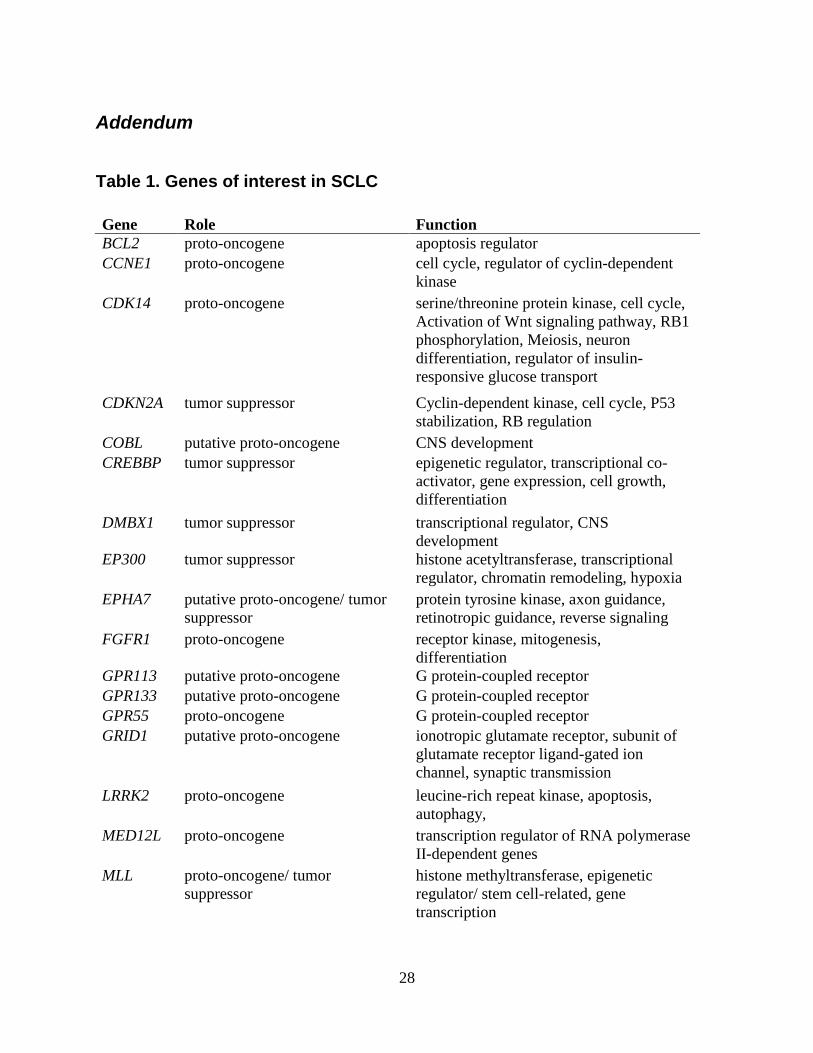
28
Addendum
Table 1. Genes of interest in SCLC
Gene Role Function
BCL2 proto-oncogene apoptosis regulator
CCNE1 proto-oncogene cell cycle, regulator of cyclin-dependent
kinase
CDK14 proto-oncogene serine/threonine protein kinase, cell cycle,
Activation of Wnt signaling pathway, RB1
phosphorylation, Meiosis, neuron
differentiation, regulator of insulin-
responsive glucose transport
CDKN2A tumor suppressor Cyclin-dependent kinase, cell cycle, P53
stabilization, RB regulation
COBL putative proto-oncogene CNS development
CREBBP tumor suppressor epigenetic regulator, transcriptional co-
activator, gene expression, cell growth,
differentiation
DMBX1 tumor suppressor transcriptional regulator, CNS
development
EP300 tumor suppressor histone acetyltransferase, transcriptional
regulator, chromatin remodeling, hypoxia
EPHA7 putative proto-oncogene/ tumor
suppressor
protein tyrosine kinase, axon guidance,
retinotropic guidance, reverse signaling
FGFR1 proto-oncogene receptor kinase, mitogenesis,
differentiation
GPR113 putative proto-oncogene G protein-coupled receptor
GPR133 putative proto-oncogene G protein-coupled receptor
GPR55 proto-oncogene G protein-coupled receptor
GRID1 putative proto-oncogene ionotropic glutamate receptor, subunit of
glutamate receptor ligand-gated ion
channel, synaptic transmission
LRRK2 proto-oncogene leucine-rich repeat kinase, apoptosis,
autophagy,
MED12L proto-oncogene transcription regulator of RNA polymerase
II-dependent genes
MLL proto-oncogene/ tumor
suppressor
histone methyltransferase, epigenetic
regulator/ stem cell-related, gene
transcription
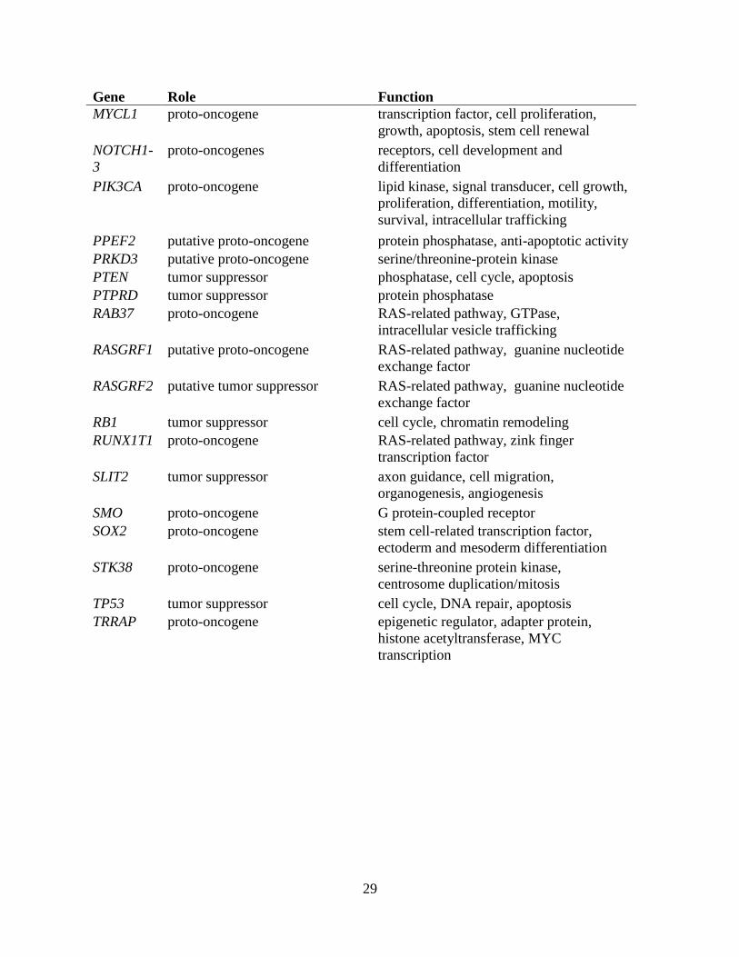
29
Gene Role Function
MYCL1 proto-oncogene transcription factor, cell proliferation,
growth, apoptosis, stem cell renewal
NOTCH1-
3
proto-oncogenes receptors, cell development and
differentiation
PIK3CA proto-oncogene lipid kinase, signal transducer, cell growth,
proliferation, differentiation, motility,
survival, intracellular trafficking
PPEF2 putative proto-oncogene protein phosphatase, anti-apoptotic activity
PRKD3 putative proto-oncogene serine/threonine-protein kinase
PTEN tumor suppressor phosphatase, cell cycle, apoptosis
PTPRD tumor suppressor protein phosphatase
RAB37 proto-oncogene RAS-related pathway, GTPase,
intracellular vesicle trafficking
RASGRF1 putative proto-oncogene RAS-related pathway, guanine nucleotide
exchange factor
RASGRF2 putative tumor suppressor RAS-related pathway, guanine nucleotide
exchange factor
RB1 tumor suppressor cell cycle, chromatin remodeling
RUNX1T1 proto-oncogene RAS-related pathway, zink finger
transcription factor
SLIT2 tumor suppressor axon guidance, cell migration,
organogenesis, angiogenesis
SMO proto-oncogene G protein-coupled receptor
SOX2 proto-oncogene stem cell-related transcription factor,
ectoderm and mesoderm differentiation
STK38 proto-oncogene serine-threonine protein kinase,
centrosome duplication/mitosis
TP53 tumor suppressor cell cycle, DNA repair, apoptosis
TRRAP proto-oncogene epigenetic regulator, adapter protein,
histone acetyltransferase, MYC
transcription
SCLC Framework - Appendix 1 30
Appendix 1: Investments in Small Cell Lung Cancer Research – NCI/NIH
Investments in Small Cell Lung Cancer Research
National Cancer Institute
National Institutes of Health
July 2013
SCLC Framework - Appendix 1 31
Table of Contents
NCI Investments in Lung Cancer Research 32
Investments in Small Cell Lung Cancer Research by NCI and Others 32
Research Projects 32
Specialized Programs of Research Excellence 36
Clinical Trials 37
Workforce Development 37
Research Resources 41
Methods 42
Addendum A: NCI-Funded Research on Neuroendocrine Tumors 44
Addendum B: International Cancer Research Partnership Investment in Small Cell 46
Lung Cancer
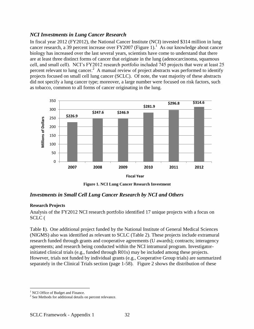
SCLC Framework - Appendix 1 32
NCI Investments in Lung Cancer Research
In fiscal year 2012 (FY2012), the National Cancer Institute (NCI) invested $314 million in lung
cancer research, a 39 percent increase over FY2007 (Figure 1).1
As our knowledge about cancer
biology has increased over the last several years, scientists have come to understand that there
are at least three distinct forms of cancer that originate in the lung (adenocarcinoma, squamous
cell, and small cell). NCI’s FY2012 research portfolio included 745 projects that were at least 25
percent relevant to lung cancer.2 A manual review of project abstracts was performed to identify
projects focused on small cell lung cancer (SCLC). Of note, the vast majority of these abstracts
did not specify a lung cancer type; moreover, a large number were focused on risk factors, such
as tobacco, common to all forms of cancer originating in the lung.
Figure 1. NCI Lung Cancer Research Investment
Investments in Small Cell Lung Cancer Research by NCI and Others
Research Projects
Analysis of the FY2012 NCI research portfolio identified 17 unique projects with a focus on
SCLC (
Table 1). One additional project funded by the National Institute of General Medical Sciences
(NIGMS) also was identified as relevant to SCLC (Table 2). These projects include extramural
research funded through grants and cooperative agreements (U awards); contracts; interagency
agreements; and research being conducted within the NCI intramural program. Investigator-
initiated clinical trials (e.g., funded through R01s) may be included among these projects.
However, trials not funded by individual grants (e.g., Cooperative Group trials) are summarized
separately in the Clinical Trials section (page 1-58). Figure 2 shows the distribution of these
1 NCI Office of Budget and Finance. 2 See Methods for additional details on percent relevance.
$226.9 $247.6 $246.9
$281.9 $296.8 $314.6
0
50
100
150
200
250
300
350
2007 2008 2009 2010 2011 2012
Mill
ion
s o
f D
olla
rs
Fiscal Year
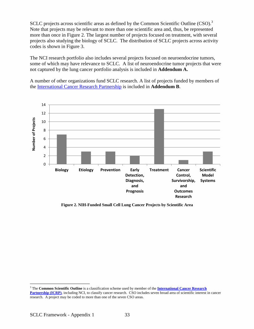
SCLC Framework - Appendix 1 33
SCLC projects across scientific areas as defined by the Common Scientific Outline (CSO).3
Note that projects may be relevant to more than one scientific area and, thus, be represented
more than once in Figure 2. The largest number of projects focused on treatment, with several
projects also studying the biology of SCLC. The distribution of SCLC projects across activity
codes is shown in Figure 3.
The NCI research portfolio also includes several projects focused on neuroendocrine tumors,
some of which may have relevance to SCLC. A list of neuroendocrine tumor projects that were
not captured by the lung cancer portfolio analysis is included in Addendum A.
A number of other organizations fund SCLC research. A list of projects funded by members of
the International Cancer Research Partnership is included in Addendum B.
Figure 2. NIH-Funded Small Cell Lung Cancer Projects by Scientific Area
3 The Common Scientific Outline is a classification scheme used by member of the International Cancer Research
Partnership (ICRP), including NCI, to classify cancer research. CSO includes seven broad area of scientific interest in cancer
research. A project may be coded to more than one of the seven CSO areas.
0
2
4
6
8
10
12
14
Biology Etiology Prevention EarlyDetection,Diagnosis,
andPrognosis
Treatment CancerControl,
Survivorship,and
OutcomesResearch
ScientificModel
Systems
Nu
mb
er
of
Pro
ject
s
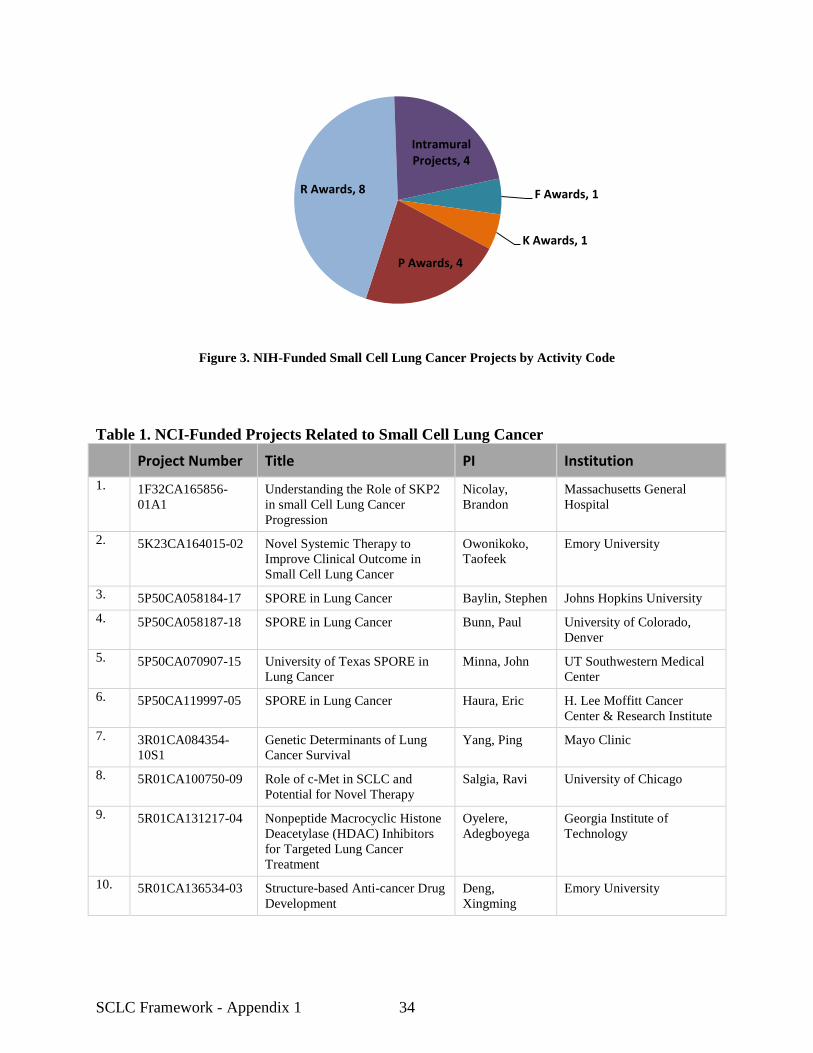
SCLC Framework - Appendix 1 34
Figure 3. NIH-Funded Small Cell Lung Cancer Projects by Activity Code
Table 1. NCI-Funded Projects Related to Small Cell Lung Cancer
Project Number Title PI Institution
1. 1F32CA165856-
01A1
Understanding the Role of SKP2
in small Cell Lung Cancer
Progression
Nicolay,
Brandon
Massachusetts General
Hospital
2. 5K23CA164015-02 Novel Systemic Therapy to
Improve Clinical Outcome in
Small Cell Lung Cancer
Owonikoko,
Taofeek
Emory University
3. 5P50CA058184-17 SPORE in Lung Cancer Baylin, Stephen Johns Hopkins University
4. 5P50CA058187-18 SPORE in Lung Cancer Bunn, Paul University of Colorado,
Denver
5. 5P50CA070907-15 University of Texas SPORE in
Lung Cancer
Minna, John UT Southwestern Medical
Center
6. 5P50CA119997-05 SPORE in Lung Cancer Haura, Eric H. Lee Moffitt Cancer
Center & Research Institute
7. 3R01CA084354-
10S1
Genetic Determinants of Lung
Cancer Survival
Yang, Ping Mayo Clinic
8. 5R01CA100750-09 Role of c-Met in SCLC and
Potential for Novel Therapy
Salgia, Ravi University of Chicago
9. 5R01CA131217-04 Nonpeptide Macrocyclic Histone
Deacetylase (HDAC) Inhibitors
for Targeted Lung Cancer
Treatment
Oyelere,
Adegboyega
Georgia Institute of
Technology
10. 5R01CA136534-03 Structure-based Anti-cancer Drug
Development
Deng,
Xingming
Emory University
F Awards, 1
K Awards, 1
P Awards, 4
R Awards, 8
Intramural Projects, 4

SCLC Framework - Appendix 1 35
Project Number Title PI Institution
11. 7R01CA148867-04 Using Mouse Models to
Understand Retinoblastoma
Initiation and Progression
MacPherson,
David
Fred Hutchinson Cancer
Research Center
12. 1R15CA161491-
01A1
Capsaicin and Small Cell Lung
Cancer Therapy
Dasgupta,
Piyali
Marshall University
13. 2R44CA162613-02 Targeted Treatment of Recurrent
Small Cell Lung Cancer with
Anti-AbnV2 Antibodies
Pang, Roy Woomera Therapeutics, Inc.
14. ZIABC010748 Clinical Investigation of Signal
Transduction Inhibitors
Dennis, Phillip National Cancer Institute
15. ZIASC000167 Molecular Pathology of
Pulmonary Carcinogenesis
Linnoila, Ilona National Cancer Institute
16. ZIASC010093 Targeting the Epigenome for
Lung Cancer Therapy
Schrump,
David
National Cancer Institute
17. ZICBC011040 Molecular Profiling of Clinical
Specimens
Edelman,
Daniel
National Cancer Institute
Table 2. Non-NCI NIH-Funded Projects Related to Small Cell Lung Cancer
Project Number Title PI Institution
1. 2R01GM079719-06 Enabling New Translational
Discoveries Using a Genomic
Data-Driven Nosology
Butte, Atul Stanford University
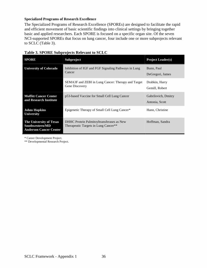
SCLC Framework - Appendix 1 36
Specialized Programs of Research Excellence
The Specialized Programs of Research Excellence (SPOREs) are designed to facilitate the rapid
and efficient movement of basic scientific findings into clinical settings by bringing together
basic and applied researchers. Each SPORE is focused on a specific organ site. Of the seven
NCI-supported SPOREs that focus on lung cancer, four include one or more subprojects relevant
to SCLC (Table 3).
Table 3. SPORE Subprojects Relevant to SCLC
SPORE Subproject Project Leader(s)
University of Colorado Inhibition of IGF and FGF Signaling Pathways in Lung
Cancer
Bunn, Paul
DeGregori, James
SEMA3F and ZEBI in Lung Cancer: Therapy and Target
Gene Discovery
Drabkin, Harry
Gemill, Robert
Moffitt Cancer Center
and Research Institute
p53-based Vaccine for Small Cell Lung Cancer Gabrilovich, Dmitry
Antonia, Scott
Johns Hopkins
University
Epigenetic Therapy of Small Cell Lung Cancer* Hann, Christine
The University of Texas
Southwestern/MD
Anderson Cancer Center
DHHC Protein Palmitoyltransferases as New
Therapeutic Targets in Lung Cancer**
Hoffman, Sandra
* Career Development Project.
** Developmental Research Project.
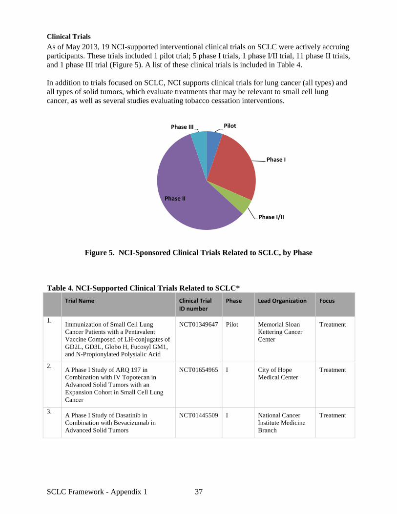
SCLC Framework - Appendix 1 37
Clinical Trials
As of May 2013, 19 NCI-supported interventional clinical trials on SCLC were actively accruing
participants. These trials included 1 pilot trial; 5 phase I trials, 1 phase I/II trial, 11 phase II trials,
and 1 phase III trial (Figure 5). A list of these clinical trials is included in Table 4.
In addition to trials focused on SCLC, NCI supports clinical trials for lung cancer (all types) and
all types of solid tumors, which evaluate treatments that may be relevant to small cell lung
cancer, as well as several studies evaluating tobacco cessation interventions.
Figure 5. NCI-Sponsored Clinical Trials Related to SCLC, by Phase
Table 4. NCI-Supported Clinical Trials Related to SCLC*
Trial Name Clinical Trial ID number
Phase Lead Organization Focus
1. Immunization of Small Cell Lung
Cancer Patients with a Pentavalent
Vaccine Composed of LH-conjugates of
GD2L, GD3L, Globo H, Fucosyl GM1,
and N-Propionylated Polysialic Acid
NCT01349647 Pilot Memorial Sloan
Kettering Cancer
Center
Treatment
2. A Phase I Study of ARQ 197 in
Combination with IV Topotecan in
Advanced Solid Tumors with an
Expansion Cohort in Small Cell Lung
Cancer
NCT01654965 I City of Hope
Medical Center
Treatment
3. A Phase I Study of Dasatinib in
Combination with Bevacizumab in
Advanced Solid Tumors
NCT01445509 I National Cancer
Institute Medicine
Branch
Treatment
Pilot
Phase I
Phase I/II
Phase II
Phase III
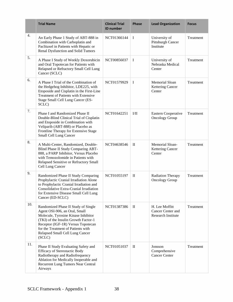
SCLC Framework - Appendix 1 38
Trial Name Clinical Trial ID number
Phase Lead Organization Focus
4. An Early Phase 1 Study of ABT-888 in
Combination with Carboplatin and
Paclitaxel in Patients with Hepatic or
Renal Dysfunction and Solid Tumors
NCT01366144 I University of
Pittsburgh Cancer
Institute
Treatment
5. A Phase I Study of Weekly Doxorubicin
and Oral Topotecan for Patients with
Relapsed or Refractory Small Cell Lung
Cancer (SCLC)
NCT00856037 I University of
Nebraska Medical
Center
Treatment
6. A Phase I Trial of the Combination of
the Hedgehog Inhibitor, LDE225, with
Etoposide and Cisplatin in the First-Line
Treatment of Patients with Extensive
Stage Small Cell Lung Cancer (ES-
SCLC)
NCT01579929 I Memorial Sloan
Kettering Cancer
Center
Treatment
7. Phase I and Randomized Phase II
Double-Blind Clinical Trial of Cisplatin
and Etoposide in Combination with
Veliparib (ABT-888) or Placebo as
Frontline Therapy for Extensive Stage
Small Cell Lung Cancer
NCT01642251 I/II Eastern Cooperative
Oncology Group
Treatment
8. A Multi-Center, Randomized, Double-
Blind Phase II Study Comparing ABT-
888, a PARP Inhibitor, Versus Placebo
with Temozolomide in Patients with
Relapsed Sensitive or Refractory Small
Cell Lung Cancer
NCT04638546 II Memorial Sloan-
Kettering Cancer
Center
Treatment
9. Randomized Phase II Study Comparing
Prophylactic Cranial Irradiation Alone
to Prophylactic Cranial Irradiation and
Consolidative Extra-Cranial Irradiation
for Extensive Disease Small Cell Lung
Cancer (ED-SCLC)
NCT01055197 II Radiation Therapy
Oncology Group
Treatment
10. Randomized Phase II Study of Single
Agent OSI-906, an Oral, Small
Molecule, Tyrosine Kinase Inhibitor
(TKI) of the Insulin Growth Factor-1
Receptor (IGF-1R) Versus Topotecan
for the Treatment of Patients with
Relapsed Small Cell Lung Cancer
(SCLC)
NCT01387386 II H. Lee Moffitt
Cancer Center and
Research Institute
Treatment
11. Phase II Study Evaluating Safety and
Efficacy of Stereotactic Body
Radiotherapy and Radiofrequency
Ablation for Medically Inoperable and
Recurrent Lung Tumors Near Central
Airways
NCT01051037 II Jonsson
Comprehensive
Cancer Center
Treatment
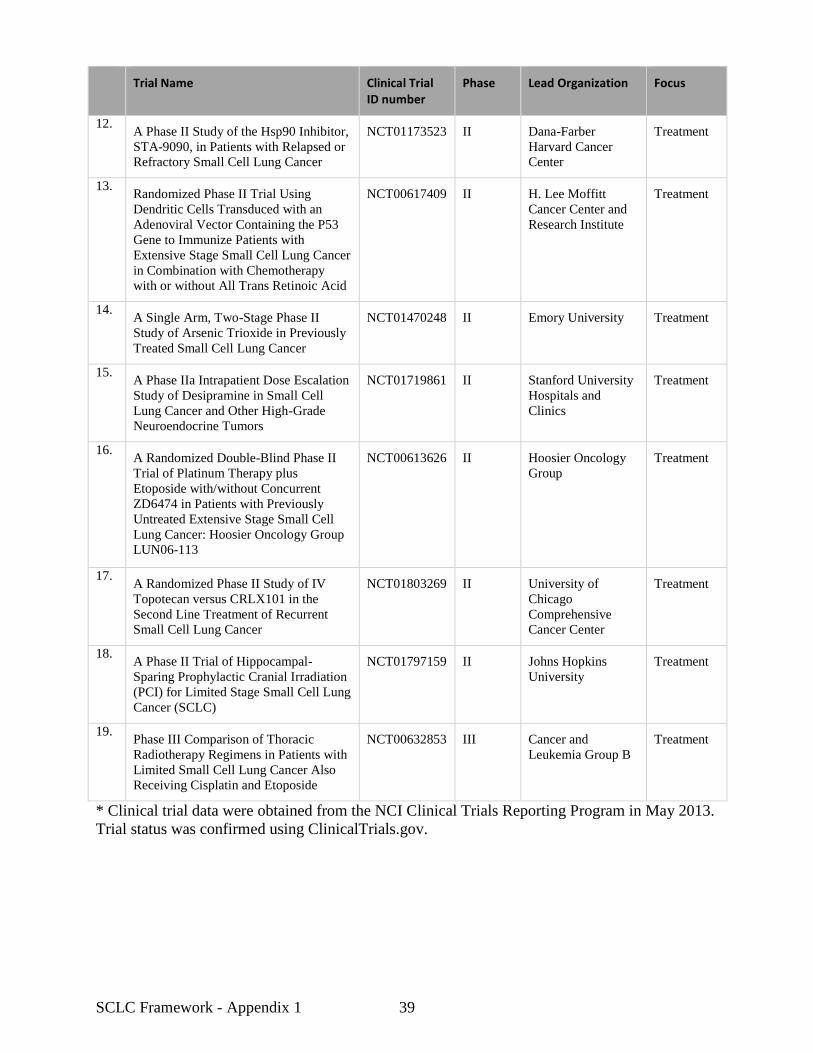
SCLC Framework - Appendix 1 39
Trial Name Clinical Trial ID number
Phase Lead Organization Focus
12. A Phase II Study of the Hsp90 Inhibitor,
STA-9090, in Patients with Relapsed or
Refractory Small Cell Lung Cancer
NCT01173523 II Dana-Farber
Harvard Cancer
Center
Treatment
13. Randomized Phase II Trial Using
Dendritic Cells Transduced with an
Adenoviral Vector Containing the P53
Gene to Immunize Patients with
Extensive Stage Small Cell Lung Cancer
in Combination with Chemotherapy
with or without All Trans Retinoic Acid
NCT00617409 II H. Lee Moffitt
Cancer Center and
Research Institute
Treatment
14. A Single Arm, Two-Stage Phase II
Study of Arsenic Trioxide in Previously
Treated Small Cell Lung Cancer
NCT01470248 II Emory University Treatment
15. A Phase IIa Intrapatient Dose Escalation
Study of Desipramine in Small Cell
Lung Cancer and Other High-Grade
Neuroendocrine Tumors
NCT01719861 II Stanford University
Hospitals and
Clinics
Treatment
16. A Randomized Double-Blind Phase II
Trial of Platinum Therapy plus
Etoposide with/without Concurrent
ZD6474 in Patients with Previously
Untreated Extensive Stage Small Cell
Lung Cancer: Hoosier Oncology Group
LUN06-113
NCT00613626 II Hoosier Oncology
Group
Treatment
17. A Randomized Phase II Study of IV
Topotecan versus CRLX101 in the
Second Line Treatment of Recurrent
Small Cell Lung Cancer
NCT01803269 II University of
Chicago
Comprehensive
Cancer Center
Treatment
18. A Phase II Trial of Hippocampal-
Sparing Prophylactic Cranial Irradiation
(PCI) for Limited Stage Small Cell Lung
Cancer (SCLC)
NCT01797159 II Johns Hopkins
University
Treatment
19. Phase III Comparison of Thoracic
Radiotherapy Regimens in Patients with
Limited Small Cell Lung Cancer Also
Receiving Cisplatin and Etoposide
NCT00632853 III Cancer and
Leukemia Group B
Treatment
* Clinical trial data were obtained from the NCI Clinical Trials Reporting Program in May 2013.
Trial status was confirmed using ClinicalTrials.gov.
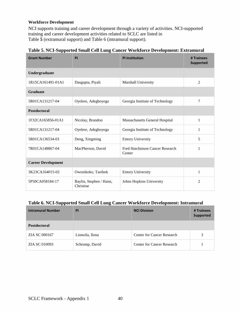
SCLC Framework - Appendix 1 40
Workforce Development
NCI supports training and career development through a variety of activities. NCI-supported
training and career development activities related to SCLC are listed in
Table 5 (extramural support) and Table 6 (intramural support).
Table 5. NCI-Supported Small Cell Lung Cancer Workforce Development: Extramural
Grant Number PI PI Institution # Trainees Supported
Undergraduate
1R15CA161491-01A1 Dasgupta, Piyali Marshall University 2
Graduate
5R01CA131217-04 Oyelere, Adegboyega Georgia Institute of Technology 7
Postdoctoral
1F32CA165856-01A1 Nicolay, Brandon Massachusetts General Hospital 1
5R01CA131217-04 Oyelere, Adegboyega Georgia Institute of Technology 1
5R01CA136534-03 Deng, Xingming Emory University 5
7R01CA148867-04 MacPherson, David Fred Hutchinson Cancer Research
Center
1
Career Development
3K23CA164015-02 Owonikoko, Taofeek Emory University 1
5P50CA058184-17 Baylin, Stephen / Hann,
Christine
Johns Hopkins University 2
Table 6. NCI-Supported Small Cell Lung Cancer Workforce Development: Intramural
Intramural Number PI NCI Division # Trainees Supported
Postdoctoral
ZIA SC 000167 Linnoila, Ilona Center for Cancer Research 3
ZIA SC 010093 Schrump, David Center for Cancer Research 1
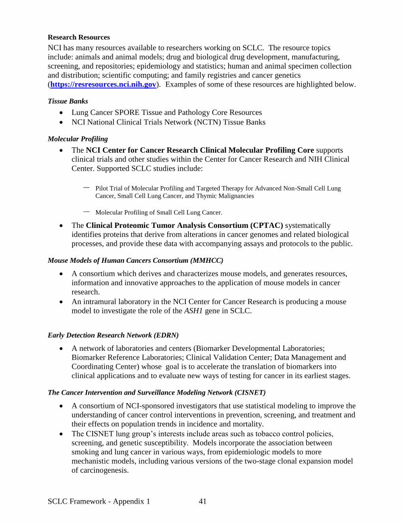
SCLC Framework - Appendix 1 41
Research Resources
NCI has many resources available to researchers working on SCLC. The resource topics
include: animals and animal models; drug and biological drug development, manufacturing,
screening, and repositories; epidemiology and statistics; human and animal specimen collection
and distribution; scientific computing; and family registries and cancer genetics
(https://resresources.nci.nih.gov). Examples of some of these resources are highlighted below.
Tissue Banks
Lung Cancer SPORE Tissue and Pathology Core Resources
NCI National Clinical Trials Network (NCTN) Tissue Banks
Molecular Profiling
The NCI Center for Cancer Research Clinical Molecular Profiling Core supports
clinical trials and other studies within the Center for Cancer Research and NIH Clinical
Center. Supported SCLC studies include:
– Pilot Trial of Molecular Profiling and Targeted Therapy for Advanced Non-Small Cell Lung
Cancer, Small Cell Lung Cancer, and Thymic Malignancies
– Molecular Profiling of Small Cell Lung Cancer.
The Clinical Proteomic Tumor Analysis Consortium (CPTAC) systematically
identifies proteins that derive from alterations in cancer genomes and related biological
processes, and provide these data with accompanying assays and protocols to the public.
Mouse Models of Human Cancers Consortium (MMHCC)
A consortium which derives and characterizes mouse models, and generates resources,
information and innovative approaches to the application of mouse models in cancer
research.
An intramural laboratory in the NCI Center for Cancer Research is producing a mouse
model to investigate the role of the ASH1 gene in SCLC.
Early Detection Research Network (EDRN)
A network of laboratories and centers (Biomarker Developmental Laboratories;
Biomarker Reference Laboratories; Clinical Validation Center; Data Management and
Coordinating Center) whose goal is to accelerate the translation of biomarkers into
clinical applications and to evaluate new ways of testing for cancer in its earliest stages.
The Cancer Intervention and Surveillance Modeling Network (CISNET)
A consortium of NCI-sponsored investigators that use statistical modeling to improve the
understanding of cancer control interventions in prevention, screening, and treatment and
their effects on population trends in incidence and mortality.
The CISNET lung group’s interests include areas such as tobacco control policies,
screening, and genetic susceptibility. Models incorporate the association between
smoking and lung cancer in various ways, from epidemiologic models to more
mechanistic models, including various versions of the two-stage clonal expansion model
of carcinogenesis.
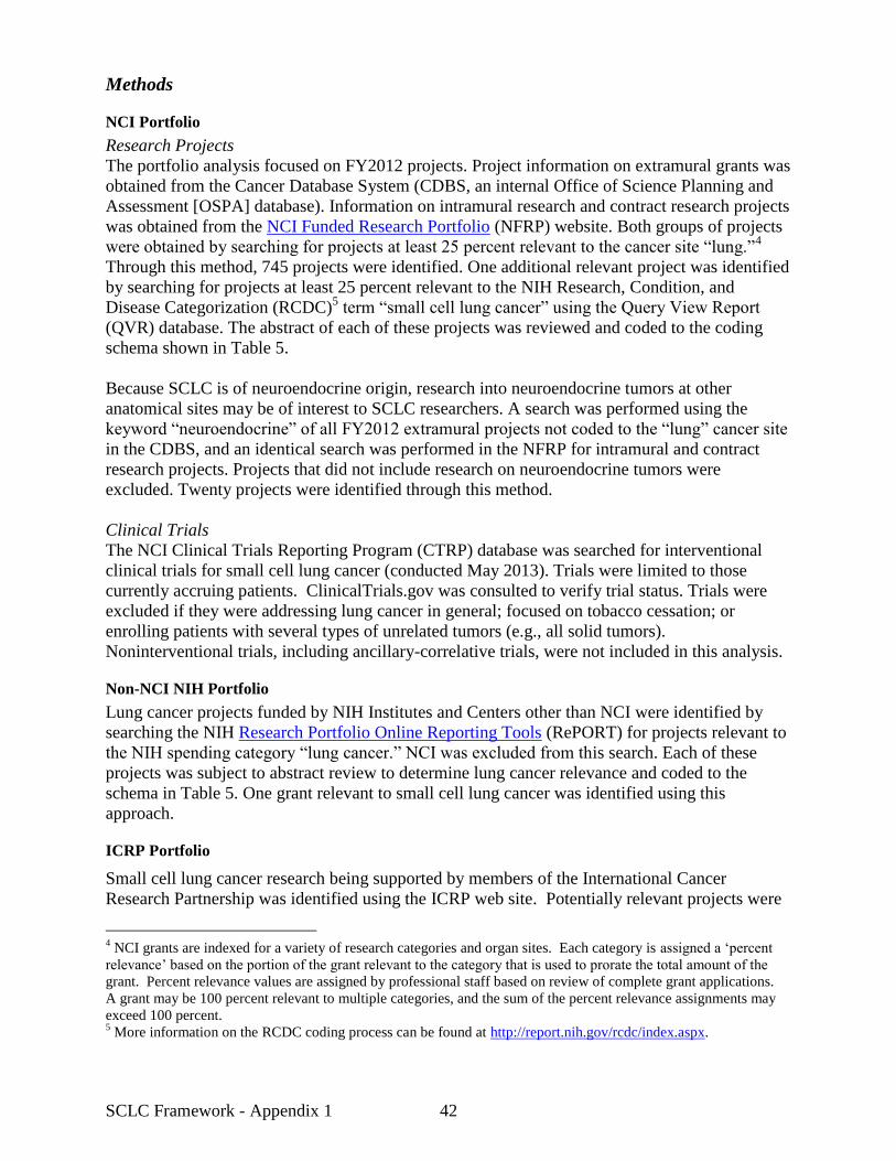
SCLC Framework - Appendix 1 42
Methods
NCI Portfolio
Research Projects
The portfolio analysis focused on FY2012 projects. Project information on extramural grants was
obtained from the Cancer Database System (CDBS, an internal Office of Science Planning and
Assessment [OSPA] database). Information on intramural research and contract research projects
was obtained from the NCI Funded Research Portfolio (NFRP) website. Both groups of projects
were obtained by searching for projects at least 25 percent relevant to the cancer site “lung.”4
Through this method, 745 projects were identified. One additional relevant project was identified
by searching for projects at least 25 percent relevant to the NIH Research, Condition, and
Disease Categorization (RCDC)5 term “small cell lung cancer” using the Query View Report
(QVR) database. The abstract of each of these projects was reviewed and coded to the coding
schema shown in Table 5.
Because SCLC is of neuroendocrine origin, research into neuroendocrine tumors at other
anatomical sites may be of interest to SCLC researchers. A search was performed using the
keyword “neuroendocrine” of all FY2012 extramural projects not coded to the “lung” cancer site
in the CDBS, and an identical search was performed in the NFRP for intramural and contract
research projects. Projects that did not include research on neuroendocrine tumors were
excluded. Twenty projects were identified through this method.
Clinical Trials
The NCI Clinical Trials Reporting Program (CTRP) database was searched for interventional
clinical trials for small cell lung cancer (conducted May 2013). Trials were limited to those
currently accruing patients. ClinicalTrials.gov was consulted to verify trial status. Trials were
excluded if they were addressing lung cancer in general; focused on tobacco cessation; or
enrolling patients with several types of unrelated tumors (e.g., all solid tumors).
Noninterventional trials, including ancillary-correlative trials, were not included in this analysis.
Non-NCI NIH Portfolio
Lung cancer projects funded by NIH Institutes and Centers other than NCI were identified by
searching the NIH Research Portfolio Online Reporting Tools (RePORT) for projects relevant to
the NIH spending category “lung cancer.” NCI was excluded from this search. Each of these
projects was subject to abstract review to determine lung cancer relevance and coded to the
schema in Table 5. One grant relevant to small cell lung cancer was identified using this
approach.
ICRP Portfolio
Small cell lung cancer research being supported by members of the International Cancer
Research Partnership was identified using the ICRP web site. Potentially relevant projects were
4 NCI grants are indexed for a variety of research categories and organ sites. Each category is assigned a ‘percent
relevance’ based on the portion of the grant relevant to the category that is used to prorate the total amount of the
grant. Percent relevance values are assigned by professional staff based on review of complete grant applications.
A grant may be 100 percent relevant to multiple categories, and the sum of the percent relevance assignments may
exceed 100 percent. 5 More information on the RCDC coding process can be found at http://report.nih.gov/rcdc/index.aspx.
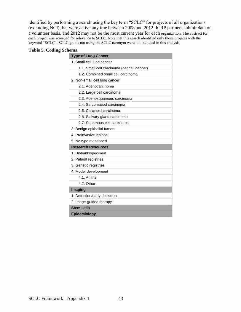
SCLC Framework - Appendix 1 43
identified by performing a search using the key term “SCLC” for projects of all organizations
(excluding NCI) that were active anytime between 2008 and 2012. ICRP partners submit data on
a volunteer basis, and 2012 may not be the most current year for each organization. The abstract for
each project was screened for relevance to SCLC. Note that this search identified only those projects with the
keyword “SCLC”; SCLC grants not using the SCLC acronym were not included in this analysis.
Table 5. Coding Schema
Type of Lung Cancer
1. Small cell lung cancer
1.1. Small cell carcinoma (oat cell cancer)
1.2. Combined small cell carcinoma
2. Non-small cell lung cancer
2.1. Adenocarcinoma
2.2. Large cell carcinoma
2.3. Adenosquamous carcinoma
2.4. Sarcomatiod carcinoma
2.5. Carcinoid carcinoma
2.6. Salivary gland carcinoma
2.7. Squamous cell carcinoma
3. Benign epithelial tumors
4. Preinvasive lesions
5. No type mentioned
Research Resources
1. Biobank/specimen
2. Patient registries
3. Genetic registries
4. Model development
4.1. Animal
4.2. Other
Imaging
1. Detection/early detection
2. Image-guided therapy
Stem cells
Epidemiology
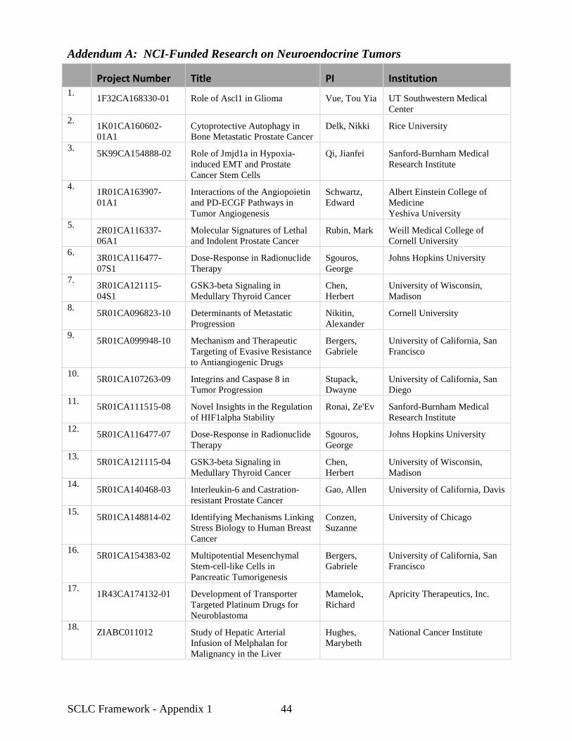
SCLC Framework - Appendix 1 44
Addendum A: NCI-Funded Research on Neuroendocrine Tumors
Project Number Title PI Institution 1.
1F32CA168330-01 Role of Ascl1 in Glioma Vue, Tou Yia UT Southwestern Medical
Center
2. 1K01CA160602-
01A1
Cytoprotective Autophagy in
Bone Metastatic Prostate Cancer
Delk, Nikki Rice University
3. 5K99CA154888-02 Role of Jmjd1a in Hypoxia-
induced EMT and Prostate
Cancer Stem Cells
Qi, Jianfei Sanford-Burnham Medical
Research Institute
4. 1R01CA163907-
01A1
Interactions of the Angiopoietin
and PD-ECGF Pathways in
Tumor Angiogenesis
Schwartz,
Edward
Albert Einstein College of
Medicine
Yeshiva University
5. 2R01CA116337-
06A1
Molecular Signatures of Lethal
and Indolent Prostate Cancer
Rubin, Mark Weill Medical College of
Cornell University
6. 3R01CA116477-
07S1
Dose-Response in Radionuclide
Therapy
Sgouros,
George
Johns Hopkins University
7. 3R01CA121115-
04S1
GSK3-beta Signaling in
Medullary Thyroid Cancer
Chen,
Herbert
University of Wisconsin,
Madison
8. 5R01CA096823-10 Determinants of Metastatic
Progression
Nikitin,
Alexander
Cornell University
9. 5R01CA099948-10 Mechanism and Therapeutic
Targeting of Evasive Resistance
to Antiangiogenic Drugs
Bergers,
Gabriele
University of California, San
Francisco
10. 5R01CA107263-09 Integrins and Caspase 8 in
Tumor Progression
Stupack,
Dwayne
University of California, San
Diego
11. 5R01CA111515-08 Novel Insights in the Regulation
of HIF1alpha Stability
Ronai, Ze'Ev Sanford-Burnham Medical
Research Institute
12. 5R01CA116477-07 Dose-Response in Radionuclide
Therapy
Sgouros,
George
Johns Hopkins University
13. 5R01CA121115-04 GSK3-beta Signaling in
Medullary Thyroid Cancer
Chen,
Herbert
University of Wisconsin,
Madison
14. 5R01CA140468-03 Interleukin-6 and Castration-
resistant Prostate Cancer
Gao, Allen University of California, Davis
15. 5R01CA148814-02 Identifying Mechanisms Linking
Stress Biology to Human Breast
Cancer
Conzen,
Suzanne
University of Chicago
16. 5R01CA154383-02 Multipotential Mesenchymal
Stem-cell-like Cells in
Pancreatic Tumorigenesis
Bergers,
Gabriele
University of California, San
Francisco
17. 1R43CA174132-01 Development of Transporter
Targeted Platinum Drugs for
Neuroblastoma
Mamelok,
Richard
Apricity Therapeutics, Inc.
18. ZIABC011012 Study of Hepatic Arterial
Infusion of Melphalan for
Malignancy in the Liver
Hughes,
Marybeth
National Cancer Institute
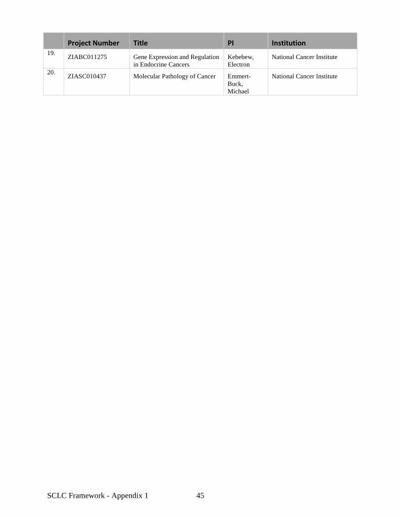
SCLC Framework - Appendix 1 45
Project Number Title PI Institution 19.
ZIABC011275 Gene Expression and Regulation
in Endocrine Cancers
Kebebew,
Electron
National Cancer Institute
20. ZIASC010437 Molecular Pathology of Cancer Emmert-
Buck,
Michael
National Cancer Institute
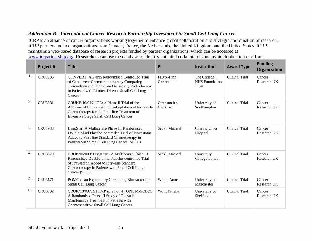
SCLC Framework - Appendix 1 46
Addendum B: International Cancer Research Partnership Investment in Small Cell Lung Cancer
ICRP is an alliance of cancer organizations working together to enhance global collaboration and strategic coordination of research.
ICRP partners include organizations from Canada, France, the Netherlands, the United Kingdom, and the United States. ICRP
maintains a web-based database of research projects funded by partner organizations, which can be accessed at
www.icrpartnership.org. Researchers can use the database to identify potential collaborators and avoid duplication of efforts.
Project # Title PI Institution Award Type Funding Organization
1. CRU2233 CONVERT: A 2-arm Randomised Controlled Trial
of Concurrent Chemo-radiotherapy Comparing
Twice-daily and High-dose Once-daily Radiotherapy
in Patients with Limited Disease Small Cell Lung
Cancer
Faivre-Finn,
Corinne
The Christie
NHS Foundation
Trust
Clinical Trial Cancer
Research UK
2. CRU3581 CRUKE/10/019: ICE: A Phase II Trial of the
Addition of Ipilimumab to Carboplatin and Etoposide
Chemotherapy for the First-line Treatment of
Extensive Stage Small Cell Lung Cancer
Ottensmeier,
Christian
University of
Southampton
Clinical Trial Cancer
Research UK
3. CRU1933 LungStar: A Multicentre Phase III Randomised
Double-blind Placebo-controlled Trial of Pravastatin
Added to First-line Standard Chemotherapy in
Patients with Small Cell Lung Cancer (SCLC)
Seckl, Michael Charing Cross
Hospital
Clinical Trial Cancer
Research UK
4. CRU3879 CRUK/06/009: LungStar - A Multicentre Phase III
Randomised Double-blind Placebo-controlled Trial
of Pravastatin Added to First-line Standard
Chemotherapy in Patients with Small Cell Lung
Cancer (SCLC)
Seckl, Michael University
College London
Clinical Trial Cancer
Research UK
5. CRU3671 POMC as an Exploratory Circulating Biomarker for
Small Cell Lung Cancer
White, Anne University of
Manchester
Clinical Trial Cancer
Research UK
6. CRU3792 CRUK/10/037: STOMP (previously OPIUM-SCLC):
A Randomised Phase II Study of Olaparib
Maintenance Treatment in Patients with
Chemosensitive Small Cell Lung Cancer
Woll, Penella University of
Sheffield
Clinical Trial Cancer
Research UK

SCLC Framework - Appendix 1 47
Project # Title PI Institution Award Type Funding Organization
7. DGOS_0387 Prospective Multicentric Optimization and Phase I/II
Study of Pretargeted Radioimmunotherapy (PRAIT)
Using Anti-CEA x Anti-HSG TF2 Bispecific
Antibody (bsMAb) and 177Lu-IMP-288 Peptide in
Patients with CEA-expressing Small Cell Lung
Carcinoma (SCLC)
Bodere, Fran
Kraeber
Centre
Hospitalier
Universitaire de
Nantes
Clinical Trial DGOS-
Ministère de la
Santé
8. IKZ 2007-3865 Treatment and Outcome for Cancer Patients aged 75
or Older: A National Population-based Study
Janssen-Heijnen,
M.L.G.
Integraal
Kankercentrum
Zuid
Clinical Trial KWF
Kankerbestrijdi
ng/Dutch
Cancer Society
9. VU 2008-4290 Randomized Trial on Chest Irradiation in Extensive
Disease Small Cell Lung Cancer. (CREST Study)
Slotman, B.J. Vrije
Universiteit
Clinical Trial KWF
Kankerbestrijdi
ng/Dutch
Cancer Society
10. BUIT 2009-
4451
Antibodies and Paraneoplastic Syndromes Related to
Neuro-endocrine Tumours
Titulaer, M.J. University of
Pennsylvania
Clinical Trial KWF
Kankerbestrijdi
ng/Dutch
Cancer Society
11. SCO179 The Role of CD45 as a Novel Prognostic Marker of
Survival and Treatment Response in Patients with
Small Cell Lung Cancer
Sethi, Tariq University of
Edinburgh
Clinical Trial Scottish
Government
Health
Directorates,
Chief Scientist
Office
12. RSG-10-071-01 Mechanisms of Small Cell Lung Carcinoma Initiation
and Maintenance
Sage, Julien Stanford
University
Research American
Cancer Society
13. CRU2742 Research Bursary: MicroRNA Expression in Small
Cell Lung Carcinoma—A Novel Means of
Classification, Predicting Prognosis and Identification
of Therapeutic Targets?
Andrews, Timothy NHS Lothian Research Cancer
Research UK
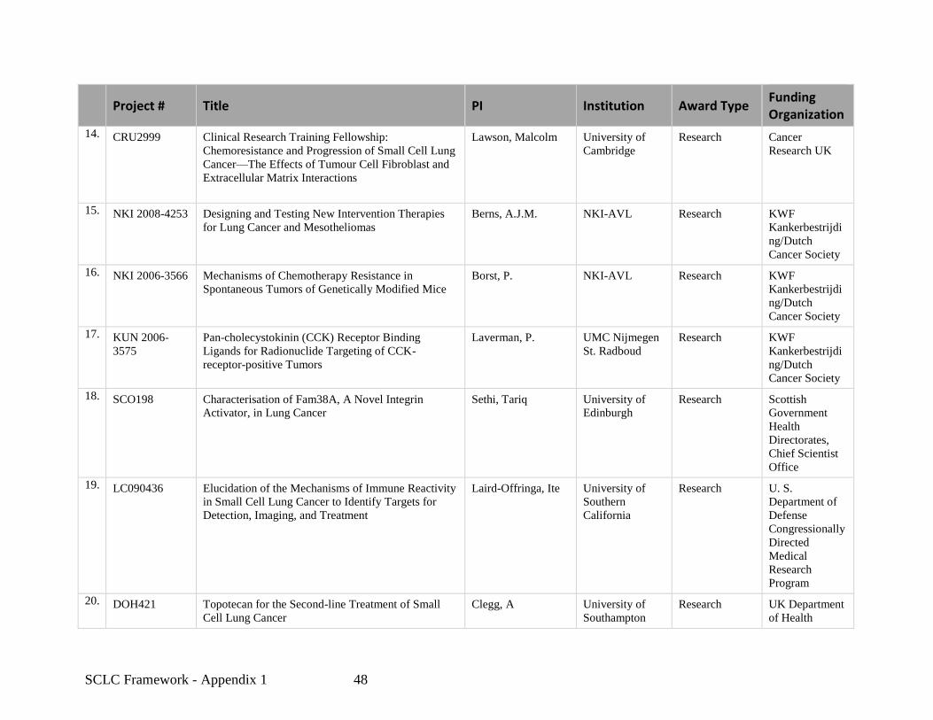
SCLC Framework - Appendix 1 48
Project # Title PI Institution Award Type Funding Organization
14. CRU2999 Clinical Research Training Fellowship:
Chemoresistance and Progression of Small Cell Lung
Cancer—The Effects of Tumour Cell Fibroblast and
Extracellular Matrix Interactions
Lawson, Malcolm University of
Cambridge
Research Cancer
Research UK
15. NKI 2008-4253 Designing and Testing New Intervention Therapies
for Lung Cancer and Mesotheliomas
Berns, A.J.M. NKI-AVL Research KWF
Kankerbestrijdi
ng/Dutch
Cancer Society
16. NKI 2006-3566 Mechanisms of Chemotherapy Resistance in
Spontaneous Tumors of Genetically Modified Mice
Borst, P. NKI-AVL Research KWF
Kankerbestrijdi
ng/Dutch
Cancer Society
17. KUN 2006-
3575
Pan-cholecystokinin (CCK) Receptor Binding
Ligands for Radionuclide Targeting of CCK-
receptor-positive Tumors
Laverman, P. UMC Nijmegen
St. Radboud
Research KWF
Kankerbestrijdi
ng/Dutch
Cancer Society
18. SCO198 Characterisation of Fam38A, A Novel Integrin
Activator, in Lung Cancer
Sethi, Tariq University of
Edinburgh
Research Scottish
Government
Health
Directorates,
Chief Scientist
Office
19. LC090436 Elucidation of the Mechanisms of Immune Reactivity
in Small Cell Lung Cancer to Identify Targets for
Detection, Imaging, and Treatment
Laird-Offringa, Ite University of
Southern
California
Research U. S.
Department of
Defense
Congressionally
Directed
Medical
Research
Program
20. DOH421 Topotecan for the Second-line Treatment of Small
Cell Lung Cancer
Clegg, A University of
Southampton
Research UK Department
of Health
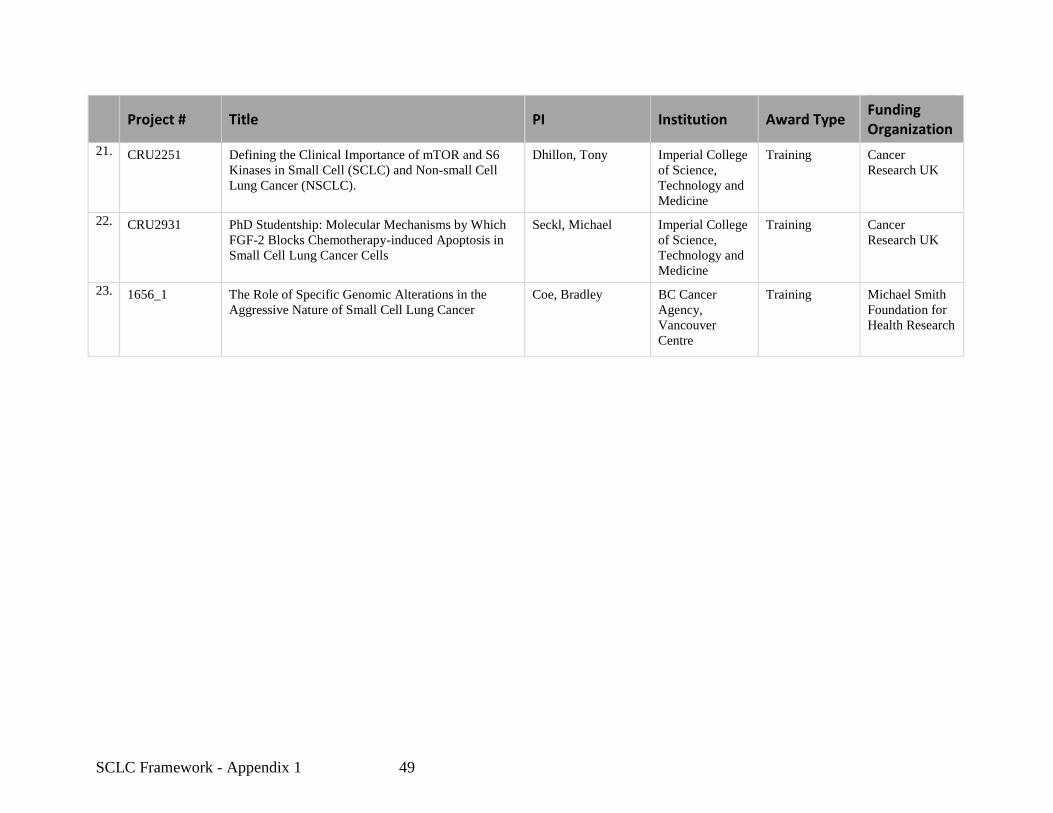
SCLC Framework - Appendix 1 49
Project # Title PI Institution Award Type Funding Organization
21. CRU2251 Defining the Clinical Importance of mTOR and S6
Kinases in Small Cell (SCLC) and Non-small Cell
Lung Cancer (NSCLC).
Dhillon, Tony Imperial College
of Science,
Technology and
Medicine
Training Cancer
Research UK
22. CRU2931 PhD Studentship: Molecular Mechanisms by Which
FGF-2 Blocks Chemotherapy-induced Apoptosis in
Small Cell Lung Cancer Cells
Seckl, Michael Imperial College
of Science,
Technology and
Medicine
Training Cancer
Research UK
23. 1656_1 The Role of Specific Genomic Alterations in the
Aggressive Nature of Small Cell Lung Cancer
Coe, Bradley BC Cancer
Agency,
Vancouver
Centre
Training Michael Smith
Foundation for
Health Research

SCLC Framework - Appendix 2 50
Appendix 2: Report from Small Cell Lung Cancer Workshop 2013
Small Cell Lung Cancer
Seizing on Opportunities to Translate Recent Research into the Clinic for New Diagnostics and Interventions
The Small Cell Lung Cancer Working Group
Clinical Trials and Translational Research Advisory Committee
June 18, 2014
SCLC Framework - Appendix 2 51
Organization of the Report
A. The Workshop
(1) Origin of the Workshop
(2) Overview of the Workshop Program
B. Current Approaches to SCLC
(1) Risk Assessment and Screening
(2) Diagnosis, Staging, and Monitoring
(3) Therapy and Resistance
C. Recent Scientific Advances and Emerging Research Questions
(1) Characterization of the SCLC Genome, Transcriptome, and Epigenome
(2) Analysis of Acquired Chemotherapy Resistance in SCLC
(3) TP53 and RB as Gatekeeper Mutations in SCLC
(4) MYC Family Members in SCLC
(5) Developmental and Stem Cell Signaling Pathways in SCLC
D. Attracting Investigators to the Field of SCLC
E. Recommended Initiatives
(1) Develop Better Research Tools for the Study of SCLC
(A) Optimize Collection of Tumor Tissues
(B) Develop New SCLC Models
(2) Assemble Comprehensive Genomic Profiling for SCLC
(3) Develop New Diagnostic Approaches
(4) Facilitate Therapeutic Development Efforts
(5) Understand Mechanisms Underlying Both the High Initial Rate of Response and the Rapid
Emergence of Drug and Radiation Resistance
F. Summary
G. References
H. List of Abbreviations
SCLC Framework - Appendix 2 52
A. The Workshop
(1) Origin of the Workshop
The Recalcitrant Cancer Research Act of 2012 (H.R. 733) requires the National Cancer Institute
(NCI) to “develop scientific frameworks” that will assist in making “progress against recalcitrant
or deadly cancers.” Small cell lung cancer (SCLC) is a recalcitrant cancer as defined by its five-
year relative survival rate of less than 7 percent and the loss of approximately 30,000 lives per
year. The NCI convened a group of experts in the field of SCLC for a workshop held in
Bethesda, Maryland on July 8-9, 2013 to evaluate research opportunities that could improve the
scientific understanding and medical control of SCLC. The group, chaired by Drs. John Minna
and Charles Rudin, included laboratory scientists, medical oncologists, surgeons, radiation
oncologists, pathologists, biostatisticians, patient advocates, and NCI staff (see Addendum 1 –
SCLC Working Group Roster).
The goals of the workshop were to identify key scientific opportunities and critical areas where
focused research efforts could have the greatest impact on prevention, detection, or disease
outcome for patients with SCLC. Workshop participants were asked to discuss recent advances
in "omics", molecular pathology, and the prospects for early detection of SCLC; key
developments in animal models for SCLC; and putative new drug targets and other areas of
vulnerability of SCLC that may lead to new therapeutic approaches.
The findings and recommendations arising from the workshop are to be discussed with the NCI's
Clinical Trials and Translational Research Advisory Committee (CTAC) and to inform the NCI
in the development of a scientific framework for SCLC in accordance with the Recalcitrant
Cancer Research Act of 2012.
(2) Overview of the Workshop Program
The workshop agenda topics included three thematic scientific sessions, a special session
focused on attracting investigators to SCLC research, and a series of smaller breakout sessions
designed to identify top research priorities and opportunities related to each of the three scientific
sessions. A final session summarized the outcomes of the breakout sessions and prioritized
recommendations. (See Addendum 2 – SCLC Workshop Agenda)
The first session focused on emerging opportunities in “omics”, molecular pathology, and early
detection for SCLC. In a series of presentations and discussions, workshop participants
reviewed the classification of neuroendocrine lung cancer molecular pathology and
epidemiology, focusing on approaches to molecular characterization and early pathogenesis of
putative precursor lesions of SCLC; current data and gaps in knowledge about the SCLC genome
and transcriptome, with emphasis on known and suspected driver oncogenes and tumor
suppressors1, 2
; recent and ongoing studies of the SCLC proteome, including potential therapeutic
targets identified through this approach3; and new data on the SCLC epigenome, defining
additional putative targets for intervention4.
The second session addressed emerging opportunities in preclinical models and on targeting
cancer stem cells in SCLC. Workshop participants reviewed and discussed patient-derived
xenograft (PDX) models as a platform for enhancing the biological understanding of SCLC and
SCLC Framework - Appendix 2 53
for therapeutic testing5; recent and ongoing genomic studies of genetically engineered mouse
models (GEMMs) of SCLC6-8
; recent and ongoing studies using SCLC GEMMs as a platform
for defining putative cells of origin for SCLC; developmental signaling pathways in SCLC; and a
relatively unbiased approach to identifying critical oncogenic drivers in lung cancer through the
use of synthetic lethal siRNA/shRNA screens.
The third session focused on emerging therapeutic opportunities, and new drug targets.
Workshop participants discussed an ongoing study at the NCI Frederick National Laboratory
assessing the relative activity of 103 oncology drugs and 420 investigational compounds of
interest against a panel of approximately 60 SCLC cell lines characterized by genomic and gene
expression profiles; recent studies and novel opportunities for immunotherapy in SCLC,
including vaccine approaches as well as agents targeting immune checkpoints9; and recent
clinical data using temozolomide both as a single agent10
and with a poly-(ADP-ribose)-
polymerase (PARP) inhibitor11
. Finally, promising opportunities for targeting Bcl-2 in SCLC12
were discussed.
In addition to the three scientific sessions, a fourth session focused on the scientific workforce in
the field of SCLC. The group discussed barriers to entry to the field, noting that despite the
relatively high incidence of SCLC, a relatively small number of scientists and clinicians are
attracted to the study of this disease. A number of ideas were proposed to attract both new and
established investigators to the study of SCLC.
This was followed by individual breakout sessions during which workshop participants proposed
specific recommendations to address scientific opportunities that had been identified during the
aforementioned discussions. On the second day of the workshop, the entire group was
reconvened to summarize the outcomes of the breakout sessions and prioritize a final set of
recommendations.
B. Current Approaches to SCLC
Clinical approaches to SCLC have not advanced significantly in three decades. Although the
focus of the workshop was on the identification of critical scientific advances and the
prioritization of research opportunities, the current standard of care provides a necessary
backdrop to the group's findings and is described in brief in this section.
(1) Risk Assessment and Screening
Although SCLC is, in most cases, a disease associated with tobacco use, little is known about
predisposing genetic or non-genetic factors that lead to the development of the disease in certain
current or former smokers but not in others. Somatic mutations accumulate during the lifetime
of an individual exposed to the carcinogens in tobacco smoke. There is a need for further study
of the germline (i.e., heritable) traits that contribute to the development of SCLC as well as the
interactions between environmental exposures and individual inherited predispositions to SCLC.
Screening for SCLC is also a challenge. There are currently no validated biomarkers that can be
measured in blood or other tissues to detect SCLC at an early stage. Furthermore, the recent
NCI-sponsored National Lung Screening Trial13-15
that proved the value of screening individuals
SCLC Framework - Appendix 2 54
at high risk of developing lung cancer with low-dose helical computed tomography (CT) also
demonstrated that screening did not improve survival for the subset of SCLC patients detected by
CT screening, unlike those with adenocarcinoma or squamous cell cancer of the lung. The
majority of patients with SCLC detected by CT screening (86% of the 125 patients) were
diagnosed with advanced stage disease, similar to the percent seen in the absence of dedicated
screening. Consistent with this distribution of stages, subsequent therapy did not evidently
prolong the survival of screened patients. These results suggest that metastatic dissemination
and/or resistance to systemic therapy may develop early in the natural history of SCLC.
(2) Diagnosis, Staging, and Monitoring
The diagnosis of SCLC, whether the patient is symptomatic or not, usually begins with histologic
confirmation of an abnormality detected on imaging studies, typically by fine needle aspirate
biopsy. Immunohistochemical evaluation employing a variety of neuroendocrine or other
markers confirms the diagnosis of SCLC. Medical history, physical examination, routine
laboratory tests, and computed tomographic scans of the chest and abdomen with infusion of
contrast material, and magnetic resonance imaging of the brain complete the initial evaluation.
For patients without evidence of disease outside one hemithorax on these studies, 18
Fluoro-
deoxyglucose positron emission tomography (PET) is useful for optimal staging, and can detect
bone metastases. Staging for patients with SCLC is most commonly categorized using the
Veterans Administration Lung Study Group system; limited-stage disease (LD), which occurs in
approximately one third of patients, is defined as SCLC confined to the hemithorax of origin, the
mediastinum, or the supraclavicular nodes, which can be encompassed within a tolerable
radiation therapy port. Extensive-stage disease (ED) SCLC has spread beyond the
supraclavicular areas and is too widespread to be included within the definition of LD. Patients
with distant metastases by definition have ED16
.
Monitoring of response to therapy is usually performed by imaging techniques capable of
providing accurate measurements of tumor size; these size measurements are interpreted by
Response Evaluation Criteria In Solid Tumors (RECIST) criteria that define categories of
response to treatment16
. PET staging now approaches a 100% level of sensitivity and greater
than 90% specificity17-20
. The use of PET scanning to both stage and follow the effect of
treatment for patients with SCLC has enhanced the accuracy by which the effectiveness of new
treatment modalities can be examined.
(3) Therapy and Resistance
Current therapeutic approaches for SCLC are of modest long-term benefit despite the
exceptionally good response to first-line therapy. Treatment for LD includes a standard first line
chemotherapy regimen21, 22
with concomitant radiation that can be encompassed in a single
radiation port23, 24
. Treatment for ED includes the same chemotherapy options, without
concomitant radiation22
. In some instances, particularly for small peripheral lung nodules,
surgery can also be considered25
.
Treatment programs for SCLC have changed little over the past three decades; the most
important advances have improved the precision of radiation therapy and have introduced better
supportive care measures, such as more effective antiemetic regimens. The generally accepted
standard for first-line systemic therapy, etoposide combined with either cisplatin or carboplatin,
SCLC Framework - Appendix 2 55
has been in use since the early 1980s23, 26-28
. An alternative first-line chemotherapy regimen,
cisplatin and irinotecan, appeared to be superior in a Phase III study conducted in Japan29
, but
these results could not be confirmed in subsequent US comparative trials22
. SCLC is an
unusually chemosensitive and radiosensitive disease, at least initially, resulting in objective
response rates of 60 to 80% in patients without substantive co-morbid conditions. However,
essentially all patients with ED, and most patients with LD, experience disease progression
within months of completing first-line therapy. A recent genome-wide association study
suggested that germline genetic variations may affect resistance to irinotecan, and thus may be
associated with decreased overall survival of SCLC patients treated with chemotherapy30
.
Certain single nucleotide polymorphisms (SNPs) that were associated with shorter overall
survival may affect the expression of transcription factors involved in the epithelial-to-
mesenchymal transition, a process by which epithelial cells lose their cell polarity and cell-cell
adhesion, and gain migratory and invasive properties that may be involved in the development of
metastases.
There is only one FDA-approved therapy for recurrent SCLC: topotecan, a topoisomerase 1
inhibitor31-33
. Recurrent SCLC is substantially less responsive to therapy than primary disease.
Response rates for topotecan are approximately 25% for relapses occurring at least 3 months
after completion of first-line therapy, and as low as 3 – 6% for progressive disease occurring at
the time of or shortly after completion of first-line therapy. Objective responses to a third line of
chemotherapy are uncommon34
. Hence, no consensus has been reached on treatment regimens
for patients whose disease has progressed after first- and second-line therapy.
Prophylactic whole brain irradiation, in the absence of detectable brain metastases, is an
important component of therapy for most limited stage, and some extensive stage, patients with
SCLC. It is typically administered to those individuals who respond well to initial treatment
shortly after completion of first-line combined modality therapy35, 36
. Prophylactic cranial
radiation therapy decreases the risk of subsequent, clinically significant brain metastases and
improves survival in patients with LD and ED24, 37
.
Approximately 100 SCLC interventional clinical trials have been registered in the
ClinicalTrials.gov database since December 2007; about one-third of which are supported by the
NCI38
. These studies include efforts to target the neuroendocrine character of SCLC, its
dependence on the PARP pathway11
, and the use of immunological interventions including
therapeutic vaccines39
, antibody radio-immunoconjugates40
, or checkpoint inhibitors intended to
stimulate anti-cancer immune responses28
.
C. Recent Scientific Advances and Emerging Research Questions
The workshop participants discussed recent advances in SCLC research across many areas
including genomics and proteomics, molecular pathology, animal models, cancer stem cells, and
new drug targets. A number of critical scientific advances and emerging research questions were
defined in the discussion.
SCLC Framework - Appendix 2 56
(1) Characterization of the SCLC Genome, Transcriptome, and Epigenome
Two recent studies have assessed the genomic landscape of SCLC using next generation
sequencing approaches, including full exome sequencing, transcriptome profiling by RNASeq,
copy number analyses, and limited whole genome sequencing to identify translocations1, 2
. In
large part because of its association with smoking, SCLC has one of the highest densities of
mutation per tumor1. Most of the mutations are of the passenger type, which means that they do
not necessarily contribute to the initiation or progression of the disease. More important are
driver mutations that directly contribute to carcinogenesis. These two reports confirmed what
had been previously proposed in smaller studies, namely that the most prevalent inactivated
tumor suppressor genes in SCLC are TP53 and RB40-42
. Concomitant inactivation of these tumor
suppressors is nearly universal in SCLC. Novel mutations were also found, such as those in
genes controlling epigenetic regulators, stem cell genes, as well as other driver mutations within
established proto-oncogene and tumor suppressor gene families (including MYC family genes,
Bcl-2, PTEN, CREBBP, FGFR1, SLIT2, and EPHA7, among others).
The number of primary SCLCs for which data have been reported at the level of full exome
sequencing comprises only 82 samples (compared with the baseline number of 500 specimens
per disease used in The Cancer Genome Atlas [TCGA] initiative) and is inadequate to
characterize the spectrum of potential oncogenic driver mutations in SCLC to include those
alterations with a frequency of occurrence below 10% with statistical significance. To highlight
this, FGFR1 amplification was detected at a rate of 6% in one study1, while such alterations were
not observed at all in the other2. Another limitation of these investigations is that many of the
samples analyzed were from surgically resected early stage and chemo-naïve patients, and do not
represent the full natural history of the disease with regard to development of metastases and
changes induced by therapeutic intervention.
Critical research questions that emerged from these discussions included: What are the critical
driver mutations present in SCLC beyond TP53 and RB inactivation? Are there molecularly
defined subsets of SCLC with distinct clinical outcomes and distinct therapeutic vulnerabilities?
Are there important molecular differences between primary SCLC and metastatic disease? Can
drivers of metastasis, the dominant cause of death in SCLC patients, be specifically targeted?
(2) Analysis of Acquired Chemotherapy Resistance in SCLC
SCLCs possess a set of specific biological characteristics. They are often fast growing and
rapidly metastatic, initially highly responsive to both chemotherapy and radiation, but often
rapidly recurrent, with recurrent disease that is markedly more resistant to therapy22
. Recurrent
SCLCs are rarely biopsied. Recurrence is expected in SCLC, and repeat biopsy is not known to
be useful in guiding decisions regarding second-line therapy. Hence, remarkably little is known,
at the molecular level, about the evolution of disease with treatment.
Questions that emerged during discussions of acquired resistance to SCLC therapy included:
What are the molecular differences between de novo chemosensitive and subsequent chemo-
resistant disease? Are these differences distinct from those in patients whose SCLCs are
resistant to initial therapy? Are the mechanisms of acquired resistance targetable using existing
drugs? To what extent can the mechanisms of acquired resistance in patients with SCLC be
phenocopied in animal models?
SCLC Framework - Appendix 2 57
(3) TP53 and RB as Gatekeeper Mutations in SCLC
One of the major advances in the preclinical modeling of SCLC was the demonstration that
targeted disruption of both TP53 and RB led to the development of lung cancer closely
resembling human SCLC in a GEMM6. This model has been subsequently refined and
revalidated, and used by a number of groups to investigate critical questions including the
molecular heterogeneity of metastases6, 43, 44
. These models have also been used to explore the
biology of SCLC, including cell of origin studies and examinations of the development of
metastases in SCLC8. As described above, recent genomic sequencing studies of SCLC have
identified a number of genes of interest that may be important in subsets of SCLC but also
reconfirmed that these two critical tumor suppressors, TP53 and RB, were jointly disrupted in the
large majority of SCLC.
Important questions that arose in discussions concerning gatekeeper mutations in SCLC: What
effects do joint loss of TP53 and RB have on the signaling circuitry of the cell, distinct from loss
of either single gene? Does concurrent loss of these two genes result in unique vulnerabilities in
SCLC (i.e. are there synthetic lethalities associated with their joint disruption)? Can these
vulnerabilities be targeted by existing or novel drugs?
(4) MYC Family Members in SCLC
Altered MYC signaling in SCLC (like that of TP53 and RB) was originally described many
years ago45
. It was confirmed and further refined in recent genomic sequencing studies of
SCLC1, 2
. New observations included identification of a recurrent in-frame fusion involving RLF
and MYCL1 in a primary SCLC tumor and four SCLC cell lines, and that siRNA targeting
MYCL1 in such lines inhibited proliferation2. In contrast to the tumor suppressors TP53 and RB,
MYC family members are activated oncogenes in SCLC and other cancers46
. Previous efforts to
design specific inhibitors of MYC signaling have been, broadly speaking, disappointing, but
many new research tools and approaches are emerging47
.
Questions regarding the role of MYC signaling in SCLC: Could a reinvigorated effort focused
on inhibition of MYC family members create novel MYC-directed therapeutics? Are there
common dependencies among MYC-driven tumors? Could MYC-targeting lead to durable
responses in SCLC?
(5) Developmental and Stem Cell Signaling Pathways in SCLC
SCLC is unusual in that it seems to appear fully formed in the lung epithelium: no defined
histologic precursor of SCLC has been described. ASCL1-dependent embryonic developmental
signaling and Hedgehog stem cell signaling pathways in particular have been implicated in
SCLC clonogenic potential48, 49
. Despite disappointing results of a randomized clinical trial of a
Hedgehog pathway inhibitor in extensive stage SCLC50
, these pathways continue to be attractive
potential targets as are other agents that target SCLC progenitor cells.
Questions regarding developmental signaling in SCLC: In which clinical context would
targeting embryonic signaling pathways have the most impact? How should such strategies be
optimally integrated with cytotoxic chemotherapy, radiation, and surgery?
SCLC Framework - Appendix 2 58
D. Attracting Investigators to the Field of SCLC
Despite the frequency of SCLC, few scientists and clinicians are attracted to the study of this
disease. Lack of improvement in the clinical course of SCLC over several decades may be a
major barrier to attracting and retaining clinical investigators. The difficulties in obtaining
sufficient quantities of human tissue for in-depth studies may also have reduced enthusiasm for
further investigative efforts in the field.
To attract both new and established investigators to the study of SCLC, the NCI is encouraged to
consider: 1) establishing dedicated funding opportunities for SCLC; 2) modifying the grant
criteria for the Specialized Programs of Research Excellence (SPOREs) to promote the study of
SCLC; 3) forming a Task Force on cross-institutional standardized tissue acquisition, utilization,
and sharing; and 4) collaborating with scientific associations such as the International
Association to Study Lung Cancer (IASLC) to co-sponsor a meeting dedicated to SCLC in which
critical collaborative projects could be proposed and planned. Moreover, making SCLC a higher
priority at national lung cancer meetings and workshops could enhance the interest of new
investigators to the disease.
E. Recommended Initiatives
In the final session the workshop participants recommended five initiatives for the NCI to
consider incorporating within its scientific framework for SCLC:
(1) Develop Better Research Tools for the Study of SCLC
There is a critical need to acquire better biospecimens to enhance the biological understanding of
SCLC, as well as mechanisms of drug and radiation sensitivity and resistance. Moreover, the
complex biology of SCLC could be understood at greater depth by developing new tumor
models that better mirror the human disease.
(A) Optimize Collection of Tumor Tissues
The diagnosis of SCLC is frequently made by cytological examination of biopsy material
obtained by fine needle aspiration; repeat biopsies, performed during distinct stages of disease
progression, are rarely attempted. The paucity of available biospecimens for this disease is
striking, and is a primary barrier to progress in SCLC research. Newer image-guided diagnostic
approaches, such as endoscopic bronchial ultrasound-guided core biopsies, can be safely
performed and yield substantially more tumor for molecular characterization. The use of these
newer biopsy approaches underscores the importance of incorporating specialists in pulmonary
medicine, cardiothoracic surgeons, and interventional radiologists (who perform the diagnostic
procedures) as active members of the multidisciplinary team of health care professionals who
care for patients with SCLC.
Beyond changing standard of care approaches to diagnostics, investigators in the field should be
encouraged to implement biopsy protocols to ensure that good quality biospecimens are obtained
under optimized conditions for banking, molecular profiling, creating xenografts, and/or cell line
derivation. Research protocols to permit well-controlled and standardized repeat biopsies over
time (and during the multiple phases of SCLC disease progression) should also be strongly
encouraged. These will provide the tumor tissues with which to answer critical questions about
SCLC Framework - Appendix 2 59
SCLC regarding the range of driver mutations involved, mechanisms of progression, acquired
resistance to therapeutics, and factors promoting metastasis.
(B) Develop New SCLC Models
The complex biology of SCLC could be understood at greater depth by developing new tumor
models that better mirror the human disease. SCLC cell lines currently used for tissue culture
studies have a number of potential deficiencies, including low growth fractions and a tendency to
proliferate as multi-cell tumor aggregates, making their use for drug screening difficult.
Furthermore, many SCLC lines do not have germline DNA available to permit certain
identification of somatic mutations, and most SCLC lines have been continuously propagated for
years using standard methods that may drastically alter their molecular composition compared
with the primary tumors from which they were derived. New techniques, including the
development of conditionally reprogrammed tumor cell lines (developed with Rho kinase
inhibitors), initiated from small tumor biopsies, offer the possibility of rapid establishment of
SCLC cell lines with both germline DNA available and molecular pedigrees much closer to
primary tumors51
. These models, especially if well-annotated clinically and developed using
sequential tumor biopsies from individual patients, could be used to study mechanisms
underlying the early evolution of drug resistance, a phenomenon that occurs regularly following
initial therapy in patients with SCLC.
In addition to new, clinically-annotated cell lines from patients with SCLC, the need also exists
for development of a larger collection of PDX models that have been derived from paired
biopsies obtained before combined modality therapy is initiated, and then at the time of disease
progression in the same patients. Such models would be of value for understanding mechanisms
of both primary and acquired drug resistance.
Current GEMMs have elucidated the cell of origin for SCLC and essential driver mutations for
this disease; however, the long latency period required for the development of SCLCs in
GEMMs has limited the broad applicability of these models, in particular for drug screening.
There is a need to improve such models by: 1) incorporating a greater degree of genetic
heterogeneity during their elaboration; 2) producing GEMMs that integrate acquisition of drug
resistance into the model development process (which would be useful for screening second line
therapies) and, 3) evaluating the effects of tobacco smoke on the carcinogenic process in
GEMMs. Recently, other models have been developed that may be suitable to study SCLC
metastases52
. In these systems, newly-developed mouse strains that lack functional B-, T-, and
NK cells (Pfp/Rag2 double-knockout) have been used to facilitate the production of mice
carrying SCLC xenografts that undergo spontaneous metastases; this model more clearly mirrors
the clinical course of SCLC.
(2) Assemble Comprehensive Genomic Profiling for SCLC
The small number of SCLCs that have been analyzed by exome or whole genome sequencing is
inadequate to define the full spectrum and distribution of driver mutations in this disease. Efforts
to characterize a much larger set of tumors from patients with SCLC, particularly from patients
entered on clinical trials, for genomic, epigenetic, and transcriptome alterations, should be
strongly encouraged. Furthermore, comparative analyses of paired biospecimens from single
individuals, obtained from chemo-sensitive and chemo-resistant disease, or from primary and
SCLC Framework - Appendix 2 60
metastatic sites, should permit a more focused description of the driver alterations associated
with changes in disease state. A comprehensive molecular analysis of specimens from the small
subset of patients with long-term survival from SCLC would also be of substantial interest.
Studies of the SCLC genomics should be accompanied by an evaluation of genetic changes in
the germline of SCLC patients as well as individuals at high risk of developing SCLC to identify
possible heritable predispositions to this disease. Finally, coordination of these complementary
efforts with a comprehensive proteomic characterization of SCLC is necessary for the validation
of novel diagnostic and therapeutic targets appropriate for intervention.
(3) Develop New Diagnostic Approaches
In view of the need for new approaches to the diagnosis and prevention of SCLC, the unique
genetic dependencies that underlie the pathogenesis of SCLC, and the multiple genetic
alterations found in the histologically “normal” lung epithelia of patients with SCLC, there is an
opportunity to expand understanding of the critical molecular changes in the lung that precede
the development of frank SCLC. Assessment of field cancerization in the normal epithelium
surrounding tumors is already ongoing in patients with adenocarcinomas of the lung; preliminary
data indicate a distinction between a noncancerous smoker’s transcriptome signature and that
from a smoker with cancer53
. Further, the failure of spiral CT screening to detect SCLC early
enough for successful intervention has focused attention on the potential to develop early tissue-
or blood-based molecular predictors of SCLC; hence, molecular profiling efforts as described
above should also include studies of tobacco-exposed but non-malignant lung tissues, including
tissues adjacent to SCLCs.
Recent improvements in non-invasive diagnostic techniques that can use circulating tumor cells
(CTCs) or DNA from blood to characterize genetic alterations specific for an individual patient’s
tumor54, 55
suggest that more sensitive screening tests for SCLC, perhaps incorporating
assessments of mutant RB and TP53 in CTCs or circulating DNA, are possible. Validation of
non-invasive methods to detect early stage SCLC or to more clearly identify molecular risk
factors in individuals with a long history of smoking could provide critical insights into the
natural history of SCLC. Using another non-invasive technique, preliminary studies indicate that
measurement of volatile compounds and DNA abnormalities in the breath of patients with lung
cancer may enable early diagnosis56
. Establishing the relevance of these tests for the early
detection and/or monitoring of SCLC will require validation in prospective clinical studies.
Another opportunity to improve the early detection of SCLC lies in the use of improved
quantitative and functional imaging with multi–detector CT, dynamic contrast-enhanced (DCE)
magnetic resonance imaging (MRI), and combined PET and CT imaging19
. These techniques
allow more reliable detection and staging of SCLCs; for example, PET-based staging appears to
be superior to conventional staging, and can significantly alter patient management, particularly
with regard to the design of radiotherapy fields20
. Major advances in the early diagnosis of
SCLC may result from complementary combinations of molecular and imaging tests designed
for use in high-risk populations. New studies are needed for individuals at high risk of
developing SCLC to ascertain, for example, whether molecular profiling of bronchial epithelial
cells or sequencing circulating DNA from blood for the hallmarks of SCLC (such as mutations in
RB or TP53) might permit early diagnosis of a pre-invasive stage of small cell neoplasia of the
lung.
SCLC Framework - Appendix 2 61
(4) Facilitate Therapeutic Development Efforts
The nearly universal loss of functional TP53 and RB tumor suppressor genes is a hallmark of
SCLC. GEMMs developed by combined knockout of these tumor suppressor genes effectively
mimic the pathologic features of this disease. Research to examine targetable vulnerabilities
associated with loss of these two genes could lead to new therapeutic approaches focused on
molecular pathways that are altered by the loss of RB and TP53 function. While it is currently
not possible to restore the activity of malfunctioning tumor suppressor genes, synthetic lethality
approaches could target multiple proteins that these suppressor genes regulate57, 58
, potentially
restoring control of cancer cell growth. An additional experimental approach involves local
delivery of tumor suppressor genes via gene therapy59
. MYC, ASCL1, and Hedgehog signaling
pathways represent other important therapeutic targets in SCLC; preclinical models suggest that
SCLCs demonstrate dramatic “addiction” to the function of these pathways. Despite prior
difficulties in developing therapies directed against transcription factors such as MYC and
ASCL1, renewed efforts to target these critical dependencies in SCLC may be appropriate
because of recent advances in chemical biology and drug screening47
.
In addition to small molecule therapeutics, new immunotherapy strategies, such as the use of
checkpoint inhibitors targeting immune suppressor mechanisms in the tumor microenvironment,
as well as therapeutic vaccine approaches, have recently been applied to the treatment of lung
cancer60, 61
. Recent results from Phase II studies suggest that the human anti-CTLA-4
monoclonal antibody ipilumumab adds to the therapeutic benefit of chemotherapy in SCLC62
.
An ongoing Phase III clinical trial that compares the etoposide/platinum combination plus or
minus ipilumumab will help to define the role of immune suppressors in SCLC patients with
extensive disease63
; results from this and other studies should be used to broaden the range of
therapeutic approaches applicable to patients with SCLC. As part of this it will be important to
define the targets of cytotoxic immune responses after breaking tolerance including whether the
immune targets include oncopeptide mutations, and also defining mechanisms of escape from
such immune surveillance.
(5) Understand Mechanisms Underlying Both High Initial Rate of Relapse and the Rapid
Emergence of Drug and Radiation Resistance
Patients with SCLC often respond very well to first-line chemo-radiotherapy; however, disease
progression almost invariably occurs within months of achieving an initial remission22
.
Recurrence is usually characterized by rapidly progressive, treatment-resistant disease.
Understanding the mechanisms underlying early therapeutic sensitivity for most SCLC patients
and the rapid molecular changes involved in the acquisition of resistance to drug and radiation
treatment are critical to improving long-term outcomes. Recent studies suggest that the
mechanisms of therapeutic response and resistance to chemo-radiotherapy for SCLCs are
pleiotropic, and include: 1) altered mRNA expression levels of several genes (ERCCI, BRCA1,
ATP7B, PKM2, TOPOI, TOPOIIA, TOPOIIB, and C-MYC)64
; 2) the expression of certain cancer
stem cell markers (CD133) that are associated with the overexpression of mitogenic
neuropeptide receptors65, 66
; 3) elevated levels of DNA repair proteins and/or activation of the
PI3K/mTOR pathway67
; and 4) overexpression of ATP-binding cassette transporters68
, among
many. However, definitive studies to elucidate molecular mechanisms of resistance, including
the genetic evolution of drug resistance patterns, await the ready availability of clinical SCLC
tumor samples obtained before and after treatment, and the development of model systems more
SCLC Framework - Appendix 2 62
reflective of acquired drug and radiation resistance in patients. Until such tumor tissues and
models are available, definitive interventions to overcome SCLC resistance, and predictive
biomarkers to guide those interventions, will remain difficult to develop. Thus, the development
of new approaches to understanding the rapid emergence of drug and radiation resistance in
SCLC using new, clinically-annotated SCLC models is of central importance if the outcome for
patients with this disease is to be improved.
F. Summary
A workshop of SCLC experts examined the recent advances in risk assessment, screening,
diagnosis, staging, monitoring, therapy and resistance of SCLC and identified new scientific
opportunities for investigation that have the potential to improve outcome for patients with this
disease. Based on an appreciation of the current state of knowledge and standard of clinical care
used in SCLC, workshop participants recommended five research opportunities for expanding
NCI’s research programs for SCLC:
(1) Building better research tools for the study of SCLC by (a) optimizing the collection of tumor
tissue specimens representing distinct phases of SCLC (from initial diagnosis to disease
recurrence following radio-chemotherapy) and (b) developing new tumor models
(conditionally reprogrammed cell lines, patient-derived xenografts, and genetically-
engineered mouse models) that reflect the phases of SCLC found in the clinic;
(2) Expanding comprehensive genomic profiling studies of clinically-annotated SCLC specimens
to improve the basic understanding of the frequency, distribution, and range of molecular
abnormalities that exist both at diagnosis and following therapeutic relapse;
(3) Investigating new diagnostic approaches for populations at high risk of developing SCLC;
(4) Focusing therapeutic development efforts on specific molecular vulnerabilities of SCLC
(tumor suppressor genes, unique genetic drivers and their pathways, neuronal
characteristics, and immunotherapy);
(5) Examining the mechanisms underlying both the high initial rate of response to primary
SCLC therapy and the rapid emergence of drug and radiation resistance following
completion of treatment.
G. References
1. Peifer M, Fernandez-Cuesta L, Sos ML, George J, Seidel D, Kasper LH, et al. Integrative
genome analyses identify key somatic driver mutations of small-cell lung cancer. Nat
Genet. 2012;44:1104-10.
2. Rudin CM, Durinck S, Stawiski EW, Poirier JT, Modrusan Z, Shames DS, et al.
Comprehensive genomic analysis identifies SOX2 as a frequently amplified gene in
small-cell lung cancer. Nat Genet. 2012;44:1111-6.
3. Byers LA, Wang J, Nilsson MB, Fujimoto J, Saintigny P, Yordy J, et al. Proteomic
profiling identifies dysregulated pathways in small cell lung cancer and novel therapeutic
targets including PARP1. Cancer Discov. 2012;2:798-811.
4. Kalari S, Jung M, Kernstine KH, Takahashi T, Pfeifer GP. The DNA methylation
landscape of small cell lung cancer suggests a differentiation defect of neuroendocrine
cells. Oncogene. 2013;32:3559-68.
SCLC Framework - Appendix 2 63
5. Daniel VC, Marchionni L, Hierman JS, Rhodes JT, Devereux WL, Rudin CM, et al. A
primary xenograft model of small-cell lung cancer reveals irreversible changes in gene
expression imposed by culture in vitro. Cancer Res. 2009;69:3364-73.
6. Meuwissen R, Linn SC, Linnoila RI, Zevenhoven J, Mooi WJ, Berns A. Induction of
small cell lung cancer by somatic inactivation of both Trp53 and Rb1 in a conditional
mouse model. Cancer Cell. 2003;4:181-9.
7. Schaffer BE, Park KS, Yiu G, Conklin JF, Lin C, Burkhart DL, et al. Loss of p130
accelerates tumor development in a mouse model for human small-cell lung carcinoma.
Cancer Res. 2010;70:3877-83.
8. Park KS, Liang MC, Raiser DM, Zamponi R, Roach RR, Curtis SJ, et al.
Characterization of the cell of origin for small cell lung cancer. Cell Cycle.
2011;10:2806-15.
9. Reck M, Bondarenko I, Luft A, Serwatowski P, Barlesi F, Chacko R, et al. Ipilimumab in
combination with paclitaxel and carboplatin as first-line therapy in extensive-disease-
small-cell lung cancer: results from a randomized, double-blind, multicenter phase 2 trial.
Ann Oncol. 2013;24:75-83.
10. Pietanza MC, Kadota K, Huberman K, Sima CS, Fiore JJ, Sumner DK, et al. Phase II
trial of temozolomide in patients with relapsed sensitive or refractory small cell lung
cancer, with assessment of methylguanine-DNA methyltransferase as a potential
biomarker. Clin Cancer Res. 2012;18:1138-45.
11. National Cancer Institute. Temozolomide With or Without Veliparib in Treating Patients
With Relapsed or Refractory Small Cell Lung Cancer In: ClinicalTrials.gov [Internet].
Bethesda (MD): National Library of Medicine (US). 2000 - [cited 2014 May 15].
Available from http://clinicaltrials.gov/show/NCT01638546 NLM Identifier:
NCT01638546.
12. Juin P, Geneste O, Gautier F, Depil S, Campone M. Decoding and unlocking the BCL-2
dependency of cancer cells. Nat Rev Cancer. 2013;13:455-65.
13. Aberle DR, DeMello S, Berg CD, Black WC, Brewer B, Church TR, et al. Results of the
two incidence screenings in the National Lung Screening Trial. N Engl J Med.
2013;369:920-31.
14. The National Lung Screening Trial Research Team, Aberle DR, Adams AM, Berg CD,
Black WC, Clapp JD, et al. Reduced lung-cancer mortality with low-dose computed
tomographic screening. N Engl J Med. 2011;365:395-409.
15. The National Lung Screening Trial Research Team, Church TR, Black WC, Aberle DR,
Berg CD, Clingan KL, et al. Results of initial low-dose computed tomographic screening
for lung cancer. N Engl J Med. 2013;368:1980-91.
16. Kalemkerian GP. Staging and imaging of small cell lung cancer. Cancer Imaging.
2011;11:253-8.
17. Oh JR, Seo JH, Hong CM, Jeong SY, Lee SW, Lee J, et al. Extra-thoracic tumor burden
but not thoracic tumor burden on (18)F-FDG PET/CT is an independent prognostic
biomarker for extensive-disease small cell lung cancer. Lung Cancer. 2013;81:218-25.
18. Faivre-Finn C, Lorigan P. Efficacy of positron emission tomography staging for small-
cell lung cancer: a systematic review and cost analysis in the Australian setting. J Thorac
Oncol. 2012;7:e25; author reply e6.
19. Nishino M, Jackman DM, Hatabu H, Janne PA, Johnson BE, Van den Abbeele AD.
Imaging of lung cancer in the era of molecular medicine. Acad Radiol. 2011;18:424-36.
SCLC Framework - Appendix 2 64
20. Ruben JD, Ball DL. The efficacy of PET staging for small-cell lung cancer: a systematic
review and cost analysis in the Australian setting. J Thorac Oncol. 2012;7:1015-20.
21. Jiang L, Yang KH, Guan QL, Mi DH, Wang J. Cisplatin plus etoposide versus other
platin-based regimens for patients with extensive small-cell lung cancer: a systematic
review and meta-analysis of randomised, controlled trials. Intern Med J. 2012;42:1297-
309.
22. Levy B, Saxena A, Schneider BJ. Systemic therapy for small cell lung cancer. J Natl
Compr Canc Netw. 2013;11:780-7.
23. Amini A, Byers LA, Welsh JW, Komaki RU. Progress in the management of limited-
stage small cell lung cancer. Cancer. 2014;120:790-8.
24. Videtic GM. The role of radiation therapy in small cell lung cancer. Curr Oncol Rep.
2013;15:405-10.
25. Anraku M, Waddell TK. Surgery for small-cell lung cancer. Semin Thorac Cardiovasc
Surg. 2006;18:211-6.
26. Aisner J, Whitacre M, Abrams J, Propert K. Doxorubicin, cyclophosphamide, etoposide
and platinum, doxorubicin, cyclophosphamide and etoposide for small-cell carcinoma of
the lung. Semin Oncol. 1986;13:54-62.
27. Turrisi AT, 3rd, Glover DJ, Mason BA. A preliminary report: concurrent twice-daily
radiotherapy plus platinum-etoposide chemotherapy for limited small cell lung cancer. Int
J Radiat Oncol Biol Phys. 1988;15:183-7.
28. William WN, Jr., Glisson BS. Novel strategies for the treatment of small-cell lung
carcinoma. Nat Rev Clin Oncol. 2011;8:611-9.
29. Noda K, Nishiwaki Y, Kawahara M, Negoro S, Sugiura T, Yokoyama A, et al. Irinotecan
plus cisplatin compared with etoposide plus cisplatin for extensive small-cell lung cancer.
N Engl J Med. 2002;346:85-91.
30. Han JY, Lee YS, Shin ES, Hwang JA, Nam S, Hong SH, et al. A genome-wide
association study of survival in small-cell lung cancer patients treated with irinotecan
plus cisplatin chemotherapy. Pharmacogenomics J. 2014;14:20-7.
31. O'Brien ME, Ciuleanu TE, Tsekov H, Shparyk Y, Cucevia B, Juhasz G, et al. Phase III
trial comparing supportive care alone with supportive care with oral topotecan in patients
with relapsed small-cell lung cancer. J Clin Oncol. 2006;24:5441-7.
32. Perez-Soler R, Glisson BS, Lee JS, Fossella FV, Murphy WK, Shin DM, et al. Treatment
of patients with small-cell lung cancer refractory to etoposide and cisplatin with the
topoisomerase I poison topotecan. J Clin Oncol. 1996;14:2785-90.
33. GlaxoSmithKline. GSK receives approval for Hycamtin® (topotecan) capsules for the
treatment of relapsed small cell lung cancer [Press Release]. 2007. Retreived from
http://www.gsk.com/media/press-releases/2007/gsk-receives-approval-for-hycamtin-
topotecan-capsules-for-the-treatment-of-relapsed-small-cell-lung-cancer.html.
34. Simos D, Sajjady G, Sergi M, Liew MS, Califano R, Ho C, et al. Third-line
chemotherapy in small-cell lung cancer: an international analysis. Clin Lung Cancer.
2014;15:110-8.
35. Slotman B, Faivre-Finn C, Kramer G, Rankin E, Snee M, Hatton M, et al. Prophylactic
cranial irradiation in extensive small-cell lung cancer. N Engl J Med. 2007;357:664-72.
36. Slotman BJ, Mauer ME, Bottomley A, Faivre-Finn C, Kramer GW, Rankin EM, et al.
Prophylactic cranial irradiation in extensive disease small-cell lung cancer: short-term
health-related quality of life and patient reported symptoms: results of an international
SCLC Framework - Appendix 2 65
Phase III randomized controlled trial by the EORTC Radiation Oncology and Lung
Cancer Groups. J Clin Oncol. 2009;27:78-84.
37. Sas-Korczynska B, Sokolowski A, Korzeniowski S. The influence of time of radio-
chemotherapy and other therapeutic factors on treatment results in patients with limited
disease small cell lung cancer. Lung Cancer. 2013;79:14-9.
38. U.S. National Library of Medicine (NLM). In: ClinicalTrials.gov [Internet]. [accessed
June 3, 2014].
39. Antonia SJ, Mirza N, Fricke I, Chiappori A, Thompson P, Williams N, et al.
Combination of p53 cancer vaccine with chemotherapy in patients with extensive stage
small cell lung cancer. Clin Cancer Res. 2006;12:878-87.
40. D'Amico D, Carbone D, Mitsudomi T, Nau M, Fedorko J, Russell E, et al. High
frequency of somatically acquired p53 mutations in small-cell lung cancer cell lines and
tumors. Oncogene. 1992;7:339-46.
41. Yuan J, Knorr J, Altmannsberger M, Goeckenjan G, Ahr A, Scharl A, et al. Expression of
p16 and lack of pRB in primary small cell lung cancer. J Pathol. 1999;189:358-62.
42. Heighway J, Betticher DC. Lung: small cell cancer. Atlas of Genetics and Cytogenetics
in Oncology and Haematology. June 2004 URL :
http://atlasgeneticsoncology.org/Tumors/LungSmallCellID5142.html.
43. Sutherland KD, Proost N, Brouns I, Adriaensen D, Song JY, Berns A. Cell of origin of
small cell lung cancer: inactivation of Trp53 and Rb1 in distinct cell types of adult mouse
lung. Cancer Cell. 2011;19:754-64.
44. Dooley AL, Winslow MM, Chiang DY, Banerji S, Stransky N, Dayton TL, et al. Nuclear
factor I/B is an oncogene in small cell lung cancer. Genes Dev. 2011;25:1470-5.
45. Little CD, Nau MM, Carney DN, Gazdar AF, Minna JD. Amplification and expression of
the c-myc oncogene in human lung cancer cell lines. Nature. 1983;306:194-6.
46. Dang CV. MYC on the path to cancer. Cell. 2012;149:22-35.
47. Fletcher S, Prochownik EV. Small-molecule inhibitors of the Myc oncoprotein. Biochim
Biophys Acta. 2014.
48. Jiang T, Collins BJ, Jin N, Watkins DN, Brock MV, Matsui W, et al. Achaete-scute
complex homologue 1 regulates tumor-initiating capacity in human small cell lung
cancer. Cancer Res. 2009;69:845-54.
49. Park KS, Martelotto LG, Peifer M, Sos ML, Karnezis AN, Mahjoub MR, et al. A crucial
requirement for Hedgehog signaling in small cell lung cancer. Nat Med. 2011;17:1504-8.
50. Belani CP, Dahlberg SE, Rudin CM, Fleisher M, Chen HX, Takebe N, et al. Three-arm
randomized phase II study of cisplatin and etoposide (CE) versus CE with either
vismodegib (V) or cixutumumab (Cx) for patients with extensive stage-small cell lung
cancer (ES-SCLC) (ECOG 1508). J Clin Oncol. 2013;31.
51. Liu X, Ory V, Chapman S, Yuan H, Albanese C, Kallakury B, et al. ROCK inhibitor and
feeder cells induce the conditional reprogramming of epithelial cells. Am J Pathol.
2012;180:599-607.
52. Muller I, Ullrich S. The PFP/RAG2 double-knockout mouse in metastasis research:
small-cell lung cancer and prostate cancer. Methods Mol Biol. 2014;1070:191-201.
53. Kadara H, Fujimoto J, Yoo SY, Maki Y, Gower AC, Kabbout M, et al. Transcriptomic
architecture of the adjacent airway field cancerization in non-small cell lung cancer. J
Natl Cancer Inst. 2014;106:dju004.

SCLC Framework - Appendix 2 66
54. Newman AM, Bratman SV, To J, Wynne JF, Eclov NC, Modlin LA, et al. An
ultrasensitive method for quantitating circulating tumor DNA with broad patient
coverage. Nat Med. 2014;20:548-54.
55. Igawa S, Gohda K, Fukui T, Ryuge S, Otani S, Masago A, et al. Circulating tumor cells
as a prognostic factor in patients with small cell lung cancer. Oncol Lett. 2014;7:1469-73.
56. Dent AG, Sutedja TG, Zimmerman PV. Exhaled breath analysis for lung cancer. J Thorac
Dis. 2013;5:S540-S50.
57. Sos ML, Dietlein F, Peifer M, Schottle J, Balke-Want H, Muller C, et al. A framework
for identification of actionable cancer genome dependencies in small cell lung cancer.
Proc Natl Acad Sci U S A. 2012;109:17034-9.
58. Weidle UH, Maisel D, Eick D. Synthetic lethality-based targets for discovery of new
cancer therapeutics. Cancer Genomics Proteomics. 2011;8:159-71.
59. Kamat CD, Shmueli RB, Connis N, Rudin CM, Green JJ, Hann CL. Poly(beta-amino
ester) nanoparticle delivery of TP53 has activity against small cell lung cancer in vitro
and in vivo. Mol Cancer Ther. 2013;12:405-15.
60. Brahmer JR. Harnessing the immune system for the treatment of non-small-cell lung
cancer. J Clin Oncol. 2013;31:1021-8.
61. Forde PM, Kelly RJ, Brahmer JR. New strategies in lung cancer: translating
immunotherapy into clinical practice. Clin Cancer Res. 2014;20:1067-73.
62. Spigel DR, Socinski MA. Rationale for chemotherapy, immunotherapy, and checkpoint
blockade in SCLC: beyond traditional treatment approaches. J Thorac Oncol.
2013;8:587-98.
63. Bristol-Myers Squibb. Trial in Extensive-Disease Small Cell Lung Cancer (ED-SCLC)
Subjects Comparing Ipilimumab Plus Etoposide and Platinum Therapy to Etoposide and
Platinum Therapy Alone. In: ClinicalTrials.gov [Internet]. Bethesda (MD): National
Library of Medicine (US). 2000 - [cited 2014 May 15]. Available from
http://clinicaltrials.gov/show/NCT01450761 NLM Identifier: NCT01450761.
64. Karachaliou N, Papadaki C, Lagoudaki E, Trypaki M, Sfakianaki M, Koutsopoulos A, et
al. Predictive value of BRCA1, ERCC1, ATP7B, PKM2, TOPOI, TOPOmicron-IIA,
TOPOIIB and C-MYC genes in patients with small cell lung cancer (SCLC) who
received first line therapy with cisplatin and etoposide. PLoS One. 2013;8:e74611.
65. Kubo T, Takigawa N, Osawa M, Harada D, Ninomiya T, Ochi N, et al. Subpopulation of
small-cell lung cancer cells expressing CD133 and CD87 show resistance to
chemotherapy. Cancer Sci. 2013;104:78-84.
66. Sarvi S, Mackinnon AC, Avlonitis N, Bradley M, Rintoul RC, Rassl DM, et al. CD133+
cancer stem-like cells in small cell lung cancer are highly tumorigenic and chemoresistant
but sensitive to a novel neuropeptide antagonist. Cancer Res. 2014;74:1554-65.
67. Cardnell RJ, Feng Y, Diao L, Fan YH, Masrorpour F, Wang J, et al. Proteomic markers
of DNA repair and PI3K pathway activation predict response to the PARP inhibitor BMN
673 in small cell lung cancer. Clin Cancer Res. 2013;19:6322-8.
68. Minami T, Kijima T, Otani Y, Kohmo S, Takahashi R, Nagatomo I, et al. HER2 as
therapeutic target for overcoming ATP-binding cassette transporter-mediated
chemoresistance in small cell lung cancer. Mol Cancer Ther. 2012;11:830-41.
SCLC Framework - Appendix 2 67
H. List of Abbreviations
ADP adenosine diphosphate
ATP adenosine triphosphate
CT computed tomography
CTAC Clinical Trials and Translational Research Advisory Committee
CTC circulating tumor cell
DCE dynamic contrast-enhanced
DNA deoxyribonucleic acid
ED extensive-stage disease
FDA Food and Drug Administration
GEMM genetically engineered mouse model
IASLC International Association to Study Lung Cancer
LD limited-stage disease
MRI magnetic resonance imaging
NCI National Cancer Institute
NK cells natural killer cells
PDX patient-derived xenograft
PET positron emission tomography
RECIST Response Evaluation Criteria In Solid Tumors
RNA ribonucleic acid
RNASeq RNA sequencing
SCLC small cell lung cancer
shRNA short hairpin RNA
siRNA small interfering RNA
SNP single nucleotide polymorphism
SPORE Specialized Programs of Research Excellence
TCGA The Cancer Genome Atlas
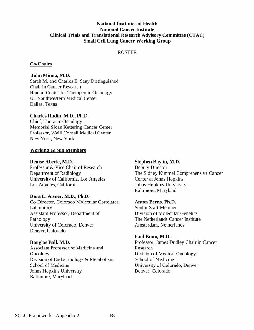
SCLC Framework - Appendix 2 68
National Institutes of Health
National Cancer Institute
Clinical Trials and Translational Research Advisory Committee (CTAC)
Small Cell Lung Cancer Working Group
ROSTER
Co-Chairs
John Minna, M.D.
Sarah M. and Charles E. Seay Distinguished
Chair in Cancer Research
Hamon Center for Therapeutic Oncology
UT Southwestern Medical Center
Dallas, Texas
Charles Rudin, M.D., Ph.D. Chief, Thoracic Oncology
Memorial Sloan Kettering Cancer Center
Professor, Weill Cornell Medical Center
New York, New York
Working Group Members
Denise Aberle, M.D.
Professor & Vice Chair of Research
Department of Radiology
University of California, Los Angeles
Los Angeles, California
Dara L. Aisner, M.D., Ph.D. Co-Director, Colorado Molecular Correlates
Laboratory
Assistant Professor, Department of
Pathology
University of Colorado, Denver
Denver, Colorado
Douglas Ball, M.D. Associate Professor of Medicine and
Oncology
Division of Endocrinology & Metabolism
School of Medicine
Johns Hopkins University
Baltimore, Maryland
Stephen Baylin, M.D. Deputy Director
The Sidney Kimmel Comprehensive Cancer
Center at Johns Hopkins
Johns Hopkins University
Baltimore, Maryland
Anton Berns, Ph.D.
Senior Staff Member
Division of Molecular Genetics
The Netherlands Cancer Institute
Amsterdam, Netherlands
Paul Bunn, M.D.
Professor, James Dudley Chair in Cancer
Research
Division of Medical Oncology
School of Medicine
University of Colorado, Denver
Denver, Colorado

SCLC Framework - Appendix 2 69
Lauren Averett Byers, M.D. Assistant Professor
Department of Thoracic/Head and Neck
Medical Oncology
Division of Cancer Medicine
The University of Texas MD Anderson
Cancer Center
Houston, Texas
David Carbone, M.D., Ph.D. Director
James Thoracic Center
The Ohio State Wexner Medical Center
Columbus, Ohio
Scott J. Dylla, Ph.D. Chief Scientific Officer
VP of Cancer Biology
Stem CentRx, Inc.
South San Francisco, California
Andrea Stern Ferris, M.B.A.
President
LUNGevity
Chicago, Illinois
Adi Gazdar, M.D. W. Ray Wallace Distinguished Chair in
Molecular Oncology Research
Department of Pathology
Hamon Center for Therapeutic Oncology
UT Southwestern Medical Center
Dallas, Texas
Eli Glatstein, M.D.
Morton M. Kligerman Professor of
Radiation Oncology
Department of Radiation Oncology
Perelman School of Medicine
University of Pennsylvania
Philadelphia, Pennsylvania
Glenwood Goss, M.D.
Professor
Department of Internal Medicine
Division of Medical Oncology
The Ottawa Hospital Cancer Centre
Ottawa, Ontario
Canada
Ramaswamy Govindan, M.D.
Professor of Medicine
Division of Medical Oncology
School of Medicine
Washington University, St. Louis
St. Louis, Missouri
Christine Hann, M.D., Ph.D. Assistant Professor of Oncology
Upper Aerodigestive Cancer Program
The Sidney Kimmel Comprehensive Cancer
Center
The Johns Hopkins University
Baltimore, Maryland
David Harpole, Jr., M.D.
Vice Chief
Division of Surgical Services
Department of Surgery
Duke University Medical Center
Durham, North Carolina
Eric Haura, M.D.
Director
Lung Cancer Center of Excellence
H. Lee Moffitt Cancer Center and Research
Institute
Tampa, Florida
Roy Herbst, M.D., Ph.D.
Chief of Medical Oncology
Yale Cancer Center
New Haven, Connecticut
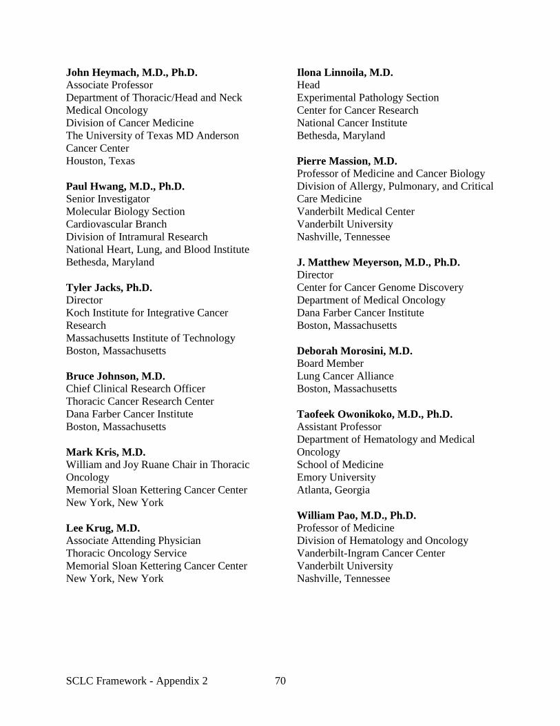
SCLC Framework - Appendix 2 70
John Heymach, M.D., Ph.D.
Associate Professor
Department of Thoracic/Head and Neck
Medical Oncology
Division of Cancer Medicine
The University of Texas MD Anderson
Cancer Center
Houston, Texas
Paul Hwang, M.D., Ph.D.
Senior Investigator
Molecular Biology Section
Cardiovascular Branch
Division of Intramural Research
National Heart, Lung, and Blood Institute
Bethesda, Maryland
Tyler Jacks, Ph.D.
Director
Koch Institute for Integrative Cancer
Research
Massachusetts Institute of Technology
Boston, Massachusetts
Bruce Johnson, M.D. Chief Clinical Research Officer
Thoracic Cancer Research Center
Dana Farber Cancer Institute
Boston, Massachusetts
Mark Kris, M.D.
William and Joy Ruane Chair in Thoracic
Oncology
Memorial Sloan Kettering Cancer Center
New York, New York
Lee Krug, M.D.
Associate Attending Physician
Thoracic Oncology Service
Memorial Sloan Kettering Cancer Center
New York, New York
Ilona Linnoila, M.D.
Head
Experimental Pathology Section
Center for Cancer Research
National Cancer Institute
Bethesda, Maryland
Pierre Massion, M.D.
Professor of Medicine and Cancer Biology
Division of Allergy, Pulmonary, and Critical
Care Medicine
Vanderbilt Medical Center
Vanderbilt University
Nashville, Tennessee
J. Matthew Meyerson, M.D., Ph.D.
Director
Center for Cancer Genome Discovery
Department of Medical Oncology
Dana Farber Cancer Institute
Boston, Massachusetts
Deborah Morosini, M.D.
Board Member
Lung Cancer Alliance
Boston, Massachusetts
Taofeek Owonikoko, M.D., Ph.D.
Assistant Professor
Department of Hematology and Medical
Oncology
School of Medicine
Emory University
Atlanta, Georgia
William Pao, M.D., Ph.D.
Professor of Medicine
Division of Hematology and Oncology
Vanderbilt-Ingram Cancer Center
Vanderbilt University
Nashville, Tennessee
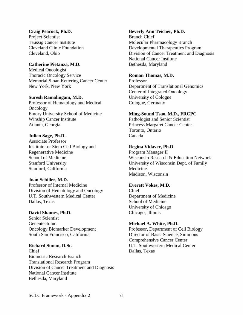
SCLC Framework - Appendix 2 71
Craig Peacock, Ph.D.
Project Scientist
Taussig Cancer Institute
Cleveland Clinic Foundation
Cleveland, Ohio
Catherine Pietanza, M.D.
Medical Oncologist
Thoracic Oncology Service
Memorial Sloan Kettering Cancer Center
New York, New York
Suresh Ramalingam, M.D.
Professor of Hematology and Medical
Oncology
Emory University School of Medicine
Winship Cancer Institute
Atlanta, Georgia
Julien Sage, Ph.D.
Associate Professor
Institute for Stem Cell Biology and
Regenerative Medicine
School of Medicine
Stanford University
Stanford, California
Joan Schiller, M.D.
Professor of Internal Medicine
Division of Hematology and Oncology
U.T. Southwestern Medical Center
Dallas, Texas
David Shames, Ph.D.
Senior Scientist
Genentech Inc.
Oncology Biomarker Development
South San Francisco, California
Richard Simon, D.Sc.
Chief
Biometric Research Branch
Translational Research Program
Division of Cancer Treatment and Diagnosis
National Cancer Institute
Bethesda, Maryland
Beverly Ann Teicher, Ph.D.
Branch Chief
Molecular Pharmacology Branch
Developmental Therapeutics Program
Division of Cancer Treatment and Diagnosis
National Cancer Institute
Bethesda, Maryland
Roman Thomas, M.D.
Professor
Department of Translational Genomics
Center of Integrated Oncology
University of Cologne
Cologne, Germany
Ming-Sound Tsao, M.D., FRCPC
Pathologist and Senior Scientist
Princess Margaret Cancer Center
Toronto, Ontario
Canada
Regina Vidaver, Ph.D.
Program Manager II
Wisconsin Research & Education Network
University of Wisconsin Dept. of Family
Medicine
Madison, Wisconsin
Everett Vokes, M.D.
Chief
Department of Medicine
School of Medicine
University of Chicago
Chicago, Illinois
Michael A. White, Ph.D.
Professor, Department of Cell Biology
Director of Basic Science, Simmons
Comprehensive Cancer Center
U.T. Southwestern Medical Center
Dallas, Texas
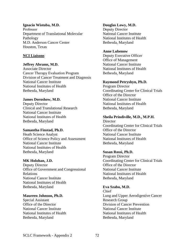
SCLC Framework - Appendix 2 72
Ignacio Wistuba, M.D.
Professor
Department of Translational Molecular
Pathology
M.D. Anderson Cancer Center
Houston, Texas
NCI Liaisons
Jeffrey Abrams, M.D.
Associate Director
Cancer Therapy Evaluation Program
Division of Cancer Treatment and Diagnosis
National Cancer Institute
National Institutes of Health
Bethesda, Maryland
James Doroshow, M.D. Deputy Director
Clinical and Translational Research
National Cancer Institute
National Institutes of Health
Bethesda, Maryland
Samantha Finstad, Ph.D. Heath Science Analyst
Office of Science Policy and Assessment
National Cancer Institute
National Institutes of Health
Bethesda, Maryland
MK Holohan, J.D.
Deputy Director
Office of Government and Congressional
Relations
National Cancer Institute
National Institutes of Health
Bethesda, Maryland
Maureen Johnson, Ph.D.
Special Assistant
Office of the Director
National Cancer Institute
National Institutes of Health
Bethesda, Maryland
Douglas Lowy, M.D. Deputy Director
National Cancer Institute
National Institutes of Health
Bethesda, Maryland
Anne Lubenow
Deputy Executive Officer
Office of Management
National Cancer Institute
National Institutes of Health
Bethesda, Maryland
Raymond Petryshyn, Ph.D.
Program Director
Coordinating Center for Clinical Trials
Office of the Director
National Cancer Institute
National Institutes of Health
Bethesda, Maryland
Sheila Prindiville, M.D., M.P.H.
Director
Coordinating Center for Clinical Trials
Office of the Director
National Cancer Institute
National Institutes of Health
Bethesda, Maryland
Susan Rossi, Ph.D.
Program Director
Coordinating Center for Clinical Trials
Office of the Director
National Cancer Institute
National Institutes of Health
Bethesda, Maryland
Eva Szabo, M.D.
Chief
Lung and Upper Aerodigestive Cancer
Research Group
Division of Cancer Prevention
National Cancer Institute
National Institutes of Health
Bethesda, Maryland
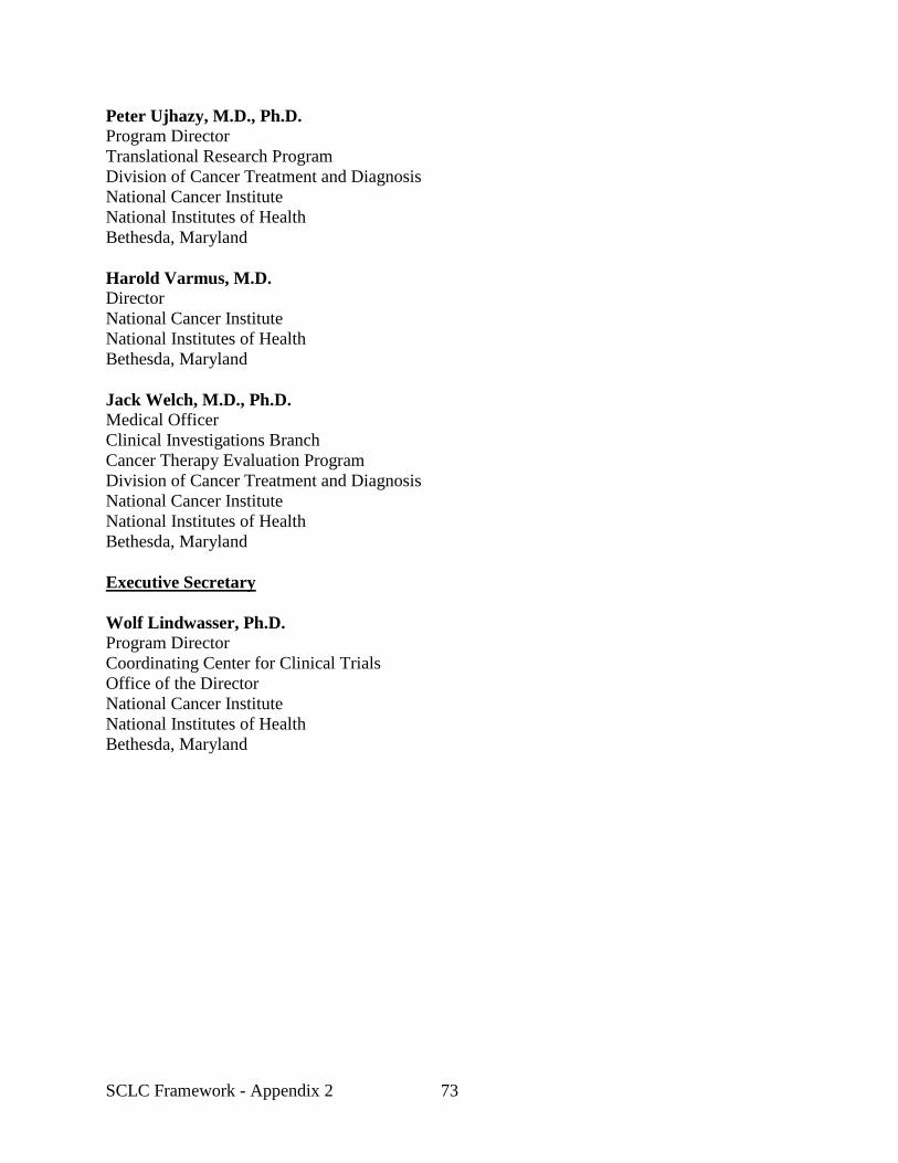
SCLC Framework - Appendix 2 73
Peter Ujhazy, M.D., Ph.D.
Program Director
Translational Research Program
Division of Cancer Treatment and Diagnosis
National Cancer Institute
National Institutes of Health
Bethesda, Maryland
Harold Varmus, M.D.
Director
National Cancer Institute
National Institutes of Health
Bethesda, Maryland
Jack Welch, M.D., Ph.D.
Medical Officer
Clinical Investigations Branch
Cancer Therapy Evaluation Program
Division of Cancer Treatment and Diagnosis
National Cancer Institute
National Institutes of Health
Bethesda, Maryland
Executive Secretary
Wolf Lindwasser, Ph.D.
Program Director
Coordinating Center for Clinical Trials
Office of the Director
National Cancer Institute
National Institutes of Health
Bethesda, Maryland
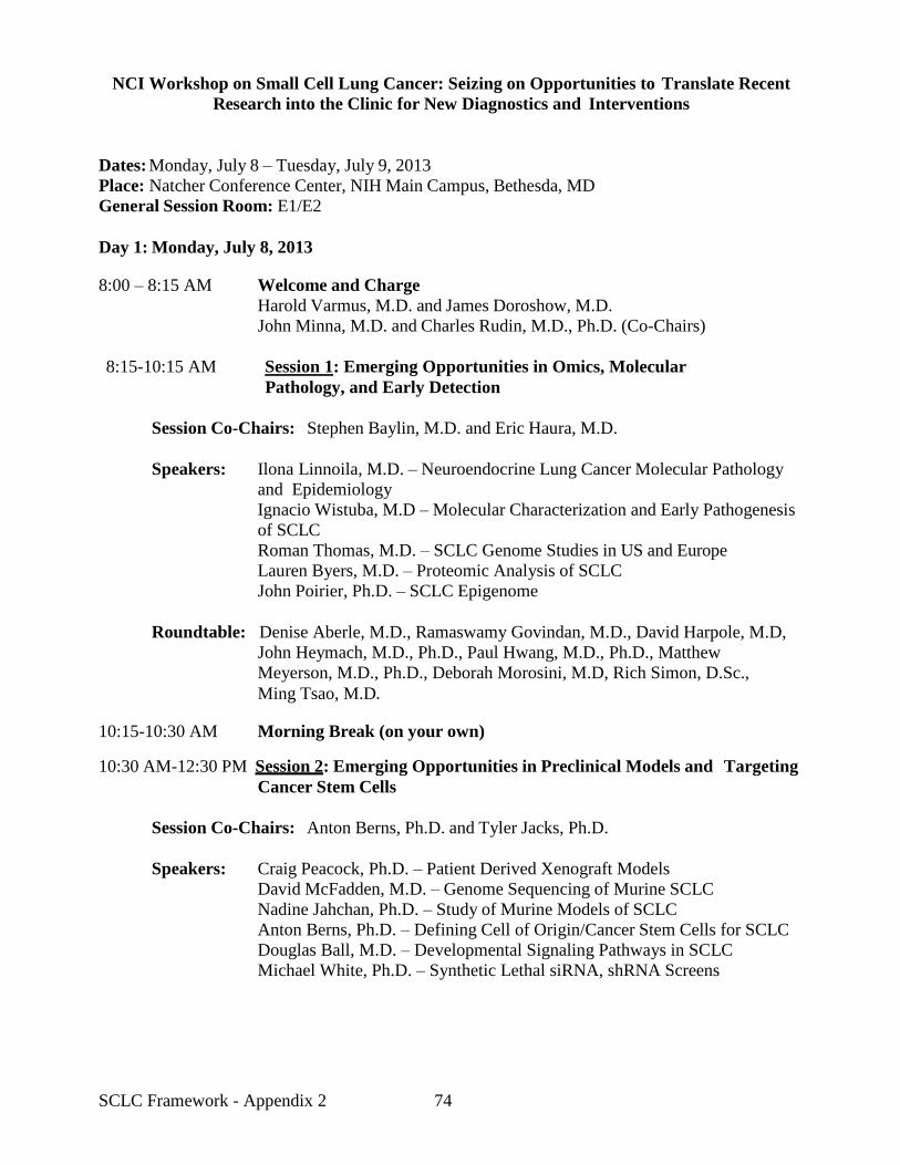
SCLC Framework - Appendix 2 74
NCI Workshop on Small Cell Lung Cancer: Seizing on Opportunities to Translate Recent
Research into the Clinic for New Diagnostics and Interventions
Dates: Monday, July 8 – Tuesday, July 9, 2013
Place: Natcher Conference Center, NIH Main Campus, Bethesda, MD
General Session Room: E1/E2
Day 1: Monday, July 8, 2013 8:00 – 8:15 AM Welcome and Charge
Harold Varmus, M.D. and James Doroshow, M.D.
John Minna, M.D. and Charles Rudin, M.D., Ph.D. (Co-Chairs)
8:15-10:15 AM Session 1: Emerging Opportunities in Omics, Molecular
Pathology, and Early Detection
Session Co-Chairs: Stephen Baylin, M.D. and Eric Haura, M.D.
Speakers: Ilona Linnoila, M.D. – Neuroendocrine Lung Cancer Molecular Pathology
and Epidemiology
Ignacio Wistuba, M.D – Molecular Characterization and Early Pathogenesis
of SCLC
Roman Thomas, M.D. – SCLC Genome Studies in US and Europe
Lauren Byers, M.D. – Proteomic Analysis of SCLC
John Poirier, Ph.D. – SCLC Epigenome
Roundtable: Denise Aberle, M.D., Ramaswamy Govindan, M.D., David Harpole, M.D,
John Heymach, M.D., Ph.D., Paul Hwang, M.D., Ph.D., Matthew
Meyerson, M.D., Ph.D., Deborah Morosini, M.D, Rich Simon, D.Sc.,
Ming Tsao, M.D. 10:15-10:30 AM Morning Break (on your own)
10:30 AM-12:30 PM Session 2: Emerging Opportunities in Preclinical Models and Targeting
Cancer Stem Cells
Session Co-Chairs: Anton Berns, Ph.D. and Tyler Jacks, Ph.D.
Speakers: Craig Peacock, Ph.D. – Patient Derived Xenograft Models
David McFadden, M.D. – Genome Sequencing of Murine SCLC
Nadine Jahchan, Ph.D. – Study of Murine Models of SCLC
Anton Berns, Ph.D. – Defining Cell of Origin/Cancer Stem Cells for SCLC
Douglas Ball, M.D. – Developmental Signaling Pathways in SCLC
Michael White, Ph.D. – Synthetic Lethal siRNA, shRNA Screens
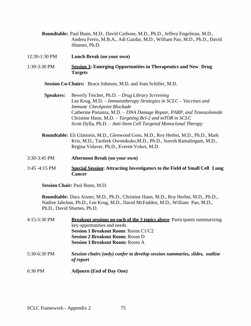
SCLC Framework - Appendix 2 75
Roundtable: Paul Bunn, M.D., David Carbone, M.D., Ph.D., Jeffrey Engelman, M.D.,
Andrea Ferris, M.B.A., Adi Gazdar, M.D., William Pao, M.D., Ph.D., David
Shames, Ph.D.
12:30-1:30 PM Lunch Break (on your own)
1:30-3:30 PM Session 3: Emerging Opportunities in Therapeutics and New Drug
Targets
Session Co-Chairs: Bruce Johnson, M.D. and Joan Schiller, M.D.
Speakers: Beverly Teicher, Ph.D. – Drug Library Screening
Lee Krug, M.D. – Immunotherapy Strategies in SCLC – Vaccines and
Immune Checkpoint Blockade
Catherine Pietanza, M.D. – DNA Damage Repair, PARP, and Temozolomide
Christine Hann, M.D. – Targeting Bcl-2 and mTOR in SCLC
Scott Dylla, Ph.D. – Anti-Stem Cell Targeted Monoclonal Therapy
Roundtable: Eli Glatstein, M.D., Glenwood Goss, M.D., Roy Herbst, M.D., Ph.D., Mark
Kris, M.D., Taofeek Owonikoko,M.D., Ph.D., Suresh Ramalingam, M.D.,
Regina Vidaver, Ph.D., Everett Vokes, M.D.
3:30-3:45 PM Afternoon Break (on your own)
3:45 -4:15 PM Special Session: Attracting Investigators to the Field of Small Cell Lung
Cancer
Session Chair: Paul Bunn, M.D.
Roundtable: Dara Aisner, M.D., Ph.D., Christine Hann, M.D., Roy Herbst, M.D., Ph.D.,
Nadine Jahchan, Ph.D., Lee Krug, M.D., David McFadden, M.D., William Pao, M.D.,
Ph.D., David Shames, Ph.D.
4:15-5:30 PM Breakout sessions on each of the 3 topics above Participants summarizing
key opportunities and needs
Session 1 Breakout Room: Room C1/C2
Session 2 Breakout Room: Room D
Session 3 Breakout Room: Room A
5:30-6:30 PM Session chairs (only) confer to develop session summaries, slides, outline
of report
6:30 PM Adjourn (End of Day One)
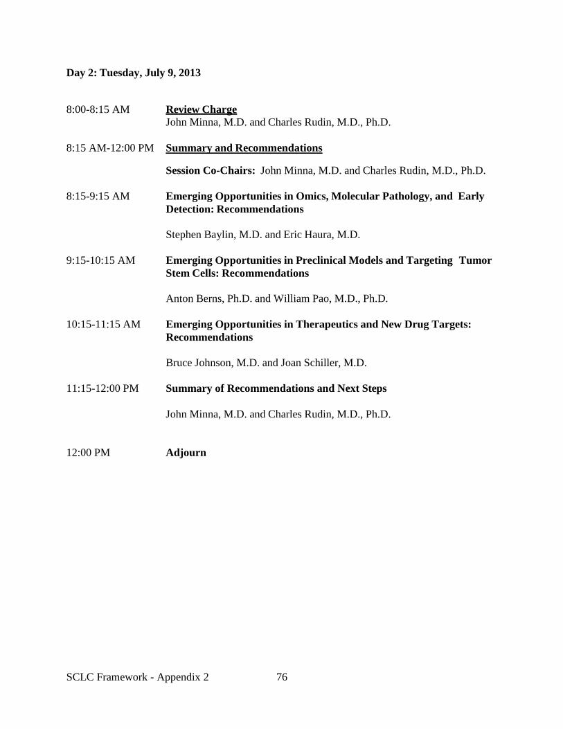
SCLC Framework - Appendix 2 76
Day 2: Tuesday, July 9, 2013
8:00-8:15 AM Review Charge
John Minna, M.D. and Charles Rudin, M.D., Ph.D.
8:15 AM-12:00 PM Summary and Recommendations
Session Co-Chairs: John Minna, M.D. and Charles Rudin, M.D., Ph.D.
8:15-9:15 AM Emerging Opportunities in Omics, Molecular Pathology, and Early
Detection: Recommendations
Stephen Baylin, M.D. and Eric Haura, M.D.
9:15-10:15 AM Emerging Opportunities in Preclinical Models and Targeting Tumor
Stem Cells: Recommendations
Anton Berns, Ph.D. and William Pao, M.D., Ph.D.
10:15-11:15 AM Emerging Opportunities in Therapeutics and New Drug Targets:
Recommendations
Bruce Johnson, M.D. and Joan Schiller, M.D.
11:15-12:00 PM Summary of Recommendations and Next Steps
John Minna, M.D. and Charles Rudin, M.D., Ph.D.
12:00 PM Adjourn