Embed Size (px)
Citation preview
ORIGINAL ARTICLE
Scintigraphic detection of TNF-driveninflammation by radiolabelledcertolizumab pegol in patientswith rheumatoid arthritisand spondyloarthritis
Philippe Carron,1 Bieke Lambert,2 Liesbet Van Praet,1 Filip De Vos,3
Gaëlle Varkas,4,5 Lennart Jans,6 Dirk Elewaut,4,5 Filip Van den Bosch1
To cite: Carron P, Lambert B,Van Praet L, et al.Scintigraphic detection ofTNF-driven inflammation byradiolabelled certolizumabpegol in patientswith rheumatoid arthritisand spondyloarthritis. RMDOpen 2016;2:e000265.doi:10.1136/rmdopen-2016-000265
▸ Prepublication history forthis paper is available online.To view these files pleasevisit the journal online(http://dx.doi.org/10.1136/rmdopen-2016-000265).
Received 12 February 2016Revised 29 March 2016Accepted 16 April 2016
For numbered affiliations seeend of article.
Correspondence toDr Filip Van den Bosch;[email protected]
ABSTRACTBackground: Biologicals are the cornerstone formany treatment algorithms in inflammatory arthritis.While tumour necrosis factor (TNF) inhibitors mayachieve important responses in ∼50% of patients withrheumatoid arthritis (RA) and spondyloarthritis (SpA),a significant fraction of patients are partial or non-responders. We hypothesised that in vivo assessmentof TNF by scintigraphy with 99mTc-radiolabelledcertolizumab pegol (CZP) might lead to a more‘evidence-based biological therapy’.Objectives: Our goal was to perform a proof-of-concept study of in vivo detection of TNF byimmunoscintigraphy of a radiolabelled TNF inhibitor inRA and SpA, and correlate this with clinical, imagingfindings and therapeutic outcome.Methods: CZP was conjugated with succinimidyl-6-hydrazino-nicotinamide and subsequently radiolabelledwith Tc99m. Whole body and static images of hands,feet and sacroiliac joints of 20 patients (5 RA; 15SpA) were acquired at 3 time points.Immunoscintigraphic findings were scoredsemiquantitatively. Subsequently, all patients weretreated with CZP.Results: In peripheral joints, clinically affected jointsor abnormal ultrasound findings were observed morefrequently (p<0.001) in the scintigraphic-positivegroup. In patients with axial SpA, bone marrowedema on MRI was detected more frequently(p<0.001) in quadrants with tracer uptake. At thepatient level, the odds of a joint remaining tenderdespite 24 weeks of CZP treatment was significantlysmaller in joints with clear tracer uptake as comparedwith those with no uptake (OR=0.42, p=0.04).Conclusions: Immunoscintigraphy with radiolabelledCZP demonstrated both axial and peripheralinflammation, and displayed good correlation withclinical features, conventional imaging and therapyresponse.Trial registration number: NCT01590966; Results.
INTRODUCTIONRheumatoid arthritis (RA) and spondyloar-thritis (SpA) are both chronic inflammatoryjoint diseases, with a combined prevalenceclose to 2%. If untreated, persistent inflam-mation may lead to functional disability, pro-gressive structural damage and potentially toalso a number of extra-articular manifesta-tions or comorbidities.1 2 Currently, theaetiology is unknown, but a combination ofpredisposing genetic factors, and person-related or environmental factors (age,gender, infectious agents, smoking, dietaryfactors) is suspected to play a role in disease
Key messages
▸ Typical joint involvement patterns in peripheraland axial disease were detected by using aimmunoscintigraphic procedure with radiola-belled certolizumab pegol (CZP): polyarticularpattern in rheumatoid arthritis, distal interpha-langeal joint (DIP) involvement and dactylitis inpsoriatic arthritis, and enthesitis and sacroiliitisin spondyloarthritis (SpA).
▸ A strong relationship with clinical involvementand immunoscintigraphic uptake was observed;tender or swollen joints or abnormal ultrasoundfindings were significantly more prevalent whenscintigraphy was positive.
▸ In patients with axial SpA, bone marrow edemaon MRI was observed more frequently in quad-rants that also showed uptake of the radiola-belled CZP.
▸ A marked relationship with therapy responsewas observed: the odds of a joint remainingtender despite 24 weeks of CZP treatment wassignificantly smaller in joints with clear traceruptake as compared to those without.
Carron P, et al. RMD Open 2016;2:e000265. doi:10.1136/rmdopen-2016-000265 1
Imaging
on 12 June 2018 by guest. Protected by copyright.
http://rmdopen.bm
j.com/
RM
D O
pen: first published as 10.1136/rmdopen-2016-000265 on 24 June 2016. D
ownloaded from
pathogenesis.3 4 Treatment strategies for SpA and RAhave changed dramatically over the last decade mainlyas a consequence of our growing knowledge of thepathogenetic role of proinflammatory cytokines, such astumour necrosis factor α (TNF-α).5 6
Immunohistological studies demonstrated the pre-sence of TNF-α and its receptors in inflamed tissues ofperipheral joints and/or sacroiliac joints (SIJs).7 Theintroduction of ‘biological agents’ targeting TNF-α, witheither monoclonal antibodies (including Fab fragments)or receptor-fusion proteins, has revolutionised our thera-peutic armamentarium. Treatments neutralising TNF-αhave the longest track record in RA and SpA; currentlyfour monoclonal antibodies (adalimumab, certolizumab,golimumab, infliximab) and one receptor-fusion protein(etanercept) are available.8 Certolizumab pegol (CZP;UCB Celltech, Slough, Berkshire, UK) is an engineeredhumanised monoclonal antibody Fc-free Fab′ fragmentwith specificity for human TNF-α, manufactured inEscherichia coli. The antibody fragment is subsequentlypurified and conjugated with high molecular weightpolyethylene glycol (PEG; 40 kDa). CZP is approvedworldwide for adult patients with moderate-to-severeactive RA, ankylosing spondylitis (AS) and psoriaticarthritis;9–11 moreover, the drug also received marketingauthorisation in Europe for the indication of non-radiographic axial SpA (axSpA).12 In addition to RA,CZP has demonstrated a positive benefit risk in Crohn’sdisease and psoriasis.13 14
In clinical trials for RA and SpA, ∼50% of patientsachieve a clinically important response such as anAmerican College of Rheumatology (ACR) 50% responserate or the Assessment of SpondyloArthritis internationalSociety (ASAS) 40% response rate.9 11 15–22 Despite anoverall impressive improvement in clinical signs andsymptoms when a TNF-blocking agent is administered,there is still a significant proportion of patients who donot reach a relevant response (primary non-responder),have insufficient improvement, or who lose an initialgood response over time (secondary non-responder).This may be due to lower levels of TNF-α expression atthe site of inflammation as it is known that there may bea large interindividual and intraindividual variability inthese levels,23 or to the fact that the disease is in fact pre-dominantly driven by other proinflammatory cytokines.In an era of evidence-based medicine, it is disappointingto realise that treatment decisions or strategies, even forexpensive biological agents, are driven by patient-reported outcomes, the difficult to standardise clinicalexaminations ( joint counts), rather aspecific laboratoryparameters (elevated C reactive protein (CRP)) or ques-tionable imaging cut-offs (erosions, sacroiliitis grade 2 bymodified New York criteria for AS). An accurate way ofpredicting a relevant clinical response to a certain tar-geted treatment would allow for a better patient selec-tion. Molecular imaging studies aiming at selectivelyvisualising TNF-α (or another suspected culprit cytokine)in vivo at the site of clinical inflammation could be an
attractive alternative to other less specific and/or invasivetechniques such as bone scintigrapy with 99mTc-labelleddiphosphonates or synovial biopsies taken by arthro-scopic procedures. This more rational approach of deter-mining the most appropriate biological treatment for anindividual patient may also have relevant consequencesfor the cost-efficacy of our currently available biologicaldrugs, and even have an impact on the design of futureclinical trials with targeted therapies. Therefore, we per-formed a proof-of-concept study to explore the possibilityof visualising TNF-driven disease in patients with activeRA and SpA using scintigraphy with Tc99m-labelled CZPas tracer, and to correlate the anti-TNF bound tracer withthe localisation of active clinical inflammation.
PATIENTS AND METHODSPatientsThe study was approved by the Medical EthicsCommittee of the Ghent University Hospital, and eachpatient gave written informed consent. The study wasconducted in compliance with International Conferenceon Harmonisation Good Clinical Practice guidelines andthe Declaration of Helsinki. (EudraCT number:2009-017998-37).We included 20 adult (18–70 years) patients with RA
(n=5), peripheral SpA (pSpA; n=6) or axSpA (n=9).Patients were recruited from the RheumatologyOutpatient Clinic of the University of Ghent, Belgium,from November 2012 until November 2013.All patients with RA fulfilled both the ACR revised
criteria for the diagnosis of RA24 and the 2010 ACR/European League Against Rheumatism (EULAR) clas-sification criteria,25 and all patients with SpA fulfilledthe current ASAS classification criteria for axSpA orpSpA.26 27 Patients with RA fulfilled Belgian reimbur-sement criteria for the initiation of anti-TNF agents(including failure of at least two disease-modifyingantirheumatic drugs, one of these being methotrexate)and had active disease Disease Acitivity Score in 28joints (DAS28≥3.7) with at least five swollen joints atscreening. Patients with pSpA had active arthritis, dac-tylitis or enthesitis at screening despite treatment withan adequate stable dose of sulfasalazine or methotrex-ate for at least 3 months, or a stable, full dose of non-steroidal anti-inflammatory drugs (NSAIDs) for at least4 weeks. Patients with axSpA were required to still haveactive disease (despite a stable, full dose of NSAIDs forat least 4 weeks), defined as a Bath AnkylosingSpondylitis Disease Activity Index (BASDAI) ≥4 (0–10)and active inflammatory lesions on MRI of the SIJs(according to the definition of a positive MRI pro-posed by the ASAS consortium) or radiographic sacroi-liitis according to the modified New York criteria.26
Prior treatment with any biological treatment was anexclusion criterion. All patients were screened forlatent tuberculosis by means of a tuberculin skin testand chest X-ray.
2 Carron P, et al. RMD Open 2016;2:e000265. doi:10.1136/rmdopen-2016-000265
RMD Open
on 12 June 2018 by guest. Protected by copyright.
http://rmdopen.bm
j.com/
RM
D O
pen: first published as 10.1136/rmdopen-2016-000265 on 24 June 2016. D
ownloaded from
Clinical and laboratory assessmentsPatients were evaluated through screening at baseline(prior to the CZP scintigraphy), prior to initiation ofCZP, and at weeks 12 and 24 after start of CZP.Evaluations were performed by the same rheumatologist(PC) and consisted of a full rheumatological examina-tion, including a 66/68 joint count, enthesitis and dacty-litis assessments as well as an evaluation of axialmetrology. At all visits, a number of patient-reported out-comes was assessed, including patient global assessment(PGA) of disease activity, PGA of pain and Short Form(SF)-36 for all patients, BASDAI and Bath AnkylosingSpondylitis Functional Index (BASFI) for patients withaxSpA, and the Health Assessment Questionnaire(HAQ) for patients with RA. During the screening 12and 24 weeks after treatment, routine blood sampleswere taken to assess potential toxicity of CZP treatment,and determine erythrocyte sedimentation rate (normallevel <20 mm/hour) and level of CRP (normal level<5 mg/L). This allowed for the calculation of validatedcomposite disease activity scores such as the DAS28 forRA, and the Ankylosing Spondylitis Disease ActivityScore (ASDAS) for patients with SpA.
Imaging assessmentsUltrasound examinationUltrasound (US) evaluation was performed at baselineand after 12 weeks treatment with CZP in patients withRA and pSpA. Systematic multiplanar grey scale (GS)and power Doppler (PD) examinations were carried outwith an ESAOTE MyLab60 using multifrequency lineartransducers (6–18 MHz). PD imaging was performed byselecting a region of interest that included the bonymargins, articular space and a variable view of surround-ing tissues (depending on the joint size). PD variableswere adjusted to the lowest permissible pulse repetitionfrequency (PRF) to maximise sensitivity. This settingresulted in a PRF between 500 and 1000 Hz dependingon the joint scanned. Low wall filters were used. Colourgain was set just below the level at which colour noiseappeared on the underlying bone (no flow should bevisualised at the bony surface). We evaluated both wrists,ankles and subtalar joints, as well as all metacarpopha-langeal (MCP) and metatarsophalangeal joints. Synovitisand tenosynovitis were assessed according to the EULARcriteria28 and the OMERACT definition.29 Synovitis andsynovial and tenosynovial vascularity were scored semi-quantitatively (grade 0–3) by PD US according toSzkudlarek et al.30 Synovitis (effusion and synovial hyper-trophy combined) in GS US was analysed semiquantita-tively as described by Scheel et al.31 Tenosynovitis in GSUS was registered as being absent (0) or present (1).
MRIAll patients with axSpA and one patient with pSpAunderwent MRI of the SIJs. Images were obtained on a1.5 T MRI unit (Avanto/Symphony, Siemens Medical,Erlangen, Germany). The SIJs were imaged in a body
flexed array coil (Siemens Medical, Erlangen,Germany). The sequence protocol included semicoronal(along long axis of the sacral bone) T1-weighted turbospin echo (slice thickness (ST) 3 mm; repetition time/echo time (TR/TE) 595/20 ms), semicoronal short tauinversion recovery (STIR) (ST 3 mm; TR/TE/inversiontime (TI) 5030/67/150 ms) and axial STIR (ST 5 mm;TR/TE/TI 7540/67/150 ms). Sacroiliitis on MRI wasscored positive or negative according to the definitionproposed by the ASAS consortium.26 Each SIJ wasdivided into four quadrants and evaluated on eightcoronal 3 mm MRI slices. Bone marrow edema (BME)was scored per quadrant as either absent or present (ontwo consecutive slices), thus providing a total scorebetween 0 and 8. BME of the SIJs was also scored usingthe Spondyloarthritis Research Consortium of Canada(SPARCC) scoring system by certified readers for theSPARCC scoring system.32 The presence of an intensesignal (comparable to blood vessels) or depth ≥1 cmanywhere within each SIJ of the six slices is given anadditional score (0=absent/1=present). Since in ourcentre 3 mm slices are used, eight slices were evaluatedand converted to a maximum score of 72. MRI of theSIJs was performed within the week before the immu-noscintigraphy was obtained.
Scintigraphic assessmentsRadiolabelling procedure of CZPLyophylised CZP was reconstituted with water for injec-tion, filtered and dialysed, and subsequently incubatedwith succinimidyl-6-hydrazino-nicotinamide (S-HYNIC,ABX GmbH, Radeberg, Germany). After removal of theunreacted S-HYNIC, a bifunctional crosslinker, the solu-tion was diluted with acetate buffer to a pH of 5 and fil-tered through a 0.22 µm membrane filter. Solutions of1.25 mg S-HYNIC CZP were dispensed in 1.0 mL glassvials stored at −80°C.For the radiolabelling procedure, a co-ligand kit con-
sisting of a tin(II) sulfate (4.66 mM, Sigma Aldrich,Steinheim, Germany) and tricine (55.81 mM, SigmaAldrich) diluted solution (50 µL) was added to theS-HYNIC CZP vial. Subsequently, 925 MBq (±10%)freshly eluated Tc99m pertechnetate was added to thisvial. This was incubated for 15 min at room temperature.Finally, physiological saline was added to dilute theradiopharmaceutical formulation in a total volume of3 mL. All handling was performed under aseptic condi-tions. Quality control was carried out by instant thinlayer chromatography with Silica gel (SilG) as stationaryphase and 0.9% NaCl solution as mobile phase. For clin-ical use, the radiochemical yield needed to exceed 90%.
Scintigraphic procedurePatients were injected intravenously with ∼740 MBqTc99m-labelled CZP (10.6 MBq/kg). All patients werescanned on the same double-headed γ-camera(BrightView, Philips Healthcare, Best, The Netherlands).Whole body (WB) images (15 cm/min scan speed,
Carron P, et al. RMD Open 2016;2:e000265. doi:10.1136/rmdopen-2016-000265 3
Imaging
on 12 June 2018 by guest. Protected by copyright.
http://rmdopen.bm
j.com/
RM
D O
pen: first published as 10.1136/rmdopen-2016-000265 on 24 June 2016. D
ownloaded from
matrix size 1024×512, pixel size 2.80 mm) were per-formed immediately following administration, at 1, 4–6and 24 hours postinjection. A standard activity of∼5 MBq Tc99m in an unshielded syringe was always inthe field of view for quantification purposes. Staticimages (5 min, matrix size 256×256, pixel size 2.33 mm)of hands and feet were acquired immediately followingthe first WB scan which was started within a couple ofminutes postinjection, at 4–6 and 24 hours postinjection.A single photon emission tomography (SPECT) wasacquired from the axial skeleton. In selected cases, aSPECT/CT was performed to depict in detail theinvolved joints.To assess the tracer accumulation in the peripheral
joints, each joint was scored semiquantitatively by usingthe following scoring system: score 0=no tracer uptake,score 1=faint uptake of the tracer and score 2=clearuptake of the tracer. The same score was applied toevery quadrant of the SIJs in patients with axSpA. Thescintigraphic result was defined as positive if there wasfaint or clear tracer uptake 4–6 hours postinjection. Asthe scintigraphic images do not provide anatomicdetails, fusion of the MRI with the nuclear image wasperformed to allow scoring of the tracer uptake perquadrant. For each individual scan, a background regionof interest (ROI) was defined within the field of view,for example, left supraclavicular region on WB, rightforearm on static images of the wrists and hands, andright distal tibia for the static images depicting theankles and feet. These results were compared with thefindings from clinical examination of each assessablejoint. The nuclear medicine physician reading theimmunoscans, and the clinician performing the clinicalexamination were blinded to each other’s observations.
Statistical analysisData were analysed using SAS V.9.3 (SAS Institute Inc,Cary, North Carolina, USA). Both a joint-based andpatient-based analysis were done. In the joint-based ana-lysis, each individual joint contributes an observation,but clustering of the joints within the patient was takeninto account in the statistical analysis. For this analysis ascintigraphic score corresponding to either faint or cleartracer uptake 4–6 hours postinjection was considered aspositive. To determine the relationship between the scin-tigraphic variable and the (clinical or US) status of thejoint at baseline, a mixed logistic regression model withpatient as random effect was used. For the predictivevalue of the scintigraphic variable on the status of thejoint 12 and 24 weeks after treatment, the data set isreduced to only those joints that were tender and/orswollen at baseline according to the variable assessed inthe analysis. A mixed logistic regression model withpatient as random effect is used on this reduced dataset. The results of the logistic regression model are sum-marised in terms of the OR and percentage of tenderand/or swollen joints in the positive and negative scinti-graphic group. In the patient-based analysis summary
statistics at the patient level was used. In this analysis thesum of the semiquantitative scintigraphic scores wasused: for patients with RA, the scores of wrists, MCP andproximal interphalangeal joint (PIP) joints were usedwhereas in pSpA, the scores of the hand distal interpha-langeal joint (DIP) joints were added; in patients withaxSpA, the sum scores of the quadrants of the SIJs wasused. To assess the predictive value of the scintigraphicvariable at baseline on the clinical improvement at 12and 24 weeks after treatment (measured by ASDAS forpatients with axSpA and pSpA; measured by DAS28 forpatients with RA), a linear regression model is used withthe scintigraphic scores included as a continuous vari-able. The results are summarised in terms of the slope.The slope describes the change in the respective clinicalscore with a unit increase in the scintigraphic variable.For all analyses p<0.05 was considered statisticallysignificant.
RESULTSPatients baseline and disease characteristics and clinicalresponse after 12 and 24 weeks treatment with CZPTable 1 depicts the baseline characteristics and the clinicalresponse after 12 and 24 weeks of treatment with CZP. Themean age±SD of patients with RA, pSpA and axSpA atbaseline was 56.2±10.1, 41.0±10.3 and 37.1±10.7 years witha mean symptom duration of 13.1±15, 12.6±14.9 and 9±6.7 years, respectively. The mean CRP level at baseline forpatients with RA, pSpA and axSpA was 10.9, 15.4 and4.7 mg/L, respectively. In general, there was a clearimprovement of signs and symptoms in all patients treatedwith CZP. Three of the five patients with RA had a goodEULAR DAS28 response, and two had a moderate EULARDAS28 response after 12 weeks treatment. At 24 weeks,loss of response was observed in one patient with RA.Three of the six patients with pSpA showed a majorASDAS improvement after 12 weeks treatment while twohad a clinically important ASDAS improvement. Onepatient with pSpA was a (primary) non-responder, andone patient with pSpA lost response after 24 weeks treat-ment. In the patients with axSpA group, two out ofnine had a major ASDAS response, and six patients had aclinically important ASDAS response. Again, one patientwith axSpA was a primary non-responder and one patientwith axSpA lost response at 24 weeks treatment. Thescintigraphic procedure was well tolerated. Noprocedure-related adverse events were observed.
Description of distinct scintigraphic patterns observed inpatients with RA and SpAIn most of the clinically involved joints of hands andfeet in patients with RA and pSpA, a tracer uptake wasimmediately visualised within minutes following theinjection, probably due to vascular hyperaemia. At 4–6 hours postinjection, more joints were showing anenhanced uptake, with the more favourablejoint-to-background ratio allowing a good anatomical
4 Carron P, et al. RMD Open 2016;2:e000265. doi:10.1136/rmdopen-2016-000265
RMD Open
on 12 June 2018 by guest. Protected by copyright.
http://rmdopen.bm
j.com/
RM
D O
pen: first published as 10.1136/rmdopen-2016-000265 on 24 June 2016. D
ownloaded from
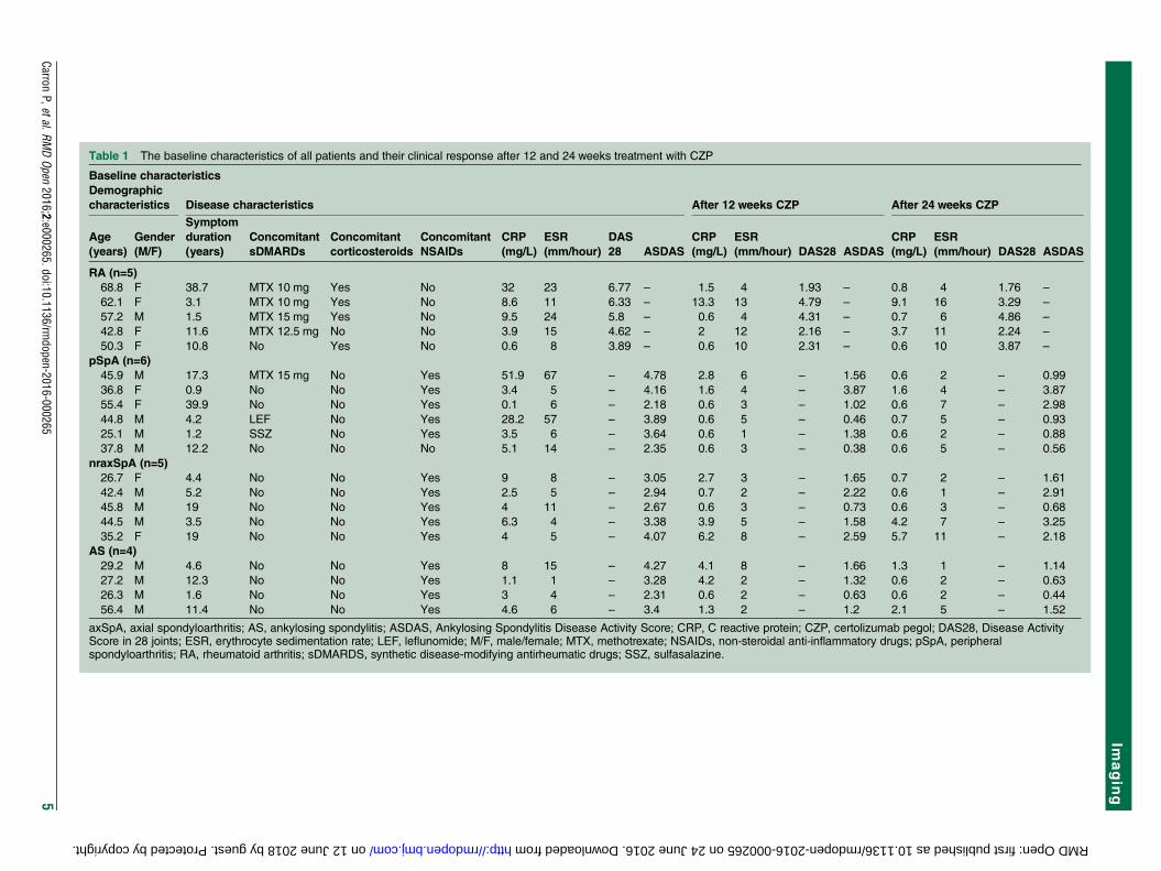
Table 1 The baseline characteristics of all patients and their clinical response after 12 and 24 weeks treatment with CZP
Baseline characteristics
Demographic
characteristics Disease characteristics After 12 weeks CZP After 24 weeks CZP
Age
(years)
Gender
(M/F)
Symptom
duration
(years)
Concomitant
sDMARDs
Concomitant
corticosteroids
Concomitant
NSAIDs
CRP
(mg/L)
ESR
(mm/hour)
DAS
28 ASDAS
CRP
(mg/L)
ESR
(mm/hour) DAS28 ASDAS
CRP
(mg/L)
ESR
(mm/hour) DAS28 ASDAS
RA (n=5)
68.8 F 38.7 MTX 10 mg Yes No 32 23 6.77 – 1.5 4 1.93 – 0.8 4 1.76 –
62.1 F 3.1 MTX 10 mg Yes No 8.6 11 6.33 – 13.3 13 4.79 – 9.1 16 3.29 –
57.2 M 1.5 MTX 15 mg Yes No 9.5 24 5.8 – 0.6 4 4.31 – 0.7 6 4.86 –
42.8 F 11.6 MTX 12.5 mg No No 3.9 15 4.62 – 2 12 2.16 – 3.7 11 2.24 –
50.3 F 10.8 No Yes No 0.6 8 3.89 – 0.6 10 2.31 – 0.6 10 3.87 –
pSpA (n=6)
45.9 M 17.3 MTX 15 mg No Yes 51.9 67 – 4.78 2.8 6 – 1.56 0.6 2 – 0.99
36.8 F 0.9 No No Yes 3.4 5 – 4.16 1.6 4 – 3.87 1.6 4 – 3.87
55.4 F 39.9 No No Yes 0.1 6 – 2.18 0.6 3 – 1.02 0.6 7 – 2.98
44.8 M 4.2 LEF No Yes 28.2 57 – 3.89 0.6 5 – 0.46 0.7 5 – 0.93
25.1 M 1.2 SSZ No Yes 3.5 6 – 3.64 0.6 1 – 1.38 0.6 2 – 0.88
37.8 M 12.2 No No No 5.1 14 – 2.35 0.6 3 – 0.38 0.6 5 – 0.56
nraxSpA (n=5)
26.7 F 4.4 No No Yes 9 8 – 3.05 2.7 3 – 1.65 0.7 2 – 1.61
42.4 M 5.2 No No Yes 2.5 5 – 2.94 0.7 2 – 2.22 0.6 1 – 2.91
45.8 M 19 No No Yes 4 11 – 2.67 0.6 3 – 0.73 0.6 3 – 0.68
44.5 M 3.5 No No Yes 6.3 4 – 3.38 3.9 5 – 1.58 4.2 7 – 3.25
35.2 F 19 No No Yes 4 5 – 4.07 6.2 8 – 2.59 5.7 11 – 2.18
AS (n=4)
29.2 M 4.6 No No Yes 8 15 – 4.27 4.1 8 – 1.66 1.3 1 – 1.14
27.2 M 12.3 No No Yes 1.1 1 – 3.28 4.2 2 – 1.32 0.6 2 – 0.63
26.3 M 1.6 No No Yes 3 4 – 2.31 0.6 2 – 0.63 0.6 2 – 0.44
56.4 M 11.4 No No Yes 4.6 6 – 3.4 1.3 2 – 1.2 2.1 5 – 1.52
axSpA, axial spondyloarthritis; AS, ankylosing spondylitis; ASDAS, Ankylosing Spondylitis Disease Activity Score; CRP, C reactive protein; CZP, certolizumab pegol; DAS28, Disease ActivityScore in 28 joints; ESR, erythrocyte sedimentation rate; LEF, leflunomide; M/F, male/female; MTX, methotrexate; NSAIDs, non-steroidal anti-inflammatory drugs; pSpA, peripheralspondyloarthritis; RA, rheumatoid arthritis; sDMARDS, synthetic disease-modifying antirheumatic drugs; SSZ, sulfasalazine.
CarronP,etal.RM
DOpen
2016;2:e000265.doi:10.1136/rmdopen-2016-000265
5
Imagin
g
on 12 June 2018 by guest. Protected by copyright. http://rmdopen.bmj.com/ RMD Open: first published as 10.1136/rmdopen-2016-000265 on 24 June 2016. Downloaded from
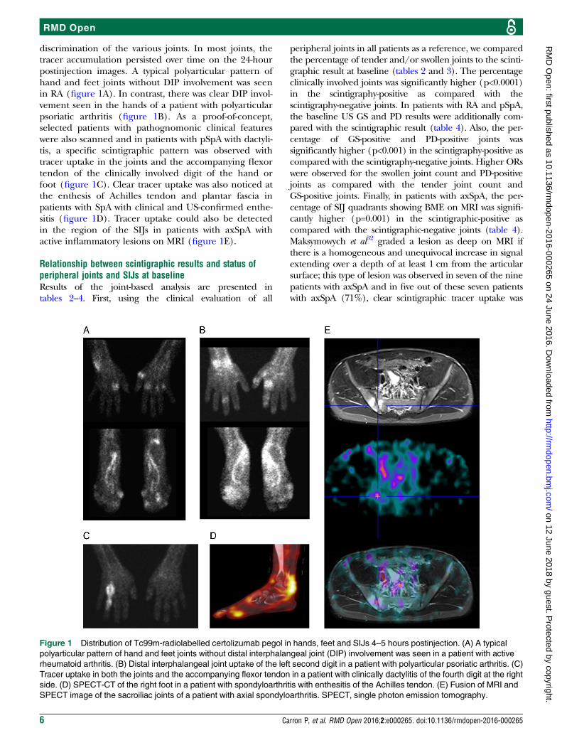
discrimination of the various joints. In most joints, thetracer accumulation persisted over time on the 24-hourpostinjection images. A typical polyarticular pattern ofhand and feet joints without DIP involvement was seenin RA (figure 1A). In contrast, there was clear DIP invol-vement seen in the hands of a patient with polyarticularpsoriatic arthritis (figure 1B). As a proof-of-concept,selected patients with pathognomonic clinical featureswere also scanned and in patients with pSpA with dactyli-tis, a specific scintigraphic pattern was observed withtracer uptake in the joints and the accompanying flexortendon of the clinically involved digit of the hand orfoot (figure 1C). Clear tracer uptake was also noticed atthe enthesis of Achilles tendon and plantar fascia inpatients with SpA with clinical and US-confirmed enthe-sitis (figure 1D). Tracer uptake could also be detectedin the region of the SIJs in patients with axSpA withactive inflammatory lesions on MRI (figure 1E).
Relationship between scintigraphic results and status ofperipheral joints and SIJs at baselineResults of the joint-based analysis are presented intables 2–4. First, using the clinical evaluation of all
peripheral joints in all patients as a reference, we comparedthe percentage of tender and/or swollen joints to the scinti-graphic result at baseline (tables 2 and 3). The percentageclinically involved joints was significantly higher (p<0.0001)in the scintigraphy-positive as compared with thescintigraphy-negative joints. In patients with RA and pSpA,the baseline US GS and PD results were additionally com-pared with the scintigraphic result (table 4). Also, the per-centage of GS-positive and PD-positive joints wassignificantly higher (p<0.001) in the scintigraphy-positive ascompared with the scintigraphy-negative joints. Higher ORswere observed for the swollen joint count and PD-positivejoints as compared with the tender joint count andGS-positive joints. Finally, in patients with axSpA, the per-centage of SIJ quadrants showing BME on MRI was signifi-cantly higher (p=0.001) in the scintigraphic-positive ascompared with the scintigraphic-negative joints (table 4).Maksymowych et al32 graded a lesion as deep on MRI ifthere is a homogeneous and unequivocal increase in signalextending over a depth of at least 1 cm from the articularsurface; this type of lesion was observed in seven of the ninepatients with axSpA and in five out of these seven patientswith axSpA (71%), clear scintigraphic tracer uptake was
Figure 1 Distribution of Tc99m-radiolabelled certolizumab pegol in hands, feet and SIJs 4–5 hours postinjection. (A) A typical
polyarticular pattern of hand and feet joints without distal interphalangeal joint (DIP) involvement was seen in a patient with active
rheumatoid arthritis. (B) Distal interphalangeal joint uptake of the left second digit in a patient with polyarticular psoriatic arthritis. (C)
Tracer uptake in both the joints and the accompanying flexor tendon in a patient with clinically dactylitis of the fourth digit at the right
side. (D) SPECT-CT of the right foot in a patient with spondyloarthritis with enthesitis of the Achilles tendon. (E) Fusion of MRI and
SPECT image of the sacroiliac joints of a patient with axial spondyloarthritis. SPECT, single photon emission tomography.
6 Carron P, et al. RMD Open 2016;2:e000265. doi:10.1136/rmdopen-2016-000265
RMD Open
on 12 June 2018 by guest. Protected by copyright.
http://rmdopen.bm
j.com/
RM
D O
pen: first published as 10.1136/rmdopen-2016-000265 on 24 June 2016. D
ownloaded from
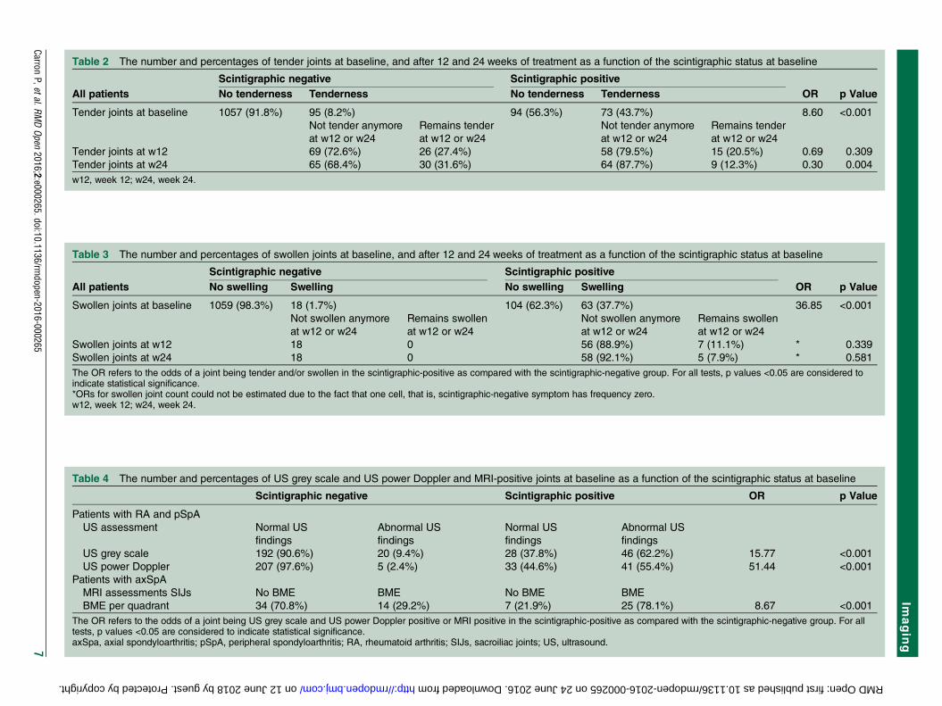
Table 4 The number and percentages of US grey scale and US power Doppler and MRI-positive joints at baseline as a function of the scintigraphic status at baseline
Scintigraphic negative Scintigraphic positive OR p Value
Patients with RA and pSpA
US assessment Normal US
findings
Abnormal US
findings
Normal US
findings
Abnormal US
findings
US grey scale 192 (90.6%) 20 (9.4%) 28 (37.8%) 46 (62.2%) 15.77 <0.001
US power Doppler 207 (97.6%) 5 (2.4%) 33 (44.6%) 41 (55.4%) 51.44 <0.001
Patients with axSpA
MRI assessments SIJs No BME BME No BME BME
BME per quadrant 34 (70.8%) 14 (29.2%) 7 (21.9%) 25 (78.1%) 8.67 <0.001
The OR refers to the odds of a joint being US grey scale and US power Doppler positive or MRI positive in the scintigraphic-positive as compared with the scintigraphic-negative group. For alltests, p values <0.05 are considered to indicate statistical significance.axSpa, axial spondyloarthritis; pSpA, peripheral spondyloarthritis; RA, rheumatoid arthritis; SIJs, sacroiliac joints; US, ultrasound.
Table 3 The number and percentages of swollen joints at baseline, and after 12 and 24 weeks of treatment as a function of the scintigraphic status at baseline
All patients
Scintigraphic negative Scintigraphic positive
OR p ValueNo swelling Swelling No swelling Swelling
Swollen joints at baseline 1059 (98.3%) 18 (1.7%) 104 (62.3%) 63 (37.7%) 36.85 <0.001
Not swollen anymore
at w12 or w24
Remains swollen
at w12 or w24
Not swollen anymore
at w12 or w24
Remains swollen
at w12 or w24
Swollen joints at w12 18 0 56 (88.9%) 7 (11.1%) * 0.339
Swollen joints at w24 18 0 58 (92.1%) 5 (7.9%) * 0.581
The OR refers to the odds of a joint being tender and/or swollen in the scintigraphic-positive as compared with the scintigraphic-negative group. For all tests, p values <0.05 are considered toindicate statistical significance.*ORs for swollen joint count could not be estimated due to the fact that one cell, that is, scintigraphic-negative symptom has frequency zero.w12, week 12; w24, week 24.
Table 2 The number and percentages of tender joints at baseline, and after 12 and 24 weeks of treatment as a function of the scintigraphic status at baseline
Scintigraphic negative Scintigraphic positive
OR p ValueAll patients No tenderness Tenderness No tenderness Tenderness
Tender joints at baseline 1057 (91.8%) 95 (8.2%) 94 (56.3%) 73 (43.7%) 8.60 <0.001
Not tender anymore
at w12 or w24
Remains tender
at w12 or w24
Not tender anymore
at w12 or w24
Remains tender
at w12 or w24
Tender joints at w12 69 (72.6%) 26 (27.4%) 58 (79.5%) 15 (20.5%) 0.69 0.309
Tender joints at w24 65 (68.4%) 30 (31.6%) 64 (87.7%) 9 (12.3%) 0.30 0.004
w12, week 12; w24, week 24.
CarronP,etal.RM
DOpen
2016;2:e000265.doi:10.1136/rmdopen-2016-000265
7
Imagin
g
on 12 June 2018 by guest. Protected by copyright. http://rmdopen.bmj.com/ RMD Open: first published as 10.1136/rmdopen-2016-000265 on 24 June 2016. Downloaded from
observed in the same quadrant of the SIJs, where theextended lesion was also located. Interestingly, in onepatients with AS with complete fusion of the SIJs (bilateralgrade 4 sacroiliitis on X-ray), who was suffering from periph-eral arthritis (and hence was included in the pSpA part ofthe study), there was an absence of BME of the SIJs onMRI. In this patient we could also not detect tracer uptakeon scintigraphy. Although this is only a solitary case, itmight suggest that in vivo detection of TNF correlates withactive inflammatory lesions on MRI and not with structuraldamage.
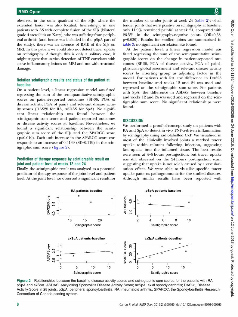
Relation scintigraphic results and status of the patient atbaselineOn a patient level, a linear regression model was fittedregressing the sum of the semiquantitative scintigraphicscores on patient-reported outcomes (SF-36, PGA ofdisease activity, PGA of pain) and relevant disease activ-ity scores (DAS28 for RA, ASDAS for SpA). No signifi-cant linear relationship was found between thescintigraphic sum score and patient-reported outcomesor disease activity scores at baseline. Nevertheless, wefound a significant relationship between the scinti-graphic sum score of the SIJs and the SPARCC score(p=0.010). Each unit increase in the SPARCC score cor-responds to an increase of 0.4139 (SE=0.119) in the scin-tigraphic sum score (figure 2).
Prediction of therapy response by scintigraphic result onjoint and patient level at weeks 12 and 24Finally, the scintigraphic result was analysed as a potentialpredictor of therapy response of the joint level and patientlevel. At the joint level, we observed a significant result for
the number of tender joints at week 24 (table 2): of alltender joints that were positive on scintigraphy at baseline,only 11.9% remained painful at week 24, compared with26.5% in the scintigraphy-negative joints (OR=0.38;p=0.030). Results for swollen joints are summarised intable 3; no significant correlation was found.At the patient level, a linear regression model was
fitted regressing the sum of the semiquantitative scinti-graphic scores on the change in patient-reported out-comes (SF-36, PGA of disease activity, PGA of pain),physician global assessment and relevant disease activityscores by inserting group as adjusting factor in themodel. For patients with RA, the difference in DAS28between baseline and weeks 12 and 24 was used andregressed on the scintigraphic sum score. For patientswith SpA, the difference in ASDAS between baselineand weeks 12 and 24 was used and regressed on the scin-tigraphic sum score. No significant relationships werefound.
DISCUSSIONWe performed a proof-of-concept study on patients withRA and SpA to detect in vivo TNF-α-driven inflammationby scintigraphy using radiolabelled CZP. We visualised inmost of the clinically involved joints a marked traceruptake within minutes following injection, suggestingfast uptake into the inflamed tissue. The best resultswere seen at 4–6 hours postinjection, but tracer uptakewas still observed on the 24 hours postinjection scan,suggesting that uptake is not solely caused by a vasculari-sation effect. We were able to visualise specific traceruptake patterns pathognomonic for the studied diseases.Although similar results have been reported with
Figure 2 Relationships between the baseline disease activity scores and scintigraphic sum scores for the patients with RA,
pSpA and axSpA. ASDAS, Ankylosing Spondylitis Disease Activity Score; axSpA, axial spondyloarthritis; DAS28, Disease
Activity Score in 28 joints; pSpA, peripheral spondyloarthritis; RA, rheumatoid arthritis; SPARCC, the Spondyloarthritis Research
Consortium of Canada scoring system.
8 Carron P, et al. RMD Open 2016;2:e000265. doi:10.1136/rmdopen-2016-000265
RMD Open
on 12 June 2018 by guest. Protected by copyright.
http://rmdopen.bm
j.com/
RM
D O
pen: first published as 10.1136/rmdopen-2016-000265 on 24 June 2016. D
ownloaded from
radiolabelled adalimumab in patients with RA,33 34 thisis—to the best of our knowledge—the first study to eval-uate immunoscintigraphic patterns in patients with dif-ferent subtypes of SpA by visualising enthesitis, dactylitisand sacroiliitis.Second, in patients with peripheral joint involvement,
we could establish a good correlation between the clini-cal evaluation and conventional imaging of the indivi-dual joints, and the tracer uptake on scintigraphy.Interestingly, this correlation was strongest for moreobjective signs of inflammation: higher ORs wereobserved for the swollen joint count and PD-positivejoints as compared with the tender joint count andGS-positive joints. At the patient level, no significant cor-relation was found between the scintigraphic sum scoreand global disease activity scores or relevant patient-reported outcomes. In patients with axSpA, the scinti-graphic findings correlated well with BME on MRIof the SIJs, which is the item that is required in theASAS definition of a positive MRI,35 and is one of theanchors of the ASAS classification criteria for axSpA.Nevertheless, the interpretation of a ‘positive MRI’ inthe context of SpA remains difficult, definitely when thepresence of BME is subtle or when there may be othercauses for the observed BME, such as mechanical stress.As a consequence, there is still an unmet need to deter-mine whether observed BME is caused by underlyingcytokine-driven inflammation in patients with axSpA. Atpresent, there is no agreement on a precise definition ofthe minimum size (area) of BME which is necessary tobe defined as ‘positive’. There is an impression that thepresence of so-called ‘deep’ BME lesions on MRI of theSIJs, defined as a homogeneous and unequivocalincrease in signal extending over at least 1 cm from thearticular surface on STIR images, would be more sugges-tive of axSpA. In our study, taking only these extendedlesions on MRI of the SIJs into account, better correla-tions between scintigraphy and MRI were found. Finally,at the patient level, there was a significant relationbetween the scintigraphic sum score of the SIJs and theSPARCC scoring system.Third, we looked whether the baseline scintigraphic
detection of TNF could predict a therapeutic responseto CZP therapy after 12–24 weeks. We need to emphasisethat this study was not designed or powered to predicttherapy response by baseline scintigraphic results.Nevertheless, we could demonstrate a significant predic-tive value of the immunoscintigraphy at the joint levelwith regard to the tender joint count: tender joints withuptake at baseline had a significantly higher probabilityof not being painful after 24 weeks of treatment. Thelack of predictive value for the other joint assessments,as well as for the global disease activity scores at thepatient level, could probably be explained by the factthat the study included only small numbers of patientswith heterogeneous inflammatory diseases. These preli-minary findings are, however, interesting because a posi-tive scintigraphic result might indicate that joint
tenderness at baseline is TNF-driven, and this resultcould serve as a more objective measurement tool fortender joints. Surprisingly, only two prospective cohortstudies in RA have assessed the use of imaging techni-ques to predict response to anti-TNF therapy. Ellegaardand colleagues measured US Doppler activity and clini-cal parameters at baseline to predict which patientswould benefit from treatment, assessed by treatment per-sistence at 1 year. They identified US Doppler activity tobe the only baseline parameter able to predict treatmentpersistence (p=0.024); baseline clinical measures,including tender and swollen joint counts, CRP, DAS28,and HAQ showed no significant association.36 Elzingaand colleagues used changes in positron emission tomo-graphy (PET) uptake 2 weeks after treatment to predictfuture treatment response, according to DAS28. A signif-icant correlation was seen between the changes in PETactivity at 2 weeks, and DAS28 at 14 and 22 weeks aftertreatment (r 0.62, p<0.05; r 0.65, p<0.01, respectively).37
In the past, only few attempts in molecular imagingusing radiolabelled monoclonal antibodies in rheumaticdiseases were made, and the earliest experiences andclinical context are excellently summarised by Malviyaet al.38 Our study is the first using a radiolabelling proce-dure with CZP, a PEGylated Fab fragment directedagainst TNF-α. One of the reasons for choosing CZP as atracer was the observation made in an animal model forarthritis using biofluorescence imaging: a greater ratioof penetration and more prolonged duration of expo-sure in inflamed versus normal tissue was described forCZP compared with adalimumab and infliximab. Onepossible explanation could be the link with PEG inCZP.39
We acknowledge that our study has several limitations,both regarding the scintigraphic technique, as well asconcerning the type of patients that was studied. Barreraet al33 published encouraging findings with radiolabelledadalimumab in 10 patients with active RA. A subset ofpatients underwent repeat imaging following administra-tion of a therapeutic dose of cold antibody. Based on thiscompetition study, the authors suggested a partial specifictargeting of TNF-α by Tc99m-labelled adalimumab.These findings were in agreement with Roimicher et al.34
We decided not to include a competition study andprioritised to first have an estimation of the radiationburden before exposing volunteering patients twice.40
We also did not perform a second immunoscintigrapyafter 12 or 24 weeks of treatment in order to evaluate thechange in tracer uptake over time. We first wanted toestablish a proof-of-concept with this new radiopharma-ceutical before endorsing more complex follow-up scinti-graphic procedures. Nevertheless, it was Conti et al41 whoshowed a positive predictive role of 99mTc-infliximabscintigraphy in therapy decision-making for patients withrefractory monoarthritis who were given intra-articularinfliximab treatment by comparing the pretherapy andpost-therapy target-to-background ratio from affectedjoints.
Carron P, et al. RMD Open 2016;2:e000265. doi:10.1136/rmdopen-2016-000265 9
Imaging
on 12 June 2018 by guest. Protected by copyright.
http://rmdopen.bm
j.com/
RM
D O
pen: first published as 10.1136/rmdopen-2016-000265 on 24 June 2016. D
ownloaded from
With regard to patient selection, this was obviously aproof-of-concept study, which included only limitednumbers of patients with different inflammatory rheu-matic diseases. We did not include a non-inflammatorycontrol group; as a consequence, we cannot excludethat the observed tracer uptake could be an aspecificphenomenon, although the good correlation with theclinical evaluation and conventional imaging resultswould argue against this. The low number of patients ineach disease subgroup evidently does not allow us tomake conclusions regarding prediction of clinicalresponse in case of a positive scintigraphy; nevertheless,the finding that individual painful joints that werescintigraphy-positive had a higher odds of becoming nottender after treatment with certolizumab could be a pro-mising find. Indeed, since treat-to-target principles havefound their way in the daily management of patientswith RA,42 a high number of tender joints might be oneof the triggers to change the therapeutic strategy: infor-mation as to whether the observed pain pattern isrelated to in vivo expression of a culprit cytokine couldpotentially avoid overtreatment with biologicals in indivi-dual cases where pain is driven by other pathophysiologi-cal mechanisms.
CONCLUSIONIn conclusion, we demonstrated that it is safe and feasi-ble to perform scintigraphy with radiolabelled CZP inpatients with different types of inflammatory arthritis,whereby specific joint involvement patterns could berecognised. Future research should confirm these preli-minary results, specifically with regard to the potentialto predict clinical response to a biological treatment tar-geting TNF. If confirmed, the technique could be a steptowards personalised medicine, where each patientreceives the right drug and the right intensity of treat-ment for as long as needed; it could allow the selectionof patients for a specific therapy in a much morerational way than the current ‘trial and error’ approach.In particular, future studies should address if a prether-apy scintigraphic approach with a radiopharmaceuticaltargeting TNF identifies the presence of the target cyto-kine in the inflammatory lesion and if positive, whetherthese patients with clear uptake of the anti-TNF tracerwould respond better to anti-TNF therapy as comparedwith strategies targeting other inflammatory pathways.This might be a crucial step in enhancing the safe andcost-effective use of expensive biological treatment byavoiding exposure of non-responders to treatments suchas anti-TNF therapy.
Author affiliations1Department of Rheumatology, Ghent University Hospital, Ghent, Belgium2Department of Nuclear Medicine, Ghent University Hospital, Ghent, Belgium3Department of Radiopharmacy, Ghent University, Ghent, Belgium4Department of Rheumatology, Ghent University Hospital, Ghent, Belgium5VIB Inflammation Research Center, Ghent University, Ghent, Belgium6Department of Radiology, Ghent University Hospital, Ghent, Belgium
Funding This research was carried out as an Investigator Initiated Studyfunded by UCB Pharma SA. UCB Pharma provided the certolizumab pegolvials and financial support for this clinical trial.
Disclaimer The presented work was initiated, conducted and performedindependently from UCB Pharma.
Competing interests None declared.
Patient consent Obtained.
Ethics approval Ethics Committee University Hospital of Ghent.
Provenance and peer review Not commissioned; externally peer reviewed.
Data sharing statement No additional data are available.
Open Access This is an Open Access article distributed in accordance withthe Creative Commons Attribution Non Commercial (CC BY-NC 4.0) license,which permits others to distribute, remix, adapt, build upon this work non-commercially, and license their derivative works on different terms, providedthe original work is properly cited and the use is non-commercial. See: http://creativecommons.org/licenses/by-nc/4.0/
REFERENCES1. Myasoedova E, Crowson CS, Kremers HM, et al. Is the incidence of
rheumatoid arthritis rising? Results from Olmsted County, Minnesota,1955–2007. Arthritis Rheum 2010;62:1576–82.
2. Bakland G, Nossent HC. Epidemiology of spondyloarthritis: a review.Curr Rheumatol Rep 2013;15:351.
3. Lundström E, Källberg H, Alfredsson L, et al. Gene-environmentinteraction between the DRB1 shared epitope and smoking in therisk of anti-citrullinated protein antibody-positive rheumatoid arthritis:all alleles are important. Arthritis Rheum 2009;60:1597–603.
4. Maciejewska Rodrigues H, Jüngel A, Gay RE, et al. Innate immunity,epigenetics and autoimmunity in rheumatoid arthritis. Mol Immunol2009;47:12–18.
5. Tak PP, Taylor PC, Breedveld FC, et al. Decrease in cellularity andexpression of adhesion molecules by anti-tumor necrosis factoralpha monoclonal antibody treatment in patients with rheumatoidarthritis. Arthritis Rheum 1996;39:1077–81.
6. Baeten D, Kruithof E, Van den Bosch F, et al. Immunomodulatoryeffects of anti-tumor necrosis factor alpha therapy on synovium inspondylarthropathy: histologic findings in eight patients from anopen-label pilot study. Arthritis Rheum 2001;44:186–95.
7. Yeremenko N, Zwerina K, Rigter G, et al. Tumor necrosis factor andinterleukin-6 differentially regulate Dkk-1 in the inflamed arthriticjoint. Arthritis Rheum 2015;67:2071–5.
8. Thalayasingam N, Isaacs JD. Anti-TNF therapy. Best Pract Res ClinRheumatol 2011;25:549–67.
9. Landewé R, Braun J, Deodhar A, et al. Efficacy of certolizumabpegol on signs and symptoms of axial spondyloarthritis includingankylosing spondylitis: 24-week results of a double-blind randomisedplacebo-controlled Phase 3 study. Ann Rheum Dis 2014;73:39–47.
10. Mease PJ, Fleischmann R, Deodhar AA, et al. Effect of certolizumabpegol on signs and symptoms in patients with psoriatic arthritis:24-week results of a phase 3 double-blind randomisedplacebo-controlled study (RAPID-PsA). Ann Rheum Dis2014;73:48–55.
11. Keystone E, Heijde Dv, Mason D Jr, et al. Certolizumab pegol plusmethotrexate is significantly more effective than placebo plusmethotrexate in active rheumatoid arthritis: findings of afifty-two-week, phase III, multicenter, randomized, double-blind,placebo-controlled, parallel-group study. Arthritis Rheum2008;58:3319–29.
12. Weir MD, Xu HH, Simon CG Jr. Strong calcium phosphatecement-chitosan-mesh construct containing cell-encapsulatinghydrogel beads for bone tissue engineering. J Biomed Mater Res A2006;77:487–96.
13. Reich K, Ortonne JP, Gottlieb AB, et al. Successful treatment ofmoderate to severe plaque psoriasis with the PEGylated Fab’certolizumab pegol: results of a phase II randomized,placebo-controlled trial with a re-treatment extension. Br J Dermatol2012;167:180–90.
14. Schreiber S. Certolizumab pegol for the treatment of Crohn’sdisease. Therap Adv Gastroenterol 2011;4:375–89.
15. van der Heijde D, Dijkmans B, Geusens P, et al. Efficacy and safetyof infliximab in patients with ankylosing spondylitis: results of arandomized, placebo-controlled trial (ASSERT). Arthritis Rheum2005;52:582–91.
10 Carron P, et al. RMD Open 2016;2:e000265. doi:10.1136/rmdopen-2016-000265
RMD Open
on 12 June 2018 by guest. Protected by copyright.
http://rmdopen.bm
j.com/
RM
D O
pen: first published as 10.1136/rmdopen-2016-000265 on 24 June 2016. D
ownloaded from
16. Davis JC, van der Heijde DM, Braun J, et al. Sustained durabilityand tolerability of etanercept in ankylosing spondylitis for 96 weeks.Ann Rheum Dis 2005;64:1557–62.
17. van der Heijde D, Kivitz A, Schiff MH, et al. Efficacy and safety ofadalimumab in patients with ankylosing spondylitis: results of amulticenter, randomized, double-blind, placebo-controlled trial.Arthritis Rheum 2006;54:2136–46.
18. Inman RD, Davis JC Jr, Heijde Dv, et al. Efficacy and safety ofgolimumab in patients with ankylosing spondylitis: results of arandomized, double-blind, placebo-controlled, phase III trial. ArthritisRheum 2008;58:3402–12.
19. Maini R, St Clair EW, Breedveld F, et al. Infliximab (chimericanti-tumour necrosis factor alpha monoclonal antibody) versusplacebo in rheumatoid arthritis patients receiving concomitantmethotrexate: a randomised phase III trial. ATTRACT Study Group.Lancet 1999;354:1932–9.
20. Moreland LW, Baumgartner SW, Schiff MH, et al. Treatment ofrheumatoid arthritis with a recombinant human tumor necrosis factorreceptor (p75)-Fc fusion protein. N Engl J Med 1997;337:141–7.
21. Weinblatt ME, Keystone EC, Furst DE, et al. Adalimumab, a fullyhuman anti-tumor necrosis factor alpha monoclonal antibody, for thetreatment of rheumatoid arthritis in patients taking concomitantmethotrexate: the ARMADA trial. Arthritis Rheum 2003;48:35–45.
22. Keystone E, Genovese MC, Klareskog L, et al. Golimumab inpatients with active rheumatoid arthritis despite methotrexatetherapy: 52-week results of the GO-FORWARD study. Ann RheumDis 2010;69:1129–35.
23. Ulfgren AK, Gröndal L, Lindblad S, et al. Interindividual andintra-articular variation of proinflammatory cytokines in patients withrheumatoid arthritis: potential implications for treatment. Ann RheumDis 2000;59:439–47.
24. Arnett FC, Edworthy SM, Bloch DA, et al. The AmericanRheumatism Association 1987 revised criteria for the classification ofrheumatoid arthritis. Arthritis Rheum 1988;31:315–24.
25. Aletaha D, Neogi T, Silman AJ, et al. 2010 Rheumatoid arthritisclassification criteria: an American College of Rheumatology/European League Against Rheumatism collaborative initiative.Arthritis Rheum 2010;62:2569–81.
26. Rudwaleit M, van der Heijde D, Landewé R, et al. The developmentof Assessment of SpondyloArthritis international Societyclassification criteria for axial spondyloarthritis (part II): validationand final selection. Ann Rheum Dis 2009;68:777–83.
27. Rudwaleit M, van der Heijde D, Landewé R, et al. The Assessmentof SpondyloArthritis International Society classification criteria forperipheral spondyloarthritis and for spondyloarthritis in general. AnnRheum Dis 2011;70:25–31.
28. Backhaus M, Burmester GR, Gerber T, et al. Guidelines formusculoskeletal ultrasound in rheumatology. Ann Rheum Dis2001;60:641–9.
29. Wakefield RJ, Balint PV, Szkudlarek M, et al. Musculoskeletalultrasound including definitions for ultrasonographic pathology.J Rheumatol 2005;32:2485–7.
30. Szkudlarek M, Court-Payen M, Jacobsen S, et al. Interobserveragreement in ultrasonography of the finger and toe joints inrheumatoid arthritis. Arthritis Rheum 2003;48:955–62.
31. Scheel AK, Hermann KG, Kahler E, et al. A novel ultrasonographicsynovitis scoring system suitable for analyzing finger jointinflammation in rheumatoid arthritis. Arthritis Rheum2005;52:733–43.
32. Maksymowych WP, Inman RD, Salonen D, et al. Spondyloarthritisresearch Consortium of Canada magnetic resonance imaging indexfor assessment of sacroiliac joint inflammation in ankylosingspondylitis. Arthritis Rheum 2005;53:703–9.
33. Barrera P, Oyen WJ, Boerman OC, et al. Scintigraphic detection oftumour necrosis factor in patients with rheumatoid arthritis. AnnRheum Dis 2003;62:825–8.
34. Roimicher L, Lopes FP, de Souza SA, et al. (99m)Tc-anti-TNF-αscintigraphy in RA: a comparison pilot study with MRI and clinicalexamination. Rheumatology (Oxford) 2011;50:2044–50.
35. Rudwaleit M, Jurik AG, Hermann KG, et al. Defining activesacroiliitis on magnetic resonance imaging (MRI) forclassification of axial spondyloarthritis: a consensualapproach by the ASAS/OMERACT MRI group. Ann Rheum Dis2009;68:1520–7.
36. Ellegaard K, Christensen R, Torp-Pedersen S, et al. UltrasoundDoppler measurements predict success of treatment withanti-TNF-α drug in patients with rheumatoidarthritis: a prospective cohort study. Rheumatology (Oxford)2011;50:506–12.
37. Elzinga EH, van der Laken CJ, Comans EF, et al. 18F-FDG PET asa tool to predict the clinical outcome of infliximab treatment ofrheumatoid arthritis: an explorative study. J Nucl Med2011;52:77–80.
38. Malviya G, Conti F, Chianelli M, et al. Molecular imaging ofrheumatoid arthritis by radiolabelled monoclonal antibodies: newimaging strategies to guide molecular therapies. Eur J Nucl Med MolImaging 2010;37:386–98.
39. Palframan R, Airey M, Moore A, et al. Use of biofluorescenceimaging to compare the distribution of certolizumab pegol,adalimumab, and infliximab in the inflamed paws of micewith collagen-induced arthritis. J Immunol Methods 2009;348:36–41.
40. Lambert B, Carron P, D’Asseler Y, et al. 99mTc labelled S-HYNICcertolizumab pegol for selecting patients for anti-TNF alphatreatment: a biodistribution and dosimetric study. Eur J Nucl MedMol Imaging 2013;40 2):S392.
41. Conti F, Malviya G, Ceccarelli F, et al. Role of scintigraphy with99mTc-infliximab in predicting the response of intraarticular infliximabtreatment in patients with refractory monoarthritis. Eur J Nucl MedMol Imaging 2012;39:1339–47.
42. Smolen JS, Landewé R, Breedveld FC, et al. EULARrecommendations for the management of rheumatoid arthritis withsynthetic and biological disease-modifying antirheumatic drugs:2013 update. Ann Rheum Dis 2014;73:492–509.
Carron P, et al. RMD Open 2016;2:e000265. doi:10.1136/rmdopen-2016-000265 11
Imaging
on 12 June 2018 by guest. Protected by copyright.
http://rmdopen.bm
j.com/
RM
D O
pen: first published as 10.1136/rmdopen-2016-000265 on 24 June 2016. D
ownloaded from