Embed Size (px)
Citation preview
PART I - 291
SCINTIGRAPHY OF GASTRIC EMPTYING
Scintigraphy of Gastric Emptying
MWJ Versleijen, Antoni van Leeuwenhoek, Amsterdam
1. IntroductionThe rate at which the stomach empties can be determined following ingestion of a meal labelled with a radiopharmaceutical that maintains a stable binding within the gastric environment. The investigation usually involves a solid test meal (e.g. a pancake) although the test meal may include a liquid component if this is indicated.Anatomically, the stomach is divided into three sections, the fundus, corpus and antrum. Physiologically, the stomach is divided into two sections, the proximal and distal stomach. The proximal stomach serves as a reservoir for solid and liquid foods and controls gastric emptying of liquids. The distal stomach contracts with a frequency of three contractions a minute, whereupon the food is pulverized and mixed with gastric secretions in coordination with pyloric closure. This is followed by gastric emptying into the duodenum. The rate of gastric emptying depends on several factors such as shape, volume, composition and the calorifi c value of the ingested meal. Feedback on caloric content via chemical receptors in the small intestine also plays an important role in gastric motor function and standardization of the test meal therefore is of key importance.Gastric emptying following a solid meal is characterised by a pre-emptying phase, or lag phase, and a subsequent linear emptying phase. During the lag phase, food is processed prior to the actual gastric emptying. Gastric emptying of a neutral, isomolar and non-caloric liquid test meal is monoexponential. The emptying phase becomes more linear as the calorifi c value of the test meal increases whilst combining a solid and liquid test meal results in delayed gastric emptying of the liquid test meal. Gastric emptying of the solid test meal is less infl uenced by a liquid test meal (including non-caloric liquids) and this should be taken into account with regard to standardizing and the determination of normal values.It has been clearly shown that gastric emptying of solid test meals in healthy pre-menopausal females is slower than that of male subjects, irrespective of the phase of the menstrual cycle. A separate set of normal values for this group is therefore recommended.
2. MethodologyThis guideline is based on available scientifi c literature on the subject, the previous guideline (Aanbevelingen Nucleaire Geneeskunde 2007), international guidelines from EANM and/or SNMMI if available and applicable to the Dutch situation.
3. Indication Suspected gastric emptying disorders (delayed, accelerated or altered empting patterns) such as with:a. Upper abdominal symptoms in patients with known pathology which may include
gastric emptying disorders such as scleroderma, diabetes mellitus, hypothyroidism, amyloidosis and myotonic dystrophy. Patients with abnormal emptying as a result of
Deel I_D.indd 291 27-12-16 14:17

PART I - 292
SCINTIGRAPHY OF GASTRIC EMPTYING
diabetes mellitus form the largest group (50% of these patients show delayed gastric emptying of solid food). Gastric emptying studies can also be used to assess the therapeutic effect of prokinetic agents (drugs which stimulate gastric contractions, e.g. metoclopramide, cisapride). See table 1 for a summary of causes related to delayed gastric emptying of solid food.
b. Upper abdominal symptoms following gastric surgery, such as antirefl ux surgery, vagotomy and gastric resection. The number of patients in this group is continually decreasing since effi cient acid inhibitory medication has been introduced (e.g. ranitidine, omeprazol). The rate of gastric emptying of liquids increases following vagotomy without pyloroplasty whilst the rate for solid food decreases slightly. Rapid gastric emptying of liquid and solid food (‘dumping’) can occur following antrectomy or vagotomy in combination with pyloroplasty. See table 2 for causes related to an increased rate of gastric emptying.
c. Severe gastroesophageal refl ux for which surgical treatment is considered.d. Patients with functional dyspepsia in which no gastric or intestinal abnormalities can
be found on ultrasound or gastroscopy.e. To determine whether generalized motility defects exist in patients with severe
constipation (a result of idiopathic chronic pseudo-obstruction).f. Upper abdominal symptoms related to the chronic use of medication causing
inhibition of motility as a side effect. Examples of medications known to cause inhibition of motility include opioid preparations (e.g. morphine and codeine) but also dopamine agonists (L-dopa).
4. Relation to other diagnostic proceduresa. Scintigraphy is the most reproducible, reliable and sensitive technique known to date
for investigating gastric emptying. b. The advantages and disadvantages of alternative methods are described briefl y
below:Radiological investigations with bariumAdvantage: local motility defects and anatomical defects can also be evaluated. Disadvantages: Reliable gastric emptying curves can only be obtained at the cost of an unacceptably high radiation dose to the patient; inaccurate quantifi cation.UltrasoundAdvantage: no radiation involved. Disadvantage: can only be carried out using liquids; inaccurate quantifi cation. Intubation methodsAdvantage: gastric secretion can also be measured. Disadvantage: diffi cult to perform; only possible using liquids; alters normal physiology.Gastric impedance studyAlthough this investigation has provided promising results, the technique is still far from perfect.Indirect methods (e.g. measurement of paracetamol absorption rates)Advantage: no radiation involved; reasonably reliable measurements. Disadvantage: only suitable for liquids; does not provide accurate information about the lag phase.
Deel I_D.indd 292 27-12-16 14:17
PART I - 293
SCINTIGRAPHY OF GASTRIC EMPTYING
5. Medical information necessary for planninga. Pattern and duration of symptoms, relationship to type of food (e.g. solids or liquids).b. Current medication.c. Details of any prior gastrointestinal surgery.d. Results of relevant previous investigations of the gastrointestinal tract.e. History of conditions associated with gastric emptying disorders (e.g. scleroderma,
diabetes mellitus, hypothyroidism, amyloidosis and myotonic dystrophy).
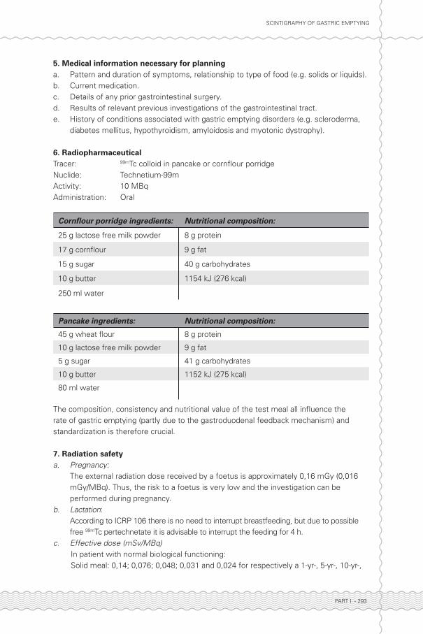
6. RadiopharmaceuticalTracer: 99mTc colloid in pancake or cornfl our porridgeNuclide: Technetium-99mActivity: 10 MBqAdministration: Oral
Cornfl our porridge ingredients: Nutritional composition:
25 g lactose free milk powder 8 g protein
17 g cornfl our 9 g fat
15 g sugar 40 g carbohydrates
10 g butter 1154 kJ (276 kcal)
250 ml water
Pancake ingredients: Nutritional composition:
45 g wheat fl our 8 g protein
10 g lactose free milk powder 9 g fat
5 g sugar 41 g carbohydrates
10 g butter 1152 kJ (275 kcal)
80 ml water
The composition, consistency and nutritional value of the test meal all infl uence the rate of gastric emptying (partly due to the gastroduodenal feedback mechanism) and standardization is therefore crucial.
7. Radiation safetya. Pregnancy:
The external radiation dose received by a foetus is approximately 0,16 mGy (0,016 mGy/MBq). Thus, the risk to a foetus is very low and the investigation can be performed during pregnancy.
b. Lactation: According to ICRP 106 there is no need to interrupt breastfeeding, but due to possible free 99mTc pertechnetate it is advisable to interrupt the feeding for 4 h.
c. Effective dose (mSv/MBq) In patient with normal biological functioning: Solid meal: 0,14; 0,076; 0,048; 0,031 and 0,024 for respectively a 1-yr-, 5-yr-, 10-yr-,
Deel I_D.indd 293 27-12-16 14:17

PART I - 294
SCINTIGRAPHY OF GASTRIC EMPTYING
15-yr old and an adult patient. Liquid meal: 0,11; 0,062; 0,039; 0,025 and 0,019 for respectively a 1-yr-, 5-yr-, 10-yr-,
15-yr old and an adult patient.
8. Patient preparation/essentials for the procedurePatient preparationa. The patient must be nil by mouth for at least 8 h prior to the investigation.b. Any medication which affects gastrointestinal motility (e.g. metoclopramide,
domperidone, cimetidine, parasympatholytics and sympathomimetics) should be discontinued for 3 days before the investigation unless the requesting physician wishes to investigate the effect of these prokinetic agents.
c. Diabetic patients should undergo the investigation in the early morning, after their usual dose of insulin. Given the fact that hyperglycemia can cause gastroparesis, the blood glucose concentration should be measured prior to the investigation. High blood glucose (>10 mmol/l) must be reported.
d. Abstinence of smoking, alcohol and caffeine intake is required for 24 h prior to the investigation.
e. It should be noted whether a female patient is postmenopausal.f. Pre-menopausal women are preferably tested in the fi rst week of their menstrual cycle
(before oestrogen and progesterone peak).
Essentials for the procedurea. Electric hob ring, frying pan, mixing jug, wooden spoon and spatula for preparation of
the test meal.b. Disposable absorbent pad, plate and cutlery.c. For dynamic imaging in sitting position; chair, preferably with adjustable back and arm
rests.
9. Acquisition and processinga. Sequential static imaging or dynamic acquisition may be performed. Sequential static
imaging is preferable since it is more patient friendly and allows for easier, more reliable attenuation correction and reconstruction of a gastric emptying curve.
b. Correction for variation in depth of the test meal within the stomach is necessary since the fundus is situated more posteriorly and the antrum more anteriorly. In sequential static imaging, this correction is preferably achieved by using a double-headed camera, or by performing anterior and posterior images in succession, and subsequently calculating the geometric mean. If a dynamic acquisition is performed, using only the anterior view, the lateral correction method should be used. When the acquisition is complete, a lateral image is obtained for depth correction. In order to obtain this image, it may be necessary to give the patient extra 99mTc colloid (4-8 MBq) in water. It can be useful to move a radioactive reference marker along the patient’s abdominal wall during acquisition. Alternatively, the entire dynamic gastric emptying study can be imaged in the LAO position, thus reducing measurement error.
c. A dual isotope study (using 4 MBq 111In DTPA in 200 ml water or orange juice in addition to the solid test meal) can be carried out if the gastric emptying of a liquid is to be investigated at the same time. It is important that down-scatter from 111In in the
Deel I_D.indd 294 27-12-16 14:17

PART I - 295
SCINTIGRAPHY OF GASTRIC EMPTYING
99mTc window is taken into account.d. Sequential static acquisition: The patient should eat the test meal within fi ve minutes
and then drink a small amount of water to fl ush the oesophagus of any remaining food. Document how much of the test meal is ingested. The fi rst image is obtained immediately post ingestion of the test meal. Subsequent images are obtained every 10-15 min for 2 h. If calculation of late retention values is desired (see paragraph ‘interpretation’), an additional static image at 3 and 4 h postprandial should be performed. A 57Co marker can be placed on the xiphoid and/or ilium to facilitate repositioning and motion correction. The patient is allowed to walk in between the acquisitions. A ROI is drawn around the stomach; the ROI in the fi rst image should include all activity present. Following correction for motion, radioactive decay and attenuation, a curve is generated using, for example, a power exponential function in the form y(t)=1-(1-e-kt). The half-time and the rate of emptying can be determined from this curve.
e. Dynamic acquisition: Anterior images are obtained with the patient positioned in front of the gamma camera sitting in a special chair (in a semi-sitting, comfortable position). The stomach should be as high as possible in the fi eld of view. The pancake is cut into small pieces and fed to the patient; dynamic imaging should begin at the same time as the meal. Make a note of the duration of the meal (t= a min). Also note the time at which the gastric emptying commences. This is when the fi rst activity appears in the small intestine (t = b min). With dynamic imaging, the pre-emptying phase (lag phase) can be calculated and is thus (b-a) minutes.
f. Several suitable frames are added together and regions of internet (ROIs) are then drawn around the stomach and the intestine (where visible). Time-activity curves are generated from the chosen frames and both ROIs in order to evaluate the rate of the gastric emptying. The curve also indicates the time at which gastric emptying commences (where the bowel curve begins to rise). The rate of gastric emptying (K) is calculated over a period of, for example, 40 min from the start of emptying using the following formula:
in which: M (b) = radioactivity in the stomach at time b
M (b+40) = radioactivity in the stomach at time b+40 min
D (b) = radioactivity in the bowel at time b
These measurements should be corrected for radioactive decay. It is also possible to calculate the gradient of the stomach curve and to express this as the caloric emptying rate. If the gastric emptying is not linear, or the curve shows plateau phases, it is better to calculate the T½ (the time at which half of the radioactivity has emptied from the stomach) or retention of activity at certain time points. Down scatter from 111In in the 99mTc window must be corrected for when using a dual isotope test meal.
Camera settings and processing: Energy: 99mTc setting, 140 keV
]/% [ 100 x 4060
x D(b)+M(b)
40)M(b+-M(b) =K hour
Deel I_D.indd 295 27-12-16 14:17
PART I - 296
SCINTIGRAPHY OF GASTRIC EMPTYING
Window: 15-20% Collimator: LEAP Computer: Sequential static imaging: 60 sec anterior and posterior images every 10
to 15 min for 2 h using a 128x128 matrix. If requested, additional images can be obtained after 3 and 4 h. The fi rst image is obtained immediately after ingestion of the meal.
Dynamic imaging: 60 sec frames beginning at the start of the meal and lasting at least
40 min following commencement of emptying or until gastric activity reaches 50% of the total ingested activity, using a 128x128 matrix.
10. Interpretation a. Several outcome parameters can be used to evaluate gastric emptying; pre-emptying
phase (lagphase), emptying half time (T50%), remaining activity at certain time points, emptying rate in percentage per hour, separate emptying rate of the proximal en distal stomach and the caloric emptying rate. Normal values for the above described pancake and porridge are described in table 3, page 299. For calorie-rich foods, the emptying phase follows a linear function of 2-3 kcal per minute.Alternatively, rest activity at certain time points (e.g. 30 min, 1, 2, 3 and 4 h post prandial) have been shown to be very useful for the evaluation of gastric emptying. Table 4, page 299, shows normal retention values that have been published as consensus recommendations in a joint report of the American Neurogastroenterology and Motility Society and the Society of Nuclear Medicine. However, standardization of study conditions and the usage of normal values that apply to the consumed test meal is crucial as normal values strongly depend on the composition, caloric value and the viscosity of the meal. Results can also differ depending on whether the study is carried out in a sitting or lying position.
b. The main source of inaccuracy when processing gastric emptying data is related to intra-gastric transport of the test meal during the acquisition. The fundus is located more posteriorly, and the antrum more anteriorly, which affects the duration of the lag phase in particular, but also the time at which gastric emptying commences. A double-headed camera allows the geometric mean to be determined from simultaneously acquired anterior and posterior images. It is also possible to calculate a geometric mean using a single-headed camera by acquiring several anterior and posterior images at given time points. Another option, although less accurate, is to acquire a lateral image at the end of the study after the patient has been given a small amount of water containing 4 MBq 99mTc colloid. The contour of the patient must be indicated on this image using a radioactive marker. This enables a correction to be made for differences in tissue attenuation between the fundus and the antrum.
c. It is often suffi cient to determine the rate of emptying for a solid test meal – more in line with physiology. In certain circumstances, for example in some diabetic patients, gastric emptying of a solid meal may be abnormal whilst that of a liquid (or semi-solid) meal can be normal. After vagotomy, gastric emptying of liquid components is often too rapid whilst that of solids is delayed or normal. Upper abdominal symptoms may be caused either by too rapid gastric emptying (dumping) or by sluggish gastric emptying.
d. Emptying of solid and liquid food must always be measured in two sessions in
Deel I_D.indd 296 27-12-16 14:17
PART I - 297
SCINTIGRAPHY OF GASTRIC EMPTYING
patients who have undergone partial gastric resection. If done in a combined session using a second radionuclide, the solid meal can be fl ushed out of the remaining part of the stomach along with the liquid.
e. The protocol for gastric emptying studies as described above should be adapted for paediatric patients. The test meal, the position of the patient during the investigation and the duration of the investigation may differ from those in adults. The normal values for a paediatric population will also differ. Though, the rate of gastric emptying can be estimated using the caloric content of the test meal.
f. Diabetic patients must maintain good glycaemic control. High blood glucose levels (>10 mmol/l) have an adverse effect on gastric emptying. When interpreting the results, it is therefore important to know whether or not a patient was hyperglycaemic during the investigation.
g. A gastric emptying study can easily be adapted in order to investigate the motility of the entire gastrointestinal tract. Since this requires images to be obtained several days following ingestion of the test meal, 111In DTPA, 67Ga citrate, 111In labelled plastic particles or 131I labelled cellulose may be used.
11. ReportThe report should include a description of blood glucose levels, the meal content, proportion of test meal ingested and time taken to do so, duration of the pre-emptying period, a description of the scintigrams and the curve, emptying parameters, normal values, and presence or absence of refl ux.
12. Literature• Akkermans LMA, Jacobs F, Smout AJPM, et al. Radionuclide measurement of normal and disturbed
gas¬tric motility. Scand J Gastroenterol 1984;19(suppl 96):19-26.
• Bennink RJ, Peeters M, et al. Comparison of total and compartmental gastric emptying and antral
motility between healthy men and women. Eur J Nucl Med 1998;25:1293-9.
• Chatterton BE, Gastric motility. In: Ell PJ, Gambhir SS, eds. Nuclear Medicine in Clinical Diagnosis and
Treatment, 3rd ed. Edinburgh: Churchill Livingstone; 2004;805-18.
• Collins PJ, Horowitz MB, Shearman DJC, Chatterton BE. Correction for tissue attenuation in
radionuclide gastric emptying studies: A comparison of a lateral image method and a geometric mean
method. Br J Radiol 1984;86:1592-610.
• Edelbroek MAL. Effect of physicochemical factors on gastric emptying and antropyloroduodenal
motility. Proefschrift, Universiteit van Utrecht, 1993.
• Geurts EJC. Hoog selectieve vagotomie. Proefschrift. Universiteit van Utrecht; 1975.
• Knight LC, Parkman HP et al. Delayed gastric emptying and decreased antral contractility in normal
premenopausal women compared with men. Am J Gastroenterol 1997;92:968-75.
• Maurer AH and Parkman HP. Update on gastrointestinal scintigraphy. Semin Nucl Med 2006;36:110-8.
• Phillips W, McMahan C, Lasher J, et al. Anterior, posterior, left anterior oblique and geometric mean
views in gastric emptying studies using a glucose solution. Eur J Nucl Med 1995;22:154-7.
• Siegel JA, Urbain J-L, et al. Biphasic nature of gastric emptying. Gut 1988;29:85-9.
• Smout AJPM, et al, Gastric emptying and postprandial symptoms after Billroth II resection. Surgery
1987;1:27-34.
• Yung BCK, Sostre S. Lag phase in solid gastric emptying: comparison of quantifi cation of physiological
and mathematical defi nitions. J Nucl Med 1993;34:1701-5.
Deel I_D.indd 297 27-12-16 14:17

PART I - 298
SCINTIGRAPHY OF GASTRIC EMPTYING
• Abell TL, Camilleri M, Donohoe K, et al. Consensus recommendations for gastric emptying scintigraphy:
a joint report of the American neurogastroenterology and motility society and the society of nuclear
medicine. J Nuc Med Techn;2008;86:44-54.
Table 1. Causes of delayed gastric emptying.
Central effects Extrinsic neuropathy Intrinsic neuropathy
Cold Autonomic neuropathy (DM) Autonomic neuropathies
Pain Multiple systeem atrophy Familial dysautonomy
Mental stress Guillain-Barré syndrome Gastroenteritis
Motor disease Spinal cord damage Idiopathic pseudo-obstruction
Migraine Amyotrophic Lateral Sclerosis
Brain stem lesions Paraneoplastic
Metabolic disorder (ketoacidosis)
Muscular diseases Post surgery Anatomical
Progressive systemic sclerosis
Vagotomy without drainage Neoplasm
Systemic Lupus Erythe-matosus
Stenosis Stenosis
Myotonic dystrophy Partial gastric resection
Dermatomyositis/Polymyositis
Pharmacological Other
Amyloidosis Opiates / anaestheticsGastro-oesophageal or duode-nogastric refl ux
Progressive muscular dystrophy
Anticholinergics Hepatic or renal failure
L-DOPA Anorexia nervosa
Beta-adrenergic agonists Ischaemia
Nicotine / Alcohol Infections (EBV, HIV etc)
Calcium antagonists Diet
NSAIDs Adrenal insuffi ciency
Deel I_D.indd 298 27-12-16 14:17
PART I - 299
SCINTIGRAPHY OF GASTRIC EMPTYING
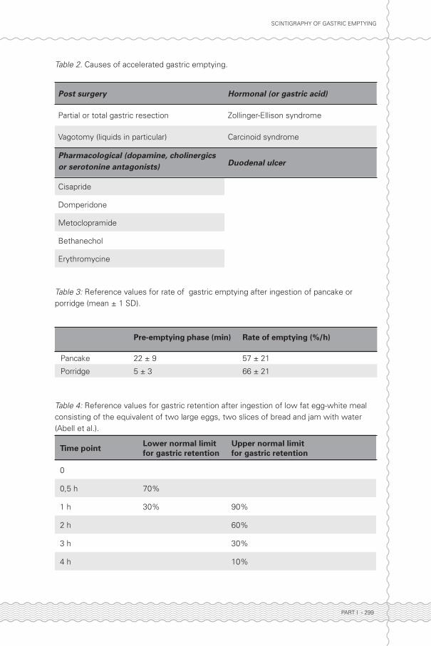
Table 2. Causes of accelerated gastric emptying.
Post surgery Hormonal (or gastric acid)
Partial or total gastric resection Zollinger-Ellison syndrome
Vagotomy (liquids in particular) Carcinoid syndrome
Pharmacological (dopamine, cholinergics or serotonine antagonists) Duodenal ulcer
Cisapride
Domperidone
Metoclopramide
Bethanechol
Erythromycine
Table 3: Reference values for rate of gastric emptying after ingestion of pancake or porridge (mean ± 1 SD).
Pre-emptying phase (min) Rate of emptying (%/h)
Pancake 22 ± 9 57 ± 21
Porridge 5 ± 3 66 ± 21
Table 4: Reference values for gastric retention after ingestion of low fat egg-white meal consisting of the equivalent of two large eggs, two slices of bread and jam with water (Abell et al.).
Time point Lower normal limit for gastric retention
Upper normal limit for gastric retention
0
0,5 h 70%
1 h 30% 90%
2 h
60%
3 h 30%
4 h 10%
Deel I_D.indd 299 27-12-16 14:17
![PATIENT MEDICATION INFORMATION · • Sympathomimetics (such as epinephrine [adrenaline], or salbutamol, albuterol or terbutaline used to treat asthma) • Growth hormone (medicine](https://img.pdfslide.net/doc/110x75/5e5027899cbbf304ec7f880c/patient-medication-information-a-sympathomimetics-such-as-epinephrine-adrenaline.jpg)