Embed Size (px)
Citation preview
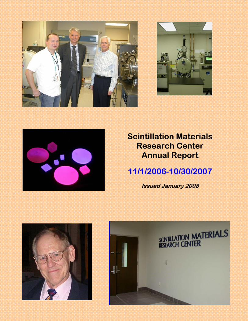
Scintillation Materials Research Center
Annual Report
11/1/2006-10/30/2007
Issued January 2008
2
Table of Contents
Contact information 3
Steering committee 4
Mission Statement 5
Brief overview of accomplishments 6
Introduction and background 7
SMRC Capabilities
Material Synthesis 8
Characterization 10
Personnel
Staff 13
Students 13
Collaborators 13
Professional activities 22
Awards 23
Patents 26
List of Publications 27
List of Presentations 28
Conference Posters
Scint 2007 30
ICCG-15 31
AVS 32
IEEE NSS-MIC 33
Financial support 34
3
Scintillator Materials Research Center
Contact information
Mailing address:
Scintillation Materials Research Center The University of Tennessee
124 Perkins Hall Knoxville, TN 37996-2000
Physical addresses:
SMRC office: 301 Science and Engineering Research Facility (SERF) Characterization Lab: 302 SERF
Synthesis Lab: 306 SERF
Telephone numbers:
Chuck Melcher: 974-0254 Merry Spurrier: 974-0267 Fax: 974-4998
E-mail:
[email protected] [email protected] [email protected]
Website:
http://www.engr.utk.edu/smrc/
4
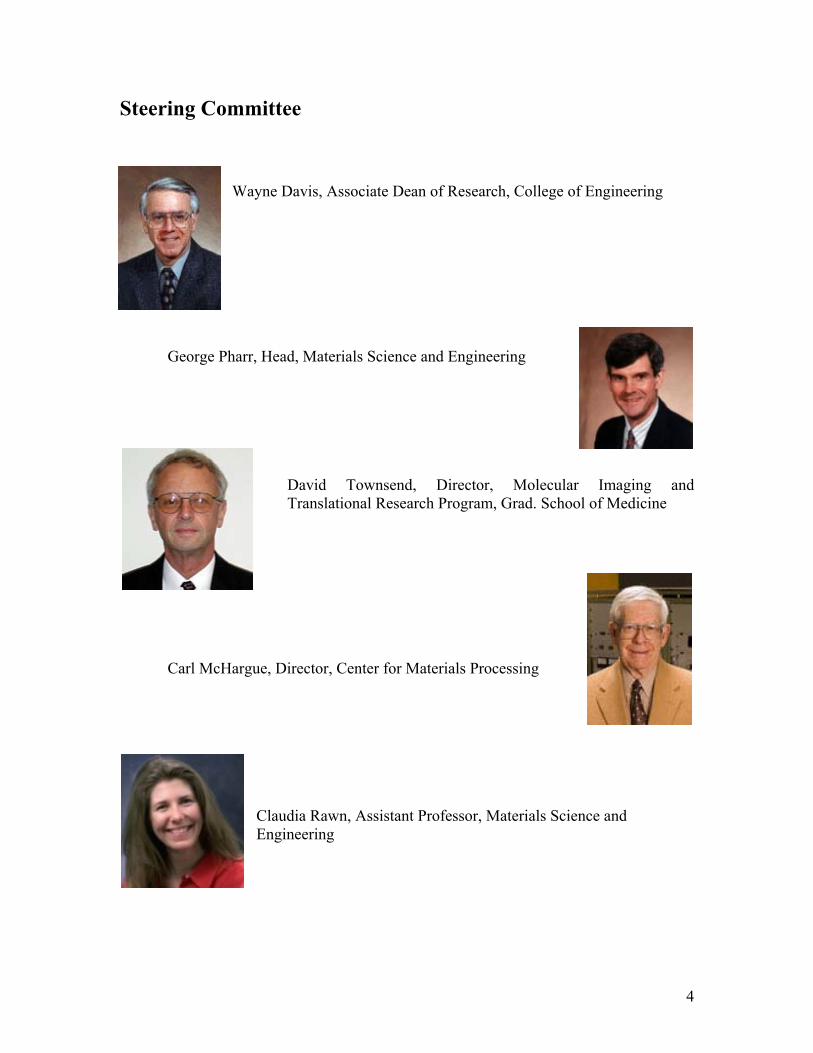
Steering Committee
Wayne Davis, Associate Dean of Research, College of Engineering
George Pharr, Head, Materials Science and Engineering
David Townsend, Director, Molecular Imaging and Translational Research Program, Grad. School of Medicine
Carl McHargue, Director, Center for Materials Processing
Claudia Rawn, Assistant Professor, Materials Science and Engineering
5
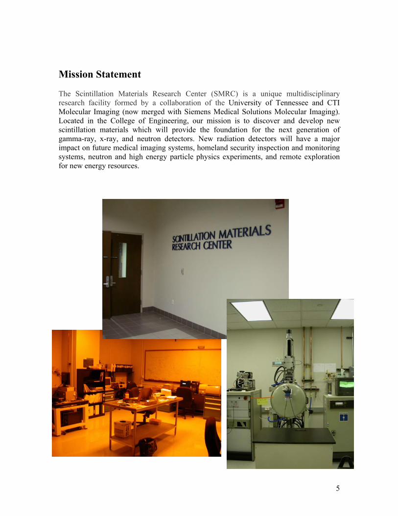
Mission Statement
The Scintillation Materials Research Center (SMRC) is a unique multidisciplinary research facility formed by a collaboration of the University of Tennessee and CTI Molecular Imaging (now merged with Siemens Medical Solutions Molecular Imaging). Located in the College of Engineering, our mission is to discover and develop new scintillation materials which will provide the foundation for the next generation of gamma-ray, x-ray, and neutron detectors. New radiation detectors will have a major impact on future medical imaging systems, homeland security inspection and monitoring systems, neutron and high energy particle physics experiments, and remote exploration for new energy resources.
6
Brief overview of 2006-2007 SMRC accomplishments
The SMRC has had a number of significant accomplishments over the October 31, 2006– October 30, 2007 center year; highlights include the following:
• Continued work with ongoing research projects o Further progress on the SMRC breakthrough in LSO growth technology,
which utilizes co-doping with optically inactive ions to optimize scintillation and growth properties
o Progress toward combinatorial synthesis of scintillator libraries • Additional projects
o Investigation of scintillation properties of neutron-sensitive glass fibers for security applications
o Contract with LBNL for work on study of proportionality of response in inorganic scintillators
• Patents o SMRC staff currently have two patents pending
• Conferences attended o Scint 2007
• Dr. Melcher served on the Organizing and Advisory committees • The Scint 2007 Proceedings will be published as an issue of the
IEEE Transactions on Nuclear Science, with Dr. Melcher as senior editor
• Oral presentations and poster; two papers accepted o AVS 5th International Symposium and Exhibition
• Poster presentation and paper accepted o ICCG-15
• Poster presentation and paper accepted o IEEE NSS-MIC
• Oral presentations and poster; three papers accepted • Dr. Melcher served as Topic Convener and Session Chair
• Awards o Dr. Chuck Melcher received the 2006 IEEE Nuclear and Plasma Sciences
Society Merit Award o Graduate students Kan Yang and Harold Rothfuss both received IEEE
awards • Publications
o SMRC staff authored or co-authored 9 papers • Participation in DOE review teams • Enhanced laboratory capability
o Acquired an additional Czochralski station o Acquisition of 3 Bridgman stations is in progress
7
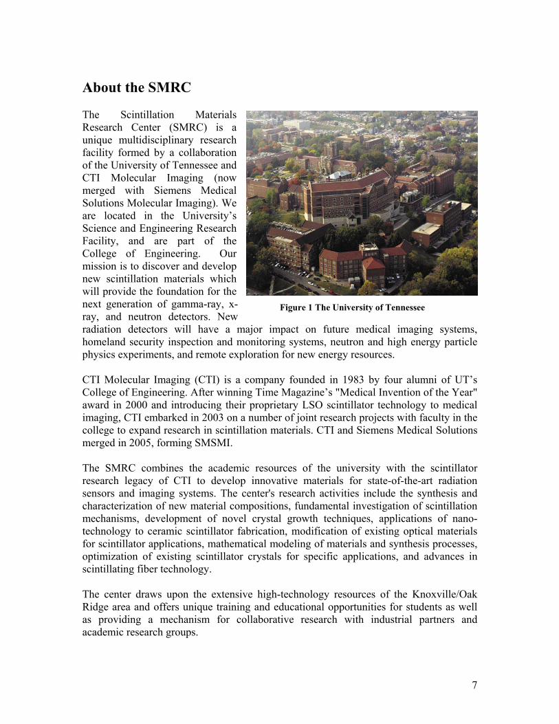
About the SMRC The Scintillation Materials Research Center (SMRC) is a unique multidisciplinary research facility formed by a collaboration of the University of Tennessee and CTI Molecular Imaging (now merged with Siemens Medical Solutions Molecular Imaging). We are located in the University’s Science and Engineering Research Facility, and are part of the College of Engineering. Our mission is to discover and develop new scintillation materials which will provide the foundation for the next generation of gamma-ray, x-ray, and neutron detectors. New radiation detectors will have a major impact on future medical imaging systems, homeland security inspection and monitoring systems, neutron and high energy particle physics experiments, and remote exploration for new energy resources. CTI Molecular Imaging (CTI) is a company founded in 1983 by four alumni of UT’s College of Engineering. After winning Time Magazine’s "Medical Invention of the Year" award in 2000 and introducing their proprietary LSO scintillator technology to medical imaging, CTI embarked in 2003 on a number of joint research projects with faculty in the college to expand research in scintillation materials. CTI and Siemens Medical Solutions merged in 2005, forming SMSMI. The SMRC combines the academic resources of the university with the scintillator research legacy of CTI to develop innovative materials for state-of-the-art radiation sensors and imaging systems. The center's research activities include the synthesis and characterization of new material compositions, fundamental investigation of scintillation mechanisms, development of novel crystal growth techniques, applications of nano-technology to ceramic scintillator fabrication, modification of existing optical materials for scintillator applications, mathematical modeling of materials and synthesis processes, optimization of existing scintillator crystals for specific applications, and advances in scintillating fiber technology. The center draws upon the extensive high-technology resources of the Knoxville/Oak Ridge area and offers unique training and educational opportunities for students as well as providing a mechanism for collaborative research with industrial partners and academic research groups.
Figure 1 The University of Tennessee
8
Research capabilities and expertise The SMRC has broad capabilities and expertise in both material synthesis and material characterization. An extensive collection of laboratory equipment permits the research staff and students to both synthesize new materials and measure their properties in-house, thus facilitating research by allowing prompt feedback.
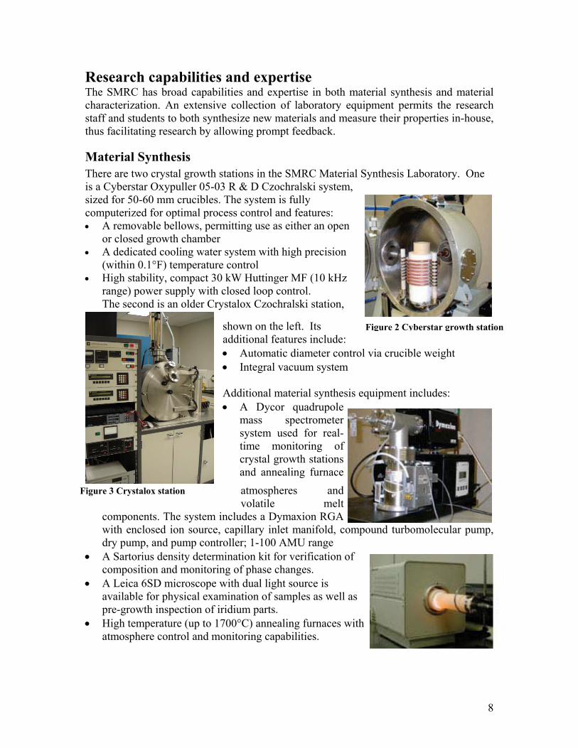
Material Synthesis There are two crystal growth stations in the SMRC Material Synthesis Laboratory. One is a Cyberstar Oxypuller 05-03 R & D Czochralski system, sized for 50-60 mm crucibles. The system is fully computerized for optimal process control and features: • A removable bellows, permitting use as either an open
or closed growth chamber • A dedicated cooling water system with high precision
(within 0.1°F) temperature control • High stability, compact 30 kW Huttinger MF (10 kHz
range) power supply with closed loop control. The second is an older Crystalox Czochralski station,
shown on the left. Its additional features include: • Automatic diameter control via crucible weight • Integral vacuum system Additional material synthesis equipment includes: • A Dycor quadrupole
mass spectrometer system used for real-time monitoring of crystal growth stations and annealing furnace atmospheres and volatile melt
components. The system includes a Dymaxion RGA with enclosed ion source, capillary inlet manifold, compound turbomolecular pump, dry pump, and pump controller; 1-100 AMU range
• A Sartorius density determination kit for verification of composition and monitoring of phase changes.
• A Leica 6SD microscope with dual light source is available for physical examination of samples as well as pre-growth inspection of iridium parts.
• High temperature (up to 1700°C) annealing furnaces with atmosphere control and monitoring capabilities.
Figure 2 Cyberstar growth station
Figure 3 Crystalox station
9
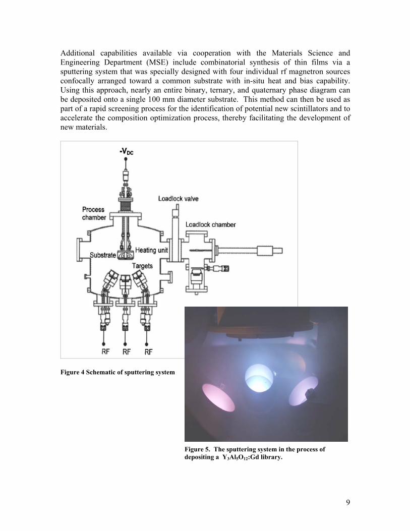
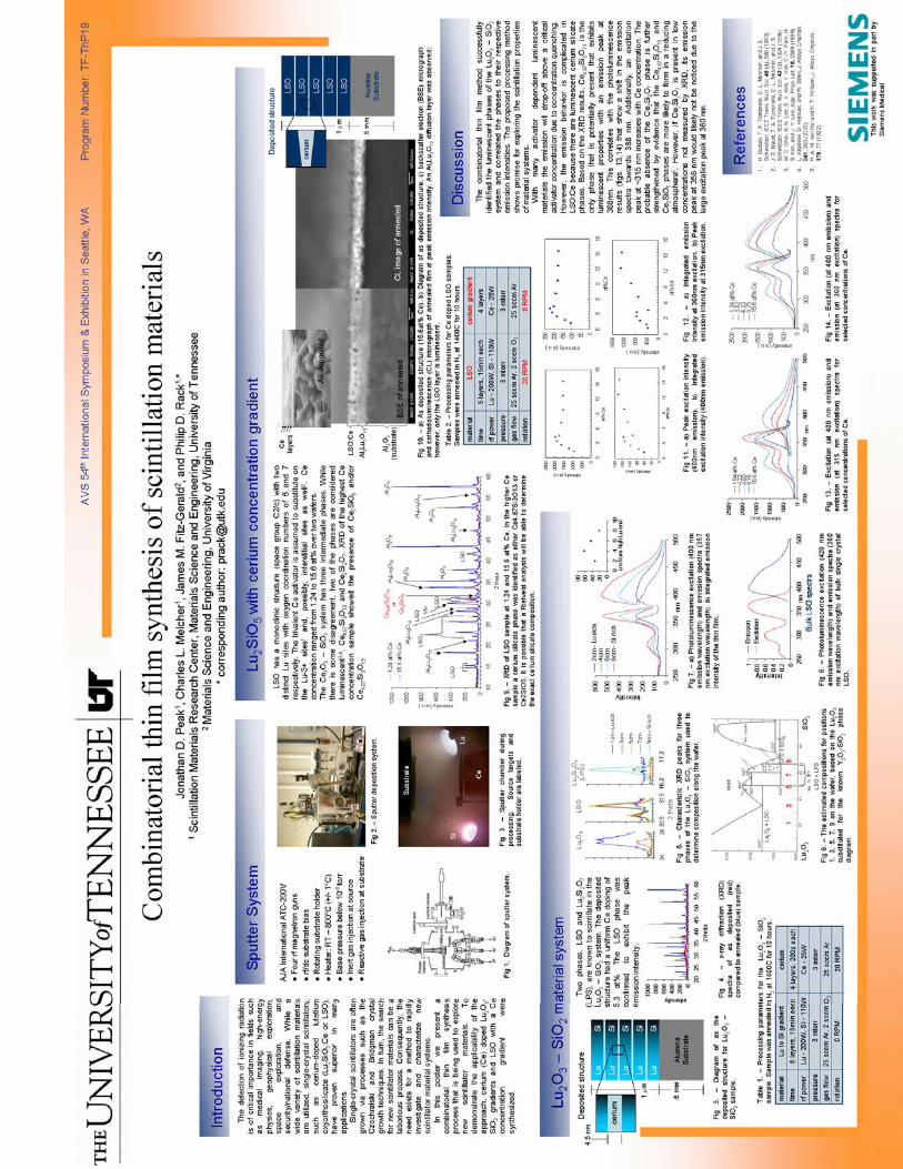
Additional capabilities available via cooperation with the Materials Science and Engineering Department (MSE) include combinatorial synthesis of thin films via a sputtering system that was specially designed with four individual rf magnetron sources confocally arranged toward a common substrate with in-situ heat and bias capability. Using this approach, nearly an entire binary, ternary, and quaternary phase diagram can be deposited onto a single 100 mm diameter substrate. This method can then be used as part of a rapid screening process for the identification of potential new scintillators and to accelerate the composition optimization process, thereby facilitating the development of new materials.
Figure 4 Schematic of sputtering system
Figure 5. The sputtering system in the process of depositing a Y3Al5O12:Gd library.
10
0
20
40
60
80
100
120
300 350 400 450 500 550 600
Emission Spectra
BGOGSO:CeLGSO:CeLSO:CeLuYAP:CeLYGSO:CeLYSO:CeYSO:Ce
Nor
mal
ized
Inte
nsity
Wavelength (nm)
0
500
1000
1500
2000
2500
0 100 200 300 400 500 600
Counts
Channel number
137Cs
FWHM=7.9%
Material Characterization
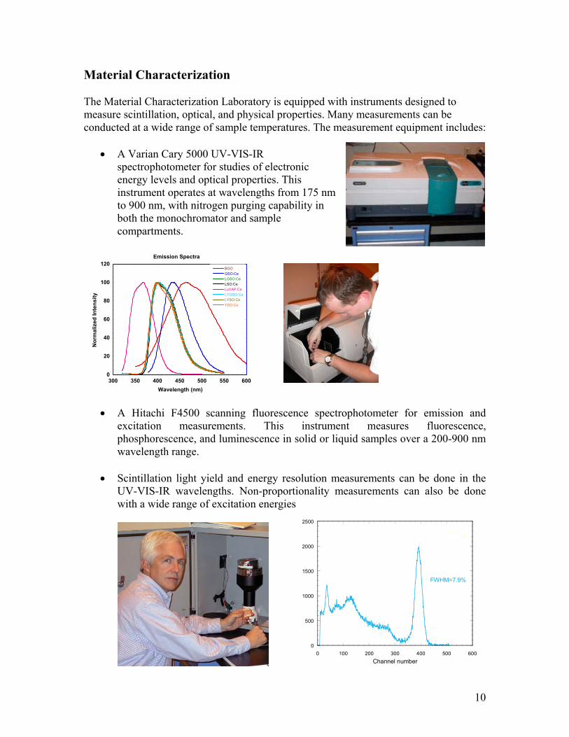
The Material Characterization Laboratory is equipped with instruments designed to measure scintillation, optical, and physical properties. Many measurements can be conducted at a wide range of sample temperatures. The measurement equipment includes:
• A Varian Cary 5000 UV-VIS-IR spectrophotometer for studies of electronic energy levels and optical properties. This instrument operates at wavelengths from 175 nm to 900 nm, with nitrogen purging capability in both the monochromator and sample compartments.
• A Hitachi F4500 scanning fluorescence spectrophotometer for emission and excitation measurements. This instrument measures fluorescence, phosphorescence, and luminescence in solid or liquid samples over a 200-900 nm wavelength range.
• Scintillation light yield and energy resolution measurements can be done in the UV-VIS-IR wavelengths. Non-proportionality measurements can also be done with a wide range of excitation energies
11
• Absolute light output measurements.
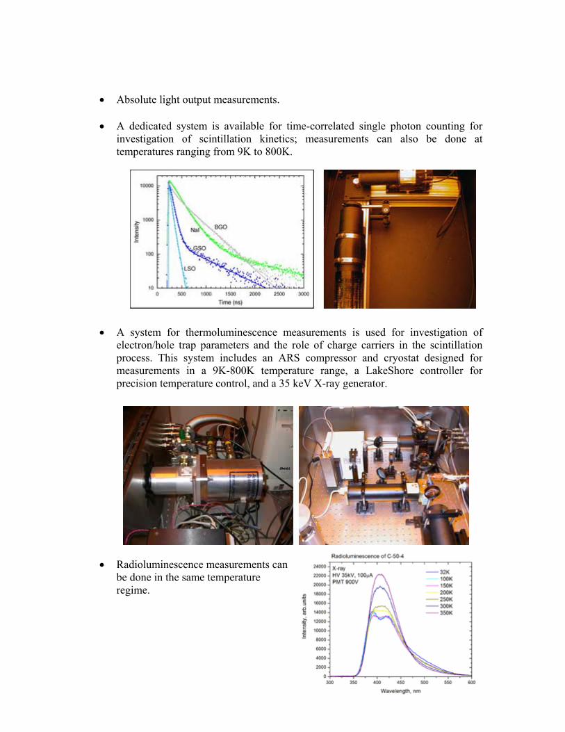
• A dedicated system is available for time-correlated single photon counting for investigation of scintillation kinetics; measurements can also be done at temperatures ranging from 9K to 800K.
• A system for thermoluminescence measurements is used for investigation of electron/hole trap parameters and the role of charge carriers in the scintillation process. This system includes an ARS compressor and cryostat designed for measurements in a 9K-800K temperature range, a LakeShore controller for precision temperature control, and a 35 keV X-ray generator.
• Radioluminescence measurements can be done in the same temperature regime.
12
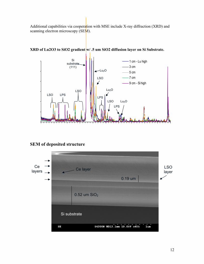
Additional capabilities via cooperation with MSE include X-ray diffraction (XRD) and scanning electron microscopy (SEM).
XRD of Lu2O3 to SiO2 gradient w/ .5 um SiO2 diffusion layer on Si Substrate.
SEM of deposited structure
Ce layers
0.19 um
0.52 um SiO2
Ce layer
Si substrate
LSOlayer
1 cm - Lu high3 cm5 cm7 cm9 cm - Si high
Si substrate
(111) Lu2O
LSO LPS
LSO
LSO
LPS
Lu2O
LSO
LPS
Lu2O
13
Personnel The Scintillation Materials Research Center fully supports two staff members, and offers partial financial support to two others, as well as supporting the laboratory activities of three additional professors. The SMRC currently supports five graduate students; three undergraduate students are employed by the center on a part-time basis to assist with sample prep, measurements, and other laboratory operations. In addition, SMRC staff have continued to develop relationships with scientists in related fields, some of whom were regular collaborators over the past year. Staff The full-time center staff: • Dr. Chuck Melcher – Director of the SMRC and Research Professor in Materials
Science and Engineering • Merry Spurrier – Research Associate III, SMRC
Partially supported staff: • Dr. Philip Rack – Associate Professor in Materials Science and Engineering • Frank Holiway - Materials Science and Engineering Accounting Specialist Adjunct faculty: • Dr. Lars Eriksson - Scientific Advisor, Siemens Medical Solutions Molecular
Imaging; Adjunct Professor, Materials Science and Engineering • Dr. Piotr Szupryczynski – Research Scientist, Siemens Medical Solutions Molecular
Imaging; Adjunct Asssistant Research Professor, Materials Science and Engineering.
Additional personnel: • Dr. George Pharr – Professor, Materials Science and Engineering, Department Head Collaborators • Dr. Bill Moses – Center for Functional Imaging, Lawrence Berkeley National
Laboratory • Dr. Marek Moszynski – Deputy Director, Soltan Institute for Nuclear Studies • Dr. Andrew Carey - Siemens Medical Solutions Molecular Imaging Students The graduate students supported by the SMRC are, in alphabetical order, Paul Cutler, Kurt Johanns, Jonathan Peak, Harold Rothfuss, and Kan Yang. They work on projects ranging from thin films to timing measurements to afterglow. The undergraduate students employed by the SMRC are Sergio de la Barrera, Brandon Goodwin, and Lucas Hofmeister. See the following pages for more detailed information on the SMRC students.
14
Paul Cutler
Graduate Student in Materials Science and Engineering* Advisor – Professor Chuck Melcher Research interests: Scintillation Materials Synthesis and Characterization Recent work has focused on: • Conversion energy nonproportionality of gamma ray scintillators using
photoexcitation light yield measurements (collaboration w/ LBNL). • Crystalox Czochralski crystal growth system installation and set-up • Effects of Gallium concentration on alpha particle scintillation in ZnO:Ga (ORNL
summer research internship) Additional activities include: • Chair of the UTK Materials Advantage organization • Recipient of the Oak Ridge Chapter of ASM scholarhip • To be the recipient of a TMS Divisional Travel Award in 2008 *Also supported by the Center for Materials Processing
15
Kurt E. Johanns
Graduate Student in Materials Science and Engineering Advisor – Professor George Pharr Research interests: - The effect of mechanical stimuli on scintillation properties.
- Simulation and experimental verification of indentation cracking in brittle materials. - The relationship between microstructure and mechanical properties of ceramics.
Recent work: - Pressure Induced Scintillation in BGO and LSO:Ce
A Nanoindenter has been used to mechanically alter the surface of both BGO and LSO:Ce in hopes of understanding any relationship between scintillation physics and residual stress.
- Hertzian Indentation of Silicon Carbide The force required to inititate a ring crack in Silicon Carbide subjected to a range of indenter radii was measured using acoustic emission.
16
Jonathan Peak
Graduate Student in Materials Science and Engineering Advisor – Professor Philip Rack Research interests: Thin Film Combinatorial Synthesis of Scintillation Materials Recent work has focused on thin-film LSO:Ce as a well-characterized sample material to use as proof-of-concept for the production of sample libraries for rapid new material screening. Recent presentations: Oral: J.D. Peak, SCINT2007, Inorganic Scintillators and their Applications, Combinatorial thin film synthesis of cerium doped scintillation materials in the lutetium oxide - silicon oxide system. Poster: J. D. Peak, C. L. Melcher, J. Fitz-Gerald, and P. D. Rack, American Vacuum Society 54th International Symposium and Exhibition, Combinatorial thin film synthesis of scintillating materials: scanning electron and cathodoluminescence imaging of thin film Lu2SiO5:Ce. See page 32 for the full text. Recent publications: J.D. Peak, C. L. Melcher, and P. D. Rack, Combinatorial thin film synthesis of cerium doped scintillation materials in the lutetium oxide - silicon oxide system, IEEE Trans. Nuc. Sci. (submitted). P. D. Rack, J. Peak, C. L. Melcher, and J. Fitz-Gerald, Scanning electron and cathodoluminescence imaging of thin film Lu2SiO5:Ce, Applied Physics Letters 91, 24 (2007).
17
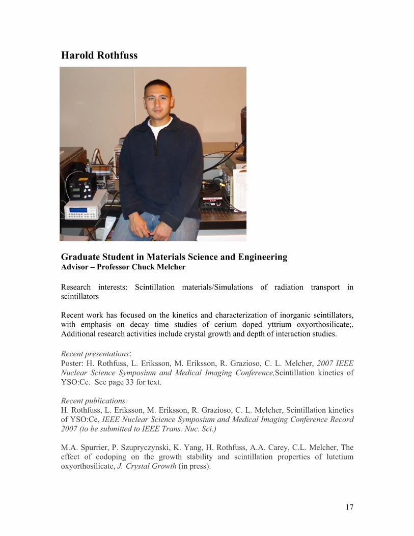
Harold Rothfuss
Graduate Student in Materials Science and Engineering Advisor – Professor Chuck Melcher Research interests: Scintillation materials/Simulations of radiation transport in scintillators Recent work has focused on the kinetics and characterization of inorganic scintillators, with emphasis on decay time studies of cerium doped yttrium oxyorthosilicate;. Additional research activities include crystal growth and depth of interaction studies. Recent presentations: Poster: H. Rothfuss, L. Eriksson, M. Eriksson, R. Grazioso, C. L. Melcher, 2007 IEEE Nuclear Science Symposium and Medical Imaging Conference,Scintillation kinetics of YSO:Ce. See page 33 for text. Recent publications: H. Rothfuss, L. Eriksson, M. Eriksson, R. Grazioso, C. L. Melcher, Scintillation kinetics of YSO:Ce, IEEE Nuclear Science Symposium and Medical Imaging Conference Record 2007 (to be submitted to IEEE Trans. Nuc. Sci.) M.A. Spurrier, P. Szupryczynski, K. Yang, H. Rothfuss, A.A. Carey, C.L. Melcher, The effect of codoping on the growth stability and scintillation properties of lutetium oxyorthosilicate, J. Crystal Growth (in press).
18
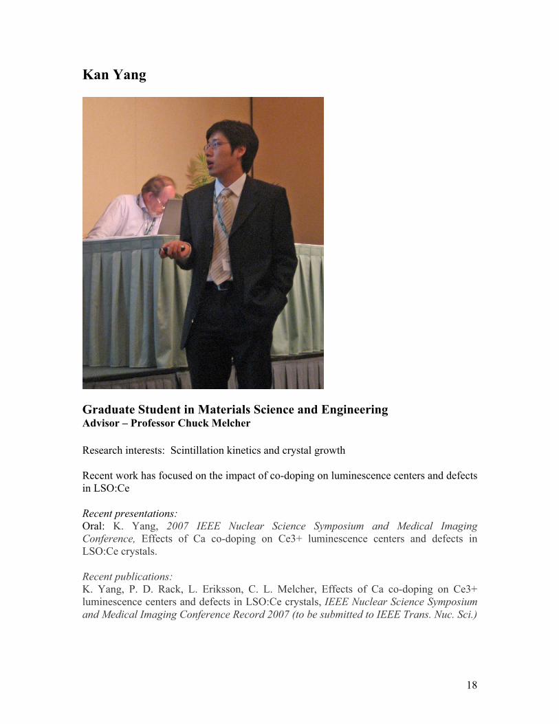
Kan Yang
Graduate Student in Materials Science and Engineering Advisor – Professor Chuck Melcher Research interests: Scintillation kinetics and crystal growth Recent work has focused on the impact of co-doping on luminescence centers and defects in LSO:Ce Recent presentations: Oral: K. Yang, 2007 IEEE Nuclear Science Symposium and Medical Imaging Conference, Effects of Ca co-doping on Ce3+ luminescence centers and defects in LSO:Ce crystals. Recent publications: K. Yang, P. D. Rack, L. Eriksson, C. L. Melcher, Effects of Ca co-doping on Ce3+ luminescence centers and defects in LSO:Ce crystals, IEEE Nuclear Science Symposium and Medical Imaging Conference Record 2007 (to be submitted to IEEE Trans. Nuc. Sci.)
19
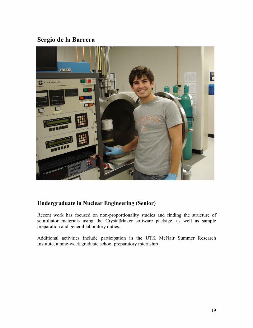
Sergio de la Barrera
Undergraduate in Nuclear Engineering (Senior)
Recent work has focused on non-proportionality studies and finding the structure of scintillator materials using the CrystalMaker software package, as well as sample preparation and general laboratory duties.
Additional activities include participation in the UTK McNair Summer Research Institute, a nine-week graduate school preparatory internship
20
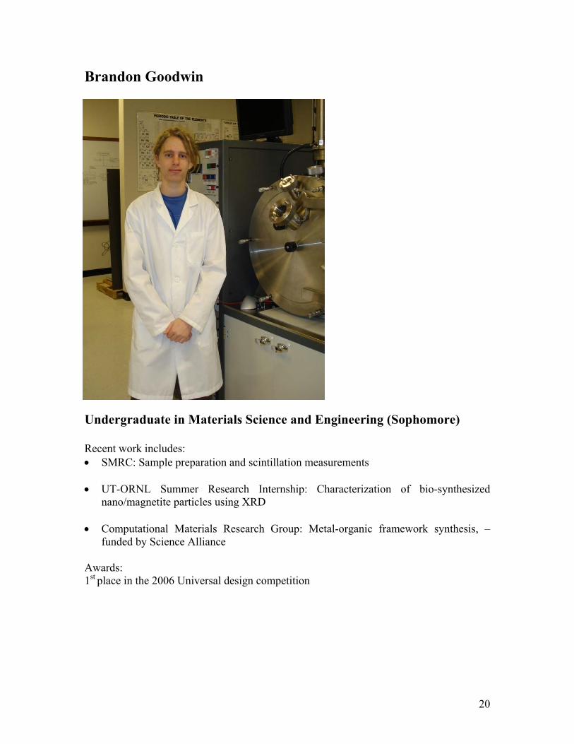
Brandon Goodwin
Undergraduate in Materials Science and Engineering (Sophomore) Recent work includes: • SMRC: Sample preparation and scintillation measurements • UT-ORNL Summer Research Internship: Characterization of bio-synthesized
nano/magnetite particles using XRD • Computational Materials Research Group: Metal-organic framework synthesis, –
funded by Science Alliance Awards: 1st place in the 2006 Universal design competition
21
Lucas Hofmeister
Undergraduate in Biomedical Engineering (Sophomore) Prior research experience includes involvement in microfluidic lab on a chip simulation of cancer microenvironments. Current work at the SMRC is primarily focused on sample preparation and density measurements; long-term research interests include materials science and synthesis with an eye toward interdisciplinary science.
22
Professional activities Editorial During the past year, Dr. Melcher has served as:
• Associate editor of IEEE Transactions on Nuclear Science • Senior editor of the Scint 2007 Conference Issue of IEEE Transactions on Nuclear
Science Conferences Dr. Melcher also served in the following capacities:
• Member of the Scint 2007 Organizing Committee • Topic Convener for the IEEE Nuclear Science Symposium and Medical Imaging
Conference • Session Chair at the IEEE Nuclear Science Symposium and Medical Imaging
Conference Review panel activities Dr. Melcher has been invited to participate in several government review panels over the last year, including: Department of Energy (NNSA) review panels: ORNL NA-22 PROGRAM REVIEW OR06-241-PD05: New Family of Scintillators Based on Novel Phosphate Glasses Jim White/Lynn Boatner/ David Singh February 8, 2007, Oak Ridge LANL NA-42 PROGRAM REVIEW Compton Backscatter Project Wendy McNeil August 16, 2007, Los Alamos ORNL NA-22 PROGRAM REVIEW OR06-PD05: Growth of Lanthanum Halide Crystals Jim White/Lynn Boatner September 27, 2007, Oak Ridge NA-22 Scintillator Materials Working Group Proliferation Deterrence Merit Review Team Workshop November 30, 2007, Boston DNDO review panel: LBNL Scintillator Discovery Project Advisory Committee CFP06–TA-01-LB02 Lawrence Berkeley National Laboratory May, 2007, Berkeley
23
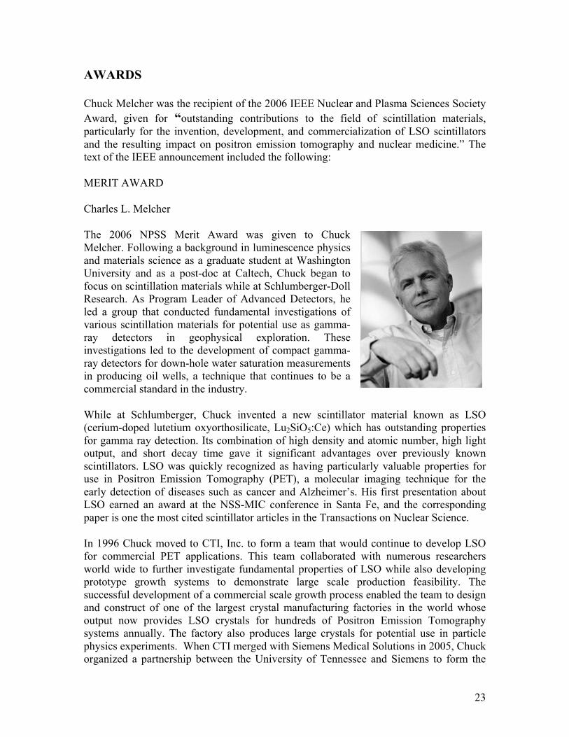
AWARDS Chuck Melcher was the recipient of the 2006 IEEE Nuclear and Plasma Sciences Society Award, given for “outstanding contributions to the field of scintillation materials, particularly for the invention, development, and commercialization of LSO scintillators and the resulting impact on positron emission tomography and nuclear medicine.” The text of the IEEE announcement included the following:
MERIT AWARD
Charles L. Melcher
The 2006 NPSS Merit Award was given to Chuck Melcher. Following a background in luminescence physics and materials science as a graduate student at Washington University and as a post-doc at Caltech, Chuck began to focus on scintillation materials while at Schlumberger-Doll Research. As Program Leader of Advanced Detectors, he led a group that conducted fundamental investigations of various scintillation materials for potential use as gamma-ray detectors in geophysical exploration. These investigations led to the development of compact gamma-ray detectors for down-hole water saturation measurements in producing oil wells, a technique that continues to be a commercial standard in the industry.
While at Schlumberger, Chuck invented a new scintillator material known as LSO (cerium-doped lutetium oxyorthosilicate, Lu2SiO5:Ce) which has outstanding properties for gamma ray detection. Its combination of high density and atomic number, high light output, and short decay time gave it significant advantages over previously known scintillators. LSO was quickly recognized as having particularly valuable properties for use in Positron Emission Tomography (PET), a molecular imaging technique for the early detection of diseases such as cancer and Alzheimer’s. His first presentation about LSO earned an award at the NSS-MIC conference in Santa Fe, and the corresponding paper is one the most cited scintillator articles in the Transactions on Nuclear Science.
In 1996 Chuck moved to CTI, Inc. to form a team that would continue to develop LSO for commercial PET applications. This team collaborated with numerous researchers world wide to further investigate fundamental properties of LSO while also developing prototype growth systems to demonstrate large scale production feasibility. The successful development of a commercial scale growth process enabled the team to design and construct of one of the largest crystal manufacturing factories in the world whose output now provides LSO crystals for hundreds of Positron Emission Tomography systems annually. The factory also produces large crystals for potential use in particle physics experiments. When CTI merged with Siemens Medical Solutions in 2005, Chuck organized a partnership between the University of Tennessee and Siemens to form the
24
Scintillation Materials Research Center (SMRC). He joined the faculty of the Materials Science and Engineering Department at the University of Tennessee and became Director of the Center. The SMRC is a groundbreaking example of a cooperative partnership between industry and academia, providing unique research opportunities for engineering students and an integrated approach for the commercial realization of innovations in scintillation materials.
LSO has become the standard against which new scintillator materials are often compared. During the 15 years since its introduction, no scintillator has yet equaled its combination of high light yield, fast decay time, high density and atomic number, and environmental stability. Chuck not only discovered and patented LSO and carried out much of the initial basic research on its properties, but he also led its development to the industrial production level and its widespread implementation in positron emission tomography (PET). It is arguably the most commercially successful scintillator of the last 20 years, now used in nearly half of the clinical PET scanners currently manufactured as well as in the vast majority of small animal PET scanners. The discovery and commercialization of LSO is often mentioned as one of the major developments in nuclear medical imaging of the last few decades. In addition, it is now under consideration for the next generation of high energy physics calorimeters.
Chuck has been an active member of the IEEE and the NPSS for many years. In addition to numerous program committees, he has served on the Radiation Instrumentation Steering Committee and the Constitution and Bylaws Committee. He currently serves as Vice Chair and Chair-elect of the Radiation Instrumentation Technical Committee. In addition, he serves as Associate Editor of the Transactions on Nuclear Science.
25
Student Awards
As excerpted from the College of Engineering newsletter below, graduate students Kan Yang and Harold Rothfuss both received awards at the 2007 IEEE Nuclear Science Symposium.
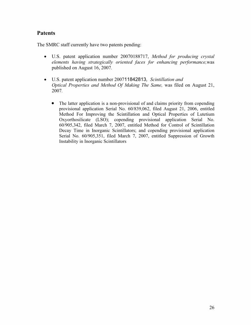
Two materials science and engineering graduate students affiliated with the Scintillation Materials Research Center (SMRC) were recently honored at the 2007 IEEE Nuclear Science Symposium. Kan Yang was awarded a Trainee Grant based on his presentation "Effects of Ca Co-doping on Ce3+ Luminescence Centers and Defects in LSO:Ce Crystals" and Harold Rothfuss was runner-up in the Nuclear and Plasma
Sciences Society Best Student Paper Contest for his presentation "Scintillation Kinetics of YSO:Ce."
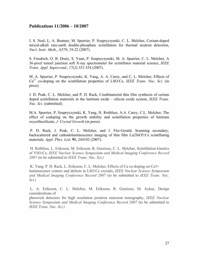
In addition, graduate student Paul Cutler was recently informed that he will be the recipient of a 2008 TMS Divisional Travel Award.
26
Patents The SMRC staff currently have two patents pending:
• U.S. patent application number 20070188717, Method for producing crystal elements having strategically oriented faces for enhancing performance,was published on August 16, 2007.
• U.S. patent application number 200711842813, Scintillation and
Optical Properties and Method Of Making The Same, was filed on August 21, 2007.
• The latter application is a non-provisional of and claims priority from copending
provisional application Serial No. 60/839,062, filed August 21, 2006, entitled Method For Improving the Scintillation and Optical Properties of Lutetium Oxyorthosilicate (LSO); copending provisional application Serial No. 60/905,342, filed March 7, 2007, entitled Method for Control of Scintillation Decay Time in Inorganic Scintillators; and copending provisional application Serial No. 60/905,351, filed March 7, 2007, entitled Suppression of Growth Instability in Inorganic Scintillators
27
Publications 11/2006 – 10/2007
J. S. Neal, L. A. Boatner, M. Spurrier, P. Szupryczynski, C. L. Melcher, Cerium-doped mixed-alkali rare-earth double-phosphate scintillators for thermal neutron detection, Nucl. Instr. Meth., A579, 19-22 (2007).
S. Friedrich, O. B. Drury, S. Yuan, P. Szupryczynski, M. A. Spurrier, C. L. Melcher, A 36-pixel tunnel junction soft X-ray spectrometer for scintillator material science, IEEE Trans. Appl. Supercond., 17(2) 351-354 (2007).
M. A. Spurrier, P. Szupryczynski, K. Yang, A. A. Carey, and C. L. Melcher, Effects of Ca2+ co-doping on the scintillation properties of LSO:Ce, IEEE Trans. Nuc. Sci. (in press).
J. D. Peak, C. L. Melcher, and P. D. Rack, Combinatorial thin film synthesis of cerium doped scintillation materials in the lutetium oxide – silicon oxide system, IEEE Trans. Nuc. Sci. (submitted).
M.A. Spurrier, P. Szupryczynski, K. Yang, H. Rothfuss, A.A. Carey, C.L. Melcher, The effect of codoping on the growth stability and scintillation properties of lutetium oxyorthosilicate, J. Crystal Growth (in press).
P. D. Rack, J. Peak, C. L. Melcher, and J. Fitz-Gerald, Scanning secondary, backscattered and cathodoluminescence imaging of thin film Lu2SiO5:Ce scintillating materials. Appl. Phys. Lett. 91, 244102 (2007). H. Rothfuss, L. Eriksson, M. Eriksson, R. Grazioso, C. L. Melcher, Scintillation kinetics of YSO:Ce, IEEE Nuclear Science Symposium and Medical Imaging Conference Record 2007 (to be submitted to IEEE Trans. Nuc. Sci.) K. Yang, P. D. Rack, L. Eriksson, C. L. Melcher, Effects of Ca co-doping on Ce3+ luminescence centers and defects in LSO:Ce crystals, IEEE Nuclear Science Symposium and Medical Imaging Conference Record 2007 (to be submitted to IEEE Trans. Nuc. Sci.) L. A. Eriksson, C. L. Melcher, M. Eriksson, R. Grazioso, M. Aykac, Design considerations of phoswich detectors for high resolution positron emission tomography, IEEE Nuclear Science Symposium and Medical Imaging Conference Record 2007 (to be submitted to IEEE Trans. Nuc. Sci.)
28
Presentations Invited Conference Presentation Prof. Chuck Melcher was invited to give the scientific summary at SCINT2007, Inorganic Scintillators and their Applications, Winston-Salem NC, June 2007 Oral Presentations J. D. Peak, SCINT2007, Inorganic Scintillators and their Applications, Combinatorial thin film synthesis of cerium doped scintillation materials in the lutetium oxide - silicon oxide system. K. Yang, 2007 IEEE Nuclear Science Symposium and Medical Imaging Conference, Effects of Ca co-doping on Ce3+ luminescence centers and defects in LSO:Ce crystals. L. A. Eriksson, 2007 IEEE Nuclear Science Symposium and Medical Imaging Conference, Design considerations of phoswich detectors for high resolution positron emission tomography. Poster Presentations M. A. Spurrier, SCINT2007, Inorganic Scintillators and their Applications, Effects of Ca2+ codoping on the scintillation properties of LSO:Ce. M. A. Spurrier, 15th International Conference for Crystal Growth, The effect of codoping on the growth stability and scintillation properties of lutetium oxyorthosilicate. J. D. Peak, American Vacuum Society 54th International Symposium and Exhibition, Scanning secondary, backscattered and cathodoluminescence imaging of thin film Lu2SiO5:Ce scintillating materials. H. Rothfuss, 2007 IEEE Nuclear Science Symposium and Medical Imaging Conference, Scintillation kinetics of YSO:Ce. Review meetings and reports In the interest of maintaining a close and effective working relationship, we held regularly scheduled review meetings with Siemens personnel, at which the previous month’s activities, results, and accomplishments are presented.
29
SMRC Poster Presentations
2006-2007
30
0
0.2
0.4
0.6
0.8
1
1.2
300 400 500 600 700
LSO:CeLSO:Ce,Ca
Inte
nsity
(nor
mal
ized
)
Wavelength (nm)
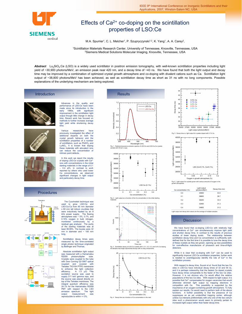
Effects of Ca2+ co-doping on the scintillation properties of LSO:Ce
M.A. Spurrier1, C. L. Melcher1, P. Szupryczynski1,2, K. Yang1, A. A. Carey2,
1Scintillation Materials Research Center, University of Tennessee, Knoxville, Tennessee, USA2Siemens Medical Solutions Molecular Imaging, Knoxville, Tennessee, USA
IEEE 9th International Conference on Inorganic Scintillators and their Applications, 2007, Winston-Salem NC, USA
Abstract Lu2SiO5:Ce (LSO) is a widely used scintillator in positron emission tomography, with well-known scintillation properties including light yield of ~30,900 photons/MeV, an emission peak near 420 nm, and a decay time of ~43 ns. We have found that both the light output and decay time may be improved by a combination of optimized crystal growth atmosphere and co-doping with divalent cations such as Ca. Scintillation light output of ~38,800 photons/MeV has been achieved, as well as scintillation decay time as short as 31 ns with no long components. Possible explanations of the underlying mechanism are being explored.
Introduction
Procedures
Discussion
We have found that co-doping LSO:Ce with relatively high concentrations of Ca2+ can simultaneously improve light yield and shorten decay time, in contrast to the results of previous studies of lower doping levels. The relationship between scintillation decay time and Ca concentration is sufficiently well-defined that for the first time it is possible to tune the decay time of these crystals as they are grown, opening up new possibilities for cost-effective manufacture of phoswich and time-of-flight detectors.
While it is clear that co-doping with Ca2+ can be used to significantly improve LSO:Ce scintillation properties, further work is needed to unambiguously identify the role of Ca2+ in the scintillation process.
With respect to decay time, Suzuki et al. found that the two Cesites in LSO:Ce have individual decay times of 32 and 54 ns, and it is perhaps noteworthy that the fastest Ca doped crystals have decay times comparable to the faster of the two Ce sites. However, it is not obvious why Ca would affect the relative populations of the two Ce sites. With respect to light output, it is possible that Ca2+ compensates for oxygen vacancies that would otherwise diminish light output by trapping electrons in competition with Ce. This possibility is supported by the importance of an oxygen-containing growth atmosphere. To fully explain our results, Ca would need to perform both of the above functions. A further possibility is that the presence of Ca suppresses an as yet unidentified trapping center, although unless Ca interacts preferentially with only one of the two cerium sites such a phenomenon would seem to primarily pertain to increased light output rather than faster decay time.
The Czochralski technique was used to grow LSO:Ce and LSO:Ce:Ca from 60 mm diameter x 60 mm tall iridium crucibles that were inductively heated by an 8 kHz power supply. The flowing atmosphere was ~ 0%, 0.7%, and 0.15% oxygen in bulk nitrogen, monitored continuously by a residual gas analyzer. The purity of the starting materials was at least 99.99%. The boules were 32 mm in diameter and ~ 100 mm long.
Scintillation decay times were measured by the time-correlated single photon technique originated by Bollinger and Thomas.
Absolute scintillation light output was measured with a Hamamatsu R2059 photomultiplier tube. Crystals were coupled to the tube with Dow Corning Q2-3067 optical couplant and covered with Tetratex TX3104 PTFE membrane to enhance the light collection efficiency. A 1.0 µCi 22Na excitation source was used to supply 511 keV gamma rays, and the source was placed directly on top of the Tetratex membrane. The integral quantum efficiency was 24.1% for the Hamamatsu R2059 PMT with respect to the LSO emission spectrum. The light output measurement was reproducible to within +/-3%.
Results
Fig. 5. Decay time vs. light output for crystals doped with 0.1% Ce.
Decay time as a function of Ca concentration in the melt.
Advances in the quality and performance of LSO:Ce have been made since its introduction in the early 1990s, with significant improvement in the scintillation light output though little change in decay time. Recent work has focused on methods to further increase average light yield while shortening decay time.
Various researchers have previously investigated the effect of additional dopants on both the crystal growth behavior and the scintillation properties of a number of scintillators, such as PbWO4 and LuAlO3. It is known that doping these materials with aliovalent ions can reduce the concentration of intrinsic point defects.
In this work we report the results of doping LSO:Ce crystals with Ca2+
ions with concentrations in the initial starting materials in the range of 0.1 – 0.4 at%. In contrast to work reported by others who used lower Ca concentrations, we observed significant changes in light output and particularly decay time.
Fig. 1. Radioluminescence emission spectra of LSO:Ce and LSO:Ce,Ca crystalsexcited with 30 keV x-rays.
Fig. 2. Light output as a function of the Ca concentration in the initial startingmaterials
Fig. 3. Scintillation decay of LSO:Ce crystals with 0, 0.1, 0.2, 0.3, and 0.4% Ca doping. All samples had single exponential decays with no long components.
30
35
40
45
0 0.1 0.2 0.3 0.4
Dec
ay ti
me
(ns)
Ca concentration (%)
LSO:Ce (0.1%)
Light output and decay time values are the average of multiple samples
aperturetiming amp
MCA SCA
CFDCFD TAC
PMT PMT
source
*
stopstart
preamppreamp
gate
crystal
timing amp
Translation
Iridiumcrucible
RotationSeed crystal
CoilInsulation
Crystal
melt
0
500
1000
1500
2000
0 100 200 300 400 500 600 700 800
Coun
ts
Channel number
22Na source
LSO:Ce:Ca (0.1%)
Crystal
22Na source
MCA
H. V. Power
Pre-amp Shaping MCA
H. V.
Signal
Photomultiplier tube
Power supply
- Shapingamplifier
Teflon reflector
Fig. 4. Decay time as a function of Ca concentration in the melt.
31.0348000.4
31.3324000.3
33.3362000.2
36.7388000.1
43.0309000.0
Decay time (ns)Light output (photons/MeV)Ca concentration (%)
Fig. 6. Light output for crystals grown with various amounts of oxygen in thegrowth atmosphere
26000
28000
30000
32000
34000
36000
38000
40000
0 0.1 0.2 0.3 0.4
Ligh
t out
put (
phot
ons/
MeV
)
Ca concentration (%)
LSO:Ce (0.1%)
28
30
32
34
36
38
40
42
44
25000 27500 30000 32500 35000 37500 40000
0.0% Ca 0.1% Ca 0.2% Ca 0.3% Ca 0.4% Ca
Dec
ay ti
me
(ns)
Light output (photons/MeV)
26000
28000
30000
32000
34000
36000
38000
40000
0 0.05 0.1 0.15 0.2Li
ght o
utpu
t (ph
oton
s/M
eV)
Oxygen in growth atmosphere (%)
LSO (Ce=0.1%, Ca=0.1%)
10
100
1000
104
0 50 100 150 200
0.0% Ca0.1% Ca0.2% Ca0.3% Ca0.4% Ca
Cou
nts
Time (ns)
31
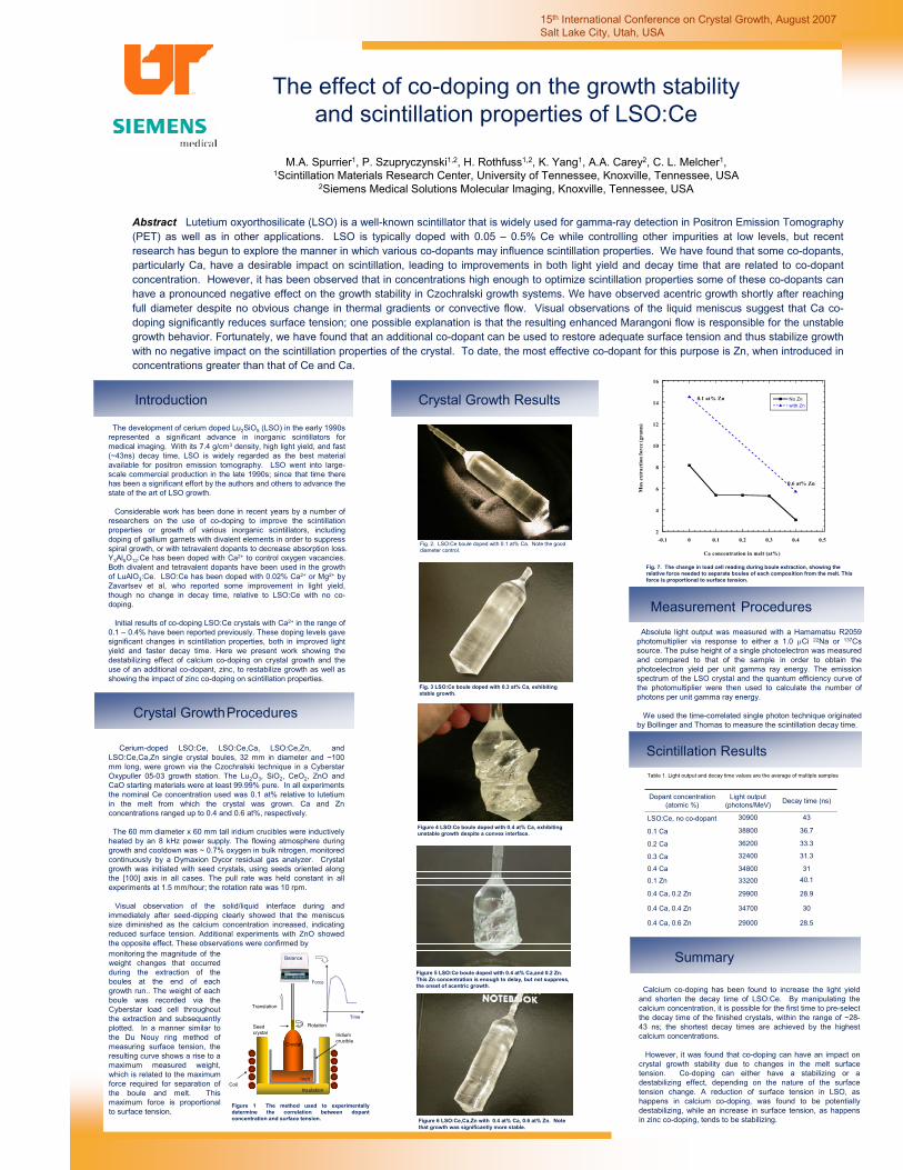
The effect of co-doping on the growth stability and scintillation properties of LSO:Ce
M.A. Spurrier1, P. Szupryczynski1,2, H. Rothfuss1,2, K. Yang1, A.A. Carey2, C. L. Melcher1, 1Scintillation Materials Research Center, University of Tennessee, Knoxville, Tennessee, USA
2Siemens Medical Solutions Molecular Imaging, Knoxville, Tennessee, USA
15th International Conference on Crystal Growth, August 2007 Salt Lake City, Utah, USA
Abstract Lutetium oxyorthosilicate (LSO) is a well-known scintillator that is widely used for gamma-ray detection in Positron Emission Tomography (PET) as well as in other applications. LSO is typically doped with 0.05 – 0.5% Ce while controlling other impurities at low levels, but recent research has begun to explore the manner in which various co-dopants may influence scintillation properties. We have found that some co-dopants, particularly Ca, have a desirable impact on scintillation, leading to improvements in both light yield and decay time that are related to co-dopant concentration. However, it has been observed that in concentrations high enough to optimize scintillation properties some of these co-dopants can have a pronounced negative effect on the growth stability in Czochralski growth systems. We have observed acentric growth shortly after reaching full diameter despite no obvious change in thermal gradients or convective flow. Visual observations of the liquid meniscus suggest that Ca co-doping significantly reduces surface tension; one possible explanation is that the resulting enhanced Marangoni flow is responsible for the unstable growth behavior. Fortunately, we have found that an additional co-dopant can be used to restore adequate surface tension and thus stabilize growth with no negative impact on the scintillation properties of the crystal. To date, the most effective co-dopant for this purpose is Zn, when introduced in concentrations greater than that of Ce and Ca.
Introduction
Crystal GrowthProcedures
Summary
Calcium co-doping has been found to increase the light yield and shorten the decay time of LSO:Ce. By manipulating the calcium concentration, it is possible for the first time to pre-select the decay time of the finished crystals, within the range of ~28-43 ns; the shortest decay times are achieved by the highest calcium concentrations.
However, it was found that co-doping can have an impact on crystal growth stability due to changes in the melt surface tension. Co-doping can either have a stabilizing or a destabilizing effect, depending on the nature of the surface tension change. A reduction of surface tension in LSO, as happens in calcium co-doping, was found to be potentially destabilizing, while an increase in surface tension, as happens in zinc co-doping, tends to be stabilizing.
Cerium-doped LSO:Ce, LSO:Ce,Ca, LSO:Ce,Zn, and LSO:Ce,Ca,Zn single crystal boules, 32 mm in diameter and ~100 mm long, were grown via the Czochralski technique in a CyberstarOxypuller 05-03 growth station. The Lu2O3, SiO2, CeO2, ZnO and CaO starting materials were at least 99.99% pure. In all experiments the nominal Ce concentration used was 0.1 at% relative to lutetium in the melt from which the crystal was grown. Ca and Zn concentrations ranged up to 0.4 and 0.6 at%, respectively.
The 60 mm diameter x 60 mm tall iridium crucibles were inductively heated by an 8 kHz power supply. The flowing atmosphere during growth and cooldown was ~ 0.7% oxygen in bulk nitrogen, monitored continuously by a Dymaxion Dycor residual gas analyzer. Crystal growth was initiated with seed crystals, using seeds oriented along the [100] axis in all cases. The pull rate was held constant in all experiments at 1.5 mm/hour; the rotation rate was 10 rpm.
Visual observation of the solid/liquid interface during and immediately after seed-dipping clearly showed that the meniscus size diminished as the calcium concentration increased, indicating reduced surface tension. Additional experiments with ZnO showed the opposite effect. These observations were confirmed by
Crystal Growth Results
The development of cerium doped Lu2SiO5 (LSO) in the early 1990s represented a significant advance in inorganic scintillators formedical imaging. With its 7.4 g/cm3 density, high light yield, and fast (~43ns) decay time, LSO is widely regarded as the best material available for positron emission tomography. LSO went into large-scale commercial production in the late 1990s; since that time there has been a significant effort by the authors and others to advance the state of the art of LSO growth.
Considerable work has been done in recent years by a number of researchers on the use of co-doping to improve the scintillation properties or growth of various inorganic scintillators, including doping of gallium garnets with divalent elements in order to suppress spiral growth, or with tetravalent dopants to decrease absorption loss. Y3Al5O12:Ce has been doped with Ca2+ to control oxygen vacancies. Both divalent and tetravalent dopants have been used in the growth of LuAlO3:Ce. LSO:Ce has been doped with 0.02% Ca2+ or Mg2+ by Zavartsev et al, who reported some improvement in light yield, though no change in decay time, relative to LSO:Ce with no co-doping.
Initial results of co-doping LSO:Ce crystals with Ca2+ in the range of 0.1 – 0.4% have been reported previously. These doping levels gave significant changes in scintillation properties, both in improved light yield and faster decay time. Here we present work showing the destabilizing effect of calcium co-doping on crystal growth and the use of an additional co-dopant, zinc, to restabilize growth as well as showing the impact of zinc co-doping on scintillation properties.
Table 1. Light output and decay time values are the average of multiple samples
monitoring the magnitude of the weight changes that occurred during the extraction of the boules at the end of each growth run.. The weight of each boule was recorded via the Cyberstar load cell throughout the extraction and subsequently plotted. In a manner similar to the Du Nouy ring method of measuring surface tension, the resulting curve shows a rise to a maximum measured weight, which is related to the maximum force required for separation of the boule and melt. This maximum force is proportional to surface tension.
28.5290000.4 Ca, 0.6 Zn
30347000.4 Ca, 0.4 Zn
28.9299000.4 Ca, 0.2 Zn
40.1332000.1 Zn
31348000.4 Ca
31.3324000.3 Ca
33.3362000.2 Ca
36.7388000.1 Ca
4330900LSO:Ce, no co-dopant
Decay time (ns)Light output (photons/MeV)
Dopant concentration (atomic %)
Fig. 2. LSO:Ce boule doped with 0.1 at% Ca. Note the gooddiameter control.
Fig. 3 LSO:Ce boule doped with 0.3 at% Ca, exhibiting stable growth.
Figure 4 LSO:Ce boule doped with 0.4 at% Ca, exhibiting unstable growth despite a convex interface.
Figure 6 LSO:Ce,Ca,Zn with 0.4 at% Ca, 0.6 at% Zn. Note that growth was significantly more stable.
Scintillation Results
Measurement ProceduresAbsolute light output was measured with a Hamamatsu R2059
photomultiplier via response to either a 1.0 µCi 22Na or 137Cssource. The pulse height of a single photoelectron was measured and compared to that of the sample in order to obtain the photoelectron yield per unit gamma ray energy. The emission spectrum of the LSO crystal and the quantum efficiency curve of the photomultiplier were then used to calculate the number of photons per unit gamma ray energy.
We used the time-correlated single photon technique originated by Bollinger and Thomas to measure the scintillation decay time.
Figure 5 LSO:Ce boule doped with 0.4 at% Ca,and 0.2 Zn. This Zn concentration is enough to delay, but not suppress, the onset of acentric growth.
Fig. 7. The change in load cell reading during boule extraction, showing the relative force needed to separate boules of each composition from the melt. This force is proportional to surface tension.
Figure 1 The method used to experimentally determine the correlation between dopant concentration and surface tension.
Coil
Translation
Iridiumcrucible
RotationSeed crystal
Insulation
Crystal
melt
Balance
Force
Time
2
4
6
8
10
12
14
16
-0.1 0 0.1 0.2 0.3 0.4 0.5
No Znwith Zn
Max
ext
ract
ion
forc
e (g
ram
s)
Ca concentration in melt (at%)
0.1 at% Zn
0.6 at% Zn
32
33
N24-196Scintillation Kinetics of YSO:CeH. Rothfuss1,2,3, L. Eriksson3,4,5, M. Eriksson4, R. Grazioso3, C. L. Melcher1,2
1Scintillation Materials Research Center, University of Tennessee, Knoxville, TN, United States2Materials Science and Engineering, University of Tennessee, Knoxville, Tn, United States3Molecular Imaging, Siemens Medical Solutions, Knoxville, TN, United States4Clinical Neuroscience Division, Karolinska Institute, Stockholm, Sweden5Physics Department, University of Stockholm, Stockholm, Sweden
Siemens Molecular ImagingInnovation is in our genes.
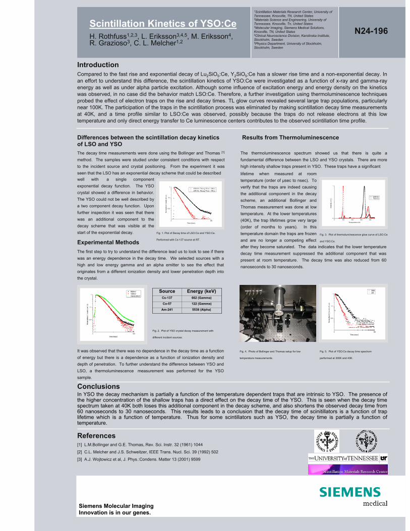
IntroductionCompared to the fast rise and exponential decay of Lu2SiO5:Ce, Y2SiO5:Ce has a slower rise time and a non-exponential decay. In an effort to understand this difference, the scintillation kinetics of YSO:Ce were investigated as a function of x-ray and gamma-ray energy as well as under alpha particle excitation. Although some influence of excitation energy and energy density on the kinetics was observed, in no case did the behavior match LSO:Ce. Therefore, a further investigation using thermoluminescence techniques probed the effect of electron traps on the rise and decay times. TL glow curves revealed several large trap populations, particularly near 100K. The participation of the traps in the scintillation process was eliminated by making scintillation decay time measurements at 40K, and a time profile similar to LSO:Ce was observed, possibly because the traps do not release electrons at this low temperature and only direct energy transfer to Ce luminescence centers contributes to the observed scintillation time profile.
ConclusionsIn YSO the decay mechanism is partially a function of the temperature dependent traps that are intrinsic to YSO. The presence of the higher concentration of the shallow traps has a direct effect on the decay time of the YSO. This is seen when the decay time spectrum taken at 40K both loses this additional component in the decay scheme, and also shortens the observed decay time from 60 nanoseconds to 30 nanoseconds. This results leads to a conclusion that the decay time of scinitillators is a function of trap lifetime which is a function of temperature. Thus for some scintillators such as YSO, the decay time is partially a function of temperature.
References[1] L.M.Bollinger and G.E. Thomas, Rev. Sci. Instr. 32 (1961) 1044
[2] C.L. Melcher and J.S. Schweitzer, IEEE Trans. Nucl. Sci. 39 (1992) 502
[3] A.J. Wojtowicz et al, J. Phys.:Condens. Matter 13 (2001) 9599
Differences between the scintillation decay kinetics of LSO and YSOThe decay time measurements were done using the Bollinger and Thomas [1]
method. The samples were studied under consistent conditions with respect to the incident source and crystal positioning. From the experiment it was seen that the LSO has an exponential decay scheme that could be described
Experimental Methods
well with a single component exponential decay function. The YSO crystal showed a difference in behavior. The YSO could not be well described by a two component decay function. Upon further inspection it was seen that there was an additional component to the decay scheme that was visible at the start of the exponential decay.
The first step to try to understand the difference lead us to look to see if there was an energy dependence in the decay time. We selected sources with a high and low energy gamma and an alpha emitter to see the effect that originates from a different ionization density and lower penetration depth into the crystal.
5538 (Alpha)Am-241
122 (Gamma)Co-57
662 (Gamma) Cs-137
Energy (keV)Source
It was observed that there was no dependence in the decay time as a function of energy but there is a dependence as a function of ionization density and depth of penetration. To further understand the difference between YSO and LSO, a thermoluminescence measurement was performed for the YSO sample.
Results from Thermoluminescence
The thermoluminescence spectrum showed us that there is quite a fundamental difference between the LSO and YSO crystals. There are more high intensity shallow traps present in YSO. These traps have a significant
lifetime when measured at room temperature (order of µsec to nsec). To verify that the traps are indeed causing the additional component in the decay scheme, an additional Bollinger and Thomas measurement was done at low temperature. At the lower temperatures (40K), the trap lifetimes grow very large (order of months to years). In this temperature domain the traps are frozen and are no longer a competing effectafter they become saturated. The data indicates that the lower temperature decay time measurement suppressed the additional component that was present at room temperature. The decay time was also reduced from 60 nanoseconds to 30 nanoseconds.
Fig. 1. Plot of Decay time of LSO:Ce and YSO:Ce.
Performed with Cs-137 source at RT.
Fig. 2. Plot of YSO crystal decay measurement with
different incident sources.
Fig. 3. Plot of thermoluminescence glow curve of LSO:Ce
and YSO:Ce.
Fig. 5. Plot of YSO:Ce decay time spectrum
performed at 300K and 40K.
Fig. 4. Photo of Bollinger and Thomas setup for low
temperature measurements.
34
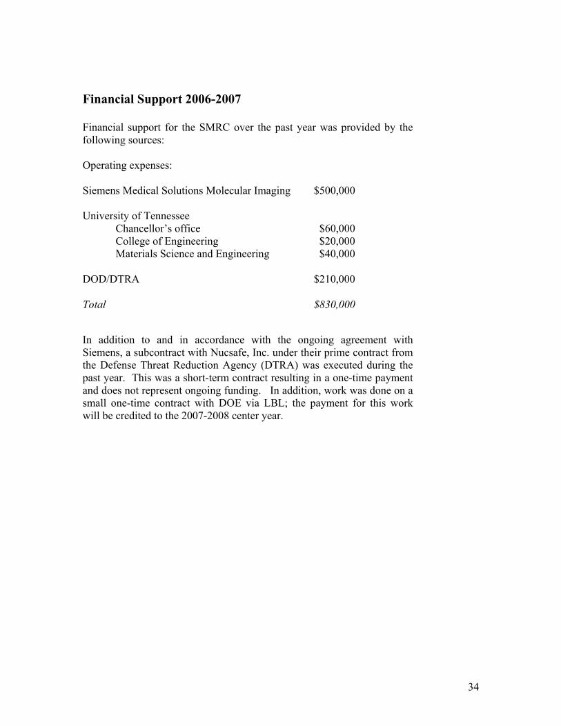
Financial Support 2006-2007 Financial support for the SMRC over the past year was provided by the following sources: Operating expenses: Siemens Medical Solutions Molecular Imaging $500,000 University of Tennessee Chancellor’s office $60,000 College of Engineering $20,000 Materials Science and Engineering $40,000 DOD/DTRA $210,000 Total $830,000
In addition to and in accordance with the ongoing agreement with Siemens, a subcontract with Nucsafe, Inc. under their prime contract from the Defense Threat Reduction Agency (DTRA) was executed during the past year. This was a short-term contract resulting in a one-time payment and does not represent ongoing funding. In addition, work was done on a small one-time contract with DOE via LBL; the payment for this work will be credited to the 2007-2008 center year.
35
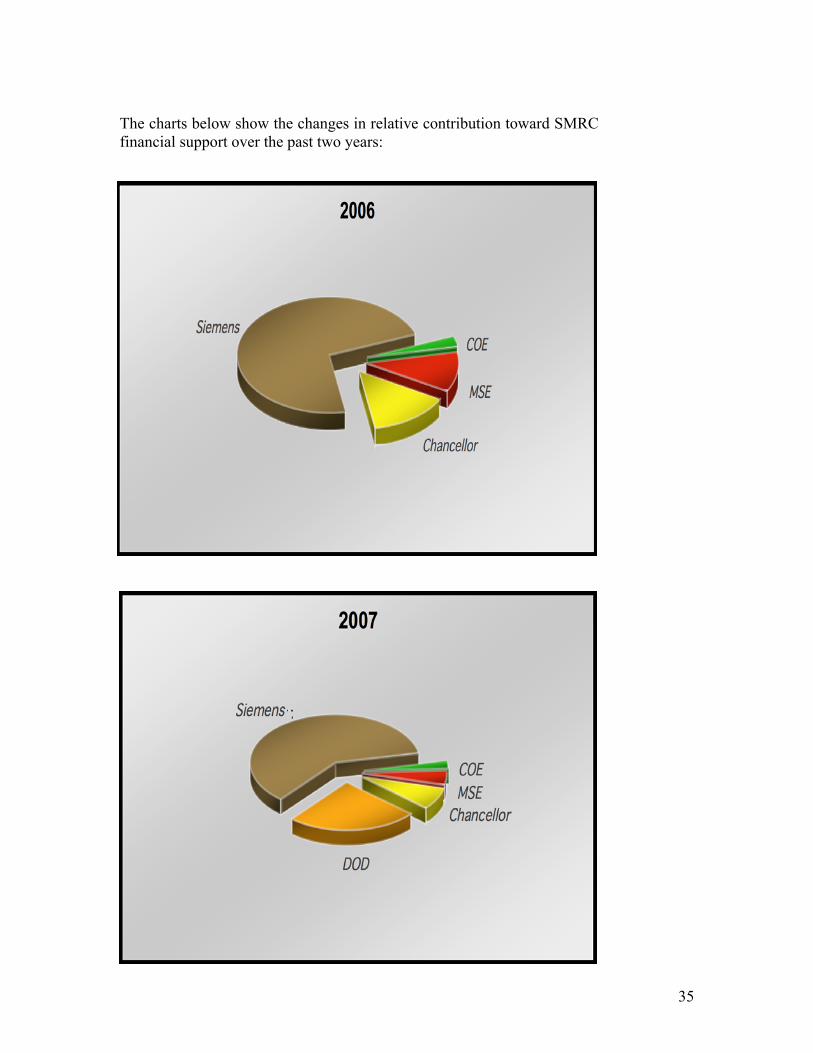
The charts below show the changes in relative contribution toward SMRC financial support over the past two years: