Embed Size (px)
Citation preview
Kuby Immunology SEVENTH EDITION
CHAPTER 11 T-Cell Activation, Differentiation,
and Memory
Copyright © 2013 by W. H. Freeman and Company
Scott Abrams, Ph.D. Professor of Oncology, x4375
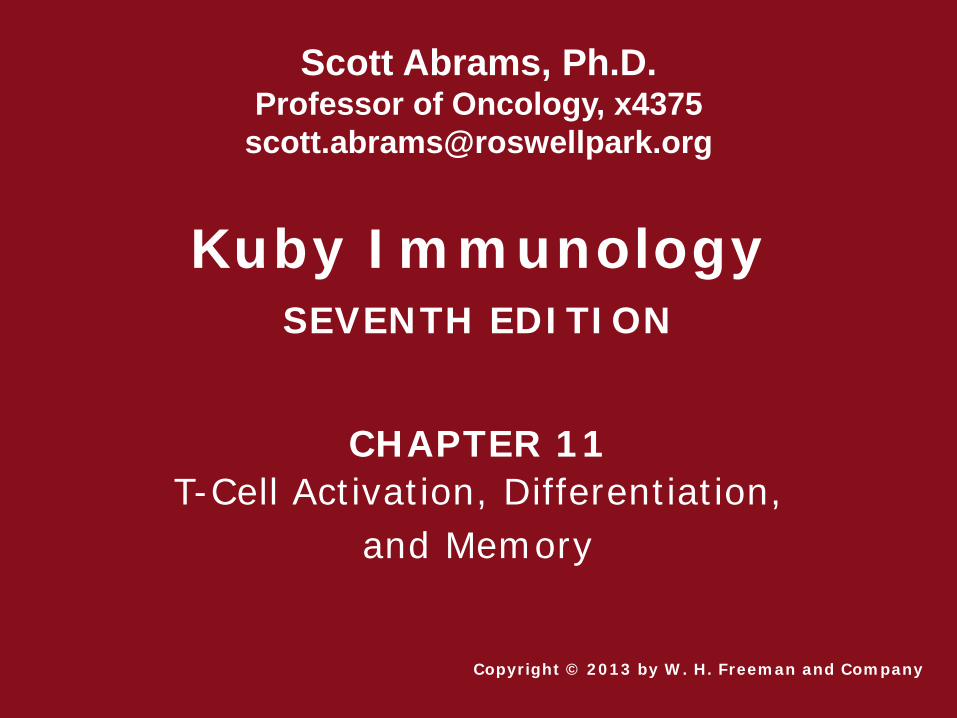
T Cell-Dendritic Cell Interactions

Role of the Antigen Presenting Cell in the Activation of Naïve T Cells
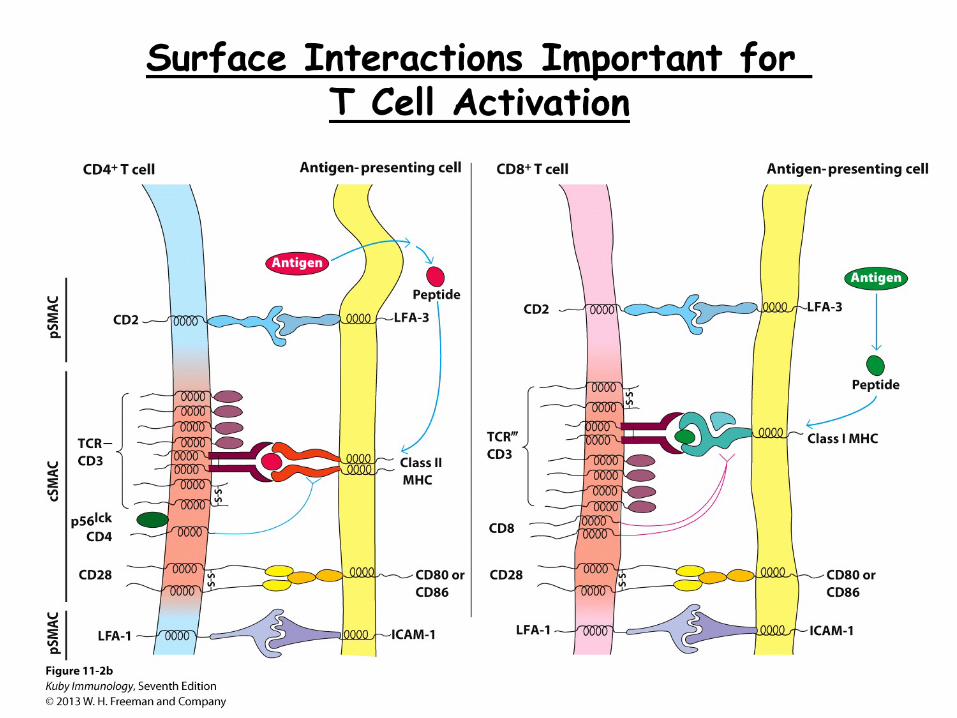
Dendritic cells are essential for the induction of the naïve T cell response, and do so through regulation of three major events known as the 3-signal model: 1. Recognition of MHC-peptide complex (& conjugate
formation) i. antigen processing ii. antigen presentation iii. co-receptors (CD4 or CD8) iv. adhesion (LFA-1/ICAM-1; CD2/LFA-3)
2. positive co-stimulation (CD28/CD80 or CD86)
3. cytokine production (e.g., IL-2)
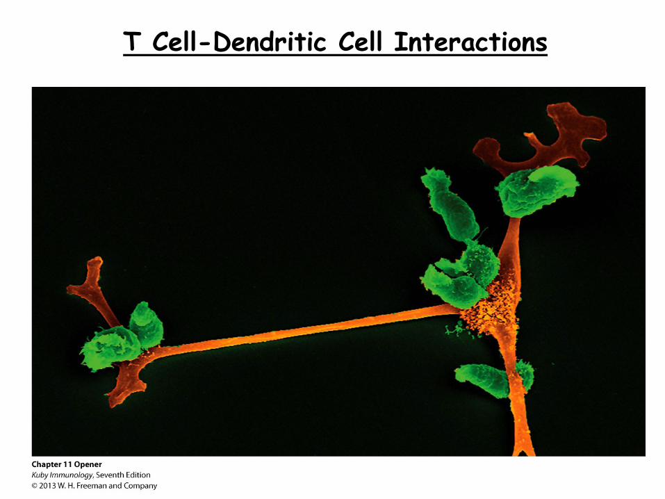
Signal 1: T Cell Recognition of MHC-Peptide Complexes
Antigen specificity is governed by the TCR, which recognizes an antigenic peptide in the context of self-MHC, a concept known as MHC restriction. MHC restriction is absolutely essential to ensure and instruct immune reactivity against ‘alterations of self’. MHC class I=heavy/light pair MHC class II=similar size pair

Summary: The Generation of MHC-Peptide Complexes for T Cell Receptor Recognition
1 MHC class I and class II molecules deliver peptides to the cell
surface from two distinct intracellular compartments. 2 Peptides presented by MHC class I molecules are generated
within the cytosolic compartment (aka, endogenous pathway) 3 Peptides presented by MHC class II molecules are generated in
acidified endocytic vesicles (aka, exogenous pathway). 4 Cross-presentation allows exogenous proteins to be presented on
both MHC class I and II molecules (i.e., via dendritic cells, which are highly effective).
5 CD8+ T cells recognize MHC class I-peptide complexes 6 CD4+ T cells recognize MHC class II-peptide complexes
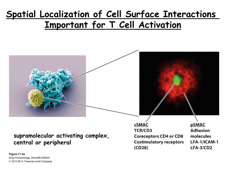
Spatial Localization of Cell Surface Interactions Important for T Cell Activation
supramolecular activating complex, central or peripheral

Summary: 3-Signal Model for T Cell Activation
From Naïve T Cell State to T Cell Activation (role of the APC and costimulation)
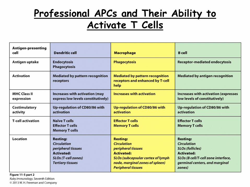
Professional APCs and Their Ability to Activate T Cells
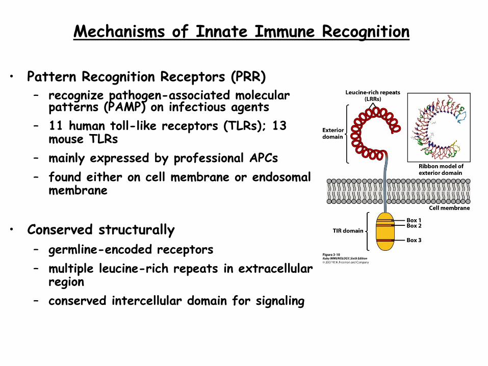
Mechanisms of Innate Immune Recognition • Pattern Recognition Receptors (PRR)
– recognize pathogen-associated molecular patterns (PAMP) on infectious agents
– 11 human toll-like receptors (TLRs); 13 mouse TLRs
– mainly expressed by professional APCs – found either on cell membrane or endosomal
membrane
• Conserved structurally – germline-encoded receptors – multiple leucine-rich repeats in extracellular
region – conserved intercellular domain for signaling
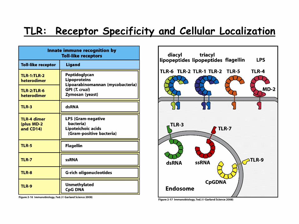
TLR: Receptor Specificity and Cellular Localization
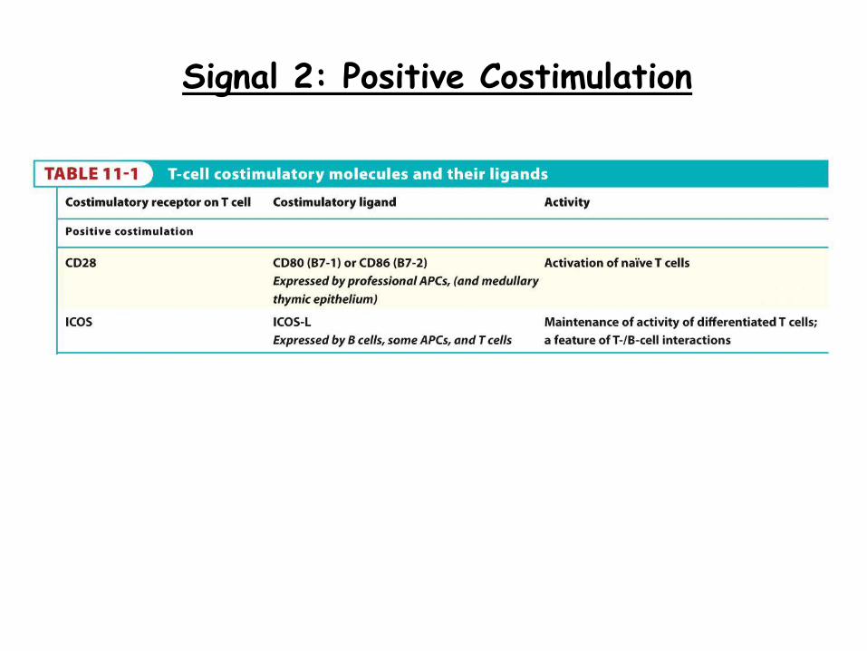
Signal 2: Positive Costimulation
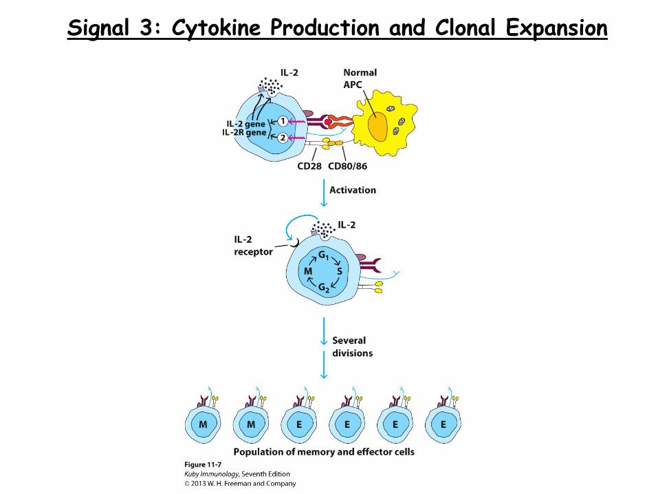
Signal 3: Cytokine Production and Clonal Expansion
Regulation of the T Cell Response
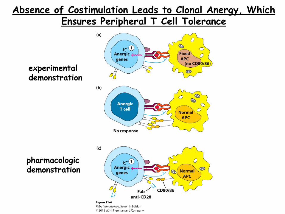
Absence of Costimulation Leads to Clonal Anergy, Which Ensures Peripheral T Cell Tolerance
experimental demonstration
pharmacologic demonstration
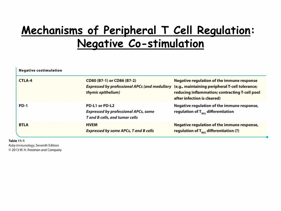
Mechanisms of Peripheral T Cell Regulation: Negative Co-stimulation
From T Cell Activation to Differentiation (or Polarization)
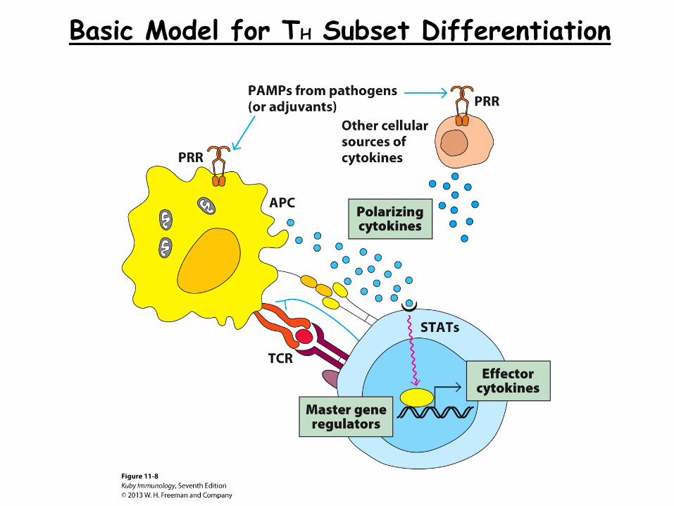
Basic Model for TH Subset Differentiation
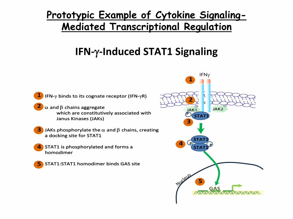
IFN-γ-Induced STAT1 Signaling
P PSTAT1
STAT1
P STAT1
Nucleus
GAS
1. IFN-γ binds to its cognate receptor (IFN-γR)
2. α and β chains aggregate1. which are constitutively associated with
Janus Kinases (JAKs)
3. JAKs phosphorylate the α and β chains, creating a docking site for STAT1
4. STAT1 is phosphorylated and forms a homodimer
5. STAT1:STAT1 homodimer binds GAS site
1
2
3
4
5
1
2
3
4
5
Prototypic Example of Cytokine Signaling- Mediated Transcriptional Regulation
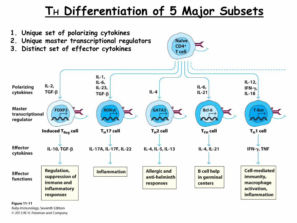
TH Differentiation of 5 Major Subsets
1. Unique set of polarizing cytokines 2. Unique master transcriptional regulators 3. Distinct set of effector cytokines

Cross-Regulation of TH Subsets: Critical to Ensure Expression of the Appropriate T Cell Response
example with Th1-Th2 paradigm
From T Cell Activation to Differentiation to Memory
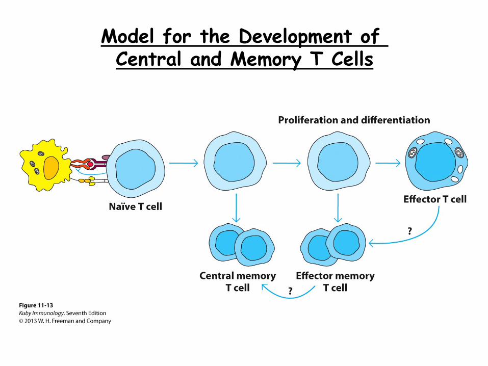
Model for the Development of Central and Memory T Cells
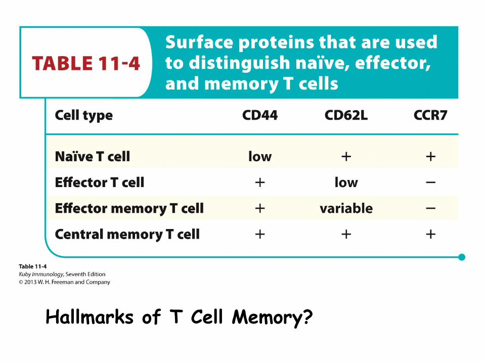
Hallmarks of T Cell Memory?
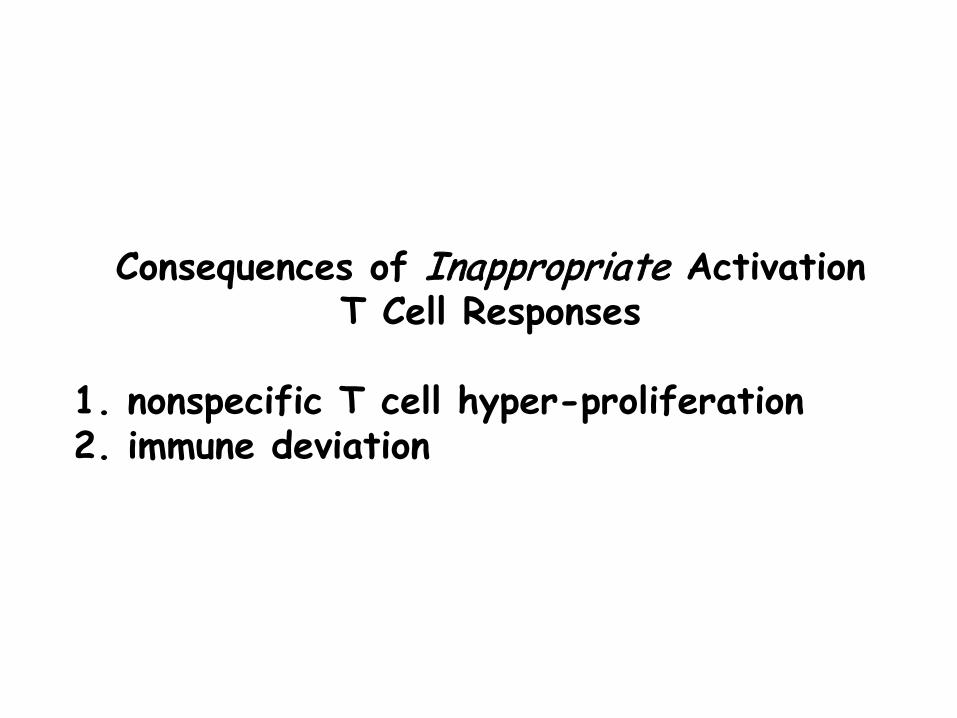
Consequences of Inappropriate Activation T Cell Responses
1. nonspecific T cell hyper-proliferation 2. immune deviation
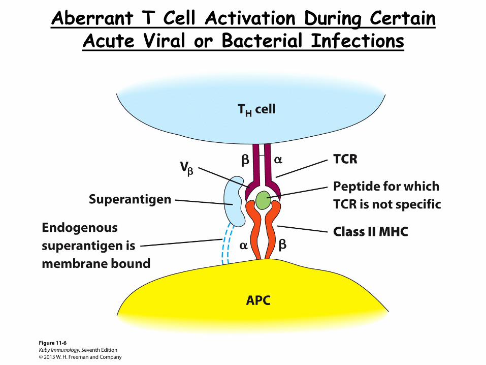
Aberrant T Cell Activation During Certain Acute Viral or Bacterial Infections
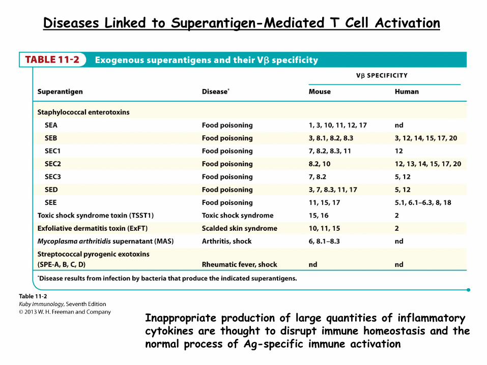
Diseases Linked to Superantigen-Mediated T Cell Activation
Inappropriate production of large quantities of inflammatory cytokines are thought to disrupt immune homeostasis and the normal process of Ag-specific immune activation

Immune Deviation: Pathologic Consequences of the Wrong TH Subset Response
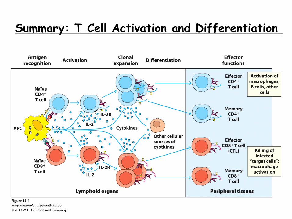
Summary: T Cell Activation and Differentiation
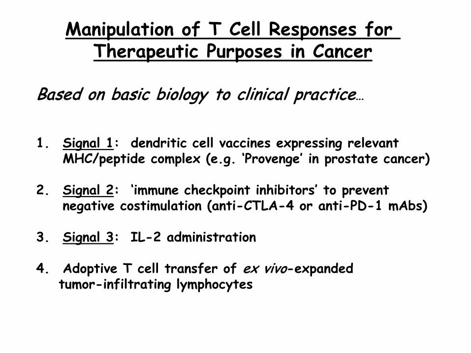
Manipulation of T Cell Responses for Therapeutic Purposes in Cancer
Based on basic biology to clinical practice… 1. Signal 1: dendritic cell vaccines expressing relevant
MHC/peptide complex (e.g. ‘Provenge’ in prostate cancer)
2. Signal 2: ‘immune checkpoint inhibitors’ to prevent negative costimulation (anti-CTLA-4 or anti-PD-1 mAbs)
3. Signal 3: IL-2 administration
4. Adoptive T cell transfer of ex vivo-expanded tumor-infiltrating lymphocytes
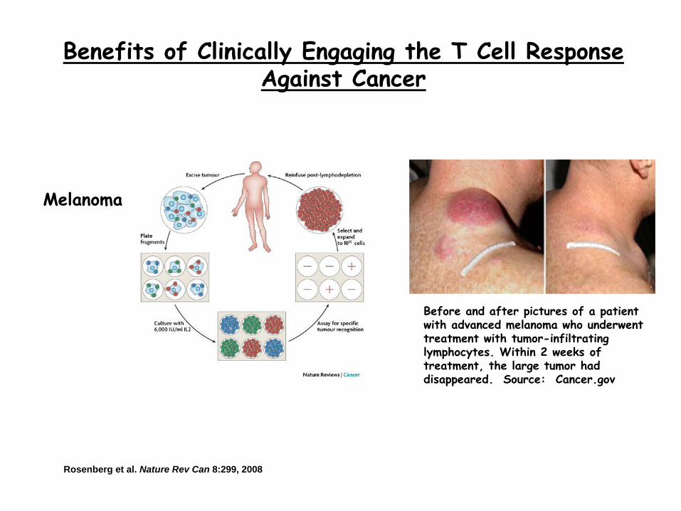
Benefits of Clinically Engaging the T Cell Response Against Cancer
Before and after pictures of a patient with advanced melanoma who underwent treatment with tumor-infiltrating lymphocytes. Within 2 weeks of treatment, the large tumor had disappeared. Source: Cancer.gov
Melanoma
Rosenberg et al. Nature Rev Can 8:299, 2008


![Roswell [reparado]](https://img.pdfslide.net/doc/110x75/55c2611abb61eb356a8b47e9/roswell-reparado.jpg)